calcium and magnesium homeostasis winston w. k. … and magnesium homeostasis ... intrapartum...
TRANSCRIPT
1
Calcium and Magnesium Homeostasis Winston W. K. Koo*
Carman and Ann Adams Department of Pediatrics Wayne State University
Detroit, MI
Reginald C. Tsang† Department of Pediatrics University of Cincinnati
Cincinnati, OH * Corresponding author Department of Pediatrics, Hutzel Hospital 4707 St Antoine Blvd Detroit, MI 48201 E mail: [email protected] Phone: 313-7457231 Fax: 313-9930198 † Department of Pediatrics CCHMC, MLC 12001 Cincinnati 45229 E mail: [email protected] Phone: 513-6369595 Fax: 513-6369596
2
OUTLINE • Introduction • Tissue distribution • Circulating concentrations
o Calcium o Magnesium
• Physiologic control o Parathyroid Hormone o Calcitonin o Vitamin D o Non classic control of calcium and magnesium homeostasis
• Disturbances in serum mineral concentrations o Hypocalcemia
Pathophysiology Diagnosis Management
o Hypercalcemia Pathophysiology Diagnosis Management
o Hypomagnesemia Pathophysiology Diagnosis Management
o Hypermagnesemia Pathophysiology Diagnosis Management
• Skeletal manifestations of disturbed mineral homeostasis o Pathophysiology o Diagnosis o Treatment and prevention o Monitoring and follow up

3
INTRODUCTION Calcium (Ca) is the most abundant mineral in the body and, together with
phosphorus (P), forms the major inorganic constituent of bone. Magnesium (Mg) is the fourth most abundant cation and is the second most common intracellular electrolyte in the body. The maintenance of Ca and Mg homeostasis requires a complex interaction of hormonal and non-hormonal factors; adequate functioning of various body systems, in particular, the renal, gastrointestinal and skeletal systems; and adequate dietary intake. From a clinical perspective, mineral homeostasis is reflected in the maintenance of circulating concentrations of Ca and Mg in the normal range, and integrity of the skeleton.
In the circulation, the amount of Ca and Mg is <1% of their respective total body content; however, disturbances in serum concentrations of these minerals are associated with disturbances of physiologic function manifested by numerous clinical symptoms and signs. Chronic and severely lowered serum concentrations of these minerals also may reflect the presence of a deficiency state.
At all ages, the total body content of Ca and Mg in the skeleton are about 99% and 60% respectively. Thus, the skeleton is a reservoir for mineral homeostasis in addition to its role providing structural and mechanical support; disturbances in mineral homeostasis can result in osteopenia and rickets in infants and children, and osteomalacia and osteoporosis in adults.
The mechanisms to maintain mineral homeostasis in neonates are the same as for children and adults. However, the newborn infant has a number of unique challenges to maintain mineral homeostasis during adaptation to extrauterine life and to continue the rapid rate of growth. These include an abrupt discontinuation of high rate of intrauterine accretion of Ca (~120 mg/kg/d) and Mg (~4 mg/kg/d) during the third trimester, a smaller skeletal reservoir available for mineral homeostasis, a delay in establishment of adequate nutrient intake for at least a few days or longer, particularly in the sick and preterm infants, and high requirement for Ca and Mg for the most rapid period of postnatal skeletal growth, with an average gain in length of >25 cm during the first year. There also may be diminished end organ responsiveness to hormonal regulation of mineral homeostasis, although the functional capacity of the gut and kidney improves rapidly within days after birth. The effects of these issues are exaggerated in infants with heritable disorders of mineral metabolism such as extracellular calcium sensing receptor (CaR) mutations, and in infants who have experienced adverse antenatal events such as maternal diabetes, intrapartum problems such as perinatal asphyxia or maternal Mg therapy, or postnatal problems such as immature function of multiple organs from premature birth.
Increased understanding of the physiology and molecular basis of mineral metabolism allows a better understanding of the pathophysiology of the resultant clinical disorder. This in turn allows a more rational management to minimize the adverse impact from disturbed mineral homeostasis and to prevent iatrogenic causes precipitating or prolonging these problems.
4
TISSUE DISTRIBUTION In the fetus, about 80% of minerals are accrued between 25 weeks of gestation
and term. During this period, the estimated daily accretion per kilogram fetal body weight is 2.3 to 2.98 mmol (92 to 119 mg) Ca and 0.1 to 0.14 mmol (2.51 to 3.44 mg) Mg. The peak accretion rates occur at 36 to 38 weeks of gestation. In newborn term infants, the total body Ca and Mg contents average approximately 28 g and 0.7 g respectively (1,2).
After birth, 99% of total body Ca is in bone. The tissue distribution of Mg varies according to the extent of bone mineralization and the rate of soft tissue growth. Near the end of the third trimester, however, about 60% of the body’s Mg is in bone, 20% is in muscle, and most of the remainder is found in the intracellular space of other tissues. CIRCULATING CONCENTRATION Calcium (Ca, 1 mmol/L = 4 mg/dL)
Serum Ca occurs in three forms: approximately 40% is bound, predominantly to albumin; approximately 10% is chelated and complexed to small molecules such as bicarbonate, phosphate, or citrate; and approximately 50% is ionized. Complexed and ionized Ca are ultrafilterable.
Total Ca concentrations (tCa) in cord sera increase with increasing gestational age. Serum tCa may be as high as 3 mmol/L in cord blood of infants born at term, and they are significantly higher than paired maternal values at delivery (3-6). Serum tCa reaches a nadir during the first 2 days after birth (7-13); thereafter, concentrations increase and stabilize at a level generally above 2 mmol/L (14). In infants exclusively fed human milk, the mean serum tCa increases from 2.3 to 2.7 mmol/L over the first 6 months postnatally. Normally, serum tCa in children and adults remains stable, with a diurnal range of less than 0.13 mmol/L. During the third trimester of pregnancy a modest reduction in maternal serum tCa concentration (average 0.1 mmol/L) is associated with a decrease in serum albumin concentration.
Serum ionized Ca (iCa) concentration is the best indicator of physiologic blood Ca activity. Measurement of serum iCa is firmly established in clinical medicine, and simple, rapid, and direct determination of iCa from whole blood, plasma and serum by ion-selective electrodes are freely available. Whole blood iCa analyzers are gaining popularity because they are adaptable for "point of care" testing. However, some differences exist in values from different iCa analyzers particularly for whole blood iCa values (15), as a result of differences in the design of the reference electrode, formulation of calibrating solutions, and the lack of a reference system for iCa. Thus normative data should be generated according to the subject’s age, the instrument and type of sample used for iCa measurement.
Serum iCa decreases in the presence of high serum albumin, P, bicarbonate, and heparin. Serum iCa increases with increased Mg, and is inversely related to blood pH. The effect of the latter may be minimized by the immediate analysis of serum samples for iCa. Freezing serum samples in 5% CO2-containing tubes may minimize the impact of pH variations if measurement of iCa is delayed for 1 week.
5
One report showed a wide range of cord whole blood iCa of 0.4-1.85 mmol/L from apparently normal pregnancies (16). This is a much wider range compared from multiple reports based on cord sera, although the range for whole blood iCa becomes much narrower within hours after birth and similar to serum iCa values. Cord serum iCa increases with increasing gestational age and is higher than values in paired maternal sera. In healthy term neonates, serum iCa averages 1.25 mmol/L with 95% confidence limits of 1.1-1.4 mmol/L (4.4-5.6 mg/dl) and there is a decline in serum iCa in the first 48 hr of life with a nadir at 24 hr (17). Serum iCa generally changes in parallel with tCa in healthy humans. However, serum iCa is stable and normal during pregnancy in contrast to a slight reduction in tCa. Serum tCa and iCa are correlated in infants and adults but is inadequate to predict one from the other with sufficient accuracy.
The concentration of iCa is critical to many important biological functions with the extracellular Ca concentration normally maintained within a narrow range. The Ca ion is well established as an intracellular second messenger, but identification of the extracellular CaR has established that iCa also functions as a messenger outside cells. Ca homeostasis also involves the maintenance of an extremely large Ca concentration gradient across the cellular plasma membrane. In the cell, distribution of Ca is not uniform. The cytosolic compartment contains 50 to 150 nmol of Ca per liter of water; a larger intramitochondrial Ca pool contains 500 to 10,000 nmol of Ca per liter cell water. In contrast, the concentration of iCa in extracellular fluid is 1 million nmol/L (1 mmol/L). There are at least two adenosine triphosphate-dependent mechanisms involved in the maintenance of the Ca concentration gradient across the plasma membrane. The measurement of intracellular Ca continues to improve with better instrumentation and probes, but is not freely available. Magnesium (Mg, 1 mmol/L = 2.4 mg/dL)
Approximately 30% of serum Mg is in the protein-bound form, with the remainder in the ultrafilterable portion. Seventy to 80% of ultrafilterable Mg is in ionic form, the remainder being complexed to anions, particularly phosphate, citrate, and oxalate. Cord serum total Mg (tMg) is higher than paired maternal values. Serum tMg of 0.92 ± 0.13 mmol/L (mean ± 2 SD) in children is slightly higher than the adult values of 0.88 ± 0.13 mmol/L (18). Ion-selective electrodes are being used in the measurement of ionized Mg (iMg) in whole blood and sera. iMg concentrations average 62% to 70% of the tMg concentration in cord and postnatal sera. Cord serum iMg is also higher than that in maternal serum (19-21). The clinical role of iMg (versus tMg) in a number of disease states appears limited (22).
Cellular Mg content of most tissues is 6 to 9 mmol/kg wet weight, and most of this Mg is localized in membrane structures (e.g., microsome, mitochondria, plasma membrane). The much smaller pool of free Mg in the cell is maintained at about 1 mmol/L and is in an exchanging equilibrium with membrane-bound Mg. This unbound intracellular Mg has a critical role in cellular physiology and catalyzes enzymatic processes concerned with the transfer, storage, and use of energy. Intracellular Mg usually remains stable despite wide fluctuations in serum Mg. In Mg-deficient states, however, the intracellular content of Mg can be low despite normal serum concentrations. The measurement of intracellular Mg continues to improve with better instrumentation and probes but is not freely available.
6
PHYSIOLOGIC CONTROL Calciotropic hormones, including parathyroid hormone (PTH) and 1,25
dihydroxyvitamin D [1,25(OH)2D], and possibly calcitonin (CT), appear to maintain Ca homeostasis by intermodulation of their physiologic effects on each other and on the classic target organs: kidney, intestine and bone. Dietary intake of Ca, Mg, P and other nutrients including sodium, glucose and protein also may significantly contribute to the regulation of mineral homeostasis. PTH serves as the major component of the rapid response to hypocalcemia, whereas 1,25(OH)2D, with its major effect on elevating intestinal absorption of Ca, is responsible for a slower but more sustained contribution to the maintenance of normocalcemia. CT, on the other hand, appears to function in the opposite role to PTH but with the capacity to stimulate the production of 1,25(OH)2D, which in theory may serve an additional regulatory role in the maintenance of Ca homeostasis.
In contrast, the control of Mg homeostasis by calciotropic hormones under physiologic conditions appears to be limited. However, Mg is critical to the maintenance of Ca homeostasis since Mg regulates the production and secretion of PTH, acts as a cofactor for the 25 hydroxyvitamin D 1α hydroxylase enzyme in the production of 1,25(OH)2D, and maintains adequate sensitivity of target tissues to PTH. Furthermore, Mg is considered as mimic/antagonist of Ca as it often functions synergistically with Ca, yet competes with Ca in the gut and kidney for transport and other metabolic pathways. PARATHYROID HORMONE (PTH)
In humans, parathyroid glands are derived from the 3rd and 4th pharyngeal pouch. The PTH gene, along with the genes for insulin, β-globulin, and CT, is located on chromosome 11p15 (23), and restriction site polymorphisms near the PTH gene have been detected. The initial translational product of the mRNA is a 115-amino-acid prepro-PTH. Prepro-PTH then undergoes proteolytic cleavage in endoplasmic reticulum to remove a 25 residual amino-terminal signal sequence to form pro-PTH. The prohormone-specific region is cleaved further during subsequent intracellular processing to generate the 84-amino-acid secreted form of the intact hormone with a relative molecular mass (Mr) of 9,500. PTH is synthesized by the chief cells and stored in secretory granules. It is colocated and secreted with chromogranin A, a protein that may act in autocrine- or paracrine-regulated release of PTH.
About 50% of the newly generated PTH is proteolytically degraded intracellularly and some of the inactive fragments are also secreted. After release into the circulation, the intact PTH molecule has a serum half-life of 5 to 8 minutes and undergoes a series of cleavages by endopeptidases in the liver and kidney. The amino-terminal fragments contain the biologically active fractions, with the 1–34 fragment having the most calcemic activity; modifications at the amino terminal, particularly at the first two residuals, can abolish its biologic activity. The midregion and carboxyl-terminal fragments are biologically inert, although the latter may have some in vitro biological activity.
Circulating immunoreactive PTH is a complex mixture of intact 1-84 PTH, multiple peptide fragments from the amino- and carboxyl-terminals, and mid-molecular regions. Normally, there are greater amounts of middle and carboxyl fragments than intact hormone in the circulation because of metabolic breakdown of the short-lived, intact hormone, coupled with glandular secretion of inactive fragments. The fragments
7
are cleared from the blood virtually exclusively by glomerular filtration. Intact PTH and amino-terminal fragments constitute <20% of PTH immunoreactivity in the peripheral circulation. PTH molecules reactive in the widely used commercial immunoradiometric assays (IRMA) designed to detect both amino- and carboxy- terminal epitopes of the peptide have been considered as “intact” PTH (IPTH) assays. However, there have been reports that the large 7-84 fragment of PTH is also detected by these assays. This large fragment is biologically inactive and present in greater concentrations in uremic states or hyperparathyroidism. The conventional IPTH technique increases the PTH concentration by 30 to 50%, compared to the latest chemiluminescence or IRMA techniques that measure the “whole” or “bio-intact” PTH. Therefore, the treatment of secondary hyperparathyroidism based on data from conventional IPTH assays theoretically may lead to overtreatment and oversuppression of biologically active PTH, although the clinical significance of this possibility remains to be defined. In any case, consistency of the PTH assay methodology, and serial measurements are critical to the interpretation and management of pathologic states.
PTH concentrations in cord blood frequently are low and do not correlate with PTH concentrations in maternal sera (4,24). Earlier studies of higher levels of bioactive PTH in cord sera from cytochemical assay may be related to elevated concentrations of parathyroid hormone-related protein (PTHrP), since PTH-like bioactivity was tightly correlated with levels of PTHrP in sheep and pig. Small amounts (about 5%) of perfused fragments (35-84, 44-68, and 65-84 amino acids), but probably not the whole PTH molecule, are reported to cross the human placenta. Serum PTH concentrations increase postnatally coincident with the fall in serum Ca in both term and preterm infants (4,24-27). The rise in serum IPTH is greater for preterm infants with hypocalcemia compared to term infants reflecting appropriate PTH response. Serum PTH concentrations are similar for children and adults but are increased in the elderly. Serum concentrations of intact PTH as measured by IRMA showed no change during normal pregnancy. In adults, serum intact PTH is present in picomolar concentrations. It has a significant circadian periodicity, spontaneous episodic pulsatility with distinct peak property and a significant temporal coupling with serum iCa and phosphorus concentrations and prolactin secretion.
PTH effects on end-organ systems appear to be mediated through its binding to specific receptors. The type 1 PTH/PTHrP receptor has been identified in bone, cartilage, kidney, intestine, aorta, urinary bladder, adrenal gland, brain and skeletal muscle. It binds equally to PTH and PTHrP, and belongs to a superfamily of guanine-nucleotide-binding (G) protein-coupled cell membrane receptors (GPCR) including those for CT, secretin, growth hormone-releasing hormone, corticotrophin-releasing hormone, glucagon, vasoactive intestinal polypeptide and others. Another PTH receptor (type 2) responds only to PTH although its main endogenous ligand appears to be a 39 amino acid peptide, hypothalamic tubular infundibular peptide (TIP-39). It has been found in the brain, pancreas and intestines but the physiologic significance of this receptor remains ill defined.
The gene for the PTH/PTHrP receptor is located on chromosome 3p21.1-p24.2. It contains 17 exons and encodes a mature glycoprotein of 593 amino acids (28). The type 1 PTH receptor consists of extended extracellular, ligand-binding amino-terminal and intracellular G protein–associated carboxyl-terminal domains and seven transmembrane
8
domains. Signal transduction mediated by G proteins results in multiple second messenger pathways to effect both stimulatory and inhibitory end organ responses. The strongest and best-characterized second messenger signaling pathway is the PTH stimulated coupling of the type 1 PTH receptor to Gs class protein (composed of 3 subunits: α, β, and γ, and is encoded by GNAS1 gene localized to 20q13.3) which activates adenyl cyclase, an enzyme that generates cyclic AMP. However, coupling of type 1 PTH receptor to the Gq class protein activates phospholipase C that generates inositol phosphate (IP3) and diacylglycerol (DAG). These second messengers in turn lead to stimulation of protein kinases A and C and Ca transport channels and result in a variety of hormone-specific tissue responses.
In physiologic terms, PTH acts synergistically with 1,25(OH)2D and is the most important regulator of extracellular Ca concentration. PTH acts directly on bone and kidney, and indirectly on intestine. Immediate control of blood Ca is probably due to PTH induced mobilization of Ca from bone, and increased renal distal tubular reabsorption of Ca. PTH also decreases proximal tubular and thick ascending limb reabsorption of sodium, Ca, phosphate, and bicarbonate. PTH effects on kidney are mediated primarily through stimulation of sodium/calcium exchange, calcium transport proteins and renal 25 OHD-1α-hydroxylase, but a decrease in sodium dependent phosphate co-transporter, NPT-2. Maintenance of steady state calcium balance is probably from increased intestinal Ca absorption secondary to increased 1,25(OH)2D production. PTH increases acutely within minutes the rate of Ca release from bone into blood. Chronic effects of PTH are to increase the numbers of osteoblasts and osteoclasts and to increase the remodeling of bone. Continuous exposure to elevated concentrations of PTH leads to increased osteoclastic resorption of bone. In contrast to its classic action on Ca mobilization from bone, the amino terminal fragments of PTH and PTHrP, and small pulses of PTH have an anabolic effect on bone, independent of its resorptive action. Other PTH effects on bone include enhanced collagen synthesis, activities of alkaline phosphatase, ornithine and citrate decarboxylases, and glucose-6-phosphate dehydrogenase; DNA, protein and phospholipid synthesis.
Extracellular Ca is the most potent regulator of PTH secretion and is mediated by the cell-surface CaR, which detects minute perturbations in the extracellular iCa concentration and responds with alterations in cellular function that normalize iCa. Thus, iCa functions as extracellular as well as intracellular messenger. The human CaR gene is located on chromosome 3q13.3-q21 and encodes a cell-surface protein of 1,078 amino acids. The CaR gene is developmentally upregulated, and CaR transcripts are present in numerous tissues including chief cells of the parathyroid glands, kidneys (in particular the thick ascending limb), brain and nerve terminals, breast, lung, intestine, adrenal and skin, and also the precursor and mature osteoblasts and osteoclasts. CaR is a member of the GPCR superfamily with a seven-member membrane-spanning domain. It contains at least seven exons, of which six encode the large (-600 amino acid) amino-terminal extracellular domain and/or its upstream untranslated regions, while a single exon codes for the remainder of the receptor including a cytoplasmic carboxy-terminal intracellular domain. Signal transduction mediated by G proteins results in activation of phospholipase C that generates IP3 and DAG, and subsequent stimulation of protein kinase C and Ca transport channels.
9
Low or falling serum Ca concentrations result in active secretion of preformed PTH within seconds. There is a sigmoidal type of PTH secretion in response to decreased serum Ca, which is most pronounced when serum Ca is in the mildly hypocalcemic range. PTH secretion is 50% of maximal at a serum iCa of about 1 mmol/L (4 mg/dL); this is considered as the calcium set point for PTH secretion (29). Sustained hypocalcemia increases PTH mRNA within hours. Protracted hypocalcemia leads within days to cellular replication and increase gland mass. High serum Ca suppresses PTH secretion via activation of CaR. It in turn activates phospholipase C and generation of IP3 and DAG, and probably increases the proteolytic destruction of preformed PTH. Hyperphosphatemia stimulates PTH secretion, probably by lowering the serum Ca concentration.
In the kidney, CaR decreases the basal and PTH-stimulated paracellular reabsorption of Ca, Mg and sodium via multiple mechanisms including inhibition of cAMP accumulation; stimulation of phospholipase A2 activity, thereby promoting the release of free arachidonic acid that is metabolized via the lipooxygenase pathway to P450 metabolites that inhibit the activities of NaK2Cl cotransporter and the K+ channel; and may affect renal water regulation by inhibition of vasopressin-abated water flow. In chronic renal failure, downregulation in the expression of renal CaR may account for the development of secondary hyperparathyroidism (30), and downregulation of PTH receptors may account for the skeletal resistance to the calcemic effect of PTH (31). Extracellular Ca exerts numerous other actions on parathyroid function, including modulation of the intracellular degradation of PTH, cellular respiration, membrane voltage, and the hexose monophosphate shunt.
Maintenance of Ca homeostasis through other organs also may be possible, for example, through the presence of CaR in intestinal cells (32), and probable modulation of CT secretion from changes in intracellular Ca (33). Furthermore, expression of the CaR in gastrin-secreting G-cells and acid-secreting parietal cells, together with data indicating that the CaR exhibits selectivity for L-aromatic amino acids, would appear to provide a molecular explanation for amino acid sensing in the gastrointestinal tract, regulation of PTH secretion and urinary Ca excretion, and the physiologic interaction between Ca and protein metabolism.
Decrease in serum Mg concentration stimulates PTH secretion (34,35), although chronic hypomagnesemia inhibits secretion of PTH (34,36). Hypomagnesemia is also associated with an increased target tissue resistance to PTH probably from inactivity of adenylate cyclase, a Mg-requiring enzyme. Hypermagnesemia rapidly decreases the secretion of PTH in vivo in human subjects, and PTH concentration remains depressed despite concomitant hypocalcemia, presumably in part due to stimulation of CaR by other divalent cations such as Mg. Vitamin D and its metabolites 25 hydroxyvitamin D (25 OHD) and 1,25(OH)2D, acting through vitamin D receptors, decrease the level of PTH mRNA. Additional systemic factors (growth hormone, insulin-like growth factor 1, estrogen, progesterone, CT, cortisol, catecholamines, prostaglandins, and somatostatin) and local factors (interleukin-l) modulate PTH secretion and function, although their role in the regulation of Ca and Mg metabolism under physiological conditions is not clear.

10
CALCITONIN (CT) Calcitonin is secreted primarily from the thyroid C cells and also from many
extrathyroidal tissues including placenta, brain, pituitary, mammary gland and other tissues. Developmentally, CT-containing cells and parathyroid gland cells are thought to derive from the same tissue source as the neural crest. In the rat, the number of thyroid C cells and secretion of CT increase from fetal life to suckling, a period of rapid growth (37). There is probably no placental crossover of CT; the human placental tissue is able to produce CT in response to the presence of Ca in the culture medium. In human neonates, the CT content in crude tissue preparations of thyroid is larger than that of the adult thyroid (38).
There are two calcitonin genes, α and β, located on chromosome 11p15.2 near the genes for β-globulin and PTH. Two different RNA molecules are transcribed from the α gene. It comprises of 6 exons with the fourth exon translated into the precursor for CT, and fifth is translated into the precursor for CT gene-related peptide-I (CGRP-I). The initial translational product of the mRNA is prepro-CT, a 141 amino-acid peptide. It is cleaved by endopeptidase at the endoplasmic reticulum to form pro-CT, a 13 kD 116 amino-acid peptide. CT (between 60th and 91st position of the pro-CT peptide) and equimolar amounts of non-CT secretory peptides, corresponding to the flanking peptides linked to the amino and carboxy terminals of the prohormone, are generated during precursor processing. Further structural modifications to the CT molecule occur intracellularly prior to release into the circulation. These include formation of a disulfide bridge between two cysteine remnants in position 1 and 7, and hydroxylation of the C terminal proline residue; both are essential for binding of CT to its receptor. The CT monomer is a 32 amino-acid peptide (Mr 3,400). CGRP-I is synthesized wherever the CT mRNA is expressed, e.g., in medullary carcinoma of thyroid, although there is no translational product from CGRP-I sequence.
The β or CGRP-II gene is transcribed into the mRNA for CGRP predominantly in nerve fibers in the central and peripheral nervous system, blood vessels, thyroid and parathyroid glands, liver, spleen, heart, lung, and possibly bone marrow. CGRP, a 37-amino-acid peptide (Mr 4,000), is also generated from the larger precursor molecule pro-CGRP, a 103 amino-acid peptide. Seventy-five amino-terminal residues of each preprohormone for CT and CGRP are predicted to be identical.
Classic bioactivity of human calcitonin (hCT) is present in the full 32 amino-acid structure or its smaller fragments, such as hCT 8-32 and hCT 9-32; the ring structure of CT enhances, but is not essential for, hormone action. Basic amino acid substitutions confer a helical structure in this region as found in salmon and other non-mammalian CT result in greater potency in lowering serum Ca, and probably longer circulating half life. The kidney appears to be the dominant organ in the metabolism of human CT. A small percentage of the metabolic clearance rate of CT in humans may be accounted for by enzymatic degradation in blood. Injected hCT monomer disappears from the blood in vivo with a t1/2 of approximately 10 min; in contrast, the t1/2 of hCT in plasma incubated in vitro at 370C may be longer than 20 hr (39). Depending on the animal species, other sites such as liver, intestine, and bone may be involved in the metabolism of CT.
Circulating immunoreactive CT and CGRP are a heterogeneous mixture of different molecular forms and are recognized as long as the antigenic epitopes recognized by the antiserum are expressed. Immunoreactive CT or CGRP concentration is expressed
11
in gravimetric or molar equivalents of synthetic CT or CGRP. Sample preparation with initial extraction, gel chromatography, and high-performance liquid chromatography separation, and the use of two-site immunoassay can improve the sensitivity and specificity of CT measurements. Serum CT concentrations during pregnancy are variable. They are high at birth compared to paired maternal CT concentrations (40). Serum CT further increases during the first few days after birth and may reach levels fivefold to tenfold higher than adult CT concentrations. Serum CT concentrations decrease progressively during infancy; however, in preterm infants up to 3 months after birth, the mean serum CT concentrations may remain twice the adult value. There is also a small peak of serum CT concentration during late childhood. In human adults, the basal serum CT concentration may be lower in women than in men, but the concentration is not affected by old age. The CT secretory response to Ca infusion is lower in women, and with old age. In human adults, serum CT and CGRP concentrations are found in the picomolar range. Diurnal variability has been reported for serum CGRP but not for serum CT. In normal individuals, larger precursor molecules of CT such as procalcitonin are not detected.
CT function is mediated by binding to receptors linked to G proteins, a member of the GPCR superfamily, and activation of adenylate cyclase and phospholipase C (41). CT receptors (CTR) have been identified in the central nervous system, testes, skeletal muscle, lymphocytes, and the placenta. The function of CTR can be influenced by accessory proteins, receptor isoforms, genetic polymorphisms, developmental and/or transcriptional regulation, feedback inhibition, and the specific cellular or tissue background. The CTR gene is located on chromosome 7q2l.2-q21.3 and encodes a 490 amino acid G protein-linked receptor with seven transmembrane domains. Two isoforms of human CTR arise by alternative splicing of an exon of 48 nucleotides that encodes a l6-amino-acid insertion within the first intracellular loop. The isoform with the insertion (hCTR-l) activates only adenylate cyclase, whereas the other isoform (hCTR-2) activates both adenylate cyclase and phospholipase C. CGRP functions are also mediated by receptors (42).
Presence of receptor-activity-modifying proteins (RAMP) can post-translationally modify the initially orphan calcitonin receptor-like (CL) receptor and CTR to exhibit different receptor function i.e., functional modification of G protein-coupled receptors is possible. RAMP 1, -2, -3 thus far identified are single transmembrane domain proteins having intracellular C-terminal of up to 10 amino acids and extracellular N-terminal of about 120 amino acids. Non-covalent association of the RAMP with the CL receptor or the CTR results in heterodimeric RAMP/receptor complexes at the cell surface. CL receptor when coexpressed with RAMP1 functions as a CGRP receptor. Whereas CL receptor/RAMP2 and CL receptor/RAMP3 are adrenomedullin (AM) 1 and AM2 receptor subtypes respectively. CTR with 60% homology to the CL receptor predominantly recognize CT in the absence of RAMP. When a CTR was coexpressed with RAMP1, it transforms into an amylin/CGRP receptor. When a CTR coexpressed with RAMP3, it interacts only with amylin. Thus, two Class II G protein-coupled receptors, the CL receptor and CTR, are associated with three RAMP to form high affinity receptors for CGRP, adrenomedullin, or amylin.
12
Secretion of CT is stimulated by an increase in serum Ca and Mg concentrations and by gastrin, glucagon, and cholecystokinin, along with several other structural analogs of these hormones (e.g., pentagastrin, prostaglandin E2), glucocorticoid, norepinephrine, and CGRP; and suppressed by hypocalcemia, propranolol and other adrenergic antagonists, somatostatin, chromogranin A and vitamin D. CT gene transcription is positively regulated by glucocorticoid and negatively regulated by protein kinase C, Ca and vitamin D. Calcitonin may activate the l-hydroxylase system independent of PTH (43), whereas 1,25(OH)2D decreases CT gene expression in adult rats but is ineffective in 13-day-old suckling rats (44). The latter observation may be related to fewer 1,25(OH)2D receptors in C cells of immature rats. Calcitonin induces refractoriness to its own actions from down regulation in the number, and functional reduction of receptor mRNA is a well known phenomenon. Clinically, it is manifested as the “escape” phenomenon during therapy with calcitonin.
In humans, changes in Ca (and P) metabolism are not seen despite extreme variations in CT production. In the neonate, there is neither an identifiable hypocalcemic response to the postnatal surge in serum CT nor a blunting of CT secretion in the presence of hypocalcemia. In adults, there are no definite effects attributable to CT deficiency, for example, totally thyroidectomized patients receiving only replacement thyroxin; or CT excess, for example, patients with medullary carcinoma of thyroid, except for the chronic suppression of bone remodeling. The clinical significance of CT is related to its use as a tumor marker in the management of medullary carcinoma of the thyroid, and its pharmacological effect to inhibit osteoclast-mediated bone resorption and to increase renal Ca clearance. The pharmacological activities of CT are useful for the suppression of bone resorption in Paget disease, for limited use in the treatment of osteoporosis, and for early phase treatment of severe hypercalcemia. In addition, CT also increases renal clearance of Mg, P, and sodium and free water clearance. The net effect of CT is a lowering of serum Ca and P concentrations. Thus, the bioactivity of CT on calcium metabolism frequently is opposite that of PTH; CT probably modulates the effect of PTH on target organs.
The non-calcium related actions of CT and associated molecules are increasingly being expanded. For example, CT and CTR may play an important role in a variety of processes as wide ranging as embryonic development and sperm function/physiology. In addition, pro-CT detectable in the plasma is not produced in C-cells of the thyroid and is being explored as a marker of bacterial induced inflammation/sepsis. Production of pro-CT after exposure to bacterial endotoxin and inflammatory cytokines TNF and IL-6 appears to be primarily from neuroendocrine cells in the lung and intestine. Cells of neuroendocrine origin express all proteins related to CT (CGRP-I and -II and amylin) derived from the same family of genes and it is speculated that “inflammatory” pro-CT may be coded by the same gene family. There are no enzymes in the plasma that could break down pro-CT, and when it is secreted into the circulation, it has a t1/2 of 25-30 h, thus increasing serum pro-CT. After administration of endotoxin, the peak circulating concentrations of TNF, IL-6, pro-CT and C reactive protein occur at about 90 min, 180 min, 6 to 8 h, and 24 h respectively.
CGRP primarily affects catecholamine release, vascular tone and blood pressure, and cardiac contractility. Its clinical role probably also lies in its potential pharmacological effect. The influence of CGRP on Ca and P homeostasis is minor

13
compared to that of CT. However, amylin, a pancreatic islet-derived or synthetic 37 amino-acid peptide, is a member of the CGRP family with a potent hypocalcemic effect despite sharing only 15% of its amino acid sequence with human CT. The hypocalcemic effect of amylin is probably mediated by the CT receptors on osteoclasts, and it is 100-fold more potent than CGRP (45). Both CT and CGRP inhibit gastric acid secretion and food intake. VITAMIN D
Vitamin D (Mr 384) can be obtained from diet or synthesized endogenously. It must undergo several metabolic transformations primarily in the liver and kidney to form the physiologically most important metabolite, 1,25(OH)2D, which functions as a hormone in the maintenance of mineral homeostasis. Under in vivo conditions, there are over 30 other vitamin D metabolites, with and without putative functions.
Dietary vitamin D (1 µg = 40 IU) is derived from plants as ergocalciferol (vitamin D2) and from animals as cholecalciferol (vitamin D3). Dietary vitamin D is absorbed from the duodenum and jejunum into lymphatics, and about 50% of the vitamin D in chylomicron is transferred to vitamin D-binding protein (DBP) in blood before uptake by the liver.
In animals, vitamin D3 can be synthesized endogenously in the skin (46). During exposure to sunlight, the high-energy UV photons (290-315 nm) penetrate the epidermis and photochemically cleave the bond between carbons 9 and 10 of the sterol B ring of 7-dehydrocholesterol (7 DHC or provitamin D3) to produce previtamin D3. It then undergoes a thermally induced isomerization to vitamin D3 that takes 2-3 days to reach completion. Thus, cutaneous synthesis of vitamin D3 continues for many hours after a single sun exposure. Previtamin D3 is photo-labile; continued exposure to sunlight causes the isomerization of previtamin D3 to biologically inert products, principally to lumisterol. No more than 10-20% of the initial provitamin D3 concentrations ultimately end up as previtamin D3, thus preventing excessive production of previtamin D3 and vitamin D3.
Vitamin D3 synthesis in the skin is directly dependent on the amount of sunlight exposure and affected by time of day, season and latitude. Peak sunlight at midday, in summer and lower latitudes are optimal conditions, the amount of skin area exposed, and duration of sunlight exposure, directly affect the vitamin D3 synthesis. Melanin in the skin competes with 7 DHC for ultraviolet photons, but the production of vitamin D3 can be compensated by increasing the duration of sunlight exposure; use of topical sunscreen blocks ultraviolet photons; and aging decreases the capacity for cutaneous synthesis of vitamin D3.
The term "vitamin D" is frequently used generically to describe vitamins D2 and D3 and, correspondingly, their metabolites. In mammals, vitamins D2 and D3 appear to metabolize along the same pathway, and there is little functional difference between their metabolites. However, differences in affinity to DBP and receptors between the parent vitamins D2 and D3 and their metabolites, support the contention that vitamin D3 is more bioavailable than D2.
In the circulation, vitamin D and its metabolites are protein bound, mainly to DBP (about 85%) and to albumin (about 15%). The DBP gene is located on chromosome 4q11-13. It is a member of the albumin multi-gene family of proteins that includes albumin and α-fetoprotein. DBP is an approximately 53 kD globulin in the human and its

14
X-ray crystallographic structure has been determined (47). Plasma DBP concentration (4-8 µM) is ~20 folds higher than that of the total circulating vitamin D metabolites (~10-
7 M) i.e., it is normally <5% saturated with vitamin D metabolite. The amount of unbound or free 25 OHD and 1,25(OH)2D, important in determining bioactivity, is <1% of the total concentration.
In the liver, vitamin D is hydroxylated at carbon 25 to 25-hydroxyvitamin D (25 OHD). Quantitatively, 25 OHD (1 nmol/L = 0.4 ng/mL) is the most abundant vitamin D metabolite in the circulation and is a useful index of vitamin D reserve. Regulation of 25-hydroxylase activity is limited and there are few limitations to the production of 25 OHD. However, in vivo administration of 1,25(OH)2D (48) inhibits hepatic production of 25 OHD and Ca deficiency (49) increases the metabolic clearance of 25 OHD with subsequently decreased circulating 25 OHD.
In the kidney, 25 OHD is hydroxylated further to 1,25(OH)2D by 25-OHD-1α-hydroxylase (CYP 1α), and to 24R,25-dihydroxyvitamin D (24,25(OH)2D) by 25-OHD-24R-hydroxylase (CYP 24). This occurs primarily in the mitochondria of renal proximal tubules. The genes for these enzymes have been localized to chromosome 12q13-14 and 20q13.3 respectively. The human gene encoding the CYP 1α is 5 kb in length, located on chromosome 12, and comprises nine exons and eight introns; its exon/intron organization is similar to other cloned mitochondrial P450 enzymes.
The activity of 1α-hydroxylase and therefore production of 1,25(OH)2D are tightly regulated. It is the rate-limiting hormonally regulated step in the bioactivation of vitamin D. PTH increases transcriptional activity of the CYP 1α gene promoter and increases mRNA for 1,25(OH)2D. Decrease in serum or dietary Ca or P increases mRNA for and serum concentration of 1,25(OH)2D independent of PTH (49-51). However, hypophosphatemia in renal wasting disorders does not elicit appropriate phosphate conservation or an increase in 1,25(OH)2D production. These disorders include X-linked hypophosphatemic rickets (XLH), autosomal-dominant hypophosphatemic rickets (ADHR), and tumor-induced osteomalacia. They have similar phenotypic manifestations characterized by hypophosphatemia, decreased renal phosphate reabsorption, normal (and thus inappropriately low) or low serum calcitriol concentrations, normal serum Ca and PTH, and defective skeletal mineralization.
XLH results from mutations in the PHEX (phosphate regulating gene with homologies to endopeptidases on the X chromosome, Xp22.1) gene, encoding a membrane-bound endopeptidase (52), whereas ADHR is associated with mutations of the gene encoding fibroblast growth factor (FGF)-23 and linked to chromosome 12p13.3 (53). The latter is a small heat-sensitive molecule of <25 kD that inhibits sodium-dependent phosphate wasting and probably inhibits CYP 1α. The endopeptidase, PHEX, degrades native FGF-23 which provides the biochemical link among these clinical syndromes. XLH rickets also has been associated with mutations in CLCN5, a voltage-gated chloride channel gene located on Xp11.22.
Other factors that enhance 1,25(OH)2D production include estrogen, prolactin, growth hormone, insulin-like growth factor-I, and PTHrP. 1,25(OH)2D production is feedback regulated and is inhibited by chronic deficiency or low circulating Mg (36). Mg deficiency also lowers serum 1,25(OH)2D response to low-Ca diet but does not appear to limit 1,25(OH)2D production in animals (54). The effect of Mg on 1,25(OH)2D metabolism is presumably related in part to its role as a cofactor of the 1 α-hydroxylase
15
enzyme. In contrast to the rapid increase within minutes in the secretion and serum concentration of PTH, measurable alteration in serum 1,25 (OH)2D concentrations usually occurs only hours after exposure to an appropriate stimulus. Extrarenal production of 1,25(OH)2D in macrophages, particularly in granulomatous disease states, may not be tightly regulated; is stimulated by γ-interferon (55), but is not responsive to changes in dietary calcium intake.
The degradation of 1,25(OH)2D is also tightly regulated. 1,25(OH)2D strongly induces the enzyme 25 hydroxyvitamin D-24 hydroxylase (CYP 24) in all target cells for vitamin D. CYP 24 catalyzes several steps of 1,25(OH)2D degradation, collectively known as the C24 oxidation pathway, which starts with 24-hydroxylation and culminates in the formation of the biliary excretory form, calcitroic acid. CYP 24 expression is inhibited by PTH and by dietary phosphate restriction. In kidney and intestine in particular, upregulation of the 24-hydroxylase enzyme in response to 1,25(OH)2D treatment is rapid and occurs within 4 hours (56). Physiologic production of 24R,25(OH)2D is therefore an important means to regulate the circulating concentration of 1,25(OH)2D and catabolism of vitamin D, although it may have a role in bone integrity and fracture healing in the chick model. Most of the other vitamin D metabolites are derived primarily from further metabolic alterations to 25 OHD and 1,25(OH)2D through oxidation or side chain cleavage and have poorly defined physiologic function. However, many analogues of vitamin D metabolites are been studied for the numerous potential pharmacological actions that involve less calcemic-inducing and greater maturation and differentiation effects.
Like other steroid hormones, 1,25(OH)2D function is mediated primarily through modulation of the cellular genome by binding to specific nuclear receptors (vitamin D receptor, VDR), a 424-amino-acid phosphoprotein which X-ray crystallographic structure has been determined (47). The VDR gene contains nine exons and is located on chromosome 12q13-14 near the site of the gene for 25 OHD-lα-hydroxylase (57). VDR is a member of the subfamily of nuclear receptors with a ligand binding domain that binds classic hormones that include thyroid hormone, androgens, estrogens, progesterone, glucocorticoids, aldosterone, hormonal forms of vitamin A and 1,25(OH)2D. It has several functional domains including a 110 residual N-terminal DNA-binding domain with two zinc fingers, a C-terminal hormone-binding domain, and a hinge region important for nuclear localization. The VDR interacts with the 9-cis retinoic acid nuclear receptor retinoid-X-receptor (RXR) to form a heterodimeric RXR-VDR complex that binds to specific DNA sequences, termed vitamin D responsive elements (VDREs). After 1,25(OH)2D binds to the receptor, it induces conformational changes (58) that result in the recruitment of a multitude of transcriptional coactivators that stimulate the transcription of target genes. VDR also can adopt a dual role of acting as a repressor in the absence of ligand and then subsequently as a coactivator when ligand binds. VDR is upregulated by 1,25(OH)2D at both the mRNA and protein levels and is increased during growth, gestation, and lactation but shows an age-dependent decrease in mature animals and humans, supporting the notion that VDR may be up- or down-regulated, depending on Ca needs.
Although 1,25(OH)2D regulates over 60 genes whose actions include those associated with calcium homeostasis and immune responses, as well as cellular growth, differentiation, and apotosis, two basic clinical functions define the major classic actions
16
of vitamin D. The first is that vitamin D is required to prevent rickets in children and osteomalacia in adults. The second is the prevention of hypocalcemic tetany. These functions are maintained by 1,25(OH)2D through its effect on a number of target tissues, primarily intestine, kidney and bone, with modulating effects from other hormones including PTH and CT.
The genomic action of 1,25(OH)2D can be preceded by more rapid nongenomic actions that occurs in minutes and involve membrane-associated events such as increased Ca transport, and protein kinase C (PKC) and mitogen-activated protein kinase (MAPK) activation. This non-genomic rapid increase in cytosolic Ca within seconds to minutes is reported to occur in various cell types from intestine, parathyroid, osteoblast, myocytes and leukemic cells (59).
Quantification of vitamin D and its metabolites has been achieved by several different methods including high-performance liquid chromatography, with detection by ultraviolet absorbance or binding assays, and immunoassays based on antibodies raised to vitamin D metabolite conjugates. Values from different laboratories cannot be compared without making direct comparison of their assay procedures. Interlaboratory coefficients of variation for the measurement of 25-OHD, 24,25(OH)2D, and l,25(OH)2D may range between 35% and 52%. Furthermore, differences between vitamins D2 and D3 in their affinity to the vitamin D binding protein and receptors, and different chromatographic behavior on various preparative chromatographic systems demand that great care be taken with assay techniques when dealing with patients who have significant vitamin D2 intake. To ensure reliable results, appropriate vitamin D standards must be used for standard curve generation in performing competitive protein binding assays of these compounds.
Maternofetal transfer of vitamin D and its metabolites varies, depending on the species. In humans, the cord serum vitamin D concentration is very low and may be undetectable; the 25 OHD concentration is directly correlated with, but is lower than, maternal values, consistent with placental crossover of this metabolite; 1,25(OH)2D concentrations also are lower than maternal values, but there is no agreement on the maternofetal relationship of this and other dihydroxylated vitamin D metabolites (3,60-62). However, the placenta, like the kidney, produces 1,25(OH)2D, making it difficult to ascertain just how much fetal 1,25(OH)2D results from placental crossover versus placental synthesis. 24,25(OH)2D also crosses the placenta and its concentration in the sera of mothers and newborns varies with the seasons, being highest in autumn. It appears that the human fetus receives the bulk of its vitamin D already metabolized to 25 OHD.
Seasonal and racial variations in serum 25 OHD concentrations occur, presumably from variations in endogenous production. Serum 25 OHD as with 24,25(OH)2D is lower in winter. These changes may be reflected in cord serum values. In normal adults, serum 1,25(OH)2D concentrations are relatively constant and maintained within approximately 20% of the overall 24-h mean, and show no seasonal variation consistent with the tightly regulated lα-hydroxylase activity. African-American mothers, infants and young children tend to have lower 25 OHD and higher 1,25(OH)2D concentrations than whites. Serum 1,25(OH)2D in the newborn become elevated within 24 h after delivery and appears to vary according to Ca and P intake.
17
The circulating t1/2 of vitamin D is about 24 hours and for 25 OHD is 2-3 weeks, although the latter t1/2 is decreased in vitamin D-deficient individuals. 1,25(OH)2D has a much shorter t1/2 of 3 to 6 hours. Metabolites of 25 OHD and 1,25(OH)2D may undergo enterohepatic circulation after exposure to intestinal β-glucuronidase. The physiological role of enterohepatic circulation of vitamin D metabolites has not been precisely quantitated. NONCLASSIC CONTROL OF CALCIUM AND MAGNESIUM HOMEOSTASIS
Factors other than the classic calciotropic hormones: PTH, CT, 1,25(OH)2D, whether acting systemically or locally on multiple effector organs are increasingly being recognized as important to the maintenance of mineral homeostasis in certain circumstances. The ultimate effect on mineral homeostasis often involves bone formation and/or bone resorption, and flux of Ca and Mg between extracellular fluid and bone, with or without direct involvement by calciotropic hormones. Skeletal health, particularly in the growing skeleton, requires the integrated actions of classic calciotropic hormones, endocrine modulators of growth, numerous cytokines and growth factors and their receptors, as well as their endogenous modulators.
Many factors such as growth hormone, insulin-like growth factor-I, estrogen, progesterone, cortisol, and tumor necrosis factor (TNF), can affect the secretion or function of one or more of the calciotropic hormones. In turn, many factors such as insulin-like growth factor-I, transforming growth factor-β1, interleukins 1, 2, 4 and 6, TNF-α, and interferon (INF)-γ can be modulated by calciotropic hormones.
Local factors such as transforming growth factor-β1, lymphotoxin, TNF-α, INF-γ and interleukins 1 and 6 act in a paracrine (i.e., cell-to-cell) or autocrine (i.e., cell-to-own cell) fashion may influence Ca flux of bone cells. The effects on Ca flux based on these interactions are probably more important under pathologic situations. INF-γ from activated macrophages (55,63) stimulates 25-OHD-1α-hydroxylase mRNA and enzyme production, with little or no feedback inhibition by 1,25(OH)2D, which potentially may compromise Ca homeostasis.
Interaction between systemic and local factors can occur, and some factors such as PTHrP may act both systemically and locally (64). PTHrP, is also known as PTH-like peptide, PTH-like protein, or human humoral hypercalcemic factor. PTHrP and PTH genes appear to be members of the same gene family. PTHrP cDNA encodes a 177-amino-acid protein consisting of a 36-amino-acid precursor segment and a 141-amino-acid mature peptide. The mature PTHrP contains several structural or functional domains. The N-terminal 1-13 region has eight of 13 residues similar to PTH. The amino acids 34-111 segment is highly conserved among species while amino acid 118 to the C-terminus is poorly conserved. PTHrP gene expression is found in an extensive variety of endocrine and non-endocrine tissues. PTHrP biological activity and immunoreactivity for PTHrP mRNA have been found in many tissues, by as early as 7 weeks of gestation, including the fetus, placenta, lactating breasts, and milk in human and in various tissues in the sheep (65) and pig (66). Both PTH and PTHrP appear to bind to the same G-protein-linked receptor. Synthetic and recombinant PTHrPs can mimic the effects of PTH on the classic PTH target organs, involving activation of adenylate cyclase and other second messenger systems.
Several PTHrP assays with varying sensitivities and specificities have been developed which account for the variability reported between assays (67). The stability
18
of PTHrP in plasma samples may be enhanced if sample collection is done in the presence of protease inhibitors. Circulating immunoreactive PTHrP concentrations are low or undetectable in normal subjects. Serum PTHrP is increased during pregnancy (5,6) and is similar to or lower than umbilical cord PTHrP concentrations. In cord sera, PTHrP concentrations are 10-15 fold higher than that of PTH. Amniotic fluid PTHrP concentrations at mid-gestation and at term are 13-16 fold higher than the cord or maternal levels (68), and the concentration of PTHrP in milk is 100-fold higher. PTHrP concentrations correlate positively with total milk calcium (69).
PTHrP concentrations in the circulation of individuals with humoral hypercalcemia of malignancy (HHM) are elevated (67). The amino-terminal fragment PTHrP 1-74 appears to be specific for HHM, whereas the carboxy-terminal fragment PTHrP 109-138 is elevated in the serum of patients with HHM or renal failure. The levels of PTHrP in these patients are similar to the concentration of PTH (10-12 to 10-11 mol/L).
Clinically, PTHrP is the humoral mediator secreted by tumors that results in the syndrome of HHM and the measurement of PTHrP is of clinical utility primarily as a tumor marker in HHM. Physiologically, PTHrP is an important paracrine regulator of several tissue-specific functions that may directly or indirectly affect fetal and neonatal mineral homeostasis, probably through its effect on smooth muscle relaxation, placental calcium transport, lactation, fetal bone development and in the control of cellular growth and differentiation. DISTURBANCES IN SERUM MINERAL CONCENTRATIONS HYPOCALCEMIA
Neonatal hypocalcemia may be defined as a serum tCa concentration <2 mmol/L (8 mg/dL) in term infants and 1.75 mmol/L (7 mg/dL) in preterm infants with iCa below 1.0 to 1.1 mmol/L (4.0 to 4.4 mg/dL), depending on the particular ion-selective electrode used. Whole blood iCa show similar values to serum iCa and are often used to determine hypocalcemia. However, the appropriate range used is also subject to the type of instrument used (15).
The definition of hypocalcemia is based on the clinical perspective, because serum Ca concentrations are maintained within narrow ranges under normal circumstances, and the potential risk for disturbances of physiologic function increases as the serum Ca concentration decreases below the normal range. Furthermore, improvements in physiologic function e.g., changes in cardiac contractility, blood pressure, and heart rate, are reported in hypocalcemic infants undergoing Ca therapy (70-72), and higher mortality rate has been reported for children with hypocalcemia in pediatric intensive care settings (73).
Clinically, there are two peaks in the occurrence of neonatal hypocalcemia. An early form typically occurs during the first few days after birth, with the lowest concentrations of serum Ca being reached at 24 to 48 hours of age; late neonatal hypocalcemia occurs toward the end of the first week. These findings reflect in part the traditional clinical practice of screening for biochemical abnormalities in small or sick hospitalized infants during the first few days, and in symptomatic infants during hospitalization and after hospital discharge. However, the nadir of the serum Ca concentration may occur at less than 12 hours (9-12) or not until some weeks after birth (74,75), and that many neonates, particularly those with genetic defects in Ca
19
metabolism, may be hypocalcemic but remain asymptomatic and undetected during the early neonatal period. This also may contribute to the less frequent diagnosis of late neonatal hypocalcemia compared to early neonatal hypocalcemia. Additionally, increased understanding of the mechanisms of disturbed Ca metabolism would support the approach to neonatal hypocalcemia based on risk factors and pathophysiologic basis rather than the traditional “early” or “late” onset. Pathophysiology
Multiple risk factors for neonatal hypocalcemia (Table 1) support the existence of varied and frequently interrelated pathophysiologic mechanisms (Table 2). However, the pathophysiologic mechanisms are not fully defined for all cases of hypocalcemia. In most cases of neonatal hypocalcemia, there is a decrease in both tCa and iCa, although iCa may be decreased without lowering tCa.
There are common bases for the occurrence of hypocalcemia particularly for “early” onset hypocalcemia. These include the abrupt discontinuation of placental Ca supply at birth, limited or no dietary calcium, transient limited increase in the serum PTH concentration, possibly end organ resistance to PTH and 1,25(OH)2D and elevated serum CT concentration. Many illnesses may preclude early enteral feeding but many clinicians do not use parenteral nutrition that contains Ca for one or more days after birth, thus increasing the risk for hypocalcemia. Even in healthy term infants, the amount of calcium retention from milk feeds probably is less than 20 mg/kg body weight on the first day, rising to about 45 to 60 mg or more/kg on the third day; these amounts are significantly lower than the daily in utero Ca accretion of over 100 mg/kg during the third trimester (2).
Hypocalcemia (in varying degree of severity) may occur in association with “transient” congenital hypoparathyroidism (TCHP) i.e., suppression of fetal and neonatal parathyroid function from maternal hyperparathyroidism (74,75) and maternal use of high doses of calcium carbonate (76) as antacid, and thus, impaired PTH response to the interruption of placental supply of Ca at birth. Neonatal hypocalcemia is often the first manifestation that leads to the diagnosis of maternal hyperparathyroidism.
In the neonate, hypocalcemia frequently occurs in the presence of rising concentration of PTH in the circulation. It is possible this represents either a relative inadequate response of the parathyroid gland or end organ resistance to PTH. Resistance to pharmacological doses of 1,25(OH)2D demonstrated in vitro (77) and in vivo in infants (11,12) also might contribute to hypocalcemia.
Despite the hypocalcemic effects of CT, the role of CT in the development of neonatal hypocalcemia remains uncertain. Serum CT concentrations continued to increase after birth in neonates of normal and diabetic pregnancies (9,26) irrespective of the variation in serum Ca; in neonates with birth asphyxia (9); and in preterm infants (25). The stimulus for the postnatal rise in serum CT, despite falling serum Ca, is unknown. There are conflicting reports on the effect of Ca supplementation to suppress the postnatal surge in CT secretion. However, serum CT is increased after an intravenous bolus of Ca during exchange blood transfusion (78).
The above problems are exaggerated in the preterm infant and accounts for the inverse relationship between the frequency of hypocalcemia versus birth weight and gestational age; over 50% of preterm very-low-birth-weight neonates may have hypocalcemia (10-12). Infants with intrauterine growth retardation may have
20
hypocalcemia if they are also preterm or have birth asphyxia; otherwise, there is apparently no increased incidence of hypocalcemia related to growth retardation per se (13).
Hypomagnesemia may be contributory to hypocalcemia in infants of mothers with insulin-dependent diabetes (79) , although gestational diabetes may (80) or may not (81) have disturbed mineral metabolism. Both hypocalcemia and hypomagnesemia may be the result of a common insult from the diabetic pregnancy, and rigid control of maternal glucose levels during pregnancy may significantly diminish these complications (82). Severe and persistent cause of hypomagnesemia from any cause can result in hypocalcemia (see hypomagnesemia section). Deficiency of various minerals including Ca and Mg, and trace minerals such as zinc can result from chronic intestinal malabsorption and fistula or enterostomy loss. During infancy, congenital or acquired short bowel syndrome or any chronic diarrheal condition, especially if associated with steatorrhea are leading causes of malabsorption and possibly impaired enterohepatic circulation of vitamin D and vitamin D metabolites.
Excessive P load can result in hypocalcemia. Cow-milk ingestion and even with “humanized” cow-milk-derived formulas (83,84) with "lower" P content compared with cow milk, but higher compared with human milk; and cereals which typically have high P content are typical sources of dietary P load. Accidental overdose of oral phosphate supplement (85) or phosphate-containing enema (86) are less frequent causes of excessive P load. Neonatal hypocalcemia from impaired synthesis or secretion of PTH in the newborn may be secondary to maternal hypercalcemia or to developmental defects of parathyroid gland. A variety of mutations of PTH or CaR genes, some with Mendelian modes of inheritance, can affect the synthesis, metabolism, and function of PTH and result in hypocalcemia.
Relative inadequacy or transient nature of the PTH response to the abrupt withdrawal of the placental transfer of Ca contributes to the fall in serum Ca after birth. This also may be responsible for the hypocalcemia induced from exchange transfusion using citrated blood (78,87), or feeding of the relatively high P content of cow-milk formula (83,84). The ability of the neonatal parathyroids to respond to hypocalcemic stress increases with postnatal age. Neonates with TCHP may have prolonged hypocalcemia that requires treatment until late infancy or early childhood, and hypoparathyroidism may recur in later childhood (88-90).
Hypoparathyroidism in the infant is a heterogeneous group of disorders and may occur sporadically or with differing Mendelian modes of inheritance (91-93). Synthesis of defective PTH can occur in the autosomal dominant form with a point mutation in the signal peptide-encoding region for the prepro-PTH. The autosomal recessive form is associated with a mutation in the donor splice site leading to transcriptional loss of the second axon and prevention of translation. The X-linked recessive form is associated with embryonic dysgenesis of parathyroid glands. Hypoparathyroidism from fetal parathyroid hypoplasia or dysgenesis usually requires life long treatment to prevent hypocalcemia.
Deletion of chromosome 22q11.2 is associated with varied phenotypic manifestation including DiGeorge and velocardiofacial/Shprintzen syndromes. Both syndromes may represent different degrees of the same disorder with partial or complete
21
absence of derivatives of the third and fourth pharyngeal pouches, and possibly the fifth pouch, and are often associated with defective development of the third, fourth, and sixth aortic arches. It is estimated that up to 30% of these patients may have hypoparathyroidism although far fewer patients develop hypocalcemia (94). Delayed motor development, cognition and neurodevelopment, and behavior and temperament problems are frequently reported in >50% of affected patients (95,96). Early screening and intervention for these problems are advised. Multiple other organ system (94,97) may be involved and include some combination of congenital heart disease, primarily involving the aortic arch, decreased T-cell number or function, and possibly thyroid C-cell deficiency. DiGeorge association may be inherited in an autosomal dominant fashion (98).
Dysregulation of PTH can result from activating mutations of CaR with reduction in EC50 (concentration of extracellular Ca required to elicit half of the maximal increase in intracellular inositol phosphate) to suppress PTH synthesis. It is manifested as autosomal dominant or sporadic cases of hypocalcemia with hypercalciuria (99,100). The latter is an effect of the mutated CaR in the kidneys. Hypocalcemia is usually mild and asymptomatic, and diagnosis is often delayed beyond the neonatal period, although hypocalcemia was likely present during the immediate newborn period.
Relative defective response to PTH can result in neonatal hypocalcemia. Inactivating mutation of the type 1 PTH receptor gene, as documented in Blomstrand’s chondrodystrophy, is present in the prenatally lethal form of short limb dwarfism (101). Theoretically this defective response to PTH may result in hypocalcemia but the regulation of serum Ca has not been evaluated in vivo.
Impaired end organ response to PTH occurs with chronic hypomagnesemia and may involve simultaneous impairment in both PTH and 1,25(OH)2D pathways (36). End organ unresponsiveness to PTH associated with genetic defect is classically manifested as pseudohypoparathyroidism type 1a (PHP-1a) or Albright's hereditary osteodystrophy. The biochemical basis of the defect is proximal to cyclic AMP production (102). It is inherited in an autosomal dominant fashion with heterozygous inactivating mutations in the maternal GNAS1 exons that encode the α-subunit of the stimulating G protein (Gsα). The gene GNASl is located on chromosome 20q13.3 and encodes 13 exons that are alternatively spliced to yield four Gsα proteins. Multiple mutations have been reported and include abnormalities in splice junctions associated with deficient mRNA production and point mutations that result in diminished amount and activity of the G proteins. The inactivating mutation of the gene impairs the production of the adenylate cyclase second messenger system, leading to resistance to multiple hormones (including PTH, vasopressin, and thyrotropin) that activate Gsα. Clinical manifestations include short stature, round face, brachymetacarpals and brachymetatarsals, dental dysplasia, subcutaneous calcifications, abnormalities in taste, smell, hearing, and vision, and developmental delay. Biochemical abnormalities include hypocalcemia, hyperphosphatemia, increased circulating PTH, and insensitivity to the administration of exogenous PTH (unaltered urinary Ca, P, and cAMP) in the absence of compromised renal function. The extent of resistance to other hormones is variable and the complete biochemical picture is usually not evident until 2 to 3 years after birth.
Parent-specific methylation with parental imprinting of the GNAS1 gene, involving selective inactivation of either the maternal or paternal allele is possible and

22
leads to different phenotypic expression. In the case of the Gsα gene, it is paternally imprinted (silenced) so that the disease PHP-1a is not inherited from the father carrying the defective allele but only from the mother (103). However, the defective allele is not imprinted or silenced in all tissues and reflects haplotype insufficiency. For example, PHP type 1b is characterized by isolated resistance to PTH without the accompanying skeletal manifestations. Paternal isodisomy of chromosome 20q in patients that lack the maternal-specific methylation pattern within GNAS1 results in normal Gsα protein and activity in the fibroblast but not in the renal proximal tubules (104). There is a third type, PHP-1c reported in a few patients that differs from PHP-1a only in having normal erythrocyte levels of Gsα; presumably there is a post-Gsα defect in adenyl cyclase stimulation. All type 1 PHP individuals show a deficient urinary cAMP response to the administration of exogenous PTH. Whereas, individuals with pseudo-pseudo-hypoparathyroidism (PPHP) have typical clinical manifestation of PHP-1a but have normal serum Ca and normal response of urinary cAMP to exogenous PTH. The mutated GNAS1 gene is inherited from the father i.e., paternal imprinting, with suppression of the mutant copy in selected tissues and there is a 50% reduction but not absent Gsα subunit.
Infants with neonatal hypocalcemia seizures and “transient” biochemical features of pseudohypoparathyroidism have been reported (105). These infants have elevated serum PTH and P with hypocalcemia at diagnosis. Administration of exogenous human PTH (1-34) showed little phosphaturic effect although there was brisk response in plasma and urine cAMP and alkaline phosphatase. After initial treatment for hypocalcemia, the serum Ca and PTH spontaneously normalized before 6 months of age.
Maternal anticonvulsant therapy with phenytoin and phenobarbital also may result in neonatal hypocalcemia presumably from increased clearance of vitamin D secondary to the induction of hepatic cytochrome P450 enzyme system. However, other maternal factors including seasonal variation in sunlight exposure, increased maternal age and parity, and poor socioeconomic status, may contribute to development of neonatal hypocalcemia, presumably in part from varied and probably deficient maternal vitamin D. Furthermore, there is no seasonal variation in the rate of early neonatal hypocalcemia (106) despite seasonal variation in maternal and fetal vitamin D status, as indicated by maternal and cord 25 OHD concentrations. Thus, maternal vitamin D or Mg deficiency probably predisposes to but is not the primary cause of hypocalcemia in the neonate.
Malignant infantile osteopetrosis may present with neonatal hypocalcemia presumably reflecting continued Ca uptake from unopposed bone formation (107). Rapid replenishment of nutrients in severe deficiency, including after prolonged starvation, often leads to disturbed blood biochemistries including hypo-kalemia, -phosphatemia, -magnesemia and -calcemia. This is known as "the refeeding syndrome" or "hungry bone syndrome” with excessively rapid shift of electrolytes and minerals intracellularly in various tissues, in particular, muscle and bone (108,109).
The pathophysiology in some situations with hypocalcemia remains ill defined. About 40% of infants with severe diarrhea from rotavirus have hypocalcemia and it resolves with symptomatic support and improvement in diarrhea (110). Mitochondrial fatty acid disorders have been associated with severe metabolic anomalies including hypoglycemia, hypocalcemia, hyperkalemia and metabolic acidosis, and organ dysfunction including hepatic and cardiac failure (111).
23
Decreases in serum iCa can occur without decreases in serum tCa. Agents that complex Ca in the blood would be expected to decrease iCa. Such agents include citrate, which is used as an anticoagulant for blood storage. During "exchange blood transfusion", iCa can decrease to 0.5 mmol/L in spite of administration of conventional amounts of Ca (i.e., 0.5 to 1 mL of 10% Ca gluconate for each 100 mL of blood exchanged) during the transfusion. Increased levels of long-chain free fatty acids from intravenous lipid emulsion can complex Ca and reduce iCa in vitro; thus hypocalcemia potentially can occur with excessive rate of intravenous lipid infusion. Alkalosis can result in shifts of Ca from the ionized state to the protein-bound fraction. Because alkalosis per se increases neuromuscular hyperirritability, the combination of decreased serum iCa and alkalosis may precipitate clinical tetany in an infant with borderline serum Ca status. In clinical practice, administration of sodium bicarbonate in the therapy of metabolic acidosis often occurs in situations with high risk of hypocalcemia such as prematurity or perinatal asphyxia, but whether it has an independent role in the development of hypocalcemia is not known. The mechanisms for hypocalcemia in some situations are not known. For example, neonates with severe hyperbilirubinemia tend to have lower ionized Ca (112), the use of phototherapy may be associated with hypocalcemia (113), and infants born to narcotic-using mothers are reported to have a lower serum iCa if they manifest withdrawal symptoms (114). Diagnosis (Table 3)
Suspicion of hypocalcemia must be confirmed by measurement of serum tCa and iCa since clinical manifestations are many and varied and may be indistinguishable from other common neonatal diseases. Confirmation of hypocalcemia as the cause of clinical manifestations is its reversibility when serum tCa or iCa has been normalized.
The less mature the infant, the more subtle and varied are the clinical manifestations and the infant is frequently asymptomatic. Clinical manifestations may include irritability, jitteriness or lethargy, feeding poorly with and without feeding intolerance, abdominal distension, apnea, cyanosis, and seizures, which may be confused with manifestations of hypoglycemia, sepsis, meningitis, anoxia, intracranial bleeding, and narcotic withdrawal. The degree of irritability of the infants does not appear to correlate with serum Ca values. Frank convulsions are seen more commonly with “late” neonatal hypocalcemia. In newborn infants, the classic signs of tetany from peripheral hyperexcitability of motor nerves including carpopedal spasm (spasm of the wrists and ankles, Trousseau sign), facial spasm (Chvostek sign) and laryngospasm (spasm of the vocal cords) are uncommon.
The level of iCa that determines which feature of tetany will be manifested varies among individuals and will be affected by other components of the extracellular fluid, e.g., hypomagnesemia and alkalosis lower, whereas hypokalemia and acidosis raise, the threshold for tetany. At physiologic concentrations of hydrogen and potassium ion, tetany may develop in older infants at an iCa less than 0.8 mmol/L (3.2 mg/dL); and will almost always be manifested (with the possible exception of preterm infants), at an iCa less than 0.6 mmol/L (2.4 mg/dL). If serum albumin concentrations are normal, the corresponding serum tCa concentrations usually are less than 1.8 mmol/L (7.2 mg/dL). In the preterm infant, serum iCa may not decrease to the same extent as tCa, presumably in part because of the sparing effect of lower serum albumin and acidosis found frequently in these infants, which tend to increase iCa. This also may partially explain
24
the frequent lack of clinical signs of hypocalcemia in preterm infants. The measurement of electrocardiographic QT intervals, corrected for heart rate, and standard nomogram relating serum tCa and total protein to iCa, have little value for the prediction of neonatal serum iCa. Serum tCa is correlated with iCa but is also inadequate for the prediction of one from the other. Management (Table 4)
Symptomatic hypocalcemia, manifested as seizures for example, should be treated promptly with parenteral Ca. It is possible that neonatal hypocalcemia may resolve spontaneously. However, asymptomatic hypocalcemia probably also should be corrected, as Ca potentially can alter important cellular functions where Ca serves either as a first or second messenger in cellular activity.
Any neonate with seizures should have blood drawn for diagnostic tests before therapy. Intravenous administration of Ca salts is the most effective and most rapid means of elevating serum Ca concentrations. Gradual or abrupt decrease in heart rate during the infusion is an indication to slow or stop the infusion. In neonates, 10% Ca gluconate [0.45 mmol (18 mg) elemental Ca/kg] can effectively increase serum iCa, heart rate, cardiac contractility and blood pressure (70-72). In children, small equimolar doses [0.07 mmol (2.8 mg) elemental Ca/kg] of 10% Ca chloride compared to 10% Ca gluconate may result in higher mean arterial blood pressure with a slightly greater mean increase (0.06 mmol/L, 0.2 mg/dL) in the measured serum iCa (115). Prolonged use of Ca chloride in high doses may be associated with acidosis and probably should be avoided. With intravenous Ca therapy, bolus infusion may be associated with a transient slight decrease in blood pH and serum P, and with hypercalcemia. Continuous infusion probably is more efficacious than intermittent therapy, because renal loss of Ca may be greater with the latter method; a dose of 1.25 to 2.0 mmol (50 to 80 mg) elemental Ca/kg/d has been used successful in the treatment and prevention of neonatal hypocalcemia. Intravenous Ca supplement should be rapidly weaned, or replaced with Ca containing parenteral nutrition if the infant is not expected to tolerate enteral feeding.
Arterial infusion of Ca in high concentrations potentially is fraught with many dangers and should be avoided. Massive sloughing of soft tissue may occur in the distribution of the arterial supply; for example, inadvertent administration into a mesenteric artery theoretically can lead to necrosis of intestinal tissues. If an umbilical venous catheter is used, the tip should be in the inferior vena cava and not intracardiac, as administration of Ca directly into the heart may result in arrhythmia. Parenteral nutrition solutions containing standard mineral (including calcium) content can be safely infused through appropriately positioned umbilical venous or umbilical arterial catheters. Direct admixture of Ca preparation with bicarbonate or phosphate solution will result in precipitation and must be avoided.
25
Oral Ca supplement at the similar dosage as parenteral Ca [1.87 mmol (75 mg) elemental Ca/kg per day in four to six divided doses] should be started if the infant is expected to tolerate it, and the serum Ca is normalizing after the initial intravenous Ca therapy. Oral Ca preparations generally contains higher Ca concentration than intravenous preparations, for example, Ca glubionate, gluceptate, and carbonate have respectively 2.88, 2.25 and 2.5 mmol (115, 90, 200 mg) elemental Ca per 5 mL, and are useful for infants, particularly those requiring fluid restriction. All oral Ca preparations are hypertonic. This effect can be lowered by administrating oral Ca supplement with feeds. Syrup base oral Ca preparations also have high sucrose content that may constitute a significant carbohydrate load for small preterm infants and may be associated with an increase in frequency of bowel movements. Alternately, an intravenous preparation can be used orally if the fluid volume is tolerated. Treatment of asymptomatic hypocalcemia can be instituted with oral Ca supplement in the same dosage.
The duration of supplemental Ca therapy depends on the underlying cause of hypocalcemia and usually lasts several days for most cases of neonatal hypocalcemia, or may be prolonged as in the case of hypocalcemia caused by malabsorption or hypoparathyroidism. The serum Ca concentrations should be measured daily during the first few days of treatment and for 1 or 2 days after discontinuation, until serum tCa and iCa concentrations are stabilized. Persistently low serum Ca concentrations should prompt further investigations even in the absence of suspicious history or physical features associated with pathologic causes of hypocalcemia.
Vitamin D metabolites, 1,25(OH)2D at 0.05-0.2 ug/kg/d, intravenously or orally and 1α-hydroxyvitamin D at 0.33 ug bid orally; and exogenous PTH have been used in the treatment of neonatal hypocalcemia. However, there is no practical advantage to the use of these agents in place of Ca for the treatment of acute hypocalcemia.
For severe persistent hypocalcemia, vitamin D or one of its analogs is often used in addition to Ca supplementation. The use of 1,25(OH)2D is preferred because it can raise serum Ca within 1 to 2 days after initiation of therapy and leaves no residual effects within several days of its discontinuation. Vitamin D has slower onset of action of 2 to 4 weeks and the residual effect also lasts several weeks after its discontinuation, thus making dosage adjustment more difficult.
Successful management of neonatal hypocalcemia also depends on the resolution, if possible, of the primary cause of hypocalcemia. For example, a poor response to Ca therapy often may result from concurrent Mg deficiency. Hypomagnesemia, if present, must be treated to obtain maximal response to Ca therapy. In phosphate-induced hypocalcemia, high-phosphate formulas and solids should be discontinued, and human milk or a low-phosphate formula should be substituted. Use of aluminum hydroxide gel to bind intestinal phosphate should be avoided because of potential risk for aluminum toxicity (116).
Early milk feeding and the use of calcium-containing parenteral nutrition within hours after birth are the best means to minimize the development and recurrence of hypocalcemia, and may negate the need for use of Ca supplementation. Delaying premature delivery and minimize perinatal asphyxia, judicious use of bicarbonate therapy and mechanical ventilation, for example, during intentional induction of alkalosis in the treatment of persistent pulmonary hypertension are also useful measures to minimize neonatal hypocalcemia. Maintenance of normal maternal vitamin D status with
26
exogenous vitamin D supplement, if needed, in theory may be helpful in maintaining normal fetal vitamin D status and may secondarily prevent hypocalcemia in some neonates. Early feeding and provision of Ca to the gut in the neonate may be important in enhancing the ability of vitamin D metabolites to prevent hypocalcemia.
Pharmacological prevention of neonatal hypocalcemia has focused primarily on the prophylactic use of Ca salts or vitamin D metabolites. In newborn infants, Ca supplementation results in sustained lowering of serum IPTH concentrations compared to unsupplemented controls (27). Theoretically, Ca supplementation may decrease the metabolic stress from hypocalcemia and minimize the potential for depletion of tissue Ca stores. Early studies used up to 1.8 to 2.0 mmol (72 to 80 mg) /kg/d of oral Ca supplement and about half this amount intravenously to prevent hypocalcemia. However, it should be noted that a similar amount of Ca can be provided from an intake of 150 to 200 mL/kg/d of standard term infant formula or human milk. Standard preterm infant formula can provide almost 5 mmol (200 mg) of Ca/kg/d and parenteral nutrition with 1.25-1.5 mmol (50-60 mg) Ca/ 100 mL can easily provide 1.5 mmol (60 mg) of Ca/kg/d. These amounts of Ca are well tolerated as they have been the standard practice in most neonatal nurseries for over a decade. Early feeding or parenteral nutrition must be considered as the best means to prevent neonatal hypocalcemia, particularly for the preterm infant. Vitamin D3 and its metabolites have been used in attempts to prevent neonatal hypocalcemia with variable degrees of success. In small preterm infants, serum Ca was normalized only at pharmacological doses of 1,25(OH)2D.
Complications of hypocalcemia vary with the clinical manifestations, and may be related to the therapy and underlying pathophysiology. Acute complications are associated with clinical manifestations including seizure, apnea, cyanosis and hypoxia, bradycardia and hypotension. Therapy related complications such as cardiac arrhythmia, arterial spasm and tissue necrosis, extravasation of Ca solution, can be avoided by continuous ECG monitoring during Ca infusion, avoiding infusion of Ca into arterial line and checking for venous patency before Ca infusion. There is also a risk for metastatic calcification from aggressive Ca treatment in the presence of hyperphosphatemia. In situations where PTH is absent or non-functional, its protective hypocalciuric action cannot occur; therefore raising markedly the serum Ca concentration may cause hypercalciuria, renal stones, nephrocalcinosis, and possible renal damage. These complications have been reported during therapy in patients with activating CaR mutation, even while the patients are normocalcemic (100). Isolated transient hypocalcemia even in symptomatic cases have not been associated with long term sequelae. Long term outcome depends on the underlying cause, for example, patients with 22q11.2 deletion syndromes frequently have defects of multiple organ systems and neurodevelopmental delay unrelated to hypocalcemia (94-97).
Regular clinical follow-up and laboratory monitoring such as for serum Ca and IPTH, are necessary in infants with "transient" hypoparathyroidism since some of these infants are at risk for "recurrence" of hypoparathyroidism and hypocalcemia as late as adolescence (88-90).

27
HYPERCALCEMIA Hypercalcemia in infants occurs much less frequently than hypocalcemia.
However, it is increasingly being diagnosed because serum Ca is usually part of a panel of chemistry tests, and because of increasing knowledge of its pathogenesis. Hypercalcemia is present when serum tCa >2.75 mmol/L (11 mg/dL) or when iCa is >1.4 mmol/L (5.6 mg/dL), depending on the particular ion-selective electrode used. In pathologic hypercalcemia, elevation of serum iCa usually occurs simultaneously with elevation of tCa; however, elevated tCa may occur without elevation of iCa. Elevation of protein available to bind Ca (e.g., prolonged application of tourniquet before venipuncture, with resultant transudation of plasma water into tissues, shown in adult patients with multiple myeloma, and possibly adrenal insufficiency) may result in elevation of serum tCa. A change in serum albumin of 1 g/dL generally results in a parallel change in tCa of about 0.2 mmol/L. Conversely, reduced albumin binding of Ca may result in normal serum tCa in the presence of elevated iCa. Pathophysiology (Table 5)
Hypercalcemia may occur within hours after birth or delayed for weeks or months. It may result from increased intestinal or renal Ca absorption, increased bone turnover, or from iatrogenic causes.
In neonatal intensive care setting, hypercalcemia is often iatrogenic from inadequate provision of dietary phosphate during and after hospitalization, as with the use of low or no phosphate parenteral nutrition or feeding human milk without fortifier in very low birth weight infants (117-121). Phosphate deficiency or hypophosphatemia stimulates 1α-hydroxylase and synthesis of 1,25(OH)2D, which enhances intestinal absorption and renal reabsorption of Ca and P. Increased Ca absorbed in the presence of increased 1,25(OH)2D cannot be deposited in bone in the absence of phosphate and contributes to hypercalcemia. Hypercalcemia is more likely if there is concomitant use of Ca supplement, a common practice for the prevention or treatment of hypocalcemia in preterm infants. Decreased renal Ca excretion in the neonate or from underlying illness also may exaggerate the extent of hypercalcemia.
Neonatal hyperparathyroidism frequently results in marked hypercalcemia. It may be a sporadic congenital occurrence or show a Mendelian inheritance, or it may be secondary to maternal hypocalcemia.
Hereditary primary hyperparathyroidism manifested in neonates is associated with inactivating mutations of CaR. The severity of hypercalcemia is related to the extent of CaR mutation. Mild hypercalcemia (serum tCa <3.0 mmol/L, 12 mg/dL) associated with heterozygous mutated CaR is manifested clinically in most patients with familial hypocalciuric hypercalcemia (FHH). The normal urinary Ca excretion despite hypercalcemia is an effect of the mutated CaR in the kidneys. Serum PTH is usually within the normal range but is higher than expected for the degree of hypercalcemia. FHH has been reported in patients from 2 hours to 82 years of age and is usually diagnosed in infants as part of a screening procedure after diagnosis of a family member with hypercalcemia or familial multiple endocrine neoplasia. It is inherited as an autosomal dominant trait with a high degree of penetrance (122). There usually is significant hypophosphatemia and a modest increase in serum Mg concentration, and functional parathyroid glands are needed for full expression. Neonatal hyperparathyroidism associated with FHH that resolves spontaneously over several
28
months has been reported (123). More severe hypercalcemia with serum tCa of 3 to 3.3 mmol/l (12 to 13 mg/dl) has been attributed to coexpression of the normal and mutated CaR, with the latter having a functional equivalent of a “dominant negative” effect. The most marked hypercalcemia (serum Ca >4 mmol/L, 16 mg/dL) occurs in neonatal severe hyperparathyroidism with homozygous inactivating germ-line mutations of the CaR gene. This severe disorder can be lethal within the first few weeks after birth (124,125).
Activating mutations of the PTH/PTHrP receptor gene in Jansen metaphyseal dysplasia presumably have the receptor defects in the kidney, bone, and chondrocytes at the growth plate. The clinical manifestations include postnatal-onset short limb dwarfism with radiographic rachitic changes, and mild hypercalcemia occurs in about 50% of the affected patients (126).
Neonatal hyperparathyroidism may be secondary to various causes of maternal hypocalcemia including maternal hypoparathyroidism (127), maternal (128) or neonatal (129) renal tubular acidosis. Presence of metabolic acidosis independently increases bone resorption, enhances the renal effects of hyperparathyroidism, and the hypercalcemic effects is augmented by decreased renal excretory capacity of the neonate.
Elevated serum PTHrP and hypercalcemia are found in increasing number of infants with a variety of tumors (130-133) including malignant hepatic sarcoma, infantile fibrosarcoma, renal adenoma and rhabdoid tumors. There is also associated mortality in some cases although the relative contribution to death from hypercalcemia versus the underlying disease is not clear.
Hypercalcemia was reported in 34% of neonates and infants from intermittent high dose vitamin D (600,000 IU each 3 to 5 months) prophylaxis (134). Hypercalcemia also has been reported in infants given human milk with very high vitamin D content (7000 IU/L), from high dose vitamin D therapy for maternal hypoparathyroidism, from milks with excessive vitamin D fortification from errors during processing, and in preterm infants given chronic vitamin D supplementation in addition to high-Ca and high-P milk formula. Neonates with extensive subcutaneous fat necrosis often have a history of perinatal asphyxia and may develop hypercalcemia after a period of low or normal serum Ca concentrations (135). There is an anecdotal report that body cooling for the treatment of birth asphyxia could augment the development of subcutaneous fat necrosis. Hypercalcemia is reported to occur between 2 and 16 weeks, most commonly at 6 to 7 weeks after the development of subcutaneous fat necrosis. Increased prostaglandin E activity, increased release of Ca from fat and other tissues, and unregulated production of 1,25(OH)2D from macrophages infiltrating fat necrotic lesions, have been postulated to be responsible for the hypercalcemia in this condition. Histiocytic disorders and disseminated tuberculosis with septic shock and hemophagocytic syndrome may be complicated with hypercalcemia in infants; whether this is also related to non-renal production of 1,25(OH)2D is not known. Vitamin A toxicity is associated with hypercalcemia presumably secondary to the retinoic acid stimulation of osteoclastic activity and bone resorption. In infants, vitamin A toxicity in infants may occur at intakes as low as 2100 IU/100 kcal and can be fatal (136).
Hypercalcemia may develop before and during thyroxine therapy of infants with congenital agoitrous hypothyroidism (137). In theory, deficient CT response to Ca loading, or an increased degradation of CT, may be responsible for the hypercalcemia.
29
Neonatal hypercalcemia is reported in other situations in which the pathophysiology remains uncertain. Idiopathic infantile hypercalcemia, often considered as part of Williams syndrome, is associated with varying manifestations including hypercalcemia, mental retardation, elfin facies, and supravalvular aortic stenosis. There also may be prenatal and postnatal growth failure. The presence of hypercalcemia in infants with Williams syndrome is variable, and serum Ca may be normal, but the presence of nephrocalcinosis and soft tissue calcifications in some of these infants suggests that hypercalcemia may have occurred previously. An exaggerated response to pharmacological doses of vitamin D2 and a blunted CT response to Ca loading and PTH infusion may contribute to the pathogenesis of hypercalcemia in idiopathic infantile hypercalcemia. Several genetic defects in idiopathic infantile hypercalcemia, including hemizygosity at the elastin gene on the long arm of chromosome 7 have been reported (138,139). No mutation of the CT/CGRP gene has been detected. However, the cellular mechanism that leads to the phenotypic expression remains unknown.
Severe infantile hypophosphatasia is associated with hypercalcemia. It is a rare autosomal recessive disorder associated with decreased synthesis of tissue nonspecific alkaline phosphatase from a deletion or point mutation in its gene located on chromosome 1. These patients have severe bone demineralization, low serum alkaline phosphatase, and elevated urinary pyrophosphate and phosphoethanolamine. The condition may be lethal in utero or shortly after birth because of inadequate bony support of the thorax and skull, although milder phenotypes are compatible with survival to adulthood (140).
Microdeletion of long arm of chromosome 4 has been associated with hypercalcemia and cardiac failure (141).
Blue diaper syndrome is a rare familial disorder with impaired intestinal transport of tryptophan. The blue discoloration of the urine results from the hydrolysis and oxidation of urinary indican, an end product of intestinal degradation of unabsorbed tryptophan and hepatic metabolism of its intermediate metabolites. Blue discoloration of the urine has been reported within weeks after birth, although hypercalcemia and nephrocalcinosis are not reported until some months after birth. Glycogen storage disease type 1a and congenital lactase deficiency and congenital sucrase-isomaltase deficiency with chronic diarrhea have been associated with hypercalcemia and nephrocalcinosis. Hypercalcemia apparently resolves without specific treatment following treatment for disaccharidase deficiency.
Transient hypercalcemia occurs in infants during extracorporeal membrane oxygenation (ECMO) therapy varying in frequency from <5% to about 30%, depending on whether the cut off point used is >2.5 or >2.25 mmol (12 mg or 11 mg/dL) respectively (142,143). Diagnosis (Table 6)
Neonates with hypercalcemia may be asymptomatic despite the onset of hypercalcemia at birth. In these cases, there are often delays of weeks or months before diagnosis is made, coincidental to a chemistry panel screening during the course of other illness or because of hypercalcemia in another family member.
Presence of family history of Ca disorder or anatomic anomalies (e.g., elfin facies, evidence of congenital heart disease, subcutaneous fat necrosis) on physical examination of the infant may be helpful in arriving at the diagnosis.
30
Symptoms and signs frequently are nonspecific and include lethargy, irritability, poor feeding with or without feeding intolerance, constipation, polyuria, dehydration, and failure to thrive. Hypertension associated with hypercalcemia in adults also may occur in infants, although it may be in part related to treatment related relative fluid overload as in many infants who require ECMO therapy. Management (Table 7)
Therapy depends on the extent of elevation of serum Ca and whether the infant is symptomatic. For mildly elevated serum tCa (<12 mg/dL) in the presence of iatrogenic cause e.g., phosphate free parenteral nutrition or the use of Ca supplement without any dietary phosphate intake, resolution of the underlying cause should resolve the problem. Dietary P deficiency induced hypercalcemia is becoming less common with the increasing use of commercial fortifier for human milk fed preterm infants, and the use of high Ca- and high P- containing infant formula and parenteral nutrition for the preterm infant. In patients with low serum P concentrations, large amounts of phosphate supplement may cause hypocalcemia and the possibility of metastatic calcification. Phosphate supplement given orally may result in diarrhea.
With moderate to severe hypercalcemia, the initial treatment is nonspecific with expansion of extracellular fluid compartment (10 to 20 mL/kg of 0.9% sodium chloride intravenously) and furosemide (2 mg/kg)-induced diuresis. Care should be taken to avoid fluid and electrolyte imbalance with careful monitoring of fluid balance and serum Ca, Mg, sodium, potassium, and osmolality at 6- to 12-hour intervals. Furosemide therapy may be repeated at 4- to 6-hour intervals. Prolonged diuresis also requires replacement of Mg losses.
Minimal information is available on the use of hormonal and other drug therapy for neonatal hypercalcemia. Non-mammalian source of CT e.g., salmon CT (4 to 8 IU/kg every 12 hours, subcutaneously or intramuscularly), has greater hypocalcemic effect and longer duration of action, compared with recombinant hCT. However, salmon CT has greater potential for allergic reaction and induction of antibody formation. The hypocalcemic effect decreases after a few days of any CT treatment. Steroid (prednisone 0.5 to 1 mg/kg per day) therapy may result in significant problems including hypertension, hyperglycemia and gastrointestinal hemorrhage, and thus are not recommended for long-term therapy. Bisphosphonates, oral etidronate (25 mg bid) and intravenous pamidronate (0.5 mg/kg) have been used for hypercalcemia in the mother and neonate. Long term use of pamidronate in infants and children with osteogenesis imperfecta decreases serum iCa with compensatory increase in PTH (144). The effects on growth plate, bone production and mineralization remains unknown and its use should be restricted to acute short-term therapy. Dialysis in the neonate is not without technical or metabolic complications. Rarely, parathyroidectomy may be necessary, although it is not always effective. Development of calcimimetic agents able to amplify the sensitivity of the CaR to iCa and suppress PTH levels, with a resulting decrease in blood iCa, offer potential for non-invasive therapy of hypercalcemia.
Treatment for chronic conditions also includes restriction of dietary intake of vitamin D and Ca, and minimizing exposure to sunlight to decrease endogenous vitamin D production. A low-Ca, low-vitamin D3, low-iron infant formula is available for the management of hypercalcemia in infants (Calcilo XD, Abbott Laboratories, Columbus, OH). This formula contains only trace amounts of Ca <10 mg/100 kcal) and no vitamin
31
D. Long-term use of this formula alone will lead to calcium depletion; iatrogenic vitamin D deficiency is also a concern in this situation, and both can result in deleterious consequences.
Complications of hypercalcemia vary with the clinical manifestations at presentation. Persistent hypercalcemia may result in ectopic calcification, which involves the ectopic deposition of a solid phase of calcium and phosphate in walls of blood vessels, and in connective tissue about the joints, gastric mucosa, renal parenchyma and cornea, especially when accompanied by normal or elevated levels of serum P. Prolonged therapy, such as severe limitation of Ca and vitamin D intake, may be associated with hypocalcemia and bone demineralization (145). Severe hypercalcemia can be fatal, although some of the infants have other potentially lethal underlying conditions. Long term complications usually depend on the underlying cause of hypercalcemia, including failure to thrive and nephrocalcinosis.
Neonatal hypercalcemia may not develop until some weeks after the onset of the insult and may resolve spontaneously, as in subcutaneous fat necrosis. Therefore, serum Ca should be monitored at regular intervals in certain situations to determine the onset of hypercalcemia and to determine the continue need for treatment. Family screening for hypercalcemia should be done unless a specific non-familial cause for hypercalcemia is established in the index case. HYPOMAGNESEMIA
Hypomagnesemia is present when serum tMg is <0.6 mmol/L (1.5 mg/dL). There are no data on the level of iMg during hypomagnesemia. Tissue Mg deficiency, however, may be present despite normal serum Mg concentrations. Pathophysiology (Table 8)
Decreased tissue accretion of Mg is a major cause of hypomagnesemia. The compensatory response at birth to abrupt termination of placental transfer of Mg will be impaired if there is reduced tissue Mg. The severity and prevalence of hypomagnesemia in infants of insulin-dependent diabetic mothers are directly related to the severity of maternal diabetes, which is thought to reflect the severity of maternal Mg deficiency (146). Mg infusion in infants results in greater increases in serum Ca and PTH in those with initially low serum Mg concentrations, and in children with insulin-dependent diabetes, compared to normal control subjects.
Maternal hyperparathyroidism has been associated with neonatal hypomagnesemia (147). Negative Mg balances may occur with hyperparathyroidism which may account for neonatal hypomagnesemia. Alternately, neonatal hypoparathyroidism in this situation may lead to hypomagnesemia from reduced PTH mobilization of bone Mg to extracellular fluid.
In theory, chronic maternal Mg deficiency from any cause may result in decreased tissue Mg accretion for the fetus. Hypomagnesemia occurs more frequently in infants with intrauterine growth retardation (IUGR) compared to infants with appropriate weight-for-gestation.
32
Intestinal resection, particularly of the jejunum and ileum, the major sites of Mg absorption, malabsorption and rapid intestinal transit time may lead to Mg deficiency and hypomagnesemia. Mg content in bile, gastric fluid, and pancreatic secretion varies from 0.2 to 5.0 mmol/L (0.5 to 12 mg/dL). Diarrheal Mg content may be as high as 7.1 mmol/L (17 mg/dL). Thus, chronic losses from diarrhea, intestinal fistula or enterostomy may be associated with significant Mg loss.
Infants with congenital biliary atresia and neonatal hepatitis may have low serum Mg concentrations. This is thought to be partly related to increase aldosterone-related renal Mg losses.
Hypomagnesemia can occur as primary defect in Mg transport in the intestine or kidney or in conjunction with a variety of inherited hypokalemic salt-losing tubulopathies.
Mutation in a member of the long transient receptor potential channel protein (TRPM6), a bifunctional protein that combines Ca- and Mg-permeable cation channel properties with protein kinase activity, expressed in intestinal epithelia and kidney tubules, can result in hypomagnesemia (148). Genetic mapping and analysis of a balanced translocation breakpoint have localized some cases of recessively inherited familial hypomagnesemia to chromosome 9q (149).
Renal tubulopathies may be subclassified further into a hypercalciuric group consistent with the classic Bartter syndrome, which usually presents in infancy with failure to thrive and episodes of dehydration. Mutations in PCLN-1, which encodes the renal tight junction protein paracellin-1 (claudin-16), resulting in impaired tubular reabsorption of Mg and Ca in the thick ascending limb of Henle's loop have been reported (150). These patients typically present with urinary tract infection, polyuria, hematuria, hypomagnesemia, hypercalciuria, nephrocalcinosis and progressive renal failure. A variant syndrome with hypocalciuria is thought to present later with short stature, substantially lower serum Mg, and more episodes of tetany.
Secondary defects in renal tubular reabsorption of Mg may result from extracellular fluid expansion caused by excessive glucose, sodium, or fluid intake, or from osmotic diuresis, diuretics such as furosemide, high doses of aminoglycosides such as gentamicin, and ibuprofen overdose.
Increased phosphate intake may lead to decreased Mg absorption, and infants on high-phosphate milk preparations have lowered serum Mg concentrations. Elevation of serum phosphate concentrations decreases serum Mg. Whether these changes are related to decreased Mg absorption or through the shift of Mg from extracellular to intracellular compartments are not well defined. In infants with uremia, serum Mg concentrations may be decreased, possibly in relation to higher blood phosphate concentrations (151). Patients with renal failure, however, become hypermagnesemic at Mg loads that do not affect people with normal renal function.
Exchange blood transfusions using citrate as anticoagulant result in complexing of citrate with Mg, which leads to hypomagnesemia, especially after multiple exchanges (87,152). Diagnosis
Suspicion of hypomagnesemia must be confirmed by measurement of serum tMg and iMg if available since clinical manifestations are many and varied and may be indistinguishable from other common neonatal diseases. The less mature the infant, the
33
more subtle and varied are the clinical manifestations, and the infants frequently are asymptomatic.
The typical deficit required to produce symptomatic hypomagnesemia is approximately 0.5 to 1.0 mmol (12 to 24 mg)/kg of body weight. However, critical assessment of Mg deficiency is difficult because more than 99% of total body Mg is found in intracellular fluids or is complexed in the skeleton. It has been proposed that high Mg retention after a Mg load may be a test to reflect Mg deficiency. Infants generally retain large amounts of infused Mg, however, and there are large variations in response; the clinical utility of this test thus appears limited in infancy. Confirmation of hypomagnesemia as the cause of clinical manifestations is its reversibility when serum tMg or iMg has been normalized.
Hypomagnesemia associated with malabsorption, or increased losses from the gut or kidney, also are at risk for concurrent hypocalcemia, hypokalemia and possible disturbance of acid-base status. The loss of other nutrients such as zinc also may be considerable. Symptoms and signs of hypomagnesemia, which often coexists with hypocalcemia, may be indistinguishable. Thus, simultaneous measurement of serum Ca (total and ionized if available), phosphorus, potassium, sodium, chloride and bicarbonate, urea nitrogen and creatinine, and zinc status may be indicated. Measurement of urine and intestinal fluid content of Mg also may be helpful in diagnosis and management. Additional investigations would depend on the underlying etiology, and the status of other nutrients also may need to be considered.
Typically, hypomagnesemia is associated with decreased circulating PTH concentrations, decreased production of active vitamin D metabolites, in particular 1,25(OH)2D, and resistance to PTH and 1,25(OH)2D. When hypomagnesemia coexists with hypocalcemia, a trial infusion of 6 mg elemental Mg/kg over 1 hour with pre- and post-infusion measurement of total and ionized Ca and PTH may be helpful in the diagnosis of the primary defect. An increase in serum PTH after Mg infusion is indicative of hypoparathyroidism and hypocalcemia secondary to Mg deficiency, whereas no change or a decrease in serum PTH supports the diagnosis of hypocalcemia unrelated to Mg deficiency. Management
Clinical manifestations of symptomatic hypomagnesemia such as seizures should be treated promptly with parenteral Mg. Asymptomatic hypomagnesemia probably also should be corrected, as Mg potentially can alter important cellular functions and may lead secondarily, to hypocalcemia with its attendant complications. Hypocalcemia occurring under this circumstance is corrected only when the Mg disturbance is corrected.
Any neonate with seizures should have blood drawn for diagnostic tests before therapy. The treatment of choice for acute hypomagnesemic seizures is 50% Mg sulfate (MgSO4·7H2O), 0.05 to 0.1 mL/kg (0.1 to 0.2 mmol/kg or 2.5 to 5.0 mg/kg elemental Mg) given by slow intravenous infusion over 15 to 20 minutes, or by intramuscular route. The frequency of Mg administration depends on the clinical response and the rate of increase in serum Mg. Repeat doses may be given at 2 to 12 h intervals. Infants receiving parenteral Mg therapy should receive continuous cardiorespiratory monitoring. Serum Mg concentrations should be measured daily or more frequently as clinically indicated to evaluate efficacy and safety and until values are stable.
34
Concomitantly, oral Mg supplements can be started if oral fluids are tolerated. Fifty percent Mg sulfate can be given at a dose of 0.2 mL/kg per day. In specific Mg malabsorption, daily oral doses of 1 mL/kg per day may be required. Oral Mg salts are not well absorbed, and large doses may cause diarrhea. The maintenance Mg supplement should be diluted fivefold to sixfold to allow for more frequent administration, maximizing gut absorption and minimizing side effects. Some oral preparations of Mg (e.g., Mg L-lactate dihydrate), especially those in a sustained-release form, may have greater bioavailability than other sources of Mg (e.g., Mg oxide, hydroxide, citrate). Practical experience with the use of Mg salts other than Mg sulfate in infancy is limited, however.
Potassium and zinc deficiency frequently coexists with Mg-deficient states, especially when there are abnormal gastrointestinal losses or malabsorption. Appropriate replacement therapy is needed. Treatment of underlying disorders (e.g., closure of gastrointestinal fistula) should be pursued actively. Chronic Mg therapy is needed if the underlying cause persists, such as genetic defect in Mg transport.
Complications of hypomagnesemia vary with the clinical manifestations, and may be related to therapy and underlying pathophysiology. Prolonged dietary Mg deprivation in human adults leads to personality change, tremor, muscle fasciculations, spontaneous carpopedal spasm, and generalized spasticity as well as hypomagnesemia, hypocalcemia, and hypokalemia. Mg depletion in pregnant rats results in fetal mortality, malformations, hypomagnesemia, decreased skeletal Mg content, hemolytic anemia, hypoproteinemia, and edema.
In infants, acute complications associated with clinical manifestations including seizure, apnea, cyanosis and hypoxia, bradycardia and hypotension. Possible complications of intravenous infusion include systemic hypotension, and prolongation or even blockade of sinoauricular or atrioventricular conduction. Isolated transient hypomagnesemia even in symptomatic cases has not been associated with long term sequelae. Long term outcome of neonatal hypomagnesemia depends on the underlying cause and adequacy of therapy, and severe neurodevelopmental deficit has been reported presumably from suboptimal therapy and recurrent seizures. HYPERMAGNESEMIA
Hypermagnesemia is present when serum Mg is more than 1.04 mmol/L (>2.5 mg/dL). There are insufficient data to define hypermagnesemia based on the measurement of serum iMg alone. Pathophysiology (Table 9)

35
Hypermagnesemia may result from a combination of excessive Mg load and a relatively low capacity for renal excretion of Mg. Neonatal hypermagnesemia most commonly occurs after maternal Mg sulfate administration for preeclampsia. In mothers given Mg sulfate, serum Mg concentrations have been reported from 1.1 to 5.8 mmol/L (2.6 to 14.0 mg/dL), with umbilical cord serum Mg concentrations from 0.8 to 4.8 mmol/L (2.0 to 11.5 mg/dL) (153,154); concomitant maternal hypocalcemia also may occur secondary to decreased serum PTH concentrations. Variations in parenteral Mg intake (118,119,155) resulting from high Mg content or high rate of infusion of parenteral nutrition fluids may result in hypermagnesemia, particularly in critically ill neonates. The use of Mg-containing antacids or enemas can cause hypermagnesemia. Prematurity and perinatal asphyxia may aggravate hypermagnesemia, presumably because of decreased renal Mg excretion. Diagnosis
Most neonates with hypermagnesemia, particularly preterm infants, are asymptomatic, even at serum Mg concentrations of more than 1.25 mmol/L (3 mg/dL) (118,119,153-155). Clinical signs may not correlate with serum Mg concentrations, although there does appear to be a correlation with the duration of maternal Mg sulfate therapy, possibly representing tissue Mg content. With judicious use of Mg sulfate in the mother, however, signs of Mg intoxication should be uncommon in the infant. In adults with hypermagnesemia, hypotension and urinary retention occur at serum Mg concentrations of 1.67 to 2.5 mmol/L (4.0 to 6.0 mg/dL); central nervous system depression, hyporeflexia, and electrocardiographic abnormalities (i.e., increased atrioventricular and ventricular conduction time) at 2.5 to 5.0 mmol/L (6.0 to 12.0 mg/dL); and respiratory depression, coma, and cardiac arrest above 5.0 mmol/L (12.0 mg/dL). Clinical signs of neuromuscular depression with floppiness and lethargy, and respiratory depression are frequent manifestations of severe neonatal hypermagnesemia. Acute hypotonia, apnea, hypotension, and refractory bradycardia mimicking septic shock syndrome has been reported in premature infants accidentally overdosed with Mg in parenteral nutrition (156).
Serum Ca concentrations may be normal, increased or decreased and should be measured in all infants with suspected hypermagnesemia. Hypermagnesemia might in theory displace bound Ca in the circulation and lead to elevation of serum iCa concentration. Hypermagnesemia may suppress PTH and 1,25(OH)2D production and may result in lower serum Ca concentrations (157,158). Rickets has been reported when maternal Mg therapy is prolonged (e.g., in tocolysis to prevent preterm delivery). It is speculated that excess Mg interferes with normal mineralization of fetal bone.
In newborn infants, a delay in passage of meconium (i.e., meconium plug syndrome) has been thought to be related to neonatal hypermagnesemia. In pregnant and newborn rats and dogs, however, hypermagnesemia does not have an effect on intestinal motility or the consistency of meconium. Management
In asymptomatic infants with normal renal function, serum Mg generally return to normal within several days after adequate hydration and nutritional support, and elimination of further Mg intake. These infants should be cared for in a facility that can provide cardiorespiratory support in case additional complications develop.
36
For symptomatic infants, intravenous Ca given in the same dosage as for treatment of hypocalcemia may be useful for acute therapy, since Ca is a direct antagonist of Mg. Loop diuretics (e.g., furosemide) with adequate fluid intake may hasten Mg excretion. Exchange blood transfusion with citrated blood is an effective treatment for severely depressed hypermagnesemic infants. Citrated donor blood is particularly useful because the complexing action of citrate will expedite removal of Mg from the infant. Peritoneal-and hemo-dialysis may be considered in refractory patients.
In infants, acute complications are associated with clinical manifestations including respiratory depression and hypoxia, bradycardia, and hypotension; and potential complications associated with therapy such as exchange transfusion. Isolated transient hypermagnesemia even in symptomatic cases has not been associated with long term sequelae. SKELETAL MANIFESTATIONS OF DISTURBED MINERAL HOMEOSTASIS Pathophysiology (Table 10)
Skeletal manifestations of disturbed mineral metabolism in infants usually present as osteopenia or rachitic changes on standard radiograph. True fetal or congenital rickets is rare. It may result from severe maternal nutritional osteomalacia associated with Ca and vitamin D deficiency, maternal hypo- or hyper-parathyroidism, or prolonged maternal treatment with Mg sulfate or phosphate-containing enemas (74,75,159-163).
The most frequent cause of skeletal abnormalities in infancy is nutritional deficiency although it may occur secondary to disorders of metabolism of multiple organs including gut, pancreas, liver, kidney. In the Western world, rickets and osteopenia presenting during infancy occur most frequently in small preterm infants, and may occur in more than 30% of extremely low-birth-weight (< 1 kg) infants. The rate of occurrence depends on the nutrient intake and is associated most frequently with prolonged low-Ca and/or low-P parenteral nutrition and prolonged intake of soy formula or unfortified human milk (2). The primary underlying cause in preterm infants appears to be mineral deficiency, particularly Ca and P, which was demonstrated over 2 decades ago (164) and confirmed by many investigators (2); vitamin D deficiency is of secondary importance. Preterm infants fed unfortified human milk or standard milk formula for term infants often have low serum concentrations of 25OHD. The major reason for the low serum 25OHD in these situations is the increased metabolism of 25OHD with mineral deficiency. Unfortunately, vitamin D deficiency secondary to inadequate mineral deficiency is still frequently misdiagnosed as the primary cause of osteopenia, fracture and rickets in preterm infants, and treated with more vitamin D supplement without improving the mineral and general nutritional support.
Chronic diuretic therapy, commonly used in infants with bronchopulmonary dysplasia may aggravate the calcium deficiency. Contamination of nutrients with toxins such as aluminum is an added risk factor (116). The extent, however, to which each specific risk factor responsible for the development of osteopenia, fractures and rickets is difficult to define in critically ill infants receiving multiple therapies and suboptimal nutritional support (2). Isolated nutritional deficiency of copper and ascorbic acid has been reported in preterm infants with clinical and radiographic manifestations similar to rickets.
In infants born at term, insufficient endogenous production or exogenous supply of vitamin D is important in the cause of rickets and osteopenia. In one report, almost all

37
children with vitamin D deficiency have ethnocultural risk factors and 80% of the mothers are also vitamin D deficient (165). However, calcium deficiency also is important in older infants and young children (166). Clinical risk factors thus include prolonged exclusive human milk feeding, limited sunshine exposure, macrobiotic diet, and prolonged total parenteral nutrition.
Acquired and heritable forms of rickets that develop despite adequate availability of vitamin D usually are associated with renal tubular disorders and metabolic defects in vitamin D and PTH metabolism. Both hypo- and hyper- phosphatasia are autosomal recessive disorders associated with disturbed bone resorption and formation. These causes of rickets are rare, but their skeletal manifestations may present during infancy. Diagnosis
A history of significant nutritional defect in the mother, either from self-selected dietary restriction or cultural habits e.g., extensive covering of the body with lack of sunlight exposure, or family history of metabolic disorders and disturbed bone mineral metabolism, should raise the awareness of the potential for nutritional and skeletal problems in both the mother and infant.
Infants with congenital rickets may be asymptomatic at birth leading to a delay in diagnosis unless investigations are performed as part of the work up for disturbances in maternal mineral metabolism. Most postnatal cases of rickets and osteopenia are diagnosed incidentally during the radiographic investigation of skeletal complications such as fractures, or non-skeletal problems such as respiratory illness. Radiographic features such as generalized bone demineralization and widening, cupping, and fraying of the distal metaphyses confirm the presence of osteopenia and rickets. Traditionally, the assessment of osteopenia and rickets is based on radiographic changes (166-169). The introduction of dual energy X-ray absorptiometry (DXA) allows a more accurate quantification of the degree of bone mineralization (170,171), although its role in the diagnosis of bone disorders remains to be defined.
Classic clinical features of rickets such as severe skeletal deformities, including kyphoscoliosis and bowing of the legs, may not be present if the diagnosis is made early in infancy, before significant growth and weight-bearing have occurred. This is particularly true for the preterm infant whose skeletal problem typically is diagnosed between 2 and 6 months postnatally (172). With the current practice of early discharge of preterm infants from Neonatal Units, it is possible that some nutritional rickets could be diagnosed after hospital discharge, and if there are associated fractures, it may be misdiagnosed as child abuse, as is the case with fractures from other medical illnesses. Clinical hypotonia is probably due to a decrease in intracellular phosphate pool of skeletal muscle.
Serial biochemical changes (117,173,174) commonly include persistently low serum inorganic phosphate, elevated serum alkaline phosphatase activity more than 5 times the normal adult upper limit, and other bone turnover markers in serum and urine also can be elevated. Serum Ca is usually normal except in late severe nutritional vitamin D deficiency rickets. Vitamin D deficiency as indicated by low or undetectable serum 25 OHD is possible; however it is more likely in preterm infants to be secondary to mineral deficiency. There may be elevated serum 1,25(OH)2D and IPTH. The elevated PTH and 1,25(OH)2D still may be relatively insufficient to maintain Ca and P homeostasis if the Ca and P intake remain low. Urine changes may reflect increased serum IPTH with
38
increased urine P excretion and Ca conservation. However, in chronic P deficiency, urine findings may reflect changes of P deficiency related PTH resistance, in which case, urine P would be minimal while there is calcuria. Measurement of specific trace mineral status may be useful if deficiency is suspected (175,176). Additional investigations are needed if inherited renal tubular disorders or disorders of vitamin D and PTH metabolism are suspected. Treatment and prevention
Rickets and fractures from nutritional deficiencies respond well to adequate nutrient intake. The best treatment for nutritional osteopenia, fractures and rickets is prevention. For preterm infants, the use of high Ca- and high P- parenteral nutrition, until establishment of enteral feeding with human milk containing commercial fortifier, or formulas designed specifically for preterm infants is appropriate (2,177,178). Human milk alone is likely to be inadequate in a number of nutrients including protein, sodium, Ca, P, and possibly other nutrients for the needs of the very small preterm infant. All human milk fed small preterm infants, particularly those with birth weights less than 1500g should receive commercial fortifier containing multiple nutrients in human milk during their hospital stay and probably post hospital discharge.
The use of Ca and P supplementation alone is inappropriate for the treatment or prevention of osteopenia with or without fractures or rickets, since bone growth requires protein and multiple other nutrients for matrix formation and mineralization. In addition, further large increases in Ca and P intake beyond the current recommended intake is probably not advisable, because of the risks of bezoar and even intestinal obstruction with excessive oral intake, and hyperphosphatemia with intravenous intake. In preterm infants, most fractures have significant callus formation at diagnosis and only require splinting support; short term analgesia is needed if the fracture is recent and without callus formation.
Human milk or standard infant formulas should provide adequate amounts of Ca and P for healthy term infants. Other appropriate nutrients also should be introduced during latter half of infancy to maintain adequate nutritional status for all infants.
In preterm infants receiving infant formulas with lower Ca and P content (compared to the preterm infant formulas currently available in USA), normal vitamin D status as indicated by serum 25 OHD concentrations has been reported in infants who received daily supplement of 400 to 2000 IU vitamin D. However, enterally fed preterm infants given adequate volumes of high Ca- high P- and vitamin D fortified preterm infant formula, or human milk fortified with commercial human milk fortifiers, a daily total intake of 400 IU vitamin D should be adequate and additional vitamin D supplementation may be excessive.
Prophylactic vitamin D supplementation has been recommended for all breastfed term infants (179) since adequate endogenous production of vitamin D cannot be assured with infants living in varied geographic regions, and in families with varied ethnocultural practices.
For infants who require parenteral nutrition as the major source of nutritional support, 25 to 40 IU vitamin D/dL of parenteral nutrition solution with a maximum total daily intake of 400 IU vitamin D is sufficient to maintain vitamin D status regardless of the gestational age of the infant (180).

39
For infants with established nutritional vitamin D deficiency, a daily supplementation of 400 IU vitamin D (181) in addition to adequate overall nutritional support is also adequate. Provision of pharmacologic doses of vitamin D to the infants is associated with hypercalcemia, nephrocalcinosis and hypertension.
Specific therapies are required for inherited renal tubular disorders and for disorders of vitamin D and PTH metabolism, and usually include one or more of the following: Ca, phosphate and 1,25(OH)2D. Monitoring and Follow up
The goal is for the affected infants to grow normally without residual defect. Regular clinical assessment and growth measurements are essential. On follow up during infancy (172,173,181), there was no major residual physical deformity with the use of 400 IU vitamin D for the treatment of rickets for infants born at term or preterm. Skeletal maturation as assessed by ossification centers of the wrists for preterm infants, is similar to term infants at 1 year of age (172). However, long-term linear growth in the extremely low-birth-weight infants may remain delayed suggesting that bone mineral status in the smallest preterm infants still may be suboptimal (182) despite the relatively uncommon occurrence of radiographic rickets and fractures on follow-up. Current data on DXA measured TB BMC data in the small preterm infant are difficult to interpret because of inconsistencies in the techniques used among different studies (183,184). Biochemical monitoring of nutritional rickets includes measurement of serum Ca, P and alkaline phosphatase and avoiding hypercalciuria (<0.15 mmol, 6 mg/kg/d especially in human milk fed preterm infants) at q 1 to 2 week intervals until stable then at 1 to 2 month intervals. Bone turnover markers, IPTH, vitamin D metabolites and any other abnormal biochemical parameters should be monitored at 1 to 2 month intervals. All biochemical monitoring should be continued until standard radiographs show completion of healing and remodeling of skeletal defects. Radiographs of the wrists and the fracture site/s should be taken at 2 to 4 month intervals. Standardization of the DXA measurement in infants with serial measurements at 2 to 3 month intervals should provide an added means to better understand bone mineral status in the developing skeleton (185,186). Screening for other affected family members and molecular studies may be warranted in heritable conditions. Other specific monitoring would depend on the underlying cause of the skeletal defect.
40
REFERENCES 1. Ziegler EE, O’Donnell AM, Nelson SE, et al. Body composition of the reference
fetus. Growth 1976; 40:329-341 2. Koo WWK, Steichen JJ. Osteopenia and rickets of prematurity. In Polin R, Fox
W, eds. Fetal and neonatal physiology, 2nd ed. Philadelphia, WB Saunders, 1998:2335-2349
3. Steichen JJ, Tsang RC, Gratton TL, et al. Vitamin D homeostasis in the perinatal period: 1,25-dihydroxyvitamin D in maternal, cord and neonatal blood. N Engl J Med 1980; 302:315-319
4. Saggese G, Baroncelli GI, Bertelloni S, et al. Intact parathyroid hormone levels during pregnancy, in healthy term neonates and in hypocalcemic preterm infants. Acta Paediatr Scand 1991; 80:36-41
5. Thiebaud D, Janisch S, Koelbl H, et al. Direct evidence of a parathyroid related protein gradient between the mother and the newborn in humans. Bone Miner 1993; 23:213-221
6. Seki K, Wada S, Nagata N, et al. Parathyroid hormone-related protein during pregnancy and the perinatal period. Gynecol Obstet Invest 1994; 37:83-86
7. Tsang RC, Oh W. Neonatal hypocalcemia in low birth weight infants. Pediatrics 1970; 45:773-781
8. Snodgrass GJ, Stemmler L, Went J, et al. Interrelations of plasma calcium, inorganic phosphate, magnesium and protein over the first week of life. Arch Dis Child 1973; 48:279-285
9. Venkataraman PS, Tsang RC, Chen IW, et al. Pathogenesis of early neonatal hypocalcemia: studies of serum calcitonin, gastrin, and plasma glucagon. J Pediatr 1987; 110:599-603
10. David L, Salle BL, Putet G, et al. Serum immunoreactive calcitonin in low birth weight infants. Description of early changes; effect of intravenous calcium infusion; relationships with early changes in serum calcium, phosphorus, magnesium, parathyroid hormone and gastrin levels. Pediatr Res 1981; 15:803-814
11. Venkataraman PS, Tsang RC, Steichen JJ, et al. Early Neonatal hypocalcemia in extremely preterm infants: high incidence, early onset, and refractoriness to supraphysiologic dose of calcitrol. Am J Dis Child 1986; 140:1004-1008
12. Koo WWK, Tsang RC, Poser JW, et al. Elevated serum calcium and osteocalcin levels from calcitriol in preterm infants. A prospective randomized study. Am J Dis Child 1986; 140:1152-1158
13. Nelson N, Finnstrom O, Larsson L. Plasma ionized calcium, phosphate and magnesium in preterm and small for gestational age infants. Acta Paediatr Scand 1989; 78:351-357
14. Soldin S, Hicks JM. Calcium and ionized calcium. Pediatric reference ranges. Washington, DC: American Association for Clinical Chemistry Press 1995:38-39
15. Murthy JN, Hicks JM, Soldin SJ. Evaluation of i-STAT portable clinical analyzer in a neonatal and pediatric intensive care unit. Clin Biochem 1997; 30:385-389
16. Dollberg S, Bauer R, Lubetzky R, et al. A reappraisal of neonatal blood chemistry reference ranges using the Nova M electrodes. Am J Perinatol 2001; 18:433-440
17. Loughead JL, Mimouni F, Tsang RC. Serum ionized calcium concentrations in normal neonates. Am J Dis Child 1988; 142:516-518

41
18. Lowenstein FW, Stanton MF. Serum magnesium levels by age, sex and two racial groups in the United States, First National Health and Nutrition Examination Survey (NHANES I), 1971–1974. J Am Coll Nutr 1986; 5:399-414
19. Handwerker SM, Altura BT, Jones KY, et al. Maternal–fetal transfer of ionized serum magnesium during the stress of labor and delivery: a human study. J Am Coll Nutr 1995; 14:376-381
20. Koo B, Sauser K, Hammami M, et al. Neonatal magnesium homeostasis with and without maternal magnesium treatment. Clin Chem 1996; 42:S309
21. Marcus JC, Valencia GB, Altura BT, et al. Serum ionized magnesium in premature and term infants. Pediatr Neurol 1998; 18:311-314
22. Sanders GT, Huijgen HJ, Sanders R. Magnesium in disease: a review with special emphasis on the serum ionized magnesium. Clin Chem Lab Med 1999; 37:1011-1033
23. Kronenberg HM, Igarashi T, Freeman MW, et al. Structure and expression of the human parathyroid hormone gene. Recent Prog Horm Res 1986; 42:641-663
24. Rubin LP, Posillico JT, Anast CS, et al. Circulating levels of biologically active and immunoreactive intact parathyroid hormone in human newborns. Pediatr Res 1991; 29:201-207
25. Venkataraman PS, Blick KE, Fry HD, et al. Postnatal changes in calcium-regulating hormones in very-low-birth-weight infants. Effect of early neonatal hypocalcemia and intravenous calcium infusion on serum parathyroid hormone and calcitonin homeostasis. Am J Dis Child 1985; 139:913-916
26. Mimouni F, Loughead J, Tsang R, et al. Postnatal surge in serum calcitonin concentrations: no contribution to neonatal hypocalcemia in infants of diabetic mothers. Pediatr Res 1990; 28:493-495
27. Dilena BA, White GH. The responses of plasma ionised calcium and intact parathyrin to calcium supplementation in preterm infants. Acta Paediatr Scand 1991; 80:1098-1100
28. Gelbert L, Schipani EA, Juppner H, et al. Chromosomal localization of the parathyroid hormone/parathyroid hormone-related protein receptor gene to human chromosome 3p21.1-p24. 2. J Clin Endocrinol Metab 1994; 79:1046-1048
29. Brown EM. Four-parameter model of the sigmoidal relationship between parathyroid hormone release and extracellular calcium concentration in normal and abnormal parathyroid tissue. J Clin Endocrinol Metab 1983; 56:572-581
30. Mathias RS, Nguyen HT, Zhang MY, et al. Reduced expression of the renal calcium-sensing receptor in rats with experimental chronic renal insufficiency. J Am Soc Nephrol 1998; 9:2067-2074
31. Drueke TB. Abnormal skeletal response to parathyroid hormone and the expression of its receptor in chronic uremia. Pediatr Nephrol 1996; 10:348-350
32. Chattopadhyay N, Cheng I, Rogers K, et al. Identification and localization of extracellular Ca(2+)-sensing receptor in rat intestine. Am J Physiol 1998; 274:G122-G130
33. Eckert RW, Scherubl H, Petzelt C, et al. Rhythmic oscillations of cytosolic calcium in rat C-cells. Mol Cell Endocrinol 1989; 64:267-270
34. Zofkova I, Kancheva RL. The relationship between magnesium and calciotropic hormones. Magnesium Res 1995; 8:77-84

42
35. Toffaletti J, Cooper DL, Lobaugh B. The response of parathyroid hormone to specific changes in either ionized calcium, ionized magnesium, or protein-bound calcium in humans. Metabolism 1991; 40:814-818
36. Fatemi S, Ryzen E, Flores J, et al. Effect of experimental human magnesium depletion on parathyroid hormone secretion and 1,25-dihydroxyvitamin D metabolism. J Clin Endocrinol Metab 1991; 73:1067-1072
37. Garel JM, Besnard P, Rebut-Bonneton C. C cell activity during the prenatal and postnatal periods in the rat. Endocrinology 1981; 109:1573-1577
38. Wolfe HJ, DeLellis RA, Volkel EF, et al. Distribution of calcitonin-containing cells in the normal neonatal human thyroid gland: a correlation of morphology with peptide content. J Clin Endocrinol Metab 1975; 41:1076-1081
39. Huwyler R, Born W, Ohnhaus EE, et al. Plasma kinetics and urinary excretion of exogenous human and salmon calcitonin in man. Am J Physiol 1979; 236:E15-E19
40. Samaan NA, Anderson GD, Adam-Mayne ME. Immunoreactive calcitonin in the mother, neonate, child and adult. Am J Obstet Gynecol 1975; 121:622-625
41. Purdue BW, Tilakaratne N, Sexton PM. Molecular pharmacology of the calcitonin receptor. Receptors Channels 2002; 8:243-255
42. Juaneda C, Dumont Y, Quirion R. The molecular pharmacology of CGRP and related peptide receptor subtypes. Trends Pharmacol Sci 2000; 21:432-438
43. Shinki T, Ueno Y, DeLuca HF, et al. Calcitonin is a major regulator for the expression of renal 25-hydroxyvitamin D3-1 alpha-hydroxylas gene in normocalcemic rats. Proc Natl Acad Sci 1999; 96:8253-8258
44. Besnard P, el M’Selmi A, Jousset U, et al. Effects of 1,25-dihydroxycholecalciferol and calcium on calcitonin mRNA levels in suckling rats. Mol Cell Endocrinol 1991; 79:45-52
45. Wimalawansa SJ, Gunasekera RD, Datta HK. Hypocalcemic actions of amylin amide in humans. J Bone Miner Res 1992; 7:1113-1116
46. Holick MF. Photosynthesis of vitamin D in the skin: effect of environmental and life-style variables. Fed Proc 1987; 46:1876-1882
47. Mizwicki MT, Norman AW. Two key proteins of the vitamin D endocrine system come into crystal focus: comparison of the x-ray structures of the nuclear receptor for 1 alpha, 24(OH)2 vitamin D3, the plasm vitamin D binding protein, and their ligands. J Bone Mineral Res 2003; 18:795-806
48. Bell NH, Shaw S, Turner RT. Evidence that 1,25-dihydroxyvitamin D3 inhibits the hepatic production of 25-hydroxyvitamin D in man. J Clin Invest 1984; 74:1540-1544
49. Clements MR, Johnson L, Fraser DR. A new mechanism for induced vitamin D deficiency in calcium deprivation. Nature 1987; 325:62-65
50. Yoshida T, Yoshida N, Monkawa T, et al. Dietary phosphorus deprivation induces 25-hydroxyvitamin D(3) 1 alpha-hydroxylase gene expression. Endocrinology 2001; 142:1720-1726
51. Portale AA, Halloran BP, Morris RC Jr. Physiologic regulation of the serum concentration of 1,25-dihydroxyvitamin D by phosphorus in normal man. J Clin Invest 1989; 83:1494-1499
43
52. The HYP consortium. A gene (PEX) with homologies to endopeptidases is mutated in patients with X-linked hypophosphatemic rickets. Nature Genetics 1995; 11:130-136
53. Shimada T, Kakitani M, Yamazaki Y, et al. Targeted ablation of Fgf23 demonstrates an essential physiological role of FGF23 in phosphate and vitamin D metabolism. J Clin Invest 2004; 113:561-568
54. Weaver VM, Welsh J. 1,25-Dihydroxycholecalciferol and the genesis of hypocalcaemia in magnesium-deficient chicks. Magnes Res 1990; 3:171-177
55. Adams JS, Sharma OP, Gacad MA, et al. Metabolism of 25-hydroxyvitamin D3 by cultured pulmonary alveolar macrophages in sarcoidosis. J Clin Invest 1983; 72:1856-1860
56. Shinki T, Jin CH, Nishimura A, et al. Parathyroid hormone inhibits 25 hydroxyvitamin D3-24-hydroxylase mRNA expression stimulated by 1α,25-dihydroxyvitamin D3 in rat kidney but not in intestine. J Biol Chem 1992; 267:13757-13762
57. Miyamoto K, Kesterson RA, Yamamoto H, et al. Structural organization of the human vitamin D receptor chromosomal gene and its promoter. Mol Endocrinol 1997; 11:1165-1179
58. Yamada S, Yamamoto K, Masuno H, et al. Three-dimensional structure-function relationship of vitamin D and vitamin D receptor model. Steroids 2001; 66:177-187
59. Norman AW, Bishop JE, Bula CM, et al. Molecular tools for study of genomic and rapid signal transduction responses initiated by 1 alpha, 25(OH)(2)-vitamin D(3). Steroids 2002; 67:457-466
60. Verity CM, Burman D, Beadle PC, et al. Seasonal changes in perinatal vitamin D metabolism: maternal and cord blood biochemistry in normal pregnancies. Arch Dis Child 1981; 56:943-948
61. Hollis BW, Pittard WB III. Evaluation of the total fetomaternal vitamin D relationship at term: evidence for racial differences. J Clin Endocrinol Metab 1984; 59:652-657
62. Nehama H, Weintroub S, Eisenberg Z, et al. Seasonal variation in paired maternal newborn serum 25 hydroxyvitamin D and 24,25 dihydroxyvitamin D concentrations in Israel. Isr J Med Sci 1987; 23:274-277
63. Overbergh L, Decallonne B, Valckx D, et al. Identification and immune regulation of 25-hydroxyvitamin D-1-alpha-hydroxylase in murine macrophages. Clin Exp Immunol 2000; 120:139-146
64. Martin TJ, Moseley JM, Williams ED. Parathyroid hormone-related protein: hormone and cytokine. J Endocrinol 1997; 154:S23-S37
65. MacIsaac RJ, Caple JW, Danks JA, et al. Ontogeny of parathyroid hormone-related protein in the ovine parathyroid gland. Endocrinology 1991; 129:757-764
66. Abbas SK, Ratcliff WA, Moniz C, et al. The role of parathyroid hormone-related protein in calcium homeostasis in the fetal pig. Exp Physiol 1994; 79:527-536
67. Bilezikian JP. Clinical utility of assays for parathyroid hormone-related protein. Clin Chem 1992; 38:179-181
68. Dvir R, Golander A, Jaccard N, et al. Amniotic fluid and plasma levels of parathyroid hormone-related protein and hormonal modulation of its secretion by amniotic fluid cells. Eur J Endocrinol 1995; 133:277-282
44
69. Law F, Moate PJ, Leaver DD, et al. Parathyroid hormone-related protein in milk and its correlation with bovine milk calcium. J Endocrinol 1991; 128:21-26
70. Salsburey DJ, Brown DR. Effect of parenteral calcium treatment on blood pressure and heart rate in neonatal hypocalcemia. Pediatrics 1982; 69:605-609
71. Mirro R, Brown DR. Parenteral calcium treatment shortens the left ventricular systolic time intervals of hypocalcemic neonates. Pediatr Res 1984; 18:71-73
72. Venkataraman PS, Wilson DA, Sheldon RE, et al. Effect of hypocalcemia on cardiac function in very-low-birth-weight preterm neonates: studies of blood ionized calcium, echocardiography and cardiac effect of intravenous calcium therapy. Pediatrics 1985; 76:543-550
73. Broner CW, Stidham GL, Westenkirchner DF, et al. Hypermagnesemia and hypocalcemia as predictors of high mortality in critically ill pediatric patients. Crit Care Med 1990; 18:921-928
74. Hanukoglu A, Chalen S, Kowardski AA. Late onset hypocalcemia, rickets and hypoparathyroidism in an infant of a mother with hyperparathyroidism. J Pediatr 1988; 112:751-754
75. Thomas AK, McVie R, Levine SN. Disorders of maternal calcium metabolism implicated by abnormal calcium metabolism in the neonate. Am J Perinatol 1999; 16:515-520
76. Robertson WC Jr. Calcium carbonate consumption during pregnancy: an unusual cause of neonatal hypocalcemia. J Child Neurol 2002; 17:853-855
77. Ravid A, Koren R, Rotem C, et al. Mononuclear cells from human neonates are partially resistant to the action of 1,25 dihydroxyvitamin D. J Clin Endocrinol Metab 1988; 67:755-759
78. Dincsoy MY, Tsang RC, Laskarzewski P, et al. Serum calcitonin response to administration of calcium in newborn infants during exchange blood transfusion. J Pediatr 1982; 100:782-786
79. Mimouni F, Tsang, RC, Hertzberg VS, et al. Polycythemia, hypomagnesemia and hypocalcemia in infants of diabetic mothers. Am J Dis Child 1986; 140:798-800
80. Banerjee S, Mimouni FB, Mehta R, et al. Lower whole blood ionized magnesium concentrations in hypocalcemic infants of gestational diabetic mothers. Magnes Res 2003; 16:127-130
81. Sarkar S, Watman J, Seigel WM, et al. A prospective controlled study of neonatal morbidities in infants born at 36 weeks or more gestation to Women with diet-controlled gestational diabetes (GDM-class Al). J Perinatol 2003; 23:223-228
82. Schwartz R, Teramo KA. Effects of diabetic pregnancy on the fetus and newborn. Semin Perinatol 2000; 24:120-135
83. Specker B, Tsang R, Ho M, et al. Low serum calcium and high parathyroid hormone levels in neonates fed humanized cow's milk-based formula. Am J Dis Child 1991; 145:941-945
84. Venkataraman PS, Tsang RC, Greer FR, et al. Late infantile tetany and secondary hyperparathyroidism in infants fed humanized cow milk formula. Longitudinal follow-up. Am J Dis Child 1985; 139:664-668
85. Perlman JM. Fatal hyperphosphatemia after oral phosphate overdose in a premature infant. Am J Health Syst Pharm 1997; 54:2488-2490

45
86. Davis RF, Eichner JM, Bleyuer WA, et al. Hypocalcemia, hyperphosphatemia and dehydration following a single hypertonic phosphate enema. J Pediatr 1977; 90:484-485
87. Dincsoy MY, Tsang RC, Laskarzewski P, et al. The role of postnatal age and magnesium on parathyroid hormone responses during “exchange” blood transfusion in the newborn period. J Pediatr 1982; 100:277-283
88. Bainbridge R, Mughal Z, Mimouni F, et al. Transient congenital hypoparathyroidism: how transient is it? J Pediatr 1988; 111:866-868
89. Kooh SW, Binet A. Partial hypoparathyroidism: a variant of transient congenital hypoparathyroidism. Am J Dis Child 1991; 145:877-880
90. Greig F, Paul E, DiMartino-Nardi J, et al. Transient congenital hypoparathyroidism: resolution and recurrence in chromosome 22q11 deletion. J Pediatr 1996; 128:563-567
91. Arnold A, Horst SA, Gardella TJ, et al. Mutation of the signal peptide-encoding region of the preproparathyroid hormone gene in familial isolated hypoparathyroidism. J Clin Invest 1990; 86:1084-1087
92. Bilous RW, Murty G, Parkinson DB, et al. Autosomal dominant familial hypoparathyroidism, sensorineural deafness, and renal dysplasia. N Engl J Med 1992; 327:1069-1074
93. Sunthornthepvarakul T, Churesigaew S, Ngowngarmratana S. A novel mutation of the signal peptide of the preproparathyroid hormone gene associated with autosomal recessive familial isolated hypoparathyroidism. J Clin Endocrinol Metab 1999; 84:3792-3796
94. Taylor SC, Morris G, Wilson D, et al. Hypoparathyroidism and 22q11 deletion syndrome. Arch Dis Child 2003; 88:520-522
95. Swillen A, Devriendt K, Legius E, et al. The behavioural phenotype in velo-cardio-facial syndrome (VCFS): from infancy to adolescence. Genet Couns 1999; 10:79-88
96. Gerdes M, Solot C, Wang PP, et al. Taking advantage of early diagnosis: preschool children with the 22q11. 2 deletion. Genet Med 2001; 3:40-44
97. Greenhalgh KL, Aligianis IA, Bromilow G, et al. 22q11 deletion: a multisystem disorder requiring multidisciplinary input. Arch Dis Child 2003; 88:523-524
98. Keppen LD, Fasules JW, Burks AW, et al. Confirmation of autosomal dominant transmission of the DiGeorge malformation complex. J Pediatr 1988; 113:506-508
99. Baron J, Winer KK, Yanovski JA, et al. Mutations in the Ca2+-sensing receptor gene cause autosomal dominant and sporadic hypoparathyroidism. Hum Mol Genet 1996; 5:601-606
100. Pearce SH, Williamson C, Kifor O, et al. A familial syndrome of hypocalcemia with hypercalciuria due to mutations in the calcium sensing receptor. N Engl J Med 1996; 335:1115-1122
101. Zhang P, Jobert AS, Couvineau A, et al. A homozygous inactivating mutation in the parathyroid hormone/parathyroid hormone-related peptide receptor causing Blomstrand chondrodysplasia. J Clin Endocrinol Metab 1998; 83:3365-3368
102. Ringel MD, Schwindinger WF, Levine MA. Clinical implications of genetic defects in G proteins: The molecular basis of McCune Albright syndrome and Albright hereditary osteodystrophy. Medicine 1996; 75:171-184
46
103. Linglart A, Carel JC, Garabedian M, et al. GNAS1 lesions in pseudohypoparathyroidism Ia and Ic: genotype phenotype relationship and evidence of the maternal transmission of the hormonal resistance. J Clin Endocrinol Metab 2002; 87:189-97
104. Bastepe M, Lane AH, Juppner H. Paternal uniparental isodisomy of chromosome 20q and the resulting changes in GNAS1 methylation as a plausible cause of pseudohypoparathyroidism. Am J Hum Genet 2001; 68:1283-1289
105. Minagawa M, Yasuda T, Kobayashi Y, et al. Transient pseudohypoparathyroidism of the neonate. Eur J Endocrinol 1995; 133:151-155
106. Mimouni F, Mimouni CP, Loughead JL, et al. A case control study of hypocalcemia in high risk neonates: racial, but no seasonal differences. J Am Coll Nutr 1991; 10:196-199
107. Srinivasan M, Abinun M, Cant AJ, et al. Malignant infantile osteopetrosis presenting with neonatal hypocalcemia. Arch Dis Child Fetal Neonatal Ed 2000; 83:F21-F23
108. Crook MA, Hally V, Panteli JV. The importance of the refeeding syndrome. Nutrition 2001; 17:632-637
109. Weinsier RL, Krumdieck CL. Death resulting from overzealous total parenteral nutrition:the refeeding syndrome. Am J Clin Nutr 1980; 34:393-399
110. Foldenauer A, Vossbeck S, Pohlandt F. Neonatal hypocalcaemia associated with rotavirus diarrhoea. Eur J Pediatr 1998; 157:838-842
111. Wasant P, Matsumoto I, Naylor E, et al. Mitochondrial fatty acid oxidation disorders in Thai infants: a report of 3 cases. J Med Assoc Thai 2002; 85 Suppl 2:S710-719
112. Sarici SU, Serdar MA, Erdem G, et al. Evaluation of plasma ionized magnesium levels in neonatal hyperbilirubinemia. Pediatr Res 2004; 55:243-247
113. Romagnoli C, Polidori G, Cataldi L, et al. Phototherapy induced hypocalcemia. J Pediatr 1979; 94:815-816
114. Oleske JM. Experience with 118 infants born to narcotic-using mothers: does a lower serum ionized calcium level contribute to the symptoms of withdrawal? Clin Pediatr 1977; 16:418-423
115. Broner CW, Stidham GL, Westenkirchner DF, et al. A prospective, randomized, double-blind comparison of calcium chloride and calcium gluconate therapies for hypocalcemia in critically ill children. J Pediatr 1990; 117:986-989
116. Koo WWK, Kaplan LA. Aluminum and bone disorders: with specific reference to aluminum contamination of infant nutrients. J Am Coll Nutr 1988; 7:199-214
117. Koo WWK, Antony G, Stevens LH. Continuous nasogastric phosphorus infusion in hypophosphatemic rickets of prematurity. Am J Dis Child 1984; 138:172-175
118. Koo WWK, Tsang RC, Steichen JJ, et al. Parenteral nutrition for infants: effect of high versus low calcium and phosphorus content. J Pediatr Gastroenterol Nutr 1987; 6:96-104
119. Koo WWK, Tsang RC, Succop P, et al. Minimal vitamin D and high calcium and phosphorus needs of preterm infants receiving parenteral nutrition. J Pediatr Gastroenterol Nutr 1989; 8:225-233
120. Lyon AJ, McIntosh N, Wheeler K, et al. Hypercalcemia in extremely low birthweight infants. Arch Dis Child 1984; 59:1141-1144

47
121. Kimura S, Nose O, Seino Y, et al. Effects of alternate and simultaneous administrations of calcium and phosphorus on calcium metabolism in children receiving total parenteral nutrition. J Parenter Enteral Nutr 1986; 10:513-516
122. Pearce SH, Trump D, Wooding C, et al. Calcium-sensing receptor mutations in familial benign hypercalcaemia and neonatal hyperparathyroidism. J Clin Invest 1995; 96:2683-2692
123. Wilkinson H, James J. Self limiting neonatal primary hyperparathyroidism associated with familial hypocalciuric hypercalcemia. Arch Dis Child 1993; 69:319-321
124. Ross AJ, Cooper A, Attie MF, et al. Primary hyperparathyroidism in infancy. J Pediatr Surg 1986; 21:493-499
125. Pollak M, Chou Y, Marx S, et al. Familial hypocalcuric hypercalcemia and neonatal severe hyperparathyroidism. Effects of mutant gene dosage on phenotype. J Clin Invest 1994; 93:1108-1112
126. Schipani E, Langman C, Parfitt AM, et al. Constitutively activated receptors for parathyroid hormone and parathyroid hormone-related peptides in Jansen’s metaphyseal chondrodysplasia. N Engl J Med 1996; 335:708-714
127. Loughead J, Mughal F, Mimouni F, et al. Spectrum and natural history of congenital hyperpapathyroidism secondary to maternal hypocalcemia. Am J Perinatol 1990; 7:350-355
128. Savani R, Mimouni F, Tsang R. Maternal and neonatal hyperparathyroidism as a consequence of maternal renal tubular acidosis. Pediatrics 1993; 91:661-663
129. Rodriguez-Soriano J, Garcia-Fuentes M, Vallo A, et al. Hypercalcemia in neonatal distal renal tubular acidosis. Pediatr Nephrol 2000; 14:354-355
130. Lakhdir F, Lawson D, Schatz D. Fatal parathyroid hormone-related protein-induced humoral hypercalcemia of malignancy in a 3-month old infant. Eur J Pediatr 1994; 153:718-720
131. Michigami T, Yamato H, Mushiake S, et al. Hypercalcemia associated with infantile fibrosarcoma producing parathyroid hormone-related protein. J Clin Endocrinol Metab 1996; 81:1090-1095
132. Mahoney C, Cassady C, Weinberger E, et al. Humoral hypercalcemia due to an occult renal adenoma. Pediatr Nephrol 1997; 11:339-342
133. Amar AM, Tomlinson G, Green DM, et al. Clinical presentation of rhabdoid tumors of the kidney. J Pediatr Hematol Oncol 2001; 23:105-108
134. Markestad T, Hesse V, Siebenhuner M, et al. Intermittent high-dose vitamin D prophylaxis during infancy: effect on vitamin D metabolites, calcium, and phosphorus. Am J Clin Nutr 1987; 46:652-658
135. Hicks MJ, Levy ML, Alexander J, et al. Subcutaneous fat necrosis of the newborn and hypercalcemia: case report and review of the literature. Pediatr Dermatol 1993; 10:271-276
136. Bush ME, Dahms BB. Fatal hypervitaminosis A in a neonate. Arch Pathol Lab Med 1984; 108:838-842
137. Tau C, Garabedian M, Farriaux JP, et al. Hypercalcemia in infants with congenital hypothyroidism and its relation to vitamin D and thyroid hormones. J Pediatr 1986; 109:808-814
48
138. Telvi L, Pinard J, Ion R, et al. De novo t(X; 21) (q28; q11) in a girl with phenotypic features of Williams-Beuren syndrome. J Med Genet 1992; 29:747-749
139. Nickerson E, Greenberg F, Keating MT, et al. Deletions of the elastin gene at 7q11.23 occur in 90% of patients with Williams syndrome. Am J Hum Genet 1995; 56:1156-1161
140. Whyte MP. Hypophosphatasia and the role of alkaline phosphatase in skeletal mineralization. Endocr Rev 1994; 15:439-461
141. Strehle EM, Ahmed OA, Hameed M, et al. The 4q-Syndrome. Genet Couns 2001; 12:327-339
142. Zwischenberger JB, Nguyen TT, Upp JR Jr, et al. Complications of neonatal extracorporeal membrane oxygenation. Collective experience from the Extracorporeal Life Support Organizations. J Thorac Cardiovas Surg 1994; 107:838-848
143. Fridriksson JH, Helmrath MA, Wessel JJ, et al. Hypercalcemia associated with extracorporeal life support in neonates. J Pediatr Surg 2001; 36:493-497
144. Rauch F, Plotkin H, Travers R, et al. Osteogenesis imperfecta types I, III, and IV: effect of pamidronate therapy on bone and mineral metabolism. J Clin Endocrinol Metab 2003; 88:986-992
145. Mathias RS. Rickets in an infant with Williams syndrome. Pediatr Nephrol 2000; 14:489-492
146. Mimouni F, Tsang RC. Perinatal magnesium metabolism: personal data and challenges for the 1990s. Magnesium Res 1991; 4:109-117
147. Monteleone JA, Lee JB, Tashjian AH Jr, et al. Transient neonatal hypocalcemia, hypomagnesemia and high serum parathyroid hormone with maternal hyperparathyroidism. Ann Intern Med 1975; 82:670-672
148. Schlingmann KP, Weber S, Peters M, et al. Hypomagnesemia with secondary hypocalcemia is caused by mutations in TRPM6, a new member of the TRPM gene family. Nat Genet 2002; 31:166-170
149. Walder RY, Shalev H, Brennan TM, et al. Familial hypomagnesemia maps to chromosome 9q, not to the X chromosome: genetic linkage mapping and analysis of a balanced translocation breakpoint. Hum Mol Genet 1997; 6:1491-1497
150. Weber S, Schneider L, Peters M, et al. Novel paracellin-1 mutations in 25 families with familial hypomagnesemia with hypercalciuria and nephrocalcinosis. J Am Soc Nephrol 2001; 12:1872-1881
151. Ghazali S, Hallett RJ, Barratt TM. Hypomagnesemia in uremic infants. J Pediatr 1972; 81:747-750
152. Bajpai PC, Sugden D, Stern L, et al. Serum ionic magnesium in exchange transfusion. J Pediatr 1967; 70:193-199
153. Stone SR, Pritchard JA. Effect of maternally administered magnesium sulfate on the neonate. Obstet Gynecol 1970; 35:574-577
154. Lipsitz PJ. The clinical and biochemical effects of excess magnesium in the newborn. Pediatrics 1971; 47:501-509
155. Koo WWK, Fong T, Gupta JM. Parenteral nutrition in infants. Aust Paediatr J 1980; 16:169-174
49
156. Ali A, Walentik C, Mantych GJ, et al. Iatrogenic acute hypermagnesemia after total parenteral nutrition infusion mimicking septic shock syndrome:two case reports. Pediatrics 2003; 112:e70-72
157. Cholst IN, Steinberg SF, Tropper PJ, et al. The influence of hypermagnesemia on serum calcium and parathyroid hormone levels in human subjects. N Engl J Med 1984; 310:1221-1225
158. Donovan EF, Tsang RC, Steichen JJ, et al. Neonatal hypermagnesemia: effect on parathyroid hormone and calcium homeostasis. J Pediatr 1980; 96:305-310
159. Lamm CI, Norton KI, Murphy RJ, et al. Congenital rickets associated with magnesium sulfate infusion for tocolysis. J Pediatr 1988; 113:1078-1082
160. Russell JGB, Hill LF. True fetal rickets. Br J Radiol 1974; 47:732-734 161. Park W, Paust H, Kaufmann HJ, et al. Osteomalacia of the mother – rickets of the
newborn. Eur J Pediatr 1987; 146:292-293 162. Anatoliotaki M, Tsilimigaki A, Tsekoura T, et al. Congenital rickets due to
maternal vitamin D deficiency in a sunny island of Greece. Acta Paediatr 2003; 92:389-391
163. Rimensberger P, Schubiger G, Willi U. Congenital rickets following repeated administration of phosphate enemas in pregnancy: a case report. Eur J Pediatr 1992; 151:54-56
164. Steichen JJ, Gratton T, Tsang RC. Osteopenia of prematurity: the cause and possible treatment. J Pediatr 1980; 96:528-534
165. Nozza JM, Rodda CP. Vitamin D deficiency in mothers of infants with rickets. Med J Aust 2001; 175:253-255
166. Thacher TD, Fischer PR, Pettifor JM, et al. A comparison of calcium, vitamin D, or both for nutritional rickets in Nigerian children. New Engl J Med 1999; 341:563-568
167. Koo WWK, Gupta JM, Nayanar VV, et al. Skeletal changes in premature infants. Arch Dis Child 1982; 57:447-52
168. James JR, Congdon PJ, Truscott J, et al. Osteopenia of prematurity. Arch Dis Child 1986; 61:871-876
169. Lyon AJ, McIntosh N, Wheeler K, et al. Radiological rickets in extremely low birthweight infants. Pediatr Radiol 1987; 17:56-58
170. Koo WWK, Hammami M, Hockman EM. Use of fan beam dual energy X-ray absorptiometry to measure body composition of piglets. J Nutr 2002; 132:1380-1383
171. Chauhan S, Koo WWK, Hammami M, et al. Fan beam dual energy X-ray absorptiometry body composition measurements in piglets. J Amer Coll Nutr 2003; 22:408-414
172. Koo WWK, Sherman R, Succop P, et al. Fractures and rickets in very low birth weight infants: conservative management and outcome. J Pediatr Orthop 1989; 9:326-330
173. Koo WWK, Sherman R, Succop P, et al. Serum vitamin D metabolites in very low birth weight infants with and without rickets and fractures. J Pediatr 1989; 114:1017-1022
174. Koo WWK. Laboratory assessment of nutritional metabolic bone disease in infants. Clin Biochem 1996; 29:429–438
50
175. Koo WWK, Succop P, Hambidge KM. Serum alkaline phosphatase and serum zinc concentrations in preterm infants with rickets and fractures. Am J Dis Child 1989; 143:1342-1345
176. Koo WWK, Succop P, Hambidge KM. Sequential concentrations of copper and ceruloplasmin in serum from preterm infants with rickets and fractures. Clin Chem 1991; 37:556-559
177. Koo WWK, Warren L. Calcium and bone health in infants. Neonatal Netw 2003; 22:23-37
178. Koo WWK, McLaughlin K, Saba M. Nutrition support for the neonatal intensive care patients. In: The ASPEN nutrition support practice manual: Washington DC, American Society for Parenteral and Enteral Nutrition (In press)
179. Gartner LM, Greer FR on behalf of Section on Breastfeeding and Committee on Nutrition, American Academy of Pediatrics: Prevention of rickets and vitamin D deficiency: new guidelines for vitamin D intake. Pediatrics 2003; 111:908-910
180. Koo WWK, Tsang RC, Steichen JJ, et al. Vitamin D requirement in infants receiving parenteral nutrition. J Parenteral Enteral Nutr 1987; 11:172-176
181. Venkataraman PS, Tsang RC, Buckley DD, et al. Elevation of serum 1,25-dihydroxyvitamin D in response to physiologic doses of vitamin D in vitamin D deficient infants. J Pediatr 1983; 103:416-419
182. Hack M, Taylor HG, Klein N, et al. School-age outcomes in children with birth weights under 750 g. N Engl J Med 1994; 331:753-759
183. Koo WWK, Hockman EM, Hammami M. Dual energy X ray absorptiometry measurements in small subjects: conditions affecting clinical measurements. J Amer Coll Nutr 2004; 23:212-219
184. Koo WWK, Hammami M, Shypailo RJ, et al. Bone and body composition measurements of small subjects: discrepancies from software for fan-beam dual energy X-ray absorptiometry. J Amer Coll Nutr (In press)
185. Koo WWK, Walters J, Bush AJ, et al. Dual energy x-ray absorptiometry studies of bone mineral status in newborn infants. J Bone Mineral Res 1996; 11:997-1002
186. Koo WWK, Bush AJ, Walters J, et al. Postnatal development of bone mineral status during infancy. J Amer Coll Nutr 1998; 17:65-70

51
Table 1. Risk factors for neonatal hypocalcemia Maternal
• Insulin-dependent diabetes • Hyperparathyroidism • Vitamin D or magnesium deficiency • Medication: calcium antacid and anticonvulsant (?) • Narcotic use (?)
Peripartum • Birth asphyxia
Infant • Intrinsic
o Prematurity o Malabsorption o Malignant infantile osteopetrosis o Parathyroid hormone: impaired synthesis, secretion, regulation or
responsiveness o Mitochondrial fatty acid disorder (?)
• Extrinsic o Diet
Inadequate calcium Excess phosphorus
o Enema: phosphate o Exchange transfusion with citrated blood o Infectious diarrhea (?) o Clinical therapy (?): phototherapy, alkali, high rate of intravenous lipid
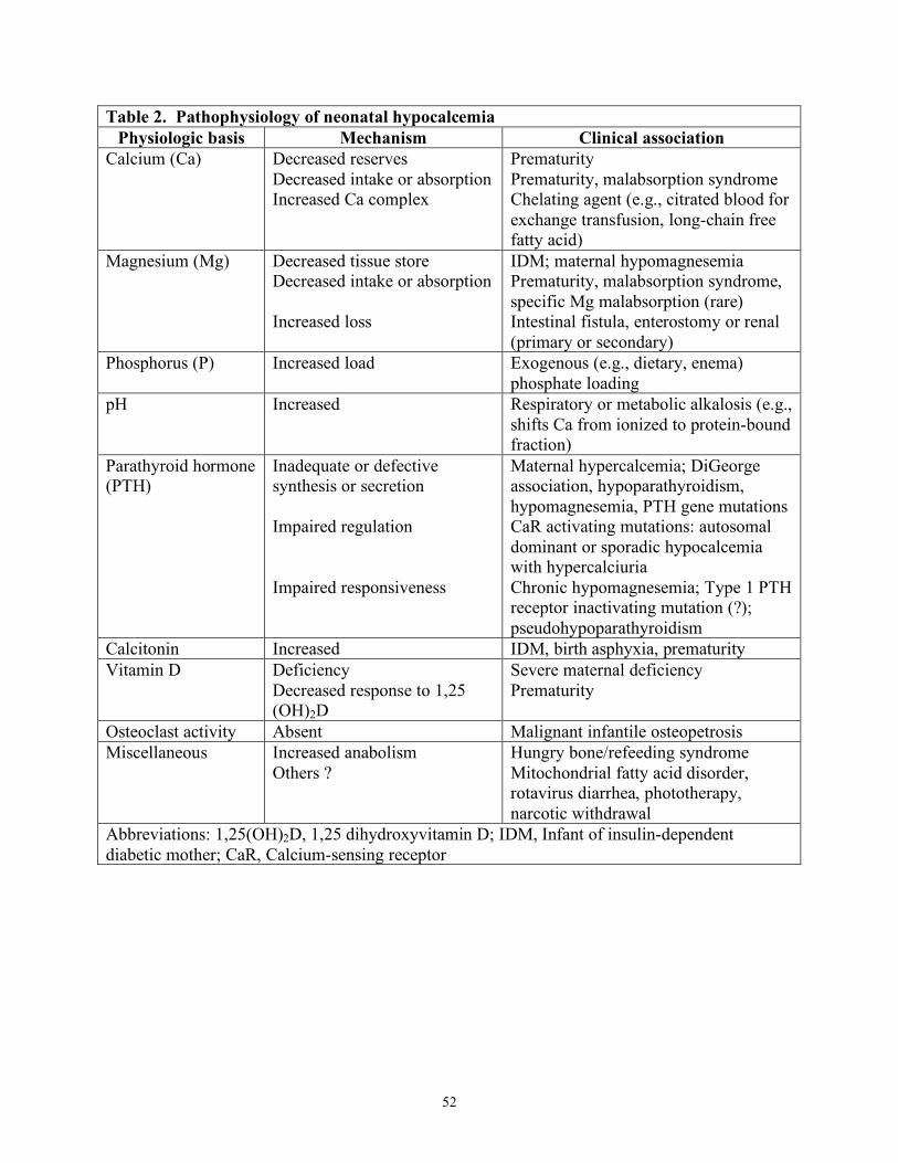
52
Table 2. Pathophysiology of neonatal hypocalcemia Physiologic basis Mechanism Clinical association
Calcium (Ca) Decreased reserves Decreased intake or absorption Increased Ca complex
Prematurity Prematurity, malabsorption syndrome Chelating agent (e.g., citrated blood for exchange transfusion, long-chain free fatty acid)
Magnesium (Mg) Decreased tissue store Decreased intake or absorption Increased loss
IDM; maternal hypomagnesemia Prematurity, malabsorption syndrome, specific Mg malabsorption (rare) Intestinal fistula, enterostomy or renal (primary or secondary)
Phosphorus (P) Increased load Exogenous (e.g., dietary, enema) phosphate loading
pH Increased Respiratory or metabolic alkalosis (e.g., shifts Ca from ionized to protein-bound fraction)
Parathyroid hormone (PTH)
Inadequate or defective synthesis or secretion Impaired regulation Impaired responsiveness
Maternal hypercalcemia; DiGeorge association, hypoparathyroidism, hypomagnesemia, PTH gene mutations CaR activating mutations: autosomal dominant or sporadic hypocalcemia with hypercalciuria Chronic hypomagnesemia; Type 1 PTH receptor inactivating mutation (?); pseudohypoparathyroidism
Calcitonin Increased IDM, birth asphyxia, prematurity Vitamin D Deficiency
Decreased response to 1,25 (OH)2D
Severe maternal deficiency Prematurity
Osteoclast activity Absent Malignant infantile osteopetrosis Miscellaneous Increased anabolism
Others ? Hungry bone/refeeding syndrome Mitochondrial fatty acid disorder, rotavirus diarrhea, phototherapy, narcotic withdrawal
Abbreviations: 1,25(OH)2D, 1,25 dihydroxyvitamin D; IDM, Infant of insulin-dependent diabetic mother; CaR, Calcium-sensing receptor

53
Table 3. Diagnostic workup for neonatal hypocalcemia History
• Screen for risk factors (See Table 1) Physical Examination
• General examination with focus on peripheral and central nervous and cardiovascular systems
• Associated features e.g., infant of a diabetic mother, prematurity, birth asphyxia, congenital heart disease, pseudohypoparathyroidism etc
Investigations1,2
• Serum total and ionized calcium (tCa and iCa), magnesium and phosphorus, total protein and albumin, and simultaneous “intact” or “whole” parathyroid hormone
• Acid-base status • Complete blood count (lymphocyte count) • Electrocardiogram (Q-Tc >0.4 sec or Qo-Tc >0.2 sec) • Chest X-ray (thymic shadow, aortic arch) • Urine calcium, phosphorus, magnesium and creatinine • Meconium and urine screen for narcotics • Maternal serum total and ionized calcium, magnesium and phosphorus, urine
calcium and phosphorus, if suspect maternal or heritable Ca disorder, particularly in persistent neonatal hypocalcemia
• Additional workup as indicated: vitamin D metabolites, T cell number and function, malabsorption studies, response to exogenous parathyroid hormone (PTH), molecular genetic studies (deletion of 22q11.2, PTH receptor and end organ responsiveness abnormalities, and calcium-sensing receptor defects, etc) and family screening
1. If serum tCa and iCa are normal, diagnostic workup should focus on non-calcium
related causes of clinical symptomatology, e.g., serum glucose, sepsis workup, screen for excretion of illicit drugs, neuroimaging studies etc
2. Resolution of clinical symptomatology when serum tCa or iCa has been normalized confirms the role of hypocalcemia
3. Maternal and family screening for calcium disorders is indicated in the absence of specific diagnosis for the neonatal hypocalcemia.
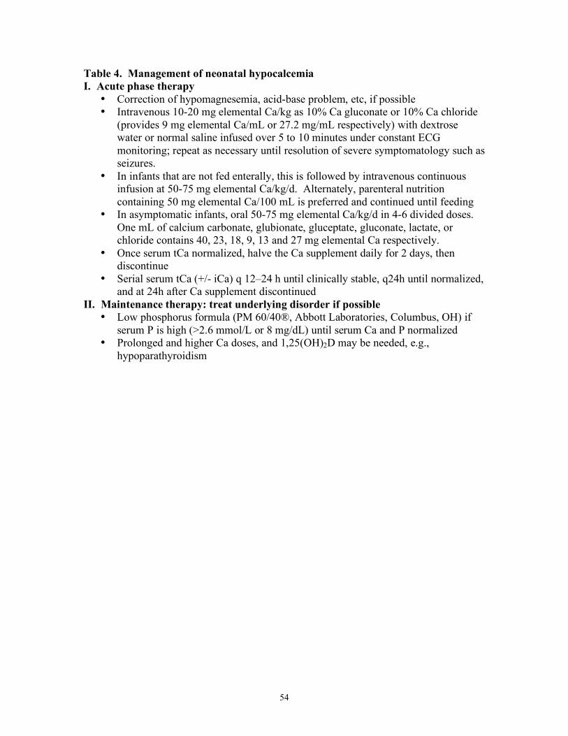
54
Table 4. Management of neonatal hypocalcemia I. Acute phase therapy
• Correction of hypomagnesemia, acid-base problem, etc, if possible • Intravenous 10-20 mg elemental Ca/kg as 10% Ca gluconate or 10% Ca chloride
(provides 9 mg elemental Ca/mL or 27.2 mg/mL respectively) with dextrose water or normal saline infused over 5 to 10 minutes under constant ECG monitoring; repeat as necessary until resolution of severe symptomatology such as seizures.
• In infants that are not fed enterally, this is followed by intravenous continuous infusion at 50-75 mg elemental Ca/kg/d. Alternately, parenteral nutrition containing 50 mg elemental Ca/100 mL is preferred and continued until feeding
• In asymptomatic infants, oral 50-75 mg elemental Ca/kg/d in 4-6 divided doses. One mL of calcium carbonate, glubionate, gluceptate, gluconate, lactate, or chloride contains 40, 23, 18, 9, 13 and 27 mg elemental Ca respectively.
• Once serum tCa normalized, halve the Ca supplement daily for 2 days, then discontinue
• Serial serum tCa (+/- iCa) q 12–24 h until clinically stable, q24h until normalized, and at 24h after Ca supplement discontinued
II. Maintenance therapy: treat underlying disorder if possible • Low phosphorus formula (PM 60/40®, Abbott Laboratories, Columbus, OH) if
serum P is high (>2.6 mmol/L or 8 mg/dL) until serum Ca and P normalized • Prolonged and higher Ca doses, and 1,25(OH)2D may be needed, e.g.,
hypoparathyroidism
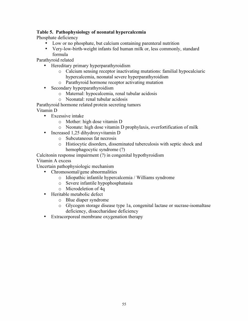
55
Table 5. Pathophysiology of neonatal hypercalcemia Phosphate deficiency
• Low or no phosphate, but calcium containing parenteral nutrition • Very-low-birth-weight infants fed human milk or, less commonly, standard
formula Parathyroid related
• Hereditary primary hyperparathyroidism o Calcium sensing receptor inactivating mutations: familial hypocalciuric
hypercalcemia, neonatal severe hyperparathyroidism o Parathyroid hormone receptor activating mutation
• Secondary hyperparathyroidism o Maternal: hypocalcemia, renal tubular acidosis o Neonatal: renal tubular acidosis
Parathyroid hormone related protein secreting tumors Vitamin D
• Excessive intake o Mother: high dose vitamin D o Neonate: high dose vitamin D prophylaxis, overfortification of milk
• Increased 1,25 dihydroxyvitamin D o Subcutaneous fat necrosis o Histiocytic disorders, disseminated tuberculosis with septic shock and
hemophagocytic syndrome (?) Calcitonin response impairment (?) in congenital hypothyroidism Vitamin A excess Uncertain pathophysiologic mechanism
• Chromosomal/gene abnormalities o Idiopathic infantile hypercalcemia / Williams syndrome o Severe infantile hypophosphatasia o Microdeletion of 4q
• Heritable metabolic defect o Blue diaper syndrome o Glycogen storage disease type 1a, congenital lactase or sucrase-isomaltase
deficiency, disaccharidase deficiency • Extracorporeal membrane oxygenation therapy

56
Table 6. Diagnostic workup for neonatal hypercalcemia History
• Familial or maternal disturbances in calcium (Ca) or phosphorus (P) metabolism • Gestational age, difficult labor, extracorporeal membrane oxygenation therapy
(ECMO) and pre-ECMO therapy • Intake of calcium, phosphorus, vitamins D and A: mother and infant
Physical Examination • General examination with focus on growth parameters, hydration status, heart
rate, blood pressure, cornea for band keratopathy (rare) • Associated features (e.g., subcutaneous fat necrosis, elfin facies, congenital heart
disease, developmental delay) Investigations
• Serum total and ionized Ca, magnesium, P, creatinine (Cr), total protein and albumin, alkaline phosphatase (total and bone specific), simultaneous “intact” or “whole” parathyroid hormone (PTH), 25 hydroxyvitamin D and 1,25 dihydroxyvitamin D
• Acid-base status • Urine Ca, P, Cr, amino-acids • X-ray of chest, hands and long bones • Ultrasound of kidneys and abdomen, ophthalmologic examination,
electrocardiogram (shortened QT interval, bradycardia) for complications • If above do not yield diagnosis, other tests depend on associated history and
symptomatology o Parental (both parents) serum and urine Ca, P, Cr o Molecular studies o Family screening depends on the primary diagnosis o Serum PTH-related protein and screen for occult tumor o Screen for metabolic defects, unusual dietary supplement
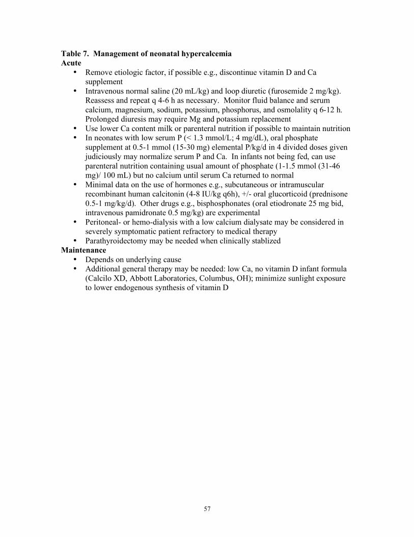
57
Table 7. Management of neonatal hypercalcemia Acute
• Remove etiologic factor, if possible e.g., discontinue vitamin D and Ca supplement
• Intravenous normal saline (20 mL/kg) and loop diuretic (furosemide 2 mg/kg). Reassess and repeat q 4-6 h as necessary. Monitor fluid balance and serum calcium, magnesium, sodium, potassium, phosphorus, and osmolality q 6-12 h. Prolonged diuresis may require Mg and potassium replacement
• Use lower Ca content milk or parenteral nutrition if possible to maintain nutrition • In neonates with low serum P (< 1.3 mmol/L; 4 mg/dL), oral phosphate
supplement at 0.5-1 mmol (15-30 mg) elemental P/kg/d in 4 divided doses given judiciously may normalize serum P and Ca. In infants not being fed, can use parenteral nutrition containing usual amount of phosphate (1-1.5 mmol (31-46 mg)/ 100 mL) but no calcium until serum Ca returned to normal
• Minimal data on the use of hormones e.g., subcutaneous or intramuscular recombinant human calcitonin (4-8 IU/kg q6h), +/- oral glucorticoid (prednisone 0.5-1 mg/kg/d). Other drugs e.g., bisphosphonates (oral etiodronate 25 mg bid, intravenous pamidronate 0.5 mg/kg) are experimental
• Peritoneal- or hemo-dialysis with a low calcium dialysate may be considered in severely symptomatic patient refractory to medical therapy
• Parathyroidectomy may be needed when clinically stablized Maintenance
• Depends on underlying cause • Additional general therapy may be needed: low Ca, no vitamin D infant formula
(Calcilo XD, Abbott Laboratories, Columbus, OH); minimize sunlight exposure to lower endogenous synthesis of vitamin D
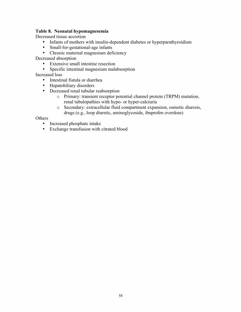
58
Table 8. Neonatal hypomagnesemia Decreased tissue accretion
• Infants of mothers with insulin-dependent diabetes or hyperparathyroidism • Small-for-gestational-age infants • Chronic maternal magnesium deficiency
Decreased absorption • Extensive small intestine resection • Specific intestinal magnesium malabsorption
Increased loss • Intestinal fistula or diarrhea • Hepatobiliary disorders • Decreased renal tubular reabsorption
o Primary: transient receptor potential channel protein (TRPM) mutation, renal tubulopathies with hypo- or hyper-calciuria
o Secondary: extracellular fluid compartment expansion, osmotic diuresis, drugs (e.g., loop diuretic, aminoglycoside, ibuprofen overdose)
Others • Increased phosphate intake • Exchange transfusion with citrated blood

59
Table 9. Neonatal hypermagnesemia Increased load
• Maternal magnesium sulfate administration • Neonatal magnesium therapy
o Parenteral nutrition o Antacid o Enema
Decreased excretion • Prematurity and asphyxia

60
Table 10. Risk factors for the development of osteopenia and rickets in infants In utero
• Severe maternal nutritional osteomalacia (i.e., vitamin D +/- calcium deficiency) • Maternal hypoparathyroidism and hyperparathyroidism • Prolonged maternal magnesium or phosphate treatment • Birth weight <1 kg
Postnatal • Prolonged organ dysfunction: intestine, kidney, liver, pancreas • Nutritional
o Preterm infants Prolonged low calcium and/or low phosphate total parenteral
nutrition Soy formula or unfortified human milk for small preterm infants Chronic loop diuretic therapy given to preterm infants
o Term infants Vitamin D deficiency Calcium deficiency Macrobiotic diet
o Toxin contamination: aluminum (?) • Inherited Defects
o Renal tubular disorder o Disorders of vitamin D or parathyroid hormone metabolism o Hypo- or hyper- phosphatasia