review article oxidative stress and salvia miltiorrhiza in...
TRANSCRIPT
Review ArticleOxidative Stress and Salvia miltiorrhiza inAging-Associated Cardiovascular Diseases
Cheng-Chieh Chang,1 Yu-Chun Chang,2 Wen-Long Hu,1,3,4 and Yu-Chiang Hung1,5
1Department of Chinese Medicine, Kaohsiung Chang Gung Memorial Hospital and Chang Gung University College of Medicine,Kaohsiung, Taiwan2Department of Applied Cosmetology, Kao Yuan University, Kaohsiung, Taiwan3Kaohsiung Medical University, College of Medicine, Kaohsiung, Taiwan4Fooyin University College of Nursing, Kaohsiung, Taiwan5School of Chinese Medicine for Post Baccalaureate, I-Shou University, Kaohsiung, Taiwan
Correspondence should be addressed to Yu-Chiang Hung; [email protected]
Received 31 May 2016; Accepted 15 September 2016
Academic Editor: Capucine Trollet
Copyright © 2016 Cheng-Chieh Chang et al. This is an open access article distributed under the Creative Commons AttributionLicense, which permits unrestricted use, distribution, and reproduction in any medium, provided the original work is properlycited.
Aging-associated cardiovascular diseases (CVDs) have some risk factors that are closely related to oxidative stress. Salviamiltiorrhiza (SM) has been used commonly to treat CVDs for hundreds of years in the Chinese community. We aimed toexplore the effects of SM on oxidative stress in aging-associated CVDs. Through literature searches using Medicine, PubMed,EMBASE, Cochrane library, CINAHL, and Scopus databases, we found that SM not only possesses antioxidant, antiapoptotic, andanti-inflammatory effects but also exerts angiogenic and cardioprotective activities. SM may reduce the production of reactiveoxygen species by inhibiting oxidases, reducing the production of superoxide, inhibiting the oxidative modification of low-density lipoproteins, and ameliorating mitochondrial oxidative stress. SM also increases the activities of catalase, manganesesuperoxide dismutase, glutathione peroxidase, and coupled endothelial nitric oxide synthase. In addition, SM reduces the impact ofischemia/reperfusion injury, prevents cardiac fibrosis after myocardial infarction, preserves cardiac function in coronary disease,maintains the integrity of the blood-brain barrier, and promotes self-renewal and proliferation of neural stem/progenitor cells instroke. However, future clinical well-designed and randomized control trials will be necessary to confirm the efficacy of SM inaging-associated CVDs.
1. Introduction
Cardiovascular diseases (CVDs) are a group of disordersrelated to the heart or blood vessels. Major CVDs includestroke, ischemic heart disease, cardiomyopathy, rheumaticheart disease, hypertensive heart disease, endocarditis, atrialfibrillation, aortic aneurysm, and peripheral arterial disease[1]. Global life expectancy increased from 65.3 years in 1990to 71.5 years in 2013. At the same time, the numbers ofdeaths from noncommunicable diseases increased steadily[2]. CVDs are the leading form of noncommunicable diseases[2]. In 2012 and 2013, 17.3 million deaths worldwide resultedfromCVDs [3]. Among these deaths, coronary artery diseaseand stroke contributed most to the total global burden of
CVDs [1]. It is estimated that 90% of CVDs are preventable[4].The Framingham andWorldHealth OrganizationMON-ICA studies found several risk factors for CVDs (e.g., age,smoking, physical inactivity, unhealthy diet, obesity, familyhistory, hypertension, diabetes mellitus, and hyperlipidemia)[5–10]. Some of these risk factors are immutable; however,many important risk factors are modifiable. When relevantrisk factors decrease, the incidence and mortality of CVDsimproved.
Most CVD risk factors are related to oxidative stress.Reactive oxygen species (ROS) are themain cause of oxidativestress and are highly reactive with proteins, lipids, and DNA,damaging these cellular components [11]. Under normal con-ditions, the production of ROS during aerobic metabolism
Hindawi Publishing CorporationOxidative Medicine and Cellular LongevityVolume 2016, Article ID 4797102, 11 pageshttp://dx.doi.org/10.1155/2016/4797102
2 Oxidative Medicine and Cellular Longevity
NO
Coupled eNOS
Uncoupled eNOS
Oxidase
Catalase
Glutathione peroxidase
Superoxide
Tan IIA [57, 58, 100, 118]Danshensu [65]
Enhancement of enzyme activityInhibition of enzyme activity
Tan IIA [59, 60]Sal B [108]SM injection B [68]DLA [108]
Hydrophilic polysaccharide of SM [54]Sal B [133]; Sal A [113]SM injection [43, 68]Danshensu [53]Water-soluble polysaccharide [116]Tan IIA [37, 109]
Danshensu [53]Tan IIA [43, 109, 130]
SM injection [43]Hydrophilic polysaccharide of SM [54]
peroxynitrite
Danshensu [53]Hydrophilic polysaccharide of SM [54]
Hydrogen peroxide
O2
O2
H2O2SOD(SOD1,SOD2,SOD3)
H2O + O2
2H2O
ONOO−
BH4/BH2↓
BH4/BH2↑
∙
O2
−
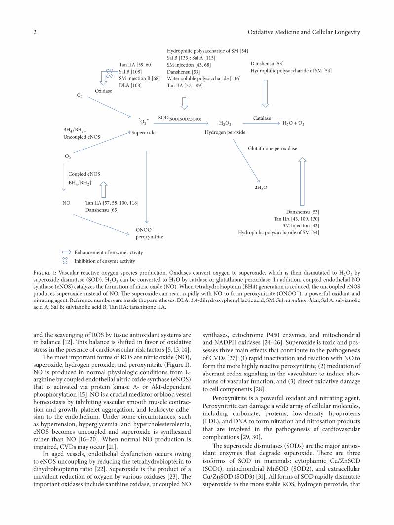
Figure 1: Vascular reactive oxygen species production. Oxidases convert oxygen to superoxide, which is then dismutated to H2O2 bysuperoxide dismutase (SOD). H2O2 can be converted to H2O by catalase or glutathione peroxidase. In addition, coupled endothelial NOsynthase (eNOS) catalyzes the formation of nitric oxide (NO). When tetrahydrobiopterin (BH4) generation is reduced, the uncoupled eNOSproduces superoxide instead of NO. The superoxide can react rapidly with NO to form peroxynitrite (ONOO−), a powerful oxidant andnitrating agent. Reference numbers are inside the parentheses.DLA: 3,4-dihydroxyphenyl lactic acid; SM: Salviamiltiorrhiza; Sal A: salvianolicacid A; Sal B: salvianolic acid B; Tan IIA: tanshinone IIA.
and the scavenging of ROS by tissue antioxidant systems arein balance [12]. This balance is shifted in favor of oxidativestress in the presence of cardiovascular risk factors [5, 13, 14].
The most important forms of ROS are nitric oxide (NO),superoxide, hydrogen peroxide, and peroxynitrite (Figure 1).NO is produced in normal physiologic conditions from L-arginine by coupled endothelial nitric oxide synthase (eNOS)that is activated via protein kinase A- or Akt-dependentphosphorylation [15]. NO is a crucialmediator of blood vesselhomeostasis by inhibiting vascular smooth muscle contrac-tion and growth, platelet aggregation, and leukocyte adhe-sion to the endothelium. Under some circumstances, suchas hypertension, hyperglycemia, and hypercholesterolemia,eNOS becomes uncoupled and superoxide is synthesizedrather than NO [16–20]. When normal NO production isimpaired, CVDs may occur [21].
In aged vessels, endothelial dysfunction occurs owingto eNOS uncoupling by reducing the tetrahydrobiopterin todihydrobiopterin ratio [22]. Superoxide is the product of aunivalent reduction of oxygen by various oxidases [23]. Theimportant oxidases include xanthine oxidase, uncoupled NO
synthases, cytochrome P450 enzymes, and mitochondrialand NADPH oxidases [24–26]. Superoxide is toxic and pos-sesses three main effects that contribute to the pathogenesisof CVDs [27]: (1) rapid inactivation and reaction with NO toform the more highly reactive peroxynitrite; (2) mediation ofaberrant redox signaling in the vasculature to induce alter-ations of vascular function, and (3) direct oxidative damageto cell components [28].
Peroxynitrite is a powerful oxidant and nitrating agent.Peroxynitrite can damage a wide array of cellular molecules,including carbonate, proteins, low-density lipoproteins(LDL), and DNA to form nitration and nitrosation productsthat are involved in the pathogenesis of cardiovascularcomplications [29, 30].
The superoxide dismutases (SODs) are the major antiox-idant enzymes that degrade superoxide. There are threeisoforms of SOD in mammals: cytoplasmic Cu/ZnSOD(SOD1), mitochondrial MnSOD (SOD2), and extracellularCu/ZnSOD (SOD3) [31]. All forms of SOD rapidly dismutatesuperoxide to the more stable ROS, hydrogen peroxide, that
Oxidative Medicine and Cellular Longevity 3
Major hydrophilic phenolic acids of SM
Major lipophilic terpenoids of SM
Miltirone
OHHO
O
OH
Caffeic acid
HO
HO OHOH
O
Danshensu
OO
Cryptotanshinone
O
OO
O
Dihydrotanshinone
OO
Tanshinone IIA
O
HO
HO
HO
HO OH
OH
OH
OHO
O
O
O
O
OR
O
Lithospermic acid B
O
HO
HO
HO
O
O O
O
OH
OH
Lithospermic acid
OO
HO
HO
OH
O
O
O OH OH
OH
HO
Salvianolic acid A
HOOH
O O
O
O
O
OH
OH
OHOH
OH
Salvianolic acid B
OR1 OR2
CO2HCO2H
OO
Tanshinone I
O
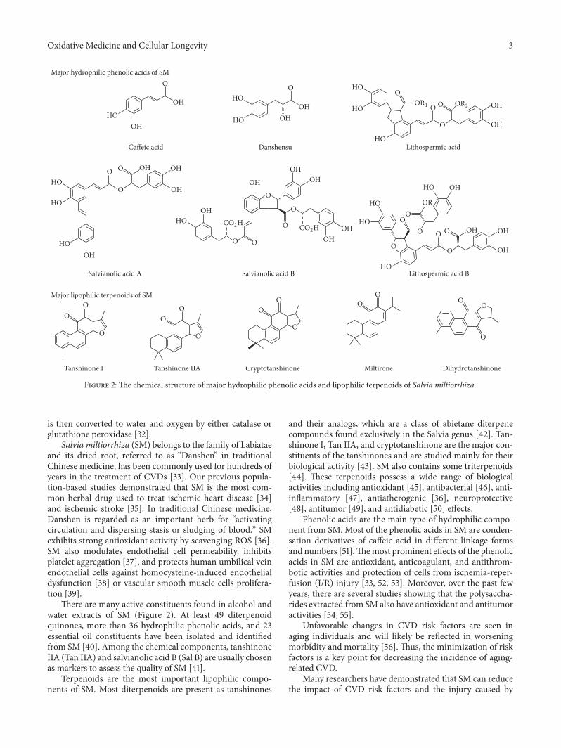
Figure 2: The chemical structure of major hydrophilic phenolic acids and lipophilic terpenoids of Salvia miltiorrhiza.
is then converted to water and oxygen by either catalase orglutathione peroxidase [32].
Salvia miltiorrhiza (SM) belongs to the family of Labiataeand its dried root, referred to as “Danshen” in traditionalChinese medicine, has been commonly used for hundreds ofyears in the treatment of CVDs [33]. Our previous popula-tion-based studies demonstrated that SM is the most com-mon herbal drug used to treat ischemic heart disease [34]and ischemic stroke [35]. In traditional Chinese medicine,Danshen is regarded as an important herb for “activatingcirculation and dispersing stasis or sludging of blood.” SMexhibits strong antioxidant activity by scavenging ROS [36].SM also modulates endothelial cell permeability, inhibitsplatelet aggregation [37], and protects human umbilical veinendothelial cells against homocysteine-induced endothelialdysfunction [38] or vascular smooth muscle cells prolifera-tion [39].
There are many active constituents found in alcohol andwater extracts of SM (Figure 2). At least 49 diterpenoidquinones, more than 36 hydrophilic phenolic acids, and 23essential oil constituents have been isolated and identifiedfrom SM [40]. Among the chemical components, tanshinoneIIA (Tan IIA) and salvianolic acid B (Sal B) are usually chosenas markers to assess the quality of SM [41].
Terpenoids are the most important lipophilic compo-nents of SM. Most diterpenoids are present as tanshinones
and their analogs, which are a class of abietane diterpenecompounds found exclusively in the Salvia genus [42]. Tan-shinone I, Tan IIA, and cryptotanshinone are the major con-stituents of the tanshinones and are studied mainly for theirbiological activity [43]. SM also contains some triterpenoids[44]. These terpenoids possess a wide range of biologicalactivities including antioxidant [45], antibacterial [46], anti-inflammatory [47], antiatherogenic [36], neuroprotective[48], antitumor [49], and antidiabetic [50] effects.
Phenolic acids are the main type of hydrophilic compo-nent from SM. Most of the phenolic acids in SM are conden-sation derivatives of caffeic acid in different linkage formsand numbers [51].Themost prominent effects of the phenolicacids in SM are antioxidant, anticoagulant, and antithrom-botic activities and protection of cells from ischemia-reper-fusion (I/R) injury [33, 52, 53]. Moreover, over the past fewyears, there are several studies showing that the polysaccha-rides extracted from SM also have antioxidant and antitumoractivities [54, 55].
Unfavorable changes in CVD risk factors are seen inaging individuals and will likely be reflected in worseningmorbidity and mortality [56]. Thus, the minimization of riskfactors is a key point for decreasing the incidence of aging-related CVD.
Many researchers have demonstrated that SM can reducethe impact of CVD risk factors and the injury caused by
4 Oxidative Medicine and Cellular Longevity
oxidative stress. This article provides an overview of SM interms of its ability to reduce oxidative stress-induced injuryand aging-related risk factors associatedwith CVD. Studies ofthe effects of SM on the two most frequent CVDs, coronaryartery disease and stroke, are also reviewed.
2. Materials and Methods
The current review focuses on the role of oxidative stress andSM (Danshen) in aging-associated CVDs. Literature searcheswere done using theMedicine, PubMed, EMBASE, Cochranelibrary, CINAHL, and Scopus databases, and the contents ofthe identified articles were summarized.
3. Results and Discussion
3.1. Hypertension. Hypertension is the most readily modifi-able risk factor for CVDs [57]. Oxidative stress, an aberrantvascular redox system, and endothelial dysfunction cancontribute to hypertension [13, 28]. In traditional Chinesemedicine, Danshen is the most frequently prescribed singleherb for hypertension [58]. Tan IIA has a vasodilatory effectthrough restoring eNOS coupling by increasing the ratioof tetrahydrobiopterin to dihydrobiopterin and reducingthe production of superoxide by inhibiting the expressionof NOX4, a member of the NADPH oxidase family [59,60]. In addition to reducing ROS, Tan IIA protects againstendothelial cell damage by decreasing the Bax/Bcl-2 ratio andinhibiting caspase-3 activation [61]. The water extract of SMthat contains lithospermic acid B (also named tanshinoateB) and Sal B exhibits an antihypertensive effect through theinhibition of angiotensin-converting enzyme or the reninangiotensin system [62–65]. Sal B and danshensu, the majorhydrophilic constituents of SM, can regulate vascular toneand reduce blood pressure by activating of the calcium-activated big potassium (BKCA) channel [66, 67]. In addition,SM injection can decrease plasma levels of endothelin-1 andthromboxane B2 [68].
3.2. Smoking. Cigarette smoking is an important and revers-ible risk factor for CVDs but is ranked lower than hyperten-sion because of the widespread implementation of smoke-free legislation [69]. However, in some countries, smokingremains the third leading risk factor forCVDs, behind dietaryrisks and hypertension. Smokers return to the risk level ofnever-smokers after cessation of smoking for at least 10 years[69].
Smoking may enhance oxidative stress not only throughthe production of ROS but also through weakening of theantioxidant defense systems [70]. Smoking-associated CVDsinclude abdominal aortic aneurysm, peripheral artery dis-ease, unheralded coronary death, and subarachnoid hem-orrhage [71]. SalA attenuates the formation of aortic aneu-rysms in apolipoprotein E-deficient mice by selectivelyinhibiting matrix metalloproteinase-9 (MMP-9) to maintainthe integrity of blood vessels [72]. A crude extract of SM dila-tes isolated rat femoral arteries by opening tetraethylammo-nium-sensitive potassium channels in smooth muscle cells[73] and produces a vasorelaxant effect in the rat knee joint
through the release of calcitonin gene-related peptide and theendothelium-derived relaxant factorsNO and prostaglandins[74]. One recent study revealed that the injection of Danshenroot could suppress cigarette smoking-induced lung inflam-mation by decreasing the levels of interleukin- (IL-) 8, IL-6,and tumor necrosis factor- (TNF-) 𝛼 in Sprague-Dawley rats[75].
3.3. Hyperglycemia. Hyperglycemia and diabetes mellitus arestrong, significant, and independent risk factors for CVDs[76, 77]. Hyperglycemia induces oxidative stress in diabeticpatients, and the overproduction of ROS contributes tothe development of CVDs [78, 79]. Peroxynitrite plays animportant role in the pathogenesis of diabetic CVD compli-cations through oxidative and nitrosative stress [29]. In thepresence of hyperglycemia, vascular remodeling is aug-mented by uncoupled eNOS [80], increases in endothelialsuperoxide levels that inhibit vascular smooth muscle Na-K-ATPase activity [81], and downregulation of transient recep-tor potential cation channel subfamily V member 4 that reg-ulates vascular function [82]. The hydrophilic extract of SMclearly ameliorates oxidative stress stimulated by hypergly-cemia in diabetic patients with coronary heart disease [83].The induction of vascular endothelial growth factor (VEGF)expression by high glucose levels is also reversed by the SMhydrophilic extract through amelioration of mitochondrialoxidative stress [84].
Sal B is the main bioactive component in the SM hydro-philic extract [85]. The tanshinones are insulin sensitizersthat enhance the activity of insulin on tyrosine phosphory-lation through the activation of Akt and extracellular signal-regulated kinase (ERK)1/2 and by glycogen synthase kinase(GSK)3𝛽 and glucose transporter (GLUT)4 translocation[86]. Furthermore, the hydrophilic polysaccharide of SMprotects against the development of type 2 diabetes by atten-uating insulin resistance through increases in the activities ofcatalase, MnSOD, and glutathione peroxidase in rats [54].
3.4. Hyperlipidemia. Hyperlipidemia is the most importantrisk factor for atherosclerosis and a major cause of CVDs [87,88]. Increased transcytosis of lipoproteins is the initial eventin atherogenesis. ROS generated by activated inflammatorycells and the production of oxidized lipoproteins are keypoints for atherosclerotic plaque erosion and rupture [89].We showed that Tan IIA exhibits a strong antiatheroscle-rotic effect associated with reduced vascular cell adhe-sion molecule- (VCAM-) 1, intercellular adhesion molecule-(ICAM-) 1, and CX3CL1 expression through inhibition of theNF-𝜅B signaling pathway in human vascular endothelial cells[90]. Sal B inhibits LDL oxidation and neointimal hyper-plasia in endothelium-denuded hypercholesterolemic rabbitsthrough inhibition of ROS production [91]. Magnesium tan-shinoate B, an important aqueous component of SM, can alsoinhibit oxidative modification of LDLs, prevent the uptakeof LDLs by macrophages [92], and protect endothelial cellsagainst oxidized lipoprotein-induced apoptosis [93].
Oxidative Medicine and Cellular Longevity 5
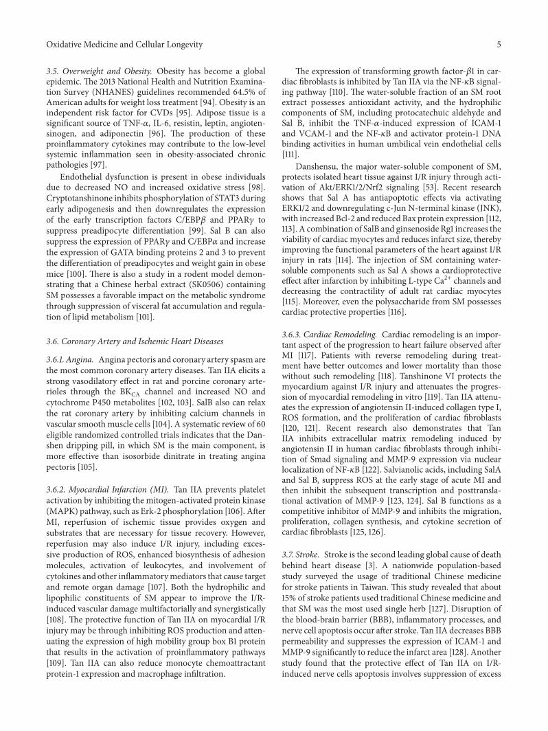
3.5. Overweight and Obesity. Obesity has become a globalepidemic. The 2013 National Health and Nutrition Examina-tion Survey (NHANES) guidelines recommended 64.5% ofAmerican adults for weight loss treatment [94]. Obesity is anindependent risk factor for CVDs [95]. Adipose tissue is asignificant source of TNF-𝛼, IL-6, resistin, leptin, angioten-sinogen, and adiponectin [96]. The production of theseproinflammatory cytokines may contribute to the low-levelsystemic inflammation seen in obesity-associated chronicpathologies [97].
Endothelial dysfunction is present in obese individualsdue to decreased NO and increased oxidative stress [98].Cryptotanshinone inhibits phosphorylation of STAT3 duringearly adipogenesis and then downregulates the expressionof the early transcription factors C/EBP𝛽 and PPAR𝛾 tosuppress preadipocyte differentiation [99]. Sal B can alsosuppress the expression of PPAR𝛾 and C/EBP𝛼 and increasethe expression of GATA binding proteins 2 and 3 to preventthe differentiation of preadipocytes and weight gain in obesemice [100]. There is also a study in a rodent model demon-strating that a Chinese herbal extract (SK0506) containingSM possesses a favorable impact on the metabolic syndromethrough suppression of visceral fat accumulation and regula-tion of lipid metabolism [101].
3.6. Coronary Artery and Ischemic Heart Diseases
3.6.1. Angina. Angina pectoris and coronary artery spasm arethe most common coronary artery diseases. Tan IIA elicits astrong vasodilatory effect in rat and porcine coronary arte-rioles through the BKCA channel and increased NO andcytochrome P450 metabolites [102, 103]. SalB also can relaxthe rat coronary artery by inhibiting calcium channels invascular smoothmuscle cells [104]. A systematic review of 60eligible randomized controlled trials indicates that the Dan-shen dripping pill, in which SM is the main component, ismore effective than isosorbide dinitrate in treating anginapectoris [105].
3.6.2. Myocardial Infarction (MI). Tan IIA prevents plateletactivation by inhibiting the mitogen-activated protein kinase(MAPK) pathway, such as Erk-2 phosphorylation [106]. AfterMI, reperfusion of ischemic tissue provides oxygen andsubstrates that are necessary for tissue recovery. However,reperfusion may also induce I/R injury, including exces-sive production of ROS, enhanced biosynthesis of adhesionmolecules, activation of leukocytes, and involvement ofcytokines and other inflammatorymediators that cause targetand remote organ damage [107]. Both the hydrophilic andlipophilic constituents of SM appear to improve the I/R-induced vascular damage multifactorially and synergistically[108]. The protective function of Tan IIA on myocardial I/Rinjury may be through inhibiting ROS production and atten-uating the expression of high mobility group box B1 proteinthat results in the activation of proinflammatory pathways[109]. Tan IIA can also reduce monocyte chemoattractantprotein-1 expression and macrophage infiltration.
The expression of transforming growth factor-𝛽1 in car-diac fibroblasts is inhibited by Tan IIA via the NF-𝜅B signal-ing pathway [110]. The water-soluble fraction of an SM rootextract possesses antioxidant activity, and the hydrophiliccomponents of SM, including protocatechuic aldehyde andSal B, inhibit the TNF-𝛼-induced expression of ICAM-1and VCAM-1 and the NF-𝜅B and activator protein-1 DNAbinding activities in human umbilical vein endothelial cells[111].
Danshensu, the major water-soluble component of SM,protects isolated heart tissue against I/R injury through acti-vation of Akt/ERK1/2/Nrf2 signaling [53]. Recent researchshows that Sal A has antiapoptotic effects via activatingERK1/2 and downregulating c-Jun N-terminal kinase (JNK),with increased Bcl-2 and reduced Bax protein expression [112,113]. A combination of SalB and ginsenoside Rg1 increases theviability of cardiac myocytes and reduces infarct size, therebyimproving the functional parameters of the heart against I/Rinjury in rats [114]. The injection of SM containing water-soluble components such as Sal A shows a cardioprotectiveeffect after infarction by inhibiting L-type Ca2+ channels anddecreasing the contractility of adult rat cardiac myocytes[115]. Moreover, even the polysaccharide from SM possessescardiac protective properties [116].
3.6.3. Cardiac Remodeling. Cardiac remodeling is an impor-tant aspect of the progression to heart failure observed afterMI [117]. Patients with reverse remodeling during treat-ment have better outcomes and lower mortality than thosewithout such remodeling [118]. Tanshinone VI protects themyocardium against I/R injury and attenuates the progres-sion of myocardial remodeling in vitro [119]. Tan IIA attenu-ates the expression of angiotensin II-induced collagen type I,ROS formation, and the proliferation of cardiac fibroblasts[120, 121]. Recent research also demonstrates that TanIIA inhibits extracellular matrix remodeling induced byangiotensin II in human cardiac fibroblasts through inhibi-tion of Smad signaling and MMP-9 expression via nuclearlocalization of NF-𝜅B [122]. Salvianolic acids, including SalAand Sal B, suppress ROS at the early stage of acute MI andthen inhibit the subsequent transcription and posttransla-tional activation of MMP-9 [123, 124]. Sal B functions as acompetitive inhibitor of MMP-9 and inhibits the migration,proliferation, collagen synthesis, and cytokine secretion ofcardiac fibroblasts [125, 126].
3.7. Stroke. Stroke is the second leading global cause of deathbehind heart disease [3]. A nationwide population-basedstudy surveyed the usage of traditional Chinese medicinefor stroke patients in Taiwan. This study revealed that about15% of stroke patients used traditional Chinese medicine andthat SM was the most used single herb [127]. Disruption ofthe blood-brain barrier (BBB), inflammatory processes, andnerve cell apoptosis occur after stroke. Tan IIA decreases BBBpermeability and suppresses the expression of ICAM-1 andMMP-9 significantly to reduce the infarct area [128]. Anotherstudy found that the protective effect of Tan IIA on I/R-induced nerve cells apoptosis involves suppression of excess
6 Oxidative Medicine and Cellular Longevity
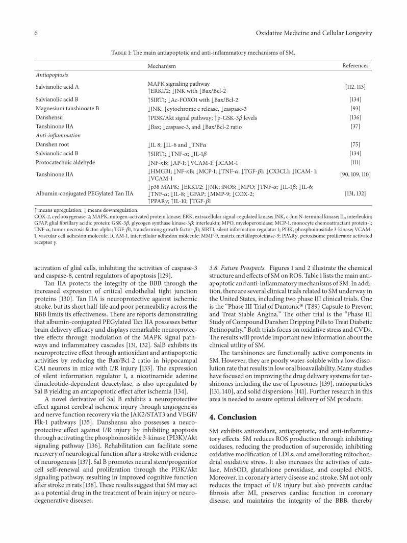
Table 1: The main antiapoptotic and anti-inflammatory mechanisms of SM.
Mechanism ReferencesAntiapoptosis
Salvianolic acid A MAPK signaling pathway↑ERK1/2; ↓JNK with ↓Bax/Bcl-2 [112, 113]
Salvianolic acid B ↑SIRT1; ↓Ac-FOXO1 with ↓Bax/Bcl-2 [134]Magnesium tanshinoate B ↓JNK, ↓cytochrome c release, ↓caspase-3 [93]Danshensu ↑PI3K/Akt signal pathway; ↑p-GSK-3𝛽 levels [136]Tanshinone IIA ↓Bax; ↓caspase-3, and ↓Bax/Bcl-2 ratio [37]Anti-inflammationDanshen root ↓IL 8; ↓IL-6 and ↓TNF𝛼 [75]Salvianolic acid B ↑SIRT1; ↓TNF-𝛼; ↓IL-1𝛽 [134]Protocatechuic aldehyde ↓NF-𝜅B; ↓AP-1; ↓VCAM-1; ↓ICAM-1 [111]
Tanshinone IIA ↓HMGB1; ↓NF-𝜅B; ↓MCP-1; ↓TNF-𝛼; ↓TGF-𝛽1; ↓CX3CL1; ↓ICAM- 1;↓VCAM-1 [90, 109, 110]
Albumin-conjugated PEGylated Tan IIA↓p38 MAPK; ↓ERK1/2; ↓JNK; iNOS; ↓MPO; ↓TNF-𝛼; ↓IL-1𝛽; ↓IL-6;↓TNF-𝛼; ↓IL-8; ↓GFAP; ↓MMP-9; ↓COX-2;↑PPAR𝛾; ↑IL-10; ↑TGF-𝛽1
[131, 132]
↑means upregulation; ↓means downregulation.COX-2, cyclooxygenase-2; MAPK,mitogen-activated protein kinase; ERK, extracellular signal-regulated kinase; JNK, c-JunN-terminal kinase; IL, interleukin;GFAP, glial fibrillary acidic protein; GSK-3𝛽, glycogen synthase kinase-3𝛽; interleukin; MPO, myeloperoxidase; MCP-1, monocyte chemoattractant protein-1;TNF-𝛼, tumor necrosis factor-alpha; TGF-𝛽1, transforming growth factor-𝛽1; SIRT1, silent information regulator 1; PI3K, phosphoinositide 3-kinase; VCAM-1, vascular cell adhesion molecule; ICAM-1, intercellular adhesion molecule; MMP-9, matrix metalloproteinase-9; PPAR𝛾, peroxisome proliferator activatedreceptor 𝛾.
activation of glial cells, inhibiting the activities of caspase-3and caspase-8, central regulators of apoptosis [129].
Tan IIA protects the integrity of the BBB through theincreased expression of critical endothelial tight junctionproteins [130]. Tan IIA is neuroprotective against ischemicstroke, but its short half-life and poor permeability across theBBB limits its effectiveness. There are reports demonstratingthat albumin-conjugated PEGylated Tan IIA possesses betterbrain delivery efficacy and displays remarkable neuroprotec-tive effects through modulation of the MAPK signal path-ways and inflammatory cascades [131, 132]. SalB exhibits itsneuroprotective effect through antioxidant and antiapoptoticactivities by reducing the Bax/Bcl-2 ratio in hippocampalCA1 neurons in mice with I/R injury [133]. The expressionof silent information regulator 1, a nicotinamide adeninedinucleotide-dependent deacetylase, is also upregulated bySal B yielding an antiapoptotic effect after ischemia [134].
A novel derivative of Sal B exhibits a neuroprotectiveeffect against cerebral ischemic injury through angiogenesisand nerve function recovery via the JAK2/STAT3 and VEGF/Flk-1 pathways [135]. Danshensu also possesses a neuro-protective effect against I/R injury by inhibiting apoptosisthrough activating the phosphoinositide 3-kinase (PI3K)/Aktsignaling pathway [136]. Rehabilitation can facilitate somerecovery of neurological function after a stroke with evidenceof neurogenesis [137]. Sal B promotes neural stem/progenitorcell self-renewal and proliferation through the PI3K/Aktsignaling pathway, resulting in improved cognitive functionafter stroke in rats [138].These results suggest that SMmay actas a potential drug in the treatment of brain injury or neuro-degenerative diseases.
3.8. Future Prospects. Figures 1 and 2 illustrate the chemicalstructure and effects of SMonROS. Table 1 lists themain anti-apoptotic and anti-inflammatorymechanisms of SM. In addi-tion, there are several clinical trials related to SMunderway inthe United States, including two phase III clinical trials. Oneis the “Phase III Trial of Dantonic� (T89) Capsule to Preventand Treat Stable Angina.” The other trial is the “Phase IIIStudy ofCompoundDanshenDripping Pills to TreatDiabeticRetinopathy.” Both trials focus on oxidative stress and CVDs.The results will provide important new information about theclinical utility of SM.
The tanshinones are functionally active components inSM. However, they are poorly water-soluble with a low disso-lution rate that results in loworal bioavailability.Many studieshave focused on improving the drug delivery systems for tan-shinones including the use of liposomes [139], nanoparticles[131, 140], and solid dispersions [141]. Further research in thisarea is needed to assure optimal delivery of SM products.
4. Conclusion
SM exhibits antioxidant, antiapoptotic, and anti-inflamma-tory effects. SM reduces ROS production through inhibitingoxidases, reducing the production of superoxide, inhibitingoxidative modification of LDLs, and ameliorating mitochon-drial oxidative stress. It also increases the activities of cata-lase, MnSOD, glutathione peroxidase, and coupled eNOS.Moreover, in coronary artery disease and stroke, SM not onlyreduces the impact of I/R injury but also prevents cardiacfibrosis after MI, preserves cardiac function in coronarydisease, and maintains the integrity of the BBB, thereby
Oxidative Medicine and Cellular Longevity 7
promoting neural stem/progenitor cell self-renewal and pro-liferation following a stroke.Therefore, SM can be an effectiveagent for the prevention and treatment of CVDs. However,in accordance with in vitro and in vivo laboratory evidence,well-designed clinical studies are necessary to confirm theefficacy of SM in the treatment of CVDs.
Competing Interests
The authors declare no competing interests.
Authors’ Contributions
Cheng-Chieh Chang and Yu-Chun Chang contributedequally to this work.
Acknowledgments
This work was supported partly by National Science ResearchGrant of Taiwan (NSC-96-2320-B-182-023-MY2) and ChangGung Memorial Hospital (CMRPG83011; CMRPD32027).
References
[1] A. E. Moran, G. A. Roth, J. Narula, and G. A. Mensah, “1990–2010 Global cardiovascular disease atlas,” Global Heart, vol. 9,no. 1, pp. 3–16, 2014.
[2] GBD 2013 Mortality and Causes of Death Collaborators,“Global, regional, and national age-sex specific all-cause andcause-specific mortality for 240 causes of death, 1990–2013:a systematic analysis for the Global Burden of Disease Study2013,”The Lancet, vol. 385, no. 9963, pp. 117–171, 2015.
[3] G. A. Roth, M. D. Huffman, A. E. Moran et al., “Global andregional patterns in cardiovascularmortality from 1990 to 2013,”Circulation, vol. 132, no. 17, pp. 1667–1678, 2015.
[4] H. C.McGill Jr., C. A.McMahan, and S. S. Gidding, “Preventingheart disease in the 21st century: implications of the Patho-biological Determinants of Atherosclerosis in Youth (PDAY)study,” Circulation, vol. 117, no. 9, pp. 1216–1227, 2008.
[5] J. R. Margolis, R. F. Gillum, M. Feinleib, R. C. Brasch, and R. R.Fabsitz, “Community surveillance for coronary heart disease:the framingham cardiovascular disease survey: methods andpreliminary results,”American Journal of Epidemiology, vol. 100,no. 6, pp. 425–436, 1974.
[6] R. F. Gillum, M. Feinleib, J. R. Margolis, R. R. Fabsitz, and R.C. Brasch, “Community surveillance for cardiovascular disease:the Framingham cardiovascular disease survey. Some method-ological problems in the community study of cardiovasculardisease,” Journal of Chronic Diseases, vol. 29, no. 5, pp. 289–299,1976.
[7] W. B. Kannel, D. McGee, and T. Gordon, “A general cardiovas-cular risk profile: the Framingham study,”TheAmerican Journalof Cardiology, vol. 38, no. 1, pp. 46–51, 1976.
[8] R. F. Gillum, R. R. Fabsitz, M. Feinleib, P. A.Wolf, J. R.Margolis,and R. C. Brasch, “Community surveillance for cerebrovasculardisease: the Framingham Cardiovascular Disease Survey,” Pub-lic Health Reports, vol. 93, no. 5, pp. 438–442, 1978.
[9] U. Keil, “The worldwide WHO MONICA project: results andperspectives,”Gesundheitswesen, vol. 67, supplement 1, pp. S38–S45, 2005.
[10] C. W. Tsao and R. S. Vasan, “Cohort Profile: the FraminghamHeart Study (FHS): overview of milestones in cardiovascularepidemiology,” International Journal of Epidemiology, vol. 44,no. 6, pp. 1800–1813, 2015.
[11] C. A. Papaharalambus and K. K. Griendling, “Basic mecha-nisms of oxidative stress and reactive oxygen species in cardio-vascular injury,” Trends in Cardiovascular Medicine, vol. 17, no.2, pp. 48–54, 2007.
[12] B. Frei, “Reactive oxygen species and antioxidant vitamins:mechanisms of action,” The American Journal of Medicine, vol.97, no. 3, pp. 5S–13S, 1994.
[13] Q. N. Dinh, G. R. Drummond, C. G. Sobey, and S. Chrissobolis,“Roles of inflammation, oxidative stress, and vascular dysfunc-tion in hypertension,” BioMed Research International, vol. 2014,Article ID 406960, 11 pages, 2014.
[14] D. Tousoulis, A. Briasoulis, N. Papageorgiou et al., “Oxidativestress and endothelial function: therapeutic interventions,”Recent Patents on Cardiovascular Drug Discovery, vol. 6, no. 2,pp. 103–114, 2011.
[15] M. Khazaei, F. Moien-afshari, and I. Laher, “Vascular endothe-lial function in health and diseases,” Pathophysiology, vol. 15, no.1, pp. 49–67, 2008.
[16] A. Bouloumie, J. Bauersachs, W. Linz et al., “Endothelialdysfunction coincides with an enhanced nitric oxide synthaseexpression and superoxide anion production,” Hypertension,vol. 30, no. 4, pp. 934–941, 1997.
[17] F. Cosentino, K. Hishikawa, Z. S. Katusic, and T. F. Luscher,“High glucose increases nitric oxide synthase expression andsuperoxide anion generation in human aortic endothelial cells,”Circulation, vol. 96, no. 1, pp. 25–28, 1997.
[18] U. Landmesser, S. Dikalov, S. R. Price et al., “Oxidation oftetrahydrobiopterin leads to uncoupling of endothelial cellnitric oxide synthase in hypertension,” The Journal of ClinicalInvestigation, vol. 111, no. 8, pp. 1201–1209, 2003.
[19] S. Kawashima and M. Yokoyama, “Dysfunction of endothe-lial nitric oxide synthase and atherosclerosis,” Arteriosclerosis,Thrombosis, and Vascular Biology, vol. 24, no. 6, pp. 998–1005,2004.
[20] K. A. Pritchard Jr., L. Groszek, D. M. Smalley et al., “Nativelow-density lipoprotein increases endothelial cell nitric oxidesynthase generation of superoxide anion,” Circulation Research,vol. 77, no. 3, pp. 510–518, 1995.
[21] K. Chen, R. N. Pittman, and A. S. Popel, “Nitric oxide in thevasculature: where does it come from and where does it go? Aquantitative perspective,”Antioxidants and Redox Signaling, vol.10, no. 7, pp. 1185–1198, 2008.
[22] Y.-M. Yang, A. Huang, G. Kaley, and D. Sun, “eNOS uncouplingand endothelial dysfunction in aged vessels,” American Journalof Physiology—Heart and Circulatory Physiology, vol. 297, no. 5,pp. H1829–H1836, 2009.
[23] I. Fridovich, “Superoxide anion radical (O⋅2), superoxide dismu-tases, and related matters,” The Journal of Biological Chemistry,vol. 272, pp. 18515–18517, 1997.
[24] L. S. Terada, I. R. Willingham, M. E. Rosandich, J. A. Leff, G.W. Kindt, and J. E. Repine, “Generation of superoxide anionby brain endothelial cell xanthine oxidase,” Journal of CellularPhysiology, vol. 148, no. 2, pp. 191–196, 1991.
[25] R.M.Clancy, J. Leszczynska-Piziak, and S. B. Abramson, “Nitricoxide, an endothelial cell relaxation factor, inhibits neutrophilsuperoxide anion production via a direct action on the NADPHoxidase,”The Journal of Clinical Investigation, vol. 90, no. 3, pp.1116–1121, 1992.
8 Oxidative Medicine and Cellular Longevity
[26] J.-M. Li and A. M. Shah, “Differential NADPH- versus NADH-dependent superoxide production by phagocyte-type endothe-lial cell NADPH oxidase,” Cardiovascular Research, vol. 52, no.3, pp. 477–486, 2001.
[27] J.-M. Li and A. M. Shah, “Endothelial cell superoxide genera-tion: regulation and relevance for cardiovascular pathophysi-ology,” American Journal of Physiology—Regulatory Integrativeand Comparative Physiology, vol. 287, no. 5, pp. R1014–R1030,2004.
[28] M. Y. Lee and K. K. Griendling, “Redox signaling, vascularfunction, and hypertension,” Antioxidants & Redox Signaling,vol. 10, no. 6, pp. 1045–1059, 2008.
[29] P. Pacher and C. Szabo, “Role of peroxynitrite in the patho-genesis of cardiovascular complications of diabetes,” CurrentOpinion in Pharmacology, vol. 6, no. 2, pp. 136–141, 2006.
[30] Z. Cao and Y. Li, “Potent inhibition of peroxynitrite-inducedDNA strand breakage by ethanol: possible implications forethanol-mediated cardiovascular protection,” PharmacologicalResearch, vol. 50, no. 1, pp. 13–19, 2004.
[31] T. Fukai and M. Ushio-Fukai, “Superoxide dismutases: role inredox signaling, vascular function, and diseases,” Antioxidants& Redox Signaling, vol. 15, no. 6, pp. 1583–1606, 2011.
[32] J. Han, V. V. Shuvaev, and V. R. Muzykantov, “Catalaseand superoxide dismutase conjugated with platelet-endothelialcell adhesion molecule antibody distinctly alleviate abnormalendothelial permeability caused by exogenous reactive oxygenspecies and vascular endothelial growth factor,” The Journal ofPharmacology and ExperimentalTherapeutics, vol. 338, no. 1, pp.82–91, 2011.
[33] X. Wang, S. L. Morris-Natschke, and K.-H. Lee, “New develop-ments in the chemistry and biology of the bioactive constituentsof Tanshen,”Medicinal Research Reviews, vol. 27, no. 1, pp. 133–148, 2007.
[34] Y.-C. Hung, Y.-J. Tseng,W.-L. Hu et al., “Demographic and pre-scribing patterns of Chinese herbal products for individualizedtherapy for ischemic heart disease in Taiwan: population-basedstudy,” PLoS ONE, vol. 10, no. 8, Article ID e0137058, 2015.
[35] I. L. Hung, Y. C. Hung, L. Y. Wang et al., “Chinese herbalproducts for ischemic stroke,”The American Journal of ChineseMedicine, vol. 43, no. 7, pp. 1365–1379, 2015.
[36] J. Fu, H. Huang, J. Liu, R. Pi, J. Chen, and P. Liu, “TanshinoneIIA protects cardiac myocytes against oxidative stress-triggereddamage and apoptosis,” European Journal of Pharmacology, vol.568, no. 1–3, pp. 213–221, 2007.
[37] J.-Q. Liu, T.-F. Lee, M. Miedzyblocki, G. C. F. Chan, D. L.Bigam, and P.-Y. Cheung, “Effects of tanshinone IIA, a majorcomponent of Salvia miltiorrhiza, on platelet aggregation inhealthy newborn piglets,” Journal of Ethnopharmacology, vol.137, no. 1, pp. 44–49, 2011.
[38] K. Chan, S. H. Chui, D. Y. L.Wong,W. Y. Ha, C. L. Chan, and R.N. S. Wong, “Protective effects of Danshensu from the aqueousextract of Salvia miltiorrhiza (Danshen) against homocysteine-induced endothelial dysfunction,” Life Sciences, vol. 75, no. 26,pp. 3157–3171, 2004.
[39] Y.-C. Hung, P.-W. Wang, T.-L. Pan, G. Bazylak, and Y.-L.Leu, “Proteomic screening of antioxidant effects exhibited byRadix Salvia miltiorrhiza aqueous extract in cultured rat aorticsmooth muscle cells under homocysteine treatment,” Journal ofEthnopharmacology, vol. 124, no. 3, pp. 463–474, 2009.
[40] H. Pang, L.Wu,Y. Tang,G. Zhou,C.Qu, and J.Duan, “Chemicalanalysis of the herbal medicine salviae miltiorrhizae radix etrhizoma (Danshen),”Molecules, vol. 21, no. 1, article 51, 2016.
[41] J.-Z. Song, S.-L. Li, Y. Zhou, C.-F. Qiao, S.-L. Chen, and H.-X.Xu, “A novel approach to rapidly explore analytical markers forquality control of Radix Salviae Miltiorrhizae extract granulesby robust principal component analysis with ultra-high per-formance liquid chromatography-ultraviolet-quadrupole time-of-flight mass spectrometry,” Journal of Pharmaceutical andBiomedical Analysis, vol. 53, no. 3, pp. 279–286, 2010.
[42] Y. Zhang, P. Jiang, M. Ye, S.-H. Kim, C. Jiang, and J. Lu, “Tan-shinones: sources, pharmacokinetics and anti-cancer activities,”International Journal of Molecular Sciences, vol. 13, no. 10, pp.13621–13666, 2012.
[43] C.-Y. Su, Q.-L. Ming, K. Rahman, T. Han, and L.-P. Qin,“Salvia miltiorrhiza: traditional medicinal uses, chemistry, andpharmacology,”Chinese Journal of NaturalMedicines, vol. 13, no.3, pp. 163–182, 2015.
[44] Y.-B. Wu, Z.-Y. Ni, Q.-W. Shi et al., “Constituents from Salviaspecies and their biological activities,” Chemical Reviews, vol.112, no. 11, pp. 5967–6026, 2012.
[45] X.-L. Niu, K. Ichimori, X. Yang et al., “Tanshinone II-Ainhibits low density lipoprotein oxidation in vitro,” Free RadicalResearch, vol. 33, no. 3, pp. 305–312, 2000.
[46] D.-S. Lee, S.-H. Lee, J.-G. Noh, and S.-D. Hong, “Antibacterialactivities of cryptotanshinone and dihydrotanshinone I from amedicinal herb, Salvia miltiorrhiza bunge,” Bioscience, Biotech-nology and Biochemistry, vol. 63, no. 12, pp. 2236–2239, 1999.
[47] S. Y. Kim, T. C. Moon, H. W. Chang, K. H. Son, S. S. Kang,and H. P. Kim, “Effects of tanshinone I isolated from Salviamiltiorrhiza Bunge on arachidonic acid metabolism and in vivoinflammatory responses,” Phytotherapy Research, vol. 16, no. 7,pp. 616–620, 2002.
[48] T. Liu, H. Jin, Q.-R. Sun, J.-H. Xu, andH.-T. Hu, “The neuropro-tective effects of tanshinone IIA on 𝛽-amyloid-induced toxicityin rat cortical neurons,”Neuropharmacology, vol. 59, no. 7-8, pp.595–604, 2010.
[49] M. A. Mosaddik, “In vitro cytotoxicity of Tanshinones iso-lated from Salvia miltiorrhiza Bunge against P388 lymphocyticleukemia cells,” Phytomedicine, vol. 10, no. 8, pp. 682–685, 2003.
[50] F. Qiu, G. Wang, R. Zhang, J. Sun, J. Jiang, and Y. Ma, “Effectof danshen extract on the activity of CYP3A4 in healthyvolunteers,” British Journal of Clinical Pharmacology, vol. 69, no.6, pp. 656–662, 2010.
[51] R.-W. Jiang, K.-M. Lau, P.-M. Hon, T. C. W. Mak, K.-S. Woo,and K.-P. Fung, “Chemistry and biological activities of caffeicacid derivatives from Salvia miltiorrhiza,” Current MedicinalChemistry, vol. 12, no. 2, pp. 237–246, 2005.
[52] G. Ge, Q. Zhang, J. Ma et al., “Protective effect of Salviamiltiorrhiza aqueous extract on myocardium oxidative injuryin ischemic-reperfusion rats,” Gene, vol. 546, no. 1, pp. 97–103,2014.
[53] J. Yu, L. Wang, M. Akinyi et al., “Danshensu protects isolatedheart against ischemia reperfusion injury through activationof Akt/ERK1/2/Nrf2 signaling,” International Journal of Clinicaland Experimental Medicine, vol. 8, no. 9, pp. 14793–14804, 2015.
[54] W. Zhang, L. Zheng, Z. Zhang, and C.-X. Hai, “Protectiveeffect of a water-soluble polysaccharide from SalviamiltiorrhizaBunge on insulin resistance in rats,”Carbohydrate Polymers, vol.89, no. 3, pp. 890–898, 2012.
[55] Y.-Y. Jiang, L. Wang, L. Zhang et al., “Characterization, antiox-idant and antitumor activities of polysaccharides from Salviamiltiorrhiza Bunge,” International Journal of Biological Macro-molecules, vol. 70, pp. 92–99, 2014.
Oxidative Medicine and Cellular Longevity 9
[56] D. A. Rhoades, T. K. Welty, W. Wang et al., “Aging and theprevalence of cardiovascular disease risk factors in older Amer-ican Indians: the strong heart study,” Journal of the AmericanGeriatrics Society, vol. 55, no. 1, pp. 87–94, 2007.
[57] D. Peiris, S. R. Thompson, A. Beratarrechea et al., “Behaviourchange strategies for reducing blood pressure-related diseaseburden: findings from a global implementation research pro-gramme,” Implementation Science, vol. 10, no. 1, 2015.
[58] P.-R. Yang, W.-T. Shih, Y.-H. Chu, P.-C. Chen, and C.-Y.Wu, “Frequency and co-prescription pattern of Chinese herbalproducts for hypertension in Taiwan: A Cohort Study,” BMCComplementary and Alternative Medicine, vol. 15, no. 1, article163, 2015.
[59] Z.-W. Zhou, X.-L. Xie, S.-F. Zhou, and C. G. Li, “Mechanismof reversal of high glucose-induced endothelial nitric oxidesynthase uncoupling by tanshinone IIA in human endothelialcell line EA.hy926,” European Journal of Pharmacology, vol. 697,no. 1–3, pp. 97–105, 2012.
[60] P. Wang, X. Wu, Y. Bao et al., “Tanshinone IIA prevents cardiacremodeling through attenuating NAD (P)H oxidase-derivedreactive oxygen species production in hypertensive rats,” DiePharmazie, vol. 66, no. 7, pp. 517–524, 2011.
[61] L.-Q. Jia, G.-L. Yang, L. Ren et al., “Tanshinone IIA reducesapoptosis induced by hydrogen peroxide in the humanendothelium-derived EA.hy926 cells,” Journal of Ethnopharma-cology, vol. 143, no. 1, pp. 100–108, 2012.
[62] D. G. Kang, H. Oh, H. T. Chung, and H. S. Lee, “Inhibition ofangiotensin converting enzyme by lithospermic acid B isolatedfrom radix Salviae miltiorrhiza Bunge,” Phytotherapy Research,vol. 17, no. 8, pp. 917–920, 2003.
[63] D. G. Kang, Y. G. Yun, J. H. Ryoo, and H. S. Lee, “Anti-hypertensive effect of water extract of Danshen on renovascularhypertension through inhibition of the renin angiotensin sys-tem,” American Journal of Chinese Medicine, vol. 30, no. 1, pp.87–93, 2002.
[64] X. Ouyang, K. Takahashi, K. Komatsu et al., “Protective effectof Salvia miltiorrhiza on angiotensin II-induced hypertrophicresponses in neonatal rat cardiac cells,” Japanese Journal ofPharmacology, vol. 87, no. 4, pp. 289–296, 2001.
[65] S. W. S. Leung, D.-Y. Zhu, and R. Y. K. Man, “Effects ofthe aqueous extract of Salvia Miltiorrhiza (danshen) and itsmagnesium tanshinoate B-enriched form on blood pressure,”Phytotherapy Research, vol. 24, no. 5, pp. 769–774, 2010.
[66] F. F. Y. Lam, S. W. Seto, Y. W. Kwan, J. H. K. Yeung, and P.Chan, “Activation of the iberiotoxin-sensitive BKCa channels bysalvianolic acid B of the porcine coronary artery smoothmusclecells,” European Journal of Pharmacology, vol. 546, no. 1–3, pp.28–35, 2006.
[67] Y. Tang, M. Wang, C. Chen, X. Le, S. Sun, and Y. Yin,“Cardiovascular protection with danshensu in spontaneouslyhypertensive rats,” Biological & Pharmaceutical Bulletin, vol. 34,pp. 1596–1601, 2011.
[68] Z. Xia, J. Gu, D. M. Ansley, F. Xia, and J. Yu, “Antioxidanttherapy with Salvia miltiorrhiza decreases plasma endothelin-1and thromboxane B2 after cardiopulmonary bypass in patientswith congenital heart disease,” The Journal of Thoracic andCardiovascular Surgery, vol. 126, no. 5, pp. 1404–1410, 2003.
[69] K. Pirie, R. Peto, G. K. Reeves, J. Green, and V. Beral, “The21st century hazards of smoking and benefits of stopping: aprospective study of onemillion women in the UK,”The Lancet,vol. 381, no. 9861, pp. 133–141, 2013.
[70] B. Isik, A. Ceylan, and R. Isik, “Oxidative stress in smokers andnon-smokers,” Inhalation Toxicology, vol. 19, no. 9, pp. 767–769,2007.
[71] M. Pujades-Rodriguez, J. George, A. D. Shah et al., “Heteroge-neous associations between smoking and a wide range of initialpresentations of cardiovascular disease in 1937 360 people inEngland: lifetime risks and implications for risk prediction,”International Journal of Epidemiology, vol. 44, no. 1, pp. 129–141,2015.
[72] T. Zhang, J. Xu, D. Li et al., “Salvianolic acid A, a matrixmetalloproteinase-9 inhibitor of Salvia miltiorrhiza, attenuatesaortic aneurysm formation in apolipoprotein E-deficient mice,”Phytomedicine, vol. 21, no. 10, pp. 1137–1145, 2014.
[73] F. F. Y. Lam, J. H. K. Yeung, and J. H. Y. Cheung, “Mechanismsof the dilator action of Danshen (Salvia miltiorrhiza) on rat iso-lated femoral artery,” Journal of Cardiovascular Pharmacology,vol. 46, no. 3, pp. 361–368, 2005.
[74] F. Y. Lam, S. C. W. Ng, J. H. Y. Cheung, and J. H. K. Yeung,“Mechanisms of the vasorelaxant effect of Danshen (Salviamiltiorrhiza) in rat knee joints,” Journal of Ethnopharmacology,vol. 104, no. 3, pp. 336–344, 2006.
[75] F. W. Yi-ju Cheng, Y. Hu, M.-X. Wu, J. Du, and M.-L. Cheng,“Compound injection of Danshen root suppresses cigarettessmoking-induced lung inflammation: a SD rat model,” Inflam-mation and Cell Signaling, vol. 2, no. 4, pp. 1–6, 2015.
[76] D. E. Singer, D.M.Nathan, K.M.Anderson, P.W. F.Wilson, andJ. C. Evans, “Association ofHbA1cwith prevalent cardiovasculardisease in the original cohort of the Framingham heart study,”Diabetes, vol. 41, no. 2, pp. 202–208, 1992.
[77] W. B. Kannel and D. L. McGee, “Diabetes and glucose toleranceas risk factors for cardiovascular disease: The FraminghamStudy,” Diabetes Care, vol. 2, no. 2, pp. 120–126, 1979.
[78] A. Aydin, H. Orhan, A. Sayal, M. Ozata, G. Sahin, and A.Isimer, “Oxidative stress and nitric oxide related parameters intype II diabetes mellitus: effects of glycemic control,” ClinicalBiochemistry, vol. 34, no. 1, pp. 65–70, 2001.
[79] G. S. Dave and K. Kalia, “Hyperglycemia induced oxidativestress in type-1 and type-2 diabetic patients with and withoutnephropathy,” Cellular and Molecular Biology, vol. 53, no. 5, pp.68–78, 2007.
[80] N. Sasaki, T. Yamashita, T. Takaya et al., “Augmentation ofvascular remodeling by uncoupled endothelial nitric oxidesynthase in amousemodel of diabetesmellitus,”Arteriosclerosis,Thrombosis, and Vascular Biology, vol. 28, no. 6, pp. 1068–1076,2008.
[81] S. Gupta, E. Chough, J. Daley et al., “Hyperglycemia increasesendothelial superoxide that impairs smooth muscle cell Na+-K+-ATpase activity,”American Journal of Physiology—Cell Phys-iology, vol. 282, no. 3, pp. C560–C566, 2002.
[82] K. Monaghan, J. McNaughten, M. K. McGahon et al., “Hyper-glycemia and diabetes downregulate the functional expressionof TRPV4 channels in retinal microvascular endothelium,”PLoS ONE, vol. 10, no. 6, article e0128359, 2015.
[83] Q. Qian, S. Qian, P. Fan, D. Huo, and S. Wang, “Effect ofSalvia miltiorrhiza hydrophilic extract on antioxidant enzymesin diabetic patients with chronic heart disease: a randomizedcontrolled trial,” Phytotherapy Research, vol. 26, no. 1, pp. 60–66, 2012.
[84] S. Qian, D. Huo, S. Wang, and Q. Qian, “Inhibition of glucose-induced vascular endothelial growth factor expression bySalvia miltiorrhiza hydrophilic extract in humanmicrovascular
10 Oxidative Medicine and Cellular Longevity
endothelial cells: evidence for mitochondrial oxidative stress,”Journal of Ethnopharmacology, vol. 137, no. 2, pp. 985–991, 2011.
[85] M. Huang, P. Wang, S. Xu et al., “Biological activities ofsalvianolic acid B from Salvia miltiorrhiza on type 2 diabetesinduced by high-fat diet and streptozotocin,” PharmaceuticalBiology, vol. 53, no. 7, pp. 1058–1065, 2015.
[86] S. H. Jung, H. J. Seol, S. J. Jeon, K. H. Son, and J. R. Lee, “Insulin-sensitizing activities of tanshinones, diterpene compounds ofthe root of Salvia miltiorrhiza Bunge,” Phytomedicine, vol. 16,no. 4, pp. 327–335, 2009.
[87] National Cholesterol Education Program (NCEP) Expert Panelon Detection-Evaluation and Treatment of High Blood Choles-terol in Adults (Adult Treatment Panel III), “Third Report ofthe National Cholesterol Education Program (NCEP) expertpanel on detection, evaluation, and treatment of high bloodcholesterol in adults (Adult Treatment Panel III) final report,”Circulation, vol. 106, no. 25, pp. 3143–3421, 2002.
[88] K.Wouters, R. Shiri-Sverdlov, P. J. van Gorp, M. van Bilsen, andM. H. Hofker, “Understanding hyperlipidemia and atheroscle-rosis: lessons from genetically modified apoe and LDLR mice,”Clinical Chemistry and Laboratory Medicine, vol. 43, no. 5, pp.470–479, 2005.
[89] M. Hulsmans and P. Holvoet, “The vicious circle betweenoxidative stress and inflammation in atherosclerosis,” Journal ofCellular andMolecularMedicine, vol. 14, no. 1-2, pp. 70–78, 2010.
[90] C.-C. Chang, C.-F. Chu, C.-N. Wang et al., “The anti-atherosclerotic effect of tanshinone IIA is associated with theinhibition of TNF-𝛼-induced VCAM-1, ICAM-1 and CX3CL1expression,” Phytomedicine, vol. 21, no. 3, pp. 207–216, 2014.
[91] T.-L. Yang, F.-Y. Lin, Y.-H. Chen et al., “Salvianolic acidB inhibits low-density lipoprotein oxidation and neointimalhyperplasia in endothelium-denuded hypercholesterolaemicrabbits,” Journal of the Science of Food and Agriculture, vol. 91,no. 1, pp. 134–141, 2011.
[92] O. Karmin, E. G. Lynn, R. Vazhappilly, K. K. Au-Yeung, D. Y.Zhu, and Y. L. Siow, “Magnesium tanshinoate B (MTB) inhibitslow density lipoprotein oxidation,” Life Sciences, vol. 68, no. 8,pp. 903–912, 2001.
[93] K. K. W. Au-Yeung, O. Karmin, P. C. Choy, D.-Y. Zhu, andY. L. Siow, “Magnesium tanshinoate B protects endothelialcells against oxidized lipoprotein-induced apoptosis,”CanadianJournal of Physiology and Pharmacology, vol. 85, no. 11, pp. 1053–1062, 2007.
[94] J. Stevens, E. E. Oakkar, Z. Cui, J. Cai, and K. P. Truesdale, “USadults recommended for weight reduction by 1998 and 2013obesity guidelines, NHANES 2007–2012,” Obesity, vol. 23, no.3, pp. 527–531, 2015.
[95] P. Poirier and R. H. Eckel, “Obesity and cardiovascular disease,”Current Atherosclerosis Reports, vol. 4, no. 6, pp. 448–453, 2002.
[96] B. L. Wajchenberg, “Subcutaneous and visceral adipose tissue:their relation to the metabolic syndrome,” Endocrine Reviews,vol. 21, no. 6, pp. 697–738, 2000.
[97] S. Nishimura, I. Manabe, and R. Nagai, “Adipose tissueinflammation in obesity and metabolic syndrome,” DiscoveryMedicine, vol. 8, no. 41, pp. 55–60, 2009.
[98] P. Poirier, T. D. Giles, G. A. Bray et al., “Obesity and cardiovas-cular disease: pathophysiology, evaluation, and effect of weightloss,” Arteriosclerosis, Thrombosis, and Vascular Biology, vol. 26,no. 5, pp. 968–976, 2006.
[99] N. Rahman, M. Jeon, H.-Y. Song, and Y.-S. Kim, “Cryp-totanshinone, a compound of Salvia miltiorrhiza inhibits pre-adipocytes differentiation by regulation of adipogenesis-related
genes expression via STAT3 signaling,” Phytomedicine, vol. 23,no. 1, pp. 58–67, 2016.
[100] P. Wang, S. Xu, W. Li et al., “Salvianolic acid B inhibited ppar𝛾expression and attenuated weight gain in mice with high-fatdiet-induced obesity,”Cellular Physiology and Biochemistry, vol.34, no. 2, pp. 288–298, 2014.
[101] Y. Tan, M. A. Kamal, Z.-Z. Wang, W. Xiao, J. P. Seale, and X.Qu, “Chinese herbal extracts (SK0506) as a potential candidatefor the therapy of themetabolic syndrome,”Clinical Science, vol.120, no. 7, pp. 297–305, 2011.
[102] G. B.Wu, E. X. Zhou, andD. X. Qing, “TanshinoneII(A) elicitedvasodilation in rat coronary arteriole: roles of nitric oxide andpotassium channels,” European Journal of Pharmacology, vol.617, no. 1–3, pp. 102–107, 2009.
[103] Y. Yang, F. Cai, P.-Y. Li et al., “Activation of high conduc-tance Ca2+-activated K+ channels by sodium tanshinoneII-Asulfonate (DS-201) in porcine coronary artery smooth musclecells,” European Journal of Pharmacology, vol. 598, no. 1–3, pp.9–15, 2008.
[104] F. F. Y. Lam, J. H. K. Yeung, Y. W. Kwan, K. M. Chan, and P. M.Y. Or, “Salvianolic acid B, an aqueous component of danshen(Salviamiltiorrhiza), relaxes rat coronary artery by inhibition ofcalcium channels,” European Journal of Pharmacology, vol. 553,no. 1–3, pp. 240–245, 2006.
[105] Y. Jia, F. Huang, S. Zhang, and S.-W. Leung, “Is danshen(Salviamiltiorrhiza) dripping pillmore effective than isosorbidedinitrate in treating angina pectoris?A systematic reviewof ran-domized controlled trials,” International Journal of Cardiology,vol. 157, no. 3, pp. 330–340, 2012.
[106] F. Maione, V. De Feo, E. Caiazzo, L. De Martino, C. Cicala, andN. Mascolo, “Tanshinone IIA, a major component of Salviamilthorriza Bunge, inhibits platelet activation via Erk-2 signal-ing pathway,” Journal of Ethnopharmacology, vol. 155, no. 2, pp.1236–1242, 2014.
[107] D. L. Carden and D. N. Granger, “Pathophysiology of ischae-mia-reperfusion injury,” Journal of Pathology, vol. 190, no. 3, pp.255–266, 2000.
[108] J.-Y. Han, J.-Y. Fan, Y. Horie et al., “Ameliorating effectsof compounds derived from Salvia miltiorrhiza root extracton microcirculatory disturbance and target organ injury byischemia and reperfusion,” Pharmacology & Therapeutics, vol.117, no. 2, pp. 280–295, 2008.
[109] H. Hu, C. Zhai, G. Qian et al., “Protective effects of tanshinoneIIA on myocardial ischemia reperfusion injury by reducingoxidative stress, HMGB1 expression, and inflammatory reac-tion,”Pharmaceutical Biology, vol. 53, no. 12, pp. 1752–1758, 2015.
[110] Z. H. Ren, Y. H. Tong, W. Xu, J. Ma, and Y. Chen, “TanshinoneII A attenuates inflammatory responses of rats with myocardialinfarction by reducing MCP-1 expression,” Phytomedicine, vol.17, no. 3-4, pp. 212–218, 2010.
[111] Z. Zhou, Y. Liu, A.-D. Miao, and S.-Q. Wang, “Protocatechuicaldehyde suppresses TNF-𝛼-induced ICAM-1 and VCAM-1expression in human umbilical vein endothelial cells,” EuropeanJournal of Pharmacology, vol. 513, no. 1-2, pp. 1–8, 2005.
[112] T. Xu, X. Wu, Q. Chen et al., “The anti-apoptotic and cardio-protective effects of salvianolic acid a on rat cardiomyocytesfollowing ischemia/reperfusion by DUSP-mediated regulationof the ERK1/2/JNK pathway,” PLoS ONE, vol. 9, no. 7, Article IDe102292, 2014.
[113] H. Fan, L. Yang, F. Fu et al., “Cardioprotective effects of salviano-lic acid a on myocardial ischemia-reperfusion injury in vivo
Oxidative Medicine and Cellular Longevity 11
and in vitro,” Evidence-Based Complementary and AlternativeMedicine, vol. 2012, Article ID 508938, 9 pages, 2012.
[114] Y. Deng, M. Yang, F. Xu et al., “Combined salvianolic acid Band ginsenoside Rg1 exerts cardioprotection against ischemia/reperfusion injury in rats,” PLoS ONE, vol. 10, no. 8, Article IDe0135435, 2015.
[115] Y. Gao, K. Zhang, F. Zhu et al., “Salvia miltiorrhiza (Danshen)inhibits L-type calcium current and attenuates calcium tran-sient and contractility in rat ventricular myocytes,” Journal ofEthnopharmacology, vol. 158, pp. 397–403, 2014.
[116] M. Song, L. Huang, G. Zhao, and Y. Song, “Beneficial effectsof a polysaccharide from Salvia miltiorrhiza on myocardialischemia-reperfusion injury in rats,” Carbohydrate Polymers,vol. 98, no. 2, pp. 1631–1636, 2013.
[117] M. A. Konstam, D. G. Kramer, A. R. Patel, M. S. Maron, andJ. E. Udelson, “Left ventricular remodeling in heart failure:current concepts in clinical significance and assessment,” JACC:Cardiovascular Imaging, vol. 4, no. 1, pp. 98–108, 2011.
[118] J. R. D. A. R. Reis Filho, J. N. Cardoso, C. M. D. R. Cardoso, andA. C. Pereira-Barretto, “Reverse cardiac remodeling: a markerof better prognosis in heart failure,” Arquivos Brasileiros deCardiologia, vol. 104, no. 6, pp. 502–506, 2015.
[119] A. Yagi and S. Takeo, “Anti-inflammatory constituents, aloesinand aloemannan in Aloe species and effects of tanshinon VIin Salvia miltiorrhiza on heart,” Yakugakuzasshi: Journal of thePharmaceutical Society of Japan, vol. 123, no. 7, pp. 517–532, 2003.
[120] P. Chan, J.-C. Liu, L.-J. Lin et al., “Tanshinone IIA inhibitsangiotensin II-induced cell proliferation in rat cardiac fibrob-lasts,” The American Journal of Chinese Medicine, vol. 39, no. 2,pp. 381–394, 2011.
[121] L. Yang, X.-J. Zou, X. Gao et al., “Sodium tanshinone IIA sul-fonate attenuates angiotensin II-induced collagen type I expres-sion in cardiac fibroblasts in vitro,” Experimental andMolecularMedicine, vol. 41, no. 7, pp. 508–516, 2009.
[122] S. Mao, W. Li, N. Qa’aty, M. Vincent, M. Zhang, and A. Hinek,“Tanshinone IIA inhibits angiotensin II induced extracellularmatrix remodeling in human cardiac fibroblasts—implicationsfor treatment of pathologic cardiac remodeling,” InternationalJournal of Cardiology, vol. 202, pp. 110–117, 2016.
[123] B. Jiang, W. Wu, M. Li et al., “Cardioprotection and matrixmetalloproteinase-9 regulation of salvianolic acids on myocar-dial infarction in rats,” Planta Medica, vol. 75, no. 12, pp. 1286–1292, 2009.
[124] B. Jiang, D. Li, Y. Deng et al., “Salvianolic acid A, a novel matrixmetalloproteinase-9 inhibitor, prevents cardiac remodeling inspontaneously hypertensive rats,” PLoS ONE, vol. 8, no. 3,Article ID e59621, 2013.
[125] B. Jiang, J. Chen, L. Xu et al., “Salvianolic acid B functionedas a competitive inhibitor of matrix metalloproteinase-9 andefficiently prevented cardiac remodeling,” BMC Pharmacology,vol. 10, article 10, 2010.
[126] Y. Wang, F. Xu, J. Chen et al., “Matrix metalloproteinase-9induces cardiac fibroblast migration, collagen and cytokinesecretion: inhibition by salvianolic acid B from Salvia miltior-rhiza,” Phytomedicine, vol. 19, no. 1, pp. 13–19, 2011.
[127] C. C. Chang, Y. C. Lee, C. C. Lin et al., “Characteristics oftraditional Chinese medicine usage in patients with stroke inTaiwan: a nationwide population-based study,” Journal of Ethno-pharmacology, vol. 186, pp. 311–321, 2016.
[128] C. Tang,H. Xue, C. Bai, R. Fu, andA.Wu, “The effects of Tanshi-none IIA on blood-brain barrier and brain edema after transient
middle cerebral artery occlusion in rats,” Phytomedicine, vol. 17,no. 14, pp. 1145–1149, 2010.
[129] L. Zhou, S. C. Bondy, L. Jian et al., “Tanshinone IIA attenuatesthe cerebral ischemic injury-induced increase in levels of GFAPand of caspases-3 and -8,” Neuroscience, vol. 288, pp. 105–111,2015.
[130] X. Yang, J. Yan, and J. Feng, “Treatment with tanshinone IIAsuppresses disruption of the blood-brain barrier and reducesexpression of adhesion molecules and chemokines in exper-imental autoimmune encephalomyelitis,” European Journal ofPharmacology, vol. 771, pp. 18–28, 2016.
[131] X. Liu, M. Ye, C. An, L. Pan, and L. Ji, “The effect of cationicalbumin-conjugated PEGylated tanshinone IIA nanoparticleson neuronal signal pathways and neuroprotection in cerebralischemia,” Biomaterials, vol. 34, no. 28, pp. 6893–6905, 2013.
[132] X. Liu, C. An, P. Jin, X. Liu, and L. Wang, “Protective effects ofcationic bovine serum albumin-conjugated PEGylated tanshi-none IIA nanoparticles on cerebral ischemia,” Biomaterials, vol.34, no. 3, pp. 817–830, 2013.
[133] Y.-F. Jiang, Z.-Q. Liu, W. Cui et al., “Antioxidant effect ofsalvianolic acid B on hippocampal CA1 neurons in mice withcerebral ischemia and reperfusion injury,” Chinese Journal ofIntegrative Medicine, vol. 21, no. 7, pp. 516–522, 2015.
[134] H. Lv, L.Wang, J. Shen et al., “Salvianolic acid B attenuates apop-tosis and inflammation via SIRT1 activation in experimentalstroke rats,” Brain Research Bulletin, vol. 115, pp. 30–36, 2015.
[135] H. Zhu, L. Zou, J. Tian, G. Du, and Y. Gao, “SMND-309, a novelderivative of salvianolic acid B, protects rat brains ischemiaand reperfusion injury by targeting the JAK2/STAT3 pathway,”European Journal of Pharmacology, vol. 714, no. 1–3, pp. 23–31,2013.
[136] C. Guo, Y. Yin, J. Duan et al., “Neuroprotective effect and under-lying mechanism of sodium danshensu [3-(3,4-dihydroxy-phenyl) lactic acid from Radix and Rhizoma Salviae miltior-rhizae = Danshen] against cerebral ischemia and reperfusioninjury in rats,” Phytomedicine, vol. 22, no. 2, pp. 283–289, 2015.
[137] G.-L.Ming andH. Song, “Adult neurogenesis in themammalianbrain: significant answers and significant questions,” Neuron,vol. 70, no. 4, pp. 687–702, 2011.
[138] P. Zhuang, Y. Zhang, G. Cui et al., “Direct stimulation of adultneural stem/progenitor cells in vitro and neurogenesis in vivoby salvianolic acid B,” PLoSONE, vol. 7, no. 4, Article ID e35636,2012.
[139] T. A. Elbayoumi andV. P. Torchilin, “Current trends in liposomeresearch,”Methods inMolecular Biology, vol. 605, pp. 1–27, 2010.
[140] Y. Cai, W. Zhang, Z. Chen, Z. Shi, H. Chengwei, and M. Chen,“Recent insights into the biological activities and drug deliverysystems of tanshinones,” International Journal of Nanomedicine,vol. 11, pp. 121–130, 2016.
[141] X. Zhao, X. Liu, L. Gan, C. Zhou, and J. Mo, “Preparationand physicochemical characterizations of tanshinone IIA soliddispersion,” Archives of Pharmacal Research, vol. 34, no. 6, pp.949–959, 2011.

Submit your manuscripts athttp://www.hindawi.com
Stem CellsInternational
Hindawi Publishing Corporationhttp://www.hindawi.com Volume 2014
Hindawi Publishing Corporationhttp://www.hindawi.com Volume 2014
MEDIATORSINFLAMMATION
of
Hindawi Publishing Corporationhttp://www.hindawi.com Volume 2014
Behavioural Neurology
EndocrinologyInternational Journal of
Hindawi Publishing Corporationhttp://www.hindawi.com Volume 2014
Hindawi Publishing Corporationhttp://www.hindawi.com Volume 2014
Disease Markers
Hindawi Publishing Corporationhttp://www.hindawi.com Volume 2014
BioMed Research International
OncologyJournal of
Hindawi Publishing Corporationhttp://www.hindawi.com Volume 2014
Hindawi Publishing Corporationhttp://www.hindawi.com Volume 2014
Oxidative Medicine and Cellular Longevity
Hindawi Publishing Corporationhttp://www.hindawi.com Volume 2014
PPAR Research
The Scientific World JournalHindawi Publishing Corporation http://www.hindawi.com Volume 2014
Immunology ResearchHindawi Publishing Corporationhttp://www.hindawi.com Volume 2014
Journal of
ObesityJournal of
Hindawi Publishing Corporationhttp://www.hindawi.com Volume 2014
Hindawi Publishing Corporationhttp://www.hindawi.com Volume 2014
Computational and Mathematical Methods in Medicine
OphthalmologyJournal of
Hindawi Publishing Corporationhttp://www.hindawi.com Volume 2014
Diabetes ResearchJournal of
Hindawi Publishing Corporationhttp://www.hindawi.com Volume 2014
Hindawi Publishing Corporationhttp://www.hindawi.com Volume 2014
Research and TreatmentAIDS
Hindawi Publishing Corporationhttp://www.hindawi.com Volume 2014
Gastroenterology Research and Practice
Hindawi Publishing Corporationhttp://www.hindawi.com Volume 2014
Parkinson’s Disease
Evidence-Based Complementary and Alternative Medicine
Volume 2014Hindawi Publishing Corporationhttp://www.hindawi.com