compound astragalus and salvia miltiorrhiza extracts modulate mapk regulated tgf smad signaling in...
TRANSCRIPT

7232019 Compound Astragalus and Salvia Miltiorrhiza Extracts Modulate MAPK Regulated TGF Smad Signaling in Hepatocellhellip
httpslidepdfcomreaderfullcompound-astragalus-and-salvia-miltiorrhiza-extracts-modulate-mapk-regulated 110
Compound Astragalus and Salvia miltiorrhiza extracts modulateMAPK-regulated TGF-βSmad signaling in hepatocellular carcinomaby multi-target mechanism
Alex Boye 12 Chao Wu 1 Yufeng Jiang Jiyu Wang Jiajun Wu Xiaochuan Yang Yan Yang n
Department of Pharmacology and Institute of Natural Medicine Anhui Medical University Hefei 230032 China
a r t i c l e i n f o
Article historyReceived 21 December 2014
Received in revised form
1 April 2015
Accepted 13 April 2015Available online 29 April 2015
Keywords
CASE
Hepatocellular carcinoma
HepG2
HSC
MAPK
TGF-β1
a b s t r a c t
Ethnopharmacological relevance Astragalus membranaceus Bunge (Leguminosae) and Salvia miltiorrhizaBunge (Lamiaceae) are two important Chinese herbs with a long history of extensive ethnobotanical
usage in the treatment of liver-related diseases over many centuries Presently these two herbs are being
used either as a single herbal formulation or a composite formula for the treatment of liver related
conditions In response recent studies on these two herbs have focused on elucidating their mechanisms
of action particularly with regards to their anti-hepatocarcinogenic effects Previously we have reported
that Compound Astragalus and Salvia miltiorrhiza extract (CASE) a synergized composite extract from
Astragalus membranaceus and Salvia miltiorrhiza ameliorates liver 1047297brosis and hepatocellular carcinoma
(HCC) by modulating the TGF-βSmad pathway Meanwhile MAPK activation and MAPK-dependent
linker phosphorylation of Smad23 and their preferential nuclear import are crucial for overall oncogenic
role of TGF-βSmad signaling in HCC To elucidate further we studied the effect of CASE on the MAPK
pathway and how it affects MAPK-dependent regulation of TGF-βSmad signaling using both cell and
animal models of HCC
Materials and methods We used immuno1047298uorescence and western blot techniques to monitor effect of
CASE on the activation of the MAPKs (pERK pJNK and pp38) in TGF-β1-stimulated hepatic stellate cells
(HSCs) HepG2 cells and also diethylnitrosamine (DEN)-induced HCC in rats Also phosphorylation and
subcellular distribution of pSmad23 Smad4 and Imp78 in TGF-β1-stimulated HSC and HepG2 cells
were monitored The expression of pERK pJNK pp38 and PAI-1 gene were monitored by using western
blot technique The effect of CASE on domain-speci1047297c phosphorylation of Smad23 and their subcellular
distribution and the expression of Smad4 and its subcellular distribution in TGF-β1-stimulated HSCs and
HepG2 cells were evaluated by using immuno1047298uorescence technique And the expression of Imp78 and
their subcellular distribution were assessed by both immuno1047298uorescence and western blot techniques
while PAI-1 gene expression was assessed by western blot
Results In vitro CASE in a concentration-dependent manner increased the expression of pp38 but
decreased the expression of pERK and pJNK however in vivo CASE in a dose dependent manner
decreased the expression of pERK pJNK as well as pp38 Also CASE concentration dependently inhibited
pSmad2CL pSmad3L Smad4 Imp78 and their nuclear import it had no effect on pSmad3C in HepG2
cells signi1047297cantly decreased PAI-1 gene expression in both in vitro and in vivo
Conclusions CASE blocked MAPK activation MAPK-dependent linker phosphorylation of Smad23
Smad4 expression Imp7 expression and their nuclear import leading to signi1047297cant down-regulation of
PAI-1 gene expression further highlighting the multi-target anti-HCC effect of CASE and its potential
drug candidatureamp 2015 Elsevier Ireland Ltd All rights reserved
Contents lists available at ScienceDirect
jo ur na l ho me pa ge wwwelseviercomlocatejep
Journal of Ethnopharmacology
httpdxdoiorg101016jjep201504013
0378-8741amp 2015 Elsevier Ireland Ltd All rights reserved
n Corresponding author
E-mail address yangyanahmueducn (Y Yang)1 Alex Boye and Chao Wu contributed equally to this work2 Also af 1047297liated to the Department of Biomedical Sciences University of Cape Coast Ghana
Journal of Ethnopharmacology 169 (2015) 219ndash228
7232019 Compound Astragalus and Salvia Miltiorrhiza Extracts Modulate MAPK Regulated TGF Smad Signaling in Hepatocellhellip
httpslidepdfcomreaderfullcompound-astragalus-and-salvia-miltiorrhiza-extracts-modulate-mapk-regulated 210
1 Introduction
Astragalus membranaceus Bunge (Leguminosae) and Salvia mil-
tiorrhiza Bunge (Lamiaceae) are two Chinese herbs with a long
recorded history of use as a hepatoprotective medicine spanning
many centuries (Roxas and Jurenka 2007) As a result the past
decades have seen increased scienti1047297c investigations on these two
herbs particularly their ef 1047297cacy and safety in cell and animal models
of diseases including liver 1047297brosis and hepatocellular carcinoma (HCC)(Chan et al 2009 Chen et al 2011 Roxas and Jurenka 2007 Zhang et
al 2013) as well as some bioactivity and phytochemical pro1047297le of
some of their active components (Cho and Leung 2007a b Shao et al
2004 Shen et al 2006) Our study group (Yang et al 2008) together
with others ( Jun 2004 Lee et al 2006) designed a synergized formula
comprising astragaloside astragalus polysaccharide and salvianolic
acid the active compounds from Astragalus membranaceus and Salvia
miltiorrhiza respectively known as Compound Astragalus and Salvia
miltiorrhiza extract (CASE) using orthogonal studies At the mechan-
istic level CASE was shown in both in vitro and in vivo studies to
modulate the TGF-βSmad signaling pathway to inhibit TGF-β-speci1047297c
target gene expression in liver1047297brosis and HCC and these effects led to
amelioration of HCC phenotypic hallmarks (Cell proliferation cell
migration and invasion) (Yang et al 2008 Liu et al 2010 Rui et al
2012 Rui et al 2014 Hu et al 2014) Meanwhile the overall onco
genic signaling output of dysregulated TGF-βSmad in HCC is sub-
stantially augmented by the mitogen activated protein kinase (MAPK)
pathway (Giehl et al 2007 Zhang 2009) through linker-speci1047297c
phosphorylation of Smad23 and their preferential nuclear relocation
(Fuentealba et al 2007 Hata and Davis 2009 Kretzschmar et al
1999) This MAPK-dependent linker phosphorylation of Smad23 and
their subsequent nuclear import are crucial for MAPK-regulated TGF-
βSmad signaling in HCC (Hayashida et al 2003 Yoshida et al 2014)
In an earlier study by our group CASE was shown to inhibit TGF-β1-
induced activation of JNK and JNK-dependent linker phosphorylation
of Smad23 in myo1047297broblast (Yang et al 2008) On the basis of these
previous results regarding JNK and our recent studies on MAPK-
Speci1047297c inhibitors (PD98059 [ERK-Speci1047297c inhibitor] SP600125 [JNK-
Speci1047297c inhibitor] and SB203580 [p38-Speci1047297c inhibitor]) whichmodulated the TGF-βSmad signaling in HCC (Boye et al 2015) we
strongly suspect that CASE may target the entire MAPK pathway in a
manner similar to the MAPK-Speci1047297c inhibitors to abrogate TGF-β
Smad signaling
Consequently we hypothesize that CASE may modulate the entire
MAPK (ERK JNK and p38) pathway particularly MAPK activation and
MAPK-dependent linker phosphorylation of Smad23 and their
nuclear import in order to truncate the MAPK-regulated TGF-βSmad
signaling in HCC To test the above hypothesis we studied the effect
of CASE on the MAPK-regulated TGF-βSmad signaling using bothin vitro (HSC and HepG2 cells) and in vivo (DEN-induced HCC in rats)
models of HCC
2 Materials and methods
21 Preparation of astragaloside astragalus polysaccharide and
CASE
The herbs of Astragalus membranaceus Bunge (Leguminosae) and
Salvia miltiorhiza Bunge (Lamiaceae) were purchased from Bozhou
Crude Drug Market (Anhui province China) and authenticated by
Professor Xiaoxiang Zhang (Department of Pharmaceutical Engineer-
ing of Hefei University of Technology) who is a specialist in tradi-
tional Chinese herbal medicine Voucher specimens were deposited
at the Traditional Chinese medicine specimen room (Anhui Univer-
sity of Chinese Traditional Medicine Hefei Anhui Province China)
The process of extracting and preparing the three components of
CASE were done as previously described (Yang et al 2008 Liu et al
2010) Brie1047298y they are described below
211 Preparation of astragaloside and astragalus polysaccharide
A 10 kg quantity of chopped dried roots of Astragalus membra-
naceus were extracted by using 90 ethanol three times at 3 h in
each case followed by drying under low pressure A yield of
175 kg of powdered extract obtained was dissolved in 17 L of
water 1047297ltered and the aqueous portion chromatographed onpolystyrene resin (D101 03ndash125 mm Nankai Chemical Factory
Tianjin China) Further the aqueous portion was sequentially
eluted with water 40 and 70 ethanol followed by drying under
low pressure 1047297nally yielding 33 g of dry powder By using the
colorimetric method previously described by Tang (2004) the
purity of the astragaloside was estimated at 674
To prepare astragalus polysaccharide the residue from the three
times 90 ethanol extraction of the chopped roots of Astragalus
membranaceus was twice decocted with 10 L of water for 1 h each time
After 1047297ltration of the decoction the 1047297ltrate was concentrated to 5 L in a
vacuum desiccator at 70 1C Astragalus polysaccharide was precipitated
by using 90 ethanol followed by dissolution in water Again 90
ethanol was added to the astragalus polysaccharide solution followed by
retrieval of the astragalus polysaccharide by the method previouslydescribed (Wu et al 2001) Subsequently the resulting sediment was
washed twice by using 80 ethanol followed by drying under low
pressure yielding 231 g dry powder By using the phenolndashsulfuric
method as previously described (Zhang et al 2001) the purity of
astragalus polysaccharide was estimated at 554 Both astragaloside and
astragalus polysaccharides were stored at ndash80 1C until use
212 Preparation of salvianolic acid
The extraction of salvianolic acid from Salvia miltiorrhizae was
done by following the method previously described by Lee et al
(2006) Concisely a 400 g of the dried roots of Salvia miltiorrhizae
was powdered and extracted with 1 L of distilled water at 80 1C for
2 h The resulting infusion was 1047297ltered and lyophilized (Virtis
freeze ndash
mobile NY) yielding 100 g of light brownish dry powderBy using a colorimetric method previously described by Ye (2006)
the purity of salvianolic acid was estimated at 4832 The extract
was stored at ndash80 1C until use
213 Preparation of CASE
CASE was prepared by following the method previously described
(Yang et al 2008) Brie1047298y the powdered forms of astragalosides
astragalus polysaccharide and salvianolic acids were dissolved in
05 sodium carboxymethylcellulose (CMC-Na) according to a stan-
dard ratio (701185) in weight of crude herbs
22 Animal model of HCC and CASE treatment
Matured and healthy male Spraguendash
Dawley rats of body weight(180ndash200 g) were purchased from Xipuer-bikai Company (Shanghai
China) The rats were housed in conventional cages at 20ndash22 1C
supplied with standard laboratory chow and water ad libitum and
kept at a 12 h lightdark cycle The rats were maintained under these
conditions for at least 1-week for acclimatization before the com-
mencement of experiments The handling and use of the rats in the
study were carried out in accordance with the guidelines for the
humane treatment of animals set out by the Association of Laboratory
Animal Sciences and the Center for Laboratory Animal Sciences at the
Anhui Medical University The rats were randomly divided into 1047297ve
groups of 10 rats each the control group the DEN treatment group
and three CASE treatment groups The rats in the control group were
given normal animal chow water and 05 CMC-Na by gavage the
rats in the DEN group in addition to daily animal chow and water
A Boye et al Journal of Ethnopharmacology 169 (2015) 219ndash 228220
7232019 Compound Astragalus and Salvia Miltiorrhiza Extracts Modulate MAPK Regulated TGF Smad Signaling in Hepatocellhellip
httpslidepdfcomreaderfullcompound-astragalus-and-salvia-miltiorrhiza-extracts-modulate-mapk-regulated 310
were treated with 02 DEN dissolved in 05 CMC-Na in the morning
by gavage 5 times a week for 12 or 16 weeks to induce HCC and the
rats in the CASE groups in addition to daily normal animal chow and
water were concurrently treated with 02 DEN in 05 CMC-Na in
the morning by gavage 1047297ve times a week and CASE (60 120 or
240 mgkg respectively) in the afternoon by gavage per day for 12 or
16 weeks The rats were sacri1047297ced two weeks post-DEN treatment
(12th or 16th-week) One lobe of liver from each rat in each group was
harvested and stored at a temperature of 801C until use
23 Cell origin culture and treatment of cells (hepatic stellate cells
[HSCs] and HepG2 cells) with CASE
The use of animals in this study was approved by the Animal Ethics
Committee of Anhui Medical University HSCs were isolated from the
liver of normal male SpraguendashDawley rats (450ndash500 g) by using
collagenase and sequential Pronase-E digestion as previously described
(Date et al 2000) The isolated HSCs were490 pure (As determined
microscopically based on vitamin A droplet-dependent auto-1047298uores-
cence) and had 499 viability (Trypan blue exclusion test) The human
hepatocellular carcinoma HepG2 cell line was purchased from The
Chinese Academy of Sciences Cell Bank (Shanghai China) The HSCs
and HepG2 Cells were grown as sub-con1047298uent monolayer cultures inDulbeccos modi1047297ed Eagles medium (DMEM Gibco Rockville MD US
A) supplemented with 10 fetal bovine serum (FBS Sijiqing Zhejiang
Tianhang Biological Technology Co LTD Zhejiang China) and main-
tained in a humidi1047297ed 5 CO2 incubator at 37 1C The experiments were
performed at the log phase of growth after the cells had been plated for
24 h HSC andor HepG2 cells were starved for 24 h in serum-free
medium and in the absence or presence of CASE (20 40 or 80 mgml)
and subsequently treated with TGF-β1 (9 pmolL) for 1 h The cells of the
control group were treated with an equal volume of serum-free
medium
24 Western blot analysis
To detect the effect of CASE on the expression of PAI-1 Impor-tin78 and phosphorylation of MAPKs (ERK JNK and p38) HSC and
or HepG2 cells were seeded at a density of 1 106 cells25 cm2
culture 1047298asks and then treated under the indicated conditions Total
proteins from HSC and or HepG2 cells and frozen liver tissue were
extracted by using Western blot and IP cell lysis liquid (Beyotime
Shanghai China) as previously described (Wu et al 2014) Proteins
were separated by sodium dodecyl sulfatepolyacrylamide gel elec-
trophoresis (SDSPAGE) transferred onto polyvinylidence di1047298uoride
(PVDF) membranes (Millipore Bedford MA USA) by wet transfer
method blocked in 5 skim milk powder dissolved in Tris-buffered
saline solution01 Tween 20 (TBST) incubated with the primary
antibody overnight at 4 1C washed 3 times with TBST for 10 min
each time incubated with corresponding secondary antibody for 2 h
at room temperature washed 3 times with TBST for 10 min eachtime and 1047297nally the membranes were developed by using the ECL
chemiluminescence system (Amersham Piscataway NJ USA) Pri-
mary antibodies used in this study included plasminogen activator
inhibitor 1 (PAI-1 rabbit anti-PAI-1 antibody) (Santa Cruz Biotech-
nology Santa Cruz CA USA) phospho-ERK12 and ERK12 phos-
pho-JNK12 and JNK phospho-p38 and p38 (rabbit anti-phospho-
ERK12 anti-ERK12 anti-phospho-JNK12 anti-JNK12 anti-
phospho-p38 and anti-p38 kinase antibodies) (Cell Signaling Tech-
nology Beverly MA USA) Importin7 and 8 (Imp78 rabbit anti-
Importin7 and anti-Importin8 antibodies) (Abcam Cambridge UK)
and glyceraldehyde phosphate dehydrogenase (GAPDH) (mouse
anti-GAPDH) (Cell Signaling Technology Beverly MA USA) Densito-
metric analysis was carried out by using Quantity One software (Bio-
Rad California USA)
25 Immuno 1047298uorescence analysis
To detect the effects of CASE on intracellular localization of
Smads and Importin78 HSCs andor HepG2 cells were seeded on
slides in a 24-well plate and then treated under the indicated
conditions The cells were 1047297xed with 4 paraformaldehyde for
30 min permeabilized with 01 saponin for 10 min and blocked
with 05 bovine serum albumin in phosphate buffer saline (PBS)
for 30 min at 4 1C then incubated with each primary antibodyovernight at 4 1C washed 3 times with PBS for 5 min each time
incubated with corresponding 1047298uorescein isothiocyanate (FITC)-
conjugated secondary antibody for 2 h at room temperature
washed 3 times with PBS for 5 min each time incubated with
406-diamidino-2-phrnylindole (DAPI Sigma) for 10 min at room
temperature for nuclear staining Finally slides were mounted
with 80 phosphoglycerol viewed and photographed under a
1047298uorescence microscope (Olympus Tokyo Japan) Primary anti-
bodies used in this experiment included Smad23 phosphorylated
at the C-terminal region (rabbit anti-pSmad2C and pSmad3C
antibodies) (Cell Signaling Technology Beverly MA USA)
Smad23 phosphorylated at the link region (rabbit anti-pSmad2L
and pSmad3L antibodies) (A gift from Prof K Matsuzaki Kansai
Medical University Japan) Smad4 (mouse anti-Smad4 antibody)
(Santa Cruz Biotechnology Santa Cruz CA USA) and Importin7
and 8 (Imp78 rabbit anti-Importin7 and anti-Importin8 antibo-
dies) (Abcam Cambridge UK) At least 100 stained cells were
analyzed per sample in each experiment
26 Statistical analyses
Data were expressed as mean7standard deviation (SD) Statistical
analyses were performed by SPSS 110 for Windows (SPSS Inc
Chicago IL USA) Experimental and control groups were compared
by one-way ANOVA P o005 was considered statistically signi1047297cant
27 Theorycalculations
Transforming growth factor beta (TGF-β) is a multi-functional
and a ubiquitous cytokine crucial in all cellular developmental and
homeostatic processes as well as disease pathogenesis It employs
basically two major signaling modes (Canonical and Non-canoni-
cal) in almost all the cellular processes it partakes Whiles the
former signaling mode involves mediation by Smad proteins the
latter involves crosstalk with other signaling pathways of which
mitogen activated protein kinase (MAPK) pathway is integral
Many therapeutic modalities including but not limited to TGF-β-
Receptor inhibitors have been designed to abrogate dysregulated
canonical TGF-β signaling in diseases such as HCC but so far they
have proved comparatively ineffective due to the potential of TGF-β to reactivate its non-canonical compensatory pathways mainly
regulated in part by the MAPKs (ERK JNK and p38) As a result the
MAPK pathway has become a possible target for investigations to
prospect for new targets for therapy since it regulates the
oncogenic arm of the TGF-β signaling in cancer To this end we
designed this study to investigate the effect of CASE a potential
anti-HCC herbal drug on the MAPK-regulated TGF-βSmad path-
way on the basis of our previous 1047297nding on CASE regarding the
TGF-βSmad pathway where CASE ameliorated liver 1047297brosis and
HCC progression by modulating the TGF-βSmad pathway
It is envisioned that the future success of CASE as a potential
anti-HCC candidate drug will not only justify its long uneventful
folk use in China but also provide a much cheaper and readily
available alternative to conventional HCC drug therapies
A Boye et al Journal of Ethnopharmacology 169 (2015) 219ndash 228 221
7232019 Compound Astragalus and Salvia Miltiorrhiza Extracts Modulate MAPK Regulated TGF Smad Signaling in Hepatocellhellip
httpslidepdfcomreaderfullcompound-astragalus-and-salvia-miltiorrhiza-extracts-modulate-mapk-regulated 410
3 Results
31 CASE down-regulated TGF- β 1-induced activation of pERK and
pJNK but up-regulated pp38 in both HSCs and HepG2 cells
TGF-β1 induced activation of ERK1 but not ERK2 in HSCs when
compared to control however CASE (20 40 and 80 mgml respec-
tively) concentration dependently decreased TGF-β1-induced up-
regulation of ERK1 whiles at the same time completely abolishedthe activation of ERK2 (Fig 1(1)) Similarly TGF-β1 induced
activation of JNK12 in HSCs compared to control however prior
treatment of HSCs with CASE (20 40 and 80 mgml respectively)
concentration dependently decreased the hitherto increased TGF-β1-induced activation of JNK12 (Fig 1(2)) TGF-β1 stimulation of
HSCs produced increased activation of pp38 compared to control
but interestingly CASE potentiated TGF-β1-induced activation of
pp38 in HSCs (Fig 1(3)) Similarly TGF-β1 induced the expression
of pERK pJNK and pp38 in HepG2 cells after incubation of HepG2
cells with exogenous TGF-β1 However pretreatment of HepG2
cells with CASE (20 40 and 80 mgml respectively) before stimula-
tion of HepG2 cells with TGF-β1 showed a concentration-
dependent inhibition of the activation and expression of pERK
(Fig 1(4)) With respect to pJNK TGF-β1 stimulation produced an
increase in pJNK activation compared to control group Though
prior CASE treatment of HepG2 cells before TGF-β1 stimulation
inhibited pJNK activation and expression it was only signi1047297cant at
a lower concentration (20 40 mgml) (Fig1(5)) Interestingly CASE
potentiated TGF-β1-induced activation and expression of pp38 inHepG2 cells similar to HSCs (Fig 1(6))
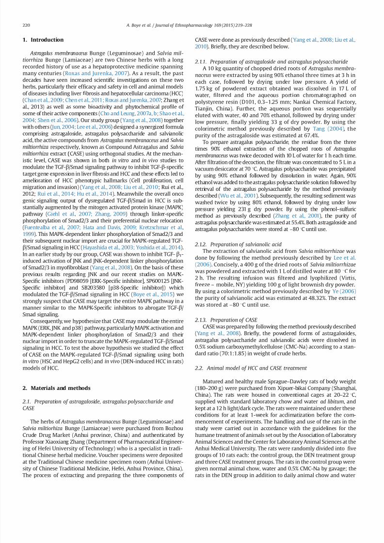
32 CASE decreased TGF- β 1-induced domain-speci 1047297c
phosphorylation of Smad23 and nuclear translocation of Smad4 in
both HSCs and HepG2 cells
Stimulation of HSCs with exogenous TGF-β1 resulted in increased
expression of phosphorylated pSmad2C pSmad2L and oncogenic
pSmad3L however prior treatment of HSC cells with CASE (20 40
and 80mgml respectively) before TGF-β1 stimulation led to a
Fig 1 Effect of CASE on TGF-β1-induced activation of pERK pJNK and pp38 in HSCs ( Fig 1(1) (2) and (3)) and HepG2 Cells (Fig 1(4) (5) and (6)) The HSCs andor HepG2
cells were seeded at a density of 1106 cells25 cm2 culture 1047298asks then cultured with 10 FBS in 95 air and 5 CO2 at 37 1C The cells were starved for 24 h in serum-free
medium and in the absence or presence of CASE (20 40 or 80 μgml) when the HSCs andor HepG2 cells attached to 70ndash80 of monolayers subsequently cells were treated
with TGF-β1 (9 pmolL) for 1 h Total proteins of the cells were extracted by using Western blot and IP cell lysis liquid Expression of ERK12 JNK12 and pp38 were monitored
by western blot using anti-pERK12 anti-pJNK12 and anti-pp38 antibodies respectively Intensities of pERK12 pJNK12 and pp38 bands were normalized to ERK12 JNK12
and p38 respectively of the corresponding treatment groups The data presented are based on at least three independent experiments (P o001 compared with control
group P o001 compared with TGF-β1 group)
A Boye et al Journal of Ethnopharmacology 169 (2015) 219ndash 228222
7232019 Compound Astragalus and Salvia Miltiorrhiza Extracts Modulate MAPK Regulated TGF Smad Signaling in Hepatocellhellip
httpslidepdfcomreaderfullcompound-astragalus-and-salvia-miltiorrhiza-extracts-modulate-mapk-regulated 510
concentration-dependent decrease in the TGF-β1-induced phosphor-
ylation of pSmad2C pSmad2L and oncogenic pSmad3L (Fig 2(1)) In a
similar manner treatment of HepG2 cells with TGF-β1 (9 pmolL)
produced increased domain-speci1047297c phosphorylation and expression
of pSmad2C pSmad2L oncogenic pSmad3L and Smad4 (Fig 2(2) and
(3)) But pretreatment of HepG2 cells with CASE (20 40 and 80 μgml
respectively) before TGF-β1 stimulation led to the reversal of the
above observations Importantly CASE reduced nuclear import of
Smad4 by enhancing its cytoplasmic retention and also enhanced
the phosphorylation of tumor suppressor pSmad3C speci1047297cally in
HepG2 cells (Fig 2(2) and (3))
33 CASE decreased TGF- β 1-induced expression of Imp78 and their
subcellular localization in both HSCs and HepG2 cells
TGF-β1 stimulated HSCs produced not only increased protein
expression of Imp78 but also enhanced nuclear relocation of Imp7
8 compared to control (Fig 3(1) and (2)) But CASE treatment beforeTGF-β1 stimulation produced a concentration-dependent decrease in
Imp78 expression and also reduced signi1047297cantly their nuclear reloca-
tion especially Imp7 (Fig 3(5)) Importantly CASE (40 and 80 mgml)
was most effective Similarly TGF-β1-stimulated HepG2 cells produced
increased expression of Imp78 and also enhanced nucleo-cytoplasmic
distribution But prior treatment of HepG2 cells with CASE before TGF-
β1 stimulation produced not only reduction in the expressions of
Imp78 (Fig 3-(3) (4)) but also reduced their nuclear relocation
particularly Imp7 (Fig 3(6))
34 CASE repressed TGF- β 1-induced expression of PAI-1 gene in
HSCs and HepG2 cells
TGF-β1 (9 pmolL) stimulation of HSCs and HepG2 cells inseparate experiments produced increased PAI-1 gene expression
compared to control groups but upon prior CASE treatment before
TGF-β1 stimulation produced a concentration dependent repres-
sion of PAI-1 gene expression in the studied cell lines in a reverse
manner (Fig 4(1) and (2))
35 CASE decreased DEN-induced activation of pERK pJNK and pp38
in rats
DEN time dependently increased the activation and expression
of pERK12 pJNK12 and pp38 in rat livers However concurrent
treatment of rats with DEN (05 ml100 g bodyweight) in the
morning and CASE (60 120 and 240 mgkg respectively) in the
afternoon 1047297
rst for a period of 12 weeks and subsequently for aperiod of 16 weeks showed dose dependent reduction in the DEN-
induced activation and expression of pERK12 pJNK12 and pp38
(Fig 5) With the exception of CASE (60 mgkg) which produced
weak inhibitory effect on DEN-induced activation and expression
of pJNK12 at the end of the 12 weeks (Fig 5(2)) CASE (60 120 and
240 mgkg respectively) dose dependently decreased DEN-
induced activation and expression of pERK pJNK and pp38 at
both 12th and 16th weeks (Fig 5) CASE (120 mgkg) was most
effective in decreasing DEN-induced activation and expression of
pERK12 and pJNK12 at both the 12th and 16th weeks whiles
CASE (240 mgkg) was only effective in decreasing the activation
and expression of pp38 at both periods (Fig 5(3) and (6))
4 Discussion
We herein present a study further elucidating the molecular
mechanisms of CASE as a potential anti-HCC herbal medicine Essen
tially we report that CASE modulates the MAPK-regulated TGF-βSmad
signaling via inhibition of oncogenic MAPK-dependent linker phosphor-
ylation of Smad23 in HSCs (A key hepatic cell implicated in liver 1047297brosis
(Puche et al 2013) and HepG2 cells (A kind of human hepatoma cell
line) resulting in down-regulation of PAI-1 gene Precisely CASE also
inhibited the MAPK pathway in DEN-induced HCC rats which provided
a strong rationale for us to probe further how the inhibition of the
MAPK pathway will affect the overall TGF-βSmad signaling The MAPK
pathway is one of the important signaling collaborators of TGF-β that
enhances overall oncogenic TGF-βSmad signaling output in cancer
(Giehl et al 2007 Moustakas and Heldin 2005) Indeed the cross-
Fig 2 Effect of CASE on TGF-β1-induced domain-speci1047297c phosphorylation of
Smad23 Smad4 expressions and their nuclear translocation in HSCs ( Fig 2(1))
and HepG2 cells (Fig 2(2) and (3)) HSCs andor HepG2 cells were starved for 24 h
in serum-free medium and in the absence or presence of CASE (20 40 or 80 μgml)
when the cells attached to 30ndash40 of monolayers in 24-holes culture plate
respectively subsequently the cells were treated with TGF-β1 (9 pmolL) for 1 h
The cells were then 1047297xed permeabilized blocked and incubated with each primary
antibody then corresponding 1047298uorescein isothiocyanate (FITC)-Conjugated sec-
ondary antibody ultimately viewed and photographed using a 1047298uorescence
microscope Fig 2(3)) HepG2 cells were starved for 24 h in serum-free medium
and in the absence or presence of CASE (20 40 or 80 μgml) when the cells attached
to 30ndash40 of monolayers in 24-holes culture plate respectively subsequently cells
were treated with TGF-β1 (9 pmolL) for 1 h The cells were then 1047297
xed permeabi-lized blocked and incubated with each primary antibody then corresponding
1047298uorescein isothiocyanate (FITC)-Conjugated secondary antibody incubated with
40 6-diamidino-2-phrnylindole (DAPI) for nuclear staining ultimately viewed and
photographed using a 1047298uorescence microscope C C-terminal L Linker region
A Boye et al Journal of Ethnopharmacology 169 (2015) 219ndash 228 223
7232019 Compound Astragalus and Salvia Miltiorrhiza Extracts Modulate MAPK Regulated TGF Smad Signaling in Hepatocellhellip
httpslidepdfcomreaderfullcompound-astragalus-and-salvia-miltiorrhiza-extracts-modulate-mapk-regulated 610
signaling between the MAPK pathway and the TGF-βSmad signaling is
now a common phenomenon reported in many cancer subtypes For
instance the MAPKs have been shown to be activated by TGF-β in
various cancer subtypes including HCC (Zhang 2009) and the TGF-β-
induced activation of the MAPKs have been shown to increase MAPK-
dependent linker phosphorylation of Smad23 (Furukawa et al 2003
Yoshida et al 2005) which incidentally promotes oncogenesis (Nagata et
al 2009) Notably activation of ERK12 and JNK was shown to be crucial
for TGF-β-induced Smad4-independent signaling (Giehl et al 2007)
which means that the MAPK pathway can signi1047297cantly promote TGF-βsignaling even in the absence of Smad4 (A common Smad protein which
mediates canonical TGF-β signaling) via the linker phosphorylation of
Smad23 (Non-canonical TGF-β signaling) Targeted inhibition of the
MAPK pathway speci1047297cally inhibition of MAPK-dependent linker phos-
phorylation of the R-Smads may signi1047297cantly abrogate TGF-βSmad
signaling in cancer Quiet recently by using MAPK-Speci1047297c Inhibitors
(PD98059 [ERK-Speci1047297c inhibitor] SP600125 [JNK-Speci1047297c inhibitor] and
SB203580 [p38-Speci1047297c inhibitor]) our study group reported on effective
modulation of the MAPK-regulated TGF-βSmad signaling in HCC
leading to decreased PAI-1 gene expression and near blockade of HCC
phenotypes (Cell proliferation migration and invasion) (Boye et al
2015) In this study CASE inhibited hitherto increased TGF-β1-induced
activation of pERK and pJNK in HSC and HepG2 cells and these results
were consistent in both HSC and HepG2 cells This shows that CASE
Fig 3 Effect of CASE on TGF-β1-induced expression of Importin7 (Imp7) and Importin8 (Imp8) in HSCs (Fig 3(1) (2) and (5)) and HepG2 cells (Fig 3(3) (4) and (6)) The
HSCs and HepG2 cells were seeded at a density of 1106 cells25 cm2 culture 1047298asks then cultured with 10 FBS in 95 air and 5 CO2 at 37 1C The cells were starved for
24 h in serum-free medium and in the absence or presence of CASE (20 40 or 80 μgml) when the HSCs andor HepG2 cells attached to 70ndash80 of monolayers subsequently
cells were treated with TGF-β1 (9 pmolL) for 1 h Total Imp78 proteins of the cells were extracted by using Western blot and IP cell lysis liquid Expression of Imp7 and Imp8
were monitored by IB using anti-Importin 7 and anti-Importin 8 antibodies respectively Intensities of Imp7 and Imp8 bands were normalized to glyceraldehyde-3-
phosphate-dehydrogenase (GAPDH) of the corresponding treatment groups The ratio of the Imp7 and Imp8 to GAPDH without exogenous TGF- β1 was assigned a value of 1
And Fig 3(5) and (6)) HSC andor HepG2 cells were starved for 24 h in serum-free medium and in the absence or presence of CASE (20 40 or 80 μgml) when the cells
attached to 30ndash40 of monolayers in 24-holes culture plate respectively subsequently cells were treated with TGF- β1 (9 pmolL) for 1 h The cells were then 1047297xed
permeabilized blocked and incubated with each primary antibody then corresponding 1047298uorescein isothiocyanate (FITC)-conjugated secondary antibody incubated with 4
6-diamidino-2-phrynylindole (DAPI) for nuclear staining ultimately viewed and photographed using a 1047298uorescence microscope The data presented are based on at least
three independent experiments (P o005 P o001 compared with control group P o005 P o001 compared with TGF-β1 group)
A Boye et al Journal of Ethnopharmacology 169 (2015) 219ndash 228224
7232019 Compound Astragalus and Salvia Miltiorrhiza Extracts Modulate MAPK Regulated TGF Smad Signaling in Hepatocellhellip
httpslidepdfcomreaderfullcompound-astragalus-and-salvia-miltiorrhiza-extracts-modulate-mapk-regulated 710
in vitro target ERKJNK-dependent MAPK signaling in the studied cell
lines however CASE increased pp38 in both cell lines even more than
the model group which perhaps indicates that CASE variably modulates
the MAPK pathway in vitro Activation of the MAPKs by TGF-β and
subsequent MAPK-dependent linker phosphorylation of Smad23enhances nuclear import of pSmad23 to promote TGF-β signaling In
view of this we sought to 1047297nd out how CASE-dependent inhibition of
the MAPKs in HSC and HepG2 cells will affect the down and upstream
mediators of the TGF-βSmad signaling speci1047297cally we monitored
domain-speci1047297c phosphorylation of Smad23 expression of Smad4
and Imp78 and their nuclear import
Brie1047298y TGF-β signaling begins with ligand activation of constituted
TGF-β receptor type 2 (TβRII) which then trans-phosphorylate TGF-βreceptor type 1 (TβRI) The phosphorylated TβRI in turn phosphorylate
receptor-dependent Smad proteins (Smad2 and Smad3) at the C-
terminal to facilitate their oligomerization with Smad4 to form a
complex (Smad234 complex) which preferentially relocates into the
nucleus (Matsuzaki 2012) In the nucleus pSmad23 directly partakes
in the execution of TGF-β-mediated transcription of target genes
through association with transcriptional effectors (Promoters and
repressors) (Matsuzaki 2012 Murata et al 2014) Thus the Smads
act as both signaling mediators (Messengers) and transcriptional
effectors to determine the fate of TGF-β-speci1047297c target gene transcrip-
tion of which PAI-1 gene is integral Critical to both canonical and non-
canonical TGF-β signaling is the nuclear import of pSmad23 as a
complex with Smad4 (Smad234 complex) or through Smad4-
independent mechanism via MAPK-dependent linker phosphorylation
of Smad23 Essentially any of the two import mechanisms mayinvolve Imp78 For instance (Chen and Xu 2011) have shown that
importin proteins (Imp78) are indispensable for nuclear import of
pSmad23 and Smad4 It was also shown that nuclear import of
pSmad23 is a critical step in TGF-β signaling transduction (Xu et al
2007) Further it was shown that knockdown of importin 78
markedly impaired nuclear import of pSmad23 in response to TGF-
β-dependent gene regulation which is critical for embryonic develop-
ment and cellular homeostasis but detrimental in dysregulated TGF-βsignaling in cancer (Xu et al 2007) Also (Xu et al 2007) have
intimated that since Imp78 is critical for nuclear import of pSmad23
it is highly probable that pharmacological inhibition of this nuclear
import factor may disrupt dysregulated TGF-βSmad signaling in
cancer Accordingly TGF-β1 stimulation of HSCs and HepG2 cells
produced increased linker phosphorylation of Smad23 increased
expression of Smad4 and Imp78 and their nuclear import and this
correlated with increased PAI-1 gene expression in both cell lines
(Fig 4(1) and (2)) Interestingly CASE-dependent inhibition of pERK
and pJNK in both cell lines correlated with CASE-dependent inhibition
of linker phosphorylation of Smad23 decreased expression of Smad4
and Imp78 and their nuclear import For example in HepG2 cells
CASE signi1047297cantly decreased nuclear import of Smad4 by enhancing
its cytoplasmic retention (Fig 2(2) and (3)) Also CASE treatment
inhibited Imp78 expression compared with model (TGF-β1-stimu-
lated HSCs and HepG2 cells) and control groups Precisely CASE
decreased both cytoplasmic and nuclear expression of Imp7 but only
decreased cytoplasmic expression of Imp8 compared to model and
control groups in both HSC and HepG2 cells and these effects
correlated with decreased pSmad23 and Smad4 expressions (Fig 2
(1) (2) and (3)) which suggest that CASE-dependent inhibition of pSmad23 and Smad4 nuclear import may be mediated through Imp7
inhibition Correspondingly CASE-dependent decrease in nuclear
import of pSmad23 Smad4 and Imp7 led to signi1047297cant decrease in
PAI-1 gene expression
Severally PAI-1 gene has been reported as one of the important
target genes of TGF-β signaling which has been shown to account for
most of the pathological roles of TGF-β signaling in cancer (Derynck
and Zhang 2003 Tahashi et al 2002) Functionally PAI-1 gene acts as
the main inhibitor of the urokinase-type plasminogen activator system
and it stimulates cell migration and invasion by disrupting cellular
adhesion and enhancing basement membrane degradation (Gutierrez
et al 2000) Indeed overexpression of PAI-1 gene has been implicated
in multiple forms of 1047297brosis including liver 1047297brosis (Gramling and
Church 2010) a key risk factor of HCC In our study both TGF-β1-stimulated HSC and HepG2 cells showed increased expression of PAI-1
gene This observation is consistent with our previous reports regard-
ing the ability of CASE to modulate TGF-βSmad signaling leading to
decrease in PAI-1 gene expression in myo1047297broblast (Yang et al 2008)
keloid 1047297broblast (He et al 2012) HepG2 cells (Hu et al 2014 Liu et al
2010) and DEN-induced HCC in rats (Rui et al 2014) Clearly from the
present results CASE selectively modulate ERKJNK-dependent linker
phosphorylation of Smad23 in vitro in part this observation con1047297rms
our earlier report concerning JNKMAPK-regulated TGF-βSmad sig-
naling in myo1047297broblast (Yang et al 2008) where CASE in a concen-
tration dependent manner inhibited JNK and JNK-dependent linker
phosphorylation of Smad23
To further con1047297rm or otherwise the ability of CASE to modulate the
MAPK pathway we studied the effect of CASE on DEN-induced
Fig 4 Effect of CASE on TGF-β1-induced expression of plasminogen activator
inhibitor 1 (PAI-1) in HSCs (Fig 4(1)) and HepG2 cells (Fig 4(2)) The HSCs andor
HepG2 cells were seeded at a density of 1 106 cells25 cm2 culture 1047298asks then
cultured with 10 FBS in 95 air and 5 CO2 at 37 1
C The cells were starved for24 h in serum-free medium and in the absence or presence of CASE (20 40 or
80 μgml) when the HSCs andor HepG2 cells attached to 70 ndash80 of monolayers
subsequently cells were treated with TGF-β1 (9 pmolL) for 1 h Total proteins of the
cells were extracted by using Western blot and IP cell lysis liquid Expression of PAI-
1 gene was monitored by western blot using anti-PAI-1 Ab Intensities of PAI-1
bands were normalized to glyceraldehyde-3-phosphate-dehydrogenase (GAPDH) of
the corresponding treatment groups The ratio of the PAI-1 gene to GAPDH without
exogenous TGF-β1 was assigned a value of 1 The data presented are based on at
least three independent experiments (P o005 compared with control group
P o005 P o001 compared with TGF-β1 group)
A Boye et al Journal of Ethnopharmacology 169 (2015) 219ndash 228 225
7232019 Compound Astragalus and Salvia Miltiorrhiza Extracts Modulate MAPK Regulated TGF Smad Signaling in Hepatocellhellip
httpslidepdfcomreaderfullcompound-astragalus-and-salvia-miltiorrhiza-extracts-modulate-mapk-regulated 810
activation of the MAPK pathway From our in vivo study treatment of
rats with DEN for 12 weeks led to increased activation and expression
of pERK pJNK and pp38 but extending DEN treatment period to 16
weeks even saw a further increase in the average expression of pERK
pJNK and pp38 (Fig 5(4) (5) and (6)) indicating a direct causal
relationship between duration of DEN treatment and MAPK activation
These results indicate that increase in MAPK activation and expression
may correlate with HCC progression But concomitant treatment of
rats with DEN in the morning and CASE in the afternoon reversed the
above relationship for instance at 12 weeks not only did CASE (120 or
240 mgkg) decreased the expression of pERK pJNK and pp38
compared to the model group but also decreased the average
expression level of each MAPK even more than the average for the
control group (Fig 5(1) (2) and (3)) showing that CASE potently
modulates the MAPK pathway and that the MAPK pathway could be
yet another important target of CASE At 16 weeks though CASE
decreased the expression of the MAPKs compared to the model group
it was however not different from that of the control group ( Fig 5(4)
(5) and (6)) indicating that MAPK activation and expression were
indeed related to duration of DEN treatment The in vivo inhibition of
pp38 by CASE (Fig 5(3) and (6)) contradicts our in vitro results
regarding pp38 But this observation probably suggest that CASE
Fig 5 Effect of CASE on diethyl nitrosamine (DEN)-induced activation of pERK pJNK and pp38 in rats The proteins were extracted from frozen liver tissues from 12th week
(Fig 5(1) (2) and (3)) and 16th week (Fig 5(4) (5) and (6)) DEN treated rats Phosphorylated (p) ERK12 pJNK12 pp38 and ERK JNK p38 were analyzed by western blot
using anti-pERK12 pJNK12 pp38 and ERK12 JNK12 p38 Abs and anti-glyceraldehyde phosphate dehydrogenase (GAPDH) Ab Intensities of pERK12 pJNK12 pp38
bands were normalized to those of ERK12 JNK12 p38 of the corresponding treatment groups The ratio of the pERK12 pJNK12 and pp38 proteins to ERK12 JNK12 and
p38 respectively of normal group was assigned a value of 1 Each value represents mean7SD nfrac143 P o005 P o001 compared with control group P o005 P o001
compared with DEN group
A Boye et al Journal of Ethnopharmacology 169 (2015) 219ndash 228226
7232019 Compound Astragalus and Salvia Miltiorrhiza Extracts Modulate MAPK Regulated TGF Smad Signaling in Hepatocellhellip
httpslidepdfcomreaderfullcompound-astragalus-and-salvia-miltiorrhiza-extracts-modulate-mapk-regulated 910
modulate pERK pJNK and pp38 in vivo by the same mechanism which
may involve some unknown factors in the tumor microenvironment
since the tumor microenvironment was the only major factor absent
in the in vitro study or it may be possible that while CASE employs the
same mechanism to modulate pERK and pJNK both in in vitro andin vivo it however employs different mechanisms to modulate pp38
in in vitro and in vivo settings Indeed these results further show that
CASE does not only target the upstream and downstream mediators
(TGF-β ligand TβRI TβRII Smad2CL Smad3L Smad4 and PAI-1) of
TGF-βSmad signaling as previously reported (He et al 2012 Hu et al
2014 Liu et al 2010 Rui et al 2014 Wu et al 2014 Yang et al 2008)
but also targets the MAPK pathway Fundamentally our in vivo results
agree in part with (Nagata et al 2009) with regard to JNK activation
by DEN treatment however our results concerning pERK and pp38
are inconsistent with their 1047297nding For instance Nagata et al (2009)
had shown that after 86-days DEN treatment of rats only pJNK was
activated whiles pERK and pp38 remained inactive (Un-phosphory-
lated) but in our study we found that increasing DEN treatment to 12
or 16 weeks produced signi1047297cant activation of all the MAPKs (pERKpJNK and pp38) We report that activation of the MAPKs by DEN
treatment is time-dependent and may relate directly to HCC
progression
Put together the present results provide a strong rationale for
further elucidation of the MAPK pathway as a key target for
therapy against HCC Essentially CASE has shown a multi-target
potential by modulating both the MAPK and TGF-βSmad signaling
pathways (Fig 6) in a manner that highlights its potential drug
candidature However our results raise some further questions for
example which speci1047297c subcellular location (Cytoplasm or
nucleus) does CASE modulate the MAPK pathway and how does
it affect HCC phenotypic manifestations such as cell proliferation
dysregulated apoptosis cell migration and invasion These ques-
tions are informed by the fact that spatio-temporal regulation of
the MAPK pathway is crucial for many pathways implicated in HCC
including multi-functional TGF-βSmad signaling
5 Conclusion
CASE blocked MAPK activation and MAPK-dependent linker
phosphorylation of Smad23 inhibited Smad4 and Imp78
blocked nuclear import of Smad234 via Imp7 modulation and
down-regulated PAI-1 gene expression and these results do not
only demonstrate the potential of the MAPK-regulated TGF-β
Smad pathway as an important target for therapy against HCC but
also highlights the potential drug candidature of CASE and con-
1047297rmation of its ethnobotanical usage
Con1047298icts of interest
No potential con1047298icts of interest
Acknowledgments
We thank Prof K Matsuzaki (Department of Gastroenterologyand Hepatology Kansai Medical University Osaka Japan) for
providing us with the following Abs Anti-pSmad2L and Anti-
pSmad3L Also this study was supported by the National Natural
Science Foundation of China (No 81073098 and no 81374012)
References
Boye A Kan H Wu C Jiang Y Yang X He S Yang Y 2015 MAPK inhibitorsdifferently modulate TGF-betaSmad signaling in HepG2 cells Tumour Biol httpdxdoiorg101007s13277-014-3002-x
Chan W Durairajan S Lu J Wang Y Xie L Kum W Koo I Yung K Li M2009 Neuroprotective effects of astragaloside IV in 6-hydroxydopamine-treated primary nigral cell culture Neurochem Int 55 414ndash422
Chen R Shao H Lin S Zhang J Xu K 2011 Treatment with astragalusmembranaceus produces antioxidative effects and attenuates intestinal mucosa
injury induced by intestinal ischemia-reperfusion in rats Am J Chin Med 39879ndash887
Chen X Xu L 2011 Mechanism and regulation of nucleocytoplasmic traf 1047297cking of Smad Cell Biosci 1 40
Cho WC Leung KN 2007a In vitro and in vivo anti-tumor effects of astragalusmembranaceus Cancer Lett 252 43ndash54
Cho WC Leung KN 2007b In vitro and in vivo immunomodulating andimmunorestorative effects of astragalus membranaceus J Ethnopharmacol113 132ndash141
Date M Matsuzaki K Matsushita M Tahashi Y Furukawa F Inoue K 2000Modulation of transforming growth factor function in hepatocytes and hepaticstellate cells in rat liver injury Gut 46 719ndash724
Derynck R Zhang YE 2003 Smad-dependent and Smad-independent pathwaysin TGF-β family signalling Nature 425 577ndash584
Fuentealba L Eivers E Ikeda A Hurtado C Kuroda H Pera E De Robertis E2007 Integrating patterning signals WntGSK3 regulates the duration of theBMPSmad1 signal Cell 131 980ndash993
Furukawa F Matsuzaki K Mori S Tahashi Y Yoshida K Sugano Y Yamagata
H Matsushita M Seki T Inagaki Y Nishizawa M Fujisawa J Inoue K2003 p38 MAPK mediates 1047297brogenic signal through Smad3 phosphorylation inrat myo1047297broblasts Hepatology 38 879ndash889
Giehl K Imamichi Y Menke A 2007 Smad4-independent TGF-β signaling intumor cell migration Cells Tissues Org 185 123ndash130
Gramling MW Church FC 2010 Plasminogen activator inhibitor-1 is anaggregate response factor with pleiotropic effects on cell signaling in vasculardisease and the tumor microenvironment Thromb Res 125 377ndash381
Gutierrez LS Schulman A Brito-Robinson T Noria F Ploplis VA Castellino FJ2000 Tumor development is retarded in mice lacking the gene for urokinase-type plasminogen activator or its inhibitor plasminogen activator inhibitor-1Cancer Res 60 5839ndash5847
Hata A Davis B 2009 Control of microRNA biogenesis by TGFbeta signalingpathway-a novel role of Smads in the nucleus Cytokine Growth Factor Rev 20517ndash521
Hayashida T Decaestecker M Schnaper H 2003 Cross-talk between ERK MAPkinase and Smad signaling pathways enhances TGF-beta-dependent lsre-sponses in human mesangial cel FASEB J 17 1576ndash1578
He S Yang Y Liu X Huang W Zhang X Yang S 2012 Compound Astragalus
and Salvia miltiorrhiza extract inhibits cell proliferation invasion and collagen
Fig 6 Fig 6 shows an illustrative summary of the multi-target mechanism by
which CASE modulates the MAPK-regulated TGF-βSmad signaling in hepatocel-lular carcinoma (HCC) CASE blocks TGF-β-induced activation of ERK JNK and p38
to abrogate MAPK-dependent linker phosphorylation of Smad23 and their sub-
sequent nuclear translocation Along the canonical loop CASE inhibits Smad4
expression formation of Smad234 and their nuclear translocation via inhibition of
Imp78 CASE-dependent disruption of nuclear import of Smad23 and Smad4 leads
to down-regulation of PAI-1 gene expression Cytoplasm (C) C-terminal phos-
phorylated Smad (pSmadC) Linker phosphorylated Smad (pSmadL) Nucleus (N)
Phosphorylated Smad (pSmad) Receptor mediated Smad (R-Smad) Transforming
growth factor receptor type 1 and 2 (TβRI and TβRII)
A Boye et al Journal of Ethnopharmacology 169 (2015) 219ndash 228 227
7232019 Compound Astragalus and Salvia Miltiorrhiza Extracts Modulate MAPK Regulated TGF Smad Signaling in Hepatocellhellip
httpslidepdfcomreaderfullcompound-astragalus-and-salvia-miltiorrhiza-extracts-modulate-mapk-regulated 1010
synthesis in keloid 1047297broblasts by mediating transforming growth factor‐βSmad pathway Br J Dermatol 166 564ndash574
Hu X Rui W Wu C He S Jiang J Zhang X Yang Y 2014 CompoundAstragalus and Salvia Miltiorrhiza extracts suppress hepatocarcinogenesis bymodulating transforming growth factor‐βSmad signaling J GastroenterolHepatol 29 1284ndash1291
Jun T 2004 Determination of total astragalosides in astragalus by colormetricmethod J Anhui Tradit Chin Med Coll 5 017
Kretzschmar M Doody J Timokhina I Massagueacute J 1999 A mechanism of repression of TGFbetaSmad signaling by oncogenic Ras Genes Dev 13804ndash816
Lee TY Chang HH Wang GJ Chiu JH Yang YY Lin HC 2006 Water‐
solubleextract of Salvia miltiorrhiza ameliorates carbon tetrachloride‐mediated hepa-tic apoptosis in rats J Pharm Pharm 58 659ndash665
Liu X Yang Y Zhang X Xu S He S Huang W Roberts MS 2010 CompoundAstragalus and Salvia miltiorrhiza extract inhibits cell invasion by modulatingtransforming growth factor‐βSmad in HepG2 cell J Gastroenterol Hepatol 25420ndash426
Matsuzaki K 2012 Smad phosphoisoform signals in acute and chronic liver injurysimilarities and differences between epithelial and mesenchymal cells CellTissue Res 347 225ndash243
Moustakas A Heldin C-H 2005 Non-Smad TGF-β signals J Cell Sci 1183573ndash3584
Murata M Yoshida K Yamaguchi T Matsuzaki K 2014 Linker phosphorylationof Smad3 promotes 1047297bro-carcinogenesis in chronic viral hepatitis of hepato-cellular carcinoma World J Gastroenterol 20 15018ndash15027
Nagata H Hatano E Tada M Murata M Kitamura K Asechi H Narita MYanagida A Tamaki N Yagi S 2009 Inhibition of c‐ Jun NH2‐terminal kinaseswitches Smad3 signaling from oncogenesis to tumor‐suppression in rat
hepatocellular carcinoma Hepatology 49 1944ndash
1953Puche JE Saiman Y Friedman SL 2013 Hepatic stellate cells and liver 1047297brosisCompr Physiol 3 1473ndash1492
Roxas M Jurenka J 2007 Colds and in1047298uenza a review of diagnosis andconventional botanical and nutritional considerations Altern Med Rev 1225ndash48
Rui W Xie L Liu X He S Wu C Zhang X Zhang L Yang Y 2014 CompoundAstragalus and Salvia miltiorrhiza extract suppresses hepatocellular carcinomaprogression by inhibiting 1047297brosis and PAI-1 mRNA transcription J Ethnophar-macol 151 198ndash209
Tang J 2004 Determination of total astragalosides in astragalus by colorimetricmethod J Anhui Tradit Chin Med Coll 23 37ndash38
Shao BM Xu W Dai H Tu P Li Z Gao XM 2004 A study on the immunereceptors for polysaccharides from the roots of astragalus membranaceus aChinese medicinal herb Biochem Biophys Res Commun 320 1103ndash1111
Shen P Liu MH Ng TY Chan YH Yong EL 2006 Differential effects of iso1047298avones from astragalus membranaceus and Pueraria thomsonii on theactivation of PPARalpha PPARgamma and adipocyte differentiation in vitro
J Nutr 136 899ndash905
Tahashi Y Matsuzaki K Yoshida K Furukawa F Sugano Y Matsushita MHimeno Y Inagaki Y Inoue K 2002 Differential regulation of TGF‐β signal in
hepatic stellate cells between acute and chronic rat liver injury Hepatology 3549ndash61
Wu C Jiang J Boye A Jiang Y Yang Y 2014 Compound Astragalus and Salviamiltiorrhiza extract suppresses rabbits hypertrophic scar by modulating theTGF-betaSmad signal Dermatology 229 363ndash368
Wu YP Cao Y Cao YY 2001 Technology optimization of extracting activitycomponents from Astragalus membranaceus Lishizen Med Mater Med Res 12876ndash877
Xu L Yao X Chen X Lu P Zhang B Ip YT 2007 Msk is required for nuclearimport of TGF-βBMP-activated Smads J cell Biol 178 981ndash994
Yang Y Yang S Chen M Zhang X Zou Y Zhang X 2008 Compound Astragalus
and Salvia miltiorrhiza extract exerts anti-1047297brosis by mediating TGF-βSmadsignaling in myo1047297broblasts J Ethnopharmacol 118 264ndash270
Ye Y 2006 Comparative study on the determination of salvianolic acids content bycolorimetery and HPLC J Zhejiang Univ Tradit Chin Med 30 350 ndash351
Yoshida K Matsuzaki K Mori S Tahashi Y Yamagata H Furukawa F Seki TNishizawa M Fujisawa J Okazaki K 2005 Transforming growth factor-betaand platelet-derived growth factor signal via c-Jun N-terminal kinase-depen-dent Smad23 phosphorylation in rat hepatic stellate cells after acute liverinjury The American journal of pathology 166 1029ndash1039
Yoshida K Murata M Yamaguchi T Matsuzaki K 2014 TGF-βSmad signalingduring hepatic 1047297bro-carcinogenesis (review) Int J Oncol 45 1363ndash1371
Zhang SS He FY Xu XY 2001 Comparative studies on the content of ingredients of polysaccharides in the Buyanghuanwutang and single materials
J Human Coll Tradit Chin Med 21 26Zhang YE 2009 Non-Smad pathways in TGF-β signaling Cell Res 19 128ndash139
Glossary
Phosphorylation Addition of a phosphorus atom to a protein or a moleculeSmad proteins A class of proteins that mediate transforming growth factor beta
(TGF-β) signaling transductionPhosphorylated Smad A Smad protein that has received a phosphorus atom from a
phosphorus carrier moleculeLinker phosphorylated Smad A Smad protein phosphorylated at a region in
between its N-terminal and C-terminalCanonical TGF-β Signaling It is Smad-mediated TGF-β signaling transduction
involving Smad2 Smad3 and Smad4 complex formation leading to theirnuclear translocation and subsequent transcription of TGF-β-target mRNAs
Importin protein 7 and 8 (Imp78) They are proteins mostly in the cytoplasm thatfacilitate nuclear entry of other proteins
Mitogen activated protein kinase (MAPK) They are kinase proteins that facilitatetransduction of extracellular signals into the nucleus
Non-canonical TGF-β signaling All TGF-β signaling transduction that does notinvolve Smad4 or non-Smad TGF-β signaling
A Boye et al Journal of Ethnopharmacology 169 (2015) 219ndash 228228
7232019 Compound Astragalus and Salvia Miltiorrhiza Extracts Modulate MAPK Regulated TGF Smad Signaling in Hepatocellhellip
httpslidepdfcomreaderfullcompound-astragalus-and-salvia-miltiorrhiza-extracts-modulate-mapk-regulated 210
1 Introduction
Astragalus membranaceus Bunge (Leguminosae) and Salvia mil-
tiorrhiza Bunge (Lamiaceae) are two Chinese herbs with a long
recorded history of use as a hepatoprotective medicine spanning
many centuries (Roxas and Jurenka 2007) As a result the past
decades have seen increased scienti1047297c investigations on these two
herbs particularly their ef 1047297cacy and safety in cell and animal models
of diseases including liver 1047297brosis and hepatocellular carcinoma (HCC)(Chan et al 2009 Chen et al 2011 Roxas and Jurenka 2007 Zhang et
al 2013) as well as some bioactivity and phytochemical pro1047297le of
some of their active components (Cho and Leung 2007a b Shao et al
2004 Shen et al 2006) Our study group (Yang et al 2008) together
with others ( Jun 2004 Lee et al 2006) designed a synergized formula
comprising astragaloside astragalus polysaccharide and salvianolic
acid the active compounds from Astragalus membranaceus and Salvia
miltiorrhiza respectively known as Compound Astragalus and Salvia
miltiorrhiza extract (CASE) using orthogonal studies At the mechan-
istic level CASE was shown in both in vitro and in vivo studies to
modulate the TGF-βSmad signaling pathway to inhibit TGF-β-speci1047297c
target gene expression in liver1047297brosis and HCC and these effects led to
amelioration of HCC phenotypic hallmarks (Cell proliferation cell
migration and invasion) (Yang et al 2008 Liu et al 2010 Rui et al
2012 Rui et al 2014 Hu et al 2014) Meanwhile the overall onco
genic signaling output of dysregulated TGF-βSmad in HCC is sub-
stantially augmented by the mitogen activated protein kinase (MAPK)
pathway (Giehl et al 2007 Zhang 2009) through linker-speci1047297c
phosphorylation of Smad23 and their preferential nuclear relocation
(Fuentealba et al 2007 Hata and Davis 2009 Kretzschmar et al
1999) This MAPK-dependent linker phosphorylation of Smad23 and
their subsequent nuclear import are crucial for MAPK-regulated TGF-
βSmad signaling in HCC (Hayashida et al 2003 Yoshida et al 2014)
In an earlier study by our group CASE was shown to inhibit TGF-β1-
induced activation of JNK and JNK-dependent linker phosphorylation
of Smad23 in myo1047297broblast (Yang et al 2008) On the basis of these
previous results regarding JNK and our recent studies on MAPK-
Speci1047297c inhibitors (PD98059 [ERK-Speci1047297c inhibitor] SP600125 [JNK-
Speci1047297c inhibitor] and SB203580 [p38-Speci1047297c inhibitor]) whichmodulated the TGF-βSmad signaling in HCC (Boye et al 2015) we
strongly suspect that CASE may target the entire MAPK pathway in a
manner similar to the MAPK-Speci1047297c inhibitors to abrogate TGF-β
Smad signaling
Consequently we hypothesize that CASE may modulate the entire
MAPK (ERK JNK and p38) pathway particularly MAPK activation and
MAPK-dependent linker phosphorylation of Smad23 and their
nuclear import in order to truncate the MAPK-regulated TGF-βSmad
signaling in HCC To test the above hypothesis we studied the effect
of CASE on the MAPK-regulated TGF-βSmad signaling using bothin vitro (HSC and HepG2 cells) and in vivo (DEN-induced HCC in rats)
models of HCC
2 Materials and methods
21 Preparation of astragaloside astragalus polysaccharide and
CASE
The herbs of Astragalus membranaceus Bunge (Leguminosae) and
Salvia miltiorhiza Bunge (Lamiaceae) were purchased from Bozhou
Crude Drug Market (Anhui province China) and authenticated by
Professor Xiaoxiang Zhang (Department of Pharmaceutical Engineer-
ing of Hefei University of Technology) who is a specialist in tradi-
tional Chinese herbal medicine Voucher specimens were deposited
at the Traditional Chinese medicine specimen room (Anhui Univer-
sity of Chinese Traditional Medicine Hefei Anhui Province China)
The process of extracting and preparing the three components of
CASE were done as previously described (Yang et al 2008 Liu et al
2010) Brie1047298y they are described below
211 Preparation of astragaloside and astragalus polysaccharide
A 10 kg quantity of chopped dried roots of Astragalus membra-
naceus were extracted by using 90 ethanol three times at 3 h in
each case followed by drying under low pressure A yield of
175 kg of powdered extract obtained was dissolved in 17 L of
water 1047297ltered and the aqueous portion chromatographed onpolystyrene resin (D101 03ndash125 mm Nankai Chemical Factory
Tianjin China) Further the aqueous portion was sequentially
eluted with water 40 and 70 ethanol followed by drying under
low pressure 1047297nally yielding 33 g of dry powder By using the
colorimetric method previously described by Tang (2004) the
purity of the astragaloside was estimated at 674
To prepare astragalus polysaccharide the residue from the three
times 90 ethanol extraction of the chopped roots of Astragalus
membranaceus was twice decocted with 10 L of water for 1 h each time
After 1047297ltration of the decoction the 1047297ltrate was concentrated to 5 L in a
vacuum desiccator at 70 1C Astragalus polysaccharide was precipitated
by using 90 ethanol followed by dissolution in water Again 90
ethanol was added to the astragalus polysaccharide solution followed by
retrieval of the astragalus polysaccharide by the method previouslydescribed (Wu et al 2001) Subsequently the resulting sediment was
washed twice by using 80 ethanol followed by drying under low
pressure yielding 231 g dry powder By using the phenolndashsulfuric
method as previously described (Zhang et al 2001) the purity of
astragalus polysaccharide was estimated at 554 Both astragaloside and
astragalus polysaccharides were stored at ndash80 1C until use
212 Preparation of salvianolic acid
The extraction of salvianolic acid from Salvia miltiorrhizae was
done by following the method previously described by Lee et al
(2006) Concisely a 400 g of the dried roots of Salvia miltiorrhizae
was powdered and extracted with 1 L of distilled water at 80 1C for
2 h The resulting infusion was 1047297ltered and lyophilized (Virtis
freeze ndash
mobile NY) yielding 100 g of light brownish dry powderBy using a colorimetric method previously described by Ye (2006)
the purity of salvianolic acid was estimated at 4832 The extract
was stored at ndash80 1C until use
213 Preparation of CASE
CASE was prepared by following the method previously described
(Yang et al 2008) Brie1047298y the powdered forms of astragalosides
astragalus polysaccharide and salvianolic acids were dissolved in
05 sodium carboxymethylcellulose (CMC-Na) according to a stan-
dard ratio (701185) in weight of crude herbs
22 Animal model of HCC and CASE treatment
Matured and healthy male Spraguendash
Dawley rats of body weight(180ndash200 g) were purchased from Xipuer-bikai Company (Shanghai
China) The rats were housed in conventional cages at 20ndash22 1C
supplied with standard laboratory chow and water ad libitum and
kept at a 12 h lightdark cycle The rats were maintained under these
conditions for at least 1-week for acclimatization before the com-
mencement of experiments The handling and use of the rats in the
study were carried out in accordance with the guidelines for the
humane treatment of animals set out by the Association of Laboratory
Animal Sciences and the Center for Laboratory Animal Sciences at the
Anhui Medical University The rats were randomly divided into 1047297ve
groups of 10 rats each the control group the DEN treatment group
and three CASE treatment groups The rats in the control group were
given normal animal chow water and 05 CMC-Na by gavage the
rats in the DEN group in addition to daily animal chow and water
A Boye et al Journal of Ethnopharmacology 169 (2015) 219ndash 228220
7232019 Compound Astragalus and Salvia Miltiorrhiza Extracts Modulate MAPK Regulated TGF Smad Signaling in Hepatocellhellip
httpslidepdfcomreaderfullcompound-astragalus-and-salvia-miltiorrhiza-extracts-modulate-mapk-regulated 310
were treated with 02 DEN dissolved in 05 CMC-Na in the morning
by gavage 5 times a week for 12 or 16 weeks to induce HCC and the
rats in the CASE groups in addition to daily normal animal chow and
water were concurrently treated with 02 DEN in 05 CMC-Na in
the morning by gavage 1047297ve times a week and CASE (60 120 or
240 mgkg respectively) in the afternoon by gavage per day for 12 or
16 weeks The rats were sacri1047297ced two weeks post-DEN treatment
(12th or 16th-week) One lobe of liver from each rat in each group was
harvested and stored at a temperature of 801C until use
23 Cell origin culture and treatment of cells (hepatic stellate cells
[HSCs] and HepG2 cells) with CASE
The use of animals in this study was approved by the Animal Ethics
Committee of Anhui Medical University HSCs were isolated from the
liver of normal male SpraguendashDawley rats (450ndash500 g) by using
collagenase and sequential Pronase-E digestion as previously described
(Date et al 2000) The isolated HSCs were490 pure (As determined
microscopically based on vitamin A droplet-dependent auto-1047298uores-
cence) and had 499 viability (Trypan blue exclusion test) The human
hepatocellular carcinoma HepG2 cell line was purchased from The
Chinese Academy of Sciences Cell Bank (Shanghai China) The HSCs
and HepG2 Cells were grown as sub-con1047298uent monolayer cultures inDulbeccos modi1047297ed Eagles medium (DMEM Gibco Rockville MD US
A) supplemented with 10 fetal bovine serum (FBS Sijiqing Zhejiang
Tianhang Biological Technology Co LTD Zhejiang China) and main-
tained in a humidi1047297ed 5 CO2 incubator at 37 1C The experiments were
performed at the log phase of growth after the cells had been plated for
24 h HSC andor HepG2 cells were starved for 24 h in serum-free
medium and in the absence or presence of CASE (20 40 or 80 mgml)
and subsequently treated with TGF-β1 (9 pmolL) for 1 h The cells of the
control group were treated with an equal volume of serum-free
medium
24 Western blot analysis
To detect the effect of CASE on the expression of PAI-1 Impor-tin78 and phosphorylation of MAPKs (ERK JNK and p38) HSC and
or HepG2 cells were seeded at a density of 1 106 cells25 cm2
culture 1047298asks and then treated under the indicated conditions Total
proteins from HSC and or HepG2 cells and frozen liver tissue were
extracted by using Western blot and IP cell lysis liquid (Beyotime
Shanghai China) as previously described (Wu et al 2014) Proteins
were separated by sodium dodecyl sulfatepolyacrylamide gel elec-
trophoresis (SDSPAGE) transferred onto polyvinylidence di1047298uoride
(PVDF) membranes (Millipore Bedford MA USA) by wet transfer
method blocked in 5 skim milk powder dissolved in Tris-buffered
saline solution01 Tween 20 (TBST) incubated with the primary
antibody overnight at 4 1C washed 3 times with TBST for 10 min
each time incubated with corresponding secondary antibody for 2 h
at room temperature washed 3 times with TBST for 10 min eachtime and 1047297nally the membranes were developed by using the ECL
chemiluminescence system (Amersham Piscataway NJ USA) Pri-
mary antibodies used in this study included plasminogen activator
inhibitor 1 (PAI-1 rabbit anti-PAI-1 antibody) (Santa Cruz Biotech-
nology Santa Cruz CA USA) phospho-ERK12 and ERK12 phos-
pho-JNK12 and JNK phospho-p38 and p38 (rabbit anti-phospho-
ERK12 anti-ERK12 anti-phospho-JNK12 anti-JNK12 anti-
phospho-p38 and anti-p38 kinase antibodies) (Cell Signaling Tech-
nology Beverly MA USA) Importin7 and 8 (Imp78 rabbit anti-
Importin7 and anti-Importin8 antibodies) (Abcam Cambridge UK)
and glyceraldehyde phosphate dehydrogenase (GAPDH) (mouse
anti-GAPDH) (Cell Signaling Technology Beverly MA USA) Densito-
metric analysis was carried out by using Quantity One software (Bio-
Rad California USA)
25 Immuno 1047298uorescence analysis
To detect the effects of CASE on intracellular localization of
Smads and Importin78 HSCs andor HepG2 cells were seeded on
slides in a 24-well plate and then treated under the indicated
conditions The cells were 1047297xed with 4 paraformaldehyde for
30 min permeabilized with 01 saponin for 10 min and blocked
with 05 bovine serum albumin in phosphate buffer saline (PBS)
for 30 min at 4 1C then incubated with each primary antibodyovernight at 4 1C washed 3 times with PBS for 5 min each time
incubated with corresponding 1047298uorescein isothiocyanate (FITC)-
conjugated secondary antibody for 2 h at room temperature
washed 3 times with PBS for 5 min each time incubated with
406-diamidino-2-phrnylindole (DAPI Sigma) for 10 min at room
temperature for nuclear staining Finally slides were mounted
with 80 phosphoglycerol viewed and photographed under a
1047298uorescence microscope (Olympus Tokyo Japan) Primary anti-
bodies used in this experiment included Smad23 phosphorylated
at the C-terminal region (rabbit anti-pSmad2C and pSmad3C
antibodies) (Cell Signaling Technology Beverly MA USA)
Smad23 phosphorylated at the link region (rabbit anti-pSmad2L
and pSmad3L antibodies) (A gift from Prof K Matsuzaki Kansai
Medical University Japan) Smad4 (mouse anti-Smad4 antibody)
(Santa Cruz Biotechnology Santa Cruz CA USA) and Importin7
and 8 (Imp78 rabbit anti-Importin7 and anti-Importin8 antibo-
dies) (Abcam Cambridge UK) At least 100 stained cells were
analyzed per sample in each experiment
26 Statistical analyses
Data were expressed as mean7standard deviation (SD) Statistical
analyses were performed by SPSS 110 for Windows (SPSS Inc
Chicago IL USA) Experimental and control groups were compared
by one-way ANOVA P o005 was considered statistically signi1047297cant
27 Theorycalculations
Transforming growth factor beta (TGF-β) is a multi-functional
and a ubiquitous cytokine crucial in all cellular developmental and
homeostatic processes as well as disease pathogenesis It employs
basically two major signaling modes (Canonical and Non-canoni-
cal) in almost all the cellular processes it partakes Whiles the
former signaling mode involves mediation by Smad proteins the
latter involves crosstalk with other signaling pathways of which
mitogen activated protein kinase (MAPK) pathway is integral
Many therapeutic modalities including but not limited to TGF-β-
Receptor inhibitors have been designed to abrogate dysregulated
canonical TGF-β signaling in diseases such as HCC but so far they
have proved comparatively ineffective due to the potential of TGF-β to reactivate its non-canonical compensatory pathways mainly
regulated in part by the MAPKs (ERK JNK and p38) As a result the
MAPK pathway has become a possible target for investigations to
prospect for new targets for therapy since it regulates the
oncogenic arm of the TGF-β signaling in cancer To this end we
designed this study to investigate the effect of CASE a potential
anti-HCC herbal drug on the MAPK-regulated TGF-βSmad path-
way on the basis of our previous 1047297nding on CASE regarding the
TGF-βSmad pathway where CASE ameliorated liver 1047297brosis and
HCC progression by modulating the TGF-βSmad pathway
It is envisioned that the future success of CASE as a potential
anti-HCC candidate drug will not only justify its long uneventful
folk use in China but also provide a much cheaper and readily
available alternative to conventional HCC drug therapies
A Boye et al Journal of Ethnopharmacology 169 (2015) 219ndash 228 221
7232019 Compound Astragalus and Salvia Miltiorrhiza Extracts Modulate MAPK Regulated TGF Smad Signaling in Hepatocellhellip
httpslidepdfcomreaderfullcompound-astragalus-and-salvia-miltiorrhiza-extracts-modulate-mapk-regulated 410
3 Results
31 CASE down-regulated TGF- β 1-induced activation of pERK and
pJNK but up-regulated pp38 in both HSCs and HepG2 cells
TGF-β1 induced activation of ERK1 but not ERK2 in HSCs when
compared to control however CASE (20 40 and 80 mgml respec-
tively) concentration dependently decreased TGF-β1-induced up-
regulation of ERK1 whiles at the same time completely abolishedthe activation of ERK2 (Fig 1(1)) Similarly TGF-β1 induced
activation of JNK12 in HSCs compared to control however prior
treatment of HSCs with CASE (20 40 and 80 mgml respectively)
concentration dependently decreased the hitherto increased TGF-β1-induced activation of JNK12 (Fig 1(2)) TGF-β1 stimulation of
HSCs produced increased activation of pp38 compared to control
but interestingly CASE potentiated TGF-β1-induced activation of
pp38 in HSCs (Fig 1(3)) Similarly TGF-β1 induced the expression
of pERK pJNK and pp38 in HepG2 cells after incubation of HepG2
cells with exogenous TGF-β1 However pretreatment of HepG2
cells with CASE (20 40 and 80 mgml respectively) before stimula-
tion of HepG2 cells with TGF-β1 showed a concentration-
dependent inhibition of the activation and expression of pERK
(Fig 1(4)) With respect to pJNK TGF-β1 stimulation produced an
increase in pJNK activation compared to control group Though
prior CASE treatment of HepG2 cells before TGF-β1 stimulation
inhibited pJNK activation and expression it was only signi1047297cant at
a lower concentration (20 40 mgml) (Fig1(5)) Interestingly CASE
potentiated TGF-β1-induced activation and expression of pp38 inHepG2 cells similar to HSCs (Fig 1(6))
32 CASE decreased TGF- β 1-induced domain-speci 1047297c
phosphorylation of Smad23 and nuclear translocation of Smad4 in
both HSCs and HepG2 cells
Stimulation of HSCs with exogenous TGF-β1 resulted in increased
expression of phosphorylated pSmad2C pSmad2L and oncogenic
pSmad3L however prior treatment of HSC cells with CASE (20 40
and 80mgml respectively) before TGF-β1 stimulation led to a
Fig 1 Effect of CASE on TGF-β1-induced activation of pERK pJNK and pp38 in HSCs ( Fig 1(1) (2) and (3)) and HepG2 Cells (Fig 1(4) (5) and (6)) The HSCs andor HepG2
cells were seeded at a density of 1106 cells25 cm2 culture 1047298asks then cultured with 10 FBS in 95 air and 5 CO2 at 37 1C The cells were starved for 24 h in serum-free
medium and in the absence or presence of CASE (20 40 or 80 μgml) when the HSCs andor HepG2 cells attached to 70ndash80 of monolayers subsequently cells were treated
with TGF-β1 (9 pmolL) for 1 h Total proteins of the cells were extracted by using Western blot and IP cell lysis liquid Expression of ERK12 JNK12 and pp38 were monitored
by western blot using anti-pERK12 anti-pJNK12 and anti-pp38 antibodies respectively Intensities of pERK12 pJNK12 and pp38 bands were normalized to ERK12 JNK12
and p38 respectively of the corresponding treatment groups The data presented are based on at least three independent experiments (P o001 compared with control
group P o001 compared with TGF-β1 group)
A Boye et al Journal of Ethnopharmacology 169 (2015) 219ndash 228222
7232019 Compound Astragalus and Salvia Miltiorrhiza Extracts Modulate MAPK Regulated TGF Smad Signaling in Hepatocellhellip
httpslidepdfcomreaderfullcompound-astragalus-and-salvia-miltiorrhiza-extracts-modulate-mapk-regulated 510
concentration-dependent decrease in the TGF-β1-induced phosphor-
ylation of pSmad2C pSmad2L and oncogenic pSmad3L (Fig 2(1)) In a
similar manner treatment of HepG2 cells with TGF-β1 (9 pmolL)
produced increased domain-speci1047297c phosphorylation and expression
of pSmad2C pSmad2L oncogenic pSmad3L and Smad4 (Fig 2(2) and
(3)) But pretreatment of HepG2 cells with CASE (20 40 and 80 μgml
respectively) before TGF-β1 stimulation led to the reversal of the
above observations Importantly CASE reduced nuclear import of
Smad4 by enhancing its cytoplasmic retention and also enhanced
the phosphorylation of tumor suppressor pSmad3C speci1047297cally in
HepG2 cells (Fig 2(2) and (3))
33 CASE decreased TGF- β 1-induced expression of Imp78 and their
subcellular localization in both HSCs and HepG2 cells
TGF-β1 stimulated HSCs produced not only increased protein
expression of Imp78 but also enhanced nuclear relocation of Imp7
8 compared to control (Fig 3(1) and (2)) But CASE treatment beforeTGF-β1 stimulation produced a concentration-dependent decrease in
Imp78 expression and also reduced signi1047297cantly their nuclear reloca-
tion especially Imp7 (Fig 3(5)) Importantly CASE (40 and 80 mgml)
was most effective Similarly TGF-β1-stimulated HepG2 cells produced
increased expression of Imp78 and also enhanced nucleo-cytoplasmic
distribution But prior treatment of HepG2 cells with CASE before TGF-
β1 stimulation produced not only reduction in the expressions of
Imp78 (Fig 3-(3) (4)) but also reduced their nuclear relocation
particularly Imp7 (Fig 3(6))
34 CASE repressed TGF- β 1-induced expression of PAI-1 gene in
HSCs and HepG2 cells
TGF-β1 (9 pmolL) stimulation of HSCs and HepG2 cells inseparate experiments produced increased PAI-1 gene expression
compared to control groups but upon prior CASE treatment before
TGF-β1 stimulation produced a concentration dependent repres-
sion of PAI-1 gene expression in the studied cell lines in a reverse
manner (Fig 4(1) and (2))
35 CASE decreased DEN-induced activation of pERK pJNK and pp38
in rats
DEN time dependently increased the activation and expression
of pERK12 pJNK12 and pp38 in rat livers However concurrent
treatment of rats with DEN (05 ml100 g bodyweight) in the
morning and CASE (60 120 and 240 mgkg respectively) in the
afternoon 1047297
rst for a period of 12 weeks and subsequently for aperiod of 16 weeks showed dose dependent reduction in the DEN-
induced activation and expression of pERK12 pJNK12 and pp38
(Fig 5) With the exception of CASE (60 mgkg) which produced
weak inhibitory effect on DEN-induced activation and expression
of pJNK12 at the end of the 12 weeks (Fig 5(2)) CASE (60 120 and
240 mgkg respectively) dose dependently decreased DEN-
induced activation and expression of pERK pJNK and pp38 at
both 12th and 16th weeks (Fig 5) CASE (120 mgkg) was most
effective in decreasing DEN-induced activation and expression of
pERK12 and pJNK12 at both the 12th and 16th weeks whiles
CASE (240 mgkg) was only effective in decreasing the activation
and expression of pp38 at both periods (Fig 5(3) and (6))
4 Discussion
We herein present a study further elucidating the molecular
mechanisms of CASE as a potential anti-HCC herbal medicine Essen
tially we report that CASE modulates the MAPK-regulated TGF-βSmad
signaling via inhibition of oncogenic MAPK-dependent linker phosphor-
ylation of Smad23 in HSCs (A key hepatic cell implicated in liver 1047297brosis
(Puche et al 2013) and HepG2 cells (A kind of human hepatoma cell
line) resulting in down-regulation of PAI-1 gene Precisely CASE also
inhibited the MAPK pathway in DEN-induced HCC rats which provided
a strong rationale for us to probe further how the inhibition of the
MAPK pathway will affect the overall TGF-βSmad signaling The MAPK
pathway is one of the important signaling collaborators of TGF-β that
enhances overall oncogenic TGF-βSmad signaling output in cancer
(Giehl et al 2007 Moustakas and Heldin 2005) Indeed the cross-
Fig 2 Effect of CASE on TGF-β1-induced domain-speci1047297c phosphorylation of
Smad23 Smad4 expressions and their nuclear translocation in HSCs ( Fig 2(1))
and HepG2 cells (Fig 2(2) and (3)) HSCs andor HepG2 cells were starved for 24 h
in serum-free medium and in the absence or presence of CASE (20 40 or 80 μgml)
when the cells attached to 30ndash40 of monolayers in 24-holes culture plate
respectively subsequently the cells were treated with TGF-β1 (9 pmolL) for 1 h
The cells were then 1047297xed permeabilized blocked and incubated with each primary
antibody then corresponding 1047298uorescein isothiocyanate (FITC)-Conjugated sec-
ondary antibody ultimately viewed and photographed using a 1047298uorescence
microscope Fig 2(3)) HepG2 cells were starved for 24 h in serum-free medium
and in the absence or presence of CASE (20 40 or 80 μgml) when the cells attached
to 30ndash40 of monolayers in 24-holes culture plate respectively subsequently cells
were treated with TGF-β1 (9 pmolL) for 1 h The cells were then 1047297
xed permeabi-lized blocked and incubated with each primary antibody then corresponding
1047298uorescein isothiocyanate (FITC)-Conjugated secondary antibody incubated with
40 6-diamidino-2-phrnylindole (DAPI) for nuclear staining ultimately viewed and
photographed using a 1047298uorescence microscope C C-terminal L Linker region
A Boye et al Journal of Ethnopharmacology 169 (2015) 219ndash 228 223
7232019 Compound Astragalus and Salvia Miltiorrhiza Extracts Modulate MAPK Regulated TGF Smad Signaling in Hepatocellhellip
httpslidepdfcomreaderfullcompound-astragalus-and-salvia-miltiorrhiza-extracts-modulate-mapk-regulated 610
signaling between the MAPK pathway and the TGF-βSmad signaling is
now a common phenomenon reported in many cancer subtypes For
instance the MAPKs have been shown to be activated by TGF-β in
various cancer subtypes including HCC (Zhang 2009) and the TGF-β-
induced activation of the MAPKs have been shown to increase MAPK-
dependent linker phosphorylation of Smad23 (Furukawa et al 2003
Yoshida et al 2005) which incidentally promotes oncogenesis (Nagata et
al 2009) Notably activation of ERK12 and JNK was shown to be crucial
for TGF-β-induced Smad4-independent signaling (Giehl et al 2007)
which means that the MAPK pathway can signi1047297cantly promote TGF-βsignaling even in the absence of Smad4 (A common Smad protein which
mediates canonical TGF-β signaling) via the linker phosphorylation of
Smad23 (Non-canonical TGF-β signaling) Targeted inhibition of the
MAPK pathway speci1047297cally inhibition of MAPK-dependent linker phos-
phorylation of the R-Smads may signi1047297cantly abrogate TGF-βSmad
signaling in cancer Quiet recently by using MAPK-Speci1047297c Inhibitors
(PD98059 [ERK-Speci1047297c inhibitor] SP600125 [JNK-Speci1047297c inhibitor] and
SB203580 [p38-Speci1047297c inhibitor]) our study group reported on effective
modulation of the MAPK-regulated TGF-βSmad signaling in HCC
leading to decreased PAI-1 gene expression and near blockade of HCC
phenotypes (Cell proliferation migration and invasion) (Boye et al
2015) In this study CASE inhibited hitherto increased TGF-β1-induced
activation of pERK and pJNK in HSC and HepG2 cells and these results
were consistent in both HSC and HepG2 cells This shows that CASE
Fig 3 Effect of CASE on TGF-β1-induced expression of Importin7 (Imp7) and Importin8 (Imp8) in HSCs (Fig 3(1) (2) and (5)) and HepG2 cells (Fig 3(3) (4) and (6)) The
HSCs and HepG2 cells were seeded at a density of 1106 cells25 cm2 culture 1047298asks then cultured with 10 FBS in 95 air and 5 CO2 at 37 1C The cells were starved for
24 h in serum-free medium and in the absence or presence of CASE (20 40 or 80 μgml) when the HSCs andor HepG2 cells attached to 70ndash80 of monolayers subsequently
cells were treated with TGF-β1 (9 pmolL) for 1 h Total Imp78 proteins of the cells were extracted by using Western blot and IP cell lysis liquid Expression of Imp7 and Imp8
were monitored by IB using anti-Importin 7 and anti-Importin 8 antibodies respectively Intensities of Imp7 and Imp8 bands were normalized to glyceraldehyde-3-
phosphate-dehydrogenase (GAPDH) of the corresponding treatment groups The ratio of the Imp7 and Imp8 to GAPDH without exogenous TGF- β1 was assigned a value of 1
And Fig 3(5) and (6)) HSC andor HepG2 cells were starved for 24 h in serum-free medium and in the absence or presence of CASE (20 40 or 80 μgml) when the cells
attached to 30ndash40 of monolayers in 24-holes culture plate respectively subsequently cells were treated with TGF- β1 (9 pmolL) for 1 h The cells were then 1047297xed
permeabilized blocked and incubated with each primary antibody then corresponding 1047298uorescein isothiocyanate (FITC)-conjugated secondary antibody incubated with 4
6-diamidino-2-phrynylindole (DAPI) for nuclear staining ultimately viewed and photographed using a 1047298uorescence microscope The data presented are based on at least
three independent experiments (P o005 P o001 compared with control group P o005 P o001 compared with TGF-β1 group)
A Boye et al Journal of Ethnopharmacology 169 (2015) 219ndash 228224
7232019 Compound Astragalus and Salvia Miltiorrhiza Extracts Modulate MAPK Regulated TGF Smad Signaling in Hepatocellhellip
httpslidepdfcomreaderfullcompound-astragalus-and-salvia-miltiorrhiza-extracts-modulate-mapk-regulated 710
in vitro target ERKJNK-dependent MAPK signaling in the studied cell
lines however CASE increased pp38 in both cell lines even more than
the model group which perhaps indicates that CASE variably modulates
the MAPK pathway in vitro Activation of the MAPKs by TGF-β and
subsequent MAPK-dependent linker phosphorylation of Smad23enhances nuclear import of pSmad23 to promote TGF-β signaling In
view of this we sought to 1047297nd out how CASE-dependent inhibition of
the MAPKs in HSC and HepG2 cells will affect the down and upstream
mediators of the TGF-βSmad signaling speci1047297cally we monitored
domain-speci1047297c phosphorylation of Smad23 expression of Smad4
and Imp78 and their nuclear import
Brie1047298y TGF-β signaling begins with ligand activation of constituted
TGF-β receptor type 2 (TβRII) which then trans-phosphorylate TGF-βreceptor type 1 (TβRI) The phosphorylated TβRI in turn phosphorylate
receptor-dependent Smad proteins (Smad2 and Smad3) at the C-
terminal to facilitate their oligomerization with Smad4 to form a
complex (Smad234 complex) which preferentially relocates into the
nucleus (Matsuzaki 2012) In the nucleus pSmad23 directly partakes
in the execution of TGF-β-mediated transcription of target genes
through association with transcriptional effectors (Promoters and
repressors) (Matsuzaki 2012 Murata et al 2014) Thus the Smads
act as both signaling mediators (Messengers) and transcriptional
effectors to determine the fate of TGF-β-speci1047297c target gene transcrip-
tion of which PAI-1 gene is integral Critical to both canonical and non-
canonical TGF-β signaling is the nuclear import of pSmad23 as a
complex with Smad4 (Smad234 complex) or through Smad4-
independent mechanism via MAPK-dependent linker phosphorylation
of Smad23 Essentially any of the two import mechanisms mayinvolve Imp78 For instance (Chen and Xu 2011) have shown that
importin proteins (Imp78) are indispensable for nuclear import of
pSmad23 and Smad4 It was also shown that nuclear import of
pSmad23 is a critical step in TGF-β signaling transduction (Xu et al
2007) Further it was shown that knockdown of importin 78
markedly impaired nuclear import of pSmad23 in response to TGF-
β-dependent gene regulation which is critical for embryonic develop-
ment and cellular homeostasis but detrimental in dysregulated TGF-βsignaling in cancer (Xu et al 2007) Also (Xu et al 2007) have
intimated that since Imp78 is critical for nuclear import of pSmad23
it is highly probable that pharmacological inhibition of this nuclear
import factor may disrupt dysregulated TGF-βSmad signaling in
cancer Accordingly TGF-β1 stimulation of HSCs and HepG2 cells
produced increased linker phosphorylation of Smad23 increased
expression of Smad4 and Imp78 and their nuclear import and this
correlated with increased PAI-1 gene expression in both cell lines
(Fig 4(1) and (2)) Interestingly CASE-dependent inhibition of pERK
and pJNK in both cell lines correlated with CASE-dependent inhibition
of linker phosphorylation of Smad23 decreased expression of Smad4
and Imp78 and their nuclear import For example in HepG2 cells
CASE signi1047297cantly decreased nuclear import of Smad4 by enhancing
its cytoplasmic retention (Fig 2(2) and (3)) Also CASE treatment
inhibited Imp78 expression compared with model (TGF-β1-stimu-
lated HSCs and HepG2 cells) and control groups Precisely CASE
decreased both cytoplasmic and nuclear expression of Imp7 but only
decreased cytoplasmic expression of Imp8 compared to model and
control groups in both HSC and HepG2 cells and these effects
correlated with decreased pSmad23 and Smad4 expressions (Fig 2
(1) (2) and (3)) which suggest that CASE-dependent inhibition of pSmad23 and Smad4 nuclear import may be mediated through Imp7
inhibition Correspondingly CASE-dependent decrease in nuclear
import of pSmad23 Smad4 and Imp7 led to signi1047297cant decrease in
PAI-1 gene expression
Severally PAI-1 gene has been reported as one of the important
target genes of TGF-β signaling which has been shown to account for
most of the pathological roles of TGF-β signaling in cancer (Derynck
and Zhang 2003 Tahashi et al 2002) Functionally PAI-1 gene acts as
the main inhibitor of the urokinase-type plasminogen activator system
and it stimulates cell migration and invasion by disrupting cellular
adhesion and enhancing basement membrane degradation (Gutierrez
et al 2000) Indeed overexpression of PAI-1 gene has been implicated
in multiple forms of 1047297brosis including liver 1047297brosis (Gramling and
Church 2010) a key risk factor of HCC In our study both TGF-β1-stimulated HSC and HepG2 cells showed increased expression of PAI-1
gene This observation is consistent with our previous reports regard-
ing the ability of CASE to modulate TGF-βSmad signaling leading to
decrease in PAI-1 gene expression in myo1047297broblast (Yang et al 2008)
keloid 1047297broblast (He et al 2012) HepG2 cells (Hu et al 2014 Liu et al
2010) and DEN-induced HCC in rats (Rui et al 2014) Clearly from the
present results CASE selectively modulate ERKJNK-dependent linker
phosphorylation of Smad23 in vitro in part this observation con1047297rms
our earlier report concerning JNKMAPK-regulated TGF-βSmad sig-
naling in myo1047297broblast (Yang et al 2008) where CASE in a concen-
tration dependent manner inhibited JNK and JNK-dependent linker
phosphorylation of Smad23
To further con1047297rm or otherwise the ability of CASE to modulate the
MAPK pathway we studied the effect of CASE on DEN-induced
Fig 4 Effect of CASE on TGF-β1-induced expression of plasminogen activator
inhibitor 1 (PAI-1) in HSCs (Fig 4(1)) and HepG2 cells (Fig 4(2)) The HSCs andor
HepG2 cells were seeded at a density of 1 106 cells25 cm2 culture 1047298asks then
cultured with 10 FBS in 95 air and 5 CO2 at 37 1
C The cells were starved for24 h in serum-free medium and in the absence or presence of CASE (20 40 or
80 μgml) when the HSCs andor HepG2 cells attached to 70 ndash80 of monolayers
subsequently cells were treated with TGF-β1 (9 pmolL) for 1 h Total proteins of the
cells were extracted by using Western blot and IP cell lysis liquid Expression of PAI-
1 gene was monitored by western blot using anti-PAI-1 Ab Intensities of PAI-1
bands were normalized to glyceraldehyde-3-phosphate-dehydrogenase (GAPDH) of
the corresponding treatment groups The ratio of the PAI-1 gene to GAPDH without
exogenous TGF-β1 was assigned a value of 1 The data presented are based on at
least three independent experiments (P o005 compared with control group
P o005 P o001 compared with TGF-β1 group)
A Boye et al Journal of Ethnopharmacology 169 (2015) 219ndash 228 225
7232019 Compound Astragalus and Salvia Miltiorrhiza Extracts Modulate MAPK Regulated TGF Smad Signaling in Hepatocellhellip
httpslidepdfcomreaderfullcompound-astragalus-and-salvia-miltiorrhiza-extracts-modulate-mapk-regulated 810
activation of the MAPK pathway From our in vivo study treatment of
rats with DEN for 12 weeks led to increased activation and expression
of pERK pJNK and pp38 but extending DEN treatment period to 16
weeks even saw a further increase in the average expression of pERK
pJNK and pp38 (Fig 5(4) (5) and (6)) indicating a direct causal
relationship between duration of DEN treatment and MAPK activation
These results indicate that increase in MAPK activation and expression
may correlate with HCC progression But concomitant treatment of
rats with DEN in the morning and CASE in the afternoon reversed the
above relationship for instance at 12 weeks not only did CASE (120 or
240 mgkg) decreased the expression of pERK pJNK and pp38
compared to the model group but also decreased the average
expression level of each MAPK even more than the average for the
control group (Fig 5(1) (2) and (3)) showing that CASE potently
modulates the MAPK pathway and that the MAPK pathway could be
yet another important target of CASE At 16 weeks though CASE
decreased the expression of the MAPKs compared to the model group
it was however not different from that of the control group ( Fig 5(4)
(5) and (6)) indicating that MAPK activation and expression were
indeed related to duration of DEN treatment The in vivo inhibition of
pp38 by CASE (Fig 5(3) and (6)) contradicts our in vitro results
regarding pp38 But this observation probably suggest that CASE
Fig 5 Effect of CASE on diethyl nitrosamine (DEN)-induced activation of pERK pJNK and pp38 in rats The proteins were extracted from frozen liver tissues from 12th week
(Fig 5(1) (2) and (3)) and 16th week (Fig 5(4) (5) and (6)) DEN treated rats Phosphorylated (p) ERK12 pJNK12 pp38 and ERK JNK p38 were analyzed by western blot
using anti-pERK12 pJNK12 pp38 and ERK12 JNK12 p38 Abs and anti-glyceraldehyde phosphate dehydrogenase (GAPDH) Ab Intensities of pERK12 pJNK12 pp38
bands were normalized to those of ERK12 JNK12 p38 of the corresponding treatment groups The ratio of the pERK12 pJNK12 and pp38 proteins to ERK12 JNK12 and
p38 respectively of normal group was assigned a value of 1 Each value represents mean7SD nfrac143 P o005 P o001 compared with control group P o005 P o001
compared with DEN group
A Boye et al Journal of Ethnopharmacology 169 (2015) 219ndash 228226
7232019 Compound Astragalus and Salvia Miltiorrhiza Extracts Modulate MAPK Regulated TGF Smad Signaling in Hepatocellhellip
httpslidepdfcomreaderfullcompound-astragalus-and-salvia-miltiorrhiza-extracts-modulate-mapk-regulated 910
modulate pERK pJNK and pp38 in vivo by the same mechanism which
may involve some unknown factors in the tumor microenvironment
since the tumor microenvironment was the only major factor absent
in the in vitro study or it may be possible that while CASE employs the
same mechanism to modulate pERK and pJNK both in in vitro andin vivo it however employs different mechanisms to modulate pp38
in in vitro and in vivo settings Indeed these results further show that
CASE does not only target the upstream and downstream mediators
(TGF-β ligand TβRI TβRII Smad2CL Smad3L Smad4 and PAI-1) of
TGF-βSmad signaling as previously reported (He et al 2012 Hu et al
2014 Liu et al 2010 Rui et al 2014 Wu et al 2014 Yang et al 2008)
but also targets the MAPK pathway Fundamentally our in vivo results
agree in part with (Nagata et al 2009) with regard to JNK activation
by DEN treatment however our results concerning pERK and pp38
are inconsistent with their 1047297nding For instance Nagata et al (2009)
had shown that after 86-days DEN treatment of rats only pJNK was
activated whiles pERK and pp38 remained inactive (Un-phosphory-
lated) but in our study we found that increasing DEN treatment to 12
or 16 weeks produced signi1047297cant activation of all the MAPKs (pERKpJNK and pp38) We report that activation of the MAPKs by DEN
treatment is time-dependent and may relate directly to HCC
progression
Put together the present results provide a strong rationale for
further elucidation of the MAPK pathway as a key target for
therapy against HCC Essentially CASE has shown a multi-target
potential by modulating both the MAPK and TGF-βSmad signaling
pathways (Fig 6) in a manner that highlights its potential drug
candidature However our results raise some further questions for
example which speci1047297c subcellular location (Cytoplasm or
nucleus) does CASE modulate the MAPK pathway and how does
it affect HCC phenotypic manifestations such as cell proliferation
dysregulated apoptosis cell migration and invasion These ques-
tions are informed by the fact that spatio-temporal regulation of
the MAPK pathway is crucial for many pathways implicated in HCC
including multi-functional TGF-βSmad signaling
5 Conclusion
CASE blocked MAPK activation and MAPK-dependent linker
phosphorylation of Smad23 inhibited Smad4 and Imp78
blocked nuclear import of Smad234 via Imp7 modulation and
down-regulated PAI-1 gene expression and these results do not
only demonstrate the potential of the MAPK-regulated TGF-β
Smad pathway as an important target for therapy against HCC but
also highlights the potential drug candidature of CASE and con-
1047297rmation of its ethnobotanical usage
Con1047298icts of interest
No potential con1047298icts of interest
Acknowledgments
We thank Prof K Matsuzaki (Department of Gastroenterologyand Hepatology Kansai Medical University Osaka Japan) for
providing us with the following Abs Anti-pSmad2L and Anti-
pSmad3L Also this study was supported by the National Natural
Science Foundation of China (No 81073098 and no 81374012)
References
Boye A Kan H Wu C Jiang Y Yang X He S Yang Y 2015 MAPK inhibitorsdifferently modulate TGF-betaSmad signaling in HepG2 cells Tumour Biol httpdxdoiorg101007s13277-014-3002-x
Chan W Durairajan S Lu J Wang Y Xie L Kum W Koo I Yung K Li M2009 Neuroprotective effects of astragaloside IV in 6-hydroxydopamine-treated primary nigral cell culture Neurochem Int 55 414ndash422
Chen R Shao H Lin S Zhang J Xu K 2011 Treatment with astragalusmembranaceus produces antioxidative effects and attenuates intestinal mucosa
injury induced by intestinal ischemia-reperfusion in rats Am J Chin Med 39879ndash887
Chen X Xu L 2011 Mechanism and regulation of nucleocytoplasmic traf 1047297cking of Smad Cell Biosci 1 40
Cho WC Leung KN 2007a In vitro and in vivo anti-tumor effects of astragalusmembranaceus Cancer Lett 252 43ndash54
Cho WC Leung KN 2007b In vitro and in vivo immunomodulating andimmunorestorative effects of astragalus membranaceus J Ethnopharmacol113 132ndash141
Date M Matsuzaki K Matsushita M Tahashi Y Furukawa F Inoue K 2000Modulation of transforming growth factor function in hepatocytes and hepaticstellate cells in rat liver injury Gut 46 719ndash724
Derynck R Zhang YE 2003 Smad-dependent and Smad-independent pathwaysin TGF-β family signalling Nature 425 577ndash584
Fuentealba L Eivers E Ikeda A Hurtado C Kuroda H Pera E De Robertis E2007 Integrating patterning signals WntGSK3 regulates the duration of theBMPSmad1 signal Cell 131 980ndash993
Furukawa F Matsuzaki K Mori S Tahashi Y Yoshida K Sugano Y Yamagata
H Matsushita M Seki T Inagaki Y Nishizawa M Fujisawa J Inoue K2003 p38 MAPK mediates 1047297brogenic signal through Smad3 phosphorylation inrat myo1047297broblasts Hepatology 38 879ndash889
Giehl K Imamichi Y Menke A 2007 Smad4-independent TGF-β signaling intumor cell migration Cells Tissues Org 185 123ndash130
Gramling MW Church FC 2010 Plasminogen activator inhibitor-1 is anaggregate response factor with pleiotropic effects on cell signaling in vasculardisease and the tumor microenvironment Thromb Res 125 377ndash381
Gutierrez LS Schulman A Brito-Robinson T Noria F Ploplis VA Castellino FJ2000 Tumor development is retarded in mice lacking the gene for urokinase-type plasminogen activator or its inhibitor plasminogen activator inhibitor-1Cancer Res 60 5839ndash5847
Hata A Davis B 2009 Control of microRNA biogenesis by TGFbeta signalingpathway-a novel role of Smads in the nucleus Cytokine Growth Factor Rev 20517ndash521
Hayashida T Decaestecker M Schnaper H 2003 Cross-talk between ERK MAPkinase and Smad signaling pathways enhances TGF-beta-dependent lsre-sponses in human mesangial cel FASEB J 17 1576ndash1578
He S Yang Y Liu X Huang W Zhang X Yang S 2012 Compound Astragalus
and Salvia miltiorrhiza extract inhibits cell proliferation invasion and collagen
Fig 6 Fig 6 shows an illustrative summary of the multi-target mechanism by
which CASE modulates the MAPK-regulated TGF-βSmad signaling in hepatocel-lular carcinoma (HCC) CASE blocks TGF-β-induced activation of ERK JNK and p38
to abrogate MAPK-dependent linker phosphorylation of Smad23 and their sub-
sequent nuclear translocation Along the canonical loop CASE inhibits Smad4
expression formation of Smad234 and their nuclear translocation via inhibition of
Imp78 CASE-dependent disruption of nuclear import of Smad23 and Smad4 leads
to down-regulation of PAI-1 gene expression Cytoplasm (C) C-terminal phos-
phorylated Smad (pSmadC) Linker phosphorylated Smad (pSmadL) Nucleus (N)
Phosphorylated Smad (pSmad) Receptor mediated Smad (R-Smad) Transforming
growth factor receptor type 1 and 2 (TβRI and TβRII)
A Boye et al Journal of Ethnopharmacology 169 (2015) 219ndash 228 227
7232019 Compound Astragalus and Salvia Miltiorrhiza Extracts Modulate MAPK Regulated TGF Smad Signaling in Hepatocellhellip
httpslidepdfcomreaderfullcompound-astragalus-and-salvia-miltiorrhiza-extracts-modulate-mapk-regulated 1010
synthesis in keloid 1047297broblasts by mediating transforming growth factor‐βSmad pathway Br J Dermatol 166 564ndash574
Hu X Rui W Wu C He S Jiang J Zhang X Yang Y 2014 CompoundAstragalus and Salvia Miltiorrhiza extracts suppress hepatocarcinogenesis bymodulating transforming growth factor‐βSmad signaling J GastroenterolHepatol 29 1284ndash1291
Jun T 2004 Determination of total astragalosides in astragalus by colormetricmethod J Anhui Tradit Chin Med Coll 5 017
Kretzschmar M Doody J Timokhina I Massagueacute J 1999 A mechanism of repression of TGFbetaSmad signaling by oncogenic Ras Genes Dev 13804ndash816
Lee TY Chang HH Wang GJ Chiu JH Yang YY Lin HC 2006 Water‐
solubleextract of Salvia miltiorrhiza ameliorates carbon tetrachloride‐mediated hepa-tic apoptosis in rats J Pharm Pharm 58 659ndash665
Liu X Yang Y Zhang X Xu S He S Huang W Roberts MS 2010 CompoundAstragalus and Salvia miltiorrhiza extract inhibits cell invasion by modulatingtransforming growth factor‐βSmad in HepG2 cell J Gastroenterol Hepatol 25420ndash426
Matsuzaki K 2012 Smad phosphoisoform signals in acute and chronic liver injurysimilarities and differences between epithelial and mesenchymal cells CellTissue Res 347 225ndash243
Moustakas A Heldin C-H 2005 Non-Smad TGF-β signals J Cell Sci 1183573ndash3584
Murata M Yoshida K Yamaguchi T Matsuzaki K 2014 Linker phosphorylationof Smad3 promotes 1047297bro-carcinogenesis in chronic viral hepatitis of hepato-cellular carcinoma World J Gastroenterol 20 15018ndash15027
Nagata H Hatano E Tada M Murata M Kitamura K Asechi H Narita MYanagida A Tamaki N Yagi S 2009 Inhibition of c‐ Jun NH2‐terminal kinaseswitches Smad3 signaling from oncogenesis to tumor‐suppression in rat
hepatocellular carcinoma Hepatology 49 1944ndash
1953Puche JE Saiman Y Friedman SL 2013 Hepatic stellate cells and liver 1047297brosisCompr Physiol 3 1473ndash1492
Roxas M Jurenka J 2007 Colds and in1047298uenza a review of diagnosis andconventional botanical and nutritional considerations Altern Med Rev 1225ndash48
Rui W Xie L Liu X He S Wu C Zhang X Zhang L Yang Y 2014 CompoundAstragalus and Salvia miltiorrhiza extract suppresses hepatocellular carcinomaprogression by inhibiting 1047297brosis and PAI-1 mRNA transcription J Ethnophar-macol 151 198ndash209
Tang J 2004 Determination of total astragalosides in astragalus by colorimetricmethod J Anhui Tradit Chin Med Coll 23 37ndash38
Shao BM Xu W Dai H Tu P Li Z Gao XM 2004 A study on the immunereceptors for polysaccharides from the roots of astragalus membranaceus aChinese medicinal herb Biochem Biophys Res Commun 320 1103ndash1111
Shen P Liu MH Ng TY Chan YH Yong EL 2006 Differential effects of iso1047298avones from astragalus membranaceus and Pueraria thomsonii on theactivation of PPARalpha PPARgamma and adipocyte differentiation in vitro
J Nutr 136 899ndash905
Tahashi Y Matsuzaki K Yoshida K Furukawa F Sugano Y Matsushita MHimeno Y Inagaki Y Inoue K 2002 Differential regulation of TGF‐β signal in
hepatic stellate cells between acute and chronic rat liver injury Hepatology 3549ndash61
Wu C Jiang J Boye A Jiang Y Yang Y 2014 Compound Astragalus and Salviamiltiorrhiza extract suppresses rabbits hypertrophic scar by modulating theTGF-betaSmad signal Dermatology 229 363ndash368
Wu YP Cao Y Cao YY 2001 Technology optimization of extracting activitycomponents from Astragalus membranaceus Lishizen Med Mater Med Res 12876ndash877
Xu L Yao X Chen X Lu P Zhang B Ip YT 2007 Msk is required for nuclearimport of TGF-βBMP-activated Smads J cell Biol 178 981ndash994
Yang Y Yang S Chen M Zhang X Zou Y Zhang X 2008 Compound Astragalus
and Salvia miltiorrhiza extract exerts anti-1047297brosis by mediating TGF-βSmadsignaling in myo1047297broblasts J Ethnopharmacol 118 264ndash270
Ye Y 2006 Comparative study on the determination of salvianolic acids content bycolorimetery and HPLC J Zhejiang Univ Tradit Chin Med 30 350 ndash351
Yoshida K Matsuzaki K Mori S Tahashi Y Yamagata H Furukawa F Seki TNishizawa M Fujisawa J Okazaki K 2005 Transforming growth factor-betaand platelet-derived growth factor signal via c-Jun N-terminal kinase-depen-dent Smad23 phosphorylation in rat hepatic stellate cells after acute liverinjury The American journal of pathology 166 1029ndash1039
Yoshida K Murata M Yamaguchi T Matsuzaki K 2014 TGF-βSmad signalingduring hepatic 1047297bro-carcinogenesis (review) Int J Oncol 45 1363ndash1371
Zhang SS He FY Xu XY 2001 Comparative studies on the content of ingredients of polysaccharides in the Buyanghuanwutang and single materials
J Human Coll Tradit Chin Med 21 26Zhang YE 2009 Non-Smad pathways in TGF-β signaling Cell Res 19 128ndash139
Glossary
Phosphorylation Addition of a phosphorus atom to a protein or a moleculeSmad proteins A class of proteins that mediate transforming growth factor beta
(TGF-β) signaling transductionPhosphorylated Smad A Smad protein that has received a phosphorus atom from a
phosphorus carrier moleculeLinker phosphorylated Smad A Smad protein phosphorylated at a region in
between its N-terminal and C-terminalCanonical TGF-β Signaling It is Smad-mediated TGF-β signaling transduction
involving Smad2 Smad3 and Smad4 complex formation leading to theirnuclear translocation and subsequent transcription of TGF-β-target mRNAs
Importin protein 7 and 8 (Imp78) They are proteins mostly in the cytoplasm thatfacilitate nuclear entry of other proteins
Mitogen activated protein kinase (MAPK) They are kinase proteins that facilitatetransduction of extracellular signals into the nucleus
Non-canonical TGF-β signaling All TGF-β signaling transduction that does notinvolve Smad4 or non-Smad TGF-β signaling
A Boye et al Journal of Ethnopharmacology 169 (2015) 219ndash 228228
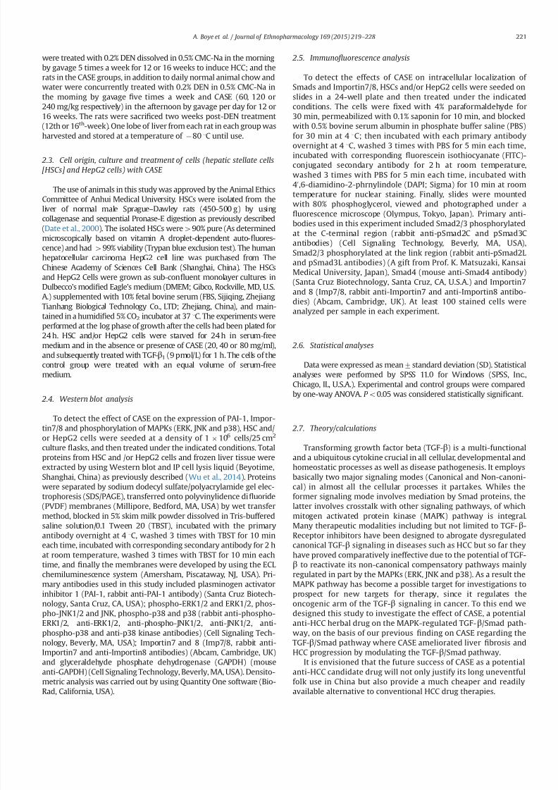
7232019 Compound Astragalus and Salvia Miltiorrhiza Extracts Modulate MAPK Regulated TGF Smad Signaling in Hepatocellhellip
httpslidepdfcomreaderfullcompound-astragalus-and-salvia-miltiorrhiza-extracts-modulate-mapk-regulated 410
3 Results
31 CASE down-regulated TGF- β 1-induced activation of pERK and
pJNK but up-regulated pp38 in both HSCs and HepG2 cells
TGF-β1 induced activation of ERK1 but not ERK2 in HSCs when
compared to control however CASE (20 40 and 80 mgml respec-
tively) concentration dependently decreased TGF-β1-induced up-
regulation of ERK1 whiles at the same time completely abolishedthe activation of ERK2 (Fig 1(1)) Similarly TGF-β1 induced
activation of JNK12 in HSCs compared to control however prior
treatment of HSCs with CASE (20 40 and 80 mgml respectively)
concentration dependently decreased the hitherto increased TGF-β1-induced activation of JNK12 (Fig 1(2)) TGF-β1 stimulation of
HSCs produced increased activation of pp38 compared to control
but interestingly CASE potentiated TGF-β1-induced activation of
pp38 in HSCs (Fig 1(3)) Similarly TGF-β1 induced the expression
of pERK pJNK and pp38 in HepG2 cells after incubation of HepG2
cells with exogenous TGF-β1 However pretreatment of HepG2
cells with CASE (20 40 and 80 mgml respectively) before stimula-
tion of HepG2 cells with TGF-β1 showed a concentration-
dependent inhibition of the activation and expression of pERK
(Fig 1(4)) With respect to pJNK TGF-β1 stimulation produced an
increase in pJNK activation compared to control group Though
prior CASE treatment of HepG2 cells before TGF-β1 stimulation
inhibited pJNK activation and expression it was only signi1047297cant at
a lower concentration (20 40 mgml) (Fig1(5)) Interestingly CASE
potentiated TGF-β1-induced activation and expression of pp38 inHepG2 cells similar to HSCs (Fig 1(6))
32 CASE decreased TGF- β 1-induced domain-speci 1047297c
phosphorylation of Smad23 and nuclear translocation of Smad4 in
both HSCs and HepG2 cells
Stimulation of HSCs with exogenous TGF-β1 resulted in increased
expression of phosphorylated pSmad2C pSmad2L and oncogenic
pSmad3L however prior treatment of HSC cells with CASE (20 40
and 80mgml respectively) before TGF-β1 stimulation led to a
Fig 1 Effect of CASE on TGF-β1-induced activation of pERK pJNK and pp38 in HSCs ( Fig 1(1) (2) and (3)) and HepG2 Cells (Fig 1(4) (5) and (6)) The HSCs andor HepG2
cells were seeded at a density of 1106 cells25 cm2 culture 1047298asks then cultured with 10 FBS in 95 air and 5 CO2 at 37 1C The cells were starved for 24 h in serum-free
medium and in the absence or presence of CASE (20 40 or 80 μgml) when the HSCs andor HepG2 cells attached to 70ndash80 of monolayers subsequently cells were treated
with TGF-β1 (9 pmolL) for 1 h Total proteins of the cells were extracted by using Western blot and IP cell lysis liquid Expression of ERK12 JNK12 and pp38 were monitored
by western blot using anti-pERK12 anti-pJNK12 and anti-pp38 antibodies respectively Intensities of pERK12 pJNK12 and pp38 bands were normalized to ERK12 JNK12
and p38 respectively of the corresponding treatment groups The data presented are based on at least three independent experiments (P o001 compared with control
group P o001 compared with TGF-β1 group)
A Boye et al Journal of Ethnopharmacology 169 (2015) 219ndash 228222
7232019 Compound Astragalus and Salvia Miltiorrhiza Extracts Modulate MAPK Regulated TGF Smad Signaling in Hepatocellhellip
httpslidepdfcomreaderfullcompound-astragalus-and-salvia-miltiorrhiza-extracts-modulate-mapk-regulated 510
concentration-dependent decrease in the TGF-β1-induced phosphor-
ylation of pSmad2C pSmad2L and oncogenic pSmad3L (Fig 2(1)) In a
similar manner treatment of HepG2 cells with TGF-β1 (9 pmolL)
produced increased domain-speci1047297c phosphorylation and expression
of pSmad2C pSmad2L oncogenic pSmad3L and Smad4 (Fig 2(2) and
(3)) But pretreatment of HepG2 cells with CASE (20 40 and 80 μgml
respectively) before TGF-β1 stimulation led to the reversal of the
above observations Importantly CASE reduced nuclear import of
Smad4 by enhancing its cytoplasmic retention and also enhanced
the phosphorylation of tumor suppressor pSmad3C speci1047297cally in
HepG2 cells (Fig 2(2) and (3))
33 CASE decreased TGF- β 1-induced expression of Imp78 and their
subcellular localization in both HSCs and HepG2 cells
TGF-β1 stimulated HSCs produced not only increased protein
expression of Imp78 but also enhanced nuclear relocation of Imp7
8 compared to control (Fig 3(1) and (2)) But CASE treatment beforeTGF-β1 stimulation produced a concentration-dependent decrease in
Imp78 expression and also reduced signi1047297cantly their nuclear reloca-
tion especially Imp7 (Fig 3(5)) Importantly CASE (40 and 80 mgml)
was most effective Similarly TGF-β1-stimulated HepG2 cells produced
increased expression of Imp78 and also enhanced nucleo-cytoplasmic
distribution But prior treatment of HepG2 cells with CASE before TGF-
β1 stimulation produced not only reduction in the expressions of
Imp78 (Fig 3-(3) (4)) but also reduced their nuclear relocation
particularly Imp7 (Fig 3(6))
34 CASE repressed TGF- β 1-induced expression of PAI-1 gene in
HSCs and HepG2 cells
TGF-β1 (9 pmolL) stimulation of HSCs and HepG2 cells inseparate experiments produced increased PAI-1 gene expression
compared to control groups but upon prior CASE treatment before
TGF-β1 stimulation produced a concentration dependent repres-
sion of PAI-1 gene expression in the studied cell lines in a reverse
manner (Fig 4(1) and (2))
35 CASE decreased DEN-induced activation of pERK pJNK and pp38
in rats
DEN time dependently increased the activation and expression
of pERK12 pJNK12 and pp38 in rat livers However concurrent
treatment of rats with DEN (05 ml100 g bodyweight) in the
morning and CASE (60 120 and 240 mgkg respectively) in the
afternoon 1047297
rst for a period of 12 weeks and subsequently for aperiod of 16 weeks showed dose dependent reduction in the DEN-
induced activation and expression of pERK12 pJNK12 and pp38
(Fig 5) With the exception of CASE (60 mgkg) which produced
weak inhibitory effect on DEN-induced activation and expression
of pJNK12 at the end of the 12 weeks (Fig 5(2)) CASE (60 120 and
240 mgkg respectively) dose dependently decreased DEN-
induced activation and expression of pERK pJNK and pp38 at
both 12th and 16th weeks (Fig 5) CASE (120 mgkg) was most
effective in decreasing DEN-induced activation and expression of
pERK12 and pJNK12 at both the 12th and 16th weeks whiles
CASE (240 mgkg) was only effective in decreasing the activation
and expression of pp38 at both periods (Fig 5(3) and (6))
4 Discussion
We herein present a study further elucidating the molecular
mechanisms of CASE as a potential anti-HCC herbal medicine Essen
tially we report that CASE modulates the MAPK-regulated TGF-βSmad
signaling via inhibition of oncogenic MAPK-dependent linker phosphor-
ylation of Smad23 in HSCs (A key hepatic cell implicated in liver 1047297brosis
(Puche et al 2013) and HepG2 cells (A kind of human hepatoma cell
line) resulting in down-regulation of PAI-1 gene Precisely CASE also
inhibited the MAPK pathway in DEN-induced HCC rats which provided
a strong rationale for us to probe further how the inhibition of the
MAPK pathway will affect the overall TGF-βSmad signaling The MAPK
pathway is one of the important signaling collaborators of TGF-β that
enhances overall oncogenic TGF-βSmad signaling output in cancer
(Giehl et al 2007 Moustakas and Heldin 2005) Indeed the cross-
Fig 2 Effect of CASE on TGF-β1-induced domain-speci1047297c phosphorylation of
Smad23 Smad4 expressions and their nuclear translocation in HSCs ( Fig 2(1))
and HepG2 cells (Fig 2(2) and (3)) HSCs andor HepG2 cells were starved for 24 h
in serum-free medium and in the absence or presence of CASE (20 40 or 80 μgml)
when the cells attached to 30ndash40 of monolayers in 24-holes culture plate
respectively subsequently the cells were treated with TGF-β1 (9 pmolL) for 1 h
The cells were then 1047297xed permeabilized blocked and incubated with each primary
antibody then corresponding 1047298uorescein isothiocyanate (FITC)-Conjugated sec-
ondary antibody ultimately viewed and photographed using a 1047298uorescence
microscope Fig 2(3)) HepG2 cells were starved for 24 h in serum-free medium
and in the absence or presence of CASE (20 40 or 80 μgml) when the cells attached
to 30ndash40 of monolayers in 24-holes culture plate respectively subsequently cells
were treated with TGF-β1 (9 pmolL) for 1 h The cells were then 1047297
xed permeabi-lized blocked and incubated with each primary antibody then corresponding
1047298uorescein isothiocyanate (FITC)-Conjugated secondary antibody incubated with
40 6-diamidino-2-phrnylindole (DAPI) for nuclear staining ultimately viewed and
photographed using a 1047298uorescence microscope C C-terminal L Linker region
A Boye et al Journal of Ethnopharmacology 169 (2015) 219ndash 228 223
7232019 Compound Astragalus and Salvia Miltiorrhiza Extracts Modulate MAPK Regulated TGF Smad Signaling in Hepatocellhellip
httpslidepdfcomreaderfullcompound-astragalus-and-salvia-miltiorrhiza-extracts-modulate-mapk-regulated 610
signaling between the MAPK pathway and the TGF-βSmad signaling is
now a common phenomenon reported in many cancer subtypes For
instance the MAPKs have been shown to be activated by TGF-β in
various cancer subtypes including HCC (Zhang 2009) and the TGF-β-
induced activation of the MAPKs have been shown to increase MAPK-
dependent linker phosphorylation of Smad23 (Furukawa et al 2003
Yoshida et al 2005) which incidentally promotes oncogenesis (Nagata et
al 2009) Notably activation of ERK12 and JNK was shown to be crucial
for TGF-β-induced Smad4-independent signaling (Giehl et al 2007)
which means that the MAPK pathway can signi1047297cantly promote TGF-βsignaling even in the absence of Smad4 (A common Smad protein which
mediates canonical TGF-β signaling) via the linker phosphorylation of
Smad23 (Non-canonical TGF-β signaling) Targeted inhibition of the
MAPK pathway speci1047297cally inhibition of MAPK-dependent linker phos-
phorylation of the R-Smads may signi1047297cantly abrogate TGF-βSmad
signaling in cancer Quiet recently by using MAPK-Speci1047297c Inhibitors
(PD98059 [ERK-Speci1047297c inhibitor] SP600125 [JNK-Speci1047297c inhibitor] and
SB203580 [p38-Speci1047297c inhibitor]) our study group reported on effective
modulation of the MAPK-regulated TGF-βSmad signaling in HCC
leading to decreased PAI-1 gene expression and near blockade of HCC
phenotypes (Cell proliferation migration and invasion) (Boye et al
2015) In this study CASE inhibited hitherto increased TGF-β1-induced
activation of pERK and pJNK in HSC and HepG2 cells and these results
were consistent in both HSC and HepG2 cells This shows that CASE
Fig 3 Effect of CASE on TGF-β1-induced expression of Importin7 (Imp7) and Importin8 (Imp8) in HSCs (Fig 3(1) (2) and (5)) and HepG2 cells (Fig 3(3) (4) and (6)) The
HSCs and HepG2 cells were seeded at a density of 1106 cells25 cm2 culture 1047298asks then cultured with 10 FBS in 95 air and 5 CO2 at 37 1C The cells were starved for
24 h in serum-free medium and in the absence or presence of CASE (20 40 or 80 μgml) when the HSCs andor HepG2 cells attached to 70ndash80 of monolayers subsequently
cells were treated with TGF-β1 (9 pmolL) for 1 h Total Imp78 proteins of the cells were extracted by using Western blot and IP cell lysis liquid Expression of Imp7 and Imp8
were monitored by IB using anti-Importin 7 and anti-Importin 8 antibodies respectively Intensities of Imp7 and Imp8 bands were normalized to glyceraldehyde-3-
phosphate-dehydrogenase (GAPDH) of the corresponding treatment groups The ratio of the Imp7 and Imp8 to GAPDH without exogenous TGF- β1 was assigned a value of 1
And Fig 3(5) and (6)) HSC andor HepG2 cells were starved for 24 h in serum-free medium and in the absence or presence of CASE (20 40 or 80 μgml) when the cells
attached to 30ndash40 of monolayers in 24-holes culture plate respectively subsequently cells were treated with TGF- β1 (9 pmolL) for 1 h The cells were then 1047297xed
permeabilized blocked and incubated with each primary antibody then corresponding 1047298uorescein isothiocyanate (FITC)-conjugated secondary antibody incubated with 4
6-diamidino-2-phrynylindole (DAPI) for nuclear staining ultimately viewed and photographed using a 1047298uorescence microscope The data presented are based on at least
three independent experiments (P o005 P o001 compared with control group P o005 P o001 compared with TGF-β1 group)
A Boye et al Journal of Ethnopharmacology 169 (2015) 219ndash 228224
7232019 Compound Astragalus and Salvia Miltiorrhiza Extracts Modulate MAPK Regulated TGF Smad Signaling in Hepatocellhellip
httpslidepdfcomreaderfullcompound-astragalus-and-salvia-miltiorrhiza-extracts-modulate-mapk-regulated 710
in vitro target ERKJNK-dependent MAPK signaling in the studied cell
lines however CASE increased pp38 in both cell lines even more than
the model group which perhaps indicates that CASE variably modulates
the MAPK pathway in vitro Activation of the MAPKs by TGF-β and
subsequent MAPK-dependent linker phosphorylation of Smad23enhances nuclear import of pSmad23 to promote TGF-β signaling In
view of this we sought to 1047297nd out how CASE-dependent inhibition of
the MAPKs in HSC and HepG2 cells will affect the down and upstream
mediators of the TGF-βSmad signaling speci1047297cally we monitored
domain-speci1047297c phosphorylation of Smad23 expression of Smad4
and Imp78 and their nuclear import
Brie1047298y TGF-β signaling begins with ligand activation of constituted
TGF-β receptor type 2 (TβRII) which then trans-phosphorylate TGF-βreceptor type 1 (TβRI) The phosphorylated TβRI in turn phosphorylate
receptor-dependent Smad proteins (Smad2 and Smad3) at the C-
terminal to facilitate their oligomerization with Smad4 to form a
complex (Smad234 complex) which preferentially relocates into the
nucleus (Matsuzaki 2012) In the nucleus pSmad23 directly partakes
in the execution of TGF-β-mediated transcription of target genes
through association with transcriptional effectors (Promoters and
repressors) (Matsuzaki 2012 Murata et al 2014) Thus the Smads
act as both signaling mediators (Messengers) and transcriptional
effectors to determine the fate of TGF-β-speci1047297c target gene transcrip-
tion of which PAI-1 gene is integral Critical to both canonical and non-
canonical TGF-β signaling is the nuclear import of pSmad23 as a
complex with Smad4 (Smad234 complex) or through Smad4-
independent mechanism via MAPK-dependent linker phosphorylation
of Smad23 Essentially any of the two import mechanisms mayinvolve Imp78 For instance (Chen and Xu 2011) have shown that
importin proteins (Imp78) are indispensable for nuclear import of
pSmad23 and Smad4 It was also shown that nuclear import of
pSmad23 is a critical step in TGF-β signaling transduction (Xu et al
2007) Further it was shown that knockdown of importin 78
markedly impaired nuclear import of pSmad23 in response to TGF-
β-dependent gene regulation which is critical for embryonic develop-
ment and cellular homeostasis but detrimental in dysregulated TGF-βsignaling in cancer (Xu et al 2007) Also (Xu et al 2007) have
intimated that since Imp78 is critical for nuclear import of pSmad23
it is highly probable that pharmacological inhibition of this nuclear
import factor may disrupt dysregulated TGF-βSmad signaling in
cancer Accordingly TGF-β1 stimulation of HSCs and HepG2 cells
produced increased linker phosphorylation of Smad23 increased
expression of Smad4 and Imp78 and their nuclear import and this
correlated with increased PAI-1 gene expression in both cell lines
(Fig 4(1) and (2)) Interestingly CASE-dependent inhibition of pERK
and pJNK in both cell lines correlated with CASE-dependent inhibition
of linker phosphorylation of Smad23 decreased expression of Smad4
and Imp78 and their nuclear import For example in HepG2 cells
CASE signi1047297cantly decreased nuclear import of Smad4 by enhancing
its cytoplasmic retention (Fig 2(2) and (3)) Also CASE treatment
inhibited Imp78 expression compared with model (TGF-β1-stimu-
lated HSCs and HepG2 cells) and control groups Precisely CASE
decreased both cytoplasmic and nuclear expression of Imp7 but only
decreased cytoplasmic expression of Imp8 compared to model and
control groups in both HSC and HepG2 cells and these effects
correlated with decreased pSmad23 and Smad4 expressions (Fig 2
(1) (2) and (3)) which suggest that CASE-dependent inhibition of pSmad23 and Smad4 nuclear import may be mediated through Imp7
inhibition Correspondingly CASE-dependent decrease in nuclear
import of pSmad23 Smad4 and Imp7 led to signi1047297cant decrease in
PAI-1 gene expression
Severally PAI-1 gene has been reported as one of the important
target genes of TGF-β signaling which has been shown to account for
most of the pathological roles of TGF-β signaling in cancer (Derynck
and Zhang 2003 Tahashi et al 2002) Functionally PAI-1 gene acts as
the main inhibitor of the urokinase-type plasminogen activator system
and it stimulates cell migration and invasion by disrupting cellular
adhesion and enhancing basement membrane degradation (Gutierrez
et al 2000) Indeed overexpression of PAI-1 gene has been implicated
in multiple forms of 1047297brosis including liver 1047297brosis (Gramling and
Church 2010) a key risk factor of HCC In our study both TGF-β1-stimulated HSC and HepG2 cells showed increased expression of PAI-1
gene This observation is consistent with our previous reports regard-
ing the ability of CASE to modulate TGF-βSmad signaling leading to
decrease in PAI-1 gene expression in myo1047297broblast (Yang et al 2008)
keloid 1047297broblast (He et al 2012) HepG2 cells (Hu et al 2014 Liu et al
2010) and DEN-induced HCC in rats (Rui et al 2014) Clearly from the
present results CASE selectively modulate ERKJNK-dependent linker
phosphorylation of Smad23 in vitro in part this observation con1047297rms
our earlier report concerning JNKMAPK-regulated TGF-βSmad sig-
naling in myo1047297broblast (Yang et al 2008) where CASE in a concen-
tration dependent manner inhibited JNK and JNK-dependent linker
phosphorylation of Smad23
To further con1047297rm or otherwise the ability of CASE to modulate the
MAPK pathway we studied the effect of CASE on DEN-induced
Fig 4 Effect of CASE on TGF-β1-induced expression of plasminogen activator
inhibitor 1 (PAI-1) in HSCs (Fig 4(1)) and HepG2 cells (Fig 4(2)) The HSCs andor
HepG2 cells were seeded at a density of 1 106 cells25 cm2 culture 1047298asks then
cultured with 10 FBS in 95 air and 5 CO2 at 37 1
C The cells were starved for24 h in serum-free medium and in the absence or presence of CASE (20 40 or
80 μgml) when the HSCs andor HepG2 cells attached to 70 ndash80 of monolayers
subsequently cells were treated with TGF-β1 (9 pmolL) for 1 h Total proteins of the
cells were extracted by using Western blot and IP cell lysis liquid Expression of PAI-
1 gene was monitored by western blot using anti-PAI-1 Ab Intensities of PAI-1
bands were normalized to glyceraldehyde-3-phosphate-dehydrogenase (GAPDH) of
the corresponding treatment groups The ratio of the PAI-1 gene to GAPDH without
exogenous TGF-β1 was assigned a value of 1 The data presented are based on at
least three independent experiments (P o005 compared with control group
P o005 P o001 compared with TGF-β1 group)
A Boye et al Journal of Ethnopharmacology 169 (2015) 219ndash 228 225
7232019 Compound Astragalus and Salvia Miltiorrhiza Extracts Modulate MAPK Regulated TGF Smad Signaling in Hepatocellhellip
httpslidepdfcomreaderfullcompound-astragalus-and-salvia-miltiorrhiza-extracts-modulate-mapk-regulated 810
activation of the MAPK pathway From our in vivo study treatment of
rats with DEN for 12 weeks led to increased activation and expression
of pERK pJNK and pp38 but extending DEN treatment period to 16
weeks even saw a further increase in the average expression of pERK
pJNK and pp38 (Fig 5(4) (5) and (6)) indicating a direct causal
relationship between duration of DEN treatment and MAPK activation
These results indicate that increase in MAPK activation and expression
may correlate with HCC progression But concomitant treatment of
rats with DEN in the morning and CASE in the afternoon reversed the
above relationship for instance at 12 weeks not only did CASE (120 or
240 mgkg) decreased the expression of pERK pJNK and pp38
compared to the model group but also decreased the average
expression level of each MAPK even more than the average for the
control group (Fig 5(1) (2) and (3)) showing that CASE potently
modulates the MAPK pathway and that the MAPK pathway could be
yet another important target of CASE At 16 weeks though CASE
decreased the expression of the MAPKs compared to the model group
it was however not different from that of the control group ( Fig 5(4)
(5) and (6)) indicating that MAPK activation and expression were
indeed related to duration of DEN treatment The in vivo inhibition of
pp38 by CASE (Fig 5(3) and (6)) contradicts our in vitro results
regarding pp38 But this observation probably suggest that CASE
Fig 5 Effect of CASE on diethyl nitrosamine (DEN)-induced activation of pERK pJNK and pp38 in rats The proteins were extracted from frozen liver tissues from 12th week
(Fig 5(1) (2) and (3)) and 16th week (Fig 5(4) (5) and (6)) DEN treated rats Phosphorylated (p) ERK12 pJNK12 pp38 and ERK JNK p38 were analyzed by western blot
using anti-pERK12 pJNK12 pp38 and ERK12 JNK12 p38 Abs and anti-glyceraldehyde phosphate dehydrogenase (GAPDH) Ab Intensities of pERK12 pJNK12 pp38
bands were normalized to those of ERK12 JNK12 p38 of the corresponding treatment groups The ratio of the pERK12 pJNK12 and pp38 proteins to ERK12 JNK12 and
p38 respectively of normal group was assigned a value of 1 Each value represents mean7SD nfrac143 P o005 P o001 compared with control group P o005 P o001
compared with DEN group
A Boye et al Journal of Ethnopharmacology 169 (2015) 219ndash 228226
7232019 Compound Astragalus and Salvia Miltiorrhiza Extracts Modulate MAPK Regulated TGF Smad Signaling in Hepatocellhellip
httpslidepdfcomreaderfullcompound-astragalus-and-salvia-miltiorrhiza-extracts-modulate-mapk-regulated 910
modulate pERK pJNK and pp38 in vivo by the same mechanism which
may involve some unknown factors in the tumor microenvironment
since the tumor microenvironment was the only major factor absent
in the in vitro study or it may be possible that while CASE employs the
same mechanism to modulate pERK and pJNK both in in vitro andin vivo it however employs different mechanisms to modulate pp38
in in vitro and in vivo settings Indeed these results further show that
CASE does not only target the upstream and downstream mediators
(TGF-β ligand TβRI TβRII Smad2CL Smad3L Smad4 and PAI-1) of
TGF-βSmad signaling as previously reported (He et al 2012 Hu et al
2014 Liu et al 2010 Rui et al 2014 Wu et al 2014 Yang et al 2008)
but also targets the MAPK pathway Fundamentally our in vivo results
agree in part with (Nagata et al 2009) with regard to JNK activation
by DEN treatment however our results concerning pERK and pp38
are inconsistent with their 1047297nding For instance Nagata et al (2009)
had shown that after 86-days DEN treatment of rats only pJNK was
activated whiles pERK and pp38 remained inactive (Un-phosphory-
lated) but in our study we found that increasing DEN treatment to 12
or 16 weeks produced signi1047297cant activation of all the MAPKs (pERKpJNK and pp38) We report that activation of the MAPKs by DEN
treatment is time-dependent and may relate directly to HCC
progression
Put together the present results provide a strong rationale for
further elucidation of the MAPK pathway as a key target for
therapy against HCC Essentially CASE has shown a multi-target
potential by modulating both the MAPK and TGF-βSmad signaling
pathways (Fig 6) in a manner that highlights its potential drug
candidature However our results raise some further questions for
example which speci1047297c subcellular location (Cytoplasm or
nucleus) does CASE modulate the MAPK pathway and how does
it affect HCC phenotypic manifestations such as cell proliferation
dysregulated apoptosis cell migration and invasion These ques-
tions are informed by the fact that spatio-temporal regulation of
the MAPK pathway is crucial for many pathways implicated in HCC
including multi-functional TGF-βSmad signaling
5 Conclusion
CASE blocked MAPK activation and MAPK-dependent linker
phosphorylation of Smad23 inhibited Smad4 and Imp78
blocked nuclear import of Smad234 via Imp7 modulation and
down-regulated PAI-1 gene expression and these results do not
only demonstrate the potential of the MAPK-regulated TGF-β
Smad pathway as an important target for therapy against HCC but
also highlights the potential drug candidature of CASE and con-
1047297rmation of its ethnobotanical usage
Con1047298icts of interest
No potential con1047298icts of interest
Acknowledgments
We thank Prof K Matsuzaki (Department of Gastroenterologyand Hepatology Kansai Medical University Osaka Japan) for
providing us with the following Abs Anti-pSmad2L and Anti-
pSmad3L Also this study was supported by the National Natural
Science Foundation of China (No 81073098 and no 81374012)
References
Boye A Kan H Wu C Jiang Y Yang X He S Yang Y 2015 MAPK inhibitorsdifferently modulate TGF-betaSmad signaling in HepG2 cells Tumour Biol httpdxdoiorg101007s13277-014-3002-x
Chan W Durairajan S Lu J Wang Y Xie L Kum W Koo I Yung K Li M2009 Neuroprotective effects of astragaloside IV in 6-hydroxydopamine-treated primary nigral cell culture Neurochem Int 55 414ndash422
Chen R Shao H Lin S Zhang J Xu K 2011 Treatment with astragalusmembranaceus produces antioxidative effects and attenuates intestinal mucosa
injury induced by intestinal ischemia-reperfusion in rats Am J Chin Med 39879ndash887
Chen X Xu L 2011 Mechanism and regulation of nucleocytoplasmic traf 1047297cking of Smad Cell Biosci 1 40
Cho WC Leung KN 2007a In vitro and in vivo anti-tumor effects of astragalusmembranaceus Cancer Lett 252 43ndash54
Cho WC Leung KN 2007b In vitro and in vivo immunomodulating andimmunorestorative effects of astragalus membranaceus J Ethnopharmacol113 132ndash141
Date M Matsuzaki K Matsushita M Tahashi Y Furukawa F Inoue K 2000Modulation of transforming growth factor function in hepatocytes and hepaticstellate cells in rat liver injury Gut 46 719ndash724
Derynck R Zhang YE 2003 Smad-dependent and Smad-independent pathwaysin TGF-β family signalling Nature 425 577ndash584
Fuentealba L Eivers E Ikeda A Hurtado C Kuroda H Pera E De Robertis E2007 Integrating patterning signals WntGSK3 regulates the duration of theBMPSmad1 signal Cell 131 980ndash993
Furukawa F Matsuzaki K Mori S Tahashi Y Yoshida K Sugano Y Yamagata
H Matsushita M Seki T Inagaki Y Nishizawa M Fujisawa J Inoue K2003 p38 MAPK mediates 1047297brogenic signal through Smad3 phosphorylation inrat myo1047297broblasts Hepatology 38 879ndash889
Giehl K Imamichi Y Menke A 2007 Smad4-independent TGF-β signaling intumor cell migration Cells Tissues Org 185 123ndash130
Gramling MW Church FC 2010 Plasminogen activator inhibitor-1 is anaggregate response factor with pleiotropic effects on cell signaling in vasculardisease and the tumor microenvironment Thromb Res 125 377ndash381
Gutierrez LS Schulman A Brito-Robinson T Noria F Ploplis VA Castellino FJ2000 Tumor development is retarded in mice lacking the gene for urokinase-type plasminogen activator or its inhibitor plasminogen activator inhibitor-1Cancer Res 60 5839ndash5847
Hata A Davis B 2009 Control of microRNA biogenesis by TGFbeta signalingpathway-a novel role of Smads in the nucleus Cytokine Growth Factor Rev 20517ndash521
Hayashida T Decaestecker M Schnaper H 2003 Cross-talk between ERK MAPkinase and Smad signaling pathways enhances TGF-beta-dependent lsre-sponses in human mesangial cel FASEB J 17 1576ndash1578
He S Yang Y Liu X Huang W Zhang X Yang S 2012 Compound Astragalus
and Salvia miltiorrhiza extract inhibits cell proliferation invasion and collagen
Fig 6 Fig 6 shows an illustrative summary of the multi-target mechanism by
which CASE modulates the MAPK-regulated TGF-βSmad signaling in hepatocel-lular carcinoma (HCC) CASE blocks TGF-β-induced activation of ERK JNK and p38
to abrogate MAPK-dependent linker phosphorylation of Smad23 and their sub-
sequent nuclear translocation Along the canonical loop CASE inhibits Smad4
expression formation of Smad234 and their nuclear translocation via inhibition of
Imp78 CASE-dependent disruption of nuclear import of Smad23 and Smad4 leads
to down-regulation of PAI-1 gene expression Cytoplasm (C) C-terminal phos-
phorylated Smad (pSmadC) Linker phosphorylated Smad (pSmadL) Nucleus (N)
Phosphorylated Smad (pSmad) Receptor mediated Smad (R-Smad) Transforming
growth factor receptor type 1 and 2 (TβRI and TβRII)
A Boye et al Journal of Ethnopharmacology 169 (2015) 219ndash 228 227
7232019 Compound Astragalus and Salvia Miltiorrhiza Extracts Modulate MAPK Regulated TGF Smad Signaling in Hepatocellhellip
httpslidepdfcomreaderfullcompound-astragalus-and-salvia-miltiorrhiza-extracts-modulate-mapk-regulated 1010
synthesis in keloid 1047297broblasts by mediating transforming growth factor‐βSmad pathway Br J Dermatol 166 564ndash574
Hu X Rui W Wu C He S Jiang J Zhang X Yang Y 2014 CompoundAstragalus and Salvia Miltiorrhiza extracts suppress hepatocarcinogenesis bymodulating transforming growth factor‐βSmad signaling J GastroenterolHepatol 29 1284ndash1291
Jun T 2004 Determination of total astragalosides in astragalus by colormetricmethod J Anhui Tradit Chin Med Coll 5 017
Kretzschmar M Doody J Timokhina I Massagueacute J 1999 A mechanism of repression of TGFbetaSmad signaling by oncogenic Ras Genes Dev 13804ndash816
Lee TY Chang HH Wang GJ Chiu JH Yang YY Lin HC 2006 Water‐
solubleextract of Salvia miltiorrhiza ameliorates carbon tetrachloride‐mediated hepa-tic apoptosis in rats J Pharm Pharm 58 659ndash665
Liu X Yang Y Zhang X Xu S He S Huang W Roberts MS 2010 CompoundAstragalus and Salvia miltiorrhiza extract inhibits cell invasion by modulatingtransforming growth factor‐βSmad in HepG2 cell J Gastroenterol Hepatol 25420ndash426
Matsuzaki K 2012 Smad phosphoisoform signals in acute and chronic liver injurysimilarities and differences between epithelial and mesenchymal cells CellTissue Res 347 225ndash243
Moustakas A Heldin C-H 2005 Non-Smad TGF-β signals J Cell Sci 1183573ndash3584
Murata M Yoshida K Yamaguchi T Matsuzaki K 2014 Linker phosphorylationof Smad3 promotes 1047297bro-carcinogenesis in chronic viral hepatitis of hepato-cellular carcinoma World J Gastroenterol 20 15018ndash15027
Nagata H Hatano E Tada M Murata M Kitamura K Asechi H Narita MYanagida A Tamaki N Yagi S 2009 Inhibition of c‐ Jun NH2‐terminal kinaseswitches Smad3 signaling from oncogenesis to tumor‐suppression in rat
hepatocellular carcinoma Hepatology 49 1944ndash
1953Puche JE Saiman Y Friedman SL 2013 Hepatic stellate cells and liver 1047297brosisCompr Physiol 3 1473ndash1492
Roxas M Jurenka J 2007 Colds and in1047298uenza a review of diagnosis andconventional botanical and nutritional considerations Altern Med Rev 1225ndash48
Rui W Xie L Liu X He S Wu C Zhang X Zhang L Yang Y 2014 CompoundAstragalus and Salvia miltiorrhiza extract suppresses hepatocellular carcinomaprogression by inhibiting 1047297brosis and PAI-1 mRNA transcription J Ethnophar-macol 151 198ndash209
Tang J 2004 Determination of total astragalosides in astragalus by colorimetricmethod J Anhui Tradit Chin Med Coll 23 37ndash38
Shao BM Xu W Dai H Tu P Li Z Gao XM 2004 A study on the immunereceptors for polysaccharides from the roots of astragalus membranaceus aChinese medicinal herb Biochem Biophys Res Commun 320 1103ndash1111
Shen P Liu MH Ng TY Chan YH Yong EL 2006 Differential effects of iso1047298avones from astragalus membranaceus and Pueraria thomsonii on theactivation of PPARalpha PPARgamma and adipocyte differentiation in vitro
J Nutr 136 899ndash905
Tahashi Y Matsuzaki K Yoshida K Furukawa F Sugano Y Matsushita MHimeno Y Inagaki Y Inoue K 2002 Differential regulation of TGF‐β signal in
hepatic stellate cells between acute and chronic rat liver injury Hepatology 3549ndash61
Wu C Jiang J Boye A Jiang Y Yang Y 2014 Compound Astragalus and Salviamiltiorrhiza extract suppresses rabbits hypertrophic scar by modulating theTGF-betaSmad signal Dermatology 229 363ndash368
Wu YP Cao Y Cao YY 2001 Technology optimization of extracting activitycomponents from Astragalus membranaceus Lishizen Med Mater Med Res 12876ndash877
Xu L Yao X Chen X Lu P Zhang B Ip YT 2007 Msk is required for nuclearimport of TGF-βBMP-activated Smads J cell Biol 178 981ndash994
Yang Y Yang S Chen M Zhang X Zou Y Zhang X 2008 Compound Astragalus
and Salvia miltiorrhiza extract exerts anti-1047297brosis by mediating TGF-βSmadsignaling in myo1047297broblasts J Ethnopharmacol 118 264ndash270
Ye Y 2006 Comparative study on the determination of salvianolic acids content bycolorimetery and HPLC J Zhejiang Univ Tradit Chin Med 30 350 ndash351
Yoshida K Matsuzaki K Mori S Tahashi Y Yamagata H Furukawa F Seki TNishizawa M Fujisawa J Okazaki K 2005 Transforming growth factor-betaand platelet-derived growth factor signal via c-Jun N-terminal kinase-depen-dent Smad23 phosphorylation in rat hepatic stellate cells after acute liverinjury The American journal of pathology 166 1029ndash1039
Yoshida K Murata M Yamaguchi T Matsuzaki K 2014 TGF-βSmad signalingduring hepatic 1047297bro-carcinogenesis (review) Int J Oncol 45 1363ndash1371
Zhang SS He FY Xu XY 2001 Comparative studies on the content of ingredients of polysaccharides in the Buyanghuanwutang and single materials
J Human Coll Tradit Chin Med 21 26Zhang YE 2009 Non-Smad pathways in TGF-β signaling Cell Res 19 128ndash139
Glossary
Phosphorylation Addition of a phosphorus atom to a protein or a moleculeSmad proteins A class of proteins that mediate transforming growth factor beta
(TGF-β) signaling transductionPhosphorylated Smad A Smad protein that has received a phosphorus atom from a
phosphorus carrier moleculeLinker phosphorylated Smad A Smad protein phosphorylated at a region in
between its N-terminal and C-terminalCanonical TGF-β Signaling It is Smad-mediated TGF-β signaling transduction
involving Smad2 Smad3 and Smad4 complex formation leading to theirnuclear translocation and subsequent transcription of TGF-β-target mRNAs
Importin protein 7 and 8 (Imp78) They are proteins mostly in the cytoplasm thatfacilitate nuclear entry of other proteins
Mitogen activated protein kinase (MAPK) They are kinase proteins that facilitatetransduction of extracellular signals into the nucleus
Non-canonical TGF-β signaling All TGF-β signaling transduction that does notinvolve Smad4 or non-Smad TGF-β signaling
A Boye et al Journal of Ethnopharmacology 169 (2015) 219ndash 228228
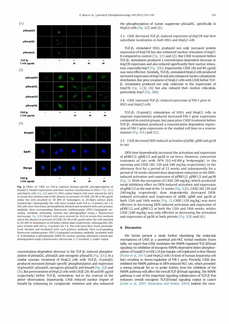
7232019 Compound Astragalus and Salvia Miltiorrhiza Extracts Modulate MAPK Regulated TGF Smad Signaling in Hepatocellhellip
httpslidepdfcomreaderfullcompound-astragalus-and-salvia-miltiorrhiza-extracts-modulate-mapk-regulated 510
concentration-dependent decrease in the TGF-β1-induced phosphor-
ylation of pSmad2C pSmad2L and oncogenic pSmad3L (Fig 2(1)) In a
similar manner treatment of HepG2 cells with TGF-β1 (9 pmolL)
produced increased domain-speci1047297c phosphorylation and expression
of pSmad2C pSmad2L oncogenic pSmad3L and Smad4 (Fig 2(2) and
(3)) But pretreatment of HepG2 cells with CASE (20 40 and 80 μgml
respectively) before TGF-β1 stimulation led to the reversal of the
above observations Importantly CASE reduced nuclear import of
Smad4 by enhancing its cytoplasmic retention and also enhanced
the phosphorylation of tumor suppressor pSmad3C speci1047297cally in
HepG2 cells (Fig 2(2) and (3))
33 CASE decreased TGF- β 1-induced expression of Imp78 and their
subcellular localization in both HSCs and HepG2 cells
TGF-β1 stimulated HSCs produced not only increased protein
expression of Imp78 but also enhanced nuclear relocation of Imp7
8 compared to control (Fig 3(1) and (2)) But CASE treatment beforeTGF-β1 stimulation produced a concentration-dependent decrease in
Imp78 expression and also reduced signi1047297cantly their nuclear reloca-
tion especially Imp7 (Fig 3(5)) Importantly CASE (40 and 80 mgml)
was most effective Similarly TGF-β1-stimulated HepG2 cells produced
increased expression of Imp78 and also enhanced nucleo-cytoplasmic
distribution But prior treatment of HepG2 cells with CASE before TGF-
β1 stimulation produced not only reduction in the expressions of
Imp78 (Fig 3-(3) (4)) but also reduced their nuclear relocation
particularly Imp7 (Fig 3(6))
34 CASE repressed TGF- β 1-induced expression of PAI-1 gene in
HSCs and HepG2 cells
TGF-β1 (9 pmolL) stimulation of HSCs and HepG2 cells inseparate experiments produced increased PAI-1 gene expression
compared to control groups but upon prior CASE treatment before
TGF-β1 stimulation produced a concentration dependent repres-
sion of PAI-1 gene expression in the studied cell lines in a reverse
manner (Fig 4(1) and (2))
35 CASE decreased DEN-induced activation of pERK pJNK and pp38
in rats
DEN time dependently increased the activation and expression
of pERK12 pJNK12 and pp38 in rat livers However concurrent
treatment of rats with DEN (05 ml100 g bodyweight) in the
morning and CASE (60 120 and 240 mgkg respectively) in the
afternoon 1047297
rst for a period of 12 weeks and subsequently for aperiod of 16 weeks showed dose dependent reduction in the DEN-
induced activation and expression of pERK12 pJNK12 and pp38
(Fig 5) With the exception of CASE (60 mgkg) which produced
weak inhibitory effect on DEN-induced activation and expression
of pJNK12 at the end of the 12 weeks (Fig 5(2)) CASE (60 120 and
240 mgkg respectively) dose dependently decreased DEN-
induced activation and expression of pERK pJNK and pp38 at
both 12th and 16th weeks (Fig 5) CASE (120 mgkg) was most
effective in decreasing DEN-induced activation and expression of
pERK12 and pJNK12 at both the 12th and 16th weeks whiles
CASE (240 mgkg) was only effective in decreasing the activation
and expression of pp38 at both periods (Fig 5(3) and (6))
4 Discussion
We herein present a study further elucidating the molecular
mechanisms of CASE as a potential anti-HCC herbal medicine Essen
tially we report that CASE modulates the MAPK-regulated TGF-βSmad
signaling via inhibition of oncogenic MAPK-dependent linker phosphor-
ylation of Smad23 in HSCs (A key hepatic cell implicated in liver 1047297brosis
(Puche et al 2013) and HepG2 cells (A kind of human hepatoma cell
line) resulting in down-regulation of PAI-1 gene Precisely CASE also
inhibited the MAPK pathway in DEN-induced HCC rats which provided
a strong rationale for us to probe further how the inhibition of the
MAPK pathway will affect the overall TGF-βSmad signaling The MAPK
pathway is one of the important signaling collaborators of TGF-β that
enhances overall oncogenic TGF-βSmad signaling output in cancer
(Giehl et al 2007 Moustakas and Heldin 2005) Indeed the cross-
Fig 2 Effect of CASE on TGF-β1-induced domain-speci1047297c phosphorylation of
Smad23 Smad4 expressions and their nuclear translocation in HSCs ( Fig 2(1))
and HepG2 cells (Fig 2(2) and (3)) HSCs andor HepG2 cells were starved for 24 h
in serum-free medium and in the absence or presence of CASE (20 40 or 80 μgml)
when the cells attached to 30ndash40 of monolayers in 24-holes culture plate
respectively subsequently the cells were treated with TGF-β1 (9 pmolL) for 1 h
The cells were then 1047297xed permeabilized blocked and incubated with each primary
antibody then corresponding 1047298uorescein isothiocyanate (FITC)-Conjugated sec-
ondary antibody ultimately viewed and photographed using a 1047298uorescence
microscope Fig 2(3)) HepG2 cells were starved for 24 h in serum-free medium
and in the absence or presence of CASE (20 40 or 80 μgml) when the cells attached
to 30ndash40 of monolayers in 24-holes culture plate respectively subsequently cells
were treated with TGF-β1 (9 pmolL) for 1 h The cells were then 1047297
xed permeabi-lized blocked and incubated with each primary antibody then corresponding
1047298uorescein isothiocyanate (FITC)-Conjugated secondary antibody incubated with
40 6-diamidino-2-phrnylindole (DAPI) for nuclear staining ultimately viewed and
photographed using a 1047298uorescence microscope C C-terminal L Linker region
A Boye et al Journal of Ethnopharmacology 169 (2015) 219ndash 228 223
7232019 Compound Astragalus and Salvia Miltiorrhiza Extracts Modulate MAPK Regulated TGF Smad Signaling in Hepatocellhellip
httpslidepdfcomreaderfullcompound-astragalus-and-salvia-miltiorrhiza-extracts-modulate-mapk-regulated 610
signaling between the MAPK pathway and the TGF-βSmad signaling is
now a common phenomenon reported in many cancer subtypes For
instance the MAPKs have been shown to be activated by TGF-β in
various cancer subtypes including HCC (Zhang 2009) and the TGF-β-
induced activation of the MAPKs have been shown to increase MAPK-
dependent linker phosphorylation of Smad23 (Furukawa et al 2003
Yoshida et al 2005) which incidentally promotes oncogenesis (Nagata et
al 2009) Notably activation of ERK12 and JNK was shown to be crucial
for TGF-β-induced Smad4-independent signaling (Giehl et al 2007)
which means that the MAPK pathway can signi1047297cantly promote TGF-βsignaling even in the absence of Smad4 (A common Smad protein which
mediates canonical TGF-β signaling) via the linker phosphorylation of
Smad23 (Non-canonical TGF-β signaling) Targeted inhibition of the
MAPK pathway speci1047297cally inhibition of MAPK-dependent linker phos-
phorylation of the R-Smads may signi1047297cantly abrogate TGF-βSmad
signaling in cancer Quiet recently by using MAPK-Speci1047297c Inhibitors
(PD98059 [ERK-Speci1047297c inhibitor] SP600125 [JNK-Speci1047297c inhibitor] and
SB203580 [p38-Speci1047297c inhibitor]) our study group reported on effective
modulation of the MAPK-regulated TGF-βSmad signaling in HCC
leading to decreased PAI-1 gene expression and near blockade of HCC
phenotypes (Cell proliferation migration and invasion) (Boye et al
2015) In this study CASE inhibited hitherto increased TGF-β1-induced
activation of pERK and pJNK in HSC and HepG2 cells and these results
were consistent in both HSC and HepG2 cells This shows that CASE
Fig 3 Effect of CASE on TGF-β1-induced expression of Importin7 (Imp7) and Importin8 (Imp8) in HSCs (Fig 3(1) (2) and (5)) and HepG2 cells (Fig 3(3) (4) and (6)) The
HSCs and HepG2 cells were seeded at a density of 1106 cells25 cm2 culture 1047298asks then cultured with 10 FBS in 95 air and 5 CO2 at 37 1C The cells were starved for
24 h in serum-free medium and in the absence or presence of CASE (20 40 or 80 μgml) when the HSCs andor HepG2 cells attached to 70ndash80 of monolayers subsequently
cells were treated with TGF-β1 (9 pmolL) for 1 h Total Imp78 proteins of the cells were extracted by using Western blot and IP cell lysis liquid Expression of Imp7 and Imp8
were monitored by IB using anti-Importin 7 and anti-Importin 8 antibodies respectively Intensities of Imp7 and Imp8 bands were normalized to glyceraldehyde-3-
phosphate-dehydrogenase (GAPDH) of the corresponding treatment groups The ratio of the Imp7 and Imp8 to GAPDH without exogenous TGF- β1 was assigned a value of 1
And Fig 3(5) and (6)) HSC andor HepG2 cells were starved for 24 h in serum-free medium and in the absence or presence of CASE (20 40 or 80 μgml) when the cells
attached to 30ndash40 of monolayers in 24-holes culture plate respectively subsequently cells were treated with TGF- β1 (9 pmolL) for 1 h The cells were then 1047297xed
permeabilized blocked and incubated with each primary antibody then corresponding 1047298uorescein isothiocyanate (FITC)-conjugated secondary antibody incubated with 4
6-diamidino-2-phrynylindole (DAPI) for nuclear staining ultimately viewed and photographed using a 1047298uorescence microscope The data presented are based on at least
three independent experiments (P o005 P o001 compared with control group P o005 P o001 compared with TGF-β1 group)
A Boye et al Journal of Ethnopharmacology 169 (2015) 219ndash 228224
7232019 Compound Astragalus and Salvia Miltiorrhiza Extracts Modulate MAPK Regulated TGF Smad Signaling in Hepatocellhellip
httpslidepdfcomreaderfullcompound-astragalus-and-salvia-miltiorrhiza-extracts-modulate-mapk-regulated 710
in vitro target ERKJNK-dependent MAPK signaling in the studied cell
lines however CASE increased pp38 in both cell lines even more than
the model group which perhaps indicates that CASE variably modulates
the MAPK pathway in vitro Activation of the MAPKs by TGF-β and
subsequent MAPK-dependent linker phosphorylation of Smad23enhances nuclear import of pSmad23 to promote TGF-β signaling In
view of this we sought to 1047297nd out how CASE-dependent inhibition of
the MAPKs in HSC and HepG2 cells will affect the down and upstream
mediators of the TGF-βSmad signaling speci1047297cally we monitored
domain-speci1047297c phosphorylation of Smad23 expression of Smad4
and Imp78 and their nuclear import
Brie1047298y TGF-β signaling begins with ligand activation of constituted
TGF-β receptor type 2 (TβRII) which then trans-phosphorylate TGF-βreceptor type 1 (TβRI) The phosphorylated TβRI in turn phosphorylate
receptor-dependent Smad proteins (Smad2 and Smad3) at the C-
terminal to facilitate their oligomerization with Smad4 to form a
complex (Smad234 complex) which preferentially relocates into the
nucleus (Matsuzaki 2012) In the nucleus pSmad23 directly partakes
in the execution of TGF-β-mediated transcription of target genes
through association with transcriptional effectors (Promoters and
repressors) (Matsuzaki 2012 Murata et al 2014) Thus the Smads
act as both signaling mediators (Messengers) and transcriptional
effectors to determine the fate of TGF-β-speci1047297c target gene transcrip-
tion of which PAI-1 gene is integral Critical to both canonical and non-
canonical TGF-β signaling is the nuclear import of pSmad23 as a
complex with Smad4 (Smad234 complex) or through Smad4-
independent mechanism via MAPK-dependent linker phosphorylation
of Smad23 Essentially any of the two import mechanisms mayinvolve Imp78 For instance (Chen and Xu 2011) have shown that
importin proteins (Imp78) are indispensable for nuclear import of
pSmad23 and Smad4 It was also shown that nuclear import of
pSmad23 is a critical step in TGF-β signaling transduction (Xu et al
2007) Further it was shown that knockdown of importin 78
markedly impaired nuclear import of pSmad23 in response to TGF-
β-dependent gene regulation which is critical for embryonic develop-
ment and cellular homeostasis but detrimental in dysregulated TGF-βsignaling in cancer (Xu et al 2007) Also (Xu et al 2007) have
intimated that since Imp78 is critical for nuclear import of pSmad23
it is highly probable that pharmacological inhibition of this nuclear
import factor may disrupt dysregulated TGF-βSmad signaling in
cancer Accordingly TGF-β1 stimulation of HSCs and HepG2 cells
produced increased linker phosphorylation of Smad23 increased
expression of Smad4 and Imp78 and their nuclear import and this
correlated with increased PAI-1 gene expression in both cell lines
(Fig 4(1) and (2)) Interestingly CASE-dependent inhibition of pERK
and pJNK in both cell lines correlated with CASE-dependent inhibition
of linker phosphorylation of Smad23 decreased expression of Smad4
and Imp78 and their nuclear import For example in HepG2 cells
CASE signi1047297cantly decreased nuclear import of Smad4 by enhancing
its cytoplasmic retention (Fig 2(2) and (3)) Also CASE treatment
inhibited Imp78 expression compared with model (TGF-β1-stimu-
lated HSCs and HepG2 cells) and control groups Precisely CASE
decreased both cytoplasmic and nuclear expression of Imp7 but only
decreased cytoplasmic expression of Imp8 compared to model and
control groups in both HSC and HepG2 cells and these effects
correlated with decreased pSmad23 and Smad4 expressions (Fig 2
(1) (2) and (3)) which suggest that CASE-dependent inhibition of pSmad23 and Smad4 nuclear import may be mediated through Imp7
inhibition Correspondingly CASE-dependent decrease in nuclear
import of pSmad23 Smad4 and Imp7 led to signi1047297cant decrease in
PAI-1 gene expression
Severally PAI-1 gene has been reported as one of the important
target genes of TGF-β signaling which has been shown to account for
most of the pathological roles of TGF-β signaling in cancer (Derynck
and Zhang 2003 Tahashi et al 2002) Functionally PAI-1 gene acts as
the main inhibitor of the urokinase-type plasminogen activator system
and it stimulates cell migration and invasion by disrupting cellular
adhesion and enhancing basement membrane degradation (Gutierrez
et al 2000) Indeed overexpression of PAI-1 gene has been implicated
in multiple forms of 1047297brosis including liver 1047297brosis (Gramling and
Church 2010) a key risk factor of HCC In our study both TGF-β1-stimulated HSC and HepG2 cells showed increased expression of PAI-1
gene This observation is consistent with our previous reports regard-
ing the ability of CASE to modulate TGF-βSmad signaling leading to
decrease in PAI-1 gene expression in myo1047297broblast (Yang et al 2008)
keloid 1047297broblast (He et al 2012) HepG2 cells (Hu et al 2014 Liu et al
2010) and DEN-induced HCC in rats (Rui et al 2014) Clearly from the
present results CASE selectively modulate ERKJNK-dependent linker
phosphorylation of Smad23 in vitro in part this observation con1047297rms
our earlier report concerning JNKMAPK-regulated TGF-βSmad sig-
naling in myo1047297broblast (Yang et al 2008) where CASE in a concen-
tration dependent manner inhibited JNK and JNK-dependent linker
phosphorylation of Smad23
To further con1047297rm or otherwise the ability of CASE to modulate the
MAPK pathway we studied the effect of CASE on DEN-induced
Fig 4 Effect of CASE on TGF-β1-induced expression of plasminogen activator
inhibitor 1 (PAI-1) in HSCs (Fig 4(1)) and HepG2 cells (Fig 4(2)) The HSCs andor
HepG2 cells were seeded at a density of 1 106 cells25 cm2 culture 1047298asks then
cultured with 10 FBS in 95 air and 5 CO2 at 37 1
C The cells were starved for24 h in serum-free medium and in the absence or presence of CASE (20 40 or
80 μgml) when the HSCs andor HepG2 cells attached to 70 ndash80 of monolayers
subsequently cells were treated with TGF-β1 (9 pmolL) for 1 h Total proteins of the
cells were extracted by using Western blot and IP cell lysis liquid Expression of PAI-
1 gene was monitored by western blot using anti-PAI-1 Ab Intensities of PAI-1
bands were normalized to glyceraldehyde-3-phosphate-dehydrogenase (GAPDH) of
the corresponding treatment groups The ratio of the PAI-1 gene to GAPDH without
exogenous TGF-β1 was assigned a value of 1 The data presented are based on at
least three independent experiments (P o005 compared with control group
P o005 P o001 compared with TGF-β1 group)
A Boye et al Journal of Ethnopharmacology 169 (2015) 219ndash 228 225
7232019 Compound Astragalus and Salvia Miltiorrhiza Extracts Modulate MAPK Regulated TGF Smad Signaling in Hepatocellhellip
httpslidepdfcomreaderfullcompound-astragalus-and-salvia-miltiorrhiza-extracts-modulate-mapk-regulated 810
activation of the MAPK pathway From our in vivo study treatment of
rats with DEN for 12 weeks led to increased activation and expression
of pERK pJNK and pp38 but extending DEN treatment period to 16
weeks even saw a further increase in the average expression of pERK
pJNK and pp38 (Fig 5(4) (5) and (6)) indicating a direct causal
relationship between duration of DEN treatment and MAPK activation
These results indicate that increase in MAPK activation and expression
may correlate with HCC progression But concomitant treatment of
rats with DEN in the morning and CASE in the afternoon reversed the
above relationship for instance at 12 weeks not only did CASE (120 or
240 mgkg) decreased the expression of pERK pJNK and pp38
compared to the model group but also decreased the average
expression level of each MAPK even more than the average for the
control group (Fig 5(1) (2) and (3)) showing that CASE potently
modulates the MAPK pathway and that the MAPK pathway could be
yet another important target of CASE At 16 weeks though CASE
decreased the expression of the MAPKs compared to the model group
it was however not different from that of the control group ( Fig 5(4)
(5) and (6)) indicating that MAPK activation and expression were
indeed related to duration of DEN treatment The in vivo inhibition of
pp38 by CASE (Fig 5(3) and (6)) contradicts our in vitro results
regarding pp38 But this observation probably suggest that CASE
Fig 5 Effect of CASE on diethyl nitrosamine (DEN)-induced activation of pERK pJNK and pp38 in rats The proteins were extracted from frozen liver tissues from 12th week
(Fig 5(1) (2) and (3)) and 16th week (Fig 5(4) (5) and (6)) DEN treated rats Phosphorylated (p) ERK12 pJNK12 pp38 and ERK JNK p38 were analyzed by western blot
using anti-pERK12 pJNK12 pp38 and ERK12 JNK12 p38 Abs and anti-glyceraldehyde phosphate dehydrogenase (GAPDH) Ab Intensities of pERK12 pJNK12 pp38
bands were normalized to those of ERK12 JNK12 p38 of the corresponding treatment groups The ratio of the pERK12 pJNK12 and pp38 proteins to ERK12 JNK12 and
p38 respectively of normal group was assigned a value of 1 Each value represents mean7SD nfrac143 P o005 P o001 compared with control group P o005 P o001
compared with DEN group
A Boye et al Journal of Ethnopharmacology 169 (2015) 219ndash 228226
7232019 Compound Astragalus and Salvia Miltiorrhiza Extracts Modulate MAPK Regulated TGF Smad Signaling in Hepatocellhellip
httpslidepdfcomreaderfullcompound-astragalus-and-salvia-miltiorrhiza-extracts-modulate-mapk-regulated 910
modulate pERK pJNK and pp38 in vivo by the same mechanism which
may involve some unknown factors in the tumor microenvironment
since the tumor microenvironment was the only major factor absent
in the in vitro study or it may be possible that while CASE employs the
same mechanism to modulate pERK and pJNK both in in vitro andin vivo it however employs different mechanisms to modulate pp38
in in vitro and in vivo settings Indeed these results further show that
CASE does not only target the upstream and downstream mediators
(TGF-β ligand TβRI TβRII Smad2CL Smad3L Smad4 and PAI-1) of
TGF-βSmad signaling as previously reported (He et al 2012 Hu et al
2014 Liu et al 2010 Rui et al 2014 Wu et al 2014 Yang et al 2008)
but also targets the MAPK pathway Fundamentally our in vivo results
agree in part with (Nagata et al 2009) with regard to JNK activation
by DEN treatment however our results concerning pERK and pp38
are inconsistent with their 1047297nding For instance Nagata et al (2009)
had shown that after 86-days DEN treatment of rats only pJNK was
activated whiles pERK and pp38 remained inactive (Un-phosphory-
lated) but in our study we found that increasing DEN treatment to 12
or 16 weeks produced signi1047297cant activation of all the MAPKs (pERKpJNK and pp38) We report that activation of the MAPKs by DEN
treatment is time-dependent and may relate directly to HCC
progression
Put together the present results provide a strong rationale for
further elucidation of the MAPK pathway as a key target for
therapy against HCC Essentially CASE has shown a multi-target
potential by modulating both the MAPK and TGF-βSmad signaling
pathways (Fig 6) in a manner that highlights its potential drug
candidature However our results raise some further questions for
example which speci1047297c subcellular location (Cytoplasm or
nucleus) does CASE modulate the MAPK pathway and how does
it affect HCC phenotypic manifestations such as cell proliferation
dysregulated apoptosis cell migration and invasion These ques-
tions are informed by the fact that spatio-temporal regulation of
the MAPK pathway is crucial for many pathways implicated in HCC
including multi-functional TGF-βSmad signaling
5 Conclusion
CASE blocked MAPK activation and MAPK-dependent linker
phosphorylation of Smad23 inhibited Smad4 and Imp78
blocked nuclear import of Smad234 via Imp7 modulation and
down-regulated PAI-1 gene expression and these results do not
only demonstrate the potential of the MAPK-regulated TGF-β
Smad pathway as an important target for therapy against HCC but
also highlights the potential drug candidature of CASE and con-
1047297rmation of its ethnobotanical usage
Con1047298icts of interest
No potential con1047298icts of interest
Acknowledgments
We thank Prof K Matsuzaki (Department of Gastroenterologyand Hepatology Kansai Medical University Osaka Japan) for
providing us with the following Abs Anti-pSmad2L and Anti-
pSmad3L Also this study was supported by the National Natural
Science Foundation of China (No 81073098 and no 81374012)
References
Boye A Kan H Wu C Jiang Y Yang X He S Yang Y 2015 MAPK inhibitorsdifferently modulate TGF-betaSmad signaling in HepG2 cells Tumour Biol httpdxdoiorg101007s13277-014-3002-x
Chan W Durairajan S Lu J Wang Y Xie L Kum W Koo I Yung K Li M2009 Neuroprotective effects of astragaloside IV in 6-hydroxydopamine-treated primary nigral cell culture Neurochem Int 55 414ndash422
Chen R Shao H Lin S Zhang J Xu K 2011 Treatment with astragalusmembranaceus produces antioxidative effects and attenuates intestinal mucosa
injury induced by intestinal ischemia-reperfusion in rats Am J Chin Med 39879ndash887
Chen X Xu L 2011 Mechanism and regulation of nucleocytoplasmic traf 1047297cking of Smad Cell Biosci 1 40
Cho WC Leung KN 2007a In vitro and in vivo anti-tumor effects of astragalusmembranaceus Cancer Lett 252 43ndash54
Cho WC Leung KN 2007b In vitro and in vivo immunomodulating andimmunorestorative effects of astragalus membranaceus J Ethnopharmacol113 132ndash141
Date M Matsuzaki K Matsushita M Tahashi Y Furukawa F Inoue K 2000Modulation of transforming growth factor function in hepatocytes and hepaticstellate cells in rat liver injury Gut 46 719ndash724
Derynck R Zhang YE 2003 Smad-dependent and Smad-independent pathwaysin TGF-β family signalling Nature 425 577ndash584
Fuentealba L Eivers E Ikeda A Hurtado C Kuroda H Pera E De Robertis E2007 Integrating patterning signals WntGSK3 regulates the duration of theBMPSmad1 signal Cell 131 980ndash993
Furukawa F Matsuzaki K Mori S Tahashi Y Yoshida K Sugano Y Yamagata
H Matsushita M Seki T Inagaki Y Nishizawa M Fujisawa J Inoue K2003 p38 MAPK mediates 1047297brogenic signal through Smad3 phosphorylation inrat myo1047297broblasts Hepatology 38 879ndash889
Giehl K Imamichi Y Menke A 2007 Smad4-independent TGF-β signaling intumor cell migration Cells Tissues Org 185 123ndash130
Gramling MW Church FC 2010 Plasminogen activator inhibitor-1 is anaggregate response factor with pleiotropic effects on cell signaling in vasculardisease and the tumor microenvironment Thromb Res 125 377ndash381
Gutierrez LS Schulman A Brito-Robinson T Noria F Ploplis VA Castellino FJ2000 Tumor development is retarded in mice lacking the gene for urokinase-type plasminogen activator or its inhibitor plasminogen activator inhibitor-1Cancer Res 60 5839ndash5847
Hata A Davis B 2009 Control of microRNA biogenesis by TGFbeta signalingpathway-a novel role of Smads in the nucleus Cytokine Growth Factor Rev 20517ndash521
Hayashida T Decaestecker M Schnaper H 2003 Cross-talk between ERK MAPkinase and Smad signaling pathways enhances TGF-beta-dependent lsre-sponses in human mesangial cel FASEB J 17 1576ndash1578
He S Yang Y Liu X Huang W Zhang X Yang S 2012 Compound Astragalus
and Salvia miltiorrhiza extract inhibits cell proliferation invasion and collagen
Fig 6 Fig 6 shows an illustrative summary of the multi-target mechanism by
which CASE modulates the MAPK-regulated TGF-βSmad signaling in hepatocel-lular carcinoma (HCC) CASE blocks TGF-β-induced activation of ERK JNK and p38
to abrogate MAPK-dependent linker phosphorylation of Smad23 and their sub-
sequent nuclear translocation Along the canonical loop CASE inhibits Smad4
expression formation of Smad234 and their nuclear translocation via inhibition of
Imp78 CASE-dependent disruption of nuclear import of Smad23 and Smad4 leads
to down-regulation of PAI-1 gene expression Cytoplasm (C) C-terminal phos-
phorylated Smad (pSmadC) Linker phosphorylated Smad (pSmadL) Nucleus (N)
Phosphorylated Smad (pSmad) Receptor mediated Smad (R-Smad) Transforming
growth factor receptor type 1 and 2 (TβRI and TβRII)
A Boye et al Journal of Ethnopharmacology 169 (2015) 219ndash 228 227
7232019 Compound Astragalus and Salvia Miltiorrhiza Extracts Modulate MAPK Regulated TGF Smad Signaling in Hepatocellhellip
httpslidepdfcomreaderfullcompound-astragalus-and-salvia-miltiorrhiza-extracts-modulate-mapk-regulated 1010
synthesis in keloid 1047297broblasts by mediating transforming growth factor‐βSmad pathway Br J Dermatol 166 564ndash574
Hu X Rui W Wu C He S Jiang J Zhang X Yang Y 2014 CompoundAstragalus and Salvia Miltiorrhiza extracts suppress hepatocarcinogenesis bymodulating transforming growth factor‐βSmad signaling J GastroenterolHepatol 29 1284ndash1291
Jun T 2004 Determination of total astragalosides in astragalus by colormetricmethod J Anhui Tradit Chin Med Coll 5 017
Kretzschmar M Doody J Timokhina I Massagueacute J 1999 A mechanism of repression of TGFbetaSmad signaling by oncogenic Ras Genes Dev 13804ndash816
Lee TY Chang HH Wang GJ Chiu JH Yang YY Lin HC 2006 Water‐
solubleextract of Salvia miltiorrhiza ameliorates carbon tetrachloride‐mediated hepa-tic apoptosis in rats J Pharm Pharm 58 659ndash665
Liu X Yang Y Zhang X Xu S He S Huang W Roberts MS 2010 CompoundAstragalus and Salvia miltiorrhiza extract inhibits cell invasion by modulatingtransforming growth factor‐βSmad in HepG2 cell J Gastroenterol Hepatol 25420ndash426
Matsuzaki K 2012 Smad phosphoisoform signals in acute and chronic liver injurysimilarities and differences between epithelial and mesenchymal cells CellTissue Res 347 225ndash243
Moustakas A Heldin C-H 2005 Non-Smad TGF-β signals J Cell Sci 1183573ndash3584
Murata M Yoshida K Yamaguchi T Matsuzaki K 2014 Linker phosphorylationof Smad3 promotes 1047297bro-carcinogenesis in chronic viral hepatitis of hepato-cellular carcinoma World J Gastroenterol 20 15018ndash15027
Nagata H Hatano E Tada M Murata M Kitamura K Asechi H Narita MYanagida A Tamaki N Yagi S 2009 Inhibition of c‐ Jun NH2‐terminal kinaseswitches Smad3 signaling from oncogenesis to tumor‐suppression in rat
hepatocellular carcinoma Hepatology 49 1944ndash
1953Puche JE Saiman Y Friedman SL 2013 Hepatic stellate cells and liver 1047297brosisCompr Physiol 3 1473ndash1492
Roxas M Jurenka J 2007 Colds and in1047298uenza a review of diagnosis andconventional botanical and nutritional considerations Altern Med Rev 1225ndash48
Rui W Xie L Liu X He S Wu C Zhang X Zhang L Yang Y 2014 CompoundAstragalus and Salvia miltiorrhiza extract suppresses hepatocellular carcinomaprogression by inhibiting 1047297brosis and PAI-1 mRNA transcription J Ethnophar-macol 151 198ndash209
Tang J 2004 Determination of total astragalosides in astragalus by colorimetricmethod J Anhui Tradit Chin Med Coll 23 37ndash38
Shao BM Xu W Dai H Tu P Li Z Gao XM 2004 A study on the immunereceptors for polysaccharides from the roots of astragalus membranaceus aChinese medicinal herb Biochem Biophys Res Commun 320 1103ndash1111
Shen P Liu MH Ng TY Chan YH Yong EL 2006 Differential effects of iso1047298avones from astragalus membranaceus and Pueraria thomsonii on theactivation of PPARalpha PPARgamma and adipocyte differentiation in vitro
J Nutr 136 899ndash905
Tahashi Y Matsuzaki K Yoshida K Furukawa F Sugano Y Matsushita MHimeno Y Inagaki Y Inoue K 2002 Differential regulation of TGF‐β signal in
hepatic stellate cells between acute and chronic rat liver injury Hepatology 3549ndash61
Wu C Jiang J Boye A Jiang Y Yang Y 2014 Compound Astragalus and Salviamiltiorrhiza extract suppresses rabbits hypertrophic scar by modulating theTGF-betaSmad signal Dermatology 229 363ndash368
Wu YP Cao Y Cao YY 2001 Technology optimization of extracting activitycomponents from Astragalus membranaceus Lishizen Med Mater Med Res 12876ndash877
Xu L Yao X Chen X Lu P Zhang B Ip YT 2007 Msk is required for nuclearimport of TGF-βBMP-activated Smads J cell Biol 178 981ndash994
Yang Y Yang S Chen M Zhang X Zou Y Zhang X 2008 Compound Astragalus
and Salvia miltiorrhiza extract exerts anti-1047297brosis by mediating TGF-βSmadsignaling in myo1047297broblasts J Ethnopharmacol 118 264ndash270
Ye Y 2006 Comparative study on the determination of salvianolic acids content bycolorimetery and HPLC J Zhejiang Univ Tradit Chin Med 30 350 ndash351
Yoshida K Matsuzaki K Mori S Tahashi Y Yamagata H Furukawa F Seki TNishizawa M Fujisawa J Okazaki K 2005 Transforming growth factor-betaand platelet-derived growth factor signal via c-Jun N-terminal kinase-depen-dent Smad23 phosphorylation in rat hepatic stellate cells after acute liverinjury The American journal of pathology 166 1029ndash1039
Yoshida K Murata M Yamaguchi T Matsuzaki K 2014 TGF-βSmad signalingduring hepatic 1047297bro-carcinogenesis (review) Int J Oncol 45 1363ndash1371
Zhang SS He FY Xu XY 2001 Comparative studies on the content of ingredients of polysaccharides in the Buyanghuanwutang and single materials
J Human Coll Tradit Chin Med 21 26Zhang YE 2009 Non-Smad pathways in TGF-β signaling Cell Res 19 128ndash139
Glossary
Phosphorylation Addition of a phosphorus atom to a protein or a moleculeSmad proteins A class of proteins that mediate transforming growth factor beta
(TGF-β) signaling transductionPhosphorylated Smad A Smad protein that has received a phosphorus atom from a
phosphorus carrier moleculeLinker phosphorylated Smad A Smad protein phosphorylated at a region in
between its N-terminal and C-terminalCanonical TGF-β Signaling It is Smad-mediated TGF-β signaling transduction
involving Smad2 Smad3 and Smad4 complex formation leading to theirnuclear translocation and subsequent transcription of TGF-β-target mRNAs
Importin protein 7 and 8 (Imp78) They are proteins mostly in the cytoplasm thatfacilitate nuclear entry of other proteins
Mitogen activated protein kinase (MAPK) They are kinase proteins that facilitatetransduction of extracellular signals into the nucleus
Non-canonical TGF-β signaling All TGF-β signaling transduction that does notinvolve Smad4 or non-Smad TGF-β signaling
A Boye et al Journal of Ethnopharmacology 169 (2015) 219ndash 228228
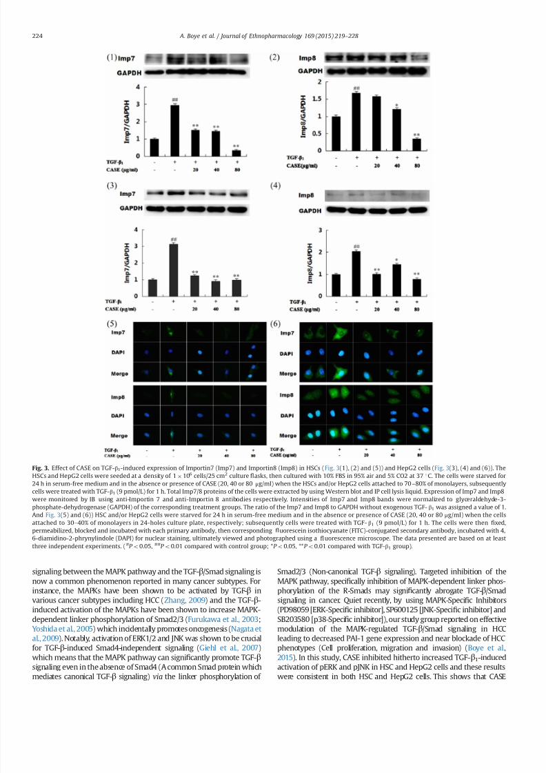
7232019 Compound Astragalus and Salvia Miltiorrhiza Extracts Modulate MAPK Regulated TGF Smad Signaling in Hepatocellhellip
httpslidepdfcomreaderfullcompound-astragalus-and-salvia-miltiorrhiza-extracts-modulate-mapk-regulated 610
signaling between the MAPK pathway and the TGF-βSmad signaling is
now a common phenomenon reported in many cancer subtypes For
instance the MAPKs have been shown to be activated by TGF-β in
various cancer subtypes including HCC (Zhang 2009) and the TGF-β-
induced activation of the MAPKs have been shown to increase MAPK-
dependent linker phosphorylation of Smad23 (Furukawa et al 2003
Yoshida et al 2005) which incidentally promotes oncogenesis (Nagata et
al 2009) Notably activation of ERK12 and JNK was shown to be crucial
for TGF-β-induced Smad4-independent signaling (Giehl et al 2007)
which means that the MAPK pathway can signi1047297cantly promote TGF-βsignaling even in the absence of Smad4 (A common Smad protein which
mediates canonical TGF-β signaling) via the linker phosphorylation of
Smad23 (Non-canonical TGF-β signaling) Targeted inhibition of the
MAPK pathway speci1047297cally inhibition of MAPK-dependent linker phos-
phorylation of the R-Smads may signi1047297cantly abrogate TGF-βSmad
signaling in cancer Quiet recently by using MAPK-Speci1047297c Inhibitors
(PD98059 [ERK-Speci1047297c inhibitor] SP600125 [JNK-Speci1047297c inhibitor] and
SB203580 [p38-Speci1047297c inhibitor]) our study group reported on effective
modulation of the MAPK-regulated TGF-βSmad signaling in HCC
leading to decreased PAI-1 gene expression and near blockade of HCC
phenotypes (Cell proliferation migration and invasion) (Boye et al
2015) In this study CASE inhibited hitherto increased TGF-β1-induced
activation of pERK and pJNK in HSC and HepG2 cells and these results
were consistent in both HSC and HepG2 cells This shows that CASE
Fig 3 Effect of CASE on TGF-β1-induced expression of Importin7 (Imp7) and Importin8 (Imp8) in HSCs (Fig 3(1) (2) and (5)) and HepG2 cells (Fig 3(3) (4) and (6)) The
HSCs and HepG2 cells were seeded at a density of 1106 cells25 cm2 culture 1047298asks then cultured with 10 FBS in 95 air and 5 CO2 at 37 1C The cells were starved for
24 h in serum-free medium and in the absence or presence of CASE (20 40 or 80 μgml) when the HSCs andor HepG2 cells attached to 70ndash80 of monolayers subsequently
cells were treated with TGF-β1 (9 pmolL) for 1 h Total Imp78 proteins of the cells were extracted by using Western blot and IP cell lysis liquid Expression of Imp7 and Imp8
were monitored by IB using anti-Importin 7 and anti-Importin 8 antibodies respectively Intensities of Imp7 and Imp8 bands were normalized to glyceraldehyde-3-
phosphate-dehydrogenase (GAPDH) of the corresponding treatment groups The ratio of the Imp7 and Imp8 to GAPDH without exogenous TGF- β1 was assigned a value of 1
And Fig 3(5) and (6)) HSC andor HepG2 cells were starved for 24 h in serum-free medium and in the absence or presence of CASE (20 40 or 80 μgml) when the cells
attached to 30ndash40 of monolayers in 24-holes culture plate respectively subsequently cells were treated with TGF- β1 (9 pmolL) for 1 h The cells were then 1047297xed
permeabilized blocked and incubated with each primary antibody then corresponding 1047298uorescein isothiocyanate (FITC)-conjugated secondary antibody incubated with 4
6-diamidino-2-phrynylindole (DAPI) for nuclear staining ultimately viewed and photographed using a 1047298uorescence microscope The data presented are based on at least
three independent experiments (P o005 P o001 compared with control group P o005 P o001 compared with TGF-β1 group)
A Boye et al Journal of Ethnopharmacology 169 (2015) 219ndash 228224
7232019 Compound Astragalus and Salvia Miltiorrhiza Extracts Modulate MAPK Regulated TGF Smad Signaling in Hepatocellhellip
httpslidepdfcomreaderfullcompound-astragalus-and-salvia-miltiorrhiza-extracts-modulate-mapk-regulated 710
in vitro target ERKJNK-dependent MAPK signaling in the studied cell
lines however CASE increased pp38 in both cell lines even more than
the model group which perhaps indicates that CASE variably modulates
the MAPK pathway in vitro Activation of the MAPKs by TGF-β and
subsequent MAPK-dependent linker phosphorylation of Smad23enhances nuclear import of pSmad23 to promote TGF-β signaling In
view of this we sought to 1047297nd out how CASE-dependent inhibition of
the MAPKs in HSC and HepG2 cells will affect the down and upstream
mediators of the TGF-βSmad signaling speci1047297cally we monitored
domain-speci1047297c phosphorylation of Smad23 expression of Smad4
and Imp78 and their nuclear import
Brie1047298y TGF-β signaling begins with ligand activation of constituted
TGF-β receptor type 2 (TβRII) which then trans-phosphorylate TGF-βreceptor type 1 (TβRI) The phosphorylated TβRI in turn phosphorylate
receptor-dependent Smad proteins (Smad2 and Smad3) at the C-
terminal to facilitate their oligomerization with Smad4 to form a
complex (Smad234 complex) which preferentially relocates into the
nucleus (Matsuzaki 2012) In the nucleus pSmad23 directly partakes
in the execution of TGF-β-mediated transcription of target genes
through association with transcriptional effectors (Promoters and
repressors) (Matsuzaki 2012 Murata et al 2014) Thus the Smads
act as both signaling mediators (Messengers) and transcriptional
effectors to determine the fate of TGF-β-speci1047297c target gene transcrip-
tion of which PAI-1 gene is integral Critical to both canonical and non-
canonical TGF-β signaling is the nuclear import of pSmad23 as a
complex with Smad4 (Smad234 complex) or through Smad4-
independent mechanism via MAPK-dependent linker phosphorylation
of Smad23 Essentially any of the two import mechanisms mayinvolve Imp78 For instance (Chen and Xu 2011) have shown that
importin proteins (Imp78) are indispensable for nuclear import of
pSmad23 and Smad4 It was also shown that nuclear import of
pSmad23 is a critical step in TGF-β signaling transduction (Xu et al
2007) Further it was shown that knockdown of importin 78
markedly impaired nuclear import of pSmad23 in response to TGF-
β-dependent gene regulation which is critical for embryonic develop-
ment and cellular homeostasis but detrimental in dysregulated TGF-βsignaling in cancer (Xu et al 2007) Also (Xu et al 2007) have
intimated that since Imp78 is critical for nuclear import of pSmad23
it is highly probable that pharmacological inhibition of this nuclear
import factor may disrupt dysregulated TGF-βSmad signaling in
cancer Accordingly TGF-β1 stimulation of HSCs and HepG2 cells
produced increased linker phosphorylation of Smad23 increased
expression of Smad4 and Imp78 and their nuclear import and this
correlated with increased PAI-1 gene expression in both cell lines
(Fig 4(1) and (2)) Interestingly CASE-dependent inhibition of pERK
and pJNK in both cell lines correlated with CASE-dependent inhibition
of linker phosphorylation of Smad23 decreased expression of Smad4
and Imp78 and their nuclear import For example in HepG2 cells
CASE signi1047297cantly decreased nuclear import of Smad4 by enhancing
its cytoplasmic retention (Fig 2(2) and (3)) Also CASE treatment
inhibited Imp78 expression compared with model (TGF-β1-stimu-
lated HSCs and HepG2 cells) and control groups Precisely CASE
decreased both cytoplasmic and nuclear expression of Imp7 but only
decreased cytoplasmic expression of Imp8 compared to model and
control groups in both HSC and HepG2 cells and these effects
correlated with decreased pSmad23 and Smad4 expressions (Fig 2
(1) (2) and (3)) which suggest that CASE-dependent inhibition of pSmad23 and Smad4 nuclear import may be mediated through Imp7
inhibition Correspondingly CASE-dependent decrease in nuclear
import of pSmad23 Smad4 and Imp7 led to signi1047297cant decrease in
PAI-1 gene expression
Severally PAI-1 gene has been reported as one of the important
target genes of TGF-β signaling which has been shown to account for
most of the pathological roles of TGF-β signaling in cancer (Derynck
and Zhang 2003 Tahashi et al 2002) Functionally PAI-1 gene acts as
the main inhibitor of the urokinase-type plasminogen activator system
and it stimulates cell migration and invasion by disrupting cellular
adhesion and enhancing basement membrane degradation (Gutierrez
et al 2000) Indeed overexpression of PAI-1 gene has been implicated
in multiple forms of 1047297brosis including liver 1047297brosis (Gramling and
Church 2010) a key risk factor of HCC In our study both TGF-β1-stimulated HSC and HepG2 cells showed increased expression of PAI-1
gene This observation is consistent with our previous reports regard-
ing the ability of CASE to modulate TGF-βSmad signaling leading to
decrease in PAI-1 gene expression in myo1047297broblast (Yang et al 2008)
keloid 1047297broblast (He et al 2012) HepG2 cells (Hu et al 2014 Liu et al
2010) and DEN-induced HCC in rats (Rui et al 2014) Clearly from the
present results CASE selectively modulate ERKJNK-dependent linker
phosphorylation of Smad23 in vitro in part this observation con1047297rms
our earlier report concerning JNKMAPK-regulated TGF-βSmad sig-
naling in myo1047297broblast (Yang et al 2008) where CASE in a concen-
tration dependent manner inhibited JNK and JNK-dependent linker
phosphorylation of Smad23
To further con1047297rm or otherwise the ability of CASE to modulate the
MAPK pathway we studied the effect of CASE on DEN-induced
Fig 4 Effect of CASE on TGF-β1-induced expression of plasminogen activator
inhibitor 1 (PAI-1) in HSCs (Fig 4(1)) and HepG2 cells (Fig 4(2)) The HSCs andor
HepG2 cells were seeded at a density of 1 106 cells25 cm2 culture 1047298asks then
cultured with 10 FBS in 95 air and 5 CO2 at 37 1
C The cells were starved for24 h in serum-free medium and in the absence or presence of CASE (20 40 or
80 μgml) when the HSCs andor HepG2 cells attached to 70 ndash80 of monolayers
subsequently cells were treated with TGF-β1 (9 pmolL) for 1 h Total proteins of the
cells were extracted by using Western blot and IP cell lysis liquid Expression of PAI-
1 gene was monitored by western blot using anti-PAI-1 Ab Intensities of PAI-1
bands were normalized to glyceraldehyde-3-phosphate-dehydrogenase (GAPDH) of
the corresponding treatment groups The ratio of the PAI-1 gene to GAPDH without
exogenous TGF-β1 was assigned a value of 1 The data presented are based on at
least three independent experiments (P o005 compared with control group
P o005 P o001 compared with TGF-β1 group)
A Boye et al Journal of Ethnopharmacology 169 (2015) 219ndash 228 225
7232019 Compound Astragalus and Salvia Miltiorrhiza Extracts Modulate MAPK Regulated TGF Smad Signaling in Hepatocellhellip
httpslidepdfcomreaderfullcompound-astragalus-and-salvia-miltiorrhiza-extracts-modulate-mapk-regulated 810
activation of the MAPK pathway From our in vivo study treatment of
rats with DEN for 12 weeks led to increased activation and expression
of pERK pJNK and pp38 but extending DEN treatment period to 16
weeks even saw a further increase in the average expression of pERK
pJNK and pp38 (Fig 5(4) (5) and (6)) indicating a direct causal
relationship between duration of DEN treatment and MAPK activation
These results indicate that increase in MAPK activation and expression
may correlate with HCC progression But concomitant treatment of
rats with DEN in the morning and CASE in the afternoon reversed the
above relationship for instance at 12 weeks not only did CASE (120 or
240 mgkg) decreased the expression of pERK pJNK and pp38
compared to the model group but also decreased the average
expression level of each MAPK even more than the average for the
control group (Fig 5(1) (2) and (3)) showing that CASE potently
modulates the MAPK pathway and that the MAPK pathway could be
yet another important target of CASE At 16 weeks though CASE
decreased the expression of the MAPKs compared to the model group
it was however not different from that of the control group ( Fig 5(4)
(5) and (6)) indicating that MAPK activation and expression were
indeed related to duration of DEN treatment The in vivo inhibition of
pp38 by CASE (Fig 5(3) and (6)) contradicts our in vitro results
regarding pp38 But this observation probably suggest that CASE
Fig 5 Effect of CASE on diethyl nitrosamine (DEN)-induced activation of pERK pJNK and pp38 in rats The proteins were extracted from frozen liver tissues from 12th week
(Fig 5(1) (2) and (3)) and 16th week (Fig 5(4) (5) and (6)) DEN treated rats Phosphorylated (p) ERK12 pJNK12 pp38 and ERK JNK p38 were analyzed by western blot
using anti-pERK12 pJNK12 pp38 and ERK12 JNK12 p38 Abs and anti-glyceraldehyde phosphate dehydrogenase (GAPDH) Ab Intensities of pERK12 pJNK12 pp38
bands were normalized to those of ERK12 JNK12 p38 of the corresponding treatment groups The ratio of the pERK12 pJNK12 and pp38 proteins to ERK12 JNK12 and
p38 respectively of normal group was assigned a value of 1 Each value represents mean7SD nfrac143 P o005 P o001 compared with control group P o005 P o001
compared with DEN group
A Boye et al Journal of Ethnopharmacology 169 (2015) 219ndash 228226
7232019 Compound Astragalus and Salvia Miltiorrhiza Extracts Modulate MAPK Regulated TGF Smad Signaling in Hepatocellhellip
httpslidepdfcomreaderfullcompound-astragalus-and-salvia-miltiorrhiza-extracts-modulate-mapk-regulated 910
modulate pERK pJNK and pp38 in vivo by the same mechanism which
may involve some unknown factors in the tumor microenvironment
since the tumor microenvironment was the only major factor absent
in the in vitro study or it may be possible that while CASE employs the
same mechanism to modulate pERK and pJNK both in in vitro andin vivo it however employs different mechanisms to modulate pp38
in in vitro and in vivo settings Indeed these results further show that
CASE does not only target the upstream and downstream mediators
(TGF-β ligand TβRI TβRII Smad2CL Smad3L Smad4 and PAI-1) of
TGF-βSmad signaling as previously reported (He et al 2012 Hu et al
2014 Liu et al 2010 Rui et al 2014 Wu et al 2014 Yang et al 2008)
but also targets the MAPK pathway Fundamentally our in vivo results
agree in part with (Nagata et al 2009) with regard to JNK activation
by DEN treatment however our results concerning pERK and pp38
are inconsistent with their 1047297nding For instance Nagata et al (2009)
had shown that after 86-days DEN treatment of rats only pJNK was
activated whiles pERK and pp38 remained inactive (Un-phosphory-
lated) but in our study we found that increasing DEN treatment to 12
or 16 weeks produced signi1047297cant activation of all the MAPKs (pERKpJNK and pp38) We report that activation of the MAPKs by DEN
treatment is time-dependent and may relate directly to HCC
progression
Put together the present results provide a strong rationale for
further elucidation of the MAPK pathway as a key target for
therapy against HCC Essentially CASE has shown a multi-target
potential by modulating both the MAPK and TGF-βSmad signaling
pathways (Fig 6) in a manner that highlights its potential drug
candidature However our results raise some further questions for
example which speci1047297c subcellular location (Cytoplasm or
nucleus) does CASE modulate the MAPK pathway and how does
it affect HCC phenotypic manifestations such as cell proliferation
dysregulated apoptosis cell migration and invasion These ques-
tions are informed by the fact that spatio-temporal regulation of
the MAPK pathway is crucial for many pathways implicated in HCC
including multi-functional TGF-βSmad signaling
5 Conclusion
CASE blocked MAPK activation and MAPK-dependent linker
phosphorylation of Smad23 inhibited Smad4 and Imp78
blocked nuclear import of Smad234 via Imp7 modulation and
down-regulated PAI-1 gene expression and these results do not
only demonstrate the potential of the MAPK-regulated TGF-β
Smad pathway as an important target for therapy against HCC but
also highlights the potential drug candidature of CASE and con-
1047297rmation of its ethnobotanical usage
Con1047298icts of interest
No potential con1047298icts of interest
Acknowledgments
We thank Prof K Matsuzaki (Department of Gastroenterologyand Hepatology Kansai Medical University Osaka Japan) for
providing us with the following Abs Anti-pSmad2L and Anti-
pSmad3L Also this study was supported by the National Natural
Science Foundation of China (No 81073098 and no 81374012)
References
Boye A Kan H Wu C Jiang Y Yang X He S Yang Y 2015 MAPK inhibitorsdifferently modulate TGF-betaSmad signaling in HepG2 cells Tumour Biol httpdxdoiorg101007s13277-014-3002-x
Chan W Durairajan S Lu J Wang Y Xie L Kum W Koo I Yung K Li M2009 Neuroprotective effects of astragaloside IV in 6-hydroxydopamine-treated primary nigral cell culture Neurochem Int 55 414ndash422
Chen R Shao H Lin S Zhang J Xu K 2011 Treatment with astragalusmembranaceus produces antioxidative effects and attenuates intestinal mucosa
injury induced by intestinal ischemia-reperfusion in rats Am J Chin Med 39879ndash887
Chen X Xu L 2011 Mechanism and regulation of nucleocytoplasmic traf 1047297cking of Smad Cell Biosci 1 40
Cho WC Leung KN 2007a In vitro and in vivo anti-tumor effects of astragalusmembranaceus Cancer Lett 252 43ndash54
Cho WC Leung KN 2007b In vitro and in vivo immunomodulating andimmunorestorative effects of astragalus membranaceus J Ethnopharmacol113 132ndash141
Date M Matsuzaki K Matsushita M Tahashi Y Furukawa F Inoue K 2000Modulation of transforming growth factor function in hepatocytes and hepaticstellate cells in rat liver injury Gut 46 719ndash724
Derynck R Zhang YE 2003 Smad-dependent and Smad-independent pathwaysin TGF-β family signalling Nature 425 577ndash584
Fuentealba L Eivers E Ikeda A Hurtado C Kuroda H Pera E De Robertis E2007 Integrating patterning signals WntGSK3 regulates the duration of theBMPSmad1 signal Cell 131 980ndash993
Furukawa F Matsuzaki K Mori S Tahashi Y Yoshida K Sugano Y Yamagata
H Matsushita M Seki T Inagaki Y Nishizawa M Fujisawa J Inoue K2003 p38 MAPK mediates 1047297brogenic signal through Smad3 phosphorylation inrat myo1047297broblasts Hepatology 38 879ndash889
Giehl K Imamichi Y Menke A 2007 Smad4-independent TGF-β signaling intumor cell migration Cells Tissues Org 185 123ndash130
Gramling MW Church FC 2010 Plasminogen activator inhibitor-1 is anaggregate response factor with pleiotropic effects on cell signaling in vasculardisease and the tumor microenvironment Thromb Res 125 377ndash381
Gutierrez LS Schulman A Brito-Robinson T Noria F Ploplis VA Castellino FJ2000 Tumor development is retarded in mice lacking the gene for urokinase-type plasminogen activator or its inhibitor plasminogen activator inhibitor-1Cancer Res 60 5839ndash5847
Hata A Davis B 2009 Control of microRNA biogenesis by TGFbeta signalingpathway-a novel role of Smads in the nucleus Cytokine Growth Factor Rev 20517ndash521
Hayashida T Decaestecker M Schnaper H 2003 Cross-talk between ERK MAPkinase and Smad signaling pathways enhances TGF-beta-dependent lsre-sponses in human mesangial cel FASEB J 17 1576ndash1578
He S Yang Y Liu X Huang W Zhang X Yang S 2012 Compound Astragalus
and Salvia miltiorrhiza extract inhibits cell proliferation invasion and collagen
Fig 6 Fig 6 shows an illustrative summary of the multi-target mechanism by
which CASE modulates the MAPK-regulated TGF-βSmad signaling in hepatocel-lular carcinoma (HCC) CASE blocks TGF-β-induced activation of ERK JNK and p38
to abrogate MAPK-dependent linker phosphorylation of Smad23 and their sub-
sequent nuclear translocation Along the canonical loop CASE inhibits Smad4
expression formation of Smad234 and their nuclear translocation via inhibition of
Imp78 CASE-dependent disruption of nuclear import of Smad23 and Smad4 leads
to down-regulation of PAI-1 gene expression Cytoplasm (C) C-terminal phos-
phorylated Smad (pSmadC) Linker phosphorylated Smad (pSmadL) Nucleus (N)
Phosphorylated Smad (pSmad) Receptor mediated Smad (R-Smad) Transforming
growth factor receptor type 1 and 2 (TβRI and TβRII)
A Boye et al Journal of Ethnopharmacology 169 (2015) 219ndash 228 227
7232019 Compound Astragalus and Salvia Miltiorrhiza Extracts Modulate MAPK Regulated TGF Smad Signaling in Hepatocellhellip
httpslidepdfcomreaderfullcompound-astragalus-and-salvia-miltiorrhiza-extracts-modulate-mapk-regulated 1010
synthesis in keloid 1047297broblasts by mediating transforming growth factor‐βSmad pathway Br J Dermatol 166 564ndash574
Hu X Rui W Wu C He S Jiang J Zhang X Yang Y 2014 CompoundAstragalus and Salvia Miltiorrhiza extracts suppress hepatocarcinogenesis bymodulating transforming growth factor‐βSmad signaling J GastroenterolHepatol 29 1284ndash1291
Jun T 2004 Determination of total astragalosides in astragalus by colormetricmethod J Anhui Tradit Chin Med Coll 5 017
Kretzschmar M Doody J Timokhina I Massagueacute J 1999 A mechanism of repression of TGFbetaSmad signaling by oncogenic Ras Genes Dev 13804ndash816
Lee TY Chang HH Wang GJ Chiu JH Yang YY Lin HC 2006 Water‐
solubleextract of Salvia miltiorrhiza ameliorates carbon tetrachloride‐mediated hepa-tic apoptosis in rats J Pharm Pharm 58 659ndash665
Liu X Yang Y Zhang X Xu S He S Huang W Roberts MS 2010 CompoundAstragalus and Salvia miltiorrhiza extract inhibits cell invasion by modulatingtransforming growth factor‐βSmad in HepG2 cell J Gastroenterol Hepatol 25420ndash426
Matsuzaki K 2012 Smad phosphoisoform signals in acute and chronic liver injurysimilarities and differences between epithelial and mesenchymal cells CellTissue Res 347 225ndash243
Moustakas A Heldin C-H 2005 Non-Smad TGF-β signals J Cell Sci 1183573ndash3584
Murata M Yoshida K Yamaguchi T Matsuzaki K 2014 Linker phosphorylationof Smad3 promotes 1047297bro-carcinogenesis in chronic viral hepatitis of hepato-cellular carcinoma World J Gastroenterol 20 15018ndash15027
Nagata H Hatano E Tada M Murata M Kitamura K Asechi H Narita MYanagida A Tamaki N Yagi S 2009 Inhibition of c‐ Jun NH2‐terminal kinaseswitches Smad3 signaling from oncogenesis to tumor‐suppression in rat
hepatocellular carcinoma Hepatology 49 1944ndash
1953Puche JE Saiman Y Friedman SL 2013 Hepatic stellate cells and liver 1047297brosisCompr Physiol 3 1473ndash1492
Roxas M Jurenka J 2007 Colds and in1047298uenza a review of diagnosis andconventional botanical and nutritional considerations Altern Med Rev 1225ndash48
Rui W Xie L Liu X He S Wu C Zhang X Zhang L Yang Y 2014 CompoundAstragalus and Salvia miltiorrhiza extract suppresses hepatocellular carcinomaprogression by inhibiting 1047297brosis and PAI-1 mRNA transcription J Ethnophar-macol 151 198ndash209
Tang J 2004 Determination of total astragalosides in astragalus by colorimetricmethod J Anhui Tradit Chin Med Coll 23 37ndash38
Shao BM Xu W Dai H Tu P Li Z Gao XM 2004 A study on the immunereceptors for polysaccharides from the roots of astragalus membranaceus aChinese medicinal herb Biochem Biophys Res Commun 320 1103ndash1111
Shen P Liu MH Ng TY Chan YH Yong EL 2006 Differential effects of iso1047298avones from astragalus membranaceus and Pueraria thomsonii on theactivation of PPARalpha PPARgamma and adipocyte differentiation in vitro
J Nutr 136 899ndash905
Tahashi Y Matsuzaki K Yoshida K Furukawa F Sugano Y Matsushita MHimeno Y Inagaki Y Inoue K 2002 Differential regulation of TGF‐β signal in
hepatic stellate cells between acute and chronic rat liver injury Hepatology 3549ndash61
Wu C Jiang J Boye A Jiang Y Yang Y 2014 Compound Astragalus and Salviamiltiorrhiza extract suppresses rabbits hypertrophic scar by modulating theTGF-betaSmad signal Dermatology 229 363ndash368
Wu YP Cao Y Cao YY 2001 Technology optimization of extracting activitycomponents from Astragalus membranaceus Lishizen Med Mater Med Res 12876ndash877
Xu L Yao X Chen X Lu P Zhang B Ip YT 2007 Msk is required for nuclearimport of TGF-βBMP-activated Smads J cell Biol 178 981ndash994
Yang Y Yang S Chen M Zhang X Zou Y Zhang X 2008 Compound Astragalus
and Salvia miltiorrhiza extract exerts anti-1047297brosis by mediating TGF-βSmadsignaling in myo1047297broblasts J Ethnopharmacol 118 264ndash270
Ye Y 2006 Comparative study on the determination of salvianolic acids content bycolorimetery and HPLC J Zhejiang Univ Tradit Chin Med 30 350 ndash351
Yoshida K Matsuzaki K Mori S Tahashi Y Yamagata H Furukawa F Seki TNishizawa M Fujisawa J Okazaki K 2005 Transforming growth factor-betaand platelet-derived growth factor signal via c-Jun N-terminal kinase-depen-dent Smad23 phosphorylation in rat hepatic stellate cells after acute liverinjury The American journal of pathology 166 1029ndash1039
Yoshida K Murata M Yamaguchi T Matsuzaki K 2014 TGF-βSmad signalingduring hepatic 1047297bro-carcinogenesis (review) Int J Oncol 45 1363ndash1371
Zhang SS He FY Xu XY 2001 Comparative studies on the content of ingredients of polysaccharides in the Buyanghuanwutang and single materials
J Human Coll Tradit Chin Med 21 26Zhang YE 2009 Non-Smad pathways in TGF-β signaling Cell Res 19 128ndash139
Glossary
Phosphorylation Addition of a phosphorus atom to a protein or a moleculeSmad proteins A class of proteins that mediate transforming growth factor beta
(TGF-β) signaling transductionPhosphorylated Smad A Smad protein that has received a phosphorus atom from a
phosphorus carrier moleculeLinker phosphorylated Smad A Smad protein phosphorylated at a region in
between its N-terminal and C-terminalCanonical TGF-β Signaling It is Smad-mediated TGF-β signaling transduction
involving Smad2 Smad3 and Smad4 complex formation leading to theirnuclear translocation and subsequent transcription of TGF-β-target mRNAs
Importin protein 7 and 8 (Imp78) They are proteins mostly in the cytoplasm thatfacilitate nuclear entry of other proteins
Mitogen activated protein kinase (MAPK) They are kinase proteins that facilitatetransduction of extracellular signals into the nucleus
Non-canonical TGF-β signaling All TGF-β signaling transduction that does notinvolve Smad4 or non-Smad TGF-β signaling
A Boye et al Journal of Ethnopharmacology 169 (2015) 219ndash 228228
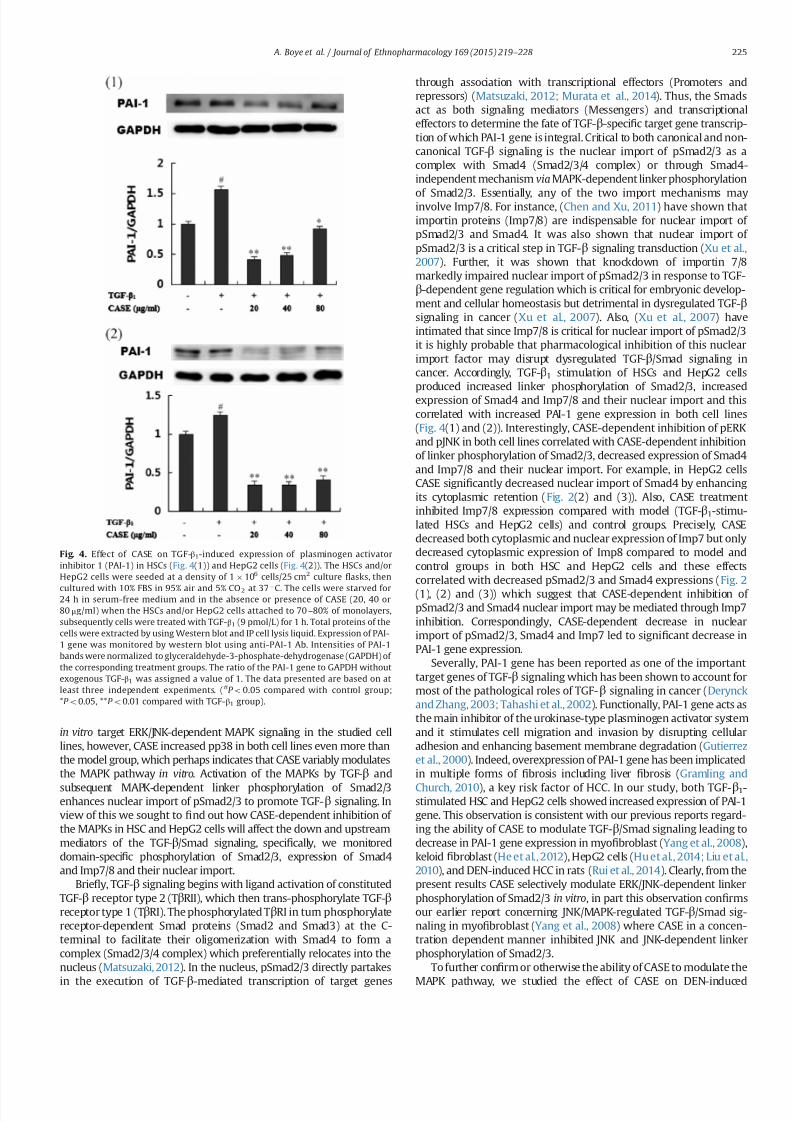
7232019 Compound Astragalus and Salvia Miltiorrhiza Extracts Modulate MAPK Regulated TGF Smad Signaling in Hepatocellhellip
httpslidepdfcomreaderfullcompound-astragalus-and-salvia-miltiorrhiza-extracts-modulate-mapk-regulated 710
in vitro target ERKJNK-dependent MAPK signaling in the studied cell
lines however CASE increased pp38 in both cell lines even more than
the model group which perhaps indicates that CASE variably modulates
the MAPK pathway in vitro Activation of the MAPKs by TGF-β and
subsequent MAPK-dependent linker phosphorylation of Smad23enhances nuclear import of pSmad23 to promote TGF-β signaling In
view of this we sought to 1047297nd out how CASE-dependent inhibition of
the MAPKs in HSC and HepG2 cells will affect the down and upstream
mediators of the TGF-βSmad signaling speci1047297cally we monitored
domain-speci1047297c phosphorylation of Smad23 expression of Smad4
and Imp78 and their nuclear import
Brie1047298y TGF-β signaling begins with ligand activation of constituted
TGF-β receptor type 2 (TβRII) which then trans-phosphorylate TGF-βreceptor type 1 (TβRI) The phosphorylated TβRI in turn phosphorylate
receptor-dependent Smad proteins (Smad2 and Smad3) at the C-
terminal to facilitate their oligomerization with Smad4 to form a
complex (Smad234 complex) which preferentially relocates into the
nucleus (Matsuzaki 2012) In the nucleus pSmad23 directly partakes
in the execution of TGF-β-mediated transcription of target genes
through association with transcriptional effectors (Promoters and
repressors) (Matsuzaki 2012 Murata et al 2014) Thus the Smads
act as both signaling mediators (Messengers) and transcriptional
effectors to determine the fate of TGF-β-speci1047297c target gene transcrip-
tion of which PAI-1 gene is integral Critical to both canonical and non-
canonical TGF-β signaling is the nuclear import of pSmad23 as a
complex with Smad4 (Smad234 complex) or through Smad4-
independent mechanism via MAPK-dependent linker phosphorylation
of Smad23 Essentially any of the two import mechanisms mayinvolve Imp78 For instance (Chen and Xu 2011) have shown that
importin proteins (Imp78) are indispensable for nuclear import of
pSmad23 and Smad4 It was also shown that nuclear import of
pSmad23 is a critical step in TGF-β signaling transduction (Xu et al
2007) Further it was shown that knockdown of importin 78
markedly impaired nuclear import of pSmad23 in response to TGF-
β-dependent gene regulation which is critical for embryonic develop-
ment and cellular homeostasis but detrimental in dysregulated TGF-βsignaling in cancer (Xu et al 2007) Also (Xu et al 2007) have
intimated that since Imp78 is critical for nuclear import of pSmad23
it is highly probable that pharmacological inhibition of this nuclear
import factor may disrupt dysregulated TGF-βSmad signaling in
cancer Accordingly TGF-β1 stimulation of HSCs and HepG2 cells
produced increased linker phosphorylation of Smad23 increased
expression of Smad4 and Imp78 and their nuclear import and this
correlated with increased PAI-1 gene expression in both cell lines
(Fig 4(1) and (2)) Interestingly CASE-dependent inhibition of pERK
and pJNK in both cell lines correlated with CASE-dependent inhibition
of linker phosphorylation of Smad23 decreased expression of Smad4
and Imp78 and their nuclear import For example in HepG2 cells
CASE signi1047297cantly decreased nuclear import of Smad4 by enhancing
its cytoplasmic retention (Fig 2(2) and (3)) Also CASE treatment
inhibited Imp78 expression compared with model (TGF-β1-stimu-
lated HSCs and HepG2 cells) and control groups Precisely CASE
decreased both cytoplasmic and nuclear expression of Imp7 but only
decreased cytoplasmic expression of Imp8 compared to model and
control groups in both HSC and HepG2 cells and these effects
correlated with decreased pSmad23 and Smad4 expressions (Fig 2
(1) (2) and (3)) which suggest that CASE-dependent inhibition of pSmad23 and Smad4 nuclear import may be mediated through Imp7
inhibition Correspondingly CASE-dependent decrease in nuclear
import of pSmad23 Smad4 and Imp7 led to signi1047297cant decrease in
PAI-1 gene expression
Severally PAI-1 gene has been reported as one of the important
target genes of TGF-β signaling which has been shown to account for
most of the pathological roles of TGF-β signaling in cancer (Derynck
and Zhang 2003 Tahashi et al 2002) Functionally PAI-1 gene acts as
the main inhibitor of the urokinase-type plasminogen activator system
and it stimulates cell migration and invasion by disrupting cellular
adhesion and enhancing basement membrane degradation (Gutierrez
et al 2000) Indeed overexpression of PAI-1 gene has been implicated
in multiple forms of 1047297brosis including liver 1047297brosis (Gramling and
Church 2010) a key risk factor of HCC In our study both TGF-β1-stimulated HSC and HepG2 cells showed increased expression of PAI-1
gene This observation is consistent with our previous reports regard-
ing the ability of CASE to modulate TGF-βSmad signaling leading to
decrease in PAI-1 gene expression in myo1047297broblast (Yang et al 2008)
keloid 1047297broblast (He et al 2012) HepG2 cells (Hu et al 2014 Liu et al
2010) and DEN-induced HCC in rats (Rui et al 2014) Clearly from the
present results CASE selectively modulate ERKJNK-dependent linker
phosphorylation of Smad23 in vitro in part this observation con1047297rms
our earlier report concerning JNKMAPK-regulated TGF-βSmad sig-
naling in myo1047297broblast (Yang et al 2008) where CASE in a concen-
tration dependent manner inhibited JNK and JNK-dependent linker
phosphorylation of Smad23
To further con1047297rm or otherwise the ability of CASE to modulate the
MAPK pathway we studied the effect of CASE on DEN-induced
Fig 4 Effect of CASE on TGF-β1-induced expression of plasminogen activator
inhibitor 1 (PAI-1) in HSCs (Fig 4(1)) and HepG2 cells (Fig 4(2)) The HSCs andor
HepG2 cells were seeded at a density of 1 106 cells25 cm2 culture 1047298asks then
cultured with 10 FBS in 95 air and 5 CO2 at 37 1
C The cells were starved for24 h in serum-free medium and in the absence or presence of CASE (20 40 or
80 μgml) when the HSCs andor HepG2 cells attached to 70 ndash80 of monolayers
subsequently cells were treated with TGF-β1 (9 pmolL) for 1 h Total proteins of the
cells were extracted by using Western blot and IP cell lysis liquid Expression of PAI-
1 gene was monitored by western blot using anti-PAI-1 Ab Intensities of PAI-1
bands were normalized to glyceraldehyde-3-phosphate-dehydrogenase (GAPDH) of
the corresponding treatment groups The ratio of the PAI-1 gene to GAPDH without
exogenous TGF-β1 was assigned a value of 1 The data presented are based on at
least three independent experiments (P o005 compared with control group
P o005 P o001 compared with TGF-β1 group)
A Boye et al Journal of Ethnopharmacology 169 (2015) 219ndash 228 225
7232019 Compound Astragalus and Salvia Miltiorrhiza Extracts Modulate MAPK Regulated TGF Smad Signaling in Hepatocellhellip
httpslidepdfcomreaderfullcompound-astragalus-and-salvia-miltiorrhiza-extracts-modulate-mapk-regulated 810
activation of the MAPK pathway From our in vivo study treatment of
rats with DEN for 12 weeks led to increased activation and expression
of pERK pJNK and pp38 but extending DEN treatment period to 16
weeks even saw a further increase in the average expression of pERK
pJNK and pp38 (Fig 5(4) (5) and (6)) indicating a direct causal
relationship between duration of DEN treatment and MAPK activation
These results indicate that increase in MAPK activation and expression
may correlate with HCC progression But concomitant treatment of
rats with DEN in the morning and CASE in the afternoon reversed the
above relationship for instance at 12 weeks not only did CASE (120 or
240 mgkg) decreased the expression of pERK pJNK and pp38
compared to the model group but also decreased the average
expression level of each MAPK even more than the average for the
control group (Fig 5(1) (2) and (3)) showing that CASE potently
modulates the MAPK pathway and that the MAPK pathway could be
yet another important target of CASE At 16 weeks though CASE
decreased the expression of the MAPKs compared to the model group
it was however not different from that of the control group ( Fig 5(4)
(5) and (6)) indicating that MAPK activation and expression were
indeed related to duration of DEN treatment The in vivo inhibition of
pp38 by CASE (Fig 5(3) and (6)) contradicts our in vitro results
regarding pp38 But this observation probably suggest that CASE
Fig 5 Effect of CASE on diethyl nitrosamine (DEN)-induced activation of pERK pJNK and pp38 in rats The proteins were extracted from frozen liver tissues from 12th week
(Fig 5(1) (2) and (3)) and 16th week (Fig 5(4) (5) and (6)) DEN treated rats Phosphorylated (p) ERK12 pJNK12 pp38 and ERK JNK p38 were analyzed by western blot
using anti-pERK12 pJNK12 pp38 and ERK12 JNK12 p38 Abs and anti-glyceraldehyde phosphate dehydrogenase (GAPDH) Ab Intensities of pERK12 pJNK12 pp38
bands were normalized to those of ERK12 JNK12 p38 of the corresponding treatment groups The ratio of the pERK12 pJNK12 and pp38 proteins to ERK12 JNK12 and
p38 respectively of normal group was assigned a value of 1 Each value represents mean7SD nfrac143 P o005 P o001 compared with control group P o005 P o001
compared with DEN group
A Boye et al Journal of Ethnopharmacology 169 (2015) 219ndash 228226
7232019 Compound Astragalus and Salvia Miltiorrhiza Extracts Modulate MAPK Regulated TGF Smad Signaling in Hepatocellhellip
httpslidepdfcomreaderfullcompound-astragalus-and-salvia-miltiorrhiza-extracts-modulate-mapk-regulated 910
modulate pERK pJNK and pp38 in vivo by the same mechanism which
may involve some unknown factors in the tumor microenvironment
since the tumor microenvironment was the only major factor absent
in the in vitro study or it may be possible that while CASE employs the
same mechanism to modulate pERK and pJNK both in in vitro andin vivo it however employs different mechanisms to modulate pp38
in in vitro and in vivo settings Indeed these results further show that
CASE does not only target the upstream and downstream mediators
(TGF-β ligand TβRI TβRII Smad2CL Smad3L Smad4 and PAI-1) of
TGF-βSmad signaling as previously reported (He et al 2012 Hu et al
2014 Liu et al 2010 Rui et al 2014 Wu et al 2014 Yang et al 2008)
but also targets the MAPK pathway Fundamentally our in vivo results
agree in part with (Nagata et al 2009) with regard to JNK activation
by DEN treatment however our results concerning pERK and pp38
are inconsistent with their 1047297nding For instance Nagata et al (2009)
had shown that after 86-days DEN treatment of rats only pJNK was
activated whiles pERK and pp38 remained inactive (Un-phosphory-
lated) but in our study we found that increasing DEN treatment to 12
or 16 weeks produced signi1047297cant activation of all the MAPKs (pERKpJNK and pp38) We report that activation of the MAPKs by DEN
treatment is time-dependent and may relate directly to HCC
progression
Put together the present results provide a strong rationale for
further elucidation of the MAPK pathway as a key target for
therapy against HCC Essentially CASE has shown a multi-target
potential by modulating both the MAPK and TGF-βSmad signaling
pathways (Fig 6) in a manner that highlights its potential drug
candidature However our results raise some further questions for
example which speci1047297c subcellular location (Cytoplasm or
nucleus) does CASE modulate the MAPK pathway and how does
it affect HCC phenotypic manifestations such as cell proliferation
dysregulated apoptosis cell migration and invasion These ques-
tions are informed by the fact that spatio-temporal regulation of
the MAPK pathway is crucial for many pathways implicated in HCC
including multi-functional TGF-βSmad signaling
5 Conclusion
CASE blocked MAPK activation and MAPK-dependent linker
phosphorylation of Smad23 inhibited Smad4 and Imp78
blocked nuclear import of Smad234 via Imp7 modulation and
down-regulated PAI-1 gene expression and these results do not
only demonstrate the potential of the MAPK-regulated TGF-β
Smad pathway as an important target for therapy against HCC but
also highlights the potential drug candidature of CASE and con-
1047297rmation of its ethnobotanical usage
Con1047298icts of interest
No potential con1047298icts of interest
Acknowledgments
We thank Prof K Matsuzaki (Department of Gastroenterologyand Hepatology Kansai Medical University Osaka Japan) for
providing us with the following Abs Anti-pSmad2L and Anti-
pSmad3L Also this study was supported by the National Natural
Science Foundation of China (No 81073098 and no 81374012)
References
Boye A Kan H Wu C Jiang Y Yang X He S Yang Y 2015 MAPK inhibitorsdifferently modulate TGF-betaSmad signaling in HepG2 cells Tumour Biol httpdxdoiorg101007s13277-014-3002-x
Chan W Durairajan S Lu J Wang Y Xie L Kum W Koo I Yung K Li M2009 Neuroprotective effects of astragaloside IV in 6-hydroxydopamine-treated primary nigral cell culture Neurochem Int 55 414ndash422
Chen R Shao H Lin S Zhang J Xu K 2011 Treatment with astragalusmembranaceus produces antioxidative effects and attenuates intestinal mucosa
injury induced by intestinal ischemia-reperfusion in rats Am J Chin Med 39879ndash887
Chen X Xu L 2011 Mechanism and regulation of nucleocytoplasmic traf 1047297cking of Smad Cell Biosci 1 40
Cho WC Leung KN 2007a In vitro and in vivo anti-tumor effects of astragalusmembranaceus Cancer Lett 252 43ndash54
Cho WC Leung KN 2007b In vitro and in vivo immunomodulating andimmunorestorative effects of astragalus membranaceus J Ethnopharmacol113 132ndash141
Date M Matsuzaki K Matsushita M Tahashi Y Furukawa F Inoue K 2000Modulation of transforming growth factor function in hepatocytes and hepaticstellate cells in rat liver injury Gut 46 719ndash724
Derynck R Zhang YE 2003 Smad-dependent and Smad-independent pathwaysin TGF-β family signalling Nature 425 577ndash584
Fuentealba L Eivers E Ikeda A Hurtado C Kuroda H Pera E De Robertis E2007 Integrating patterning signals WntGSK3 regulates the duration of theBMPSmad1 signal Cell 131 980ndash993
Furukawa F Matsuzaki K Mori S Tahashi Y Yoshida K Sugano Y Yamagata
H Matsushita M Seki T Inagaki Y Nishizawa M Fujisawa J Inoue K2003 p38 MAPK mediates 1047297brogenic signal through Smad3 phosphorylation inrat myo1047297broblasts Hepatology 38 879ndash889
Giehl K Imamichi Y Menke A 2007 Smad4-independent TGF-β signaling intumor cell migration Cells Tissues Org 185 123ndash130
Gramling MW Church FC 2010 Plasminogen activator inhibitor-1 is anaggregate response factor with pleiotropic effects on cell signaling in vasculardisease and the tumor microenvironment Thromb Res 125 377ndash381
Gutierrez LS Schulman A Brito-Robinson T Noria F Ploplis VA Castellino FJ2000 Tumor development is retarded in mice lacking the gene for urokinase-type plasminogen activator or its inhibitor plasminogen activator inhibitor-1Cancer Res 60 5839ndash5847
Hata A Davis B 2009 Control of microRNA biogenesis by TGFbeta signalingpathway-a novel role of Smads in the nucleus Cytokine Growth Factor Rev 20517ndash521
Hayashida T Decaestecker M Schnaper H 2003 Cross-talk between ERK MAPkinase and Smad signaling pathways enhances TGF-beta-dependent lsre-sponses in human mesangial cel FASEB J 17 1576ndash1578
He S Yang Y Liu X Huang W Zhang X Yang S 2012 Compound Astragalus
and Salvia miltiorrhiza extract inhibits cell proliferation invasion and collagen
Fig 6 Fig 6 shows an illustrative summary of the multi-target mechanism by
which CASE modulates the MAPK-regulated TGF-βSmad signaling in hepatocel-lular carcinoma (HCC) CASE blocks TGF-β-induced activation of ERK JNK and p38
to abrogate MAPK-dependent linker phosphorylation of Smad23 and their sub-
sequent nuclear translocation Along the canonical loop CASE inhibits Smad4
expression formation of Smad234 and their nuclear translocation via inhibition of
Imp78 CASE-dependent disruption of nuclear import of Smad23 and Smad4 leads
to down-regulation of PAI-1 gene expression Cytoplasm (C) C-terminal phos-
phorylated Smad (pSmadC) Linker phosphorylated Smad (pSmadL) Nucleus (N)
Phosphorylated Smad (pSmad) Receptor mediated Smad (R-Smad) Transforming
growth factor receptor type 1 and 2 (TβRI and TβRII)
A Boye et al Journal of Ethnopharmacology 169 (2015) 219ndash 228 227
7232019 Compound Astragalus and Salvia Miltiorrhiza Extracts Modulate MAPK Regulated TGF Smad Signaling in Hepatocellhellip
httpslidepdfcomreaderfullcompound-astragalus-and-salvia-miltiorrhiza-extracts-modulate-mapk-regulated 1010
synthesis in keloid 1047297broblasts by mediating transforming growth factor‐βSmad pathway Br J Dermatol 166 564ndash574
Hu X Rui W Wu C He S Jiang J Zhang X Yang Y 2014 CompoundAstragalus and Salvia Miltiorrhiza extracts suppress hepatocarcinogenesis bymodulating transforming growth factor‐βSmad signaling J GastroenterolHepatol 29 1284ndash1291
Jun T 2004 Determination of total astragalosides in astragalus by colormetricmethod J Anhui Tradit Chin Med Coll 5 017
Kretzschmar M Doody J Timokhina I Massagueacute J 1999 A mechanism of repression of TGFbetaSmad signaling by oncogenic Ras Genes Dev 13804ndash816
Lee TY Chang HH Wang GJ Chiu JH Yang YY Lin HC 2006 Water‐
solubleextract of Salvia miltiorrhiza ameliorates carbon tetrachloride‐mediated hepa-tic apoptosis in rats J Pharm Pharm 58 659ndash665
Liu X Yang Y Zhang X Xu S He S Huang W Roberts MS 2010 CompoundAstragalus and Salvia miltiorrhiza extract inhibits cell invasion by modulatingtransforming growth factor‐βSmad in HepG2 cell J Gastroenterol Hepatol 25420ndash426
Matsuzaki K 2012 Smad phosphoisoform signals in acute and chronic liver injurysimilarities and differences between epithelial and mesenchymal cells CellTissue Res 347 225ndash243
Moustakas A Heldin C-H 2005 Non-Smad TGF-β signals J Cell Sci 1183573ndash3584
Murata M Yoshida K Yamaguchi T Matsuzaki K 2014 Linker phosphorylationof Smad3 promotes 1047297bro-carcinogenesis in chronic viral hepatitis of hepato-cellular carcinoma World J Gastroenterol 20 15018ndash15027
Nagata H Hatano E Tada M Murata M Kitamura K Asechi H Narita MYanagida A Tamaki N Yagi S 2009 Inhibition of c‐ Jun NH2‐terminal kinaseswitches Smad3 signaling from oncogenesis to tumor‐suppression in rat
hepatocellular carcinoma Hepatology 49 1944ndash
1953Puche JE Saiman Y Friedman SL 2013 Hepatic stellate cells and liver 1047297brosisCompr Physiol 3 1473ndash1492
Roxas M Jurenka J 2007 Colds and in1047298uenza a review of diagnosis andconventional botanical and nutritional considerations Altern Med Rev 1225ndash48
Rui W Xie L Liu X He S Wu C Zhang X Zhang L Yang Y 2014 CompoundAstragalus and Salvia miltiorrhiza extract suppresses hepatocellular carcinomaprogression by inhibiting 1047297brosis and PAI-1 mRNA transcription J Ethnophar-macol 151 198ndash209
Tang J 2004 Determination of total astragalosides in astragalus by colorimetricmethod J Anhui Tradit Chin Med Coll 23 37ndash38
Shao BM Xu W Dai H Tu P Li Z Gao XM 2004 A study on the immunereceptors for polysaccharides from the roots of astragalus membranaceus aChinese medicinal herb Biochem Biophys Res Commun 320 1103ndash1111
Shen P Liu MH Ng TY Chan YH Yong EL 2006 Differential effects of iso1047298avones from astragalus membranaceus and Pueraria thomsonii on theactivation of PPARalpha PPARgamma and adipocyte differentiation in vitro
J Nutr 136 899ndash905
Tahashi Y Matsuzaki K Yoshida K Furukawa F Sugano Y Matsushita MHimeno Y Inagaki Y Inoue K 2002 Differential regulation of TGF‐β signal in
hepatic stellate cells between acute and chronic rat liver injury Hepatology 3549ndash61
Wu C Jiang J Boye A Jiang Y Yang Y 2014 Compound Astragalus and Salviamiltiorrhiza extract suppresses rabbits hypertrophic scar by modulating theTGF-betaSmad signal Dermatology 229 363ndash368
Wu YP Cao Y Cao YY 2001 Technology optimization of extracting activitycomponents from Astragalus membranaceus Lishizen Med Mater Med Res 12876ndash877
Xu L Yao X Chen X Lu P Zhang B Ip YT 2007 Msk is required for nuclearimport of TGF-βBMP-activated Smads J cell Biol 178 981ndash994
Yang Y Yang S Chen M Zhang X Zou Y Zhang X 2008 Compound Astragalus
and Salvia miltiorrhiza extract exerts anti-1047297brosis by mediating TGF-βSmadsignaling in myo1047297broblasts J Ethnopharmacol 118 264ndash270
Ye Y 2006 Comparative study on the determination of salvianolic acids content bycolorimetery and HPLC J Zhejiang Univ Tradit Chin Med 30 350 ndash351
Yoshida K Matsuzaki K Mori S Tahashi Y Yamagata H Furukawa F Seki TNishizawa M Fujisawa J Okazaki K 2005 Transforming growth factor-betaand platelet-derived growth factor signal via c-Jun N-terminal kinase-depen-dent Smad23 phosphorylation in rat hepatic stellate cells after acute liverinjury The American journal of pathology 166 1029ndash1039
Yoshida K Murata M Yamaguchi T Matsuzaki K 2014 TGF-βSmad signalingduring hepatic 1047297bro-carcinogenesis (review) Int J Oncol 45 1363ndash1371
Zhang SS He FY Xu XY 2001 Comparative studies on the content of ingredients of polysaccharides in the Buyanghuanwutang and single materials
J Human Coll Tradit Chin Med 21 26Zhang YE 2009 Non-Smad pathways in TGF-β signaling Cell Res 19 128ndash139
Glossary
Phosphorylation Addition of a phosphorus atom to a protein or a moleculeSmad proteins A class of proteins that mediate transforming growth factor beta
(TGF-β) signaling transductionPhosphorylated Smad A Smad protein that has received a phosphorus atom from a
phosphorus carrier moleculeLinker phosphorylated Smad A Smad protein phosphorylated at a region in
between its N-terminal and C-terminalCanonical TGF-β Signaling It is Smad-mediated TGF-β signaling transduction
involving Smad2 Smad3 and Smad4 complex formation leading to theirnuclear translocation and subsequent transcription of TGF-β-target mRNAs
Importin protein 7 and 8 (Imp78) They are proteins mostly in the cytoplasm thatfacilitate nuclear entry of other proteins
Mitogen activated protein kinase (MAPK) They are kinase proteins that facilitatetransduction of extracellular signals into the nucleus
Non-canonical TGF-β signaling All TGF-β signaling transduction that does notinvolve Smad4 or non-Smad TGF-β signaling
A Boye et al Journal of Ethnopharmacology 169 (2015) 219ndash 228228
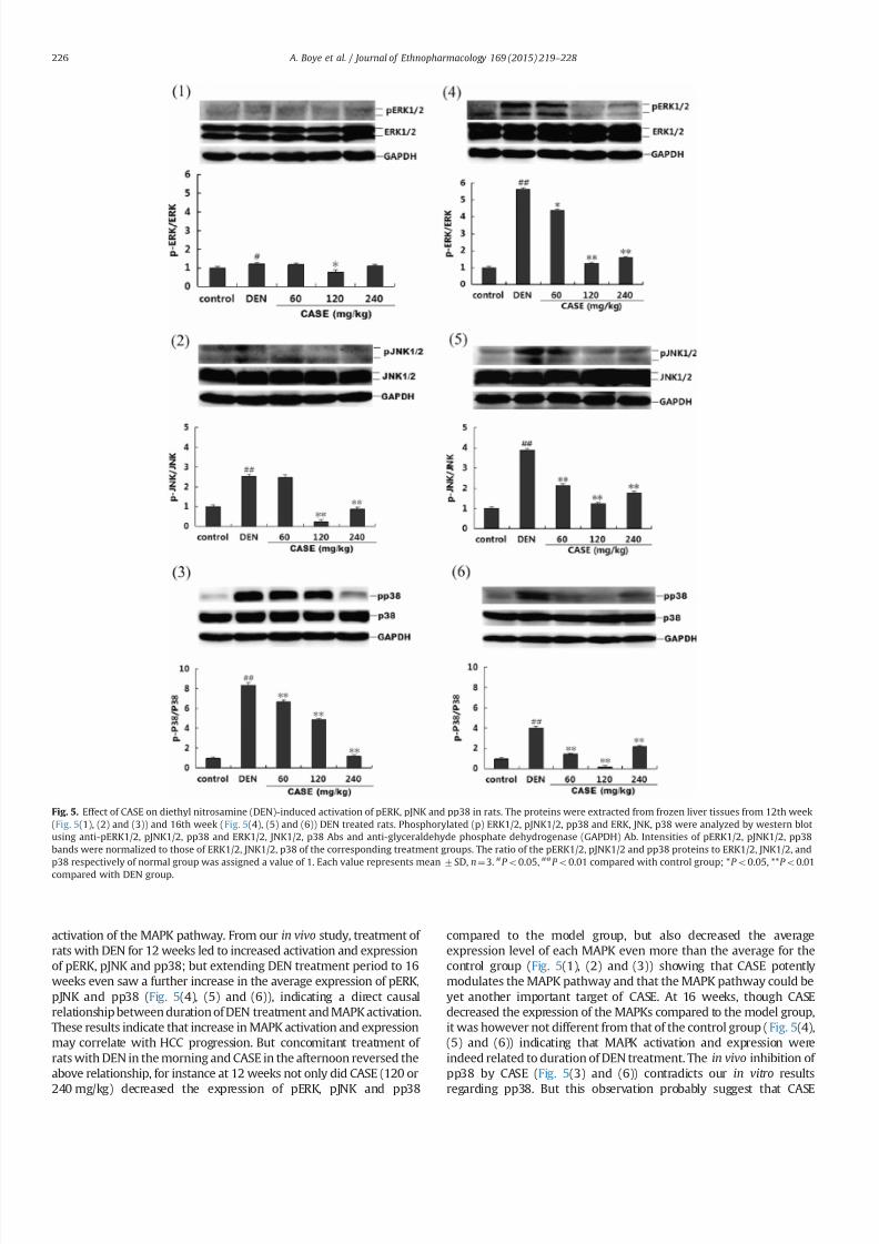
7232019 Compound Astragalus and Salvia Miltiorrhiza Extracts Modulate MAPK Regulated TGF Smad Signaling in Hepatocellhellip
httpslidepdfcomreaderfullcompound-astragalus-and-salvia-miltiorrhiza-extracts-modulate-mapk-regulated 810
activation of the MAPK pathway From our in vivo study treatment of
rats with DEN for 12 weeks led to increased activation and expression
of pERK pJNK and pp38 but extending DEN treatment period to 16
weeks even saw a further increase in the average expression of pERK
pJNK and pp38 (Fig 5(4) (5) and (6)) indicating a direct causal
relationship between duration of DEN treatment and MAPK activation
These results indicate that increase in MAPK activation and expression
may correlate with HCC progression But concomitant treatment of
rats with DEN in the morning and CASE in the afternoon reversed the
above relationship for instance at 12 weeks not only did CASE (120 or
240 mgkg) decreased the expression of pERK pJNK and pp38
compared to the model group but also decreased the average
expression level of each MAPK even more than the average for the
control group (Fig 5(1) (2) and (3)) showing that CASE potently
modulates the MAPK pathway and that the MAPK pathway could be
yet another important target of CASE At 16 weeks though CASE
decreased the expression of the MAPKs compared to the model group
it was however not different from that of the control group ( Fig 5(4)
(5) and (6)) indicating that MAPK activation and expression were
indeed related to duration of DEN treatment The in vivo inhibition of
pp38 by CASE (Fig 5(3) and (6)) contradicts our in vitro results
regarding pp38 But this observation probably suggest that CASE
Fig 5 Effect of CASE on diethyl nitrosamine (DEN)-induced activation of pERK pJNK and pp38 in rats The proteins were extracted from frozen liver tissues from 12th week
(Fig 5(1) (2) and (3)) and 16th week (Fig 5(4) (5) and (6)) DEN treated rats Phosphorylated (p) ERK12 pJNK12 pp38 and ERK JNK p38 were analyzed by western blot
using anti-pERK12 pJNK12 pp38 and ERK12 JNK12 p38 Abs and anti-glyceraldehyde phosphate dehydrogenase (GAPDH) Ab Intensities of pERK12 pJNK12 pp38
bands were normalized to those of ERK12 JNK12 p38 of the corresponding treatment groups The ratio of the pERK12 pJNK12 and pp38 proteins to ERK12 JNK12 and
p38 respectively of normal group was assigned a value of 1 Each value represents mean7SD nfrac143 P o005 P o001 compared with control group P o005 P o001
compared with DEN group
A Boye et al Journal of Ethnopharmacology 169 (2015) 219ndash 228226
7232019 Compound Astragalus and Salvia Miltiorrhiza Extracts Modulate MAPK Regulated TGF Smad Signaling in Hepatocellhellip
httpslidepdfcomreaderfullcompound-astragalus-and-salvia-miltiorrhiza-extracts-modulate-mapk-regulated 910
modulate pERK pJNK and pp38 in vivo by the same mechanism which
may involve some unknown factors in the tumor microenvironment
since the tumor microenvironment was the only major factor absent
in the in vitro study or it may be possible that while CASE employs the
same mechanism to modulate pERK and pJNK both in in vitro andin vivo it however employs different mechanisms to modulate pp38
in in vitro and in vivo settings Indeed these results further show that
CASE does not only target the upstream and downstream mediators
(TGF-β ligand TβRI TβRII Smad2CL Smad3L Smad4 and PAI-1) of
TGF-βSmad signaling as previously reported (He et al 2012 Hu et al
2014 Liu et al 2010 Rui et al 2014 Wu et al 2014 Yang et al 2008)
but also targets the MAPK pathway Fundamentally our in vivo results
agree in part with (Nagata et al 2009) with regard to JNK activation
by DEN treatment however our results concerning pERK and pp38
are inconsistent with their 1047297nding For instance Nagata et al (2009)
had shown that after 86-days DEN treatment of rats only pJNK was
activated whiles pERK and pp38 remained inactive (Un-phosphory-
lated) but in our study we found that increasing DEN treatment to 12
or 16 weeks produced signi1047297cant activation of all the MAPKs (pERKpJNK and pp38) We report that activation of the MAPKs by DEN
treatment is time-dependent and may relate directly to HCC
progression
Put together the present results provide a strong rationale for
further elucidation of the MAPK pathway as a key target for
therapy against HCC Essentially CASE has shown a multi-target
potential by modulating both the MAPK and TGF-βSmad signaling
pathways (Fig 6) in a manner that highlights its potential drug
candidature However our results raise some further questions for
example which speci1047297c subcellular location (Cytoplasm or
nucleus) does CASE modulate the MAPK pathway and how does
it affect HCC phenotypic manifestations such as cell proliferation
dysregulated apoptosis cell migration and invasion These ques-
tions are informed by the fact that spatio-temporal regulation of
the MAPK pathway is crucial for many pathways implicated in HCC
including multi-functional TGF-βSmad signaling
5 Conclusion
CASE blocked MAPK activation and MAPK-dependent linker
phosphorylation of Smad23 inhibited Smad4 and Imp78
blocked nuclear import of Smad234 via Imp7 modulation and
down-regulated PAI-1 gene expression and these results do not
only demonstrate the potential of the MAPK-regulated TGF-β
Smad pathway as an important target for therapy against HCC but
also highlights the potential drug candidature of CASE and con-
1047297rmation of its ethnobotanical usage
Con1047298icts of interest
No potential con1047298icts of interest
Acknowledgments
We thank Prof K Matsuzaki (Department of Gastroenterologyand Hepatology Kansai Medical University Osaka Japan) for
providing us with the following Abs Anti-pSmad2L and Anti-
pSmad3L Also this study was supported by the National Natural
Science Foundation of China (No 81073098 and no 81374012)
References
Boye A Kan H Wu C Jiang Y Yang X He S Yang Y 2015 MAPK inhibitorsdifferently modulate TGF-betaSmad signaling in HepG2 cells Tumour Biol httpdxdoiorg101007s13277-014-3002-x
Chan W Durairajan S Lu J Wang Y Xie L Kum W Koo I Yung K Li M2009 Neuroprotective effects of astragaloside IV in 6-hydroxydopamine-treated primary nigral cell culture Neurochem Int 55 414ndash422
Chen R Shao H Lin S Zhang J Xu K 2011 Treatment with astragalusmembranaceus produces antioxidative effects and attenuates intestinal mucosa
injury induced by intestinal ischemia-reperfusion in rats Am J Chin Med 39879ndash887
Chen X Xu L 2011 Mechanism and regulation of nucleocytoplasmic traf 1047297cking of Smad Cell Biosci 1 40
Cho WC Leung KN 2007a In vitro and in vivo anti-tumor effects of astragalusmembranaceus Cancer Lett 252 43ndash54
Cho WC Leung KN 2007b In vitro and in vivo immunomodulating andimmunorestorative effects of astragalus membranaceus J Ethnopharmacol113 132ndash141
Date M Matsuzaki K Matsushita M Tahashi Y Furukawa F Inoue K 2000Modulation of transforming growth factor function in hepatocytes and hepaticstellate cells in rat liver injury Gut 46 719ndash724
Derynck R Zhang YE 2003 Smad-dependent and Smad-independent pathwaysin TGF-β family signalling Nature 425 577ndash584
Fuentealba L Eivers E Ikeda A Hurtado C Kuroda H Pera E De Robertis E2007 Integrating patterning signals WntGSK3 regulates the duration of theBMPSmad1 signal Cell 131 980ndash993
Furukawa F Matsuzaki K Mori S Tahashi Y Yoshida K Sugano Y Yamagata
H Matsushita M Seki T Inagaki Y Nishizawa M Fujisawa J Inoue K2003 p38 MAPK mediates 1047297brogenic signal through Smad3 phosphorylation inrat myo1047297broblasts Hepatology 38 879ndash889
Giehl K Imamichi Y Menke A 2007 Smad4-independent TGF-β signaling intumor cell migration Cells Tissues Org 185 123ndash130
Gramling MW Church FC 2010 Plasminogen activator inhibitor-1 is anaggregate response factor with pleiotropic effects on cell signaling in vasculardisease and the tumor microenvironment Thromb Res 125 377ndash381
Gutierrez LS Schulman A Brito-Robinson T Noria F Ploplis VA Castellino FJ2000 Tumor development is retarded in mice lacking the gene for urokinase-type plasminogen activator or its inhibitor plasminogen activator inhibitor-1Cancer Res 60 5839ndash5847
Hata A Davis B 2009 Control of microRNA biogenesis by TGFbeta signalingpathway-a novel role of Smads in the nucleus Cytokine Growth Factor Rev 20517ndash521
Hayashida T Decaestecker M Schnaper H 2003 Cross-talk between ERK MAPkinase and Smad signaling pathways enhances TGF-beta-dependent lsre-sponses in human mesangial cel FASEB J 17 1576ndash1578
He S Yang Y Liu X Huang W Zhang X Yang S 2012 Compound Astragalus
and Salvia miltiorrhiza extract inhibits cell proliferation invasion and collagen
Fig 6 Fig 6 shows an illustrative summary of the multi-target mechanism by
which CASE modulates the MAPK-regulated TGF-βSmad signaling in hepatocel-lular carcinoma (HCC) CASE blocks TGF-β-induced activation of ERK JNK and p38
to abrogate MAPK-dependent linker phosphorylation of Smad23 and their sub-
sequent nuclear translocation Along the canonical loop CASE inhibits Smad4
expression formation of Smad234 and their nuclear translocation via inhibition of
Imp78 CASE-dependent disruption of nuclear import of Smad23 and Smad4 leads
to down-regulation of PAI-1 gene expression Cytoplasm (C) C-terminal phos-
phorylated Smad (pSmadC) Linker phosphorylated Smad (pSmadL) Nucleus (N)
Phosphorylated Smad (pSmad) Receptor mediated Smad (R-Smad) Transforming
growth factor receptor type 1 and 2 (TβRI and TβRII)
A Boye et al Journal of Ethnopharmacology 169 (2015) 219ndash 228 227
7232019 Compound Astragalus and Salvia Miltiorrhiza Extracts Modulate MAPK Regulated TGF Smad Signaling in Hepatocellhellip
httpslidepdfcomreaderfullcompound-astragalus-and-salvia-miltiorrhiza-extracts-modulate-mapk-regulated 1010
synthesis in keloid 1047297broblasts by mediating transforming growth factor‐βSmad pathway Br J Dermatol 166 564ndash574
Hu X Rui W Wu C He S Jiang J Zhang X Yang Y 2014 CompoundAstragalus and Salvia Miltiorrhiza extracts suppress hepatocarcinogenesis bymodulating transforming growth factor‐βSmad signaling J GastroenterolHepatol 29 1284ndash1291
Jun T 2004 Determination of total astragalosides in astragalus by colormetricmethod J Anhui Tradit Chin Med Coll 5 017
Kretzschmar M Doody J Timokhina I Massagueacute J 1999 A mechanism of repression of TGFbetaSmad signaling by oncogenic Ras Genes Dev 13804ndash816
Lee TY Chang HH Wang GJ Chiu JH Yang YY Lin HC 2006 Water‐
solubleextract of Salvia miltiorrhiza ameliorates carbon tetrachloride‐mediated hepa-tic apoptosis in rats J Pharm Pharm 58 659ndash665
Liu X Yang Y Zhang X Xu S He S Huang W Roberts MS 2010 CompoundAstragalus and Salvia miltiorrhiza extract inhibits cell invasion by modulatingtransforming growth factor‐βSmad in HepG2 cell J Gastroenterol Hepatol 25420ndash426
Matsuzaki K 2012 Smad phosphoisoform signals in acute and chronic liver injurysimilarities and differences between epithelial and mesenchymal cells CellTissue Res 347 225ndash243
Moustakas A Heldin C-H 2005 Non-Smad TGF-β signals J Cell Sci 1183573ndash3584
Murata M Yoshida K Yamaguchi T Matsuzaki K 2014 Linker phosphorylationof Smad3 promotes 1047297bro-carcinogenesis in chronic viral hepatitis of hepato-cellular carcinoma World J Gastroenterol 20 15018ndash15027
Nagata H Hatano E Tada M Murata M Kitamura K Asechi H Narita MYanagida A Tamaki N Yagi S 2009 Inhibition of c‐ Jun NH2‐terminal kinaseswitches Smad3 signaling from oncogenesis to tumor‐suppression in rat
hepatocellular carcinoma Hepatology 49 1944ndash
1953Puche JE Saiman Y Friedman SL 2013 Hepatic stellate cells and liver 1047297brosisCompr Physiol 3 1473ndash1492
Roxas M Jurenka J 2007 Colds and in1047298uenza a review of diagnosis andconventional botanical and nutritional considerations Altern Med Rev 1225ndash48
Rui W Xie L Liu X He S Wu C Zhang X Zhang L Yang Y 2014 CompoundAstragalus and Salvia miltiorrhiza extract suppresses hepatocellular carcinomaprogression by inhibiting 1047297brosis and PAI-1 mRNA transcription J Ethnophar-macol 151 198ndash209
Tang J 2004 Determination of total astragalosides in astragalus by colorimetricmethod J Anhui Tradit Chin Med Coll 23 37ndash38
Shao BM Xu W Dai H Tu P Li Z Gao XM 2004 A study on the immunereceptors for polysaccharides from the roots of astragalus membranaceus aChinese medicinal herb Biochem Biophys Res Commun 320 1103ndash1111
Shen P Liu MH Ng TY Chan YH Yong EL 2006 Differential effects of iso1047298avones from astragalus membranaceus and Pueraria thomsonii on theactivation of PPARalpha PPARgamma and adipocyte differentiation in vitro
J Nutr 136 899ndash905
Tahashi Y Matsuzaki K Yoshida K Furukawa F Sugano Y Matsushita MHimeno Y Inagaki Y Inoue K 2002 Differential regulation of TGF‐β signal in
hepatic stellate cells between acute and chronic rat liver injury Hepatology 3549ndash61
Wu C Jiang J Boye A Jiang Y Yang Y 2014 Compound Astragalus and Salviamiltiorrhiza extract suppresses rabbits hypertrophic scar by modulating theTGF-betaSmad signal Dermatology 229 363ndash368
Wu YP Cao Y Cao YY 2001 Technology optimization of extracting activitycomponents from Astragalus membranaceus Lishizen Med Mater Med Res 12876ndash877
Xu L Yao X Chen X Lu P Zhang B Ip YT 2007 Msk is required for nuclearimport of TGF-βBMP-activated Smads J cell Biol 178 981ndash994
Yang Y Yang S Chen M Zhang X Zou Y Zhang X 2008 Compound Astragalus
and Salvia miltiorrhiza extract exerts anti-1047297brosis by mediating TGF-βSmadsignaling in myo1047297broblasts J Ethnopharmacol 118 264ndash270
Ye Y 2006 Comparative study on the determination of salvianolic acids content bycolorimetery and HPLC J Zhejiang Univ Tradit Chin Med 30 350 ndash351
Yoshida K Matsuzaki K Mori S Tahashi Y Yamagata H Furukawa F Seki TNishizawa M Fujisawa J Okazaki K 2005 Transforming growth factor-betaand platelet-derived growth factor signal via c-Jun N-terminal kinase-depen-dent Smad23 phosphorylation in rat hepatic stellate cells after acute liverinjury The American journal of pathology 166 1029ndash1039
Yoshida K Murata M Yamaguchi T Matsuzaki K 2014 TGF-βSmad signalingduring hepatic 1047297bro-carcinogenesis (review) Int J Oncol 45 1363ndash1371
Zhang SS He FY Xu XY 2001 Comparative studies on the content of ingredients of polysaccharides in the Buyanghuanwutang and single materials
J Human Coll Tradit Chin Med 21 26Zhang YE 2009 Non-Smad pathways in TGF-β signaling Cell Res 19 128ndash139
Glossary
Phosphorylation Addition of a phosphorus atom to a protein or a moleculeSmad proteins A class of proteins that mediate transforming growth factor beta
(TGF-β) signaling transductionPhosphorylated Smad A Smad protein that has received a phosphorus atom from a
phosphorus carrier moleculeLinker phosphorylated Smad A Smad protein phosphorylated at a region in
between its N-terminal and C-terminalCanonical TGF-β Signaling It is Smad-mediated TGF-β signaling transduction
involving Smad2 Smad3 and Smad4 complex formation leading to theirnuclear translocation and subsequent transcription of TGF-β-target mRNAs
Importin protein 7 and 8 (Imp78) They are proteins mostly in the cytoplasm thatfacilitate nuclear entry of other proteins
Mitogen activated protein kinase (MAPK) They are kinase proteins that facilitatetransduction of extracellular signals into the nucleus
Non-canonical TGF-β signaling All TGF-β signaling transduction that does notinvolve Smad4 or non-Smad TGF-β signaling
A Boye et al Journal of Ethnopharmacology 169 (2015) 219ndash 228228
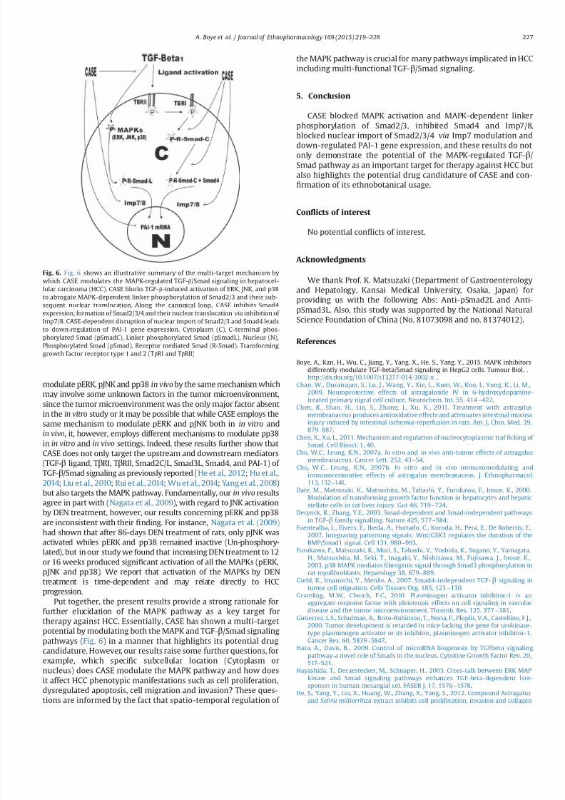
7232019 Compound Astragalus and Salvia Miltiorrhiza Extracts Modulate MAPK Regulated TGF Smad Signaling in Hepatocellhellip
httpslidepdfcomreaderfullcompound-astragalus-and-salvia-miltiorrhiza-extracts-modulate-mapk-regulated 910
modulate pERK pJNK and pp38 in vivo by the same mechanism which
may involve some unknown factors in the tumor microenvironment
since the tumor microenvironment was the only major factor absent
in the in vitro study or it may be possible that while CASE employs the
same mechanism to modulate pERK and pJNK both in in vitro andin vivo it however employs different mechanisms to modulate pp38
in in vitro and in vivo settings Indeed these results further show that
CASE does not only target the upstream and downstream mediators
(TGF-β ligand TβRI TβRII Smad2CL Smad3L Smad4 and PAI-1) of
TGF-βSmad signaling as previously reported (He et al 2012 Hu et al
2014 Liu et al 2010 Rui et al 2014 Wu et al 2014 Yang et al 2008)
but also targets the MAPK pathway Fundamentally our in vivo results
agree in part with (Nagata et al 2009) with regard to JNK activation
by DEN treatment however our results concerning pERK and pp38
are inconsistent with their 1047297nding For instance Nagata et al (2009)
had shown that after 86-days DEN treatment of rats only pJNK was
activated whiles pERK and pp38 remained inactive (Un-phosphory-
lated) but in our study we found that increasing DEN treatment to 12
or 16 weeks produced signi1047297cant activation of all the MAPKs (pERKpJNK and pp38) We report that activation of the MAPKs by DEN
treatment is time-dependent and may relate directly to HCC
progression
Put together the present results provide a strong rationale for
further elucidation of the MAPK pathway as a key target for
therapy against HCC Essentially CASE has shown a multi-target
potential by modulating both the MAPK and TGF-βSmad signaling
pathways (Fig 6) in a manner that highlights its potential drug
candidature However our results raise some further questions for
example which speci1047297c subcellular location (Cytoplasm or
nucleus) does CASE modulate the MAPK pathway and how does
it affect HCC phenotypic manifestations such as cell proliferation
dysregulated apoptosis cell migration and invasion These ques-
tions are informed by the fact that spatio-temporal regulation of
the MAPK pathway is crucial for many pathways implicated in HCC
including multi-functional TGF-βSmad signaling
5 Conclusion
CASE blocked MAPK activation and MAPK-dependent linker
phosphorylation of Smad23 inhibited Smad4 and Imp78
blocked nuclear import of Smad234 via Imp7 modulation and
down-regulated PAI-1 gene expression and these results do not
only demonstrate the potential of the MAPK-regulated TGF-β
Smad pathway as an important target for therapy against HCC but
also highlights the potential drug candidature of CASE and con-
1047297rmation of its ethnobotanical usage
Con1047298icts of interest
No potential con1047298icts of interest
Acknowledgments
We thank Prof K Matsuzaki (Department of Gastroenterologyand Hepatology Kansai Medical University Osaka Japan) for
providing us with the following Abs Anti-pSmad2L and Anti-
pSmad3L Also this study was supported by the National Natural
Science Foundation of China (No 81073098 and no 81374012)
References
Boye A Kan H Wu C Jiang Y Yang X He S Yang Y 2015 MAPK inhibitorsdifferently modulate TGF-betaSmad signaling in HepG2 cells Tumour Biol httpdxdoiorg101007s13277-014-3002-x
Chan W Durairajan S Lu J Wang Y Xie L Kum W Koo I Yung K Li M2009 Neuroprotective effects of astragaloside IV in 6-hydroxydopamine-treated primary nigral cell culture Neurochem Int 55 414ndash422
Chen R Shao H Lin S Zhang J Xu K 2011 Treatment with astragalusmembranaceus produces antioxidative effects and attenuates intestinal mucosa
injury induced by intestinal ischemia-reperfusion in rats Am J Chin Med 39879ndash887
Chen X Xu L 2011 Mechanism and regulation of nucleocytoplasmic traf 1047297cking of Smad Cell Biosci 1 40
Cho WC Leung KN 2007a In vitro and in vivo anti-tumor effects of astragalusmembranaceus Cancer Lett 252 43ndash54
Cho WC Leung KN 2007b In vitro and in vivo immunomodulating andimmunorestorative effects of astragalus membranaceus J Ethnopharmacol113 132ndash141
Date M Matsuzaki K Matsushita M Tahashi Y Furukawa F Inoue K 2000Modulation of transforming growth factor function in hepatocytes and hepaticstellate cells in rat liver injury Gut 46 719ndash724
Derynck R Zhang YE 2003 Smad-dependent and Smad-independent pathwaysin TGF-β family signalling Nature 425 577ndash584
Fuentealba L Eivers E Ikeda A Hurtado C Kuroda H Pera E De Robertis E2007 Integrating patterning signals WntGSK3 regulates the duration of theBMPSmad1 signal Cell 131 980ndash993
Furukawa F Matsuzaki K Mori S Tahashi Y Yoshida K Sugano Y Yamagata
H Matsushita M Seki T Inagaki Y Nishizawa M Fujisawa J Inoue K2003 p38 MAPK mediates 1047297brogenic signal through Smad3 phosphorylation inrat myo1047297broblasts Hepatology 38 879ndash889
Giehl K Imamichi Y Menke A 2007 Smad4-independent TGF-β signaling intumor cell migration Cells Tissues Org 185 123ndash130
Gramling MW Church FC 2010 Plasminogen activator inhibitor-1 is anaggregate response factor with pleiotropic effects on cell signaling in vasculardisease and the tumor microenvironment Thromb Res 125 377ndash381
Gutierrez LS Schulman A Brito-Robinson T Noria F Ploplis VA Castellino FJ2000 Tumor development is retarded in mice lacking the gene for urokinase-type plasminogen activator or its inhibitor plasminogen activator inhibitor-1Cancer Res 60 5839ndash5847
Hata A Davis B 2009 Control of microRNA biogenesis by TGFbeta signalingpathway-a novel role of Smads in the nucleus Cytokine Growth Factor Rev 20517ndash521
Hayashida T Decaestecker M Schnaper H 2003 Cross-talk between ERK MAPkinase and Smad signaling pathways enhances TGF-beta-dependent lsre-sponses in human mesangial cel FASEB J 17 1576ndash1578
He S Yang Y Liu X Huang W Zhang X Yang S 2012 Compound Astragalus
and Salvia miltiorrhiza extract inhibits cell proliferation invasion and collagen
Fig 6 Fig 6 shows an illustrative summary of the multi-target mechanism by
which CASE modulates the MAPK-regulated TGF-βSmad signaling in hepatocel-lular carcinoma (HCC) CASE blocks TGF-β-induced activation of ERK JNK and p38
to abrogate MAPK-dependent linker phosphorylation of Smad23 and their sub-
sequent nuclear translocation Along the canonical loop CASE inhibits Smad4
expression formation of Smad234 and their nuclear translocation via inhibition of
Imp78 CASE-dependent disruption of nuclear import of Smad23 and Smad4 leads
to down-regulation of PAI-1 gene expression Cytoplasm (C) C-terminal phos-
phorylated Smad (pSmadC) Linker phosphorylated Smad (pSmadL) Nucleus (N)
Phosphorylated Smad (pSmad) Receptor mediated Smad (R-Smad) Transforming
growth factor receptor type 1 and 2 (TβRI and TβRII)
A Boye et al Journal of Ethnopharmacology 169 (2015) 219ndash 228 227
7232019 Compound Astragalus and Salvia Miltiorrhiza Extracts Modulate MAPK Regulated TGF Smad Signaling in Hepatocellhellip
httpslidepdfcomreaderfullcompound-astragalus-and-salvia-miltiorrhiza-extracts-modulate-mapk-regulated 1010
synthesis in keloid 1047297broblasts by mediating transforming growth factor‐βSmad pathway Br J Dermatol 166 564ndash574
Hu X Rui W Wu C He S Jiang J Zhang X Yang Y 2014 CompoundAstragalus and Salvia Miltiorrhiza extracts suppress hepatocarcinogenesis bymodulating transforming growth factor‐βSmad signaling J GastroenterolHepatol 29 1284ndash1291
Jun T 2004 Determination of total astragalosides in astragalus by colormetricmethod J Anhui Tradit Chin Med Coll 5 017
Kretzschmar M Doody J Timokhina I Massagueacute J 1999 A mechanism of repression of TGFbetaSmad signaling by oncogenic Ras Genes Dev 13804ndash816
Lee TY Chang HH Wang GJ Chiu JH Yang YY Lin HC 2006 Water‐
solubleextract of Salvia miltiorrhiza ameliorates carbon tetrachloride‐mediated hepa-tic apoptosis in rats J Pharm Pharm 58 659ndash665
Liu X Yang Y Zhang X Xu S He S Huang W Roberts MS 2010 CompoundAstragalus and Salvia miltiorrhiza extract inhibits cell invasion by modulatingtransforming growth factor‐βSmad in HepG2 cell J Gastroenterol Hepatol 25420ndash426
Matsuzaki K 2012 Smad phosphoisoform signals in acute and chronic liver injurysimilarities and differences between epithelial and mesenchymal cells CellTissue Res 347 225ndash243
Moustakas A Heldin C-H 2005 Non-Smad TGF-β signals J Cell Sci 1183573ndash3584
Murata M Yoshida K Yamaguchi T Matsuzaki K 2014 Linker phosphorylationof Smad3 promotes 1047297bro-carcinogenesis in chronic viral hepatitis of hepato-cellular carcinoma World J Gastroenterol 20 15018ndash15027
Nagata H Hatano E Tada M Murata M Kitamura K Asechi H Narita MYanagida A Tamaki N Yagi S 2009 Inhibition of c‐ Jun NH2‐terminal kinaseswitches Smad3 signaling from oncogenesis to tumor‐suppression in rat
hepatocellular carcinoma Hepatology 49 1944ndash
1953Puche JE Saiman Y Friedman SL 2013 Hepatic stellate cells and liver 1047297brosisCompr Physiol 3 1473ndash1492
Roxas M Jurenka J 2007 Colds and in1047298uenza a review of diagnosis andconventional botanical and nutritional considerations Altern Med Rev 1225ndash48
Rui W Xie L Liu X He S Wu C Zhang X Zhang L Yang Y 2014 CompoundAstragalus and Salvia miltiorrhiza extract suppresses hepatocellular carcinomaprogression by inhibiting 1047297brosis and PAI-1 mRNA transcription J Ethnophar-macol 151 198ndash209
Tang J 2004 Determination of total astragalosides in astragalus by colorimetricmethod J Anhui Tradit Chin Med Coll 23 37ndash38
Shao BM Xu W Dai H Tu P Li Z Gao XM 2004 A study on the immunereceptors for polysaccharides from the roots of astragalus membranaceus aChinese medicinal herb Biochem Biophys Res Commun 320 1103ndash1111
Shen P Liu MH Ng TY Chan YH Yong EL 2006 Differential effects of iso1047298avones from astragalus membranaceus and Pueraria thomsonii on theactivation of PPARalpha PPARgamma and adipocyte differentiation in vitro
J Nutr 136 899ndash905
Tahashi Y Matsuzaki K Yoshida K Furukawa F Sugano Y Matsushita MHimeno Y Inagaki Y Inoue K 2002 Differential regulation of TGF‐β signal in
hepatic stellate cells between acute and chronic rat liver injury Hepatology 3549ndash61
Wu C Jiang J Boye A Jiang Y Yang Y 2014 Compound Astragalus and Salviamiltiorrhiza extract suppresses rabbits hypertrophic scar by modulating theTGF-betaSmad signal Dermatology 229 363ndash368
Wu YP Cao Y Cao YY 2001 Technology optimization of extracting activitycomponents from Astragalus membranaceus Lishizen Med Mater Med Res 12876ndash877
Xu L Yao X Chen X Lu P Zhang B Ip YT 2007 Msk is required for nuclearimport of TGF-βBMP-activated Smads J cell Biol 178 981ndash994
Yang Y Yang S Chen M Zhang X Zou Y Zhang X 2008 Compound Astragalus
and Salvia miltiorrhiza extract exerts anti-1047297brosis by mediating TGF-βSmadsignaling in myo1047297broblasts J Ethnopharmacol 118 264ndash270
Ye Y 2006 Comparative study on the determination of salvianolic acids content bycolorimetery and HPLC J Zhejiang Univ Tradit Chin Med 30 350 ndash351
Yoshida K Matsuzaki K Mori S Tahashi Y Yamagata H Furukawa F Seki TNishizawa M Fujisawa J Okazaki K 2005 Transforming growth factor-betaand platelet-derived growth factor signal via c-Jun N-terminal kinase-depen-dent Smad23 phosphorylation in rat hepatic stellate cells after acute liverinjury The American journal of pathology 166 1029ndash1039
Yoshida K Murata M Yamaguchi T Matsuzaki K 2014 TGF-βSmad signalingduring hepatic 1047297bro-carcinogenesis (review) Int J Oncol 45 1363ndash1371
Zhang SS He FY Xu XY 2001 Comparative studies on the content of ingredients of polysaccharides in the Buyanghuanwutang and single materials
J Human Coll Tradit Chin Med 21 26Zhang YE 2009 Non-Smad pathways in TGF-β signaling Cell Res 19 128ndash139
Glossary
Phosphorylation Addition of a phosphorus atom to a protein or a moleculeSmad proteins A class of proteins that mediate transforming growth factor beta
(TGF-β) signaling transductionPhosphorylated Smad A Smad protein that has received a phosphorus atom from a
phosphorus carrier moleculeLinker phosphorylated Smad A Smad protein phosphorylated at a region in
between its N-terminal and C-terminalCanonical TGF-β Signaling It is Smad-mediated TGF-β signaling transduction
involving Smad2 Smad3 and Smad4 complex formation leading to theirnuclear translocation and subsequent transcription of TGF-β-target mRNAs
Importin protein 7 and 8 (Imp78) They are proteins mostly in the cytoplasm thatfacilitate nuclear entry of other proteins
Mitogen activated protein kinase (MAPK) They are kinase proteins that facilitatetransduction of extracellular signals into the nucleus
Non-canonical TGF-β signaling All TGF-β signaling transduction that does notinvolve Smad4 or non-Smad TGF-β signaling
A Boye et al Journal of Ethnopharmacology 169 (2015) 219ndash 228228