research article open access yy1 suppresses … suppresses fen1 over-expression and drug resistance...
TRANSCRIPT
Wang et al. BMC Cancer (2015) 15:50 DOI 10.1186/s12885-015-1043-1
RESEARCH ARTICLE Open Access
YY1 suppresses FEN1 over-expression and drugresistance in breast cancerJianwei Wang1, Lina Zhou1,2, Zhi Li3, Ting Zhang1, Wenpeng Liu1, Zheng Liu2, Yate-Ching Yuan2, Fan Su2, Lu Xu3,Yan Wang3, Xiaotong Zhou3, Hong Xu4, Yuejin Hua4, Ying-Jie Wang5, Li Zheng2, Yue-E Teng3*
and Binghui Shen2*
Abstract
Background: Drug resistance is a major challenge in cancer therapeutics. Abundant evidence indicates that DNArepair systems are enhanced after repetitive chemotherapeutic treatments, rendering cancers cells drug-resistant.Flap endonuclease 1 (FEN1) plays critical roles in DNA replication and repair and in counteracting replication stress,which is a key mechanism for many chemotherapeutic drugs to kill cancer cells. FEN1 was previously shown to beupregulated in response to DNA damaging agents. However, it is unclear about the transcription factors thatregulate FEN1 expression in human cancer. More importantly, it is unknown whether up-regulation of FEN1 has anadverse impact on the prognosis of chemotherapeutic treatments of human cancers.
Methods: To reveal regulation mechanism of FEN1 expression, we search and identify FEN1 transcription factors orrepressors and investigate their function on FEN1 expression by using a combination of biochemical, molecular,and cellular approaches. Furthermore, to gain insights into the impact of FEN1 levels on the response of humancancer to therapeutic treatments, we determine FEN1 levels in human breast cancer specimens and correlate themto the response to treatments and the survivorship of corresponding breast cancer patients.
Results: We observe that FEN1 is significantly up-regulated upon treatment of chemotherapeutic drugs such asmitomycin C (MMC) and Taxol in breast cancer cells. We identify that the transcription factor/repressor YY1 bindsto the FEN1 promoter and suppresses the expression of FEN1 gene. In response to the drug treatments, YY1 isdissociated from the FEN1 promoter region leading over-expression of FEN1. Overexpression of YY1 in the cellsresults in down-regulation of FEN1 and sensitization of the cancer cells to MMC or taxol. Furthermore, weobserve that the level of FEN1 is inversely correlated with cancer drug and radiation resistance and withsurvivorship in breast cancer patients.
Conclusion: Altogether, our current data indicate that YY1 is a transcription repressor of FEN1 regulating FEN1levels in response to DNA damaging agents. FEN1 is up-regulated in human breast cancer and its levels inverselycorrelated with cancer drug and radiation resistance and with survivorship in breast cancer patients.
Keywords: Flap endonuclease 1 (FEN1), YY1, Over-expression, Promoter, Drug resistance
* Correspondence: [email protected]; [email protected] of Medical Oncology and Thoracic Surgery, The First Hospitalof China Medical University, No. 155 North Nanjing Street, Heping District,Shenyang 110001, China2Departments of Radiation Biology and Molecular Medicine, BeckmanResearch Institute of City of Hope, 1500 East Duarte Road, Duarte, California91010, USAFull list of author information is available at the end of the article
© 2015 Wang et al.; licensee BioMed Central. This is an Open Access article distributed under the terms of the CreativeCommons Attribution License (http://creativecommons.org/licenses/by/4.0), which permits unrestricted use, distribution, andreproduction in any medium, provided the original work is properly credited. The Creative Commons Public DomainDedication waiver (http://creativecommons.org/publicdomain/zero/1.0/) applies to the data made available in this article,unless otherwise stated.
Wang et al. BMC Cancer (2015) 15:50 Page 2 of 15
BackgroundChemotherapy is a major therapeutic treatment for cancer.The effectiveness of most current chemotherapeutic drugsfor cancer depends on the ability to induce DNA damagein hyper-proliferating cancer cells, which have inadequateDNA repair capacity. However, the development of multi-drug resistance (MDR) in cancer cells poses a major chal-lenge to chemotherapy and greatly limits the anti-cancerefficacy of chemotherapeutic drugs [1,2]. Such resistancearises in cancer cells and cancer stem-like-cells not onlybecause of the alteration in drug transport and metabolismthat results in low level of anticancer efficacy, but also be-cause of the increased tolerance for DNA lesion and en-hanced DNA replication and repair capacity [1-5]. DNArepair pathways, including base excision repair (BER), nu-cleotide excision repair (NER), mismatch repair (MMR),interstrand crosslink repair (ICL), non-homologous endjoining (NHEJ), and homologous recombination (HR),have been implicated to play important roles in modulatingthe response of human cancer to chemotherapy. Previousstudies have shown that cancer cells resistant to chemo-therapeutic drugs have abnormally high DNA repair cap-acity [6]. Furthermore, inhibition of DNA repair hassuccessfully sensitized the cancer cells to cytotoxic killingby chemotherapeutic drugs [7].Efficient DNA damage repair partly depends on the
structure-specific nuclease family members, which re-move damaged bases or nucleotides and process variousDNA intermediate structures. Flag endonuclease 1(FEN1) is an important member of this family, playing apivotal role in DNA replication and repair [8-10]. Al-though FEN1 was once widely considered a tumor sup-presser [11] based on its role in the maintenance ofgenomic stability through Okazaki fragment maturation,long-patch base excision repair [12-14], rescue of thestalled replication fork [15], and telomere maintenance[16-19], accumulated evidences now indicate that FEN1 isrequired for tumor progression [20-23]. Its expression isup-regulated in response to treatments with anti-cancerdrugs or with radiation admission, thus enhancing DNArepair pathways and contributing to cancer cells’ survivalunder genome toxic stresses [7,22,24]. Using cancer profil-ing array and immune-histochemistry, we have previouslyfound that FEN1 is clearly over-expressed in breast cancertissues [22]. In addition, FEN1 is also highly expressed inlung [25] and gastric cancer cell lines [26], as well as pros-tates cancer [21,27], neuroblastomas [28], testis, lung, andbrain tumors in situ [7]. Interestingly, FEN1 is significantlyup-regulated in mouse fibroblasts in a p53-dependentmanner under genome toxic stresses such as exposure toUV-C [29] and DNA-alkylating drugs [30]. Recently,Nikolova et al. showed that down-regulation of FEN1 ex-pression by siRNA in LN308 glioma cells increased thecells’ damage-sensitivity to methylating agents such as
methyl methane-sulfonate and temozolomide [7]. All evi-dences suggest that alteration of FEN1 expression-levelcorresponds to cellular responses to chemotherapy or ra-diation. However, the underlying mechanisms that up-regulates FEN1 upon drug treatment and confers the drugresistance to cancer cells remain unclear.Here, we identify multiple potential transcription fac-
tor binding sites in the FEN1 promoter region. UsingDNA fragments corresponding to FEN1 promoter re-gions, we pulled down the proteins bounded to theDNA fragments in the cell crude extracts prepared fromcells grown under normal cell culture conditions andidentified them using mass spectrometry. One of theoutstanding transcription factors that we have identifiedis Ying Yang 1(YY1), which plays an important role indivergent biologic processes such as embryogenesis, dif-ferentiation, cellular proliferation and cancer progression[31,32]. YY1 is well known for its dual roles in regulatinggene expression, either as activator or repressor, depend-ing upon the context in which it binds to [33-36]. In thisstudy, we found that YY1 is a repressor for FEN1 ex-pression. In response to DNA damaging agents, YY1 dis-sociated from FEN1 promoter, leading to up-regulationof FEN1 for DNA repair. Furthermore, we revealed thatthe elevated FEN1 level promotes the efficiency of DNArepair, which consequently leads to drug resistance andpoor prognostics.
MethodsDesign of the biotinylated DNA probesWe predicted the potential transcriptional factors bound tothe −300/+70 fragment of hFEN1’s promoter with the fol-lowing databases: Match1.0-public, TESS, and TFSEARCH.We found 200 transcriptional factors including NF-kB,YY1, p300, USF1, NRF-2 (Figure 1A). We designed theprobes covering the majority of the transcription factorbinding sites. The sequences of all of the probes includingProbe a, Probe b, Probe c, Probe bSNP and Probe R, whichare random sequence controls, are listed in Additional file1: Table S6. These probes were synthesized by Sangon Bio-tech (Shanghai, China).
Preparation of nuclear extractsCrude nuclear extracts from HeLa cell were prepared ac-cording to a procedure previously described [37]. In brief,the harvested cells were washed twice with ice cold PBSand resuspended in 5 package cell-volume of buffer A(10 mM HEPES [pH 7.9], 1.5 mM MgCl2, 10 mM KCl,0.5 mM dithiothreitol) containing protease inhibitor cock-tail (Roche, Indianapolis, IN, USA). NP-40 was added to afinal concentration of 0.5% and kept on ice for 10 min. Thenuclear pellet was obtained by centrifugation at 1500 rpmfor 4 min at 4°C. Then the pellet was washed by 5 packagecell-volume buffer A without NP-40. Supernatant was
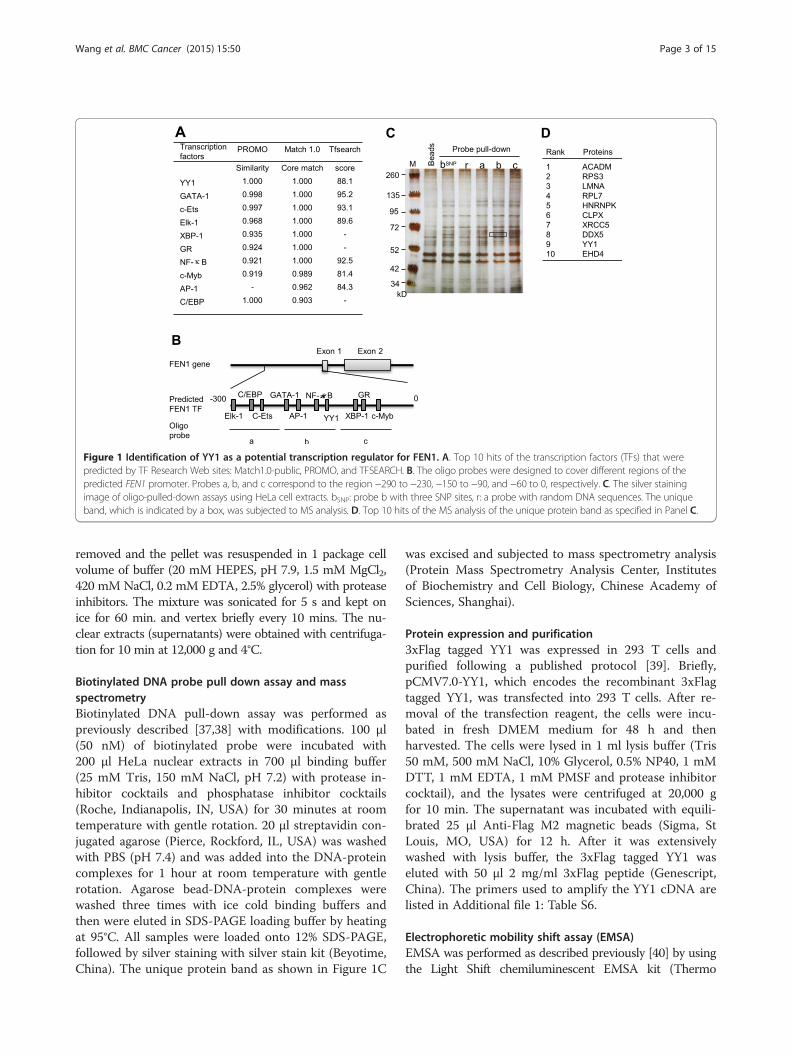
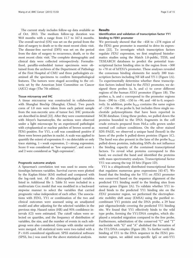
Figure 1 Identification of YY1 as a potential transcription regulator for FEN1. A. Top 10 hits of the transcription factors (TFs) that werepredicted by TF Research Web sites: Match1.0-public, PROMO, and TFSEARCH. B. The oligo probes were designed to cover different regions of thepredicted FEN1 promoter. Probes a, b, and c correspond to the region −290 to −230, −150 to −90, and −60 to 0, respectively. C. The silver stainingimage of oligo-pulled-down assays using HeLa cell extracts. bSNP: probe b with three SNP sites, r: a probe with random DNA sequences. The uniqueband, which is indicated by a box, was subjected to MS analysis. D. Top 10 hits of the MS analysis of the unique protein band as specified in Panel C.
Wang et al. BMC Cancer (2015) 15:50 Page 3 of 15
removed and the pellet was resuspended in 1 package cellvolume of buffer (20 mM HEPES, pH 7.9, 1.5 mM MgCl2,420 mM NaCl, 0.2 mM EDTA, 2.5% glycerol) with proteaseinhibitors. The mixture was sonicated for 5 s and kept onice for 60 min. and vertex briefly every 10 mins. The nu-clear extracts (supernatants) were obtained with centrifuga-tion for 10 min at 12,000 g and 4°C.
Biotinylated DNA probe pull down assay and massspectrometryBiotinylated DNA pull-down assay was performed aspreviously described [37,38] with modifications. 100 μl(50 nM) of biotinylated probe were incubated with200 μl HeLa nuclear extracts in 700 μl binding buffer(25 mM Tris, 150 mM NaCl, pH 7.2) with protease in-hibitor cocktails and phosphatase inhibitor cocktails(Roche, Indianapolis, IN, USA) for 30 minutes at roomtemperature with gentle rotation. 20 μl streptavidin con-jugated agarose (Pierce, Rockford, IL, USA) was washedwith PBS (pH 7.4) and was added into the DNA-proteincomplexes for 1 hour at room temperature with gentlerotation. Agarose bead-DNA-protein complexes werewashed three times with ice cold binding buffers andthen were eluted in SDS-PAGE loading buffer by heatingat 95°C. All samples were loaded onto 12% SDS-PAGE,followed by silver staining with silver stain kit (Beyotime,China). The unique protein band as shown in Figure 1C
was excised and subjected to mass spectrometry analysis(Protein Mass Spectrometry Analysis Center, Institutesof Biochemistry and Cell Biology, Chinese Academy ofSciences, Shanghai).
Protein expression and purification3xFlag tagged YY1 was expressed in 293 T cells andpurified following a published protocol [39]. Briefly,pCMV7.0-YY1, which encodes the recombinant 3xFlagtagged YY1, was transfected into 293 T cells. After re-moval of the transfection reagent, the cells were incu-bated in fresh DMEM medium for 48 h and thenharvested. The cells were lysed in 1 ml lysis buffer (Tris50 mM, 500 mM NaCl, 10% Glycerol, 0.5% NP40, 1 mMDTT, 1 mM EDTA, 1 mM PMSF and protease inhibitorcocktail), and the lysates were centrifuged at 20,000 gfor 10 min. The supernatant was incubated with equili-brated 25 μl Anti-Flag M2 magnetic beads (Sigma, StLouis, MO, USA) for 12 h. After it was extensivelywashed with lysis buffer, the 3xFlag tagged YY1 waseluted with 50 μl 2 mg/ml 3xFlag peptide (Genescript,China). The primers used to amplify the YY1 cDNA arelisted in Additional file 1: Table S6.
Electrophoretic mobility shift assay (EMSA)EMSA was performed as described previously [40] by usingthe Light Shift chemiluminescent EMSA kit (Thermo
Wang et al. BMC Cancer (2015) 15:50 Page 4 of 15
Fisher Scientific, Wilmington, DE, USA), purified recom-binant YY1 protein and the biotin-labeled double strandDNA. These probes, which represent the FEN1 promoterregions, include negative control Probe N, positive controlProbe P, WT FEN1 and MUT FEN1. The positive controlprobe (Probe P) is the same Probe as the Probe b used inthe biotinylated DNA pull-down assay. The MUT FEN1probe contains two mutated nucleotide residues indicatedwith low case. These probes are listed in Additional file 1:Table S6.
Chromatin immunoprecipitation (ChIP)ChIP assay was performed as described previously [40].The rabbit anti-YY1 antibody was purchased from SantaCruz Biotechnology (Santa Cruz Biotechnology, Dallas, TX,USA). The protein A/G agarose beads were purchased fromPierce (Pierce, Rockford, IL, USA) and mouse IgG conju-gated with magnetic beads were purchased from Cell Sig-naling Technology (Cell Signaling Technology, Danvers,MA, USA) as the negative control. Besides the control IgG,the amount of ACTB and FEN1 CDS DNA fragment thatwas precipitated and analyzed under same conditionsserved as an additional control for specificity of the bindingbetween the ChIP antibodies and their target genes. ChIPprimers for the FEN1 promoter, FEN1CDS and ACTB, as acontrol, are listed in Additional file 1: Table S6.
Cell culture, transfection, treatment, and flow cytometryThe 293 T, HeLa, MCF-7, MDA-MB-231 cells were ob-tained from ATCC. Cells were cultured in DMEM(Hyclone, Logan, UT, USA) supplemented with 10% fetalbovine serum (Pufei, China). 1 × 10⁶ MDA-MB-231 orMCF7 cells were seeded in 6 well-plate for 24 h at 37°C, 5%CO2, then treated with 5 μM Mytomycine C (MMC)(Sigma, St Louis, MO, USA) for 1 h. After treatment, cellswere collected 9 and 16 hours later for RT-PCR andWestern blotting to detect the YY1 and FEN1 proteinand mRNA levels, respectively. In parallel, cells weretreated with Taxol (Melone, China) in a concentrationof 20 nM for 24 h and were then collected for RT-PCRand Western blotting.The transfections were carried out according to standard
procedures using SuperFectin II DNATransfection Reagent(Pufei, China) and the EGFP intensity was measuredwith the Cytomics TM FC 500 Flow Cytometer System(Beckman Coulter, Pasadena, CA). To detect the effectsof the YY1 level in cellular response to the drugs, 239 Tcells were transfected with pcDNA3.1-YY1. The cellsurvival fractions at different time points were mea-sured by cell counting.
Western blottingWestern blotting analysis was performed according tostandard procedures using ECL detection substrate (Pierce,
Rockford, IL, USA) and the blot was exposed to the Tannon5200 System for visualization. The antibodies used in ourstudies were the rabbit polyclonal anti-YY1 antibody (SantaCruz), the rabbit monoclonal anti-FEN1 antibody (NovusBiologicals, Littleton, CO, USA), the Horseradish peroxid-ase (HRP)-conjugated anti-GAPDH (GenScript, China),and the Horseradish peroxidase (HRP)-conjugated anti-rabbit secondary antibody (Pierce, Rockford, IL, USA).
RT-PCR analysisTotal mRNA was isolated using TRIzol reagent (LifeTechnologies, Carlsbad, CA, USA). Reverse transcriptionreaction was performed using PrimeScript RT reagentkit (TaKaRa, Japan) according to the manufacturer’s in-structions. qRT-PCR was performed in a MJ Chromo 4(Bio-Rad) by using a reaction mixture with PlatinumSYBR qPCR SuperMix-UDG (Invitrogen, Carlsbad, CA,USA). All the PCR amplification was performed in tripli-cate and repeated in three independent experiments.The sequence for all of the primers for human FEN1,human YY1, and the internal control of human GAPDHand EGFP are listed in Additional file 1: Table S6.
Disease free survival analyses based on the data availablein the literatureFEN1 survival analyses were determined based onIvshina et al. [41]. In their study, the gene expressionwas profiled with 347 primary invasive breast tumorsusing Affymetrix microarray. Data were deposited toGene Expression Omnibus (GEO) database (GSE4922).The FEN1 expression ‘high’ and ‘low’ groups were segre-gated based on median expression values. Kaplan-Meiersurvival analysis was used to determine the survival differ-ences between ‘high’ and ‘low’ expression, visualized byKaplan-Meier plots and compared using Cox regressionanalysis, with p-values calculated by log-rank test using theSurvival package in R [42]. Survival analyses were per-formed on all patients, including ER+ subgroups, ER- sub-groups and ER negative and lymph node negative (ER-LN-)groups respectively for clinical interest.
Patient information and tumor specimens for prognosticoutcome analysisThe use of specimens from human subjects was approvedby the Ethics Committee of China Medical University(CMU). A total of 288 primary breast cancer patients fromthe archives of the Department of Pathology in the FirstHospital of CMU were initially recruited in the currentretrospective study. All patients included in the studywere the ones who had surgery between May 1995 andDecember 2009. Patients were selected into the studybased on the availability of complete clinical medical re-cords, follow-up data and an adequate number ofparaffin-embedded tissue blocks.
Wang et al. BMC Cancer (2015) 15:50 Page 5 of 15
The current study includes follow-up data available asof Oct. 2013. The medium follow-up duration was90.8 months with a range from 11.7 to 167.4 months.The overall survival (OS) was set on the period from thedate of surgery to death or to the most recent clinic visit.The disease-free survival (DFS) was set on the periodfrom the date of surgery to recurrence, death, or to themost recent clinic visit. The complete demographic andclinical data were collected retrospectively. Formalin-fixed, paraffin-embedded tumor specimens were ob-tained from the archives of the Department of Pathologyof the First Hospital of CMU and three pathologists ex-amined all the specimens to confirm histopathologicalfeatures. The tumors were staged according to the cri-teria set by the American Joint Committee on Cancer(AJCC) stage (The 7th edition).
Tissue microarray and IHCA tissue microarray was constructed in collaborationwith Shanghai Biochip (Shanghai, China). Two punchcores of 1.0 mm were taken from each patient samplefrom the non-necrotic area of tumor foci. IHC protocolsare described in detail [22]. After they were counterstainedwith Meyer’s haematoxylin, the sections were observedunder a light microscope by an experienced pathologistwith cytoplasmical or nuclear patches of brown scored asFEN1-positive. For YY1, a cell was considered positive ifthere were brown patches in nuclei. A scale was applied toquantify the extent of expression: 0 = no detectable or onlytrace staining, 1 = weak expression, 2 = strong expression.Score 0 was considered as “low expression”, and score 1and 2 were considered as “high expression”.
Prognostic outcome analysisA Spearman’s correlation test was used to assess rela-tionships between variables. Survival curves were plottedby the Kaplan-Meier (KM) method and compared withthe log-rank test. All the clinicopathological variableslisted in Additional file 1: Table S1 were included in amultivariate Cox model that was modified in a backwardstepwise manner to select the variables that carriedprognostic value independent of each other. The associa-tions with FEN1, YY1 or combination of the two andclinical outcomes were assessed using an unadjustedmodel and after adjusting for the selected variables in theprevious step. Hazard ratios (HR) and 95% confidence in-tervals (CI) were estimated. The cutoff values were se-lected on quartiles, and the frequency of distribution ofvariables, the size, and the number of events in each sub-group were also considered. Groups with similar survivalwere merged. All statistical tests were two-tailed with aP < 0.05 considered significant. SPSS statistical software(SPSS, Inc.) was used for the above statistical analysis.
ResultsIdentification and validation of transcription factor YY1binding to FEN1 promoterWe previously showed that the −458 to +278 region ofthe FEN1 gene promoter is essential to drive its expres-sion [22]. To investigate which transcription factorsregulate FEN1 expression, we first employed bioinfor-matics studies using the Match 1.0-public, TESS, andTESEARCH databases to predict the potential tran-scriptional factor binding sites in the region from −300to +70 nt of hFEN1’s promoter. These analyses revealedthe consensus binding elements for nearly 200 tran-scription factors including NF-kB and YY-1 (Figure 1A).To experimentally determine whether these transcrip-tion factors indeed bind to the FEN1 promoter, we de-signed three probes (a, b, and c) to cover differentregions of the human FEN1 promoter (Figure 1B). Theprobes a, b, and c correspond to the promoter regionsfrom −290 to −230, −150 to −90, and −60 to 0, respect-ively. In addition, probe bSNP contains the same regionof −150 to −90 as probe b, but includes three single nu-cleotide polymorphisms that have been reported inNCBI database. Using these probes, we pulled down theproteins bounded to the DNA fragments in the cellcrude extracts prepared from HeLa cells grown undernormal cell culture conditions. On the silver stainedSDS-PAGE, we observed a unique band (boxed) in thelanes of the probe b pulled-down proteins (Figure 1C).The band was also present in the lane of the probe bSNP
pulled-down proteins, indicating SNPs do not influencethe binding capacity of the contained transcriptionalfactors. To reveal what proteins correspond to thisband, we excised the band and identified the proteinswith mass spectrometry analyses. Transcriptional factorYY1 was among the top 10 hits (Figure 1D).YY1 is a ubiquitously distributed transcriptional factor
that regulates numerous gene expressions [43-47]. Wefound that the binding site for YY1 on FEN1 promoterwas conserved based on the sequence alignment of thepredicted YY1 binding motif to the binding sites fromvarious genes (Figure 2A). To validate whether YY1 in-deed binds to the predicted YY1 binding site on theFEN1 promoter region, we performed the electrophor-etic mobility shift assay (EMSA) using the purified re-combinant YY1 protein and the DNA probe, a 29 basepair oligonucleotide covering the predicted YY1 bindingsite. We found that YY1 effectively binds to the wildtype probe, forming the YY1/DNA complex, which dis-played a retarded migration compared to the free probe.Furthermore, substitution of the conserved “C” and “T”nucleotide with “G” and “A” abolished the formation ofthe YY1/DNA complex (Figure 2B). To further verify thebinding of YY1 to the DNA sequence in the FEN1 pro-moter region, we added non-specific IgG or anti-YY1

Figure 2 YY1 binds to the conserved YY1 binding motif in the FEN1 promoter region. A. Sequence alignment of the conserved YY1binding motif in different proteins. B. EMSA analysis of YY1 binding to the YY1 binding motif in the FEN1 promoter. Recombinant YY1 wasincubated with different biotin-labeled DNA probes. The sequences of the Probe N, Probe P, WT FEN1and MUT FEN1 can be found in Additionalfile 1: Table S6. The free probe and YY1/DNA complex were resolved in 5% native PAGE. C. EMSA assay on YY1 and FEN1 oligo in the presence ofnon-specific IgG or the anti-YY1 antibody. D. ChIP analysis of YY1 binding to the FEN1 promoter region. Specific YY1-bound DNA in MCF7 cellextracts was pulled down by an anti-YY1 antibody. The YY1-bound FEN1 sequence was amplified by PCR. The sequence for the FEN1 promoterspecific primer can be found in the Additional file 1: Table S6 as FEN1 (YY1). The PCR product was analyzed by 1% agarose electrophoresis.
Wang et al. BMC Cancer (2015) 15:50 Page 6 of 15
anti-body to the binding reaction with YY1 and WT FEN1sequence. Addition of anti-YY1 but not non-specific IgG di-minished the YY1-oligo complex (Figure 2C), suggestingthat YY1 specifically bound to the oligo sequence of FEN1promoter. We then investigated whether YY1 bound to theFEN1 promoter region in MCF7 breast cancer cells by con-ducting a chromatin immune-precipitation-PCR (ChIP-PCR) and showed that the FEN1 promoter was specificallypulled-down by an YY1-specific antibody but not the con-trol antibody (Figure 2D). The results all suggest that tran-scriptional factor YY1 binds to the FEN1 promoter.
Anti-cancer drugs release the YY1 suppression to FEN1leading to its over-expression and drug resistanceYY1 is a multifunctional protein and can work as eithera gene expression repressor or an activator [35,48]. Todetermine the roles of YY1 in regulation of FEN1 ex-pression, we exogenously overexpressed YY1 in 293 Tcells and evaluated the FEN1 protein level. We foundthat the protein level of endogenous FEN1 gradually de-creased as the amounts of the plasmid DNA transfected
into 293 T cells increased (Figure 3A). We next exam-ined whether YY1 bound to the FEN1 promoter regionand suppressed the gene expression. We sub-cloned theFEN1 promoter into the pGL4.0 plasmid, so that the ex-pression of the EGFP reporter gene was only driven by theFEN1 promoter. The Flag-tagged YY1 expression vectorand the pGL4.0-FEN1 promoter-driven EGFP vector wereco-transfected into 293 T cells. The overexpression ofFlag-tagged YY1 was confirmed by PCR and western blot(Figure 3B and C). We then measured the EGFP mRNAlevel by qPCR and the EGFP protein by flow cytometry.Our data indicated that the ectopic over-expression ofYY1 in 293 T cells considerably reduced EGFP mRNA andprotein levels (Figure 3B, D and E). Next, we determined ifa decrease in YY1 level resulted in up-regulation of FEN1expression. We knocked down YY1 in 293 T or MCF7cells by shRNA specific against YY1 sequences. We foundthat knockdown of YY1 was associated with significant in-crease in FEN1 expression level in both 293 T and MCF7cells (Figure 3F). Similar phenomenon was observed inHeLa and U251 cancer cells.
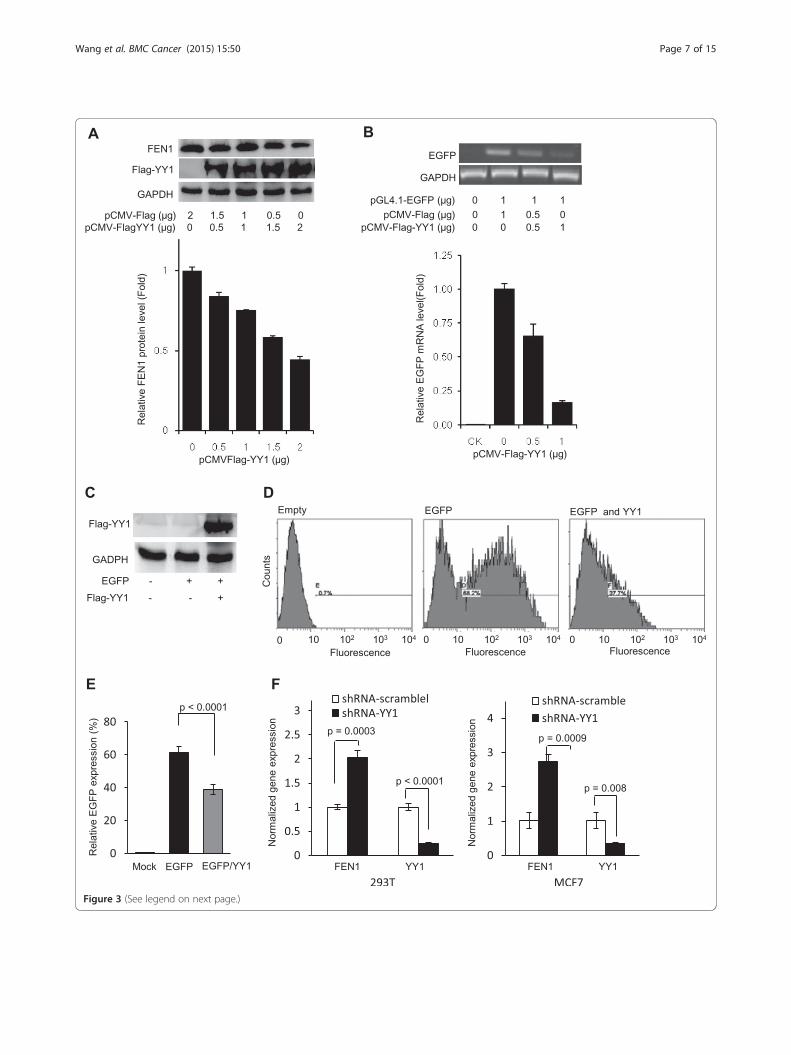
Figure 3 (See legend on next page.)
Wang et al. BMC Cancer (2015) 15:50 Page 7 of 15
(See figure on previous page.)Figure 3 Overexpression of YY1 inhibits FEN1 promoter-driven protein expression. A. YY1 was overexpressed in 293 T cells and its impacton the FEN1 protein level was evaluated by western blot using the anti-Flag or anti-FEN1 antibody. B. The pCMV-Flag-YY1 expression vector and thepGL4.0-FEN1 promoter-EGFP vector, or pGL4.0 EGFP vector was co-transfected into 293 T cells. The EGFP expression was detected by semi-quantitativePCR (Upper panel) and quantitative PCR (lower panel). C. The overexpression of Flag-tagged YY1 was confirmed by western blot using the anti-Flagantibody. D and E. EGFP protein levels with or without YY1 overexpression was measured by FACS. Panel D shows the representative FACS images.Panel E is the quantification of FACS. Values are means ± s.d. of four independent experiments. p value was calculated by the two-tail student’s t-test.F. Knockdown of YY1 in 293 T (Left Panel) and MCF7 (right panel) cells. The YY1 and FEN1 expression was measured by quantitative PCR. The mRNAlevel was normalized with corresponding mRNA level of GADPH, and the normalized mRNA level of YY1 or FEN1 in the cells treated with control siRNAwas arbitrarily set as 1. Values are means ± s.d. of three independent experiments. p value was calculated by the two-tail student’s t-test.
Wang et al. BMC Cancer (2015) 15:50 Page 8 of 15
We then tested whether DNA damaging agents andchemotherapeutic drugs relieve such a restraint, leadingto induction of FEN1 expression. We treated the breastcancer cell line MDA-MB-231 with mitomycin C(MMC) and Taxol and performed qPCR and Westernblotting to analyze the gene expression of YY1 andFEN1. We found that in response to treatments withMMC and Taxol, the mRNA level of YY1 was down-regulated by more than 2 folds, while the mRNA level ofFEN1 was up-regulated by 3 to 6 folds (Figure 4A andB). We consistently observed that the YY1 protein levelwas reduced by approximate 2 folds, while the proteinlevel of FEN1 increased by more than 2 folds. Inaddition, we tested whether the drug treatment also im-pairs the binding of the transcription factor to the FEN1promoter. Indeed, our ChIP analyses indicated that theamount of YY1 bound to the FEN1 promoter reduced by2 folds upon the MMC treatment (Figure 4C). Further-more, when we overexpressed the Flag-tagged YY1 in293 T cells (Figure 4D), we observed that the cells harbor-ing this expression plasmid became more sensitive to bothMMC and Taxol treatment (Figure 4E and F).To support the notion that different DNA damage
agents and therapeutic drugs induce FEN1 gene expres-sion, we employed an expression array of 26 cancer celllines in 13 major categories that have been treated with25 different DNA-damaging agents and therapeutic drugs(Figure 5A). The fact that FEN1 expression was high inbreast cancer cell lines was consistent with our publisheddata [22]. The Northern dot blotting results showed thatFEN1 expression levels in breast cancer cell lines, MDA-MB-4355 and MDA-MB-231, increased significantly (bymore than 8 folds) after the treatment with DNA-damagingagents, such as camptothecin, cytochalasin D, MMC, andgamma irradiation (Figure 5A and B). However, otheragents such as Etoposide, 5-fluorouracil, Aphidicoline andTaxol, induced the FEN1 expression in MDA-MB-231, butnot in MDA-MB-4355 (Figure 5A and B).
Breast cancer patients with low expression of YY1 andhigh expression of FEN1 have poor prognosticsSeeking the relevance between FEN1 expression andcancer patient outcomes, we performed survival analysis
using 5 different breast cancer patient cohorts, namelyIvshina [41], Huang [49], Pawitan [50], Sotiriou [51], andWang [52], all of which are available in the literature. Forthe data from the Ivshina [41], patients were grouped intoHigh-Risk and Low-Risk subgroups based on 2-mean cat-egorical clustering of selected significant genes for Kaplan-Meier survival analysis [41] with high and low expressionlevels of FEN1 gene to measure the number of patients liv-ing for a certain amount of time after the treatment.Kaplan-Meier analyses revealed that the under-expressionof FEN1 measured by the mRNA level was correlated withbetter disease free survival (DFS) outcome. For overall 249breast tumor samples (p = 0.0007), 211 of ER+ subgroups(p = 0.005), 34 of ER- subgroups (p = 0.03), 20 of ER-LN-subgroups (p = 0.007) all showed an inverse correlation ofFEN1 gene expression with DFS (Figure 6A). The inversecorrelation was also validated with other 4 large breastcancer cohorts (Huang et al. (n = 89; p = 0.004) [49], Pawitanet al. (n = 159; p = 5.21e-5) [50], Sotiriou et al. (n = 117 p =0.04) [51] and Wang et al. (n = 286 p = 0.02) [52]. Interest-ingly, our additional patient cohort data mining indicatedthat the difference of the survivorship between the patientswith the low expression and high expression of FEN1 in ER-and ER-/lymph node negative patient cohorts is much largerthan that in ER+ patients (Data not shown).Further seeking the association between FEN1 and YY1
expression levels and survivorship in breast cancer pa-tients, we studied a cohort that made available in the FirstHospital of China Medical University. The characteristicsof the studied cohort are summarized in Additional file 1:Table S1. After excluding cases with insufficient tumor tis-sue in tissue micro-array, FEN1 expression was detectablein 268 cases, and YY1 expression was detectable in 285cases. The expression of FEN1 was detected in 209 casesout of a total of 268 cases (78.0%) by IHC staining, whileYY1 was present in 67 cases out of a total of 285 cases(23.5%) by IHC staining. The association between FEN1and YY1 expressions with clinicopathological variables ofthe cohort is shown in Additional file 1: Table S1. No sig-nificant association between FEN1 expression and age, Tstage, N stage, stage, ER, PR, HR, Her-2, triple-negative,being ductal carcinoma in situ (Dcis), using taxane in ad-juvant therapy, or using standard therapy was found.

Figure 4 DNA damaging agents MMC and Taxol inhibit YY1 expression but induce FEN1 expression. A. and B. YY1 and FEN1 expressionin MDA-MB-231 breast cancer cell line in response to the MMC and Taxol treatment. The mRNA level (A) and protein level (B) were measured byquantitative PCR and Western blot. The left panel in B showed the quantification of Western blot results. All experiments were independentcarried out at least three times. C. Analysis of YY1 binding to the FEN1 promoter in response to the MMC treatment. Cells were treated with MMC, andthe level of YY1-bound FEN1 promoter was evaluated by the ChIP assay. The lowest DNA staining density is arbitrarily set as 1. D. western blotconfirmed the overexpression of Flag-tagged YY1 in 293 T cells. The β-actin(ACTB) was used as control. E. and F. The survivorship of 293 T cell and293 T cell harboring a YY1 expression plasmid, pCMV-Flag-YY1, or the empty vector, under treatment of mytomycine C (MMC) (Panel E) or taxol (Panel F).In both panels, the cells were treated with indicated concentrations of MMC or taxol for 48 hours. The number of survival cells was counted. The survivalrate of the untreated cells with or without YY1 overexpression was arbitrarily set as 1.
Wang et al. BMC Cancer (2015) 15:50 Page 9 of 15
However, high YY1 staining correlated significantly withER-positive cases (P = 0.007), PR cases (P = 0.000), HR-positive cases (P = 0.000), NOT-tri-negative cases (P =0.030). The correlation between FEN1 and YY1 expressionwas not significant.The 5-year overall survival rate of the cohort was 86.0%.
In a Kaplan–Meier (KM) analysis, FEN1 and YY1 expres-sions showed no prognostic significance in OS in this co-hort (P = 0.135 and 0.258, respectively). In contrast,patients with FEN1-high/YY1-low expression had signifi-cantly poor overall postoperative survival, compared withthose with other phenotypes (P = 0.027) (Figure 6B). Stage
was the only independent clinicopathological variable topredict OS (Additional file 1: Table S2.). After adjustingwith the stage, FEN1-high/YY1-low expression could stillpredict a poor OS in the multivariate Cox model (P =0.020, Additional file 1: Table S3). However, in the ER-negative or ER-negative/lymph-node-metastasis-negativesubgroups, there was no significant association betweenthe FEN1 expression level, YY1 expression level, or theircombination and OS in the CMU cohort. The similartrends were observed in association between FEN1 expres-sion, YY1 expression, their combination, and DFS whenanalyzed with KM methods. The corresponding log-
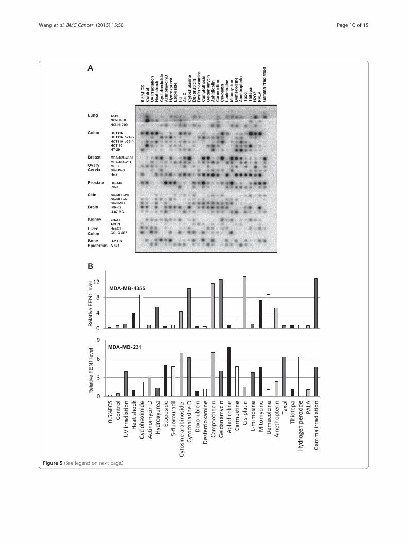
Figure 5 (See legend on next page.)
Wang et al. BMC Cancer (2015) 15:50 Page 10 of 15
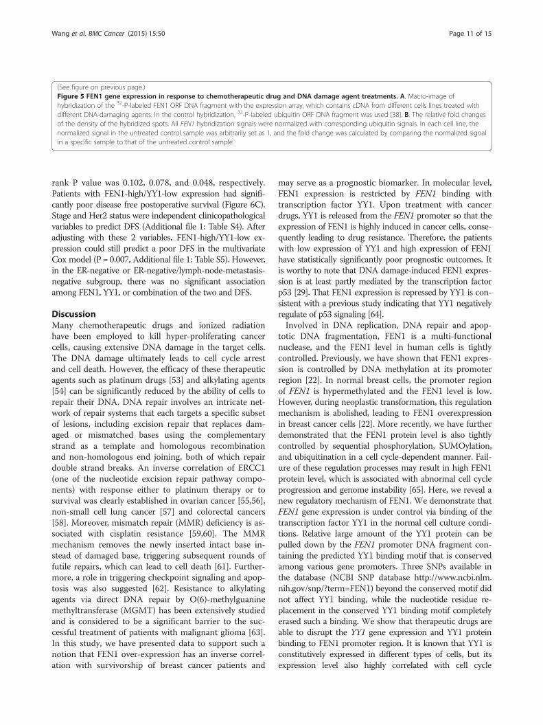
(See figure on previous page.)Figure 5 FEN1 gene expression in response to chemotherapeutic drug and DNA damage agent treatments. A. Macro-image ofhybridization of the 32-P-labeled FEN1 ORF DNA fragment with the expression array, which contains cDNA from different cells lines treated withdifferent DNA-damaging agents. In the control hybridization, 32-P-labeled ubiquitin ORF DNA fragment was used [38]. B. The relative fold changesof the density of the hybridized spots. All FEN1 hybridization signals were normalized with corresponding ubiquitin signals. In each cell line, thenormalized signal in the untreated control sample was arbitrarily set as 1, and the fold change was calculated by comparing the normalized signalin a specific sample to that of the untreated control sample.
Wang et al. BMC Cancer (2015) 15:50 Page 11 of 15
rank P value was 0.102, 0.078, and 0.048, respectively.Patients with FEN1-high/YY1-low expression had signifi-cantly poor disease free postoperative survival (Figure 6C).Stage and Her2 status were independent clinicopathologicalvariables to predict DFS (Additional file 1: Table S4). Afteradjusting with these 2 variables, FEN1-high/YY1-low ex-pression could still predict a poor DFS in the multivariateCox model (P = 0.007, Additional file 1: Table S5). However,in the ER-negative or ER-negative/lymph-node-metastasis-negative subgroup, there was no significant associationamong FEN1, YY1, or combination of the two and DFS.
DiscussionMany chemotherapeutic drugs and ionized radiationhave been employed to kill hyper-proliferating cancercells, causing extensive DNA damage in the target cells.The DNA damage ultimately leads to cell cycle arrestand cell death. However, the efficacy of these therapeuticagents such as platinum drugs [53] and alkylating agents[54] can be significantly reduced by the ability of cells torepair their DNA. DNA repair involves an intricate net-work of repair systems that each targets a specific subsetof lesions, including excision repair that replaces dam-aged or mismatched bases using the complementarystrand as a template and homologous recombinationand non-homologous end joining, both of which repairdouble strand breaks. An inverse correlation of ERCC1(one of the nucleotide excision repair pathway compo-nents) with response either to platinum therapy or tosurvival was clearly established in ovarian cancer [55,56],non-small cell lung cancer [57] and colorectal cancers[58]. Moreover, mismatch repair (MMR) deficiency is as-sociated with cisplatin resistance [59,60]. The MMRmechanism removes the newly inserted intact base in-stead of damaged base, triggering subsequent rounds offutile repairs, which can lead to cell death [61]. Further-more, a role in triggering checkpoint signaling and apop-tosis was also suggested [62]. Resistance to alkylatingagents via direct DNA repair by O(6)-methylguaninemethyltransferase (MGMT) has been extensively studiedand is considered to be a significant barrier to the suc-cessful treatment of patients with malignant glioma [63].In this study, we have presented data to support such anotion that FEN1 over-expression has an inverse correl-ation with survivorship of breast cancer patients and
may serve as a prognostic biomarker. In molecular level,FEN1 expression is restricted by FEN1 binding withtranscription factor YY1. Upon treatment with cancerdrugs, YY1 is released from the FEN1 promoter so that theexpression of FEN1 is highly induced in cancer cells, conse-quently leading to drug resistance. Therefore, the patientswith low expression of YY1 and high expression of FEN1have statistically significantly poor prognostic outcomes. Itis worthy to note that DNA damage-induced FEN1 expres-sion is at least partly mediated by the transcription factorp53 [29]. That FEN1 expression is repressed by YY1 is con-sistent with a previous study indicating that YY1 negativelyregulate of p53 signaling [64].Involved in DNA replication, DNA repair and apop-
totic DNA fragmentation, FEN1 is a multi-functionalnuclease, and the FEN1 level in human cells is tightlycontrolled. Previously, we have shown that FEN1 expres-sion is controlled by DNA methylation at its promoterregion [22]. In normal breast cells, the promoter regionof FEN1 is hypermethylated and the FEN1 level is low.However, during neoplastic transformation, this regulationmechanism is abolished, leading to FEN1 overexpressionin breast cancer cells [22]. More recently, we have furtherdemonstrated that the FEN1 protein level is also tightlycontrolled by sequential phosphorylation, SUMOylation,and ubiquitination in a cell cycle-dependent manner. Fail-ure of these regulation processes may result in high FEN1protein level, which is associated with abnormal cell cycleprogression and genome instability [65]. Here, we reveal anew regulatory mechanism of FEN1. We demonstrate thatFEN1 gene expression is under control via binding of thetranscription factor YY1 in the normal cell culture condi-tions. Relative large amount of the YY1 protein can bepulled down by the FEN1 promoter DNA fragment con-taining the predicted YY1 binding motif that is conservedamong various gene promoters. Three SNPs available inthe database (NCBI SNP database http://www.ncbi.nlm.nih.gov/snp/?term=FEN1) beyond the conserved motif didnot affect YY1 binding, while the nucleotide residue re-placement in the conserved YY1 binding motif completelyerased such a binding. We show that therapeutic drugs areable to disrupt the YY1 gene expression and YY1 proteinbinding to FEN1 promoter region. It is known that YY1 isconstitutively expressed in different types of cells, but itsexpression level also highly correlated with cell cycle

100
80
60
Sur
viva
l (%
)
C
40
20
0
140120100806040200
Per
cent
Time (months)
100
A
80
60
40
20erce
nt S
urvi
val (
%)
20
0
140120100806040200
Pe
Time (months)
100
B
80
60
40
ent S
urvi
val (
%)
20
0
140120100806040200
Per
ce
Time (months)
Figure 6 Associations between FEN1, YY1 protein, or theircombination and OS or DFS. A. FEN1 Kaplan Meier survival plotwith breast cancer patient cohort in the Ivshina data base. The blackline indicates FEN1 high expression while the red line indicates FEN1low expression (‘high’ and ‘low’ determined by median expression).Patients with FEN1 high expression: 132; patients with FEN1 lowexpression: 117, Log-rank p = 0.0007. B. Low expression of YY1 andhigh expression of FEN1 and OS in the CMU cohort. The black lineindicates YY1 low but FEN1 high expression and red line indicatesother types. Patients with YY1 low but FEN1 high expression: 154;patients with other types: 113, Log-rank p = 0.027. C. Low expression ofYY1 and high expression of FEN1 and DFS in the CMU cohort. Theblack line indicates YY1 low but FEN1 high expression and red lineindicates other types. Patients with YY1 low but FEN1 high expression:154; patients with other type: 113, Log-rank p = 0.048.
Wang et al. BMC Cancer (2015) 15:50 Page 12 of 15
progression and cell proliferation [66]. Therefore, it is pos-sible that therapeutic drugs such as MMC and taxol in-duced cell cycle arrest and contribute to down-regulationof YY1. In addition, the drug treatment may inducechanges in post-translational modifications and conform-ation of YY1 proteins or altering the FEN1 promotermethylation status, so that the interaction between YY1and FEN1 promoter is impaired. As a result, the suppres-sion of FEN1 expression by YY1 is eliminated, and FEN1 isover-expressed. Consequently, cancer cells become moreresistant to drugs due to enhanced DNA repair systems asa result of FEN1 over-expression. Moreover, after we artifi-cially over-expressed YY1 protein, the cancer cells becamemore sensitive to drugs. Transcriptional factor binding se-quence analysis of this region, using the computer pro-grams TRANSFAC, Match1.0-public, TESS, andTFSEARCH, suggested that nearly 200 transcription factorsincluding NF-κB and YY-1 might bind to the FEN1 pro-moter region. Therefore, what we have seen with YY1might be only tips of the iceberg. It is crucial to elucidatethe comprehensive network that controls FEN1 gene ex-pression under both normal and treatment conditions andto obtain a dynamic picture on how such a control mech-anism changes in response to different drug treatments.From the clinical standpoint, FEN1 is a good candidate
biomarker for prognostics of breast cancer patients basedon evidences that we made available in the current studies.From the data made available by Ivshina [41], we see aclear distinction between the disease-free survivorship ofthe patients with high expression of FEN1 and that of pa-tients with low expression of FEN1. Namely, more than80% of patients with low expression of FEN1 can survive,disease-free, for more than 10 years; however, only lessthan 55% of patients with high expression can do so. Thatmeans 25% more patients in the cohort would live for atleast 10 years longer if FEN1 expression were suppressed.The separation was very much further improved in ER-(45%) or ER- and lymph node negative patient cohorts(55%) though the cohort sizes are relatively small in the

Wang et al. BMC Cancer (2015) 15:50 Page 13 of 15
later two cases as only about 10% of breast cancer patientsare ER-. In the patients with not only ER- but also low ex-pression of FEN1 gene, more than 90% of them would beable to live for at least 10 years longer. Thus, examiningthe FEN1 expression level is very critical to the patientswith ER-. With the patient cohort from the First Hospitalof China Medical University, FEN1 and YY1 expressionswere evaluated with IHC. In that cohort, patients withFEN1-high and YY1-low expression had both statisticallysignificantly poor overall and disease-free postoperativesurvivals, a fact suggesting that FEN1 and YY1 might haveinverse impact on the survival of breast cancer patients.This is consistent with the results that we have obtainedfrom molecular studies using cultured cell lines and clin-ical drugs. Overall, the FEN1/YY1 interaction and regula-tory mechanism might be of clinical importance andshould be further investigated.
ConclusionAltogether, we demonstrate that YY1 plays a critical role inregulating FEN1 gene expression as a repressor. Reductionof YY1 levels in breast cancer cells results in overexpressionof FEN1 leading to resistance to chemotherapeutic drugs.Conversely, overexpression of YY1 in the cancer cells sup-presses FEN1 expression and sensitizes cancer cells toDNA damaging drugs. These finding provide basis for tar-geting YY1 and FEN1 for developing chemotherapy and ra-diation sensitizers.
Additional file
Additional file 1: Table S1. Correlation between FEN1 or YY1expression and clinicopathologic variables. Table S2. Univariate andmultivariate analyses of OS according to clinicopathologic variables.Table S3. Univariate and multivariate analyses of OS according tobiomarkers adjusted by selected clinicopathologic variables. Table S4.Univariate and multivariate analyses of DFS according to clinicopathologicvariables. Table S5. Univariate and multivariate analyses of DFS according tobiomarkers adjusted by selected clinicopathologic variables. Table S6.All the DNA oligonucleotides used in the current study.
AbbreviationsBER: Base excision repair; ChIP: Chromatin immunoprecipitation;DFS: Disease-free survival; DNA: Deoxyribonucleic acid; EMSA: Electrophoreticmobility shift assay; ER: Estrogen receptor; FEN1: Flap endonuclease 1;HR: Homologous recombination; ICL: Interstrand crosslink repair;IHC: Immunohistochemistry; MDR: Multidrug resistance; MMC: Mitomycin C;MMR: Mismatch repair; NER: Nucleotide excision repair; NHEJ: Non-homologousend joining; NF-kB: Nuclear factor kappa-light-chain-enhancer of activated Bcells; Nrf2: Erythroid-derived factor 2; PCR: polymerization chain reaction;SNP: Single nucleotide polymorphism; USF1: Upstream stimulatory factor 1;YY1: Yin Yang 1.
Competing interestsThe authors declare that they have no competing interests.
Authors’ contributionsBS, LZ, YH, YW, and YT conceived and designed the experiments. JW, LNZ,TZ, WL, HX performed biochemical, molecular, and cellular studies on YY1function to regulate FEN1 expression and the impact on drug resistance. ZLi,
ZLiu, YCY, FS, LX, YW, XZ analyze FEN1 expression in human breast cancerand conduct data collecting, mining, and analysis on patient prognosticoutcome. BS, LZ, YH, YW, and YT interpret data and write manuscript.All authors read and approved the final manuscript.
Authors’ informationThe authors wish it to be known that, in their opinion, the first two authors,Jianwei Wang and Lina Zhou should be regarded as joint first authors.
AcknowledgementsWe would like to thank the Bioinformatics core facility of City of HopeComprehensive Cancer Center for the additional data mining and analyses aswell as the members of the Shen laboratory for the stimulating discussions.
Grant supportThis work was supported by NSFC grant No. 81172535 to Y.E.T. and NIHgrant CA073764 to B.H.S. This work was in part by an internationalcorporation sponsored by NSFC grant No. 31210103904 to Y.J.H. and the NCICCSG P30 CA033572 to City of Hope.
Author details1College of Life Sciences, Zhejiang University, Hangzhou, China.2Departments of Radiation Biology and Molecular Medicine, BeckmanResearch Institute of City of Hope, 1500 East Duarte Road, Duarte, California91010, USA. 3Departments of Medical Oncology and Thoracic Surgery, TheFirst Hospital of China Medical University, No. 155 North Nanjing Street,Heping District, Shenyang 110001, China. 4College of Agricultural Sciencesand Biotechnology, Zhejiang University, Hangzhou, China. 5School ofMedicine, Zhejiang University, Hangzhou, China.
Received: 21 August 2014 Accepted: 26 January 2015
References1. Gottesman MM. Mechanisms of cancer drug resistance. Annu Rev Med.
2002;53:615–27.2. Zahreddine H, Borden KL. Mechanisms and insights into drug resistance in
cancer. Front Pharmacol. 2013;4:28.3. Johannessen TC, Bjerkvig R, Tysnes BB. DNA repair and cancer stem-like
cells–potential partners in glioma drug resistance? Cancer Treat Rev.2008;34(6):558–67.
4. Muller MR, Thomale J, Rajewsky MF, Seeber S. Drug resistance and DNArepair in leukaemia. Cytotechnology. 1998;27(1–3):175–85.
5. Salehan MR, Morse HR. DNA damage repair and tolerance: a role inchemotherapeutic drug resistance. Br J Biomed Sci. 2013;70(1):31–40.
6. Zheng L, Dai H, Zhou M, Li X, Liu C, Guo Z, et al. Polyploid cells rewire DNAdamage response networks to overcome replication stress-induced barriersfor tumour progression. Nat Commun. 2012;3:815.
7. Nikolova T, Christmann M, Kaina B. FEN1 is overexpressed in testis, lung andbrain tumors. Anticancer Res. 2009;29(7):2453–9.
8. Shen B, Singh P, Liu R, Qiu J, Zheng L, Finger LD, et al. Multiple butdissectible functions of FEN-1 nucleases in nucleic acid processing, genomestability and diseases. Bioessays. 2005;27(7):717–29.
9. Zheng L, Jia J, Finger LD, Guo Z, Zer C, Shen B. Functional regulation ofFEN1 nuclease and its link to cancer. Nucleic Acids Res. 2011;39(3):781–94.
10. Balakrishnan L, Bambara RA. Flap Endonuclease 1. Annu Rev Biochem.2013;82:119–38.
11. Henneke G, Friedrich-Heineken E, Hubscher U. Flap endonuclease 1: a noveltumour suppresser protein. Trends Biochem Sci. 2003;28(7):384–90.
12. Liu Y, Kao HI, Bambara RA. Flap endonuclease 1: a central component ofDNA metabolism. Annu Rev Biochem. 2004;73:589–615.
13. Liu P, Qian L, Sung JS, de Souza-Pinto NC, Zheng L, Bogenhagen DF, et al.Removal of oxidative DNA damage via FEN1-dependent long-patch baseexcision repair in human cell mitochondria. Mol Cell Biol. 2008;28(16):4975–87.
14. Zheng L, Shen B. Okazaki fragment maturation: nucleases take centre stage.J Mol Cell Biol. 2011;3(1):23–30.
15. Zheng L, Zhou M, Chai Q, Parrish J, Xue D, Patrick SM, et al. Novel functionof the flap endonuclease 1 complex in processing stalled DNA replicationforks. EMBO Rep. 2005;6(1):83–9.
16. Saharia A, Guittat L, Crocker S, Lim A, Steffen M, Kulkarni S, et al. Flapendonuclease 1 contributes to telomere stability. Curr Biol. 2008;18(7):496–500.
Wang et al. BMC Cancer (2015) 15:50 Page 14 of 15
17. Saharia A, Stewart SA. FEN1 contributes to telomere stability in ALT-positivetumor cells. Oncogene. 2009;28(8):1162–7.
18. Sampathi S, Bhusari A, Shen B, Chai W. Human flap endonuclease I is incomplex with telomerase and is required for telomerase-mediated telomeremaintenance. J Biol Chem. 2009;284(6):3682–90.
19. Saharia A, Teasley DC, Duxin JP, Dao B, Chiappinelli KB, Stewart SA. FEN1ensures telomere stability by facilitating replication fork re-initiation. J BiolChem. 2010;285(35):27057–66.
20. Zheng L, Dai H, Zhou M, Li M, Singh P, Qiu J, et al. Fen1 mutations result inautoimmunity, chronic inflammation and cancers. Nat Med. 2007;13(7):812–9.
21. LaTulippe E, Satagopan J, Smith A, Scher H, Scardino P, Reuter V, et al.Comprehensive gene expression analysis of prostate cancer reveals distincttranscriptional programs associated with metastatic disease. Cancer Res.2002;62(15):4499–506.
22. Singh P, Yang M, Dai H, Yu D, Huang Q, Tan W, et al. Overexpression andhypomethylation of flap endonuclease 1 gene in breast and other cancers.Mol Cancer Res. 2008;6(11):1710–7.
23. Shen Z. Genomic instability and cancer: an introduction. J Mol Cell Biol.2011;3(1):1–3.
24. Miyoshi T, Nagai T, Kikuchi S, Ohmine K, Nakamura M, Hanafusa T, et al.Cloning and characterization of a human BCR/ABL-positive cell line, K562/RR, resistant to the farnesyltransferase inhibition by tipifarnib. Exp Hematol.2007;35(9):1358–65.
25. Sato M, Girard L, Sekine I, Sunaga N, Ramirez RD, Kamibayashi C, et al.Increased expression and no mutation of the Flap endonuclease (FEN1)gene in human lung cancer. Oncogene. 2003;22(46):7243–6.
26. Kim JM, Sohn HY, Yoon SY, Oh JH, Yang JO, Kim JH, et al. Identification ofgastric cancer-related genes using a cDNA microarray containing novelexpressed sequence tags expressed in gastric cancer cells. Clin Cancer Res.2005;11(2 Pt 1):473–82.
27. Lam JS, Seligson DB, Yu H, Li A, Eeva M, Pantuck AJ, et al. Flapendonuclease 1 is overexpressed in prostate cancer and is associated with ahigh Gleason score. BJU Int. 2006;98(2):445–51.
28. Krause A, Combaret V, Iacono I, Lacroix B, Compagnon C, Bergeron C, et al.Genome-wide analysis of gene expression in neuroblastomas detected bymass screening. Cancer Lett. 2005;225(1):111–20.
29. Christmann M, Tomicic MT, Origer J, Kaina B. Fen1 is induced p53dependently and involved in the recovery from UV-light-induced replicationinhibition. Oncogene. 2005;24(56):8304–13.
30. Kokkinakis DM, Liu X, Neuner RD. Modulation of cell cycle and geneexpression in pancreatic tumor cell lines by methionine deprivation(methionine stress): implications to the therapy of pancreaticadenocarcinoma. Mol Cancer Ther. 2005;4(9):1338–48.
31. Castellano G, Torrisi E, Ligresti G, Malaponte G, Militello L, Russo AE, et al.The involvement of the transcription factor Yin Yang 1 in cancerdevelopment and progression. Cell Cycle. 2009;8(9):1367–72.
32. Nicholson S, Whitehouse H, Naidoo K, Byers RJ. Yin Yang 1 in humancancer. Crit Rev Oncog. 2011;16(3–4):245–60.
33. Zaravinos A, Spandidos DA. Yin yang 1 expression in human tumors. CellCycle. 2010;9(3):512–22.
34. Wang X, Feng Y, Xu L, Chen Y, Zhang Y, Su D, et al. YY1 restrained cellsenescence through repressing the transcription of p16. Biochim BiophysActa. 2008;1783(10):1876–83.
35. Gordon S, Akopyan G, Garban H, Bonavida B. Transcription factor YY1:structure, function, and therapeutic implications in cancer biology.Oncogene. 2006;25(8):1125–42.
36. Deng Z, Wan M, Cao P, Rao A, Cramer SD, Sui G. Yin Yang 1 regulates thetranscriptional activity of androgen receptor. Oncogene. 2009;28(42):3746–57.
37. Wu KK. Analysis of protein-DNA binding by streptavidin-agarose pulldown.Methods Mol Biol. 2006;338:281–90.
38. Deng WG, Zhu Y, Montero A, Wu KK. Quantitative analysis of binding oftranscription factor complex to biotinylated DNA probe by a streptavidin-agarose pulldown assay. Anal Biochem. 2003;323(1):12–8.
39. Lin W, Sampathi S, Dai H, Liu C, Zhou M, Hu J, et al. Mammalian DNA2helicase/nuclease cleaves G-quadruplex DNA and is required for telomereintegrity. EMBO J. 2013;32(10):1425–39.
40. Lin Y, Yang Y, Li W, Chen Q, Li J, Pan X, et al. Reciprocal regulation of Aktand Oct4 promotes the self-renewal and survival of embryonal carcinomacells. Mol Cell. 2012;48(4):627–40.
41. Ivshina AV, George J, Senko O, Mow B, Putti TC, Smeds J, et al. Geneticreclassification of histologic grade delineates new clinical subtypes of breastcancer. Cancer Res. 2006;66(21):10292–301.
42. Bair E, Tibshirani R. Semi-supervised methods to predict patient survivalfrom gene expression data. PLoS Biol. 2004;2(4):E108.
43. Riggs KJ, Saleque S, Wong KK, Merrell KT, Lee JS, Shi Y, et al. Yin-yang 1activates the c-myc promoter. Mol Cell Biol. 1993;13(12):7487–95.
44. Wang CC, Tsai MF, Hong TM, Chang GC, Chen CY, Yang WM, et al. Thetranscriptional factor YY1 upregulates the novel invasion suppressor HLJ1expression and inhibits cancer cell invasion. Oncogene. 2005;24(25):4081–93.
45. Lee MH, Lahusen T, Wang RH, Xiao C, Xu X, Hwang YS, et al. Yin Yang 1positively regulates BRCA1 and inhibits mammary cancer formation.Oncogene. 2012;31(1):116–27.
46. Burgess ST, Shen C, Ferguson LA, O’Neill GT, Docherty K, Hunter N, et al.Identification of adjacent binding sites for the YY1 and E4BP4 transcriptionfactors in the ovine PrP (Prion) gene promoter. J Biol Chem.2009;284(11):6716–24.
47. Athanikar JN, Badge RM, Moran JV. A YY1-binding site is required foraccurate human LINE-1 transcription initiation. Nucleic Acids Res.2004;32(13):3846–55.
48. Atchison M, Basu A, Zaprazna K, Papasani M. Mechanisms of Yin Yang 1 inoncogenesis: the importance of indirect effects. Crit Rev Oncog.2011;16(3–4):143–61.
49. Huang E, Cheng SH, Dressman H, Pittman J, Tsou MH, Horng CF, et al. Geneexpression predictors of breast cancer outcomes. Lancet.2003;361(9369):1590–6.
50. Pawitan Y, Bjohle J, Amler L, Borg AL, Egyhazi S, Hall P, et al. Geneexpression profiling spares early breast cancer patients from adjuvanttherapy: derived and validated in two population-based cohorts.Breast Cancer Res. 2005;7(6):R953–964.
51. Sotiriou C, Neo SY, McShane LM, Korn EL, Long PM, Jazaeri A, et al. Breastcancer classification and prognosis based on gene expression profiles froma population-based study. Proc Natl Acad Sci U S A. 2003;100(18):10393–8.
52. Wang Y, Klijn JG, Zhang Y, Sieuwerts AM, Look MP, Yang F, et al. Gene-expression profiles to predict distant metastasis of lymph-node-negativeprimary breast cancer. Lancet. 2005;365(9460):671–9.
53. Martin LP, Hamilton TC, Schilder RJ. Platinum resistance: the role of DNArepair pathways. Clin Cancer Res. 2008;14(5):1291–5.
54. Sarkaria JN, Kitange GJ, James CD, Plummer R, Calvert H, Weller M, et al.Mechanisms of chemoresistance to alkylating agents in malignant glioma.Clin Cancer Res. 2008;14(10):2900–8.
55. Dabholkar M, Bostick-Bruton F, Weber C, Bohr VA, Egwuagu C, Reed E.ERCC1 and ERCC2 expression in malignant tissues from ovarian cancerpatients. J Natl Cancer Inst. 1992;84(19):1512–7.
56. Kang S, Ju W, Kim JW, Park NH, Song YS, Kim SC, et al. Association betweenexcision repair cross-complementation group 1 polymorphism and clinicaloutcome of platinum-based chemotherapy in patients with epithelialovarian cancer. Exp Mol Med. 2006;38(3):320–4.
57. Olaussen KA, Dunant A, Fouret P, Brambilla E, Andre F, Haddad V, et al. DNArepair by ERCC1 in non-small-cell lung cancer and cisplatin-based adjuvantchemotherapy. N Engl J Med. 2006;355(10):983–91.
58. Metzger R, Leichman CG, Danenberg KD, Danenberg PV, Lenz HJ, Hayashi K,et al. ERCC1 mRNA levels complement thymidylate synthase mRNA levels inpredicting response and survival for gastric cancer patients receivingcombination cisplatin and fluorouracil chemotherapy. J Clin Oncol.1998;16(1):309–16.
59. Fink D, Nebel S, Norris PS, Baergen RN, Wilczynski SP, Costa MJ, et al.Enrichment for DNA mismatch repair-deficient cells during treatment withcisplatin. Int J Cancer. 1998;77(5):741–6.
60. Strathdee G, MacKean MJ, Illand M, Brown R. A role for methylation of thehMLH1 promoter in loss of hMLH1 expression and drug resistance inovarian cancer. Oncogene. 1999;18(14):2335–41.
61. Karran P, Marinus MG. Mismatch correction at O6-methylguanine residues inE. coli DNA. Nature. 1982;296(5860):868–9.
62. Yoshioka K, Yoshioka Y, Hsieh P. ATR kinase activation mediated byMutSalpha and MutLalpha in response to cytotoxic O6-methylguanineadducts. Mol Cell. 2006;22(4):501–10.
63. Verbeek B, Southgate TD, Gilham DE, Margison GP. O6-Methylguanine-DNAmethyltransferase inactivation and chemotherapy. Br Med Bull. 2008;85:17–33.
64. Sui G, el Affar B, Shi Y, Brignone C, Wall NR, Yin P, et al. Yin Yang 1 is anegative regulator of p53. Cell. 2004;117(7):859–72.

Wang et al. BMC Cancer (2015) 15:50 Page 15 of 15
65. Guo Z, Kanjanapangka J, Liu N, Liu S, Liu C, Wu Z, et al. Sequentialposttranslational modifications program FEN1 degradation during cell-cycleprogression. Mol Cell. 2012;47(3):444–56.
66. el Affar B, Gay F, Shi Y, Liu H, Huarte M, Wu S, et al. Essential dosage-dependentfunctions of the transcription factor yin yang 1 in late embryonic developmentand cell cycle progression. Mol Cell Biol. 2006;26(9):3565–81.
Submit your next manuscript to BioMed Centraland take full advantage of:
• Convenient online submission
• Thorough peer review
• No space constraints or color figure charges
• Immediate publication on acceptance
• Inclusion in PubMed, CAS, Scopus and Google Scholar
• Research which is freely available for redistribution
Submit your manuscript at www.biomedcentral.com/submit