proteins of the retinoblastoma pathway, fen1 and mgmt are...
TRANSCRIPT

Accepted Manuscript
Title: Proteins of the retinoblastoma pathway, FEN1 andMGMT are novel potential prognostic biomarkers inpancreatic adenocarcinoma
Authors: Joel Isohookana, Kirsi-Maria Haapasaari, YlermiSoini, Joni Leppanen, Peeter Karihtala
PII: S0344-0338(18)30153-5DOI: https://doi.org/10.1016/j.prp.2018.04.016Reference: PRP 52055
To appear in:
Received date: 12-2-2018Revised date: 16-4-2018Accepted date: 26-4-2018
Please cite this article as: Joel Isohookana, Kirsi-Maria Haapasaari, Ylermi Soini, JoniLeppanen, Peeter Karihtala, Proteins of the retinoblastoma pathway, FEN1 and MGMTare novel potential prognostic biomarkers in pancreatic adenocarcinoma, Pathology -Research and Practice https://doi.org/10.1016/j.prp.2018.04.016
This is a PDF file of an unedited manuscript that has been accepted for publication.As a service to our customers we are providing this early version of the manuscript.The manuscript will undergo copyediting, typesetting, and review of the resulting proofbefore it is published in its final form. Please note that during the production processerrors may be discovered which could affect the content, and all legal disclaimers thatapply to the journal pertain.

Proteins of the retinoblastoma pathway, FEN1 and MGMT are novel potential prognostic
biomarkers in pancreatic adenocarcinoma
Joel Isohookana a, Kirsi-Maria Haapasaari b, Ylermi Soini b,c, Joni Leppänen b,d, Peeter Karihtala a
a.Department of Oncology and Radiotherapy, Medical Research Center Oulu, Oulu University
Hospital and University of Oulu, Finland
b.Department of Pathology, Medical Research Center Oulu, Oulu University Hospital and
University of Oulu, Finland
c. Department of Pathology and Forensic Medicine, University of Eastern Finland, Kuopio, Finland,
Cancer Center of Eastern Finland
d.Department of Surgery, Medical Research Center Oulu, Oulu University Hospital and University
of Oulu, Finland
*Correspondence to: Peeter Karihtala, Department of Oncology and Radiotherapy, Oulu University
Hospital, PO Box 22, 90029 Oulu, Finland. e-mail [email protected]
ACCEPTED MANUSCRIP
T

2
Abstract
Background
We studied the expression of some major proteins involved in cell-cycle regulation and DNA
repair, the roles of which are not well known in pancreatic ductal adenocarcinoma (PDAC), but
which have a significant impact on carcinogenesis of many other cancers.
Methods
We immunohistochemically assessed expression levels of the cell-cycle regulators Rb1, p16 and
cyclin-dependent kinase 4 (CDK4), and the DNA repair enzymes O6-methylguanine-DNA-
alkyltransferase (MGMT) and flap endonuclease-1 (FEN1) separately in malignant tissue and
benign tissue from resection margins in 102 cases of PDAC. Nearly all (95.1%) patients had
undergone pancreaticoduodenectomy.
Results
The studied proteins showed wide but somewhat variable expression in both benign and malignant
pancreatic tissues. Strong CDK4 expression in islets of Langerhans predicted poor relapse-free
survival (RFS) (HR 2.874; 95% CI 1.261–6.550; p=0.012) and within T3–4 tumors CDK4
expression in adenocarcinoma cells also predicted poor disease-free survival (DFS) (RR 2.148; 95%
CI 1.081–4.272; p=0.029). Strong MGMT expression was associated in N1 patients with weak local
relapse-free survival (RFS), DFS and overall survival; all significantly in Cox regression analysis.
FEN1 was also an independent predictor of decreased DFS (in the whole study population) and
worse RFS (in the patients with T3–4 tumors).
Conclusions
Major cell-cycle regulators and DNA repair enzymes display notable prognostic roles in PDAC,
especially in the most aggressive cases. Based on levels in other tumor types, their expression may
also have predictive significance, but further studies are required to evaluate this.
ACCEPTED MANUSCRIP
T

3
Abbreviations
Rb = Retinoblastoma-associated protein-1
CDK4/6 = Cyclin-dependent kinases 4 and 6
MGMT = O6-methylguanine-DNA-alkyltransferase
FEN1 = Flap endonuclease-1
PDAC = Pancreatic ductal adenocarcinoma
DFS = Disease-free survival
RFS = Relapse-free survival
OS = Overall survival
TLS = Tertiary lymphoid structures
Keywords: cell cycle; DNA repair; immunohistochemistry; pancreatic cancer; survival
Introduction
The retinoblastoma (Rb) pathway is one of the key elements of cell-cycle regulation (1). During G1,
cells react to incoming extracellular signals by advancing to cell division or receding to a resting
state (G0) (2). Mutations and overexpression of the RB gene are linked to various cancers such as
non-small cell lung cancer and breast cancer (3).
Cyclin-dependent kinases 4 and 6 (CDK4/6) are needed in the phosphorylation of Rb1 protein,
leading to its inactivation, release of E2F transcription factors and consequently the expression of
genes required for progression of the cell cycle and entry to the S phase (4). Elevated CDK4/6
activity promotes tumor growth (5). The protein p16ink4 acts as a tumor suppressor by binding to
ACCEPTED MANUSCRIP
T

4
CDK4/6 and it prevents the catalytic activity of cyclin D1-CDK4/6 holoenzymes (6). Targeting
CDK4/6 in combination with the use of antiestrogens or aromatase inhibitors is a new method in the
treatment of advanced estrogen receptor-positive breast cancer and clinical studies on CDK4/6
inhibitors in connection with many cancer types are ongoing (7) (8) .
DNA replication and repair are crucial for maintaining genome stability. The DNA repair enzyme
O6-methylguanine-DNA-alkyltransferase (MGMT) protects the genome by removing mutagenic
alkyl groups from the O6 position of guanine, thus protecting cells from exogenous carcinogens. If
the alkyl group is not removed, O6 guanine is read erroneously as adenine (A) and it pairs with
thymine (T) in DNA replication. Therefore, it is possible that unrepaired lesions may cause
mutation in proto-oncogenes. Inactivation of MGMT, usually by methylation of the gene-regulatory
region, can thus trigger cell transformation into cancer cells (9). Different tumors have been noted
to be heterogeneous in MGMT expression (10). The results of several studies suggest that MGMT
has a key role in resistance to alkylating chemotherapy (11).
Flap endonuclease-1 (FEN1), a 43-kDa protein, is a structure-specific and multifunctional nuclease
(12). It is critical during DNA long-patch base excision repair (LP-BER) and Okazaki fragment
maturation during replication. FEN1 also plays essential roles in rescue of stalled replication forks,
maintenance of telomere stability, and apoptosis (13) (14). Dysregulation of FEN1 can result in
damaged genetic information coded in DNA and disarray in programmed cell cycles (15).
Increasing evidence shows that FEN1 plays a pivotal role in carcinogenesis and FEN1
overexpression has been detected in several malignancies such as testis-, non-small cell lung- and
brain cancers (16) (17).
We immunohistochemically assessed expression of the cell-cycle regulators CDK4, p16 and Rb1,
and the DNA repair enzymes MGMT and FEN1 in PDAC tissue and separately in benign tissue
ACCEPTED MANUSCRIP
T
5
from surgical resection margins. Our primary aim was to evaluate the possible prognostic value of
these poorly studied proteins and associations with traditional prognostic factors in human PDAC.
Materials and methods
Patients and samples
The material consisted of 102 surgical PDAC samples before the initiation of any treatment. All
patients were diagnosed and treated at Oulu University Hospital in 1993–2015 and the cohort
consisted of samples available from this time period. Owing to the lack of reliably representative
material, FEN1 was assessed in only 81 cases. Most (97; 95.1%) of the patients underwent
pancreaticoduodenectomy (Table 1). Immunostaining results were assessed both in adenocarcinoma
cells and separately in benign pancreatic tissues from resection margins, when available (n=21 to 86
depending on staining). In addition, we took care to examine peritumoral tissue to detect specific
peritumoral immunostaining. The specimens had been fixed in neutral formalin, embedded in
ACCEPTED MANUSCRIP
T

6
paraffin blocks and stored at the Department of Pathology, Oulu University Hospital. Fifty (49.0%)
of the patients had been diagnosed in or after 2010. During the follow-up period (median 15
months) 72 patients (70.6%) died of pancreatic cancer. Diagnoses were reviewed by a specialist
pathologist and evaluation of immunostaining was performed by experienced histopathologist
(KMH) and JI). Exact and updated patient data was acquired from medical records. During the
evaluation of immunostaining, the investigators were blind to the clinical patient data. Pathology
TNM staging data was available in 99 (97.1%) cases and clinical TNM staging alone in two (2.0%)
cases. In one case, reliable TNM staging was absent.
Immunohistochemistry
The PDAC samples and benign pancreatic tissue from resection margins were fixed in formalin and
embedded in paraffin. Sections of 3.5 μm thickness were rehydrated in a descending series of
ethanol solutions and deparaffinized in xylene. In staining for Rb1, FEN1 and MGMT, antigen
retrieval was carried out in a microwave oven in citrate buffer at pH 6 for 17 minutes for Rb1 and
12 minutes for FEN1 and MGMT. In staining for CDK4 and p16 the samples were also pretreated
in a microwave oven, but in citrate buffer at pH 9 for 17 minutes. After that, the samples were
cooled at room temperature for 20 minutes. Next, in all cases, endogenous peroxidase activity was
blocked with Dako REAL™ Peroxidase-Blocking solution (Dako S2023, Dako Denmark A/S,
Glostrup, Denmark) for 15 minutes. The samples were incubated with primary antibodies (Table 2)
at +4 ºC for 30 minutes for p16 and CDK4 staining, for 60 minutes for Rb1 and FEN1 staining, and
overnight for MGMT immunostaining. Next, the slides were incubated with secondary biotinylated
antibodies (Dako S2023, Dako Denmark A/S, Glostrup, Denmark) and immunostaining was carried
out with a NovoLink Polymer Detection System (Leica Biosystems, Newcastle, UK) or a Dako
REAL™ EnVision™ Detection System (Dako Denmark A/S, Glostrup, Denmark) according to the
instructions of the manufacturers. Between stages of the immunostaining procedure, the slides were
ACCEPTED MANUSCRIP
T

7
washed with Tris-buffered saline (TBS). The chromogen used was 3,3’-diaminobenzidine and the
slides were counterstained with Mayer’s hematoxylin and finally mounted. Negative controls were
carried out using same procedures omitting primary antibody.
Statistical analyses
For statistical analyses, immunostaining intensity (0–3) was multiplied by the percentage of stained
cells out of all PDAC cells (0–100%), resulting in a continuous variable of 0–300. Both intensity
and the extent of immunostaining were separately evaluated in nuclei and cytoplasm, and separately
in adenocarcinoma cells and cells of exo- and endocrine pancreas from resection margins. The
Mann–Whitney test was used to determine the significance of the results, with the exception of
survival analyses, where the continuous variable was divided into two classes (low or high
expression) based on the median expression of each variable.
Grade was divided into well-to-moderate differentiation or poor differentiation and T-class was
handled in statistical analyses as T1–2 or T3–4. Associations between protein levels and patient
survival were analyzed by using the Kaplan–Meier method with the log-rank test. Disease-free
survival (DFS) was calculated from the date of diagnosis to the date of the first confirmed relapse,
either local or distant. Relapse-free survival (RFS) was defined as the time from diagnosis to local
relapse. Overall survival (OS) was calculated from the date of diagnosis to the time of death from
any cause. Cox regression analysis was applied in multivariate analysis. Statistical analyses were
carried out by using IBM SPSS Statistics 24.0.0.0 software (SPSS, Armonk, NY, USA) and the
results were considered significant if the two-sided p-value was <0.05.
ACCEPTED MANUSCRIP
T

8
Results
Staining patterns in malignant tissue in PDACs
Expression of p16 was detected in less than half of the cases, both in nuclei and cytoplasm. When
present, nuclear staining intensity ranged from weak (+) to strong (+++) and in most cases the
extent of immunostaining was 5–50%. Cellular staining intensity varied from weak (+) to strong
(+++), but only 4 samples showed strong immunopositivity. The extent of cytoplasmic
immunostaining ranged from 5 to 100%. Positive staining was also detected in tumor-associated
fibroblasts (n=21). In these samples 56.7% of the cases showed no immunostaining and 5 cases
were not evaluable because of exhaustion of the blocks or the occurrence of non-representative
areas.
CDK4 expression was mainly detected in nuclei, being identified in 55 (55.0%) of the cases.
Nuclear intensity varied from weak (+) to strong (+++). Cytoplasmic CDK4 was seen in 17 of the
cases and the intensity was mainly weak in the evaluable cases. The extent of nuclear CDK4
staining varied from 5% to 50%. The extent of cytoplasmic CDK4 staining ranged between1%–
100%. A peritumoral stromal CDK4 immunoreaction was detected in 37% (n=37) of the cases.
Most samples (n=75) showed weak (+) or moderate (++) nuclear Rb1 positivity and only 13 of the
cases showed cytoplasmic staining. The magnitude of the immunoreaction varied between 1–100%
both in nuclear and cytoplasmic staining. Most cases also showed tumor-associated Rb1 in the
stroma (50.5%) and in lymphocytes (55.6%). Owing to exhaustion of the blocks or the occurrence
of non-representative areas, CDK4 and Rb1 were not evaluable in 2 and 3 cases, respectively.
ACCEPTED MANUSCRIP
T

9
Weak (+) to strong (+++) nuclear MGMT staining was detected in 82 cases (82.0%) and weak (+)
to moderate (++) staining in the cytoplasm in the majority of these cases. In nuclei, the magnitude
of staining ranged from 5 to 100% but in cytoplasm the extent was 100% in every sample. Only two
of the cases were not evaluable. All of the samples showed nuclear FEN1 staining and the intensity
varied between weak (+) and strong (+++). About two thirds (67.6%) of the cases also showed
cytoplasmic immunoreactions but the intensity was mainly weak. The extent of FEN1 staining
ranged from 1 to 90% in nuclei but in cytoplasm it was 100%. In addition, 72 out of 74 cases
(97.3%) showed FEN1 immunopositivity in tumor-associated lymphocytes, but interestingly 8 of
the cases 10.8% also showed immunoreactivity in tertiary lymphoid structures (TLSs). Seven of the
cases were not evaluable because of exhaustion of the blocks or the occurrence of non-
representative areas.
Staining patterns in benign tissue from resection margins
Expression of p16 was detected both in nuclei and cytoplasm in benign pancreatic exocrine tissue in
a minority of the cases (n=21, 33.3%) and the intensity varied mostly from weak (+) to moderate
(++). Forty-two cases showed no immunostaining at all. The extent of nuclear and cytoplasmic
staining ranged from 1 to 20%. In contrast, 58.7% (n=37) of the cases showed immunostaining in
endocrine cells of islets of Langerhans. Thirty-nine cases were not evaluable.
Exocrine pancreatic tissue showed no CDK4 immunopositivity, but in islets of Langerhans, CDK4
immunoreactivity was detected in 40 cases (46.5%). Similarly, only one case (1.2%) showed (weak)
Rb1 staining in exocrine pancreatic tissue, but in endocrine tissue in islets of Langerhans, Rb1
immunoreactivity was observed in the majority of cases (n=73, 86.9%). Weak (+) or moderate (++)
ACCEPTED MANUSCRIP
T

10
MGMT staining was observed in 27 (79.4%) cases in nuclei and in 23 of the cases in cytoplasm in
exocrine pancreatic tissue. The extent of MGMT staining varied from 5 to 80% in nuclei, but in the
cytoplasm, the magnitude was 100% in all cases. In exocrine pancreatic tissue, nuclear FEN1
expression was detected in all cases and 59% of the cases also showed cytoplasmic staining.
Sixteen to 18 cases were not evaluable because of exhaustion of the blocks or the occurrence of
non-representative areas.
Association with clinical parameters
Expression of Rb1in the nuclei of adenocarcinoma cells showed an inverse correlation with the
number of metastatic lymph nodes (p=0.024; r = -0.279). High-level nuclear MGMT1 expression in
benign pancreatic cells from resection margins was associated with lower T-class (p=0.048). Both
strong nuclear and cytoplasmic p16 immunostaining in pancreatic cancer cells were associated with
better differentiation (p=0.006 and p=0.005). High-level cytoplasmic CDK4 expression in
pancreatic cancer cells was associated with nodal involvement (p=0.043).
Survival analysis
Expression of FEN1 at any level in TLSs in peritumoral tissue was associated with shorter DFS
(p=0.007) and shorter RFS (p=0.035). In multivariate analysis (Table 4) FEN1 was the most
significant predictor of DFS (RR 2.619; 95% CI 1.132–6.059; p=0.025) when metastatic lymph
node involvement (RR 1.905; 95% CI 0.999–3.633; p=0.050) and T-class (RR 1.344; 95% CI
0.688–2.627; p=0.387) were also included in the model. In addition, in multivariate analysis FEN1
was also the most significant predictor of RFS (RR 3.758; 95% CI 1.148–12.299; p=0.029) when
ACCEPTED MANUSCRIP
T

11
metastatic lymph node involvement (RR 1.284; 95% CI 0.572–2.881; p=0.545) and T-class (RR
2.263; 95% CI 0.885–5.783; p=0.088) were also included in the model.
In benign pancreatic tissue from resection margins, CDK4 positivity in islets of Langerhans was
associated significantly with shorter RFS (p=0.001). In multivariate analysis CDK4 positivity in
islets of Langerhans was the most significant predictor of RFS (RR 2.874; 95% CI 1.261–6.550;
p=0.012) when metastatic lymph node involvement (RR 1.285; 95% CI 0.584–2.827; p=0.584) and
T-class (RR 1.289; 95% CI 0.547–3.041; p=0.562) were also included in the model. When
considering only patients with T3–T4 tumors, high-level nuclear CDK4 expression in PDAC cells
was associated with shorter DFS (p=0.049). In Cox regression analysis this was a more significant
predictor of decreased DFS than nodal involvement (for CDK4, RR 2.148; 95% CI 1.081–4.272;
p=0.029 and for N-class, RR 2.102; 95% CI 1.009–4.380; p=0.047). Likewise, although MGMT
was not connected to any survival parameters in the whole population, when we studied patients
with T3–T4 tumors, high-level nuclear MGMT expression in PDAC cells tissue was associated
with shorter OS (p=0.042), and in multivariate analysis this was more significant than nodal status.
Again, when considering only the patients with nodal involvement, high-level nuclear MGMT
expression in PDAC cells was associated with significantly shorter OS (p=0.014) and RFS
(p=0.017), and nearly significantly associated with shorter DFS (p=0.063). All these analyses were
significant when they were included in the Cox regression model along with tumor size.
Expression of p16 in tumor-associated fibroblasts was associated with shorter RFS in univariate
analysis (p=0.019), but the results of multivariate analysis did not support this finding.
ACCEPTED MANUSCRIP
T
12
Discussion
According to our results, proteins involved in cell-cycle regulation and DNA repair seem to have
central roles in the aggressiveness of PDAC. Although relatively widely studied in other
carcinomas, most of the proteins assessed in our study have been poorly studied in PDAC. One of
the strengths of the current study was the careful evaluation of expression in benign pancreatic
tissue, which, according to our results, should be a part of standard evaluation of these biomarkers.
We also had homogeneously treated (mainly with curative intention) single-institution material,
which increases the plausibility of the reported results. Weaknesses of our setting may include the
relatively long time period between cases. Also, the number of cases could have been larger.
Increased CDK4/6 activity initiates cell division and tumor growth by preventing the function of the
tumor suppressor protein Rb1. Our results from PDAC cases suggest that CDK4 overexpression in
PDAC cells increases the possibility of nodal involvement, while strong CDK4 expression in the
ACCEPTED MANUSCRIP
T

13
islets of Langerhans was connected with poor local relapse outcome (RFS). In addition, in the most
extensive tumors (T3–4), strong CDK4 expression in adenocarcinoma cells was associated with
decreased DFS. Preliminary data from some ongoing clinical trials shows that CDK4/6 inhibitors
also have activity in pancreatic cancer(8) (18). As far as we know, there are no previous studies on
the prognostic value of CDK4 in PDAC, but our current results are in line with data concerning
other cancer types (19). The association between expression of CDK4 in cells of islets of
Langerhans and poor local relapse outcome seems perplexing on the face of it. This association
probably is not causal but reflects the regeneration of islet cells after tissue destruction, an effect
which may extend to ductal cancer cells (20). Although it has not been assessed in the context of
PDAC, CDK4 is vital to regulate physiological pancreatic islet development (21).
Nuclear Rb1 expression in adenocarcinoma cells showed an inverse correlation with the number of
metastatic lymph nodes, and both strong nuclear and cytoplasmic p16 immunostaining in pancreatic
cancer cells was associated with better differentiation. This emphasizes the tumor suppressive value
of these two proteins in PDAC.
The DNA repair enzyme MGMT is physiologically expressed in all human cells, but different
tumors show heterogeneous MGMT expression (9). MGMT overexpression has been described in
various malignancies such as colon cancer, gliomas, lung cancer, breast cancer, leukemia,
lymphomas and myeloma (22). On the other hand, loss of MGMT expression through epigenetic
MGMT gene silencing due to promoter methylation has been reported, especially in glioblastoma
multiforme, where MGMT status is currently a significant predictive factor in clinical practice
involving temozolomide treatment (23) (24). In a small (n=30) cohort of PDAC patients treated
with FOLFIRINOX combination chemotherapy, MGMT expression had a tendency to reflect
poorer progression-free survival and OS (25). In another study, a specific single nucleotide
ACCEPTED MANUSCRIP
T

14
polymorphism of MGMT (IVS-44836G>A) predicted dismal overall survival in PDAC patients
(26).
Although MGMT was not of prognostic significance in the whole patient population in our
material, in the most extensive tumors (T3–4) and in those with nodal involvement MGMT
expression in cancer cells was a highly significant predictor of RFS. Among the patients with nodal
involvement, elevated nuclear MGMT expression was also associated with DFS and OS. This may
be explained by effective DNA repair where there is oxidative stress in cancerous cells, leading to
avoidance of apoptosis. Another hypothesis is that there could be an “excess” effective function of
MGMT which paradoxically also protects cancer cells, in relation to a particular adjuvant treatment.
Due to our limited data on adjuvant treatment, we could not assess this aspect. Nevertheless,
preclinical evidence suggests a role of MGMT in gemcitabine resistance, in a survivin-mediated
manner (27).
One of the most intriguing results was the highly significant interaction between FEN1 expression
in tertiary lymphoid structures and shortened DFS and RFS. Most of the samples did not have any
FEN1 expression in TLSs, but when present, the prognostic power even exceeded that of TN
classification. A previous study has suggested intratumoral TLS as a favorable prognostic indicator
in PDAC (28). Although we did not compare the presence of TLS with survival, these results
underline the importance of microenvironment and local immune response in PDAC. They also
may emphasize the immunogenic phenotype of PDAC (29). In our material, TLS were nearly
always intratumorally located. FEN1 associates with poor outcomes also in ovarian and breast
cancers (13). At least in vitro, FEN1 also confers chemoresistance against cisplatin which can be
ACCEPTED MANUSCRIP
T

15
overcome with FEN1 inhibitor (17). Although we did not have adjuvant chemotherapy data, it
would be interesting to assess if FEN1 is also linked to PDAC chemotherapeutic agents.
Conclusions
As discussed above, the expression levels of MGMT in the most high-risk cases and FEN1 in the
whole study population emerged as potential prognostic indicators of worse outcome in PDAC after
surgical treatment. We also linked CDK4 expression with worse prognosis, while Rb1 and p16
seem to have only minor roles in PDAC. There were surprisingly few associations between the
studied proteins and traditional clinicopathological parameters, which suggests that the reported
associations with survival are independent of tumor size and nodal involvement. Further studies are
required not only to assess these issues and confirm the current results, but also to evaluate if FEN1
and MGMT could serve as predictive factors for gemcitabine adjuvant therapy.
Ethical declaration
This study was approved by the Local Ethics Committee of the Ostrobothnia Hospital District
(114/2011) and the National Supervisory Authority for Welfare and Health (Dnro
9580/05.01.00.06/2010)
Conflict of interest
The authors declare that they have no conflict of interest.
ACCEPTED MANUSCRIP
T

16
Acknowledgements
We thank Riitta Vuento for technical expertise in preparation of the immunohistochemical
stainings. This work was supported by grants from the Maud Kuistila Memorial Foundation, the
Mary and Georg C. Ehrnrooth Foundation, the Finnish Anti-Tuberculosis Association and the Ida
Montin Foundation. The funding sources did not have any involvement in study design; in the
collection, analysis and interpretation of data; in the writing of the report; and in the decision to
submit the article for publication.
References
1. Burkhart DL, Sage J. Cellular mechanisms of tumour suppression by the retinoblastoma gene.
Nat Rev Cancer. 2008 Sep;8(9):671-82.
2. Sherr CJ. Cancer Cell Cycles. Science. 1996 Dec 6,;274(5293):1672-7.
3. George J, Lim JS, Jang SJ, Cun Y, Ozretic L, Kong G, et al. Comprehensive genomic profiles of
small cell lung cancer. Nature. 2015 Aug 6,;524(7563):47.
4. Radulovich N, Pham N-A, Strumpf D, Leung L, Xie W, Jurisica I, et al. Differential roles of
cyclin D1 and D3 in pancreatic ductal adenocarcinoma. Molecular cancer. 2010 Feb 1,;9(1):24.
5. Heilmann AM, Perera RM, Ecker V, Nicolay BN, Bardeesy N, Benes CH, et al. CDK4/6 and
IGF1 receptor inhibitors synergize to suppress the growth of p16INK4A-deficient pancreatic
cancers. Cancer research. 2014 Jul 15,;74(14):3947-58.
6. Medema RH, Herrera RE, Lam F, Weinberg RA. Growth Suppression by P16ink4 Requires
Functional Retinoblastoma Protein. Proceedings of the National Academy of Sciences of the United
States of America. 1995 Jul 3,;92(14):6289-93.
7. Wolff AC. CDK4 and CDK6 Inhibition in Breast Cancer - A New Standard. N Engl J Med. 2016
November 17,;375(20):1993-4.
8. www.clinicaltrials.gov
9. Zhang L, Zeng J, Zeng Z, Wang F, Wang D, Chen C, et al. MGMT in colorectal cancer: a
promising component of personalized treatment. Tumor Biol. 2016 Aug;37(8):11443-56.
10. Gerson SL. MGMT : its role in cancer aetiology and cancer therapeutics. Nature Reviews
Cancer. 2004 Apr;4(4):296-307.
ACCEPTED MANUSCRIP
T
17
11. Li Q, Guo J, Wang W, Wang D. Relationship between MGMT gene expression and treatment
effectiveness and prognosis in glioma. Oncol Lett. 2017 Jul;14(1):229-33.
12. Luo Y, Qiu Z, Tian L, Zhu G, Feng Y, Yi M, et al. Identification of novel predictive markers for
the prognosis of pancreatic ductal adenocarcinoma. Hum Pathol. 2013 Jan;44(1):69-76.
13. Abdel-Fatah TMA, Russell R, Albarakati N, Maloney DJ, Dorjsuren D, Rueda OM, et al.
Genomic and protein expression analysis reveals flap endonuclease 1 (FEN1) as a key biomarker in
breast and ovarian cancer. Molecular Oncology. 2014 Oct;8(7):1326-38.
14. Ali R, Rakha EA, Madhusudan S, Bryant HE. DNA damage repair in breast cancer and its
therapeutic implications. Pathology. 2017 Feb;49(2):156-65.
15. Guo Z, Kanjanapangka J, Liu N, Liu S, Liu C, Wu Z, et al. Sequential posttranslational
modifications program FEN1 degradation during cell-cycle progression. Molecular cell. 2012 Aug
10,;47(3):444-56.
16. Nikolova T, Christmann M, Kaina B. FEN1 is Overexpressed in Testis, Lung and Brain
Tumors. Anticancer Research. 2009 Jul 1,;29(7):2453-9.
17. He L, Luo L, Zhu H, Yang H, Zhang Y, Wu H, et al. FEN1 promotes tumor progression and
confers cisplatin resistance in non-small-cell lung cancer. Mol Oncol. 2017 Sep;11(9):1302-3.
18. Witkiewicz AK, Borja NA, Franco J, Brody JR, Yeo CJ, Mansour J, et al. Selective impact of
CDK4/6 suppression on patient-derived models of pancreatic cancer. Oncotarget. 2015 Jun
30,;6(18):15788.
19. Turner NC, Neven P, Loibl S, Andre F. Advances in the treatment of advanced oestrogen-
receptor-positive breast cancer. The Lancet. 2017 Jun 17,;389(10087):2403.
20. Lee J-H, Jo J, Hardikar AA, Periwal V, Rane SG. Cdk4 Regulates Recruitment of Quiescent β-
Cells and Ductal Epithelial Progenitors to Reconstitute β-Cell Mass. PLoS One. 2010 Jan
1,;5(1):e8653.
21. Rane SG, Dubus P, Mettus RV, Galbreath EJ, Boden G, Reddy EP, Barbacid M. Loss
of Cdk4 expression causes insulin-deficient diabetes and Cdk4 activation results in beta-isletcell
hyperplasia. Nat Genet. 1999 May;22(1):44-52.
22. Liu L, Gerson SL. Targeted modulation of MGMT: clinical implications. Clin Cancer Res. 2006
Jan 15;12(2):328-31.
23. Soejima H, Zhao W, Mukai T. Epigenetic silencing of the MGMT gene in cancer. Biochem Cell
Biol. 2005 Aug;83(4):429-37.
24. Hegi ME, Diserens AC, Gorlia T, Hamou MF, de Tribolet N, Weller M, et al. MGMT gene
silencing and benefit from temozolomide in glioblastoma. N Engl J Med. 2005 Mar
10;352(10):997-1003.
ACCEPTED MANUSCRIP
T
18
25. Vitellius C, Eymerit-Morin C, Luet D, Fizanne L, Foubert F, Bertrais S, et al. Relationship
Between the Expression of O6-Methylguanine-DNA Methyltransferase (MGMT) and p53, and the
Clinical Response in Metastatic Pancreatic Adenocarcinoma Treated with FOLFIRINOX. Clin
Drug Investig. 2017 Jul;37(7):669-77.
26. Dong X, Li Y, Hess KR, Abbruzzese JL, Li D. DNA mismatch repair gene polymorphisms
affect survival in pancreatic cancer. Oncologist. 2011;16(1):61-70.
27. Bobustuc GC, Patel A, Thompson M, Srivenugopal KS, Frick J, Weese J, et al. MGMT
inhibition suppresses survivin expression in pancreatic cancer. Pancreas. 2015 May;44(4):626-35.
28. Hiraoka N, Ino Y, Yamazaki-Itoh R, Kanai Y, Kosuge T, Shimada K.
Intratumoral tertiary lymphoid organ is a favourable prognosticator in patients with
pancreatic cancer. Br J Cancer. 2015 May 26;112(11):1782-90.
29. Bailey P, Chang DK, Nones K, Johns AL, Patch AM, Gingras MC, et al.
Genomic analyses identify molecular subtypes of pancreatic cancer. Nature. 2016 Mar
3;531(7592):47-52. doi: 10.1038/nature16965.
ACCEPTED MANUSCRIP
T

19
Figure legends
Figure 1. Kaplan–Meier curves showing disease-free survival (DFS) and relapse-free survival
(RFS) according to FEN1 expression in tertiary lymphoid structures (TLSs) in peritumoral tissue
(A, B). Kaplan–Meier curves showing RFS according to CDK4 expression in islets of Langerhans
(C) and DFS according to CDK4 expression in cancer cell nuclei (D). Kaplan–Meier curves
showing overall survival (OS) in T3–T4 cases according to MGMT expression in cancer cell nuclei
(E). Kaplan–Meier curves showing OS, DFS and RFS in node-positive cases according to MGMT
expression in cancer cell nuclei (F, G, H). Kaplan–Meier curves showing RFS according to p16
expression in tumor-associated fibroblasts (I).
Figure 2. Any (A) and no (B) FEN1 expression in tertiary lymphoid structures (TLSs). Any (C) and
no (D) CDK4 expression in islets of Langerhans in the resection margin. High (E) and low (F)
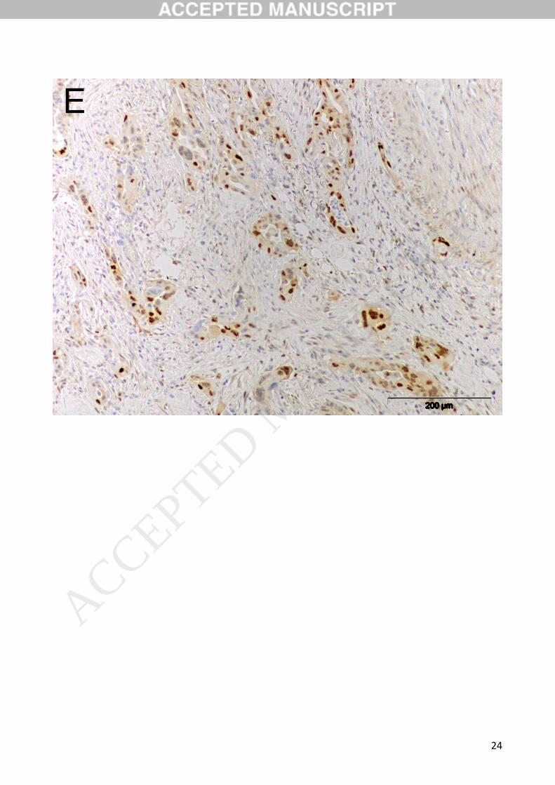
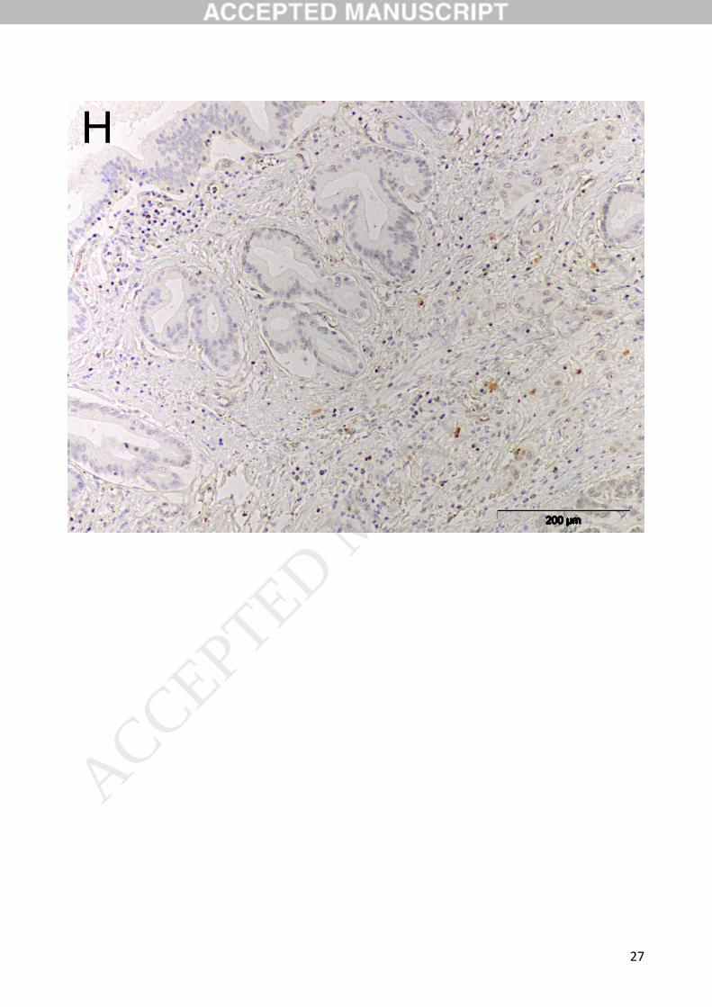
nuclear CDK4 expression in PDAC cells. High (G) and low (H) nuclear MGMT expression in
PDAC cells. Any (I) and no (J) p16 expression in tumor-associated fibroblasts. (Low magnification
×10)
ACCEPTED MANUSCRIP
T

20
ACCEPTED MANUSCRIP
T

21
Fig 1A
Fig B
ACCEPTED MANUSCRIP
T

22
Fig 2
ACCEPTED MANUSCRIP
T

23
ACCEPTED MANUSCRIP
T
24
ACCEPTED MANUSCRIP
T

25
ACCEPTED MANUSCRIP
T

26
ACCEPTED MANUSCRIP
T
27
ACCEPTED MANUSCRIP
T

28
ACCEPTED MANUSCRIP
T

29
ACCEPTED MANUSCRIP
T

30
Table 1. Patient characteristics
Characteristic N (%)
Gender
Male
Female
Not available
Age at diagnosis
<50 years
50-59 years
60-69 years
>69 years
Not available
Tumour (T)
1
52 (51%)
49 (48%)
1 (1%)
6 (5.9%)
29 (28.4%)
31 (30.4%)
29 (28.4%)
7 (6.9%)
6 (5.9%)
2 28 (27.5%)
3 57 (55.9%)
4 8 (7.8%)
Not available 3 (2.9%)
Nodal metastasis (N)
No 39 (38.2%)
Yes 61 (59.8%)
Not available 2 (2.0%)
Distant metastasis at the time
of diagnosis (M)
ACCEPTED MANUSCRIP
T

31
No 90 (88.2%)
Yes 10 (9.8%)
Not available 2 (2.0%)
Distant metastasis during
follow-up
No 83 (81.4%)
Yes 19 (18.6%)
Not available 0 (0.0%)
Grade
I 25 (24.5%)
II 42 (41.2%)
III 27 (26.5%)
Not available 8 (7.8%)
Local relapse
No 64 (62.7%)
Yes 35 (34.3%)
Not available 3 (2.9%)
Type of surgery
Palliative 5 (4.9%)
Whipple 83 (81.4%)
Other, with curative intention 14 (13.7%)
Table 2. Immunohistochemical methods
ACCEPTED MANUSCRIP
T

32
Primary antibody Manufacturer of the
primary antibody
Dilution Immunostaining
method
p16ink4
(Ref9511)
Ventana Medical
System, Inc., Tucson,
USA
1:4 Dako REAL™
EnVision™ Detection
System (Dako
Denmark A/S,
Glostrup, Denmark)
CDK4
(NBP1-31308)
Novus Biologicals,
Littleton, USA
1:100 Dako REAL™
EnVision™ Detection
System (Dako
Denmark A/S,
Glostrup, Denmark)
Rb1
(HPA050082)
Atlas Antibodies Ab,
Bromma, Sweden
1:500 Dako REAL™
EnVision™ Detection
System (Dako
Denmark A/S,
Glostrup, Denmark)
FEN1
(ab109132)
MGMT1
(ab108630)
Abcam, Cambridge,
UK
Abcam, Cambridge,
UK
1:1000
1:750
Novolink Polymer Detection Systems (Leica Biosystems, Newcastle, UK)
Dako REAL™ EnVision™ Detection
System (Dako Denmark A/S, Glostrup, Denmark)
CDK4, Cyclin-dependent kinase 4
Rb1, Retinoblastoma-associated protein-1
FEN1, Flap endonuclease-1
MGMT, O-6-methylguanine-DNA methyltransferase
Table 3. Percentages of evaluable cases showing expression of p16ink4, CDK4, Rb1, FEN1 and
MGMT.
Adenocarcinoma cells Benign exocrine pancreatic
tissue
ACCEPTED MANUSCRIP
T
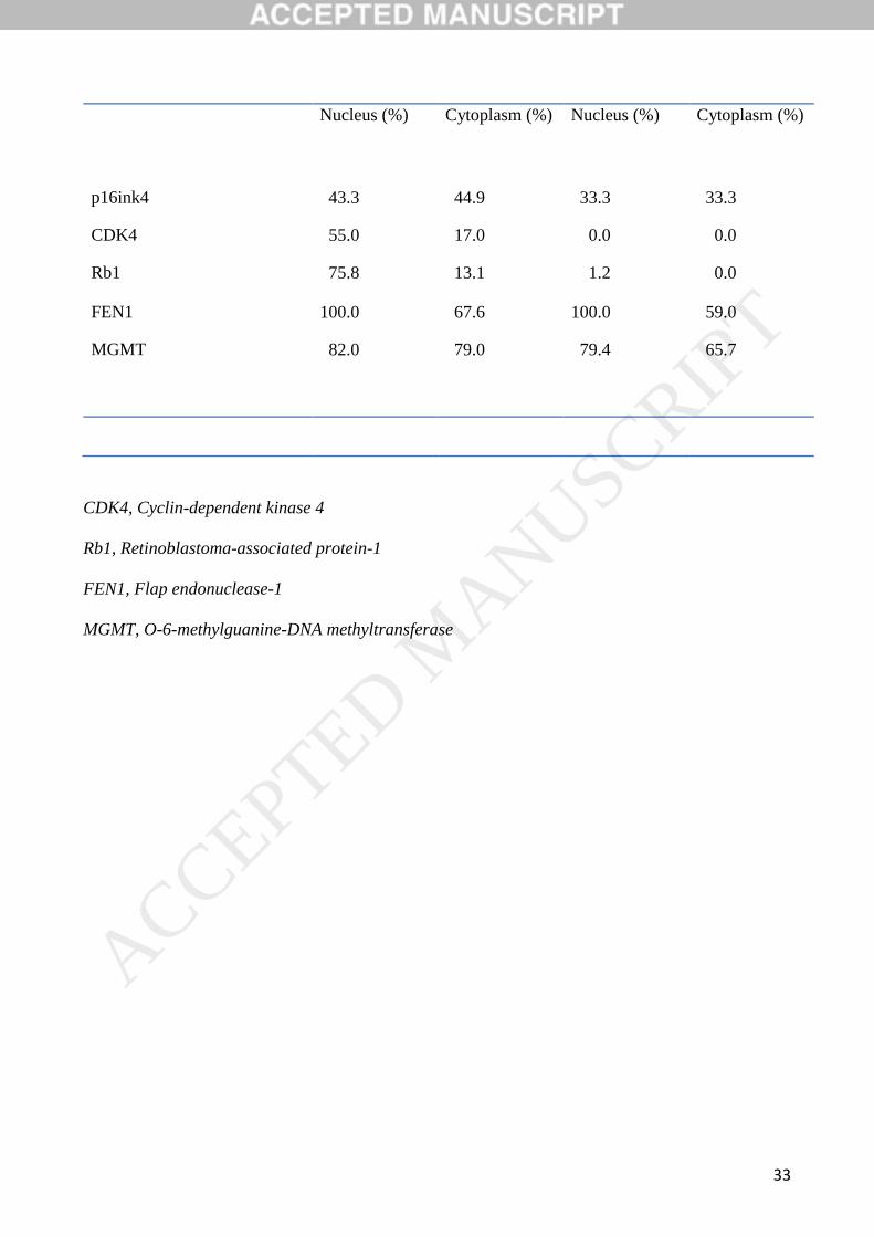
33
Nucleus (%) Cytoplasm (%) Nucleus (%) Cytoplasm (%)
p16ink4 43.3 44.9 33.3 33.3
CDK4 55.0 17.0 0.0 0.0
Rb1 75.8 13.1 1.2 0.0
FEN1 100.0 67.6 100.0 59.0
MGMT 82.0 79.0 79.4 65.7
CDK4, Cyclin-dependent kinase 4
Rb1, Retinoblastoma-associated protein-1
FEN1, Flap endonuclease-1
MGMT, O-6-methylguanine-DNA methyltransferase
ACCEPTED MANUSCRIP
T

34
Table 4. Protein expression showing independent prognostic value in multivariate analysis.
Prot
ein
Immunostaining
location
(nuclear/cytoplas
mic/other)
Endp
oint
Subgr
oup
Cox
multiva
riate
analysi
s: Risk
ratio
95
%
CI
Variabl
es
include
d in
Cox
multiva
riate
analysi
s
Kapla
n
Meier
univar
iate
analys
is: p-
value
Media
n
surviv
al in
low
expres
sion
group
(mont
hs)
Media
n
surviv
al in
high
expres
sion
group
(mont
hs)
CD
K4
Nuclear, cancer
cells
DFS T3-4 2.148 1.08
1-
4.27
2
CDK4,
N 0.029 18 7
CD
K4
Islets of
Langerhans,
benign cells
RFS - 2.874 1.26
1-
6.55
0
CDK4,
T, N 0.012 64 17
MG
MT
Nuclear, cancer
cells
OS N1 2.148 1.06
6-
4.32
9
MGMT
, T
0.032 19 12
MG
MT
Nuclear, cancer
cells
DFS N1 2.114 1.01
9-
4.38
5
MGMT
, T
0.044 16 9
MG
MT
Nuclear, cancer
cells
RFS N1 9.028 2.19
2-
37.1
79
MGMT
, T
0.002 28 14
MG
MT
Nuclear, cancer
cells
OS T3-4 1.878 0.96
2-
3.66
7
MGMT
, N
0.065 19 12
FEN
1
TLS DFS - 2.619 1.13
2-
6.05
9
FEN1,
T, N 0.025 18 6
FEN
1
TLS RFS - 3.758 1.14
8-
12.2
99
FEN1,
T, N 0.029 32 17
ACCEPTED MANUSCRIP
T

35
CDK4, Cyclin-dependent kinase 4
MGMT, O-6-methylguanine-DNA methyltransferase
FEN1, Flap endonuclease-1
TLS, Tertiary lymphoid structure
OS, Overall survival
DFS, Disease-free survival
RFS, Relapse-free survival
ACCEPTED MANUSCRIP
T