reducing tumour hypoxia via oral administration …uir.ulster.ac.uk/37069/1/offprint.pdf ·...
TRANSCRIPT
RESEARCH ARTICLE
Reducing Tumour Hypoxia via Oral
Administration of Oxygen Nanobubbles
Joshua Owen1, Conor McEwan2, Heather Nesbitt2, Phurit Bovornchutichai1,
Raymond Averre3, Mark Borden4, Anthony P. McHale2, John F. Callan2, Eleanor Stride1*
1 Oxford Institute of Biomedical Engineering, University of Oxford, Oxford, United Kingdom, 2 Biomedical
Sciences Research Institute, Ulster University, Coleraine, Northern Ireland, United Kingdom, 3 Avrox
Technologies Ltd. Copgrove, Harrogate, North Yorkshire, United Kingdom, 4 Department of Mechanical
Engineering, University of Colorado, 1111 Engineering Drive, Boulder, CO, United States of America
Abstract
Hypoxia has been shown to be a key factor inhibiting the successful treatment of solid
tumours. Existing strategies for reducing hypoxia, however, have shown limited efficacy
and/or adverse side effects. The aim of this study was to investigate the potential for reduc-
ing tumour hypoxia using an orally delivered suspension of surfactant-stabilised oxygen
nanobubbles. Experiments were carried out in a mouse xenograft tumour model for human
pancreatic cancer (BxPc-3 cells in male SCID mice). A single dose of 100 μL of oxygen satu-
rated water, oxygen nanobubbles or argon nanobubbles was administered via gavage. Ani-
mals were sacrificed 30 minutes post-treatment (3 per group) and expression of hypoxia-
inducible-factor-1α (HIF1α) protein measured by real time quantitative polymerase chain
reaction and Western blot analysis of the excised tumour tissue. Neither the oxygen satu-
rated water nor argon nanobubbles produced a statistically significant change in HIF1αexpression at the transcriptional level. In contrast, a reduction of 75% and 25% in the tran-
scriptional and translational expression of HIF1α respectively (p<0.001) was found for the
animals receiving the oxygen nanobubbles. This magnitude of reduction has been shown in
previous studies to be commensurate with an improvement in outcome with both radiation
and drug-based treatments. In addition, there was a significant reduction in the expression
of vascular endothelial growth factor (VEGF) in this group and corresponding increase in the
expression of arrest-defective protein 1 homolog A (ARD1A).
Introduction
Hypoxia, i.e. a reduction in dissolved oxygen concentration below physiologically normal lev-
els, has been identified as playing a critical role in the progression of several diseases, including
many types of cancer [1]. In solid tumours, it arises as a consequence of the rapid proliferation
and atypical vasculature of cancerous tissue. This results in the development of areas in which
oxygen demand outstrips supply [2]. Once a hypoxic environment develops within a tumour,
cell populations become resistant to many conventional chemotherapeutic agents through a
variety of adaptive survival mechanisms [3]. Similarly, hypoxia can have a significant influence
PLOS ONE | DOI:10.1371/journal.pone.0168088 December 30, 2016 1 / 12
a1111111111
a1111111111
a1111111111
a1111111111
a1111111111
OPENACCESS
Citation: Owen J, McEwan C, Nesbitt H,
Bovornchutichai P, Averre R, Borden M, et al.
(2016) Reducing Tumour Hypoxia via Oral
Administration of Oxygen Nanobubbles. PLoS ONE
11(12): e0168088. doi:10.1371/journal.
pone.0168088
Editor: Sonia Rocha, University of Dundee, UNITED
KINGDOM
Received: July 24, 2016
Accepted: November 25, 2016
Published: December 30, 2016
Copyright: © 2016 Owen et al. This is an open
access article distributed under the terms of the
Creative Commons Attribution License, which
permits unrestricted use, distribution, and
reproduction in any medium, provided the original
author and source are credited.
Data Availability Statement: All raw data files are
available from the University of Oxford ORA data
repository (doi.10.5287/bodleian:2RAeOzDeb).
Funding: The authors thank the Engineering and
Physical Sciences Research Council for supporting
this work through EP/I021795/1 and the Impact
Acceleration Scheme (https://www.epsrc.ac.uk).
John Callan thanks Norbrook Laboratories Ltd. for
an endowed chair. The funders had no role in study
design, data collection and analysis, decision to
publish, or preparation of the manuscript.
upon the effectiveness of radiotherapy, since radiation induced damage requires the formation
of reactive oxygen species, which is inhibited in the absence of oxygen [4]. Consequently hyp-
oxia is now recognised as a key determinant of successful cancer treatment [4].
Strategies for treating hypoxia have included the development of hypoxia-selective drugs
[5] and radiosensitisers such as nimorazole [6] as well as methods for directly increasing blood
oxygenation, e.g. hyperbaric oxygen therapy [7], pure oxygen or carbogen breathing, ozone
therapy [8], hydrogen peroxide injections [9] and administration of suspensions of oxygen car-
rier liquids [10]. To date, however, these approaches have delivered limited success owing to
lack of proven efficacy and/or unwanted side effects [11]. Gas microbubbles, stabilised by a
biocompatible shell have been in use as ultrasound contrast agents for several decades and
have also been investigated as an alternative means of oxygen delivery [12]. Recent studies
have demonstrated that peritoneal or intratumoral injection of oxygen-loaded microbubbles
can be used to increase systemic oxygen levels [13] and substantially increase the efficacy of
cancer treatment in animal models [14]. Encouragingly, no adverse side effects were observed
in these studies; however, the risks associated with injecting high concentrations of microparti-
cles [13] makes them unsuitable for intravenous administration and this limits their potential
for clinical translation.
A further route that has been explored involves the delivery of oxygen in the form of a stabi-
lised foam via the stomach. Oral administration of pharmaceuticals and other therapeutic
materials has considerable advantages in terms of patient acceptability, reducing the risk of
infection, cost and the quantity of material that can be delivered [15]. Frequently, however,
oral administration is associated with inefficient delivery and/or poor bioavailability (ibid.).
There are a number of reports in the literature of the use of foams or “oxygen cocktails” for the
treatment of a variety of conditions [16–18], but to the best of the authors’ knowledge there
has been very little research in this area in recent years. There have been several more recent
studies of oxygen saturated waters for reducing recovery times for athletes, but the clinical evi-
dence is controversial [19].
The aim of this study was to assess the potential of a further alternative method for reducing
tumour hypoxia using an orally delivered suspension of sub-micrometre sized oxygen nano-
bubbles stabilised by a surfactant. By encapsulating the oxygen, higher concentrations and
improved stability can be achieved than via direct dissolution in water. Oral delivery over-
comes the risks associated with direct injection of high concentrations of microbubbles (e.g.
embolism, lipid toxicity etc. [13]) and a liquid formulation should offer improved flow charac-
teristics leading to more efficient delivery compared with foam, as well as greater patient
acceptability.
Materials and Methods
All animals employed in this study were treated humanely and in accordance with licensed
procedures under the UK Animals (Scientific Procedures) Act 1986 under a UK Home Office
Licence and with ethical approval from Ulster University. Anaesthesia was performed via
intraperitoneal injection of Hypnorm/Hypnovel. Mice were euthanised by dislocation of the
neck. None of the animals utilized for this work became ill or died prior to the experimental
endpoint.
Lecithin and citric acid were purchased from Special Ingredients (Chesterfield, Derbyshire,
UK). Glycyrrhizin and glycerol were purchased from Sigma Aldrich (Gillingham, Dorset,
UK). Oxygen and argon gas cylinders were purchased from BOC gases (Guilford, Surrey, UK).
The nanobubble suspensions were prepared from a mixture of glycyrrhizin (3 mg/ml), lecithin
(3 mg/ml), citric acid (5 mg/ml) and glycerol (0.0125 ml/ml) in untreated tap water. A 250 ml
Oxygen Nanobubbles to Reduce Hypoxia
PLOS ONE | DOI:10.1371/journal.pone.0168088 December 30, 2016 2 / 12
Raymond Averre is a director of Avrox
Technologies Ltd. but neither he nor any other
associate of Avrox Technologies Ltd. had any
additional role in the study design, data collection
and analysis, decision to publish, or preparation of
the manuscript. The specific role of this author is
articulated in the ‘author contributions’ section.
Competing Interests: Raymond Averre is a director
of Avrox Technologies Ltd. Avrox Technologies
Ltd. did not fund this work but did partially fund an
earlier pilot study on oxygen nanobubbles. Joshua
Owen, Raymond Averre and Eleanor Stride are co-
inventors on a patent (Stride, E., Averre, R., Owen,
J. (2015) Nanoencapsulated Oxygen. UK Patent
Application No. 1512728.5), which has been filed in
relation to the formulation described in the paper.
Avrox Technologies Ltd. has licensed this patent.
John Callan thanks Norbrook Laboratories Ltd. for
an endowed chair. There are no further patents,
products in development or marketed products to
declare. This does not alter the authors’ adherence
to all the PLOS ONE policies on sharing data and
materials.

volume of the mixture was stirred for 2 hours (50˚C) to disperse the constituents. An aliquot
of the liquid (1 ml) was transferred to a glass vial, degassed via vacuum pump and the head-
space was refilled with either oxygen or argon. The vial was then sealed and mechanically agi-
tated for 30 s. The degassing, gas replacement and shaking process was repeated three times
for each sample. Oxygenated water was prepared by following the same procedure with only
water.
The size distribution of the particles in the suspension was measured using an AccuSizer
780 AD single particle optical sizing system (NICOMP Particle Sizing Systems, Santa Barbara,
CA) and via dynamic light scattering (Zetasizer Nano, Malvern Instruments, Malvern,
Worcestershire), hereafter referred to as SPOS and DLS respectively to detect both micro and
nanoscale particles. For SPOS, a 10 μl sample of each suspension was diluted in 50 mL of fil-
tered deionised water in a flask under mild mixing for ~30s before injection into the instru-
ment. For DLS, 20 μl of each suspension was diluted with 980 μl of filtered deionised water in
a disposable cuvette which was placed in the instrument. Three measurements were made on
three samples from each suspension with both methods.
Samples of the oxygen nanobubble suspension were also examined by transmission electron
microscopy using the method developed by Owen et al. [20] for studying microbubbles. 5 μl of
each suspension was applied to carbon film coated 300 mesh copper grids (Electron micros-
copy sciences), which had been ionized in a plasma cleaner (Harris Plasma) for 30 seconds.
Each grid was inverted for approximately 1 minute in order to allow buoyant particles to accu-
mulate on the grid surface. Grids were negatively stained by incubation on a 5ul drop of 2%
w/v uranyl acetate for 30 seconds. They were then dried using filter paper and left for approxi-
mately 1 minute. The samples were imaged at 80kV with an FEI Tecnai T12 electron micro-
scope and low-dose images were acquired at ~0.8μm underfocus with 15e-/Å2 on an FEI Eagle
CCD camera.
A CDI blood parameter monitoring system (Terumo UK Ltd. Egham, Surrey, UK) was
used to measure the oxygen content of 3 ml samples of the above suspensions over a period of
20 min. This was carried out in a closed flow loop consisting of a peristaltic pump (Gilson,
Minipuls3, Cole Parmer, Hanwell UK) operating at a flow rate of 12ml/min and a length of
Tygon tubing (with inner and outer diameters of 4 and 5.4 mm respectively) in to which the
sensor was inserted. The measurements were repeated on further samples of the oxygen nano-
bubble suspension at 37˚C to determine the impact of temperature.
The ability of the nanobubble suspensions to influence tumour hypoxia was examined in a
mouse xenograft tumour model for human pancreatic cancer. BxPc-3 cells were maintained in
RPMI-1640 medium supplemented with 10% fetal calf serum. Cells were cultured at 37˚C
under 5% CO2 in air. BxPc-3 cells (2 × 10⁶) were re-suspended in 100 μL of Matrigel (BD Bio-
sciences, Erembodegem, Belgium) and implanted subcutaneously into the rear dorsum of
male SCID mice. Cells were purchased from ATTC, LGC Standards, Teddington, UK. Inde-
pendent identification was not carried out. Tumour formation occurred approximately 2
weeks after implantation and tumour measurements were taken every other day using calli-
pers. Data from our laboratory indicate that the tumour oxygen levels for this particular model
remain relatively constant between volumes of 170 mm3 and 260 mm3, reducing the likelihood
of significant inter-subject variability (S1 Fig).
Once the tumours had reached an average volume of 256 mm3 calculated from the geometric
mean diameter using the equation tumour volume = 4πR3/3, animals were randomly distrib-
uted into three groups as follows: (i) oxygenated water (4 animals) (ii) argon nanobubbles (4
animals) and (iii) oxygen nanobubbles (8 animals). Following induction of anaesthesia via intra-
peritoneal injection of Hypnorm/Hypnovel (150μl (i.p.) of a mixture of 2:1:1; PBS: Hypnorm
(0.315 mg/ml fentanyl citrate and fluanisone 10mg/ml, VetaPharma Ltd, U.K.): Hypnovel
Oxygen Nanobubbles to Reduce Hypoxia
PLOS ONE | DOI:10.1371/journal.pone.0168088 December 30, 2016 3 / 12
(10mg/ml midazolam, Roche, UK) the oxygen partial pressure (pO2) of tumours was recorded
using an Oxylite oxygen electrode sensor (Oxford Optronics, Oxford, UK). A fibre optic probe
was inserted into a 21-gauge needle before insertion into the centre of the tumour tissue. The
needle was withdrawn and the probe readings allowed to stabilise for 5 minutes. The oxygen
level in the tumour was recorded every second for 20 min. 100 μL aliquots of oxygenated water,
the oxygen nanobubbles or the argon nanobubbles were then administered orally via gavage
and tumour oxygenation measured every second for a further 30 minutes. This period was cho-
sen to avoid the need for re-administering anaesthesia and on the basis of the results of an initial
pilot study (S2 Fig). A total of 8 repeats were conducted per group. Two separate measurements
were performed on the animals receiving oxygenated water and argon nanobubbles.
In a separate experiment, a further 9 animals (n = 3/group) were treated with the relevant
preparation and then sacrificed after 30 min later without measurement of tumour oxygen.
The tumours were harvested by surgical excision and processed for further studies. We
selected HIF1α as the primary physiological probe for oxygen delivery because the presence of
oxygen results in its rapid degradation [21, 22]. It thus provides a direct and immediate indica-
tor of increased tumour oxygen levels. In addition, we examined expression of vascular endo-
thelial growth factor (VEGF) and arrest-defective protein 1 homolog A (ARD1A). To examine
the expression of HIF1α and VEGF at a transcriptional level, RNA was extracted from tumours
using Trizol (Invitrogen, Paisley, UK) and reverse transcribed using a first strand cDNA syn-
thesis kit according to the manufacturer’s instructions (Roche, Welwyn Garden City, UK).
Real Time Quantitative PCR (RT-Q-PCR) was undertaken using SYBR green (Fermentas,
Cambridge, UK) and gene-specific primers in a Lightcycler 480 (Roche, Welwyn Garden City,
UK). Using β-actin to as a reference, expression of HIF1α and VEGF was calculated using the
comparative CT (ΔΔCT) method.
For Western blotting analysis of HIF1α and ARD1A protein expression at a translational
level, total protein was extracted using RIPA buffer (Pierce, Rockford, UK). Primary murine
antibodies employed in these studies were anti-HIF1α (Millipore, MAB5382, 1:500), ARD1A
(GeneTex, GTX125971, 1:1000), anti-β-actin (Sigma, A2228, 1:1000) and GAPDH (Cell Sig-
naling Technology Europe BV, 2118, 1:1000). Following resolution using SDS-PAGE electro-
phoresis and transfer to nitrocellulose membranes, blocking of non-specific binding was
carried out in 5% (w/v) bovine serum albumin diluted in 1x tris buffered saline containing
0.05% (v/v) Tween 20. Membranes were then incubated in the appropriate secondary anti-
body, goat anti-mouse IgG-HRP (1:10000 of the stock solution). Secondary antibodies were
purchased from Santa Cruz Biotechnology, Heidelberg, Germany. Densitometry was carried
out to quantify HIF1α and ARD1A protein expression using β-actin as a housekeeping refer-
ence. For statistical analyses, significance was assessed between pairs of data sets using a two
tailed Student’s t-test and confirmed by ANOVA.
Results and Discussion
Fig 1A and Table 1 show the particle size distribution as measured by SPOS in each suspension
immediately following preparation. As expected, the tap water contained some particulate
matter but the nanobubble suspensions contained much higher concentrations of particles.
There was no measurable difference in the particle content of water before and after sparging
with oxygen. The AccuSizer system is limited in terms of its ability to accurately size particles
smaller than 500 nm and the fact that the maximum particle counts were at this lower limit for
the oxygen and argon nanobubble suspensions indicated that they contained predominantly
smaller particles. This was supported by the DLS measurements (Fig 1B) which indicated a
population with a peak size of ~340nm in both nanobubble suspensions. The DLS results also
Oxygen Nanobubbles to Reduce Hypoxia
PLOS ONE | DOI:10.1371/journal.pone.0168088 December 30, 2016 4 / 12

indicated a sub-population of particles with mean sizes between 50 and 60 nm that were not
present in water. This was supported by the TEM images (Fig 1C and 1D). Unfortunately it
was not possible to determine the concentration of these nanoscale particles using the methods
available. Population data were not calculated for the DLS measurements on account of this
Fig 1. Mean particle size distributions measured for oxygen nanobubbles, argon nanobubbles and oxygenated water by (a) SPOS and (b) DLS.
Error bars indicate the standard deviation in each measurement (n = 9; 3 readings containing 10 runs from 3 samples of each liquid). Panels (c) and (d) show
transmission electron micrographs of a sample from the oxygen nanobubble suspension indicating the presence of nanoscale particles (scale bar in (c) is
2 μm, in (d) 200 nm).
doi:10.1371/journal.pone.0168088.g001
Table 1. Population statistics for oxygen nanobubbles, argon nanobubbles and oxygenated water as measured by SPOS.
Suspension Particle size (μm) Concentration (ml)
Mean Standard deviation Mode Median
Oxygen nanobubbles 0.86 0.70 0.63 0.66 3 x 10⁷
Argon nanobubbles 0.85 0.69 0.63 0.66 3 x 10⁷
Water 0.76 0.61 0.55 0.61 3 x 10⁶
doi:10.1371/journal.pone.0168088.t001
Oxygen Nanobubbles to Reduce Hypoxia
PLOS ONE | DOI:10.1371/journal.pone.0168088 December 30, 2016 5 / 12
biomodal distribution. The standard deviations in the individual measurements were compa-
rable in size to those obtained for microbubble suspensions in previous studies [23]. They
were deemed acceptable since the primary aim of these measurements was to identify any sig-
nificant differences in particle size and concentration between the nanobubble suspensions
that might influence the subsequent experiments.
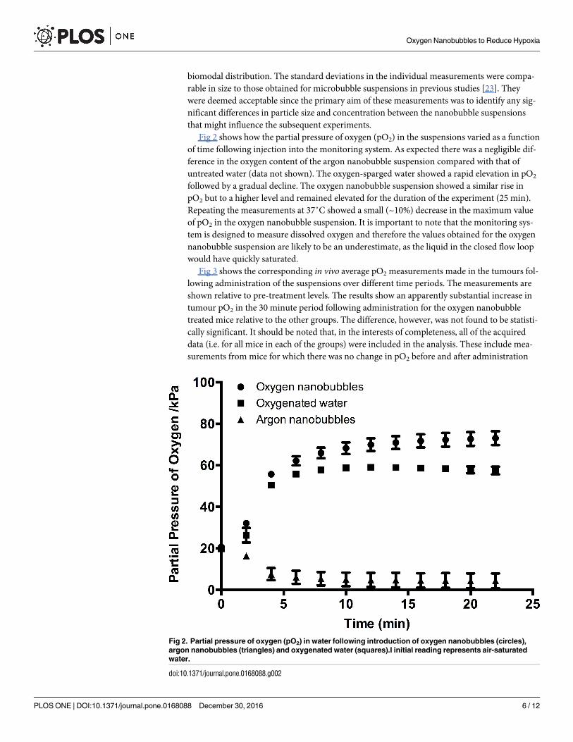
Fig 2 shows how the partial pressure of oxygen (pO2) in the suspensions varied as a function
of time following injection into the monitoring system. As expected there was a negligible dif-
ference in the oxygen content of the argon nanobubble suspension compared with that of
untreated water (data not shown). The oxygen-sparged water showed a rapid elevation in pO2
followed by a gradual decline. The oxygen nanobubble suspension showed a similar rise in
pO2 but to a higher level and remained elevated for the duration of the experiment (25 min).
Repeating the measurements at 37˚C showed a small (~10%) decrease in the maximum value
of pO2 in the oxygen nanobubble suspension. It is important to note that the monitoring sys-
tem is designed to measure dissolved oxygen and therefore the values obtained for the oxygen
nanobubble suspension are likely to be an underestimate, as the liquid in the closed flow loop
would have quickly saturated.
Fig 3 shows the corresponding in vivo average pO2 measurements made in the tumours fol-
lowing administration of the suspensions over different time periods. The measurements are
shown relative to pre-treatment levels. The results show an apparently substantial increase in
tumour pO2 in the 30 minute period following administration for the oxygen nanobubble
treated mice relative to the other groups. The difference, however, was not found to be statisti-
cally significant. It should be noted that, in the interests of completeness, all of the acquired
data (i.e. for all mice in each of the groups) were included in the analysis. These include mea-
surements from mice for which there was no change in pO2 before and after administration
Fig 2. Partial pressure of oxygen (pO2) in water following introduction of oxygen nanobubbles (circles),
argon nanobubbles (triangles) and oxygenated water (squares).I initial reading represents air-saturated
water.
doi:10.1371/journal.pone.0168088.g002
Oxygen Nanobubbles to Reduce Hypoxia
PLOS ONE | DOI:10.1371/journal.pone.0168088 December 30, 2016 6 / 12
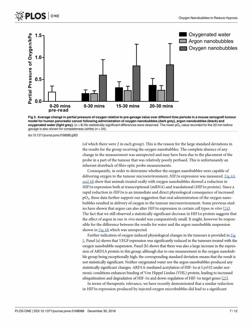
(of which there were 2 in each group). This is the reason for the large standard deviations in
the results for the group receiving the oxygen nanobubbles. The complete absence of any
change in the measurement was unexpected and may have been due to the placement of the
probe in a part of the tumour that was relatively poorly perfused. This is unfortunately an
inherent drawback of fibre optic probe measurements.
Consequently, in order to determine whether the oxygen nanobubbles were capable of
delivering oxygen to the tumour microenvironment, HIF1α expression was measured. Fig 4A
and 4B show that animals treated orally with oxygen nanobubbles showed a reduction in
HIF1α expression both at transcriptional (mRNA) and translational (HIF1α protein). Since a
rapid reduction in HIF1α is an immediate and direct physiological consequence of increased
pO2, these data further support our suggestion that oral administration of the oxygen nano-
bubbles resulted in delivery of oxygen to the tumour microenvironment. Some previous stud-
ies have shown that argon can also alter HIF1α expression in certain cell types in vitro [24].
The fact that we still observed a statistically significant decrease in HIF1α protein suggests that
the effect of argon in our in vivo model was comparatively small. It might, however be respon-
sible for the difference between the results for water and the argon nanobubble suspension
shown in Fig 4B which was unexpected.
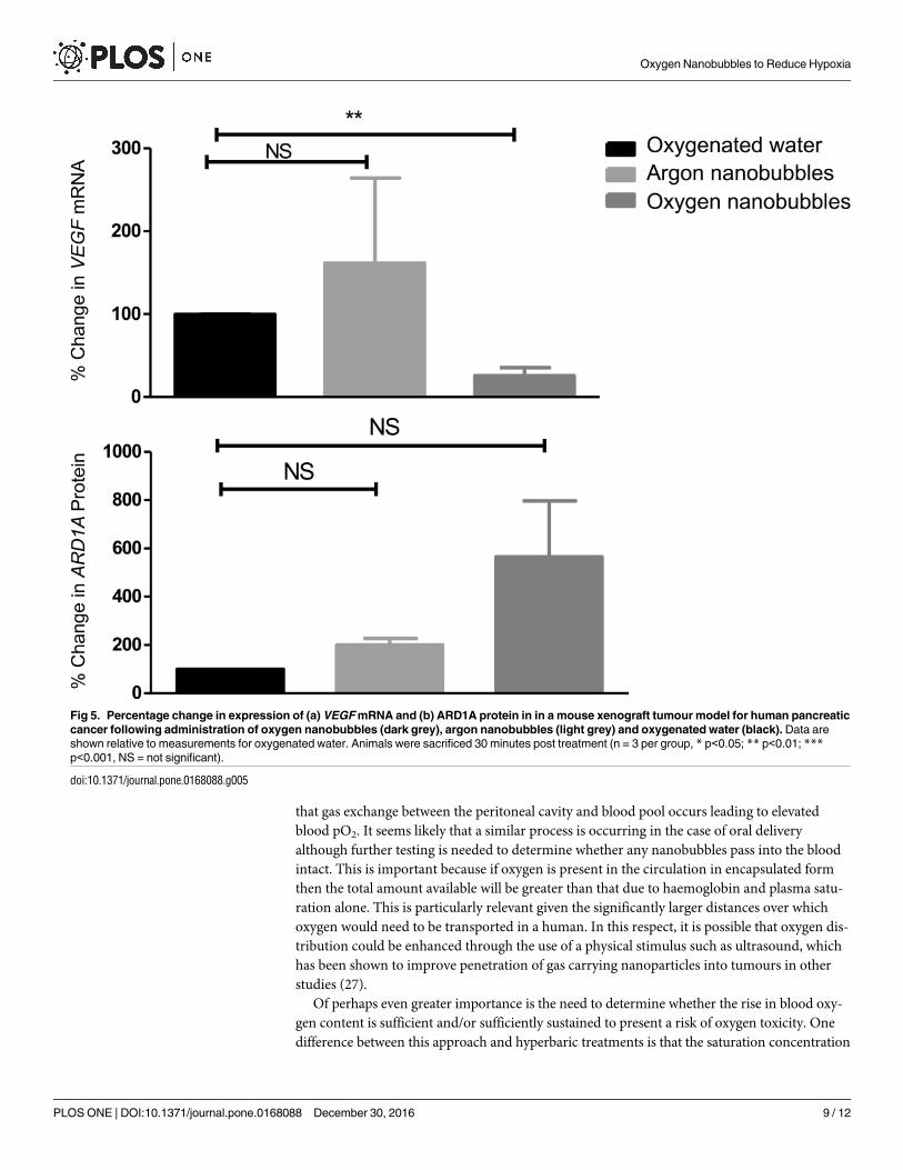
Further indication of oxygen-induced physiological changes in the tumours is provided in Fig
5. Panel (a) shows that VEGF expression was significantly reduced in the tumours treated with the
oxygen nanobubble suspension. Panel (b) shows that there was also a large increase in the expres-
sion of ARD1A protein in this group; although due to one measurement in the oxygen nanobub-
ble group being exceptionally high, the corresponding standard deviation means that the result is
not statistically significant. Neither oxygenated water nor the argon nanobubbles produced any
statistically significant changes. ARD1A-mediated acetylation of HIF-1α at Lys532 under nor-
moxic conditions enhances binding of Von Hippel Lindau (VHL) protein, leading to increased
ubiquitination and degradation of HIF-1α and down-regulation of HIF-1α target genes [25].
In terms of therapeutic relevance, we have recently demonstrated that a similar reduction
in HIF1α expression produced by injected oxygen microbubbles did lead to a significant
Fig 3. Average change in partial pressure of oxygen relative to pre-gavage value over different time periods in a mouse xenograft tumour
model for human pancreatic cancer following administration of oxygen nanobubbles (dark grey), argon nanobubbles (black) and
oxygenated water (light grey). (n = 8) No statistically significant differences were observed. The mean pO2 value recorded for the 20 min before
gavage is also shown for completeness (white) (n = 24).
doi:10.1371/journal.pone.0168088.g003
Oxygen Nanobubbles to Reduce Hypoxia
PLOS ONE | DOI:10.1371/journal.pone.0168088 December 30, 2016 7 / 12
improvement in treatment efficacy with sonodynamic therapy in the same tumour model [14].
Similarly, previous studies have indicated that the response to radiation therapy is highly
dependent upon tumour oxygenation with an increase from 2.5 mmHg (0.33 kPa) to 6 mmHg
(0.8 kPa) significantly affecting loco-regional tumour control in advanced squamous cell head
and neck carcinoma [26].
As above, the ability to reduce hypoxia via an orally delivered agent could have considerable
advantages in terms of cost, convenience and patient acceptability/compliance. Clearly, how-
ever, there are several important questions to answer: First, the results from previous studies
involving peritoneal infusions of microbubbles [13] or oxygen-carrying liquid droplets suggest
Fig 4. Expression of HIF1α at (a) a transcriptional level and (b) a translational level in a mouse xenograft tumour model for human pancreatic
cancer following administration of oxygen nanobubbles (dark grey), argon nanobubbles (light grey) and oxygenated water (black). Animals
were sacrificed 30 minutes post treatment (n = 3 per group, * p<0.05; ** p<0.01; *** p<0.001, NS = not significant).
doi:10.1371/journal.pone.0168088.g004
Oxygen Nanobubbles to Reduce Hypoxia
PLOS ONE | DOI:10.1371/journal.pone.0168088 December 30, 2016 8 / 12
that gas exchange between the peritoneal cavity and blood pool occurs leading to elevated
blood pO2. It seems likely that a similar process is occurring in the case of oral delivery
although further testing is needed to determine whether any nanobubbles pass into the blood
intact. This is important because if oxygen is present in the circulation in encapsulated form
then the total amount available will be greater than that due to haemoglobin and plasma satu-
ration alone. This is particularly relevant given the significantly larger distances over which
oxygen would need to be transported in a human. In this respect, it is possible that oxygen dis-
tribution could be enhanced through the use of a physical stimulus such as ultrasound, which
has been shown to improve penetration of gas carrying nanoparticles into tumours in other
studies (27).
Of perhaps even greater importance is the need to determine whether the rise in blood oxy-
gen content is sufficient and/or sufficiently sustained to present a risk of oxygen toxicity. One
difference between this approach and hyperbaric treatments is that the saturation concentration
Fig 5. Percentage change in expression of (a) VEGF mRNA and (b) ARD1A protein in in a mouse xenograft tumour model for human pancreatic
cancer following administration of oxygen nanobubbles (dark grey), argon nanobubbles (light grey) and oxygenated water (black). Data are
shown relative to measurements for oxygenated water. Animals were sacrificed 30 minutes post treatment (n = 3 per group, * p<0.05; ** p<0.01; ***p<0.001, NS = not significant).
doi:10.1371/journal.pone.0168088.g005
Oxygen Nanobubbles to Reduce Hypoxia
PLOS ONE | DOI:10.1371/journal.pone.0168088 December 30, 2016 9 / 12
of oxygen in tissue is not being modified because neither temperature nor pressure is altered.
Rather, the quantity of oxygen available is increased and there should therefore be selective
delivery to regions where the oxygen concentration is lower. In our treatments, we did not
notice any overt adverse effects on the animals following gavage and the absence of adverse
effects reported in previous studies of peritoneal administration of microbubbles is encouraging
[13], but this undoubtedly requires further investigation.
Conclusions
In summary, there is a need for improved methods to reduce tumour hypoxia in order to
increase the efficacy of current cancer therapies. Oral delivery of oxygen loaded nanobubbles
could potentially provide a convenient delivery vehicle to enable the transient oxygenation of
hypoxic tumours that would be compatible with existing cancer treatment regimes. In this
study, direct measurements of tumour oxygenation using a fibre optic probe, were character-
ised by very large standard deviations and did not show a statistically significant difference
between treatment groups. Examination of HIF1α and VEGF expression, however, indicated
that oxygen nanobubbles could produce a statistically significant reduction to a level that has
been associated with improved treatment outcomes in previous studies using the same pancre-
atic tumour model. Further investigation of the mechanisms by which gas transfer occurs
within the circulation and tissue is required to determine whether or not the effects observed
would be replicated in humans and whether there are any adverse side effects.
Supporting Information
S1 Fig. (a) Plot of tumour oxygenation as a function of tumour growth for the ectopic BxPC3
tumour model used in this study. (b) Plot of tumour volume against time for the tumours mea-
sured in (a). Tumour oxygen (pO2mmHg) was measured via an OxyLite oxygen electrode and
readings converted to % oxygen (n = 5).
(TIFF)
S2 Fig. (a) Representative example of measured tumour pO2 recorded every min for 30 min
after oral gavage of either (i) oxygen nanobubbles (solid black line) (ii) argon nanobubbles
(dotted black line) and (iii) oxygenated water (solid grey line) (n = 8).
(TIFF)
Acknowledgments
We thank the Engineering and Physical Sciences Research Council for supporting this work
through EP/I021795/1 and the Impact Acceleration Scheme. Eleanor Stride and Joshua Owen
thank Dr. Boon Teo for her assistance with the electron microscopy. John Callan thanks Nor-
brook Laboratories Ltd. for an endowed chair.
The unprocessed data on which this manuscript is based are available through the Univer-
sity of Oxford ORA data repository (doi.10.5287/bodleian:2RAeOzDeb)
Author Contributions
Conceptualization: ES RA APM JC.
Data curation: JO CM HN.
Formal analysis: CM HN ES JO JC.
Funding acquisition: ES.
Oxygen Nanobubbles to Reduce Hypoxia
PLOS ONE | DOI:10.1371/journal.pone.0168088 December 30, 2016 10 / 12
Investigation: CM HN ES JO JC PB.
Methodology: ES APM JC.
Project administration: ES APM JC.
Resources: ES JC APM.
Supervision: ES APM JC.
Validation: HN CM JO.
Visualization: HN PB ES.
Writing – original draft: ES.
Writing – review & editing: MB PB APM JC.
References1. Semenza GL. Oxygen sensing, homeostasis, and disease. The New England journal of medicine.
2011; 365(6):537–47. doi: 10.1056/NEJMra1011165 PMID: 21830968
2. Vaupel P, Kallinowski F, Okunieff P. Blood flow, oxygen and nutrient supply, and metabolic microenvi-
ronment of human tumors: a review. Cancer research. 1989; 49(23):6449–65. PMID: 2684393
3. Teicher BA. Hypoxia and drug resistance. Cancer metastasis reviews. 1994; 13(2):139–68. PMID:
7923547
4. Yoshimura M, Itasaka S, Harada H, Hiraoka M. Microenvironment and radiation therapy. BioMed
research international. 2013; 2013:685308. PubMed Central PMCID: PMC3591225. doi: 10.1155/
2013/685308 PMID: 23509762
5. Brown JM. Exploiting the hypoxic cancer cell: mechanisms and therapeutic strategies. Molecular medi-
cine today. 2000; 6(4):157–62. PMID: 10740254
6. Henk JM, Bishop K, Shepherd SF. Treatment of head and neck cancer with CHART and nimorazole:
phase II study. Radiotherapy and oncology: journal of the European Society for Therapeutic Radiology
and Oncology. 2003; 66(1):65–70.
7. Tibbles PM, Edelsberg JS. Hyperbaric-oxygen therapy. The New England journal of medicine. 1996;
334(25):1642–8. doi: 10.1056/NEJM199606203342506 PMID: 8628361
8. Rockwell S, Dobrucki IT, Kim EY, Marrison ST, Vu VT. Hypoxia and radiation therapy: past history,
ongoing research, and future promise. Current molecular medicine. 2009; 9(4):442–58. PubMed Cen-
tral PMCID: PMC2752413. PMID: 19519402
9. Ogawa Y, Kubota K, Ue H, Tadokoro M, Matsui R, Yamanishi T, et al. Safety and effectiveness of a
new enzyme-targeting radiosensitization treatment (KORTUC II) for intratumoral injection for low-LET
radioresistant tumors. International journal of oncology. 2011; 39(3):553–60. doi: 10.3892/ijo.2011.
1069 PMID: 21667020
10. Matsutani N, Takase B, Nogami Y, Ozeki Y, Kaneda S, Maehara T, et al. Efficacy of peritoneal oxygen-
ation using a novel artificial oxygen carrier (TRM-645) in a rat respiratory insufficiency model. Surg
Today. 2010; 40(5):451–5. doi: 10.1007/s00595-009-4104-8 PMID: 20425549
11. Martin DS, Grocott MP. Oxygen therapy in critical illness: precise control of arterial oxygenation and
permissive hypoxemia. Critical care medicine. 2013; 41(2):423–32. doi: 10.1097/CCM.
0b013e31826a44f6 PMID: 23263574
12. Kheir JN, Scharp LA, Borden MA, Swanson EJ, Loxley A, Reese JH, et al. Oxygen Gas-Filled Micropar-
ticles Provide Intravenous Oxygen Delivery. Sci Transl Med. 2012; 4(140). doi: ARTN 140ra88
13. Feshitan JA, Legband ND, Borden MA, Terry BS. Systemic oxygen delivery by peritoneal perfusion of
oxygen microbubbles. Biomaterials. 2014; 35(9):2600–6. doi: 10.1016/j.biomaterials.2013.12.070
PMID: 24439406
14. McEwan C, Owen J, Stride E, Fowley C, Nesbitt H, Cochrane D, et al. Oxygen carrying microbubbles
for enhanced sonodynamic therapy of hypoxic tumours. Journal of controlled release: official journal of
the Controlled Release Society. 2015; 203:51–6.
15. Sastry SV, Nyshadham JR, Fix JA. Recent technological advances in oral drug delivery—a review.
Pharmaceutical science & technology today. 2000; 3(4):138–45.
Oxygen Nanobubbles to Reduce Hypoxia
PLOS ONE | DOI:10.1371/journal.pone.0168088 December 30, 2016 11 / 12
16. Zanozdra NS, Nazarenko VR. [Effect of oxygen cocktail on the cardiohemodynamics in complex treat-
ment of hypertension]. Vrachebnoe delo. 1978;(3):25–8. PMID: 645030
17. Dubrovskii VI, Gotovtsev PI. [Use of the oxygen cocktail for stimulating recovery processes in athletes].
Voprosy pitaniia. 1982;(1):29–30. PMID: 7072175
18. Stetsenko GS. [Effect of an oxygen cocktail on the course of peptic ulcer]. Vrachebnoe delo. 1975;
(3):63–5. PMID: 1121840
19. Hampson NP N; Piantadosi C. Oxygenated water and athletic performance. Journal of the American
Medical Association. 2003; 290(18).
20. Owen J, Stride E. Technique for the Characterization of Phospholipid Microbubbles Coatings by Trans-
mission Electron Microscopy. Ultrasound in medicine & biology. 2015; 41(12):3253–8.
21. Baek JH, Mahon PC, Oh J, Kelly B, Krishnamachary B, Pearson M, et al. OS-9 interacts with hypoxia-
inducible factor 1alpha and prolyl hydroxylases to promote oxygen-dependent degradation of HIF-
1alpha. Molecular cell. 2005; 17(4):503–12. doi: 10.1016/j.molcel.2005.01.011 PMID: 15721254
22. Jewell UR, Kvietikova I, Scheid A, Bauer C, Wenger RH, Gassmann M. Induction of HIF-1alpha in
response to hypoxia is instantaneous. FASEB journal: official publication of the Federation of American
Societies for Experimental Biology. 2001; 15(7):1312–4.
23. Sennoga CA, Yeh JS, Alter J, Stride E, Nihoyannopoulos P, Seddon JM, et al. Evaluation of methods
for sizing and counting of ultrasound contrast agents. Ultrasound in medicine & biology. 2012; 38
(5):834–45. PubMed Central PMCID: PMC3657178.
24. Yarin YM, Amarjargal N, Fuchs J, Haupt H, Mazurek B, Morozova SV, et al. Argon protects hypoxia-,
cisplatin- and gentamycin-exposed hair cells in the newborn rat’s organ of Corti. Hearing research.
2005; 201(1–2):1–9. doi: 10.1016/j.heares.2004.09.015 PMID: 15721555
25. Lee MN, Lee SN, Kim SH, Kim B, Jung BK, Seo JH, et al. Roles of arrest-defective protein 1(225) and
hypoxia-inducible factor 1alpha in tumor growth and metastasis. Journal of the National Cancer Insti-
tute. 2010; 102(6):426–42. PubMed Central PMCID: PMC2841038. doi: 10.1093/jnci/djq026 PMID:
20194889
26. Nordsmark M, Overgaard M, Overgaard J. Pretreatment oxygenation predicts radiation response in
advanced squamous cell carcinoma of the head and neck. Radiotherapy and oncology: journal of the
European Society for Therapeutic Radiology and Oncology. 1996; 41(1):31–9.
Oxygen Nanobubbles to Reduce Hypoxia
PLOS ONE | DOI:10.1371/journal.pone.0168088 December 30, 2016 12 / 12