circulating tumour cells, circulating tumour dna and
TRANSCRIPT
Review ArticleCirculating Tumour Cells, Circulating Tumour DNA and Circulating Tumour miRNA in Blood Assays in the Different Steps of Colorectal Cancer Management, a Review of the Evidence in 2019
Niki Christou ,1,2,3 Jeremy Meyer,3 Sotirios Popeskou, 3 Valentin David ,2 Christian Toso,3 Nicolas Buchs,3 Emilie Liot,3 Joan Robert,3 Frederic Ris,3 and Muriel Mathonnet1,2
1Endocrine, General and Digestive Surgery Department, CHU de Limoges, Limoges Cedex 87042, France2 Laboratoire EA3842 Contrôle de l’Activation cellulaire, Progression Tumorale et Résistances thérapeutiques «CAPTuR», Faculté de médecine, 2 Rue du Docteur Marcland, 87025 Limoges, France
3Department of Visceral Surgery, University Hospital of Geneva, Rue Gabrielle-Perret-Gentil 4, 1211 Genève 14, Switzerland
Correspondence should be addressed to Niki Christou; [email protected]
Received 23 May 2019; Revised 2 August 2019; Accepted 17 August 2019; Published 16 December 2019
Guest Editor: Bin Duan
Copyright © 2019 Niki Christou et al. �is is an open access article distributed under the Creative Commons Attribution License, which permits unrestricted use, distribution, and reproduction in any medium, provided the original work is properly cited.
Despite many advances in the diagnosis and treatment of colorectal cancer (CRC), its incidence and mortality rates continue to make an impact worldwide and in some countries rates are mounting. Over the past decade, liquid biopsies have been the object of fundamental and clinical research with regard to the di�erent steps of CRC patient care such as screening, diagnosis, prognosis, follow-up, and therapeutic response. �ey are attractive because they are considered to encompass both the cellular and molecular heterogeneity of tumours. �ey are easily accessible and can be applied to large-scale settings despite the cost. However, liquid biopsies face drawbacks in detection regardless of whether we are testing for circulating tumour cells (CTCs), circulating tumour DNA (ctDNA), or miRNA. �is review highlights the di�erent advantages and disadvantages of each type of blood-based biopsy and underlines which speci�c one may be the most useful and informative for each step of CRC patient care.
1. Introduction
Colorectal cancer (CRC) is a frequently diagnosed cancer in developed countries, ranking third in terms of both incidence and mortality [1].
In many countries, a bowel cancer screening program is available especially in patients with speci�c risks of colorectal cancer such as patients over 50 years old or hereditary colorectal cancers [2]. Common screening methods used are: (1) stool testing for blood such as guaiac fecal occult blood test or fecal immunochemical test (FIT) [3]; (2) endoscopy [4] such as rectoscopy or colonoscopy; and (3) computed tomographic (CT) colonography, which is less invasive. For many years, a reliable diagnosis has been based on biopsies from colorectal tissues. However, biopsy results alone cannot display sensitive and speci�c information which can provide a more complete analysis of the tumour thus allowing us to
target treatment in the di�erent phases of CRC. �is is linked to the heterogeneity of tumours not only in the spatial dimen-sion but also in the temporal one [5]. Moreover, the procedure of tissue biopsy can sometimes be invasive with risk of com-plications such as pain, bleeding, infections, or perforations. During screening or diagnosing colonoscopies, the overall adverse event rate has been reported to be around 2.8 per 1000 acts [6]. Even with computed tomographic (CT) colo-nography, a recent study including 431 Japanese centres with 147,439 CT examinations showed 0.014% of colorectal per-forations [7]. In addition, tissue biopsies are also time-con-suming. A lack of speed in histologic response is an ever-increasing phenomenon due in particular to the number of demands and new therapies used, for example, chemother-apy or immunotherapy.
If we look globally at prognosis, follow-up, risk of recur-rence, therapeutic response, and combined clinical modalities,
HindawiBioMed Research InternationalVolume 2019, Article ID 5953036, 11 pageshttps://doi.org/10.1155/2019/5953036
BioMed Research International2
we see that imaging and biopsy results o¢en lack of sensitivity and speci�city while exposing the patient to speci�c risks.
As a result, research has been focused on developing more reliable and more accessible biomarkers. Blood, urine, cere-brospinal £uid, stool, and saliva were explored [8]. Despite, the technical di¤culties and cost generated to develop those tools, progress has been made in this area. �e aim of this paper is to discuss new available tools such as Circulating Tumour Cells (CTCs), Circulating Tumour DNA (ctDNA), and microRNAs (miRNAs) regarding CRC management encompassing screening, diagnosis, search for recurrences, prognosis, and prediction of therapeutic response.
2. Different Types of Liquid Biopsies
2.1. Circulating Tumour Cells (CTCs): De�nition and Methods of Detection. CTCs were �rst described by T. Ashworth, an Australian physician, in the blood of a deceased patient [9]. �ey originate from both primary tumours and metastases shedding. Di�erent biological phenotypes of CTCs exist: epithelial, mesenchymal, stem cell-like or mixed [10]. �ey are present in blood in very small quantities, vastly outnumbered by other cells, especially white blood cells. As a result, their detection needs a phase of isolation-enrichment and a second phase of detection.
All the recent CTC identi�cation devices combine these two steps (isolation-enrichment and detection) such as ISET [11], CellSearch System™ (Veridex, Raritan, NJ) [12], CTC-chip™ (Circulating tumour cell-chip) [13] or EPISPOT™ (EPithelial Immuno SPOT) [14]. Sometimes, di�erent meth-ods are used in the same device for one step: for instance, the RosetteSep™ device includes 2 methods of enrichment/
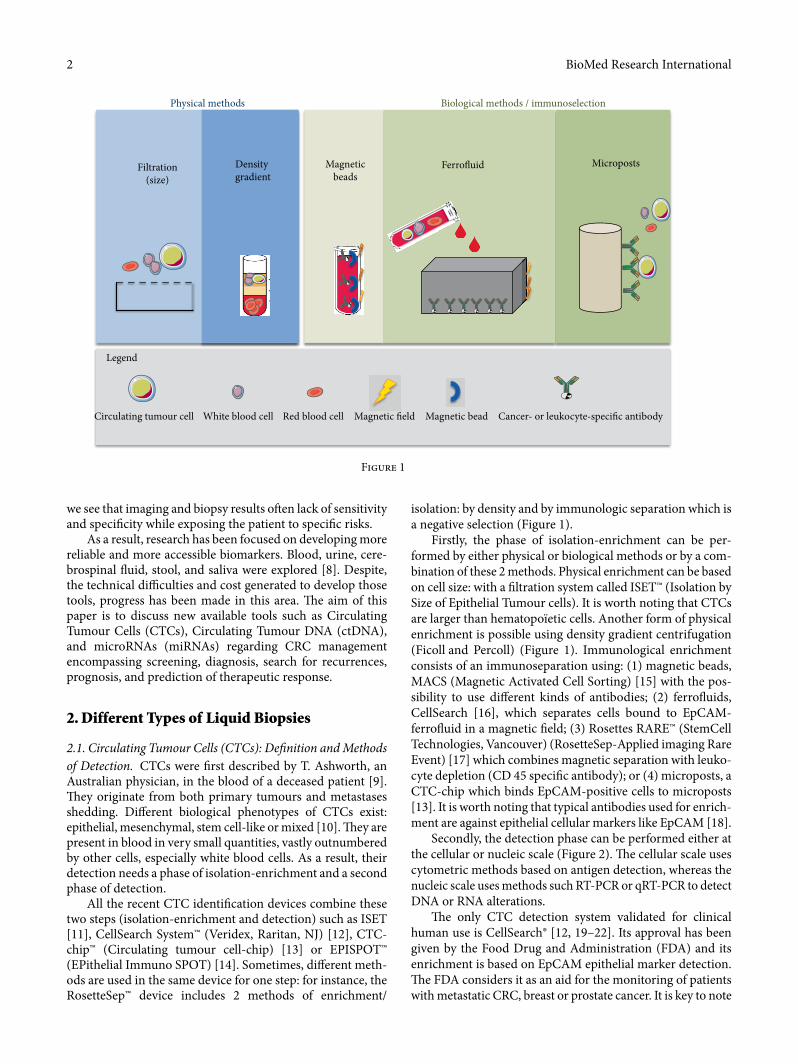
isolation: by density and by immunologic separation which is a negative selection (Figure 1).
Firstly, the phase of isolation-enrichment can be per-formed by either physical or biological methods or by a com-bination of these 2 methods. Physical enrichment can be based on cell size: with a �ltration system called ISET™ (Isolation by Size of Epithelial Tumour cells). It is worth noting that CTCs are larger than hematopoïetic cells. Another form of physical enrichment is possible using density gradient centrifugation (Ficoll and Percoll) (Figure 1). Immunological enrichment consists of an immunoseparation using: (1) magnetic beads, MACS (Magnetic Activated Cell Sorting) [15] with the pos-sibility to use di�erent kinds of antibodies; (2) ferro£uids, CellSearch [16], which separates cells bound to EpCAM-ferro£uid in a magnetic �eld; (3) Rosettes RARE™ (StemCell Technologies, Vancouver) (RosetteSep-Applied imaging Rare Event) [17] which combines magnetic separation with leuko-cyte depletion (CD 45 speci�c antibody); or (4) microposts, a CTC-chip which binds EpCAM-positive cells to microposts [13]. It is worth noting that typical antibodies used for enrich-ment are against epithelial cellular markers like EpCAM [18].
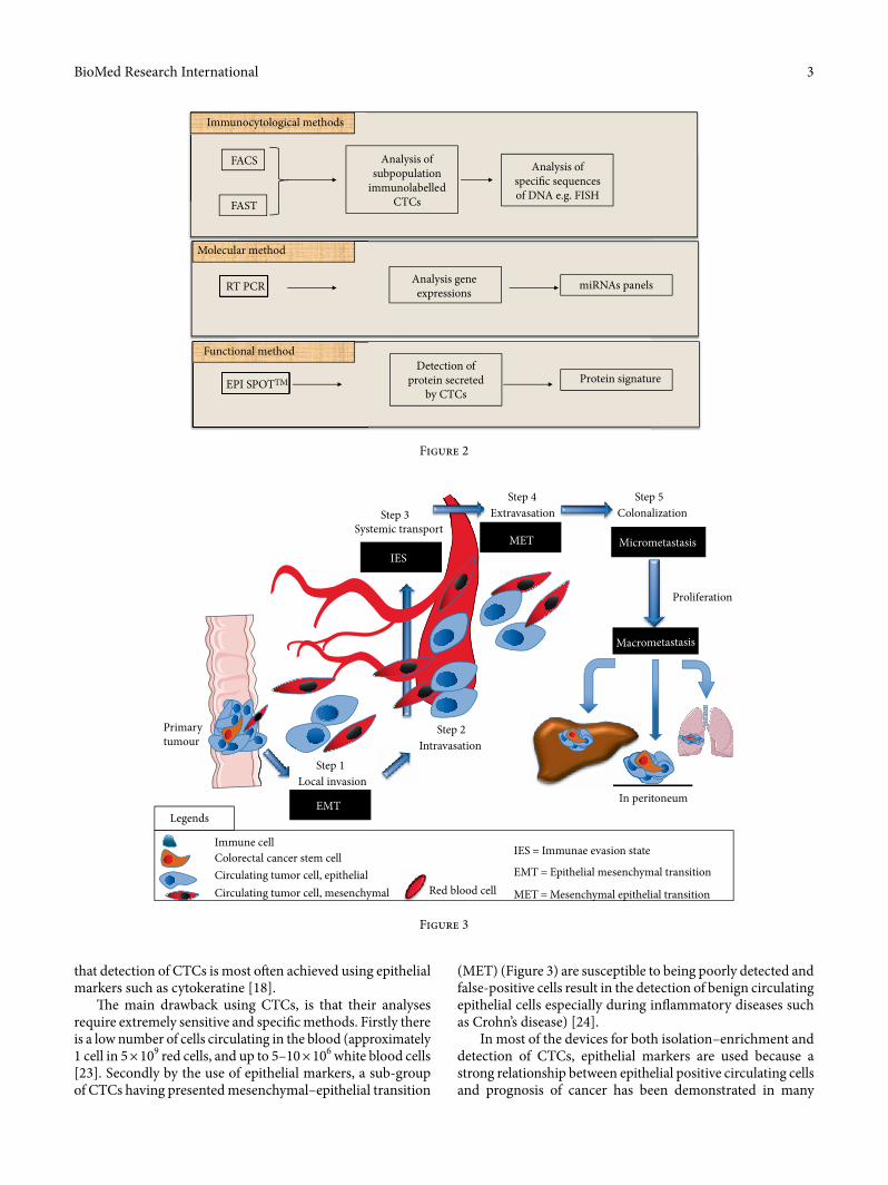
Secondly, the detection phase can be performed either at the cellular or nucleic scale (Figure 2). �e cellular scale uses cytometric methods based on antigen detection, whereas the nucleic scale uses methods such RT-PCR or qRT-PCR to detect DNA or RNA alterations.
�e only CTC detection system validated for clinical human use is CellSearch® [12, 19–22]. Its approval has been given by the Food Drug and Administration (FDA) and its enrichment is based on EpCAM epithelial marker detection. �e FDA considers it as an aid for the monitoring of patients with metastatic CRC, breast or prostate cancer. It is key to note
Physical methods Biological methods / immunoselection
Filtration(size)
Density gradient
Magneticbeads
Ferro�uid Microposts
Legend
Circulating tumour cell White blood cell Red blood cell Magnetic �eld Magnetic bead Cancer- or leukocyte-speci�c antibody
Figure 1
3BioMed Research International
that detection of CTCs is most o¢en achieved using epithelial markers such as cytokeratine [18].
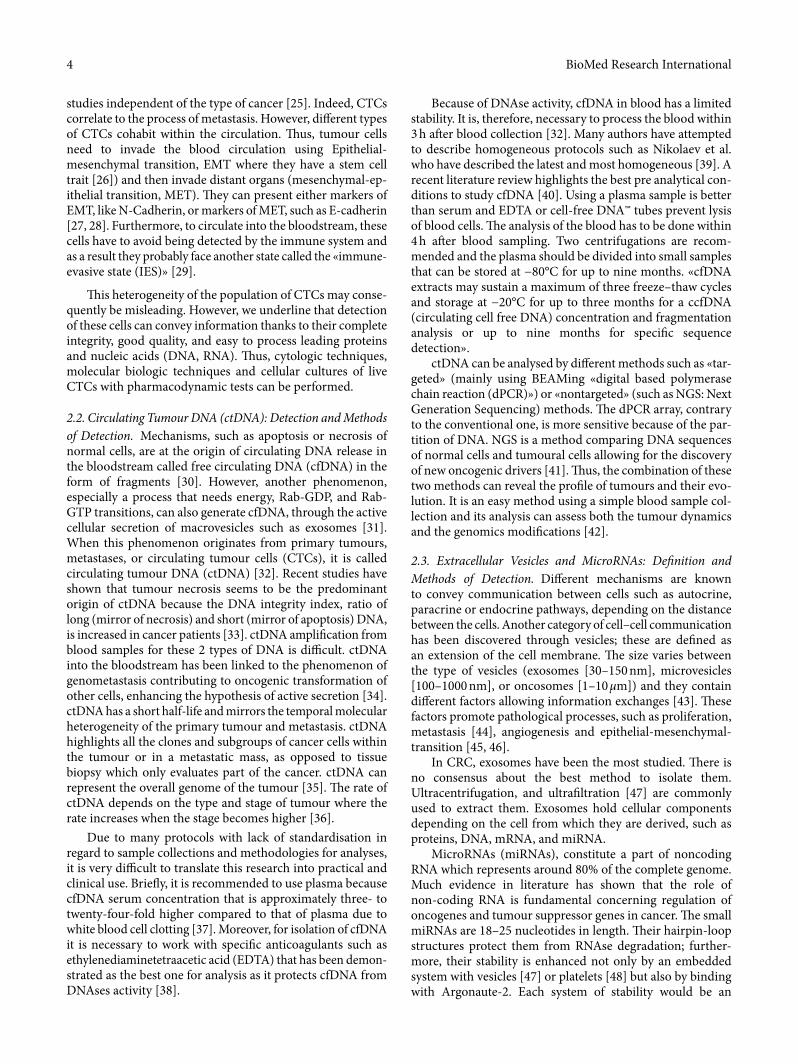
�e main drawback using CTCs, is that their analyses require extremely sensitive and speci�c methods. Firstly there is a low number of cells circulating in the blood (approximately 1 cell in 5 × 10⁹ red cells, and up to 5–10 × 106 white blood cells [23]. Secondly by the use of epithelial markers, a sub-group of CTCs having presented mesenchymal–epithelial transition
(MET) (Figure 3) are susceptible to being poorly detected and false-positive cells result in the detection of benign circulating epithelial cells especially during in£ammatory diseases such as Crohn’s disease) [24].
In most of the devices for both isolation–enrichment and detection of CTCs, epithelial markers are used because a strong relationship between epithelial positive circulating cells and prognosis of cancer has been demonstrated in many
FACS Analysis ofsubpopulation
immunolabelledCTCs
Analysis ofspecic sequencesof DNA e.g. FISH
FAST
Immunocytological methods
Molecular method
Functional method
RT PCR
EPI SPOTTM
Analysis geneexpressions
Detection ofprotein secreted
by CTCs
miRNAs panels
Protein signature
Figure 2
Proliferation
Step 3Systemic transport
ExtravasationStep 4
ColonalizationStep 5
IESMET Micrometastasis
Macrometastasis
Primarytumour
Step 1Local invasion
Step 2Intravasation
EMT In peritoneum
Immune cell
Legends
Colorectal cancer stem cellCirculating tumor cell, epithelialCirculating tumor cell, mesenchymal
IES = Immunae evasion state
EMT = Epithelial mesenchymal transition
MET = Mesenchymal epithelial transitionRed blood cell
Figure 3
BioMed Research International4
Because of DNAse activity, cfDNA in blood has a limited stability. It is, therefore, necessary to process the blood within 3 h a�er blood collection [32]. Many authors have attempted to describe homogeneous protocols such as Nikolaev et al. who have described the latest and most homogeneous [39]. A recent literature review highlights the best pre analytical con-ditions to study cfDNA [40]. Using a plasma sample is better than serum and EDTA or cell-free DNA™ tubes prevent lysis of blood cells. �e analysis of the blood has to be done within 4 h a�er blood sampling. Two centrifugations are recom-mended and the plasma should be divided into small samples that can be stored at −80°C for up to nine months. «cfDNA extracts may sustain a maximum of three freeze–thaw cycles and storage at −20°C for up to three months for a ccfDNA (circulating cell free DNA) concentration and fragmentation analysis or up to nine months for specific sequence detection».
ctDNA can be analysed by different methods such as «tar-geted» (mainly using BEAMing «digital based polymerase chain reaction (dPCR)») or «nontargeted» (such as NGS: Next Generation Sequencing) methods. �e dPCR array, contrary to the conventional one, is more sensitive because of the par-tition of DNA. NGS is a method comparing DNA sequences of normal cells and tumoural cells allowing for the discovery of new oncogenic drivers [41]. �us, the combination of these two methods can reveal the profile of tumours and their evo-lution. It is an easy method using a simple blood sample col-lection and its analysis can assess both the tumour dynamics and the genomics modifications [42].
2.3. Extracellular Vesicles and MicroRNAs: Definition and Methods of Detection. Different mechanisms are known to convey communication between cells such as autocrine, paracrine or endocrine pathways, depending on the distance between the cells. Another category of cell–cell communication has been discovered through vesicles; these are defined as an extension of the cell membrane. �e size varies between the type of vesicles (exosomes [30–150 nm], microvesicles [100–1000 nm], or oncosomes [1–10 μm]) and they contain different factors allowing information exchanges [43]. �ese factors promote pathological processes, such as proliferation, metastasis [44], angiogenesis and epithelial-mesenchymal-transition [45, 46].
In CRC, exosomes have been the most studied. �ere is no consensus about the best method to isolate them. Ultracentrifugation, and ultrafiltration [47] are commonly used to extract them. Exosomes hold cellular components depending on the cell from which they are derived, such as proteins, DNA, mRNA, and miRNA.
MicroRNAs (miRNAs), constitute a part of noncoding RNA which represents around 80% of the complete genome. Much evidence in literature has shown that the role of non-coding RNA is fundamental concerning regulation of oncogenes and tumour suppressor genes in cancer. �e small miRNAs are 18–25 nucleotides in length. �eir hairpin-loop structures protect them from RNAse degradation; further-more, their stability is enhanced not only by an embedded system with vesicles [47] or platelets [48] but also by binding with Argonaute-2. Each system of stability would be an
studies independent of the type of cancer [25]. Indeed, CTCs correlate to the process of metastasis. However, different types of CTCs cohabit within the circulation. �us, tumour cells need to invade the blood circulation using Epithelial-mesenchymal transition, EMT where they have a stem cell trait [26]) and then invade distant organs (mesenchymal-ep-ithelial transition, MET). �ey can present either markers of EMT, like N-Cadherin, or markers of MET, such as E-cadherin [27, 28]. Furthermore, to circulate into the bloodstream, these cells have to avoid being detected by the immune system and as a result they probably face another state called the «immune- evasive state (IES)» [29].
�is heterogeneity of the population of CTCs may conse-quently be misleading. However, we underline that detection of these cells can convey information thanks to their complete integrity, good quality, and easy to process leading proteins and nucleic acids (DNA, RNA). �us, cytologic techniques, molecular biologic techniques and cellular cultures of live CTCs with pharmacodynamic tests can be performed.
2.2. Circulating Tumour DNA (ctDNA): Detection and Methods of Detection. Mechanisms, such as apoptosis or necrosis of normal cells, are at the origin of circulating DNA release in the bloodstream called free circulating DNA (cfDNA) in the form of fragments [30]. However, another phenomenon, especially a process that needs energy, Rab-GDP, and Rab-GTP transitions, can also generate cfDNA, through the active cellular secretion of macrovesicles such as exosomes [31]. When this phenomenon originates from primary tumours, metastases, or circulating tumour cells (CTCs), it is called circulating tumour DNA (ctDNA) [32]. Recent studies have shown that tumour necrosis seems to be the predominant origin of ctDNA because the DNA integrity index, ratio of long (mirror of necrosis) and short (mirror of apoptosis) DNA, is increased in cancer patients [33]. ctDNA amplification from blood samples for these 2 types of DNA is difficult. ctDNA into the bloodstream has been linked to the phenomenon of genometastasis contributing to oncogenic transformation of other cells, enhancing the hypothesis of active secretion [34]. ctDNA has a short half-life and mirrors the temporal molecular heterogeneity of the primary tumour and metastasis. ctDNA highlights all the clones and subgroups of cancer cells within the tumour or in a metastatic mass, as opposed to tissue biopsy which only evaluates part of the cancer. ctDNA can represent the overall genome of the tumour [35]. �e rate of ctDNA depends on the type and stage of tumour where the rate increases when the stage becomes higher [36].
Due to many protocols with lack of standardisation in regard to sample collections and methodologies for analyses, it is very difficult to translate this research into practical and clinical use. Briefly, it is recommended to use plasma because cfDNA serum concentration that is approximately three- to twenty-four-fold higher compared to that of plasma due to white blood cell clotting [37]. Moreover, for isolation of cfDNA it is necessary to work with specific anticoagulants such as ethylenediaminetetraacetic acid (EDTA) that has been demon-strated as the best one for analysis as it protects cfDNA from DNAses activity [38].
5BioMed Research International
cell transforming sequence 2 oncogene (ECT2) from CTCs demonstrated a high potential to be a good diagnostic bio-marker of CRC thanks to both interesting sensitivity and spec-ificity, even in the cases were CEA concentrations accounted for less than 5 ng/mL (diagnostic threshold).
Several in vitro studies have highlighted specific miRNAs of CRC [59]. Recent clinical studies have confirmed the pres-ence of these unique CRC miRNAs in comparison with healthy people [60, 61]. One recent publication by Wang et al., in 2015 [62], was able to detect a panel of 3 miRNAs in plasma, miR-409-3p, miR-7, and miR-93 as good biomarkers for CRC early detection. Other studies [63, 64], showed alternate miRNAs as potential markers for screening or early diagnosis of CRC. It is important to underline that for some studies, samples were from plasma while in others they were from serum; indeed, extracted DNA quality varies according to sample type. �e most recent meta-analysis focused on blood-based miRNA analysis for diagnosis of CRC cancer was performed in 2017 by Carter et al. [65]. A literature search including articles between January 2002 and April 2016 was performed and 34 studies analysing serum or plasma miRNAs for diagnosis of CRC were included. In comparison to healthy people, the analysis showed a dysregulation of 31 miRNAs in CRC patients. Eight out of the thirty-one miRNAs were validated by more than one study: «six were up-regulated: miR-17-3p, miR-18a, miR-21, miR-29a, miR-92, and miR-106a and two were down-regulated: miR-29b and miR-145». �e main issue of this meta-analysis is that a sub-group analysis according to the type of sample (either serum or plasma) was not carried out. Another meta-analysis [66] looked at 103 studies from 2008 to 2015 with 3124 CRC patients and 2579 control healthy patients. It included not only blood-based miRNAs (plasma, and/or serum) analyses but also those of tissue and feces. A�er meta-regression analyses, this study demonstrated that serum samples were the most accurate. Moreover, multiple miRNA assays proved to be more robust in the diagnosis of CRC in contrast to single miRNA assays [67, 68]. �e accuracy was also more important in the Asian population in comparison with Caucasian people.
It appears that miRNAs are interesting prospective bio-markers but more studies are needed to confirm this in the preclinical stage, in particular within a cohort of patients who have a positive fecal immunochemical test (FIT) [69].
All in all, taking into account all the drawbacks of each biomarker, the best cancer screening method, particularly in colorectal cancer, appears at present to be CancerSEEK. It is a test developed in January 2018 by researchers in Baltimore [57] which combines the detection of ctDNA with different proteins in order to improve the signal to noise ratio.
4. Prognosis: Surveillance and Detection of Tumour Recurrence and Minimal Residual Disease
In colon cancers stages II and III, a�er radical and curative treatment, surveillance is conducted using both scanners and biological assessments (CEA biomarker) [70]. However, imag-ing is not sensitive enough and exposes patients to radiation
indirect marker of cell type origin [49]. �ey can be extracted from different tissues or biologic fluids (circulating miRNA = cmiRNA) like feces, saliva, urine or blood [50]. �is has raised the question whether miRNAs could be used as CRC biomark-ers in diagnosis or prognosis. However the lack of standardi-zation and robustness of the measuring methodology has led to contradictory results [51]. Firstly, it is difficult to extract and isolate miRNAs due to their small size and their different binding associations. Despite different ultracentrifugation methods with detergents or proteases to purify them, different variations exist highlighting the need for further studies. Secondly, various techniques are used for examination of miR-NAs, such as RT-qPCR, Microarray, and Next-Generation Sequencing (NGS) with variations on sensitivity, specificity, accuracy, ease of analysis, reproducibility, and costs. RT-qPCR has the highest sensitivity and specificity. But studies have to be performed in order to improve isolation and use of miRNAs as validated biomarkers.
3. Early Diagnosis: Screening and Tumour Burden at Diagnosis
Up to now, clinical studies have especially emphasized ctDNA as a potential biomarker for early diagnosis and screening. Methylation of gene promoters is generally the first step in oncogenesis and according to the type of genes implicated, the origin of the tumour can be determined. As a result, many studies have focused on the research of potential signatures represented by one gene [52] or the association [53, 54] of the gene promoter’s methylation profile. �e best sensitivity and specificity among these marketed tests have been found for the one called Epi proColon® 2.0 CE which detects methylated Septin9 DNA [52] demonstrating 75–81% sensitivity and 96–99% specificity. By comparison, it was found that FIT had a sensitivity around 79% and a specificity near 94% [55]. Negative FIT can be preceded by the gold standard of a screen-ing colonoscopy [56]. It is worth noting that recently, the team of Cohen et al. [57] has assessed the combined detection of circulating protein biomarkers and tumour specific mutations in circulating DNA to detect cancer in patients who already had the diagnosis of cancer (lung, ovary, stomach, colorectal, pancreas, liver, or oesophagus). �e sensitivity and specificity for colon and rectum cancers using the combined detection methods was approximately 60–70%; nevertheless, this test was not conducted in healthy patients to detect colorectal can-cer at a preclinical or asymptomatic stage.
�e difficulty to isolate and define CTCs highlights the weakness in using them in daily clinical practice for screening and early diagnosis. Searches in the Pubmed and Cochrane databases were conducted. We searched for screening CRC, using the terms «circulating tumour cells», «screening» and «colorectal cancer», and found 1131 articles, but a�er analysis of each title, none corresponded to our research. We then searched for early diagnosis of CRC, using the terms «circu-lating tumour cells», «early diagnosis» and «colorectal cancer», and found 222 articles, but only 1 study was finally included a�er rigorous analysis of all the titles [58]. �is article from Chen et al. [58], pointed out that extracted RNA of epithelial
BioMed Research International6
types, collection origins (plasma, � = 3 [93–95] or serum, � = 6 [96–101]), detection methods (PCR followed by sequencing [93, 96], spectrophotometry [97], quantitative PCR (q-PCR) [94, 95], mutant-enriched PCR (ME-PCR) [98], and real-time PCR (rt-PCR) [95, 99–101]. A¢er strati�cation on confounding factors such as type of tumour marker searched in cfDNA, tumour stage, collection origin [serum or plasma] and methods of detection [PCR, qPCR, others] and population size, it was demonstrated that cfDNA could predict both overall survival and recurrence-free survival. Recurrences were considered as distant metastasis and con�rmed by imaging.
�e role of prognostic assessment by both qualitative and quantitative analysis of ctDNA was useful in CRC at a meta-static stage [102].
5. Therapy Response
In di�erent cases, neoadjuvant radio chemotherapy (for mid-dle and low rectal cancers stages II-III), or adjuvant chemo-therapy (metastatic diseases or recurrences) are used. Di�erent combinations of drugs exist and di�erent kinds of therapies are available: chemotherapies, targeted therapies, or immunotherapies.
Heterogeneity de�nes cancers and explains why for both speci�c tumours and stages, each person will respond di�er-ently to therapies. Analysing CTCs and ctDNA in patients with CRC allows us to choose the best therapy with respect to the tumour mutations identi�ed: presence of RAS mutations, Anti-Epidermal Growth Factor Receptor (anti-EGFR) muta-tions, and BRAF mutations. �us, the ctDNA RAS mutation pro�le has provided information concerning the e�ectiveness of the use of monoclonal antibody against EGFR such as Cetuximab [103]. Since the study of Karapetis et al. in 2008 [104], Cetuximab has been shown to improve OS and PFD in patients with metastatic CRC if they have a Wild Type RAS status. �e work of Grasseli [103] has con�rmed similar rates between RAS status in ctDNA (plasma) and those of tumour issue. �ere is also agreement towards response to anti-EGFR therapy according to RAS determination between plasma and tissue. In the same way, BRAF V600E mutation occurs in approximately 5–10% of metastatic CRCs and predicts poor prognosis [105]. As a result, therapies like BRAF and MEK inhibitors have little impact in BRAF mutated metastatic CRC. But the use of combination therapies with inhibition of EGFR and MAPK (Mitogen-activated protein kinases) pathways can improve e¤cacy. �e study of Corcoran has proven this using anti EGFR antibody, BRAF inhibitor and MEK inhibitor with reduction in BRAF V600E mutation for 86% of patients with this triple therapy [106]. Furthermore, following the evolution of the ctDNA pro�le during such treatments can be used to analyse response [107] and induced genetic modi�cations [108]. All in all, it will help physicians to adapt these treat-ments over time.
A recent phase II clinical study, (PROSPECT-C trial: clin-ical trials.gov number NCT02994888) [109], monitored genetic variations of cfDNA in human plasma during Anti-EGFR treatment and brought to light the dynamics of
while CEA does not combine high sensitivity and speci�city. �e best test to detect recurrences is an increase of CEA during the �rst year of follow-up in patients treated for CRC [71].
In rectal cancers, liquid biopsies could be of interest in order to predict the response of radiochemotherapy and dis-ease recurrence; however, evidence in this domain is lacking.
4.1. Minimal Residual Disease (MRD) to Tumour Recurrences. Di�erent studies have tried to investigate this MRD with the search of both CTCs and ctDNA a¢er surgery. �e majority of them have found a progressive decrease of ctDNA in plasma a¢er colorectal resection for CRC in both adult women and men [72]; but the persistence of ctDNA in plasma just a¢er resection (approximately 3 days a¢er surgery) does not seem to correlate with further recurrences [73]. As a consequence, it is the steady decrease with time of ctDNA rates that seems to best represent the absence of residual disease. A modi�cation with increase of ctDNA is a synonym of recurrence [74]. �is has been con�rmed with rectal cancers (T3/T4/N+) treated by neoadjuvant therapy [75] in samples collected before and 4–6 weeks a¢er radiochemotherapy and surgery with sample collection 4–10 weeks postoperatively.
A clinical trial which has been initiated in our French Digestive Surgery Department (Clinical Trial: NCT02813928) aims at detecting the presence of recurrences in patients cura-tively treated for a CRC stages II and III within the 3 years of follow-up. �is study started in July 2016 with a total of 473 patients enrolled in the follow-up of 3 years. �e �rst results will be available at the end of 2020.
A meta-analysis [76] for CTC’s, including 13 studies with patients treated for CRC with chemotherapy alone or in com-bination with surgery showed that high-CTC levels a¢er treat-ment were correlated with disease progression con�rmed by imagery.
4.2. Prognosis. Two recent meta-analyses have shown the impact on prognosis of both CTCs [77] and cfDNA [78] in CRC. �e �rst, focused on CTCs [77], gathered 15 studies with 3129 patients having undergone either surgery alone or chemotherapy alone or both therapies [21, 79–92]. �e presence of CTCs was linked to a poorer overall survival and progression-free disease; however there was an important heterogeneity among all the studies therefore sub-group analyses were also done. �ey reported that positive CTC patients had signi�cant poor overall survival (OS) and progression-free disease (PFD) according to di�erent criteria (time of blood collection, detection method, median follow-up and cuto� value of CTC number). Poorer OS and PFD were demonstrated for sampling collection during treatment and not at baseline, using Cellsearch detection and not RT PCR or other methods, with a median follow-up more than 24 months, and a cuto� value of CTCs superior to 1.
�e second meta-analysis [78], included 9 studies of patients treated for CRC [93–101] with both qualitative and quantitative evaluation of cfDNA. Here also, there was notable heterogeneity: population size of studies, tumour stages, time of collection (8 before treatment, 1 a¢er treatment) marker
7BioMed Research International
[2] M. Arnold, M. S. Sierra, M. Laversanne, I. Soerjomataram, A. Jemal, and F. Bray, “Global patterns and trends in colorectal cancer incidence and mortality,” Gut, vol. 66, no. 4, pp. 683–691, 2017.
[3] J. K. Lee, E. G. Liles, S. Bent, T. R. Levin, and D. A. Corley, “Accuracy of fecal immunochemical tests for colorectal cancer: systematic review and meta-analysis,” Annals of Internal Medicine, vol. 160, no. 3, p. 171, 2014.
[4] C. Bray, L. N. Bell, H. Liang, D. Collins, and S. H. Yale, “Colorectal cancer screening,” WMJ Official Publication of the State Medical Society of Wisdom, vol. 116, no. 1, pp. 27–33, 2017.
[5] M. Gerlinger, A. J. Rowan, S. Horswell et al., “Intratumor heterogeneity and branched evolution revealed by multiregion sequencing,” �e New England Journal of Medicine, vol. 366, no. 10, pp. 883–892, 2012.
[6] S. R. Brown, T. C. Hicks, and C. B. Whitlow, “Chapter 145 - diagnostic and therapeutic colonoscopy,” Shackelford’s Surgery of the Alimentary Tract, 2 Volume Set, Content Repository Only, PhiladelphiaC. J. Yeo, Ed., pp. 168–1699, Eighth edition, 2019, http://www.sciencedirect.com/science/article/pii/B978032340232300145X.
[7] K. Nagata, K. Takabayashi, T. Yasuda et al., “Adverse events during CT colonography for screening, diagnosis and preoperative staging of colorectal cancer: a Japanese national survey,” European Radiology, vol. 27, no. 12, pp. 4970–4978, 2017.
[8] G. Siravegna, S. Marsoni, S. Siena, and A. Bardelli, “Integrating liquid biopsies into the management of cancer,” Nature Reviews Clinical Oncology, vol. 14, no. 9, pp. 531–548, 2017.
[9] T. R. Ashworth, “A case of cancer in which cells similar to those in the tumours were seen in the blood a�er death,” Australian Medical Journal, vol. 14, pp. 146–147, 1869.
[10] L. M. Millner, M. W. Linder, and R. Valdes, “Circulating tumor cells: a review of present methods and the need to identify heterogeneous phenotypes,” Annals of Clinical Laboratory Science, vol. 43, no. 3, pp. 295–304, 2013.
[11] G. Vona, A. Sabile, M. Louha et al., “Isolation by size of epithelial tumor cells: a new method for the immunomorphological and molecular characterization of circulating tumor cells,” �e American Journal of Pathology, vol. 156, no. 1, pp. 57–63, 2000.
[12] M. C. Miller, G. V. Doyle, and L. W. M. M. Terstappen, “Significance of circulating tumor cells detected by the cellsearch system in patients with metastatic breast colorectal and prostate cancer,” Journal of Oncology, vol. 2010, Article ID 617421, pp. 1–8, 2010.
[13] S. Nagrath, L. V. Sequist, S. Maheswaran et al., “Isolation of rare circulating tumour cells in cancer patients by microchip technology,” Nature, vol. 450, no. 7173, pp. 1235–1239, 2007.
[14] C. Alix-Panabières, “EPISPOT assay: detection of viable dtcs/ctcs in solid tumor patients,” Minimal Residual Disease and Circulating Tumor Cells in Breast Cancer, Springer, Berlin, HeidelbergM. Ignatiadis, C. Sotiriou, and K. Pantel, Ed., pp. 69–76, 2012.
[15] D. R. Parkinson, N. Dracopoli, B. G. Petty et al., “Considerations in the development of circulating tumor cell technology for clinical use,” Journal of Translational Medicine, vol. 10, no. 1, Article ID 138, 2012.
[16] S. Riethdorf, H. Fritsche, V. Müller et al., “Detection of circulating tumor cells in peripheral blood of patients with metastatic breast cancer: a validation study of the cell search
resistance acquisition. �is work also explained the possibility, using a mathematical model, to anticipate these variations in order to introduce personalised therapeutics.
6. Conclusion
Liquid biopsies looking at circulating tumour cells (CTCs), circulating tumour DNA (ctDNA), and miRNAs are of great interest in the management of CRC.
However, there is no consensus to define which may be the best combining both sensitivity and specificity for each step of CRC management due to the multiple differences in detection and analysis. It is necessary to standardise the methods of anal-ysis for each biomarker in order to find the best sensitivity and specificity and offer the patients tailored therapies.
Moreover, we have to keep in mind that a possible multi-tude of markers/molecules can exist: different types of cancers use different pathways but most of the time pathways involving different molecules interlock. So our search for the “Holy Grail” of CRC management should be modified. Rather than looking for the single magical solution of CRC management and thinking that a unique method (ctDNA or CTCs or miR-NAs) with a unique biomarker (in each method) for each type of cancer can exist, is wrong. �e trend is rather to combine a panel of biomarkers and different methods in each step in the management of cancers in general and understanding that some identical biomarkers may be involved whatever the type of cancer. By analysing a panel of biomarkers, we can then offer optimal management for cancer diagnosis, treatment and follow-up to our patients.
Conflicts of Interest
�e authors declare that they have no conflicts of interest.
Authors’ Contributions
N.C. conceived and designed the study. N.C., J.M., A.P., S.B., V.D., N.B., E.L., F.R., J.R., and M.M., contributed to the writing of the manuscript and to its critical revision. N.C, J.M, A.P, S.B, V.D., N.B., E.L., F.R., J.R., and M.M., approved the final version of the manuscript.
Acknowledgments
We thank Doctor Hussein Al-Akhrass for critical reading of the manuscript.
References
[1] F. Bray, J. Ferlay, I. Soerjomataram, R. L. Siegel, L. A. Torre, and A. Jemal, “Global cancer statistics 2018: GLOBOCAN estimates of incidence and mortality worldwide for 36 cancers in 185 countries,” CA: A Cancer Journal for Clinicians, vol. 68, no. 6, pp. 394–424, 2018.
BioMed Research International8
[31] B. K. �akur, H. Zhang, A. Becker et al., “Double-stranded DNA in exosomes: a novel biomarker in cancer detection,” Cell Research, vol. 24, no. 6, pp. 766–769, 2014.
[32] D. A. Haber and V. E. Velculescu, “Blood-based analyses of cancer: circulating tumor cells and circulating tumor DNA,” Cancer Discovery, vol. 4, no. 6, pp. 650–661, 2014.
[33] M. Stroun, J. Lyautey, C. Lederrey, H. E. Mulcahy, and P. Anker, “Alu repeat sequences are present in increased proportions compared to a unique gene in plasma/serum DNA,” Annals of the New York Academy of Sciences, vol. 945, no. 1, pp. 258–264, 2001.
[34] D. C. García-Olmo, C. Domínguez, M. García-Arranz et al., “Cell-free nucleic acids circulating in the plasma of colorectal cancer patients induce the oncogenic transformation of susceptible cultured cells,” Cancer Research, vol. 70, no. 2, pp. 560–567, 2010.
[35] K. C. A. Chan, “Scanning for cancer genomic changes in plasma: toward an era of personalized blood-based tumor markers,” Clinical Chemistry, vol. 59, no. 11, pp. 1553–1555, 2013.
[36] C. Bettegowda, M. Sausen, R. J. Leary et al., “Detection of circulating tumor DNA in early- and late-stage human malignancies,” Science Translational Medicine, vol. 6, no. 224, 224ra24 pages, 2014.
[37] T.-H. Lee, L. Montalvo, V. Chrebtow, and M. P. Busch, “Quantitation of genomic DNA in plasma and serum samples: higher concentrations of genomic DNA found in serum than in plasma,” Transfusion, vol. 41, no. 2, pp. 276–282, 2001.
[38] E. Beutler, T. Gelbart, and W. Kuhl, “Interference of heparin with the polymerase chain reaction,” BioTechniques, vol. 9, no. 2, p. 166, 1990, https://www.cabdirect.org/cabdirect/abstract/19930198817.
[39] S. Nikolaev, L. Lemmens, T. Koessler, J.-L. Blouin, and T. Nouspikel, “Circulating tumoral DNA: preanalytical validation and quality control in a diagnostic laboratory,” Analytical Biochemistry, vol. 542, pp. 34–39, 2018.
[40] S. El Messaoudi, F. Rolet, F. Mouliere, and A. R. �ierry, “Circulating cell free DNA: preanalytical considerations,” Clinica Chimica Acta, vol. 424, pp. 222–230, 2013.
[41] O. M. Toor, Z. Ahmed, W. Bahaj et al., “Correlation of somatic genomic alterations between tissue genomics and ctDNA employing next-generation sequencing: analysis of lung and gastrointestinal cancers,” Molecular Cancer �erapeutics, vol. 17, no. 5, pp. 1123–1132, 2018.
[42] L. A. Diaz and A. Bardelli, “Liquid biopsies: genotyping circulating tumor DNA,” Journal of Clinical Oncology, vol. 32, no. 6, pp. 579–586, 2014.
[43] D. Choi, T. H. Lee, C. Spinelli, S. Chennakrishnaiah, E. D’Asti, and J. Rak, “Extracellular vesicle communication pathways as regulatory targets of oncogenic transformation,” Seminars in Cell & Developmental Biology, vol. 67, pp. 11–22, 2017.
[44] D.-S. Choi, D.-Y. Choi, B. Hong et al., “Quantitative proteomics of extracellular vesicles derived from human primary and metastatic colorectal cancer cells,” Journal of Extracellular Vesicles, vol. 1, no. 1, p. 18704, 2012.
[45] L. J. Vella, “�e emerging role of exosomes in epithelial–mesenchymal-transition in cancer,” Front Oncology, vol. 19, no. 4, p. 361, 2014, https://www.frontiersin.org/articles/10.3389/fonc.2014.00361/full.
[46] S. EL Andaloussi, I. Mäger, X. O. Breakefield, and M. J. A. Wood, “Extracellular vesicles: biology and emerging
system,” Clinical Cancer Research, vol. 13, no. 3, pp. 920–928, 2007.
[17] B. Naume, E. Borgen, S. Tøssvik, N. Pavlak, D. Oates, and J. M. Nesland, “Detection of isolated tumor cells in peripheral blood and in BM: evaluation of a new enrichment method,” Cytotherapy, vol. 6, no. 3, pp. 244–252, 2004.
[18] S. D. Mikolajczyk, L. S. Millar, P. Tsinberg et al., “Detection of EpCAM-negative and cytokeratin-negative circulating tumor cells in peripheral blood,” Journal of Oncology, vol. 2011, Article ID 252361, p. 10, 2011.
[19] S. J. Cohen, R. K. Alpaugh, S. Gross et al., “Isolation and characterization of circulating tumor cells in patients with metastatic colorectal cancer,” Clinical Colorectal Cancer, vol. 6, no. 2, pp. 125–132, 2006.
[20] S. J. Cohen, C. J. A. Punt, N. Iannotti et al., “Relationship of circulating tumor cells to tumor response, progression-free survival, and overall survival in patients with metastatic colorectal cancer,” Journal of Clinical Oncology, vol. 26, no. 19, pp. 3213–3221, 2008.
[21] S. J. Cohen, C. J. A. Punt, N. Iannotti et al., “Prognostic significance of circulating tumor cells in patients with metastatic colorectal cancer,” Annals Oncology, vol. 20, no. 7, pp. 1223–1229, 2009.
[22] J. Sastre, M. L. Maestro, A. Gómez-España et al., “Circulating tumor cell count is a prognostic factor in metastatic colorectal cancer patients receiving first-line chemotherapy plus bevacizumab: a spanish cooperative group for the treatment of digestive tumors study,” �e Oncologist, vol. 17, no. 7, pp. 947–955, 2012.
[23] W. J. Allard, J. Matera, M. C. Miller et al., “Tumor cells circulate in the peripheral blood of all major carcinomas but not in healthy subjects or patients with nonmalignant diseases,” Clinical Cancer Research, vol. 10, no. 20, pp. 6897–6904, 2004.
[24] K. Pantel, E. Denève, D. Nocca et al., “Circulating epithelial cells in patients with benign colon diseases,” Clinical Chemistry, vol. 58, no. 5, pp. 936–940, 2012.
[25] J. S. de Bono, H. I. Scher, R. B. Montgomery et al., “Circulating tumor cells predict survival benefit from treatment in metastatic castration-resistant prostate cancer,” Clinical Cancer Research, vol. 14, no. 19, pp. 6302–6309, 2008.
[26] C. Mélin, A. Perraud, N. Christou et al., “New ex-ovo colorectal-cancer models from different SdFFF-sorted tumor-initiating cells,” Analytical and Bioanalytical Chemistry, vol. 407, no. 28, pp. 8433–8443, 2015.
[27] N. Christou, A. Perraud, S. Blondy, M.-O. Jauberteau, S. Battu, and M. Mathonnet, “�e extracellular domain of E cadherin linked to invasiveness in colorectal cancer: a new resistance and relapses monitoring serum-bio marker?” Journal Cancer Research and Clinical Oncology, vol. 143, no. 7, pp. 1177–1190, 2017.
[28] N. Christou, A. Perraud, S. Blondy, M.-O. Jauberteau, S. Battu, and M. Mathonnet, “E-cadherin: a potential biomarker of colorectal cancer prognosis (review),” Oncology Letters, vol. 13, no. 6, pp. 4571–4576, 2017.
[29] S. Jaiswal, C. H. M. Jamieson, W. W. Pang et al., “CD47 is upregulated on circulating hematopoietic stem cells and leukemia cells to avoid phagocytosis,” Cell, vol. 138, no. 2, pp. 271–285, 2009.
[30] P. Mandel and P. Metais, “Not available,” Comptes Rendus des Seances de la Societe de Biologie Filiales, vol. 142, no. 3–4, pp. 241–243, 1948.
9BioMed Research International
[63] Y. Sun, Y. Liu, D. Cogdell et al., “Examining plasma microRNA markers for colorectal cancer at different stages,” Oncotarget, vol. 7, no. 10, pp. 11434–11449, 2016.
[64] J. Yu, L. Jin, L. Jiang et al., “Serum miR-372 is a diagnostic and prognostic biomarker in patients with early colorectal cancer,” Anti-Cancer Agents in Medicinal Chemistry, vol. 16, no. 4, pp. 424–431, 2016, https://www.ingentaconnect.com/contentone/ben/acamc/2016/00000016/00000004/art00006.
[65] J. V. Carter, N. J. Galbraith, D. Yang, J. F. Burton, S. P. Walker, and S. Galandiuk, “Blood-based microRNAs as biomarkers for the diagnosis of colorectal cancer: a systematic review and meta-analysis,” British Journal of Cancer, vol. 116, no. 6, pp. 762–774, 2017.
[66] L. Yan, W. Zhao, H. Yu, Y. Wang, Y. Liu, and C. Xie, “A comprehensive meta-analysis of microRNAs for predicting colorectal cancer,” Medicine (Baltimore), vol. 95, no. 9, p. e2738, 2016.
[67] X. Luo, C. Stock, B. Burwinkel, and H. Brenner, “Identification and evaluation of plasma microRNAs for early detection of colorectal cancer,” PLoS One, vol. 8, no. 5, p. e62880, 2013.
[68] G. Zheng, L. Du, X. Yang et al., “Serum microRNA panel as biomarkers for early diagnosis of colorectal adenocarcinoma,” British Journal of Cancer, vol. 111, no. 10, pp. 1985–1992, 2014.
[69] V. Erben, M. Bhardwaj, P. Schrotz-King, and H. Brenner, “Metabolomics biomarkers for detection of colorectal neoplasms: a systematic review,” Cancers, vol. 10, no. 8, p. 246, 2018.
[70] R. Labianca, B. Nordlinger, G. D. Beretta et al., “Early colon cancer: ESMO clinical practice guidelines for diagnosis, treatment and follow-up,” Annals of Oncology, vol. 24, no. suppl 6, pp. vi64–vi72, 2013.
[71] B. Shinkins, J. N. Primrose, S. A. Pugh et al., “Serum carcinoembryonic antigen trends for diagnosing colorectal cancer recurrence in the FACS randomized clinical trial,” British Journal of Surgery, vol. 105, no. 6, pp. 658–662, 2018.
[72] M. Frattini, G. Gallino, S. Signoroni et al., “Quantitative and qualitative characterization of plasma DNA identifies primary and recurrent colorectal cancer,” Cancer Letters, vol. 263, no. 2, pp. 170–181, 2008.
[73] U. Lindforss, H. Zetterquist, N. Papadogiannakis, and H. Olivecrona, “Persistence of K-ras mutations in plasma a�er colorectal tumor resection,” Anticancer Research, vol. 25, no. 1B, pp. 657–661, 2005.
[74] T. Reinert, L. V. Schøler, R. �omsen et al., “Analysis of circulating tumour DNA to monitor disease burden following colorectal cancer surgery,” vol. 65, no. 4, pp. 625–634, 2016.
[75] J. Tie, J. D. Cohen, Y. Wang et al., “Serial circulating tumour DNA analysis during multimodality treatment of locally advanced rectal cancer: a prospective biomarker study,” Gut, vol. 68, no. 4, pp. 663–671, 2019.
[76] X. Huang, P. Gao, Y. Song et al., “Relationship between circulating tumor cells and tumor response in colorectal cancer patients treated with chemotherapy: a meta-analysis,” BMC Cancer, vol. 14, no. 1, Article ID 976, 2014.
[77] Y. Tan and H. Wu, “�e significant prognostic value of circulating tumor cells in colorectal cancer: a systematic review and meta-analysis,” Current Problems in Cancer, vol. 42, no. 1, pp. 95–106, 2018.
[78] S. Basnet, Z.-Y. Zhang, W.-Q. Liao, S.-H. Li, P.-S. Li, and H.-Y. Ge, “�e Prognostic value of circulating cell-free dna in
therapeutic opportunities,” Nature Reviews Drug Discovery, vol. 12, no. 5, pp. 347–357, 2013.
[47] L. Ruiz-López, I. Blancas, J. M. Garrido et al., “�e role of exosomes on colorectal cancer: a review,” Journal of Gastroenterology and Hepatology, vol. 33, no. 4, pp. 792–799, 2018.
[48] N. Sol and T. Wurdinger, “Platelet RNA signatures for the detection of cancer,” Cancer Metastasis Reviews, vol. 36, no. 2, pp. 263–272, 2017.
[49] J. D. Arroyo, J. R. Chevillet, E. M. Kroh et al., “Argonaute2 complexes carry a population of circulating microRNAs independent of vesicles in human plasma,” Proceedings of the National Academy of Sciences, vol. 108, no. 12, pp. 5003–5008, 2011, https://www.pnas.org/content/108/12/5003.
[50] Y. Okugawa, W. M. Grady, and A. Goel, “Epigenetic alterations in colorectal cancer: emerging biomarkers,” Gastroenterology, vol. 149, no. 5, pp. 1204–1225.e12, 2015.
[51] S. Ono, S. Lam, M. Nagahara, and D. S. B. Hoon, “Circulating microRNA biomarkers as liquid biopsy for cancer patients: pros and cons of current assays,” Journal of Clinical Medicine, vol. 4, no. 10, pp. 1890–1907, 2015.
[52] Y. N. Lamb and S. Dhillon, “Epi proColon® 2.0 CE: a blood-based screening test for colorectal cancer,” Molecular Diagnosis & �erapy, vol. 21, no. 2, pp. 225–232, 2017.
[53] S. L. Rasmussen, H. B. Krarup, K. G. Sunesen et al., “Hypermethylated DNA, a circulating biomarker for colorectal cancer detection,” PLoS One, vol. 12, no. 7, p. e0180809, 2017.
[54] J.-F. Rahier, A. Druez, L. Faugeras et al., “Circulating nucleosomes as new blood-based biomarkers for detection of colorectal cancer,” Clinical Epigenetics, vol. 9, no. 1, Article ID 53, 2017.
[55] L.-L. Song and Y.-M. Li, “Current noninvasive tests for colorectal cancer screening: an overview of colorectal cancer screening tests,” World Journal of Gastrointestinal Oncology, vol. 8, no. 11, pp. 793–800, 2016.
[56] D. K. Rex, G. A. Lehman, R. H. Hawes, T. M. Ulbright, and J. J. Smith, “Screening colonoscopy in asymptomatic average-risk persons with negative fecal occult blood tests,” Gastroenterology, vol. 100, no. 1, pp. 64–67, 1991.
[57] J. D. Cohen, L. Li, Y. Wang et al., “Detection and localization of surgically resectable cancers with a multi-analyte blood test,” Science, vol. 359, no. 6378, pp. 926–930, 2018.
[58] C.-J. Chen, W.-W. Sung, H.-C. Chen et al., “Early assessment of colorectal cancer by quantifying circulating tumor cells in peripheral blood: ECT2 in diagnosis of colorectal cancer,” International Journal of Molecular Sciences, vol. 18, no. 4, p. 743, 2017.
[59] E. Bandrés, E. Cubedo, X. Agirre et al., “Identification by Real-time PCR of 13 mature microRNAs differentially expressed in colorectal cancer and nontumoral tissues,” Molecular Cancer, vol. 19, pp. 5–29, 2006.
[60] E. K. O. Ng, W. W. S. Chong, H. Jin et al., “Differential expression of microRNAs in plasma of patients with colorectal cancer: a potential marker for colorectal cancer screening,” Gut, vol. 58, no. 10, pp. 1375–1381, 2009.
[61] Z. Huang, D. Huang, S. Ni, Z. Peng, W. Sheng, and X. Du, “Plasma microRNAs are promising novel biomarkers for early detection of colorectal cancer,” International Journal of Cancer, vol. 127, no. 1, pp. 118–126, 2010.
[62] S. Wang, J. Xiang, Z. Li et al., “A plasma microRNA panel for early detection of colorectal cancer,” International Journal of Cancer, vol. 136, no. 1, pp. 152–161, 2015.
BioMed Research International10
[92] M. J. Sotelo, J. Sastre, M. L. Maestro et al., “Role of circulating tumor cells as prognostic marker in resected stage III colorectal cancer,” Annals of Oncology, vol. 26, no. 3, pp. 535–541, 2015.
[93] V. Bazan, L. Bruno, C. Augello et al., “Molecular detection of TP53, Ki-Ras and p16INK4A promoter methylation in plasma of patients with colorectal cancer and its association with prognosis. Results of a 3-year GOIM (Gruppo Oncologico dell'Italia Meridionale) prospective study,” Annals of Oncology, vol. 17, no. suppl_7, pp. vii84–vii90, 2006.
[94] K.-L. G. Spindler, A. L. Appelt, N. Pallisgaard, R. F. Andersen, I. Brandslund, and A. Jakobsen, “Cell-free DNA in healthy individuals, noncancerous disease and strong prognostic value in colorectal cancer cancer,” International Journal of Cancer, vol. 135, no. 12, pp. 2984–2991, 2014.
[95] J.-K. Lin, P.-C. Lin, C.-H. Lin et al., “Clinical relevance of alterations in quantity and quality of plasma DNA in colorectal cancer patients: based on the mutation spectra detected in primary tumors,” Annals of Surgical Oncology, vol. 21, no. S4, pp. 680–686, 2014.
[96] B. M. Ryan, “A prospective study of circulating mutant KRAS2 in the serum of patients with colorectal neoplasia: strong prognostic indicator in postoperative follow up,” Gut, vol. 52, no. 1, pp. 101–108, 2003.
[97] H. Schwarzenbach, J. Stoehlmacher, K. Pantel, and E. Goekkurt, “Detection and monitoring of cell-free DNA in blood of patients with colorectal cancer,” Annals of the New York Academy of Sciences, vol. 1137, no. 1, pp. 190–196, 2008.
[98] C. Trevisiol, F. Di Fabio, R. Nascimbeni et al., “Prognostic value of circulating KRAS2 gene mutations in colorectal cancer with distant metastases,” �e International Journal of Biological Markers, vol. 21, no. 4, pp. 223–228, 2006.
[99] M. Wallner, A. Herbst, A. Behrens et al., “Methylation of serum DNA is an independent prognostic marker in colorectal cancer,” Clinical Cancer Research, vol. 12, no. 24, pp. 7347–7352, 2006.
[100] A. Herbst, M. Wallner, K. Rahmig et al., “Methylation of helicase-like transcription factor in serum of patients with colorectal cancer is an independent predictor of disease recurrence,” European Journal of Gastroenterology & Hepatology, vol. 21, no. 5, pp. 565–569, 2009.
[101] A. B. Philipp, D. Nagel, P. Stieber et al., “Circulating cell-free methylated DNA and lactate dehydrogenase release in colorectal cancer,” BMC Cancer, vol. 14, no. 1, Article ID 1245, 2014.
[102] S. E. Messaoudi, F. Mouliere, S. D. Manoir et al., “Circulating DNA as a strong multimarker prognostic tool for metastatic colorectal cancer patient management care,” Clinical Cancer Research, vol. 22, no. 12, pp. 3067–3077, 2016.
[103] J. Grasselli, E. Elez, G. Caratù et al., “Concordance of blood- and tumor-based detection of RAS mutations to guide anti-EGFR therapy in metastatic colorectal cancer,” Annals of Oncology, vol. 28, no. 6, pp. 1294–1301, 2017.
[104] C. S. Karapetis, S. Khambata-Ford, D. J. Jonker et al., “K-ras mutations and benefit from cetuximab in advanced colorectal cancer,” New England Journal of Medicine, vol. 359, no. 17, pp. 1757–1765, 2008.
[105] J. Tol, I. D. Nagtegaal, and C. J. A. Punt, “BRAF mutation in metastatic colorectal cancer,” New England Journal of Medicine, vol. 361, no. 1, pp. 98–99, 2009.
[106] R. B. Corcoran, T. André, T. Yoshino et al., “Efficacy and circulating tumor DNA (ctDNA) analysis of the BRAF inhibitor dabrafenib (D), MEK inhibitor trametinib (T), and
colorectal cancer: a meta-analysis,” Journal of Cancer, vol. 7, no. 9, pp. 1105–1113, 2016.
[79] C. Aggarwal, N. J. Meropol, C. J. Punt et al., “Relationship among circulating tumor cells, CEA and overall survival in patients with metastatic colorectal cancer,” Annals of Oncology, vol. 24, no. 2, pp. 420–428, 2013.
[80] U. Bork, N. N. Rahbari, S. Schölch et al., “Circulating tumour cells and outcome in non-metastatic colorectal cancer: a prospective study,” British Journal of Cancer, vol. 112, no. 8, pp. 1306–1313, 2015.
[81] P. Gazzaniga, C. Raimondi, C. Nicolazzo et al., “�e rationale for liquid biopsy in colorectal cancer: a focus on circulating tumor cells,” Expert Review of Molecular Diagnostics, vol. 15, no. 7, pp. 925–932, 2015.
[82] J. Barbazán, L. Muinelo-Romay, M. Vieito et al., “A multimarker panel for circulating tumor cells detection predicts patient outcome and therapy response in metastatic colorectal cancer,” International Journal of Cancer, vol. 135, no. 11, pp. 2633–2643, 2014.
[83] Y. Kuboki, S. Matsusaka, S. Minowa et al., “Circulating tumor cell (CTC) count and epithelial growth factor receptor expression on CTCs as biomarkers for cetuximab efficacy in advanced colorectal cancer,” Anticancer Research, vol. 33, no. 9, pp. 3905–3910, 2013.
[84] L. Seeberg, A. Waage, C. Brunborg et al., “Circulating tumor cells in patients with colorectal liver metastasis predict impaired survival,” Annals of Surgery, vol. 261, no. 1, pp. 164–171, 2015.
[85] J. Sastre, M. Vidaurreta, A. Gómez et al., “Prognostic value of the combination of circulating tumor cells plus KRAS in patients with metastatic colorectal cancer treated with chemotherapy plus bevacizumab,” Clinical Colorectal Cancer, vol. 12, no. 4, pp. 280–286, 2013.
[86] G. Galizia, M. Gemei, M. Orditura et al., “Postoperative detection of circulating tumor cells predicts tumor recurrence in colorectal cancer patients,” Journal of Gastrointestinal Surgery, vol. 17, no. 10, pp. 1809–1818, 2013.
[87] C.-Y. Lu, H.-L. Tsai, Y.-H. Uen et al., “Circulating tumor cells as a surrogate marker for determining clinical outcome to mFOLFOX chemotherapy in patients with stage III colon cancer,” British Journal of Cancer, vol. 108, no. 4, pp. 791–797, 2013.
[88] Y. Ning, D. L. Hanna, W. Zhang et al., “Cytokeratin-20 and survivin-expressing circulating tumor cells predict survival in metastatic colorectal cancer patients by a combined immunomagnetic qRT-PCR approach,” Molecular Cancer �erapeutics, vol. 14, no. 10, pp. 2401–2408, 2015.
[89] K. Hiraiwa, H. Takeuchi, H. Hasegawa et al., “Clinical significance of circulating tumor cells in blood from patients with gastrointestinal cancers,” Annals of Surgical Oncology, vol. 15, no. 11, pp. 3092–3100, 2008.
[90] J. Tol, M. Koopman, M. C. Miller et al., “Circulating tumour cells early predict progression-free and overall survival in advanced colorectal cancer patients treated with chemotherapy and targeted agents,” Annals of Oncology, vol. 21, no. 5, pp. 1006–1012, 2010.
[91] A. Romiti, S. Raffa, R. Di Rocco et al., “Circulating tumor cells count predicts survival in colorectal cancer patients,” Journal Gastrointestinal Liver Diseases JGLD, vol. 23, no. 3, pp. 279–284, 2014.
11BioMed Research International
anti-EGFR antibody panitumumab (P) in patients (pts) with BRAF V600E–mutated (BRAFm) metastatic colorectal cancer (mCRC),” Annals of Oncology, vol. 27, supplement 6, 2016.
[107] J. Tie, I. Kinde, Y. Wang et al., “Circulating tumor DNA as an early marker of therapeutic response in patients with metastatic colorectal cancer,” Annals of Oncology, vol. 26, no. 8, pp. 1715–1722, 2015.
[108] L. A. Diaz, R. T. Williams, J. Wu et al., “�e molecular evolution of acquired resistance to targeted EGFR blockade in colorectal cancers,” Nature, vol. 486, no. 7404, pp. 537–540, 2012.
[109] K. H. Khan, D. Cunningham, B. Werner et al., “Longitudinal liquid biopsy and mathematical modeling of clonal evolution forecast time to treatment failure in the prospect-c phase ii colorectal cancer clinical trial,” Cancer Discovery, vol. 8, no. 10, pp. 1270–1285, 2018.

Stem Cells International
Hindawiwww.hindawi.com Volume 2018
Hindawiwww.hindawi.com Volume 2018
MEDIATORSINFLAMMATION
of
EndocrinologyInternational Journal of
Hindawiwww.hindawi.com Volume 2018
Hindawiwww.hindawi.com Volume 2018
Disease Markers
Hindawiwww.hindawi.com Volume 2018
BioMed Research International
OncologyJournal of
Hindawiwww.hindawi.com Volume 2013
Hindawiwww.hindawi.com Volume 2018
Oxidative Medicine and Cellular Longevity
Hindawiwww.hindawi.com Volume 2018
PPAR Research
Hindawi Publishing Corporation http://www.hindawi.com Volume 2013Hindawiwww.hindawi.com
The Scientific World Journal
Volume 2018
Immunology ResearchHindawiwww.hindawi.com Volume 2018
Journal of
ObesityJournal of
Hindawiwww.hindawi.com Volume 2018
Hindawiwww.hindawi.com Volume 2018
Computational and Mathematical Methods in Medicine
Hindawiwww.hindawi.com Volume 2018
Behavioural Neurology
OphthalmologyJournal of
Hindawiwww.hindawi.com Volume 2018
Diabetes ResearchJournal of
Hindawiwww.hindawi.com Volume 2018
Hindawiwww.hindawi.com Volume 2018
Research and TreatmentAIDS
Hindawiwww.hindawi.com Volume 2018
Gastroenterology Research and Practice
Hindawiwww.hindawi.com Volume 2018
Parkinson’s Disease
Evidence-Based Complementary andAlternative Medicine
Volume 2018Hindawiwww.hindawi.com
Submit your manuscripts atwww.hindawi.com