protein-tyrosine fourthmemberof intracellular … protein-tyrosine kinases expressedin humanlung:...
TRANSCRIPT
Proc. Nadl. Acad. Sci. USAVol. 88, pp. 10411-10415, December 1991Cell Biology
Two additional protein-tyrosine kinases expressed in human lung:Fourth member of the fibroblast growth factor receptor familyand an intracellular protein-tyrosine kinase
(tyrosine kinase/growth factor/oncogenes/human genome/lung)
UWE HOLTRICH, ANDREAS BRAUNINGER, KLAUS STREBHARDT, AND HELGA RUBSAMEN-WAIGMANNChemotherapeutisches Forschungsinstitut Georg-Speyer-Haus, Paul-Ehrlich-Strasse 42-44, 6000 Frankfurt 70, Federal Republic of Germany
Communicated by Manfred Eigen, July 29, 1991
ABSTRACT The expression of protein-tyrosine kinases(PTKs; ATP:protein-tyrosine O-phosphotransferase, EC2.7.1.112) was studied in normal human lung and varioustumors by PCR followed by molecular cloning and sequenceanalysis. Six known PTKs (YES, FGR, LYN, HCK,PDGFB-R, and CSF1-R), as well as two additional members ofthis enzyme family, were detected in lung. One of the newlydiscovered sequences appears to represent a group of cytosolicPTKs. The cDNA sequence of the second unknown PTKrevealed that it is a fourth member of the fibroblast growthfactor receptor family. It was therefore called TKF (tyrosinekinase related to fibroblast growth factor receptor). Among awide variety of cells and tissues tested, including humanlymphocytes and macrophages, TKF was only found expressedin lung. Apart from normal lung, TKF expression could bedemonstrated in some tumors of lung origin, but also inmalignancies not derived from lung tissues. As fibroblastgrowth factors are generally involved in a variety of functionssuch as mitogenesis, angiogenesis, and wound healing, thespecific expression of a receptor-related gene in lung only maypoint to yet another special function of this group of proteins.
Since the original discovery that the transforming gene of atumorigenic virus, the Rous sarcoma virus, is a protein kinase(1) phosphorylating tyrosine (2), a wide variety ofenzymes ofthis class have been detected in tumor viruses as well as innormal cells. Within cells, protein-tyrosine kinases (PTKs;ATP:protein-tyrosine O-phosphotransferase, EC 2.7.1.112)are involved in signal transmission and aberrant forms ofthese genes have oncogenic potential (3).
Generally, PTKs are highly conserved in evolution. Whilethe protooncogenes ras (4) and myc (5) have been found inbacteria, functional genes of PTKs exist in yeast (6) andDrosophila (7, 8). The ancient phylogenetic origin ofPTKs ofat least 800 million years ago (9) and the high degree ofconservation of these genes suggests that tyrosine phosphor-ylation is a crucial requirement for the physiology of multi-cellular organisms and that it plays a key role in regulation ofeukaryotic cell growth.
Several PTK families can be distinguished: Members of thefirst category of PTKs such as c-SRC (10), c-fes (11), c-abl(12), c-lck (13), c-HCK (14), and c-tkl (15) are located withinthe cell either associated to the cytoplasmic side of theplasma membrane or in the cytoplasm. They are all veryclosely related over a continuous stretch of -260 amino acidscomprising the catalytic domain but are more or less diver-gent over the rest of the protein (16).A second class of PTKs are receptor tyrosine kinases.
Here, an extracellular ligand-binding domain is linked via atransmembrane region to a catalytic domain located in the
cytoplasm. The signal of the extracellular ligand is trans-duced by triggering a phosphorylation reaction of the intra-cellular domain. Receptor protein kinases are divided intothree distinct subgroups characterized by the following pro-totypes: epidermal growth factor receptor (17), the receptorsfor insulin (18, 19), and the platelet-derived growth factorreceptor (20).Some PTK oncogenes are implicated in human malignan-
cies. For example, amplification of HER2/neu is thought tobe a predictor of both overall survival and time to relapse inpatients with breast cancer (21) and the rearrangedBCR-ABLon the Philadelphia chromosome is a diagnostic karyotypicabnormality found in almost all cases ofchronic myelogenousleukemia (22).To further elucidate the physiological relevance of known
PTKs and to search for additional members of this genefamily as potential factors in carcinogenesis, mRNA fromlung tissue was amplified by the PCR using PTK-specificprimers followed by sequencing the clones. We describe herethe expression of FGR, LYN, YES, HCK, PDGFB-R, andCFS1-R in normal lung. Furthermore, two additional mem-bers of the family of PTKs from normal lung tissue weredetected. One of them (TKF) was characterized as a memberof the fibroblast growth factor receptor (FGF-R) family,while the other appears to be a member of the intracellularPTKs.*
METHODSTissue Samples. Tissue samples were obtained from 32
patients undergoing surgical resection at the Nordwest Hos-pital in Frankfurt. Whenever possible, surrounding normaltissue was also obtained. The samples were stored at -70'C.
Cell Culture. Human lymphocytes and macrophages fromperipheral blood were cultivated according to von Briesen etal. (23).
Nucleic Acids. High molecular weight DNA was extractedfrom tissues and cells according to the method of Enrietto etal. (24). Total RNA was isolated by the guanidinium isothio-cyanate/CsCl cushion technique (25). Poly(A)+ RNA wasprepared by selection on an oligo(dT)-cellulose column (26).Oligonucleotide primers were synthesized by using an Ap-plied Biosystems model 380A synthesizer with the cyano-ethyl phosphoramidate chemistry.
Northern Blot Analysis. Ten micrograms of total RNA wasseparated on agarose/formaldehyde gels (27), transferred tonitrocellulose membranes (Amersham), and hybridized un-der high stringency to the probes indicated in the text (28).
Abbreviations: PTK, protein-tyrosine kinase; FGF-R, fibroblastgrowth factor receptor; TKF, tyrosine kinase related to FGF-R.*The sequence reported in this paper has been deposited in theGenBank data base (accession no. X71157).
10411
The publication costs of this article were defrayed in part by page chargepayment. This article must therefore be hereby marked "advertisement"in accordance with 18 U.S.C. §1734 solely to indicate this fact.
Proc. Natl. Acad. Sci. USA 88 (1991)
1 2
@ t * ~~~3.3 kb
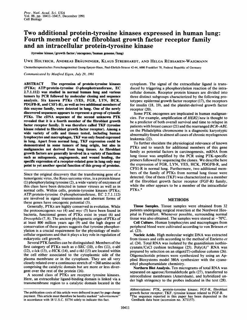
FIG. 1. Expression of the TKF gene. RNA was isolated fromadult normal and malignant human lung tissues. Samples of 6 ,ug oftotal RNAs were fractionated by electrophoresis through a 2.2 Mformaldehyde/1% agarose gel and transferred to a nylon filter. Theblots were probed under high stringency with the 260-bp T1 fragmentobtained from anchored PCR. Lanes: 1, lung carcinoma; 2, normallung.
RNA and Anchored PCR. First-strand cDNA synthesis wasdone by using a PTK-specific primer [P6(2)/0.2 ,M], 3 ,g oftotal RNA, and 10 units of Moloney murine leukemia virusreverse transcriptase (50 mM Tris HCl, pH 8.3/75 mMKCl/10 mM dithiothreitol/3 mM MgCl2/500 AM dNTPs/100,g ofbovine serum albumin per ml). In RNA PCRs halfofthefirst-strand syntheses were used as templates in 25-cyclePCRs [8.3 mM Tris-HCl, pH 8.8/41.7 mM KCl/1.25 mMMgCl2/0.01% gelatin/166.7 ,M dNTPs/0.2 ,uM each prim-er/5 units of Taq polymerase (Amersham)]. One cycle was 1min at 96°C, 2 min at 40°C, and 3 min at 70°C.For anchored PCR (29), 50 units ofterminal transferase and
100 ,M dGTP were used to add a homopolymer to 0.5-100pg of cDNA (100 mM sodium cacodylate pH 7.1/1 mMCoCl2/0.1 mM dithiothreitol/50 ,g of bovine serum albuminper ml/25 mM Tris HCl, pH 7.1). Half of a tailing reactionwas used as template in a 25-cycle PCR with 0.2 ,M each theanchor P12 and the specific P6(2) primer (10 mM Tris-HCI,pH 8.8/50 mM KCl/1.5 mM MgCl2/0.01% gelatin/0.2 mMdNTPs/5 units of Taq polymerase). One microliter (of 50 IlI)ofPCR products was used as template in a second PCR usinganother specific primer.For further analysis, all PCR products were ethanol pre-
cipitated, ligated into the Bluescript KS+ vector (Strata-gene), and sequenced (30).The following primers were used: P6(2), 5'-ATCCCAT-
AACACCACACGTC; P5(1), 5'-TTTGTCCACCGAGA-CCTGGC; P5, 5'-TCCACCGGGACCTGCGGGC; P6, 5'-CCAAAGGACCAGACGTCAGA; P12, 5'-GATTTCA-GAGAACTAAAC(dC)15; P5(3), 5'-TTTATCCACCGAGACCTGGC; P6(3), 5'-ATCCCATAGCACCACACGT.
Construction and Screening of a cDNA Library. A cDNAlibrary was constructed by the method of Gubler and Hoff-man (31) using a Pharmacia kit and 5 ,g of poly(A)+ RNAfrom lung tissue. Then, 1.8 x 10⁶ recombinant AgtlO plaqueswere screened under high stringency (28) using a 260-base-pair (bp) TKF fragment obtained from an anchored PCR witha specific activity of 5 x 10⁹ dpm/,g.DNA Sequencing. PCR products and positive clones from
the cDNA library were sequenced by the dideoxynucleotidechain-termination method (30) after subcloning into the Blue-script vector. Deletion fragments were created by the Exo IIImethod (32).
161
121181241301361
Table 1. Expression of PTKsPrimer PTK detected
P5/P6 YES, FGR, LYN, HCKP5(3)/P6(3) PDGFB-RP5(1)/P6(2) T1T1/P6(2) CSF1-RT2/P6(2) T2
The deduced amino acid sequences (single-letter code) for T1 andT2 are as follows: T1, ARNVLVTEDNVMKIADFGLARGVH-HIDYYKKTSNGRLPVKWMAPEALFDRVYTHQS-56; T2,ARNVLVSEDNVAKVSDFGLTKEASSTQDTGKLPVK-WTAPEALREKKFSTKS-51. These sequences correspond to FIKsubdomains VI-IX. The sequences of the primers are given inMethods.
Determination of Phylogenetic Relationship. Phylogeneticrelationships were determined by using the TREE program ofHUSAR (Heidelberg Unix Sequence Analysis Resource,Deutsches Krebsforschungsrentrum, Heidelberg), based onthe progressive alignment method of Feng and Doolittle (33)in a multiple sequence alignment. Thirty-three human PTKsequences were aligned over the entire catalytic domains (16)(amino acids 67-351 of TKF, corresponding to amino acids265-523 of SRC). The kinase inserts of PDGFA-R,PDGFB-R, KIT, CSF1-R, and FLT were deleted prior toalignment because of their great divergence in length andsequence.
RESULTSIdentification of PTKs in Human Lung Tissue. Aligning the
catalytic domains of known PTKs demonstrates that theirdegree of conservation is not uniform but rather alternates.Within the catalytic domain, 11 major subdomains of highconservation exist (16). Degenerate oligonucleotide primersPS and P6 from subdomains VI and IX (see Methods)corresponding to the motifs His-Arg-Asp-Leu and Asp-Val-Trp-Ser-Phe-Gly were used for PCR based on cDNA fromhuman lung tissues.The PCR products were cloned and the complete se-
quences of 212 inserts were determined. A computer homol-ogy search (European Molecular Biology Laboratory, Gen-Bank, Swiss-Prot, and National Biomedical Resource)showed that six types of clones represented known PTKs:HCK (14), FGR (34), LYN (35), YES (36), PDGFB-R (20),and CSF1-R (37). In addition, two more sequences, T1 andT2, were found (Table 1), one of which has been describedindependently by Partanen et al. (38) using the strategy ofWilks (39).
Isolation of the FGF-R-Related Gene. To clone the T1-corresponding cDNA, the sequence of T1 was elongated by60 bp into a more specific domain by anchored PCR. This260-bp Tl-specific fragment was used to screen 1.8 x 10⁶recombinant clones from a cDNA library from normal lung.
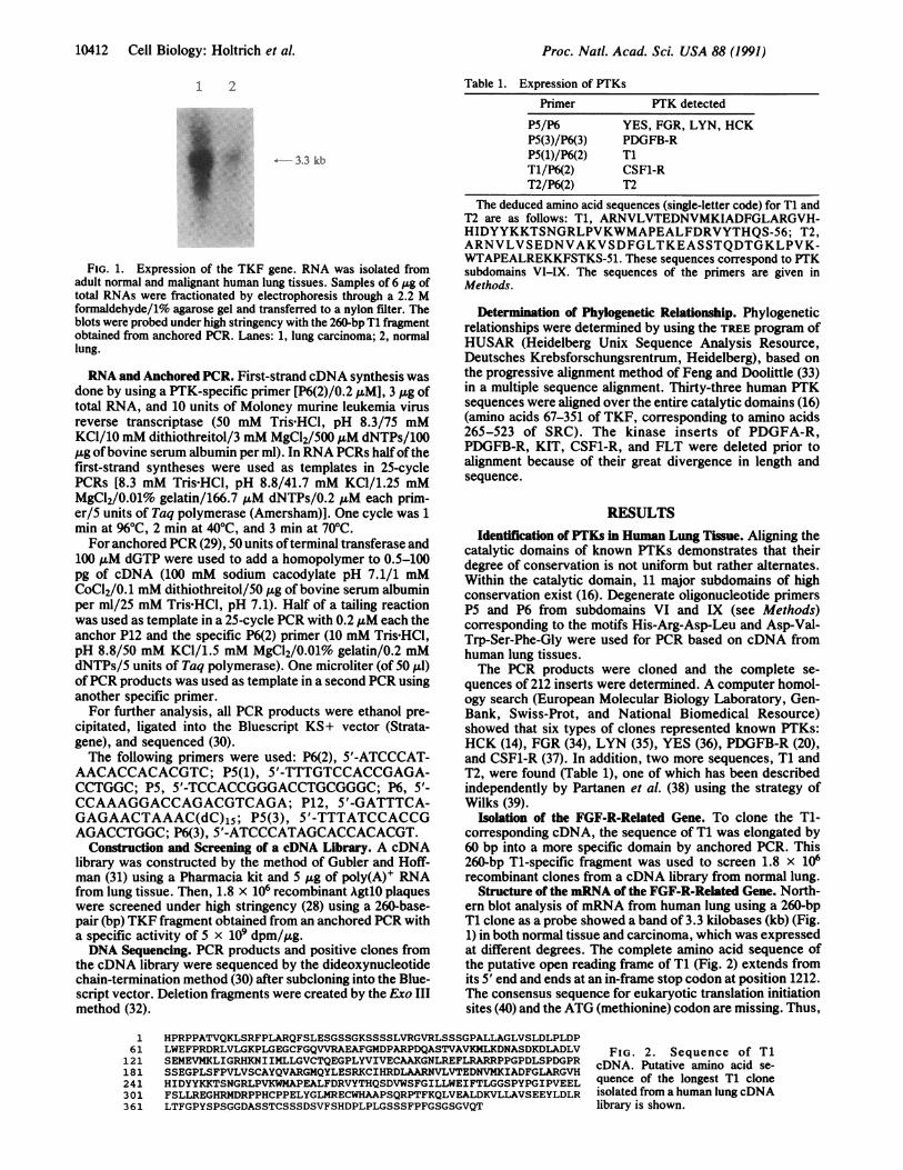
Structure of the mRNA of the FGF-R-Related Gene. North-ern blot analysis of mRNA from human lung using a 260-bpT1 clone as a probe showed a band of 3.3 kilobases (kb) (Fig.1) in both normal tissue and carcinoma, which was expressedat different degrees. The complete amino acid sequence ofthe putative open reading frame of T1 (Fig. 2) extends fromits 5' end and ends at an in-frame stop codon at position 1212.The consensus sequence for eukaryotic translation initiationsites (40) and the ATG (methionine) codon are missing. Thus,
HPRPPATVQKLSRFPLARQFSLESGSSGKSSSSLVRGVRLSSSGPALLAGLVSLDLPLDPLWEFPRDRLVLGKPLGEGCFGQVVRAEAFGMDPARPDQASTVAVKMLKDNASDKDLADLV FIG. 2. Sequence of T1SEMEVMKLIGRHKNIIMLLGVCTQEGPLYVIVECAAKGNLREFLRARRPPGPDLSPDGPR cDNA Putative amino acid se-SSEGPLSFPVLVSCAYQVARGMQYLESRKCIHRDLAARNVLVTEDNVMKIADFGLARGVH c . ta loe T1 cloneHIDYYKKTSNGRLPVKWMAPEALFDRVYTHQSDVWSFGILLWEIFTLGGSPYPGIPVEEL quence of the longest Ti cloneFSLLREGHRMDRPPHCPPELYGLMRECWHAAPSQRPTFKQLVEALDKVLLAVSEEYLDLR isolated from a human lung cDNALTFGPYSPSGGDASSTCSSSDSVFSHDPLPLGSSSFPFGSGSGVQT libray is shown.
10412 Cell Biology: Holtrich et al.

Proc. Natl. Acad. Sci. USA 88 (1991) 10413
TKFFLGBEKFGFR3
TKFFLGBEKFGFR3
TKFFLGBEKFGPR3
TKFFLGBEKFGFR3
TKFFLGBEKFGFR3
TKFFLGBEKFGFR3
TKFFLGBEKFGFR3
HPRPPATVQKLSRFPLARQ--FSLESGSSGKSSSSLVRGVRLSSS--GPALLAGLVSLDL 56
SQMAVAKLA.SIPLRRQVT--V.AD.SA.MN.GVL ... PS --.TPM ... VSEYE. 465
FSSQ ..VHKLTK.I ..R..VTV.A..S..MN.NTP... ITTRL..TADTPM.. VSEYE. 468
KGLGSP ..H.I .... ..--V.... NA.MS.NTP...IA. .. G--EGPT ..NVSE.E. 459
PLDPLWEFPRDRLVLGKPLGEGCF ,VVRAEAFGMDPARPDQASTiMLKDNASDKDL 116.E. .L ....L ... I.L.KDK.NRVTK ... I.... SD.TE... 525
.E.K K.T .V.I.KDK.KE.V .... .... D.TE 528
.A. .K. .LS.A. .T...............M.. MI.I.KD.AAKPV D.......T.... 519
ADLVSEMEVMKLIGRHKNIIMLLGVCTQEGPLYVIVECAAKGNLREFLRARRPPGPDLSP 176S. I... M..M..M A .D. Y.S Y.Q LEYCY 585
S. M. .M. A .D. Y.S Y MEY.Y 588
S. M. .M. N.. A. G .Y.A.L.Y.F 579
DGPRSSEGPLSFPVLVSCAYQVARGMQYLESRKCI RDLAARNVLVTEDNVMKIA i LA 236
NPSHNP.EQ. .SKD E. .A.K .. ..... . 645
.IN.VP.EQMT.KD ..L......E..A.Q || N ... .. 648
.TCKPP.EQ.T.KD .A.Q.. _..... .. 639
RGVHHIDYYKKTSNGRLPVKWHAPEALFDRVYTHQSDVWSFGILLWEIFTLGGSPYPGIP 296
.DINN T.............I..V.M............... 708
.D..NL. T....L.....V.M. 699
VEELFSLLREGHRMDRPPHCPPELYGLMRECWHAAPSQRPTFKQLVEALDKVL-LAVSEE 356.....K .K K.SN.TN ... MM..D ... V. D. .RIVA.TSNQ. 765
.....K .K K.AN.TN ...10..D....V. D. .RI.T.TTN.. 768
.....K .K K.AN.THD ..MI............ D..R. .TVTSTD. 759
YLDLRLTFGPYSPSGGDASSTCSSS--DSVFSHDPLPLGSSSFPFGSGSGVQT 404.SMPLDQ .... FP.TR.STC ..GE. E .EEPCLPRHPAQLANGGLKRR 825
.SQPLEQ .........YP .. P. .M.YEPCLPQYPHINGSVKT 827
.... SAP.EQ ... GGQ.TP.SS ..G-DDA... .L..PAPP.SGGSRT 806
T1 most likely does not comprise the whole mRNA ofthe newgene. The 3' noncoding region has a length of 527 bp, whichencompasses the signal for poly(A) addition at position 1656and a poly(A) sequence at its end.A computer analytic comparison of the T1 sequence re-
vealed that its putative translation product is related tocatalytic domains ofPTKs. The homology is most striking forthe family of receptor tyrosine kinases and in particular to thesubgroup of FGF-Rs. The relationship of T1 is closest to theproducts of two genes, FLG (69% homology) and BEK-(68%homology), and less for those of RET, CSF1-R, FLT, KIT,and PDGF-R. Because of this relationship to PTKs of the
FIG. 3. Comparison of cata-lytic domains of human FGF-Rs.The catalytic domains of TKF,FLG, BEK, and FGF-R3 as de-duced from the cDNA clones arealigned. Dots replace residuessimilar to the putative TKF pro-tein. Dashes represent gaps intro-duced to improve alignment. Mo-tifs referred to in the text areboxed.
FGF-R family, we named the gene TKF (tyrosine kinaserelated to the FGF-R).A comparison of the catalytic domains of FLG (41), BEK
(41), FGF-R3 (42), and TKF (Fig. 3) confirmed TKF to be amember of the PTK family. The consensus Gly-Xaa-Gly-Xaa-Xaa-Gly found in many nucleotide-binding proteins inaddition to PTKs (43) is perfectly conserved in TKF at aminoacid positions 76-81. A highly conserved lysine residue,which seems to be directly involved in the phosphotransferreaction possibly mediating proton transfer (44), is located atposition 105. Three invariant amino acids (Asp-Phe-Gly)implicated in ATP-binding are located between residue po-
Table 2. Expression of TKF in normal and malignant human tissuesNumber of Number ofsamples samples
Tissue Expression studied Tissue Expression studiedNormal tissue TumorsLung + 19 IntestineIntestine (colon, sigma, Adenocarcinoma
rectum) - 7 (mucigenous) + 3Stomach - 1 AdenocarcinomaEsophagus - 2 (not mucigenous) + 3Thymus - 1 AdenocarcinomaSpleen - 1 (not mucigenous) - 1Placenta - 1 StomachForeskin - 1 Adenocarcinoma - 1
Normal human cells EsophagusLymphocytes - Epidermoid - 2Macrophages - Brain - 3
Tumors Skeletal metastasesLung AdenocarcinomaAdenocarcinoma - 4 (breast cancer) + 1Epidermoid (partially Adenocarcinoma
differentiated) - 13 (cardia carcinoma) + 1Epidermoid (partially
differentiated) + 1Epidermoid
(undifferentiated) + 2Carcinoid (primary) + 1
Cell Biology: Holtrich et al.

Proc. Natl. Acad. Sci. USA 88 (1991)
Table 3. Relationship of FLG and BEK sequences with otherpublished sequences of genes from the FGF-R family
Sequence variations
- SRC
- YES
FYN
TYK2
FIG. 4. Phylogenetic relationships of 33 human PTK sequences.The tree was calculated as indicated in Methods. It is informativeonly concerning the branching order. Branch lengths in this figure arenot proportional to true evolutionary distances.
sitions 232 and 234. Sequences that distinguish PTKs fromserine/threonine kinases, His-Arg-Asp-Leu-Ala-Ala-Arg-Asn-Val (positions 212-220) and Pro-Val-Lys-Trp-Met-Ala-Pro (positions 254-259), are conserved in comparison toother PTKs and are identical to BEK and FLG. Compared toother PTKs (16), the kinase domain has an insertion of 14amino acids identical in length to BEK and FLG (residues172-185 of TKF).
Expression ofTKF. The expression ofTKF in various cellsand tissues is shown in Table 2. Very interestingly, TKF isspecifically expressed in lung and not in other tissues andcells, including human lymphocytes and macrophages. Inseveral tumors, however, including malignancies not derivedfrom lung, TKF was found expressed as well.
Phylogeny of the PTK-Specific Catalytic Domains of TKFand T2. The sequences of the catalytic domains of 33 human
TK14 (47)
K-Sam (49)
FGF2H (45)sbFGF (48)
FGFlaFGF-R (46)
Compared to BEK (41)aa 308/309: 3-aa insertionaa 429/430 deletedaa 37-125 deletedaa 314-361, z50% homologyaa 429/430 deletedaa 761-772 highly divergentC-terminal 48 aa deleted
Compared to FLG (41)aa 30-119 deletedaa 30-119 deletedaa 148/149 deletedaa 148/149 deletedaa 817 exchanged
aa, Amino acid(s).
PTKs (corresponding to amino acids 265-523 of pp60c-SRC)were used to determine their phylogenetic relationships (Fig.4). Five major branches can be distinguished: (i) insulinreceptors, (ii) intracellular PTKs, (iii) FGF-Rs, (iv) platelet-derived growth factor receptors, and (v) epidermal growthfactor receptors. Again, TKF is grouped with the family ofthe FGF-Rs. Within this family BEK, FLG, and FGF-R3 aremore closely related to each other than to TKF. T2, incontrast, is grouping with the intracellular PTKs and appearsto define a distinct group.
DISCUSSIONBecause of their mostly low expression rates, the role ofmany PTKs in the regulation of normal cells, and also in thedevelopment of malignant tumors, is not very well under-stood. The use of PCR technology now allows us to effi-ciently search for messages of even very low abundance.Among receptor PTKs, so far only the expression of ERB-Bhas been described in lung. We have found the expression ofadditional PTKs-HCK, YES, FGR, LYN, PDGFB-R,CSF1-R, and two additional genes in this tissue. Among thenewly discovered genes, T2 represents an unusual group ofPTKs (Fig. 4). Further studies (unpublished data) haveshown that it is related to csk, recently isolated from ratbrain.The putative TKF product is significantly homologous to
various human receptors ofthe heparin-binding growth factorfamily: BEK (41), FLG (41), FGF-R h2/h3 (45), FGF-r (46),TK 14 (47), FGF-Rs (48), and K-sam (49). A homology searchshowed that they belong to two categories (Table 3): (i)FGF-Rs of the BEK type: The putative proteins ofBEK andTK 14 are identical (100% homology) except for the occur-rence oftwo gaps inTK 14 due to an addition of 3 amino acidsat position 309 and a deletion of 2 amino acids at position 431.K-sam, which is amplified in the stomach cancer-derived cellline KATO-II (49), is 95.9% homologous to BEK at theamino acid level with two gaps. The largest gap of 88 aminoacids in K-sam occurs within the first of the three extracel-lular immunoglobulin-like domains of BEK. Thus, most ofthe structural variations ofTK' 14 and K-sam, which are bothof tumorigenic origin, occur in the extracellular domain andmight influence the ligand-mediated signal transduction byFGF. (it) FGF-R of the FLG type: FLG has been isolatedfrom a human umbilical vein endothelial cell (HUVEC)cDNA (41, 50). Comparison to a human receptor for acidicand basic FGF (46) and to a shorter form of a human FGF-R(48) shows homologies of 99.9% and 99.8%, respectively.The alignment ofFLG to diverse forms (h2/3) of a receptor
for acidic and basic FGFs (45) from a HUVEC and a placenta
10414 Cell Biology: Holtrich et al.

Proc. Natl. Acad. Sci. USA 88 (1991) 10415
library shows 100% homology and one gap, which is againlocated in the region of the immunoglobulin-like domain. Inaddition, some forms of FGF-R (h4/5) cDNA (45) wereobserved, which encode only the extracellular domain of theFGF-R, a variant that is discussed as a secreted protein. Thedifferent receptor species differ by a few amino acids in theextracellular domain. These multiple forms of FGF-R, whichare highly homologous to FLG may derive by alternativesplicing, a process that has been detected in mouse cells aswell (51).
In summary, the different representatives of the FGF-Rsare various modifications of two genes, BEK and FLG. Theirheterogeneity is mostly due to tumorigenic abnormalities orpossibly specific regulation by alternative splicing. Veryrecently, another member of the FGF-R family of humans hasbeen described (42), which was called FGF-R3. The genedescribed here, TKF, is clearly distinct from FGF-R3 andthus represents a fourth member of this family (Fig. 3).
Generally, several different but related receptors for acertain family of growth factors exist in various species.There are two different human platelet-derived growth factorreceptors (52), various receptors for insulin and insulin-likegrowth factor (53), and different FGF-R genes in chickens(54, 55) and mice (51, 56). The existence of seven differentFGFs might indicate that the FGF-R family has a particularlyhigh number of different members. In line with'this assump-tion binding analyses on cells demonstrated that differencesin the relative capacities of acidic and basic FGF to bind tothe BEK and FLG proteins are not due to different affinitiesor differences in the expression of these receptors (41).Furthermore, by affinity labeling experiments using either1251I-labeled acidic FGF or 1251I-labeled basic'FGF, a variety ofreceptor proteins were identified (45). Changes in heparin-binding FGF gene expression and receptor phenotype havealso been described during liver regeneration (57). Whetherthe specific expression of our TKF gene in lung only pointsto a very specific biochemical function remains to be deter-mined. In any case, it does not appear to be due to invadinglymphocytes and/or macrophages as both cell types havebeen found negative for TKF expression.
We are very grateful to Dr. Brede, Dr. Doermer, Dr. Rosenthal,Dr. Merz, and Dr. Bockhorn for their advice and for supplying tissuesamples. The Georg-Speyer-Haus is supported by the Bundesge-sundheitsministerium and the Hessisches Ministerium fMlr Wissen-schaft und Kunst. This work was supported by the Hermann-Schlosser-Stiftung (U.H.) and the Deutsche Forschungsgemein-schaft (A.B.).
1. Collett, M. & Erikson, R. L. (1978) Proc. Nati. Acad. Sci. USA 75,2021-2024.
2. Hunter, T. & Sefton, B. M. (1980) Proc. Nati. Acad. Sci. USA 77,1311-1315.
3. Bishop, J. M. (1987) Science 235, 305-311.4. Neuman-Silberberg, F. S., Schejter, E., Hoffman, F. M. & Shilo, B.
(1984) Cell 37, 1027-1033.5. Katzen, A. L., Kornberg, T. B. & Bishop, J. M. (1985) Cell 41, 449-456.6. Dahl, C., Biemann, H. P. & Dahl, J. (1987) Proc. Natd. Acad. Sci. USA
84, 4012-4016.7. Shilo, B. & Weinberg, R. A. (1981) Proc. Natl. Acad. Sci. USA 78,
6789-6792.8. Hafen, E., Basler, K., Edstroem, J. E. & Rubin, G. M. (1987) Science
236, 55-58.9. Yarden, Y. & Ullrich, A. (1988) Annu. Rev. Biochem. 57, 443-478.
10. Golden, A., Nemeth, S. P. & Brugge, J. S. (1986) Proc. Natl. Acad. Sci.USA 83, 852-856.
11. Barbacid, M., Beemon, K. & Devare, S. G. (1980) Proc. Natl. Acad. Sci.USA 77, 5158-5162.
12. Witte, 0. N., Rosenberg, N. & Baltimore, D. (1979) Nature (London)281, 396-398.
13. Marth, J. D., Peet, R., Krebs, E. G. & Perlmutter, R. M. (1985) Cell 43,393-404.
14. Ziegler, S. F., Marth, J. D., Lewis, D. B. & Perlmutter, R. M. (1987)Mol. Cell. Biol. 7, 2276-2285.
15. Strebhardt, K., Mullins, J. I., Bruck, C. & Rubsamen-Waigmann, H.(1987) Proc. Natl. Acad. Sci. USA 84, 8778-8782.
16. Hanks, S. K., Quinn, A. M. & Hunter, T. (1988) Science 241, 42-52.17. King, C. R., Kraus, M. H. & Aaronson, S. A. (1985) Science 229,
974-978.18. Ullrich, A., Bell, J. R., Chen, E. Y., Herrera, R., Petruzzelli, L. M.,
Dull, T. J., Gray, A., Coussens, L., Liao, Y. C., Tsubokawa, M.,Mason, A., Seeburg, P. H., Grunfeld, C., Rosen, 0. M. & Ramachan-dran, J. (1985) Nature (London) 313, 756-761.
19. Ebina, Y., Ellis, L., Jarnagin, K., Eclery, M. & Graf, L. (1985) Cell 40,747-758.
20. Yarden, Y., Escobedo, J. A., Kuang, W.-J., Yang-Feng, T. L., Daniel,T. O., Tremble, P. M., Chen, E. Y., Ando, M. E., Harkiens, R. N.,Francke, U., Fried, V. A., Ullrich, A. & Wiliams, L. T. (1986) Nature(London) 323, 226-232.
21. Slamon, D. J., Clark, G. M., Wong, S. G., Levin, W. J., Ullrich, A. &McGuire, W. L. (1987) Science 235, 177-182.
22. Witte, 0. N. & Lugo, T. G. (1989) Mol. Cell. Biol. 9, 1263-1270.23. von Briesen, H., Andreesen, R. & Rubsamen-Waigmann, H. (1990)
Virology 178, 597-602.24. Enrietto, P. J., Payne, L. N. & Hayman, M. J. (1983) Cell 35, 369-379.25. Chirgwin, J. M., Przybyla, A. E., MacDonald, R. J. & Rutter, W. J.
(1979) Biochemistry 18, 5294-5299.26. Aviv, H. & Leder, P. (1972) Proc. Natl. Acad. Sci. USA 69, 1408-1412.27. Lehrach, H., Diamond, D., Wozney, J. M. & Boedtker, H. (1977)
Biochemistry 16, 4743-4748.28. Sambrook, J., Fritsch, E. F. & Maniatis, T. (1989) Molecular Cloning:A
Laboratory Manual (Cold Spring Harbor Lab., Cold Spring Harbor, NY).29. Loh, E. Y., Elliott, J. F., Cwirla, S., Lanier, L. L. & Davis, M. M.
(1989) Science 243, 217-220.30. Sanger, F., Nicklen, S. & Coulson, A. R. (1977) Proc. NatI. Acad. Sci.
USA 74, 5463-5467.31. Gubler, U. & Hoffman, B. J. (1983) Gene 25, 263-269.32. Hoheisel, J. & Pohl, F. M. (1986) Nucleic Acids Res. 14, 3605-3608.33. Feng, D.-F. & Doolittle, R. F. (1987) J. Mol. Evol. 25, 351-360.34. Nishizawa, M., Semba, H., Yoshida, M. C., Yamamoto, T., Sasaki, M.
& Toyoshima, K. (1986) Mol. Cell. Biol. 6, 511-517.35. Yamanashi, Y., Fukushige, S.-J., Semba, K., Sukegawa, J., Miyajima,
N., Matsubara, K.-J., Yamamoto, T. & Toyoshima, K. (1987) Mol. Cell.Biol. 7, 237-243.
36. Sukegawa, J., Semba, K., Yamanashi, Y., Nishizawa, M., Miyajima, N.,Yamamoto, T. & Toyoshima, K. (1987) Mol. Cell. Biol. 7, 41-47.
37. Coussens, L., Van Beveren, C., Smith, D., Chen, E., Mitchell, R. L.,Isacke, C. M., Verma, I. M. & Ullrich, A. (1986) Nature (London) 320,277-280.
38. Partanen, J., Haikela, T. P., Alitalo, R., Lehvaslaiho, H. & Alitalo, K.(1990) Proc. Natl. Acad. Sci. USA 87, 8913-8917.
39. Wilks, A. (1989) Proc. Natl. Acad. Sci. USA 86, 1603-1607.40. Kozak, M. (1984) Nucleic Acids Res. 12, 857-872.41. Dionne, C. A., Crumley, G., Bellot, F., Kaplow, J. M., Searfoss, G.,
Ruta, M., Burgess, W. H., Jaye, M. & Schlessinger, J. (1990) EMBO J.9, 2685-2692.
42. Keegan, K., Johnson, D. E., Williams, L. T. & Hayman, M. J. (1991)Proc. Natl. Acad. Sci. USA 88, 1095-1099.
43. Wierenga, R. K. & Hol, W. G. J. (1983) Nature (London) 302, 842-844.44. Kamps, M. P., Taylor, S. S. & Sefton, B. M. (1984) Nature (London)
310, 589-592.45. Johnson, D. E., Lee, P. L., Lu, J. & Williams, L. T. (1990) Mol. Cell.
Biol. 10, 4728-4736.46. Isacchi, A., Bergonzoni, L. & Sarmientos, P. (1990) Nucleic Acids Res.
18, 1906.47. Houssaint, E., Blanquet, P. R., Champion-Arnaud, P., Gesnel, M. C.,
Torriglia, A., Courtois, Y. & Breathnach, R. (1990) Proc. Natl. Acad.Sci. USA 87, 8180-8184.
48. Itoh, N., Terachi, T., Okta, M. & Seo, M. K. (1990) Biochem. Biophys.Res. Commun. 169, 680-685.
49. Hattori, Y., Odagin, H., Nakatani, H., Miyagawa, K., Naito, K.,Sakamoto, H., Katoh, O., Yoshida, T., Sugimura, T. & Terada, M. (1990)Proc. Natl. Acad. Sci. USA 87, 5983-5987.
50. Ruta, M., Howk, R., Ricca, G., Drohan, W., Zabelshansky, M., Lau-reys, G., Barton, D. E., Francke, U., Schlessinger, J. & Givol, D. (1988)Oncogene 3, 9-15.
51. Reid, H. H., Wilks, A. F. & Bernard, 0. (1990) Proc. Natl. Acad. Sci.USA 87, 1596-1600.
52. Ullrich, A. & Schlessinger, J. (1990) Cell 61, 203-212.53. Czech, M. P. (1989) Cell 59, 235-238.54. Lee, P. L., Johnson, D. E., Cousens, L. S., Fried, V. A. & Williams,
L. T. (1989) Science 245, 57-60.55. Pasquale, E. B. & Singer, S. J. (1990) Proc. Natl. Acad. Sci. USA 86,
5449-5453.56. Kornbluth, S., Paulson, K. E. & Hanafusa, H. (1988) Mol. Cell. Biol. 8,
5541-5544.57. Hou, J. Z., Kan, M. K., McKeehan, K., McBride, G., Adamns, P. &
McKeehan, W. L. (1991) Science 251, 665-668.
Cell Biology: Holtrich et al.