inhibition of protein tyrosine phosphatases by mild oxidative … · oxidative stress and tyrosine...
TRANSCRIPT
Oxidative Stress and Tyrosine Phosphatase S-Nitrosylation
INHIBITION OF PROTEIN TYROSINE PHOSPHATASES BY MILD OXIDATIVE STRESSES IS DEPENDENT ON S-
NITROSYLATION*
Daniel M. Barrett§, Stephen M. Black#, Horia Todor§, Rupert K. Schmidt-Ullrich§†, Kathryn S. Dawson‡ and Ross B. Mikkelsen§
From the Departments of Radiation Oncology§ and Biostatistics‡, Virginia Commonwealth University, Richmond, Virginia 23298-0058 and # Department of Biomedical and Pharmaceutical
Sciences, and the International Heart Institute, the University of Montana, Missoula, MT. Running title: Oxidative Stress and Tyrosine Phosphatase S-Nitrosylation
Address correspondance to: Ross B. Mikkelsen, Massey Cancer Center, 401 College Street, Richmond, VA 23298-0058. Tel: 804-628-0857; Fax: 804-828-6042; E-mail: [email protected]
Previous studies have shown that a Ca2+ dependent nitric oxide synthase (NOS) is activated as part of a cellular response to low doses of ionizing radiation. Genetic and pharmacological inhibitor studies linked this NO signaling to the radiation-induced activation of ERK1/2. Herein, a mechanism for the radiation-induced activation of Tyr phosphorylation dependent pathways (e.g. ERK1/2) involving the inhibition of protein Tyr phosphatases (PTP) by S-nitrosylation is tested. The basis for this mechanism resides in the redox-sensitive active site Cys in PTPs. These studies also examined oxidative stress induced by low concentrations of H2O2. S-nitrosylation of total cellular PTP and immunopurified SHP-1 and SHP-2 was detected as protection of PTP enzymatic activity from alkylation by N-ethylmaleimide and reversal by ascorbate. Both radiation and H2O2 protected PTP activity from alkylation by a mechanism reversible by ascorbate and inhibited by NOS inhibitors or expression of a dominant negative mutant of NOS-1. Radiation and H2O2 stimulated a transient increase in cytoplasmic free [Ca2+]. Radiation, H2O2 and the Ca2+ ionophore, ionomycin, also stimulated NOS activity and this was associated with an enhanced S-nitrosylation of the active site Cys453 determined by isolation of S-nitrosylated wild type but not active site Cys453→Ser SHP-1
mutant by the “biotin-switch” method. Thus one consequence of oxidative stimulation of NO generation is S-nitrosylation and inhibition of PTPs critical in cellular signal transduction pathways. These results support the conclusion that a mild oxidative signal is converted to a nitrosative one due to the better redox signaling properties of NO.
Phosphotyrosine phosphatases (PTP1) compose a super family of phosphatases that hydrolyze phosphoTyr residues in proteins critically involved in several cell-signaling pathways (1-3). Their active sites are characterized by the consensus sequence (I/V)HCXAGXXR(S/T). Mutation of the active site Cys results in a catalytically inactive, dominant negative enzyme (3). The redox state of the active site Cys has been shown to be critical modulator of PTP activity (4). For example, the reversible oxidation of the active site Cys to sulfenic acid by ROS such as H2O2 inhibits PTP activity (4). A prolonged exposure to high concentrations of H2O2 can irreversibly oxidize sulfenic acid to sulfinic acid (5). Several investigators have also provided evidence suggesting that ligand-stimulated generation of ROS activates growth factor receptor Tyr kinase-dependent signal transduction pathways by inhibiting counteracting PTPs (1,2,6-12). Key evidence in these studies has included the use of
1
JBC Papers in Press. Published on January 31, 2005 as Manuscript M411523200
Copyright 2005 by The American Society for Biochemistry and Molecular Biology, Inc.
by guest on May 29, 2018
http://ww
w.jbc.org/
Dow
nloaded from

Oxidative Stress and Tyrosine Phosphatase S-Nitrosylation
fluorescent dyes to qualitatively measure the ROS generated. However, these dyes do not identify the ROS nor distinguish between ROS and RNS (reviewed in 12).
High cellular concentrations of ROS are toxic and are not generated under normal physiological conditions (13,14). This toxicity, the lack of target selectivity by ROS, and a cell’s complement of ROS scavenging mechanisms would appear to preclude ROS as physiological signal transducing molecules (12). An alternative mechanism for redox modulation of cellular signal transduction pathways proposes that the oxidative signal is converted to a nitrosative signal (11,12,15,16). In contrast to ROS, cellular NO synthesis is tightly regulated by Ca2+ dependent NOS’s, cellular NO buffering, and catabolic mechanisms (12,14,17). This is best exemplified by the catalytic requirements of these enzymes including homodimerization, 3 cosubstrates (NADPH, L-arginine, and O2), and 5 cofactors (calmodulin, heme, FAD, FMN, and tetra-hydrobiopterin) (17). In addition, NOS activities are modulated by phosphorylation and protein-protein interactions (e.g. 12,18, 19). In contrast to ROS, physiologically relevant RNS are not toxic at the intracellular concentrations generated by Ca2+ activated constitutive NOS. At these concentrations RNS are relatively specific in their cellular targets (12,17).
The most studied NO target is soluble guanylate cyclase (20). A number of recent studies have also demonstrated S-nitrosylation of protein Cys as a critical redox driven mechanism for modulation of protein function (12,15,16,21-23). The S-nitrosylation of Cys residues can modify protein activity or simply serve as a NO reservoir through the proteins they are attached to (24-26). Examination of S-nitrosylated proteins has revealed a degenerate consensus sequence X(K,R,H)C(D,E) (16,27). This consensus sequence is found in hundreds of proteins including caspases, cyclin D1, Rb, and BRAC1/2. This motif does not have to be linear; it can be derived from the tertiary structure of the protein (28). Not all proteins containing the consensus sequence become S-nitrosylated, since other factors exist that control S-nitrosylation such as the location of Cys in hydrophobic compartments. Hydrophobic environments enhance the rate of S-nitrosylation by concentrating the lipophilic O2
and NO molecules necessary for the generation of the nitrosylating species N2O3 (12,16,29). Of specific interest to the present study are the findings that exogenous RNS donors inhibit PTP activity (e.g. 9, 30-32).
In two previous studies we demonstrated that a mild oxidative stress produced at low clinically relevant doses of ionizing radiation generated a transient signal involving activation of the Ca2+ dependent NOS-1 in CHO and other epithelial cells (11,12,33). Using a combination of chemical inhibitors and genetic manipulation of NOS-1 activity, we were able to show that the radiation induced transient activation of ERK1/2 signaling in these cells was dependent on NOS activity and RNS generation.
One mechanism postulates that RNS transiently inhibit PTP by reversible oxidation of PTP active site Cys thereby shifting the relative balance of Tyr kinase/PTP activities in favor of enhanced Tyr phosphorylation. In this scenario enhanced ERK1/2 activity is the result of inhibiting PTPs that block the activities of upstream kinases (e.g. epidermal growth factor receptor, Raf kinase) and/or inhibit PTPs downstream that dephosphorylate and inactivate ERK1/2 (e.g. MKP-1 and MKP-3). Since the relative activities of PTPs are 100-1000X greater than the activities of Tyr kinases, only modest changes in PTP activity can have significant effects on the net Tyr phosphorylation state of a protein (1,2). Inhibiting PTP activity provides a mechanism for the findings that ionizing radiation enhances total cellular protein Tyr phosphorylation (34,35) and apparently activates diverse specific Tyr kinases, e.g. ERK1/2, EGFR, c-abl, and c-lyn (36-40).
In the present study we show that mild oxidative stresses such as ionizing radiation or low concentrations of H2O2 reversibly inhibit bulk cellular PTP activity and the activities of specific PTPs (SHP-1 and SHP-2). The mechanism of inhibition involves the activation of Ca2+ dependent NOS and the S-nitrosylation of the PTPs. This RNS-dependent mechanism of PTP inhibition represents one example of how an oxidative signaling event is converted into a nitrosative signal (12).
2
by guest on May 29, 2018
http://ww
w.jbc.org/
Dow
nloaded from

Oxidative Stress and Tyrosine Phosphatase S-Nitrosylation
MATERIALS AND METHODS
Cells and Plasmids. CHO-K1, MCF-7 and MRC-5SV cell lines purchased from American Type Culture Collection (Gaithersburg, MD) were cultured as previously described in RPMI 1640 supplemented with 5% fetal calf serum (33). CHO and MRC-5SV cells were transfected using the LipofectAMINE PLUS kit according to the manufacture’s protocols (Invitrogen, Carlsbad). Transfection efficiency exceeded 80% as attested by transfection with a green fluorescent protein-encoding plasmid and fluorescence microscopy. Plasmids encoding wild type SHP-1 and a dominant negative mutant with the active site 453Cys-Ser mutation were obtained from Dr. C. Susini (41). Dr. J. Pessin (42) provided wild type SHP-2. A NOS-1 dominant negative mutant (HemeRedF) has been described (11, 43). Expression of HemeRedF in CHO cells inhibits radiation-induced endogenous NOS-1 activity (11).
Pharmacological Inhibitors and Radiation Treatments. The NOS inhibitors, L-NAME at 1 mM and L-NNA at 100 nM, obtained from Sigma Chemical Co. (St. Louis MO), were added to the cells one hour prior to radiation exposure or treatment with H2O2. H2O2 was added to 10-100 µM; ionomycin (Sigma Chemical Co.) was added to a final 1µM concentration. Prior to adding H2O2 or ionomycin, cells were washed thrice and incubated for 1 hr with serum free culture medium. This minimized serum catalase activity and serum protein binding of ionomycin. Cells were irradiated at room temperature with a Picker 60Co source at a dose rate of 1.8-2.0 Gy/min and transferred to a slide warming plate to maintain temperature at 37oC. All treatments and experimental manipulations were performed under low light conditions.
S-nitrosylated Protein Purification. S-nitrosylated proteins were purified using a modification of the ‘biotin switch’ method (44). At indicated times of treatment, cells were rinsed thrice in ice cold PBS and lysed in ice-cold lysis buffer containing 50mM Tris-HCl (pH 7.5), 150mM NaCl, 1% Nonidet P-40, 0.5% deoxycholic acid, and 0.1% SDS. Cells were scraped and collected into amber polypropylene microcentrifuge tubes to further reduce light exposure. Cell lysates were spun at 13,200 rpm
for 5 min. The resulting supernatant (150 µl) was incubated for 40 min at 50°C with 450 µl of free sulfhydryl blocking buffer consisting of 250mM Hepes (pH 7.7), 1mM EDTA, 0.1 mM neocuproine, 2.5% SDS, and 20 mM methylmethanethiosulfonate as blocking agent (Pierce Chemical Co., Rockford, IL). Proteins were precipitated with 1.0 ml acetone for 30 minutes at -20°C and collected by microcentrifugation. The pellets were re-suspended in 1.0mL of -20°C acetone, centrifuged and solublized in 180 µL of HENS buffer containing 25 mM Hepes (pH 7.7), 0.1 mM EDTA, 10 µM neocuproine, 1% SDS. To each sample 60 µl of 4 mM biotin-HPDP and 2.4 µl of 100 mM ascorbate were added. Ascorbate reduces the S-NO bond (but not disulfide or sulfenic) to the free sulfhydryl and the biotin HPDP reacts specifically with and biotinylates the free sulfhydryl. After a brief vortexing, the samples were incubated for 1 hour at room temperature. Proteins were precipitated with acetone and subsequently solublized in 200 µl of HENS buffer. A small aliquot was used for protein determination and loading controls (actin or PTP Western blots) and the remaining supernatant was mixed with 400µl of neutralization buffer (20 mM Hepes, 100 mM NaCl, 1 mM EDTA, 0.5% Triton X-100, pH 7.7). Streptavidin-agarose (20 µl, 50% suspension, Sigma Chemical Co.) were added to each sample and rotated for 1 hour at 4°C. The streptavidin-agarose beads were washed 4 times with washing buffer (20 mM Hepes, 600 mM NaCl, 1 mM EDTA, 0.5% Triton X-100, pH 7.7) and once with 20 mM Hepes, 100 mM NaCl, 1 mM EDTA (pH 7.7). Proteins were eluted by incubating the beads with 60 µl of the same buffer containing 120 mM β-mercaptoethanol for 20 min to release the biotin group regenerating the protein to its original unmodified form. After centrifugation, the supernatants were mixed with 15µl of 5X Laemmli sample buffer and proteins fractionated by electrophoresis on 8% SDS polyacrylamide gels.
Immunoblotting. After electrophoresis proteins were transferred to nitrocellulose membranes and analyzed using standard Western blotting procedures (e.g. 11). All primary
3
by guest on May 29, 2018
http://ww
w.jbc.org/
Dow
nloaded from

Oxidative Stress and Tyrosine Phosphatase S-Nitrosylation
antibody incubations were performed in 5% bovine serum albumin in Tris-buffered saline with 0.1% Tween at 4°C overnight. Primary rabbit antibodies were from Santa Cruz Biotechnology: SHP-1 (sc-287) and SHP-2 (sc-424). A goat anti-actin polyclonal antibody was also obtained from Santa Cruz Biotechnology. After incubation with primary antibodies, blots were washed 5 times for 5 minutes each before secondary antibodies were added. Incubation with secondary antibody was performed in 5% dry milk solution in Tris-buffered saline with 0.1% Tween at room temperature for 1 hour. The secondary antibody, conjugated to alkaline phosphatase, was diluted according to the manufacturer’s protocols (Promega, Madison WI). After extensive washing the blots were developed with CDP-Star chemiluminescence reagent (Perkin-Elmer, Boston, MA) diluted 1:5 with deionized water and mixed with a 1:40 dilution of Nitro-Block II (Applied Biosystems, Foster City, CA).
Assay of Total and Immuno-Purified PTP Activities. Redox modulation of total cellular PTP activity was measured by modifications of an assay developed by Li and Whorton (1,45). This assay, like the protocol for purification of S-nitrosylated proteins, is based on protection of the active site Cys from NEM alkylation by reversible oxidation of the Cys by either S-nitrosylation, S-glutathiolation or sulfenic acid formation. To distinguish between S-nitrosylation and either S-glutathiolation or sulfenic acid formation, PTP activity is measured after treating NEM-treated lysates or purified PTP with either dithiothreitol to reduce all three oxidative modifications or ascorbate to selectively reduce the S-NO bond (44). Protection against NEM alkylation measured by an increase in dithiothreitol or ascorbate recoverable activity is equated to the amount of PTP inhibition obtained after exposing cells to either low doses of radiation or H2O2.
For total cellular PTP activity measurements, lysates from cells cultured in 3.5 cm dishes were prepared by washing cells once in ice-cold PBS followed by lysis in 200 µl of lysis buffer containing 1% Triton X-100, 100 mM NaCl, 25 mM Hepes (pH 7.4), 1 mM EDTA and with or without 20 mM NEM. After 10 min on ice, lysates were centrifuged and assayed for PTP activity using a commercially available PTP 96-well activity kit (Molecular Probes, Eugene, OR).
This assay procedure uses the PTP substrate, DiFMUP, and a Ser/Thr phosphatase inhibitor cocktail to insure that only PTP activity is measured. To meet the requirements of the present studies the substrate-phosphatase inhibitor mix was solublized in 0.1% Tween 20 and 25 mM MOPS (pH 7.0) plus either 25 mM dithiothreitol or 50 mM ascorbate. Cell lysates initially diluted between 1:2 and 1:10 depending on the cell preparation were further diluted by adding 5 µl to 200 µl of the complete assay buffer to reduce the NEM concentration and initiate catalytic activity. Activity was monitored over the next 10-20 min with a Packard fluorescence plate reader. A linear regression analysis and normalization to protein measured by the Bradford assay was used to calculate PTP specific activities. Figure 1A shows typical raw data and linear regression analysis used for these measurements.
SHP-1 and SHP-2 PTP activities were measured with immunoprecipitates isolated from cells transfected with plasmids expressing these PTPs. Cell lysates prepared as above with NEM were incubated with 2 µg of either anti-SHP-1 or anti-SHP-2 and protein A agarose (Oncogene Sciences, Cambridge MA) for 90 min followed by 4 washes in lysis buffer and two in the same buffer without Triton but containing Ser/Thr phosphatase inhibitors (10 mM pyrophosphate, 10 mM glycerophosphate, 20 mM NaF). Enyzme activity was initiated by adding 20 µM DIFUMP to the beads. Beads were continuously rotated at 37°C for 30 min before microcentrifugation to remove the beads. Fluorescence of the supernatants was measured. Control experiments using vanadate as a PTP inhibitor verified the selectivity of the assay and that the measured activities were linear for at least 30 min incubation. Activities were normalized with respect to cell lysate protein concentrations.
Cell Ca2+ Measurements. Cytosolic free [Ca2+] was measured with the fluorescent Ca2+ sensitive dye, fura-2, as previously described (46) but with ratiometric imaging software provided by Universal Imaging and detection with a Photometrics Sensys CCD camera. Cells on coverslips were loaded with dye by incubating cells at room temperature in serum free medium containing 2.5 µM of the membrane permeant acetoxymethyl ester derivative of fura-2 for 30-60
4
by guest on May 29, 2018
http://ww
w.jbc.org/
Dow
nloaded from

Oxidative Stress and Tyrosine Phosphatase S-Nitrosylation
min. A subsequent incubation for 60 min without dye was used to maximize ester cleavage and dye retention in the cells. Coverslips were mounted in a perfusion chamber prior to microscopic analysis.
NOS Activity. Cellular NOS activity was measured by the arginine-citrulline conversion assay using [3H]-Arginine as previously described (11).
Statistical Analysis. Each experimental figure shown is one experiment representative of at least two experiments. The data points in each figure represent the mean plus or minus the standard deviation of at least two measurements. For the analysis of data from several experiments, results were normalized with respect to a control value (e.g. t=0, or 0Gy) to facilitate comparisons. Two-sided t-tests were used to compare the considered groups. Since the sample numbers are small, it is assumed that the data is sampled from a normal distribution. The t value is calculated as: the absolute value of (mean1-mean2)/sqrt(s12/n1+ s22/n2) where n is sample size and s is the standard deviation. A p<0.05 was determined to be of statistical significance.
RESULTS
Radiation Inhibits Cellular PTP Activity by a NO-Sensitive Mechanism. Our previous investigations showed that a mild oxidative stress induced by exposing cells to low doses of ionizing radiation activated a constitutive, Ca2+ dependent NOS activity in diverse cell types (11,33). This activity was inhibited by a dominant negative mutant of NOS-1 and enhanced by expression of a wild type NOS-1 suggesting that radiation activates a NOS-1 isoform in CHO cells. Furthermore, we showed that radiation-induced activation of the ERK1/2 signaling pathway in CHO cells as revealed by measurements of enzyme activity and Tyr phosphorylation of ERK1/2 could be blocked by NOS inhibitors such as L-NAME or expression of a dominant negative mutant of NOS-1. We proposed that one mechanism for this activation might be inhibition of a counteracting PTP acting at one step of the ERK1/2 activation pathway by S-nitrosylation of the PTP active site Cys. In support of this proposal, preliminary studies showed that radiation stimulated the S-nitrosylation of the
active site Cys of two prominent cellular PTPs, SHP-1 and SHP-2 (12).
More direct evidence for this mechanism has come from measuring total cellular PTPase activity before and after irradiating cells. As described in Experimental Procedures, protection from NEM-alkylation was used to assess active site Cys modification and thus indirectly provide a measure of PTP inhibition. The protection by either S-nitrosylation, oxidation to sulfenic acid or by S-glutathiolation was revealed by treating cell lysates with the SH reducing agent, dithiothreitol. The sensitivity of S-NO to reduction by ascorbate was used to distinguish S-nitrosylation from the ascorbate-insensitive sulfenic acid formation or S-glutathiolation.
Figure 1A shows a typical time course used in the PTP assay. Specific activities were determined from the linear regression analyses and normalized with respect to protein concentration. The results in Figure 1B compare the relative total PTP specific activities before and after treatment with NEM followed by recovery with either dithiothreitol or ascorbate. Maximal cellular PTP specific activity was defined as that measured in cell lysates not treated with NEM but in the presence of 25 mM dithiothreitol. Based on this definition, 60-80% of total cellular PTP activity was irreversibly inhibited by treatment with NEM (n=8 separate experiments). The remaining 20-40% of basal PTP activity was protected from NEM alkylation and inactivation by a mechanism reversible by dithiothreitol treatment. Approximately half as much was also protected by a mechanism reversed by ascorbate treatment (17 ± 8% of total basal PTP activity, n=6). Two sided t-tests revealed statistical differences between the dithiothreitol and ascorbate treatments in six independent experiments with p values <0.01.
Cells were irradiated (5 Gy) and 5 min post-irradiation cell lysates were prepared with NEM. Maximal radiation-induced NOS activity and ERK1/2 activation in CHO cells are observed at 5 min post-irradiation (11). As shown in Figure 1B, an additional modest but significant protection from alkylation was observed following radiation and reversal by treatment with dithiothreitol or ascorbate. The increase in protection produced by a 5 Gy radiation exposure ranged between 5 and 20% of total cellular PTP activity (n=4 separate experiments, p<0.02). Dithiothreitol was
5
by guest on May 29, 2018
http://ww
w.jbc.org/
Dow
nloaded from

Oxidative Stress and Tyrosine Phosphatase S-Nitrosylation
consistently more effective than ascorbate in unmasking this radiation induced PTP inhibition. However, the p values for these comparisons ranged between 0.03 and 0.26 and thus the difference between the two reductants in their effectiveness in revealing radiation-induced PTP activity was not statistically significant.
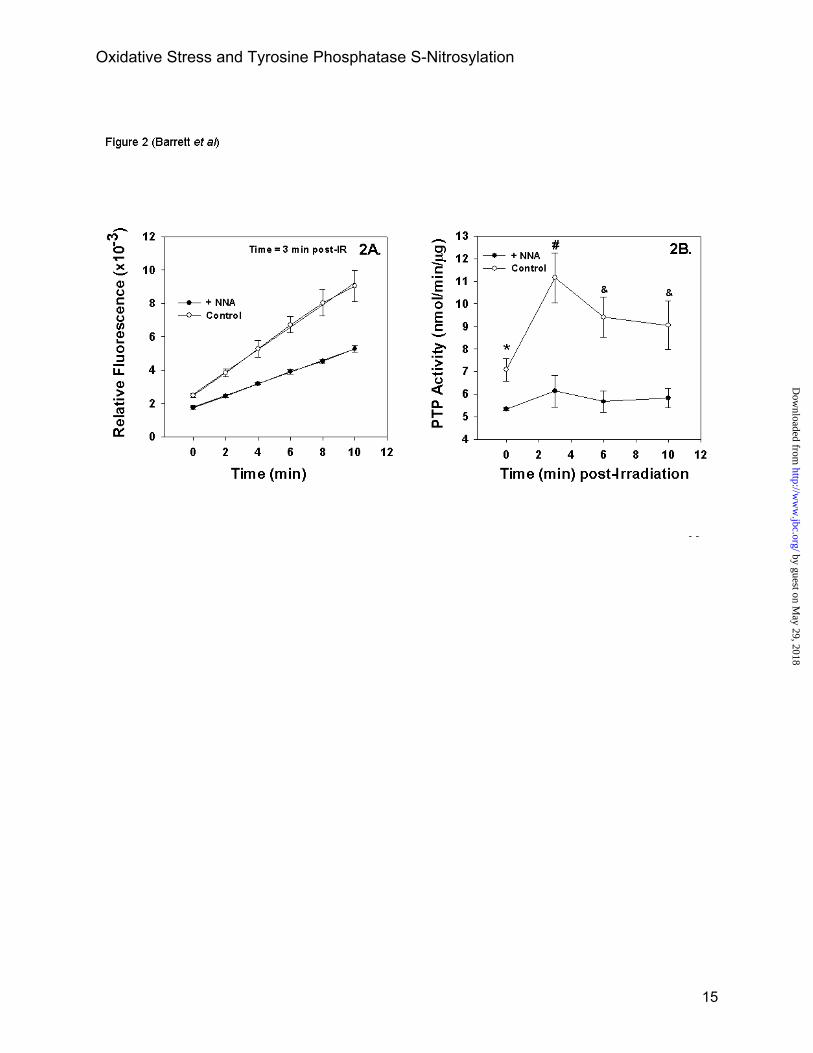
The effects of NOS inhibitors on basal and post-radiation total cellular PTP activities were also assessed. Initial experiments established that pre-incubating cells with 100 nM L-NNA for one hour prior to irradiation inhibited basal and radiation-stimulated NOS activity by 71 ± 12 % (n=3, data not shown). This compares with 50% inhibition observed previously with another NOS inhibitor, L-NAME, at 1.0 mM for one hour (11). Under these treatment conditions, at least 25% of basal cellular PTP activity insensitive to NEM is sensitive to L-NNA (25-55%, n=3, p< 0.05; e.g. Figure 2B). The radiation-stimulated protection was completely inhibited under these conditions (n =3; p < 0.001). Similar results were obtained with L-NAME (e.g. Figure 5 below). These results, combined with the partial ascorbate sensitivity in recovery of PTP activity and our previous findings demonstrating active site S-nitrosylation of the PTP, SHP-1, following radiation treatment of cells (12), support the conclusion that radiation-induced inhibition of PTP activity is a consequence of S-nitrosylation. Pharmacological inhibition of NOS also completely inhibited radiation-induced protection of PTP activities in MCF-7 breast carcinoma cells and MRC-5SV fibroblasts (data not shown).
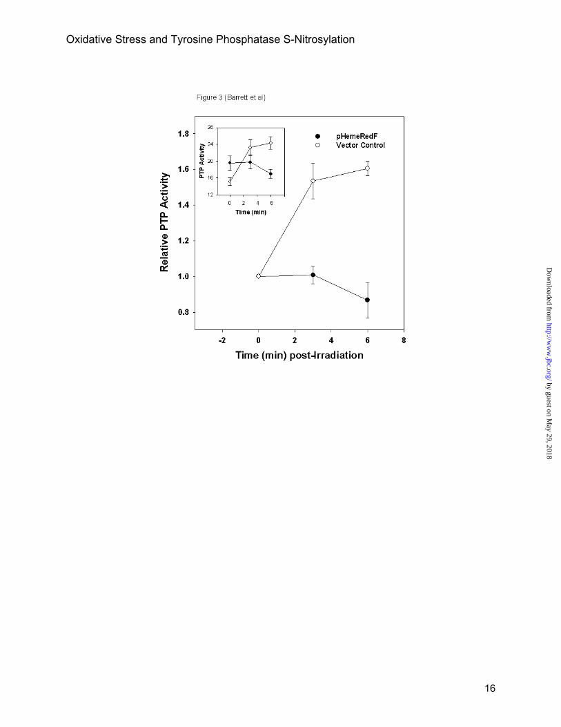
The findings with the pharmacological inhibitors were verified in CHO cells by genetic manipulation of NOS-1 activity using the dominant negative mutant, HemeRedF (11). At 48 hr post-transfection with the mutant, >80% of the radiation-stimulated NOS activity is inhibited (11). Results in Figure 3 from a single experiment representative of two performed show that expression of HemeRedF under these conditions completely blocks the transient PTP inhibition observed following radiation. In contrast to what was found with the chemical inhibitors, a prolonged 48 hr genetic inhibition of NOS activity had no significant effect on basal NEM-insensitive PTP activity with respect to vector control cells (inset to Figure 3; n=3, p<0.6). This suggests possible compensatory mechanisms for redox
protection of PTP function under conditions of sustained NOS inhibition.
SHP-1 and SHP-2 are Inhibited by a Radiation-Induced NO-Sensitive Mechanism. The activities of immunopurified PTPs, SHP-1 and SHP-2, were also measured. The PTPs were immunopurified as described in Experimental Procedures from cell lysates prepared at times directly before radiation and 5 min post-radiation. Catalytic activities were measured with the same fluorescent substrate used in the bulk PTP measurements and with the assays initiated by addition of substrate and either dithiothreitol or ascorbate to the immune complex beads. The results for both PTPs in Figure 4 are of a single representative experiment repeated 3 times with p values shown for that experiment. The results in Figure 4A with purified SHP-1 parallel those obtained with total cellular PTP measurements. After irradiation, there is an approximate 40% increase in NEM insensitive PTP activity measured with dithiothreitol present in the assay (42 ±11%, n=3 independent experiments, p<0.008). With ascorbate there is a 20% increase in NEM insensitive PTP activity following a radiation exposure (21±6%, n=3 independent experiments, p<0.02). That only 50% is recovered with ascorbate relative to dithiothreitol may in part be explained by the prolonged incubation times (30 min). The absence of a sufficiently strong reducing agent such as dithiothreitol can result in increased oxidation of active site Cys and PTP inhibition (e.g. 45). The results and statistical analysis in Panels B and C of Figure 4 demonstrate that pharmacological inhibition of NOS in CHO cells completely blocks the radiation-induced inhibition of both SHP-1 and SHP-2.
Inhibition of PTP Activity by Low Concentrations of H2O2 is Blocked by NOS Inhibitors. Previous studies have demonstrated that relatively high concentrations of H2O2 (approximately 1 mM) oxidized PTP active site Cys to sulfenic acid resulting in PTP inhibition and protection from alkylation (4,6-8). Figure 5 shows a time course of PTP protection following addition of 90 µM H2O2. There is a rapid increase in protected PTP activity that was more sustained in contrast to the transient protection following a radiation exposure. The sustained protection with H2O2 treatment compared to the transient
6
by guest on May 29, 2018
http://ww
w.jbc.org/
Dow
nloaded from

Oxidative Stress and Tyrosine Phosphatase S-Nitrosylation
protection obtained with radiation is probably the result of the continuous presence of the oxidant. The H2O2 induced protection was observed over the concentration range tested of 10 to 100 µM. At concentrations of H2O2 greater than 100 µM and in the serum free culture medium used in these studies significant cell lysis was observed. As was found in the radiation studies, the H2O2 -induced PTP protection was mostly blocked by prior incubation with the NOS inhibitor, L-NAME (Figure 5).
Similar results were obtained when the activity of SHP-1 immunopurified from H2O2 treated cells was measured (Figure 6). SHP-1 purified from H2O2 treated cells was protected from NEM alkylation by a mechanism blocked by L-NAME and reversed by treatment with either dithiothreitol or ascorbate. The H2O2 induced protection was more effectively reversed by dithiothreitol than ascorbate, similar to what was found in measurements of total NEM-insensitive PTP activity after radiation. In contrast to the results obtained for total cellular PTP activity, the H2O2 induced protection of SHP-1 was transient. The transient nature of the protection, however, fits with the findings shown below in Figure 8 that H2O2 induced S-nitrosylation of SHP-1 is also transient.
Low Concentrations of H2O2 Increase Cellular Cytoplasmic Free [Ca2+] and Stimulate NOS Activity. In our previous investigation we demonstrated that ionizing radiation activated cellular NOS-1 activity in CHO cells as assayed by an arginine to citrulline conversion assay (11). These experiments were repeated with H2O2 over the concentration range of 10- 40 µM. At these low concentrations H2O2 stimulates NOS activity about 2-3 fold comparable to what is achieved with the Ca2+ ionophore, ionomycin, or a radiation dose of 2 Gy (Figure 7A and ref.11).
Several investigations have demonstrated that low H2O2 concentrations and other mild oxidative stresses including ionizing radiation reversibly stimulate increases in cytoplasmic free [Ca2+] (e.g. 46,47). We verified this in the cell lines used in the present study at the single cell level using microscopic fluorescence measurements of the Ca2+ sensitive dye, fura-2 (Figure 7B). To simplify the analysis, the tracing of a single cell is shown for a cell population in which 18 out of 20 cells responded to the addition
of H2O2 to 10 µM with an immediate and moderate increase in intracellular [Ca2+]. The increase in [Ca2+] was sustained for at least 20 min. At H2O2 concentrations greater that 100 µM, cell lysis was observed as indicated by the release of cytoplasmic dye.
H2O2 and ionomycin stimulate S-nitrosylation of SHP-1 and SHP-2. A previous study demonstrated that ionizing radiation stimulated the S-nitrosylation of the active site Cys of SHP-1 and SHP-2 in a time course that correlates with the radiation-induced inhibition of total cellular PTP and immunopurified SHP-1 and SHP-2 activities (12, Figure 2B and Figure 4). To establish the linkage between S-nitrosylation of PTPs and their inhibition by H2O2, S-nitrosylated SHP-1 and SHP-2 were purified by the biotin switch method following treatment of cells with either H2O2 or ionomycin (Figure 8). Maximal S-nitrosylation of SHP-1 and SHP-2 was observed between 3-6 min after adding H2O2 corresponding to the time period during which these PTPs were maximally protected from NEM alkylation. Basal and H2O2 –induced S-nitrosylation were mostly inhibited by a prior 1 hour incubation with L-NAME. Following from its effects on NOS activity, it was not surprising to find that ionomycin also stimulated the S-nitrosylation of SHP-2 (Figure 8B) and SHP-1 (data not shown). We conclude from the experiments in Figures 7 and 8, that low concentrations of H2O2 like low doses of ionizing radiation induce intracellular Ca2+ transients, stimulating Ca2+ dependent NOS activity and the S-nitrosylation of PTPs, and by so doing transiently inhibiting PTP activities.
Previously, we demonstrated using cells transfected with wild type and a mutant SHP-1 with the active site Cys453 mutated to Ser that radiation-induced S-nitrosylation was exclusively targeted to Cys453 (12). These experiments were repeated with 100 µM H2O2 as the stimulant (Figure 8C). As was found with radiation, H2O2-induced S-nitrosylation of SHP-1 was undetectable in cells expressing the Cys453→Ser mutant except for the minor probably endogenous wild type component. Similar unambiguous experiments cannot be performed with the SHP-2 active site Cys mutant since overexpression of the dominant negative SHP-2 mutant inhibits endogenous NOS-1 activity (11). Over expression of wild type SHP-1 or the Cys453→Ser mutant is
7
by guest on May 29, 2018
http://ww
w.jbc.org/
Dow
nloaded from

Oxidative Stress and Tyrosine Phosphatase S-Nitrosylation
without effect on NOS activity in these cells (11). These results with SHP-1 are compatible with the effects of radiation and H2O2 on enzyme activity and provide a mechanism for how ionizing radiation or H2O2 transiently inhibit SHP-1 by S-nitrosylation.
DISCUSSION
It has been previously argued that a mild oxidative stress such as clinically relevant doses of ionizing radiation may activate cellular redox response pathways by mechanisms involving RNS such as NO rather than ROS (11). In contrast to most ROS, NO is very specific and readily reversible in its reactions, and intracellular NO levels are highly regulated by both anabolic and catabolic mechanisms. By these criteria NO represents the “prototypic redox signaling molecule” (15,16, 48). We provide evidence in the present report with H2O2 and past publications with ionizing radiation (11,33,46) that mild oxidative events by stimulating transient increases in intracellular [Ca2+] activate a Ca2+ dependent NOS. Herein we show that one consequence of oxidative stimulation of NO generation is the transient S-nitrosylation and inhibition of PTPs critical in cellular signal transduction pathways. Evidence supporting our hypothesis falls into three categories. Firstly, as already mentioned, mild oxidative events stimulate Ca2+ dependent NOS activity. Secondly we demonstrate that low doses of ionizing radiation or H2O2 protect both total cellular and selected immunopurified PTPs from NEM alkylation. Operationally this indicates that mild oxidative stresses such as ionizing radiation and H2O2 transiently inhibit PTP activities. The underlying mechanism of S-nitrosylation is revealed in part by the protection moiety’s relative sensitivity to reduction by ascorbate. The S-NO bond is selectively reduced by ascorbate whereas other modifications including oxidation to sulfenic acid and S-glutathiolation are impervious to ascorbate treatment and are only reduced with potent reducing agents such as dithiothreitol (30, 44,45). We demonstrate that a significant proportion of the protected PTP activity observed after radiation or H2O2 treatments is sensitive to ascorbate reduction (Figures 1 and 4A). Importantly, pharmacological and genetic inhibition of cellular
NOS activity completely blocked either H2O2 or radiation-induced protection of PTP activity whether measured in cell lysates or after immunopurification of selected PTPs (Figures 2-6). The third line of evidence follows from the demonstrations that mild oxidative stresses, whether induced by ionizing radiation, low [H2O2] or a Ca2+ ionophore, stimulate the S-nitrosylation of specific PTPs (Figure 8, ref. 12). The assay for S-nitrosylation depends on ascorbate reduction certifying the S-NO formation. We conclude from these three lines of evidence that mild oxidative stress induces S-nitrosylation of PTPs and as a consequence transiently inhibits PTP activity. In analyzing the data three quantitative issues need addressing. One issue concerns the relatively small but statistically significant amount of PTP activity that appears sensitive to inhibition by S-nitrosylation and induced by the mild oxidative stresses employed herein. The modest and transient inhibition (5-20% of total cellular PTP activity) must be considered in light of the relative enzymatic rates of PTPs and protein tyrosine kinases. It is the balance of the two apposing enzymatic activities that determines net phosphorylation of the target proteins. The catalytic rate constants for PTPs are, in general, 100-1000X that of kinases and thus a small change in PTP activity can have a significant impact on the net phosphorylation of a protein (1,2). This consideration is especially important for signal transduction pathways that are autocrine regulated (e.g. epidermal growth factor receptor in some tumor cells). With these cells the kinase signal is always “on” due to autocrine-generated ligand binding or receptor mutations that make the kinase constitutively active. This predicts that autocrine-stimulated Tyr phosphorylation is in large part regulated by the apposing PTP activities.
The relative intracellular juxtaposition of the activated NOS, the target PTPs and their phosphorylated targets is an additional important consideration given the limited cellular diffusion distances of most RNS (12). A localized increase in NO may S-nitrosylate all PTPs within a small intracellular volume but with considerably lesser effect further from the RNS source. Experimentally this would be measured as a small overall change in PTP activity when measured in bulk as performed here but may have important signaling consequences when localized near target
8
by guest on May 29, 2018
http://ww
w.jbc.org/
Dow
nloaded from

Oxidative Stress and Tyrosine Phosphatase S-Nitrosylation
PTPs. NOS-1 is localized in endoplasmic reticulum of some cells (49,50); there is evidence for a NOS-1 isoform in mitochondria of diverse cell types (51,52); and isoforms of NOS-1 have a PDZ domain that presumably is essential for targeting (19). The other constitutive Ca2+-activated, NOS, NOS-3, or endothelial NOS, is palmitoylated and this appears important for its plasma membrane tethering (53). PTPs are also geographically targeted within cells. PTP1B, for example, is predominantly found in the endoplasmic reticulum (54). SHP-1 has a nuclear or perinuclear localization in epithelial cells but cytoplasmic in hematopoietic cells (55). SHP-2 on the other hand interacts via its SH2 domains either directly or indirectly through adaptor proteins to plasma membrane tyrosine kinase receptors (56,57). SHP-2 also localizes in the nuclei of cells (58). Thus, the relative effect of S-nitrosylation on a specific PTP activity and the resultant effects on the phosphorylation state of target proteins will depend in part on the proximity of the PTP to the NOS that is activated (12). These same considerations apply when attempting to compare the relative effects of exogenous and endogenous ROS/RNS. The action of extracellular ROS/RNS on cells, whether added directly or with donors, is unlikely to mimic the results obtained by the localized ROS/RNS generated by cellular mechanisms.
The relative effects of dithiothreitol, ascorbate and NOS inhibition with chemical inhibitors or genetically with a dominant negative NOS mutant on PTP activities were compared. Dithiothreitol and pharmacological inhibition of NOS with L-NAME were equivalently effective in revealing PTP activity protected from NEM alkylation following either radiation or H2O2 treatments. Compared to dithiothreitol, ascorbate reduction recovered about half as much PTP activity. One explanation is that after the initial reduction of the S-nitrosylated PTP active site Cys by ascorbate, re-oxidation not reversible by ascorbate occurs during the assay. An alternative mechanism has the reversal of S-nitrosylation by the cell involving an intermediate step, S-glutathiolation (5,59). The disulfide bond formed from S-glutathiolation is insensitive to ascorbate but is readily reduced by dithiothreitol. Thus, at any specific time, the PTP pool would consist of unmodified, S-glutathiolated and S-nitrosylated
PTP with relative proportions of each dependent on the redox state of the cell. It is also worth noting that peroxynitrite, the reaction product of NO and superoxide anion, is produced at low levels after oxidative treatments (e.g. radiation, 11). Peroxynitrite can oxidize protein sulfhydryls to sulfenic acid (60).
There was some inter-experimental variability in the absolute PTP specific activities measured. Previous studies have shown that total cellular activity and the activities of individual PTPs can vary as much as 15 fold depending on cell culture density (61,62). Since we did not apply stringent precautions in regulating cell density this is a likely explanation. In the assays of specific PTPs, variations in transfection efficiency and expression levels may also be factors. Similar considerations may also apply to why L-NAME significantly inhibits total basal PTP activity but not the basal activities of purified SHP-1 or SHP-2 (Figures 4 and 6). Other explanations are possible including the relative location of the PTPs and NOS other cellular NO donors less sensitive to short term NOS inhibition, e.g. GSNO (e.g. 52). Importantly, these variations do not alter the overall conclusions that mild oxidative treatments stimulate PTP S-nitrosylation and as a result inhibit both bulk PTP activity and the activities of purified PTPs.
The focus of this report has been on ROS/RNS signaling and how this intersects with key components of tyrosine phosphorylation dependent signal transduction pathways. The analysis has been limited to measurements of bulk cellular PTP activity and the activities of 2 purified PTPs. Since PTPs are characterized by highly homologous active sites it would not be surprising to find that other PTPs are sensitive to inhibition by S-nitrosylation following a mild oxidative stress. Of particular interest may be the CDC25 family of mixed function PTPs since they are critical in regulating cell cycle traversal and cell cycle arrest following oxidative stress (63). Besides PTPs, several recent studies have demonstrated S-nitrosylation and functional modulation of a number of other proteins including Ras, Raf, some Ser/Thr phosphatases, transcription factors and caspases (e.g. 21, 48, 64-68). Based on our findings, these proteins may also be regulated during oxidative signaling by RNS-dependent pathways.
9
by guest on May 29, 2018
http://ww
w.jbc.org/
Dow
nloaded from
Oxidative Stress and Tyrosine Phosphatase S-Nitrosylation
REFERENCES
1. Meng, T.-C., Fukada, T. and Tonks, N.K. (2002) Molecular Cell 9, 387-399. 2. Ostman, A., and Bohmer, F.D. (2001) Trends in Cell Biology 11, 258-266. 3. Jackson, M. D. and J. M. Denu (2001) Chem Rev 101, 2313-40. 4. Denu, J. M. and Tanner, K. G. (1998) Biochemistry 37, 5633-42. 5. Klatt, P. and Lamas, S. (2000) Eur. J. Biochem. 267, 4928-4944. 6. Lee, S.R., Kwon, K.S., Kim, S.R. and Rhee, S.G. (1998) J. Biol. Chem. 273, 15366-15372. 7. Lee, K. and Esselman, W.J. (2002) Free Radical Biol. Med. 33, 1121-1132. 8. Barrett, W. C., DeGnore, J. P., Keng, Y. F., Zhang, Z.-Y., Yim, Y. B. and
Chock, P. B. (1999) J. Biol. Chem. 274, 34543-34546. 9. Callsen, D., Sandau, K.B. and Brune, B. (1999) Free Radical Biol. Med. 26,1544-1553. 10. Mahadev, K., Zilbering, A., Zhu, L. and Goldstein, B.J. (2001) J. Biol.Chem. 276, 21938-21942. 11. Leach, J.K., Black, S.M., Schmidt-Ullrich, R.K. and Mikkelsen, R.B. (2002)
J. Biol.Chem. 277, 15400-15406. 12. Mikkelsen, R.B. and Wardman, P. (2003) Oncogene 22, 5734-5754. 13. Freeman, B. A. and Crapo, J. D. (1982). Lab Invest 47, 412-426. 14. Dröge, W. (2001) Physiol. Rev. 82, 47-95. 15. Marshall, H.E., Merchant, K. and Stamler, J.S. (2000) FASEB J. 14, 1889-1900. 16. Hess, D.T., Matsumoto, A., Nudelman, R. and Stamler, J.S. (2001) Nature
Cell Biol. 3, E46-E49. 17. Gow, A.J. and Ischiropoulos, H. (2001) J. Cell. Physiol. 187, 277-282. 18. Alderton, W.K., Cooper, C.E. and Knowles, R.G. (2001) Biochem. J. 357, 593-615. 19. Kone B.C., Kuncewicz, T., Zhang, W. and Yu, Z.Y. (2003) Am J Physiol
Renal Physiol. 285, F178-F190. 20. Lucas, K.A., Pitari, G.M., Kazerounian, S., Ruiz-Stewart, I., Park, J.,
Schulz, S., Chepenik, K.P. and Waldman, S.A. (2000) Pharmacol Rev. 52, 375-414. 21. Foster, M.W. and Stamler, J.S. (2004) J. Biol. Chem. 279, 25891-25897. 22. Gow, A. J., Chen, Q., Hess, D.T., Day, B.J., Ischiropoulos, H. and
Stamler, J.S. (2002) J. Biol. Chem. 277, 9637-9640. 23. Martinez-Ruiz, A. and Lamas, S. (2004) Cardiovascular Res. 62, 43-52. 24. Foster, M. W., McMahon, T. J. and Stamler J.S., (2003). Trends Mol. Med. 9, 160-168. 25. Gaston, B. (1999) Biochim. Biophys. Acta 1411, 323-333. 26. Mallis, R.J. and Thomas, J.A. (2000) Arch. Biochem. Biophys. 383, 60-69. 27. Stamler, J.S., Toone, E.J., Lipton, S.A. and Sucher, N.J. (1997) Neuron 18, 691-696. 28. Perez-Mato, I., Castro, C., Ruiz, F.A., Corarales, F.J. and Mato, J.M.
(1999) J. Biol. Chem. 274, 17075-17079. 29. Nedospasov, A., Rafikov, R., Beda, N. and Nudler, E. (2000) Proc. Nat.
Acad. Sci. USA 97, 13543-13548. 30. Li, S. and Whorton, A.R. (2003) Arch. Biochem. Biophys. 410, 269-279. 31. Takakura, K., Beckman, J.S., MacMillan-Crow, L.E. and Crow, J.P. (1999)
Arch. Biochem. Biophys. 369, 197-207. 32. Kuncewicz, T., Sheta, E.A., Goldknopf, I.L., and Kone, B.C. (2003) Mol Cell Proteomics. 2, 156-
163. 33. Leach, J., Van Tuyle, G., Lin, P.-S., Schmidt-Ullrich, R. and Mikkelsen,
R.B. (2001) Cancer Res. 61, 3894-3901. 34. Tuttle, S., Horan, A.M., Koch, C.J., Held, K., Manevich, Y. and Biaglow, J.
(1998). Int. J. Rad. Oncol. Biol. Phys. 42, 833-838. 35. Uckun, F.M., Schieven, G.L., Tuel-Ahlgren, L.M., Dibirdik, I., Myers, D.E.,
10
by guest on May 29, 2018
http://ww
w.jbc.org/
Dow
nloaded from

Oxidative Stress and Tyrosine Phosphatase S-Nitrosylation
Ledbetter, J.A., and Song, C.W. (1993) Proc. Nat. Acad. Sci. (USA) 90, 252-256. 36. Stephenson, M.A., Pollock, S., Coleman, N.C., and Calderwood, S. (1994)
Cancer Res. 54, 12-15. 37. Kavanagh, B., Dent, P., Schmidt-Ullrich, R.K., Chen, P. and Mikkelsen,
R.B. (1998). Radiat. Res. 149, 579-587. 38. Schmidt-Ullrich, R.K., Contessa, J.N., Lammering, G., Amorino, G. and
Lin, P.-S. (2003) Oncogene 22, 5855-5865. 39. Yuan, Z.M., Shioya, H., Ishiko, T., Sun, X., Gu, J., Huang, Y.Y., Lu, H.,
Kharbanda, S., Weichselbaum, R. and Kufe, D. (1999) Nature 399, 814-817. 40. Yoshida, K., Weichselbaum, R., Kharbanda, S., and Kufe, D. (2000) Mol.
Cell. Biol. 20, 5370-5380. 41. Lopez, F., Esteve, J. P., Buscail, L., Delesque, N., Saint-Laurent, N., Theveniau, M., Nahmias, C.,
Vaysse, N. and Susini, C. (1997) J. Biol.Chem. 272, 24448-24454. 42. Yamauchi, K., Milarski, K. L., Saltiel, A. R. and Pessin, J. E. (1995) Proc.
Natl. Acad. Sci. (U S A) 92, 664-668. 43. Phung, Y.T. and Black, S.M. (1999) IUBMB Life 48, 333-338. 44. Jaffrey, S., Erdjument-Bromage, H., Ferris, C.D., Tempst, P. and Snyder,
S.H. (2001) Nature Cell Biol. 3, 193-197. 45. Li, S. and Whorton, A.R. (2002) Anal. Biochem. 303, 217-220. 46. Todd, D. and Mikkelsen, R.B. (1994) Cancer Res. 54, 5224-5230. 47. Colston, J.T., Chandrasekar, B., and Freeman, G.L. (2002) J. Biol. Chem. 277, 23477-23483. 48. Kroncke, K.D., Klotz, L.O., Suschek, C.V. and Sies, H. (2002) J. Biol. Chem. 277,13294-13301. 49. Xu, K.Y., Huso, D.L., Dawson, T.M., Bredt, D.S. and Becker, L.C. (1999)
Proc. Nat. Acad. Sci. (USA) 96, 657-662. 50. Hecker, M., Mulsch, A. and Busse, R. (1994) J. Neurochem 62, 1524-1529. 51. Brookes, P.S. (2004) Mitochondrion 3, 187-204. 52. Giulivi, C. (2003) Free Rad. Biol Med. 34, 397-408. 53. Yeh, D.C., Duncan, J.A., Yamashita, S. and Michel, T. (1999) J. Biol. Chem. 274, 33148-33154. 54. Haj, F.G., Verveer, P.J., Squire, A., Neel, B.G., and Bastiaens, P.I. (2002)
Science. 295, 1708-1711. 55. Craggs, G. and Kellie, S. (2001) J. Biol. Chem. 276, 23719-23725. 56. Agasie, Y.M., and Hayman, M.J. (2003) Mol. Cell. Biol. 23, 7875-7886. 57. Lechleider, R.J., Sugimoto, S., Bennett, A.M., Kashishiam, A.S., Cooper, J.A., Shoelson, S.E.,
Walsh, C.T. and Neel, B.G. (1993) J. Biol. Chem. 268, 21478-21481. 58. Yuan, L., Yu, W.-M., Yuan, Z., Haudenschild, C.C. and Qu, C.-K. (2003) J.
Biol. Chem. 278, 15208-15216 59. Padgett, C.M. and Whorton, A.R. (1998) Arch. Biochem. Biophys 358, 232-242. 60. Carballal, S., Radi, R., Kirk, M.C., Barnes, S., Freeman, B.A., and Alvarez, B.
(2003) Biochemistry 42, 9906-9914. 61. Mansbridge, J.N., Knuchel, R., Knapp, A.M. and Sutherland, R.M. (1992) J. Cell. Physiol. 151,
433-442. 62. Bleyle, L.A., Peng, Y., Ellis, C. and Mooney, R.A. (1999) Cell Signal. 11, 719-725. 63. Savitsky, P.A. and Finkel, T. (2002) J. Biol. Chem. 277, 20535-20540. 64. Deora, A.A., Hajjar, D.P., and Lander, H.M. (2000) Biochemistry 39, 9901-9908. 65. Mannick, J.B., Schonhoff, C., Papeta, N., Ghafourifar, P., Szibor, M.,
Fang, K., and Gaston B. (2001) J Cell Biol 154, 1111-1116. 66. Kim, J.-E. and Tannenbaum, S.T. (2004) J. Biol. Chem. 279, 9758-9764. 67. Marshall, H. E. and Stamler, J.S. (2002) J. Biol. Chem. 277, 34223-34228. 68. Sommer, D., Coleman, S., Swanson S.A., and Stemmer, P.M. (2002) Arch. Biochem. Biophys.
404, 271-278.
11
by guest on May 29, 2018
http://ww
w.jbc.org/
Dow
nloaded from
Oxidative Stress and Tyrosine Phosphatase S-Nitrosylation
FOOTNOTES
*This work was supported by National Institutes of Health grants, CA65896, CA72955 (RSU), CA 89055 (RBM) and HD39110, HL070061 (SMB). The costs of publication of this article were defrayed in part by the payment of page charges. This article must therefore be hereby marked “advertisement” in accordance with 18 U.S.C. Section 1734 solely to indicate this fact. †Deceased December 20, 2004 1The abbreviations used are: NOS, nitric oxide synthase; PTP, protein tyrosine phosphatase; ROS, reactive oxygen species; RNS, reactive nitrogen species; L-NAME, NG-Nitro-L-arginine methyl ester; L NNA, NG-Nitro-L-arginine; DiFUMP, 6,8-difluoro-4-methylumbelliferyl phosphate; NEM, N ethylmaleimide; Biotin-HPDP, (N-[6-(Biotinamido)hexyl]-3’-(2’pyidyldithio)propionamide); CHO, Chinese hamster ovary.
FIGURE LEGENDS
Fig. 1. Exposure of cells to ionizing radiation protects bulk cellular PTP activity from NEM alkylation. A. CHO cell lysates were prepared as described in Experimental Procedures with or without NEM. PTP activities were measured using Molecular Probes PTP assay kit after diluting the cell lysates by approximately 40 fold in assay buffer containing substrate and either 25 mM dithiothreitol or 50 mM ascorbate. Fluorescence was immediately measured in triplicate samples as a function of time and linear regression analysis was used to determine rates of PTP activity. Only results from the basal, non-irradiated lysates are shown to simplify the graph. ●, -NEM + DTT; ○, +NEM+DTT; ▼ +NEM + ascorbate. B. Cells were irradiated at 5 Gy and at 5 min post-irradiation lysates prepared, processed and PTP activity measured as described for Figure 1A. The results shown represent the average of triplicate samples ± SD from a single experiment and are representative of 6 separate experiments. The p values are for the single experiment shown comparing PTP activity with and without radiation. Fig. 2. The NOS inhibitor, NNA, enhances the sensitivity of basal PTP activity to NEM alkylation and blocks the radiation-stimulated protection of bulk PTP activity to NEM alkylation. A. The rate curves for the 3 min post-irradiation curves are shown and demonstrate that the NOS inhibitor, NNA, blocks the radiation-induced protection of PTP activity from NEM alkylation. The radiation dose was 5 Gy. B. The complete time course for radiation-induced protection of bulk PTP activity. Cells were treated with NNA as described in the text and irradiated and cell lysates prepared and processed as described in the legend to Figure 1 and the text using dithiothreitol in the assay buffer. The results shown are averages of triplicate samples ± SD of a single experiment representative of 3 independent experiments. The p values comparing with and without L-NNA are for the single experiment shown: *p<.05, #, p<.02 and &, p<.04. For both panels symbols are: ○, control; ●, + 100 nM L-NNA. Fig. 3. Inhibition of NOS activity with the dominant negative mutant, HemeRedF, enhances the sensitivity of basal PTP activity to NEM alkylation and blocks the radiation-stimulated protection of bulk PTP activity to NEM alkylation. CHO cells were transfected with 1 µg of either pHemeRedF or empty vector (11) and 48 h post transfection PTP activity was assessed as described in Figure 1 with dithiothreitol in the assay buffer. The results are presented as normalized with respect to activity at time = 0 and in the inset in absolute activities (nmol/min/µg). The results are the average of triplicate samples
12
by guest on May 29, 2018
http://ww
w.jbc.org/
Dow
nloaded from

Oxidative Stress and Tyrosine Phosphatase S-Nitrosylation
± SD from a single representative experiment performed in duplicate. Symbols are: ○, vector control; ●, pHemeRedF. Fig. 4. Radiation (5 Gy) inhibits SHP-1 and SHP-2 activities in CHO cells by a mechanism sensitive to inhibition by the NOS inhibitor, L-NAME, and revealed by either dithiothreitol or ascorbate. Cells (6 cm dishes) were transfected with 2 µg of plasmid DNA encoding SHP-1 or SHP-2 and 48 h post-transfection cells were treated with or without 1.0 mM L-NAME for 1 h and irradiated at 5 Gy. Cells were harvested at 5 min post-irradiation. Cell lysates were normalized according to protein levels, and SHP-1 or SHP-2 were immunoprecipitated and assayed as described in Experimental Procedures. A. The relative effects of dithiothreitol and ascorbate in the assay buffer on SHP-1 activity. A representative experiment of 3 performed is shown. B. The effect of L-NAME inhibition of NOS on SHP-1 activity with dithiothreitol in the assay buffer. C. The effect of L-NAME inhibition on SHP-2 activity with dithiothreitol in the assay buffer. The results in Panels B and C are from a single representative experiment performed in duplicate. The data points in all 3 Panels are the averages ± SD of triplicate samples with the p values calculated for the individual experiment shown. Fig. 5. H2O2 protects bulk PTP activity in CHO cells by a mechanism inhibited by the NOS inhibitor L-NAME. As discussed in Experimental Procedures, cells were washed 3X with medium without serum to remove catalase activities in the culture medium. Other procedures are the same as described in the legend to Figure 2 except that 90 µM H2O2 was added at time = 0 to initiate the assay. The data points from a single representative experiment are the averages of triplicate samples ± SD. Similar results were obtained in 6 other experiments. Symbols are: ○, control; ●, + 1 mM L-NAME. Fig. 6. H2O2 transiently protects SHP-1 activity from NEM alkylation by a mechanism sensitive to the NOS inhibitor L-NAME. Cells were transfected with a plasmid expressing SHP-1 and treated with L-NAME as described in the legend to Figure 4. After treatment with or without 90 µM H2O2, cell lysates were prepared at the designated times and SHP-1 was immunoaffinity purified and assayed with ascorbate (A) or dithiothreitol (B) as described in the legend to Figure 5. The results represent the average of triplicate samples ± SD from one experiment performed in duplicate. Symbols are: ○, control; ●, +1 mM L-NAME. Fig. 7. Low concentrations of H2O2 transiently stimulate increases in cytosolic free [Ca2+] (A) and NOS activity (B). Methods are described in Experimental Procedures. Methods are described in Experimental Procedures. The results in A are the averages of triplicate samples ± S.D. The p values for this experiment are: * p<.003, # p<.06, & p<.004. As discussed in the text, the tracing in Panel B is for one cell representative of 18 of 20 cells that responded to the addition of 20 µM H2O2. The protonophore, carbonyl cyanide p-trifluoromethoxy-phenylhydrazone (CCCP), was added to 10 µM at the indicated time to inhibit mitochondrial ATP generation and thereby maximize the response to ionomycin (46). Fig. 8. S-nitrosylation of SHP-1 and SHP-2 after treatment of cells with ionomycin or H2O2. CHO were transfected with SHP-1 and SHP-2 plasmids and S-nitrosylated proteins were isolated and electrophoretically fractionated as described in Experimental Procedures. Western blots were probed with the SHP-1 and SHP-2 antibodies described in Experimental Procedures. Equal loading was verified by probing for actin in Western Blots of extracts obtained just prior to the strepavidin affinity purification step. A. The Ca2+ ionophore, ionomycin, and low H2O2 concentrations stimulate S-nitrosylation of both SHP-1 and SHP-2. B. H2O2 (50 µM) stimulates S-nitrosylation of SHP-1 and SHP-2 by a mechanism inhibited by the NOS inhibitor, L-NAME. C. H2O2 (100 µM) stimulates S-nitrosylation of the active site Cys453 of SHP-1. CHO cells were transfected with either wild type or the Cys453→Ser mutant of SHP-1. Subsequent processing was as described above. The results shown are representative of three independent experiments.
13
by guest on May 29, 2018
http://ww
w.jbc.org/
Dow
nloaded from

Oxidative Stress and Tyrosine Phosphatase S-Nitrosylation
14
by guest on May 29, 2018
http://ww
w.jbc.org/
Dow
nloaded from
Oxidative Stress and Tyrosine Phosphatase S-Nitrosylation
15
by guest on May 29, 2018
http://ww
w.jbc.org/
Dow
nloaded from
Oxidative Stress and Tyrosine Phosphatase S-Nitrosylation
16
by guest on May 29, 2018
http://ww
w.jbc.org/
Dow
nloaded from

Oxidative Stress and Tyrosine Phosphatase S-Nitrosylation
17
by guest on May 29, 2018
http://ww
w.jbc.org/
Dow
nloaded from

Oxidative Stress and Tyrosine Phosphatase S-Nitrosylation
18
by guest on May 29, 2018
http://ww
w.jbc.org/
Dow
nloaded from

Oxidative Stress and Tyrosine Phosphatase S-Nitrosylation
19
by guest on May 29, 2018
http://ww
w.jbc.org/
Dow
nloaded from

Oxidative Stress and Tyrosine Phosphatase S-Nitrosylation
20
by guest on May 29, 2018
http://ww
w.jbc.org/
Dow
nloaded from

Oxidative Stress and Tyrosine Phosphatase S-Nitrosylation
21
by guest on May 29, 2018
http://ww
w.jbc.org/
Dow
nloaded from

Oxidative Stress and Tyrosine Phosphatase S-Nitrosylation
22
by guest on May 29, 2018
http://ww
w.jbc.org/
Dow
nloaded from

Dawson and Ross B. MikkelsenDaniel M. Barrett, Stephen M. Black, Horia Todor, Rupert K. Schmidt-Ullrich, Kathryn S.
S-nitrosylationInhibition of protein tyrosine phosphatases by mild oxidative stresses is dependent on
published online January 31, 2005J. Biol. Chem.
10.1074/jbc.M411523200Access the most updated version of this article at doi:
Alerts:
When a correction for this article is posted•
When this article is cited•
to choose from all of JBC's e-mail alertsClick here
by guest on May 29, 2018
http://ww
w.jbc.org/
Dow
nloaded from