prostate mri workshop j. barentsz, nijmegen, nl>Ì · 2013-12-03 · prostate mri workshop 1....
TRANSCRIPT
Prostate MRI Workshop J. Barentsz, Nijmegen, NL
Prostate MRI Workshop
1. Introduction
2. Acquisition
3. Technical issues
4. Interpretation/Reporting
5. Indications/Clinical relevance
6. Future perspectives

Prostate MRI Workshop
1. Introduction
2. Acquisition
3. Technical issues
4. Interpretation/Reporting
5. Indications/Clinical relevance
6. Future perspectives
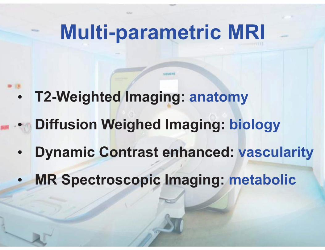
• T2-Weighted Imaging: anatomy
• Diffusion Weighed Imaging: biology
• Dynamic Contrast enhanced: vascularity
• MR Spectroscopic Imaging: metabolic
Multi-parametric MRI
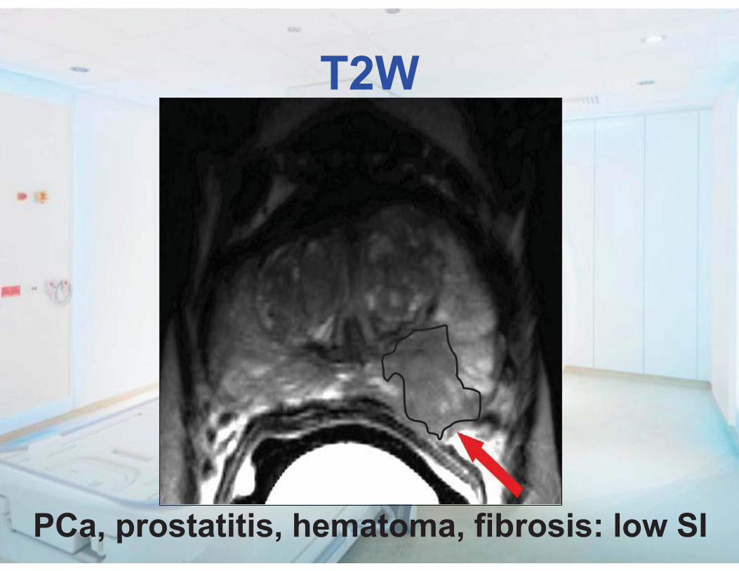
PCa, prostatitis, hematoma, fibrosis: low SI
T2W
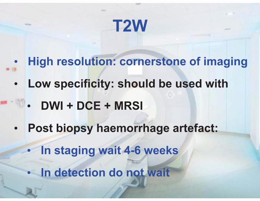
• High resolution: cornerstone of imaging
• Low specificity: should be used with • DWI + DCE + MRSI
• Post biopsy haemorrhage artefact: • In staging wait 4-6 weeks
• In detection do not wait
T2W
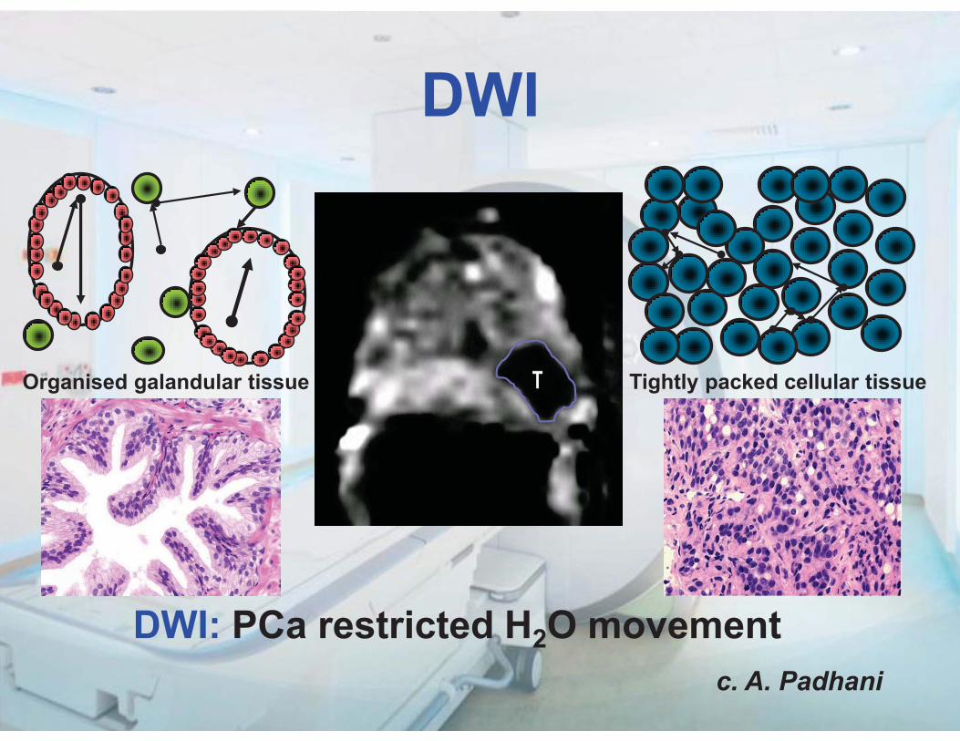
DWI: PCa restricted H2O movement
Tightly packed cellular tissue Organised galandular tissue
c. A. Padhani
DWI
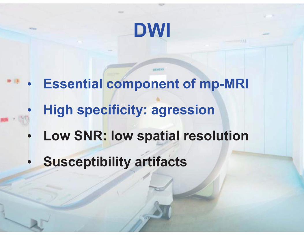
• Essential component of mp-MRI
• High specificity: agression
• Low SNR: low spatial resolution
• Susceptibility artifacts
DWI
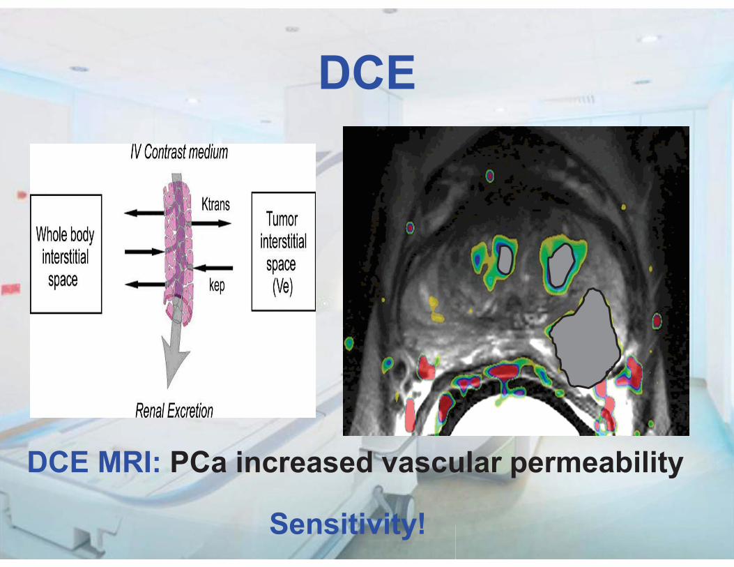
DCE MRI: PCa increased vascular permeability
Sensitivity!
DCE
DCE •High sensitivity: detection •Essential in “recurrence detection” •Low specificity: false positives •Limited standardization •No standardized calibration & analysis •Costs
MRSI
• Specificity especially in TZ • in PZ: DWI=MRSI
• Expertise is needed • Time consuming
• Requires ERC

For Ferrari drivers only?
MRSI

You need EXPERIENCE how to drive
MRSI

Prostate MRI Workshop
1. Introduction
2. Acquisition
3. Technical issues
4. Interpretation/Reporting
5. Indications/Clinical relevance
6. Future perspectives
Acquisition (minial requirements)
Good, simple, fast 1. Detection (30 minutes)
2. Staging (40 minutes)
3. Bone and Nodes (30 minutes)
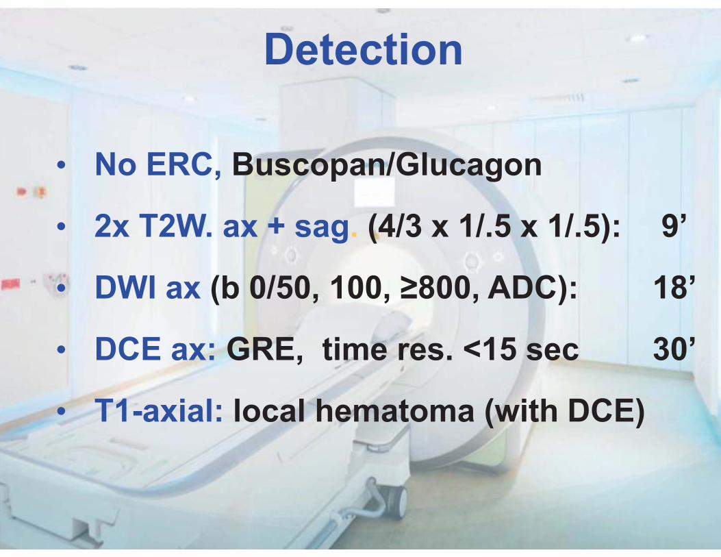
Detection
• No ERC, Buscopan/Glucagon • 2x T2W. ax + sag. (4/3 x 1/.5 x 1/.5): 9’ • DWI ax (b 0/50, 100, ≥800, ADC): 18’
• DCE ax: GRE, time res. <15 sec 30’
• T1-axial: local hematoma (with DCE)
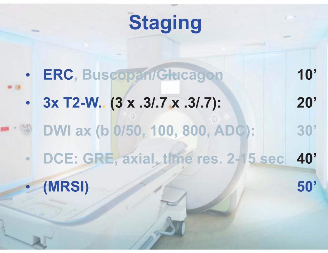
Staging
• ERC, Buscopan/Glucagon 10’ • 3x T2-W.. (3 x .3/.7 x .3/.7): 20’ • DWI ax (b 0/50, 100, 800, ADC): 30’
• DCE: GRE, axial, time res. 2-15 sec 40’
• (MRSI) 50’

Node and bone Recurrence, or PSA>15, Gl>7, DRE T3
• 3D T1-WI tSE (.9x.9x.9) 6’ • DWI cor (b 50, 800, ADC): 14’
• C-T-L spine: sag. T1WI and STIR 30’ or better: protocol presented by Padhani

Prostate MRI Workshop
1. Introduction
2. Acquisition
3. Technical issues
4. Interpretation/Reporting
5. Indications/Clinical relevance
6. Future perspectives
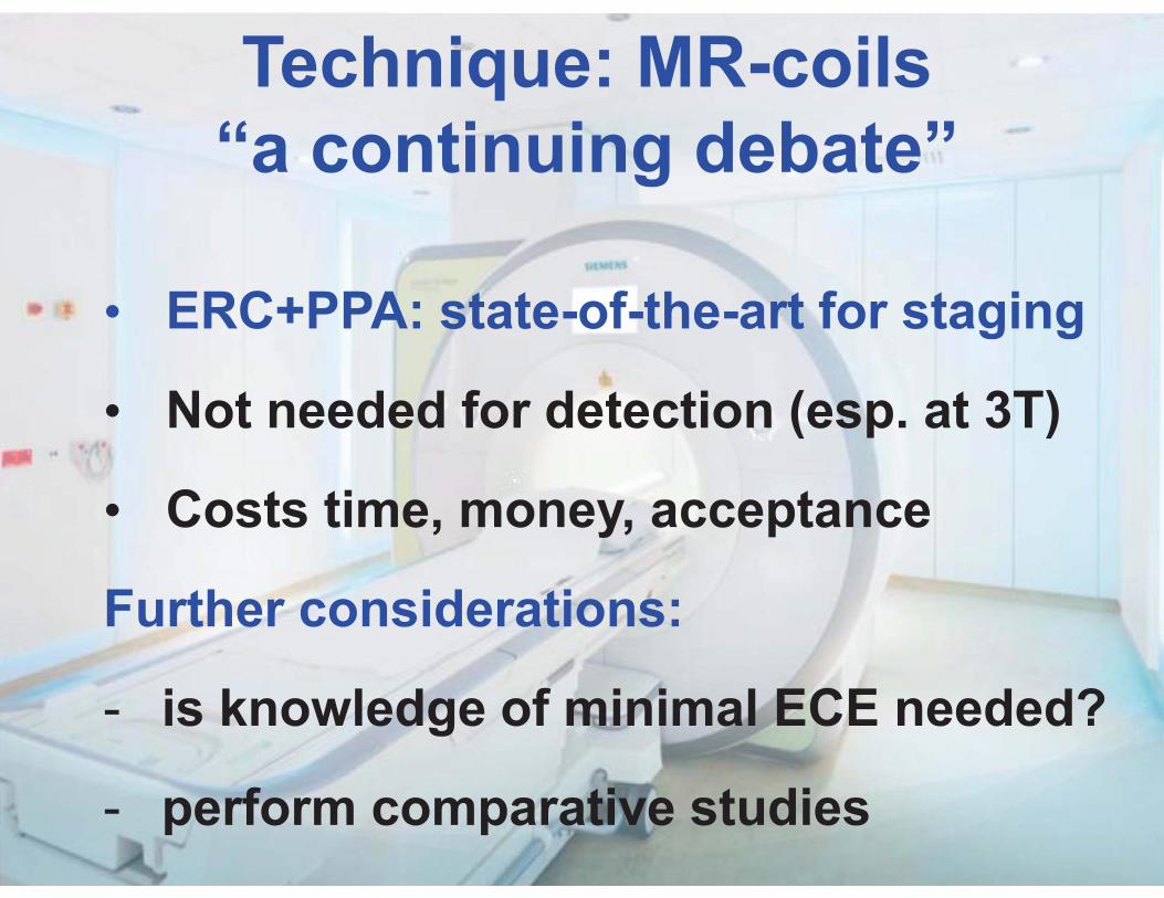
Technique: MR-coils “a continuing debate”
• ERC+PPA: state-of-the-art for staging
• Not needed for detection (esp. at 3T)
• Costs time, money, acceptance Further considerations: - is knowledge of minimal ECE needed?
- perform comparative studies
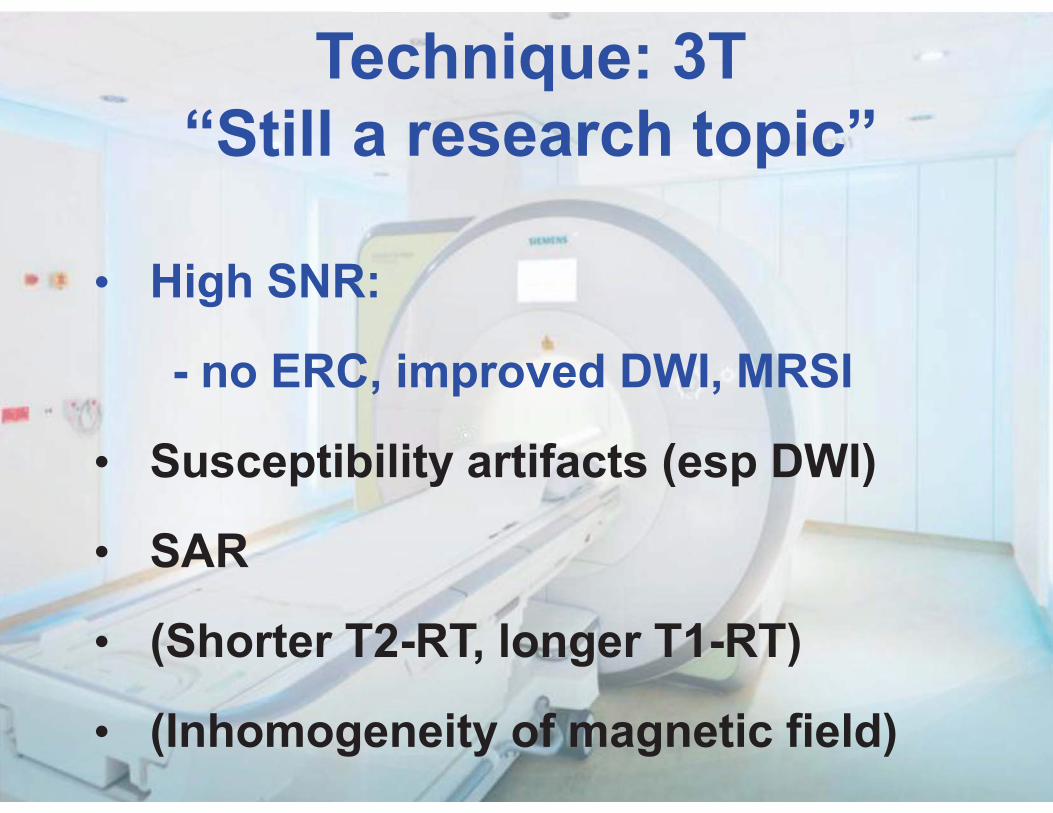
Technique: 3T “Still a research topic”
• High SNR: - no ERC, improved DWI, MRSI
• Susceptibility artifacts (esp DWI)
• SAR
• (Shorter T2-RT, longer T1-RT)
• (Inhomogeneity of magnetic field)

Prostate MRI Workshop
1. Introduction
2. Acquisition
3. Technical issues
4. Interpretation/Reporting
5. Indications/Clinical relevance
6. Future perspectives
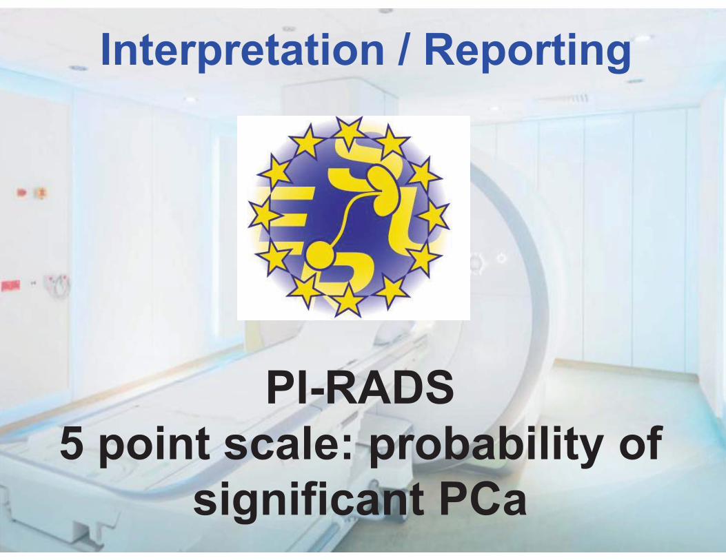
Interpretation / Reporting
PI-RADS 5 point scale: probability of
significant PCa
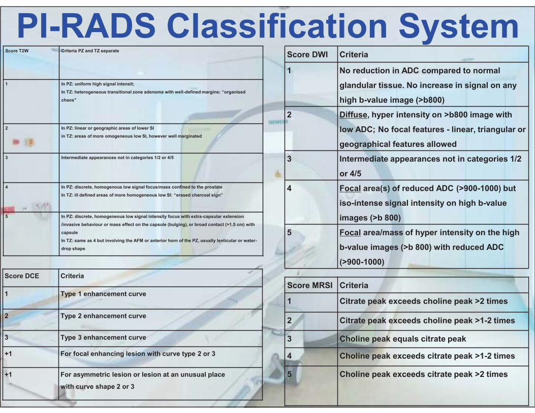
PI-RADS Classification System Score T2W Criteria PZ and TZ separate
1 In PZ: uniform high signal intensit;
In TZ: heterogeneous transitional zone adenoma with well-defined margins: “organised
chaos"
2 In PZ: linear or geographic areas of lower SI
in TZ: areas of more omogeneous low SI, however well marginated
3 Intermediate appearances not in categories 1/2 or 4/5
4 In PZ: discrete, homogenous low signal focus/mass confined to the prostate
In TZ: ill defined areas of more homogeneous low SI: “erased charcoal sign”
5 In PZ: discrete, homogeneous low signal intensity focus with extra-capsular extension
/invasive behaviour or mass effect on the capsule (bulging), or broad contact (>1.5 cm) with
capsule
In TZ: same as 4 but involving the AFM or anterior horn of the PZ, usually lenticular or water-
drop shape
Score DWI Criteria
1 No reduction in ADC compared to normal
glandular tissue. No increase in signal on any
high b-value image (>b800)
2 Diffuse, hyper intensity on >b800 image with
low ADC; No focal features - linear, triangular or
geographical features allowed
3 Intermediate appearances not in categories 1/2
or 4/5
4 Focal area(s) of reduced ADC (>900-1000) but
iso-intense signal intensity on high b-value
images (>b 800)
5 Focal area/mass of hyper intensity on the high
b-value images (>b 800) with reduced ADC
(>900-1000) Score DCE Criteria
1 Type 1 enhancement curve
2 Type 2 enhancement curve
3 Type 3 enhancement curve
+1 For focal enhancing lesion with curve type 2 or 3
+1 For asymmetric lesion or lesion at an unusual place
with curve shape 2 or 3
Score MRSI Criteria
1 Citrate peak exceeds choline peak >2 times
2 Citrate peak exceeds choline peak >1-2 times
3 Choline peak equals citrate peak
4 Choline peak exceeds citrate peak >1-2 times
5 Choline peak exceeds citrate peak >2 times
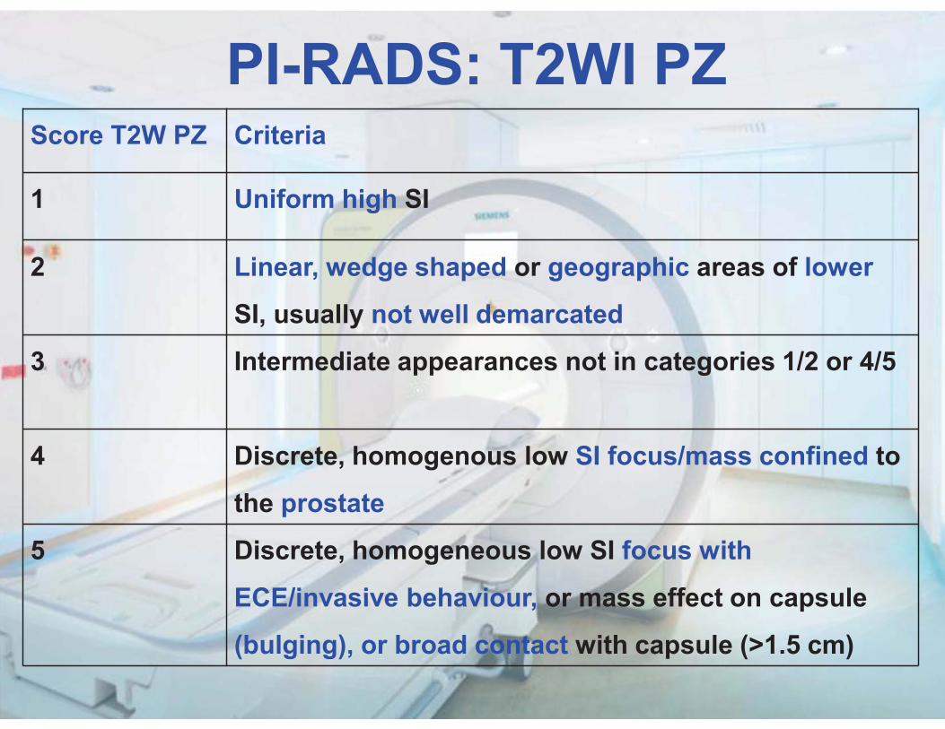
Score T2W PZ Criteria
1 Uniform high SI
2 Linear, wedge shaped or geographic areas of lower
SI, usually not well demarcated
3 Intermediate appearances not in categories 1/2 or 4/5
4 Discrete, homogenous low SI focus/mass confined to
the prostate
5 Discrete, homogeneous low SI focus with
ECE/invasive behaviour, or mass effect on capsule
(bulging), or broad contact with capsule (>1.5 cm)
PI-RADS: T2WI PZ
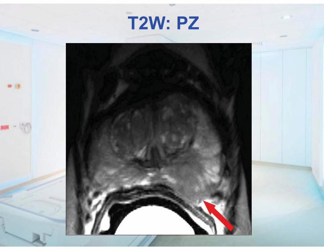
T2W: PZ
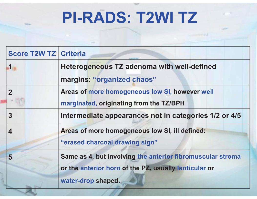
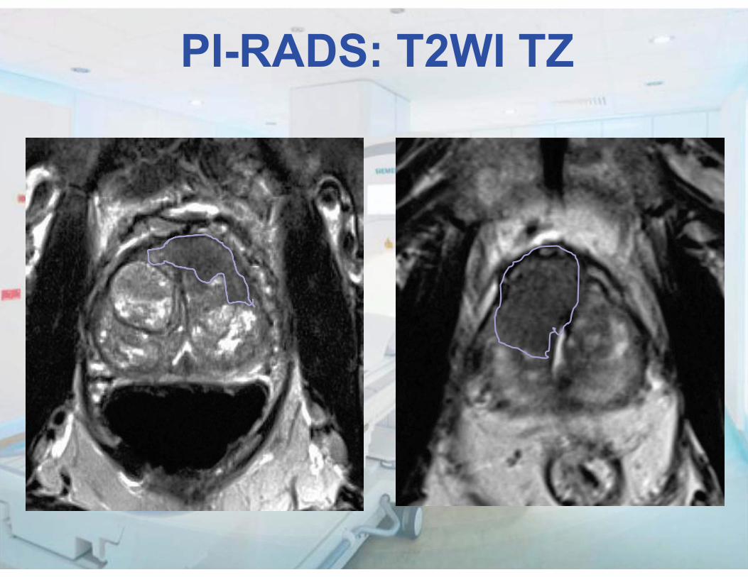
PI-RADS: T2WI TZ
Score T2W TZ Criteria
1 Heterogeneous TZ adenoma with well-defined
margins: “organized chaos”
2 Areas of more homogeneous low SI, however well
marginated, originating from the TZ/BPH
3 Intermediate appearances not in categories 1/2 or 4/5
4 Areas of more homogeneous low SI, ill defined:
“erased charcoal drawing sign”
5 Same as 4, but involving the anterior fibromuscular stroma
or the anterior horn of the PZ, usually lenticular or
water-drop shaped.
PI-RADS: T2WI TZ
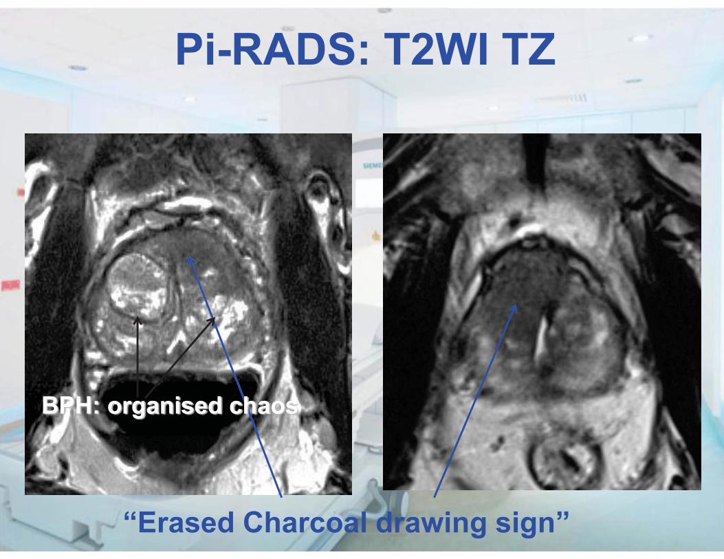
“Erased Charcoal drawing sign”
BPH: organised chaos
Pi-RADS: T2WI TZ
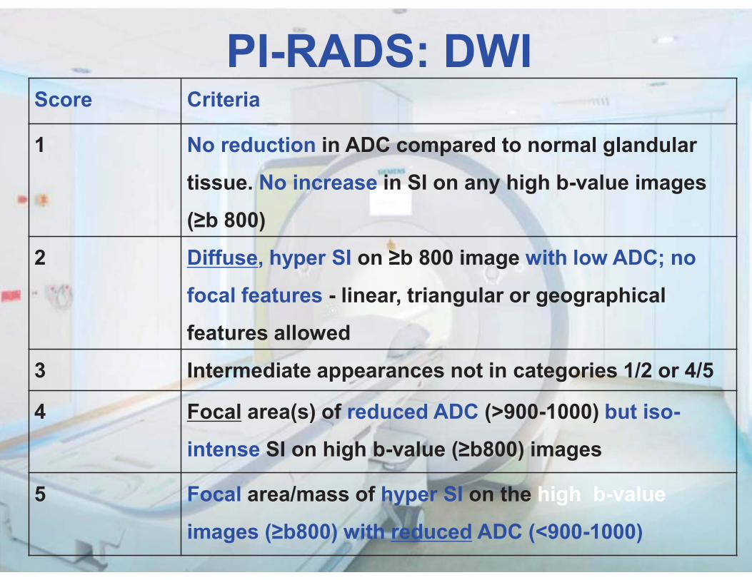
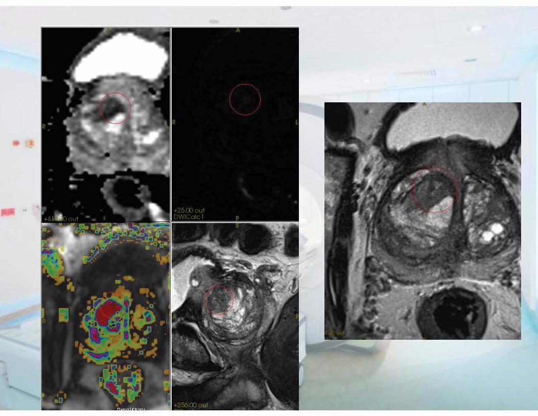
PI-RADS: DWIScore Criteria
1 No reduction in ADC compared to normal glandular
tissue. No increase in SI on any high b-value images
(≥b 800)
2 Diffuse, hyper SI on ≥b 800 image with low ADC; no
focal features - linear, triangular or geographical
features allowed
3 Intermediate appearances not in categories 1/2 or 4/5
4 Focal area(s) of reduced ADC (>900-1000) but iso-
intense SI on high b-value (≥b800) images
5 Focal area/mass of hyper SI on the high b-value
images (≥b800) with reduced ADC (<900-1000)
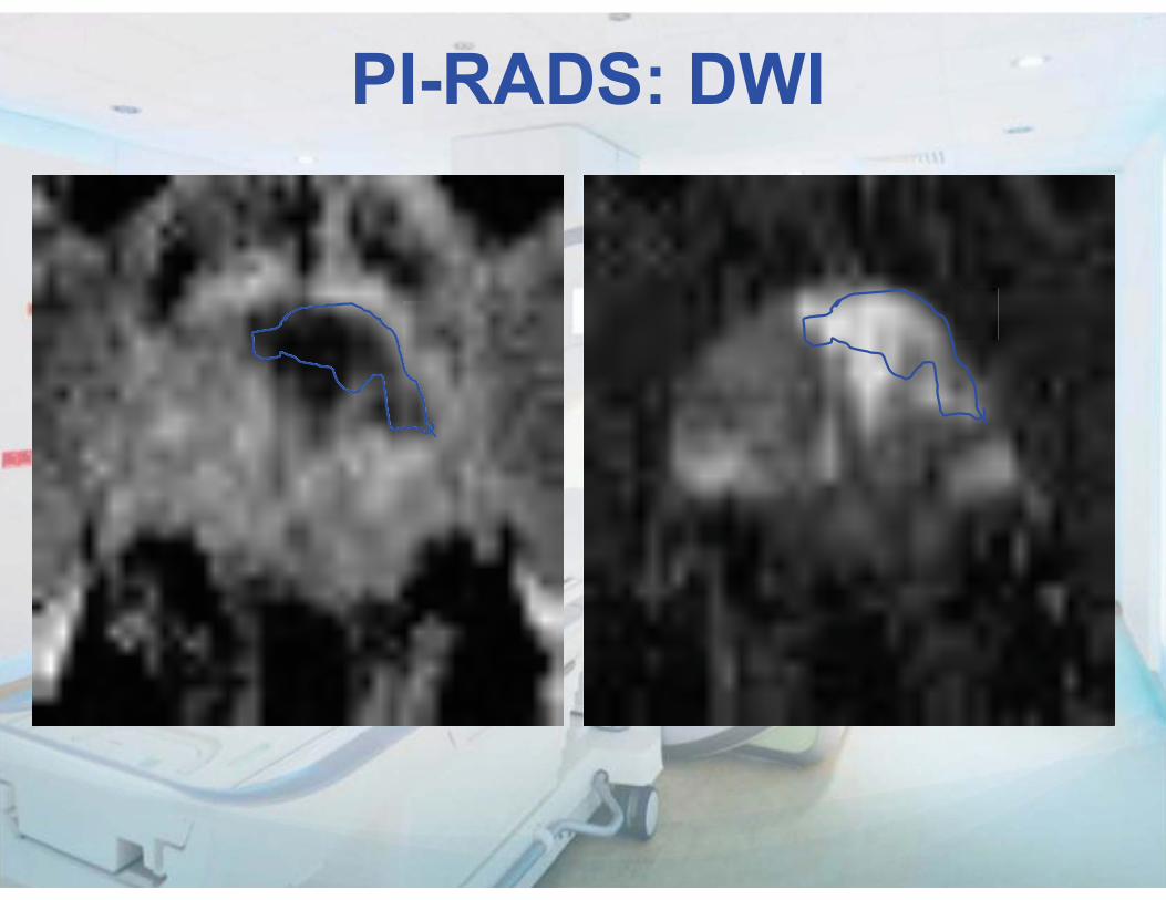
PI-RADS: DWI
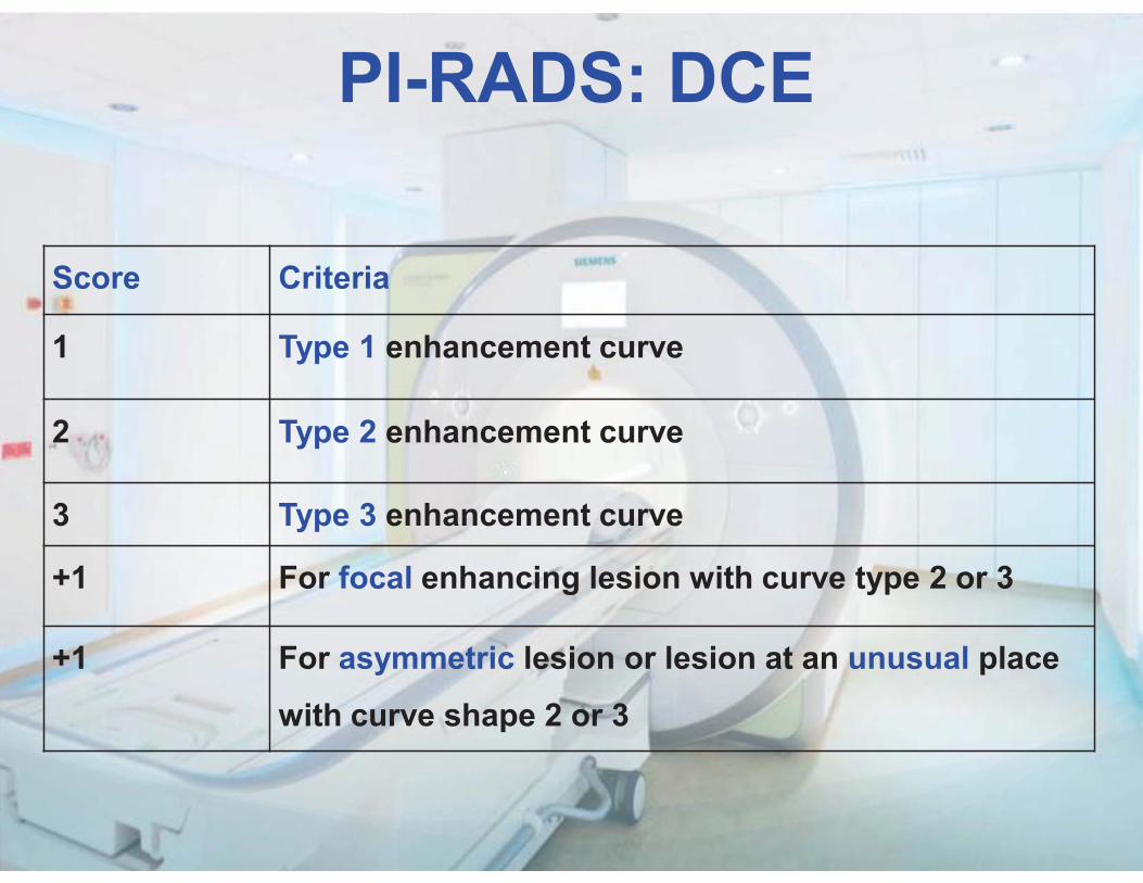
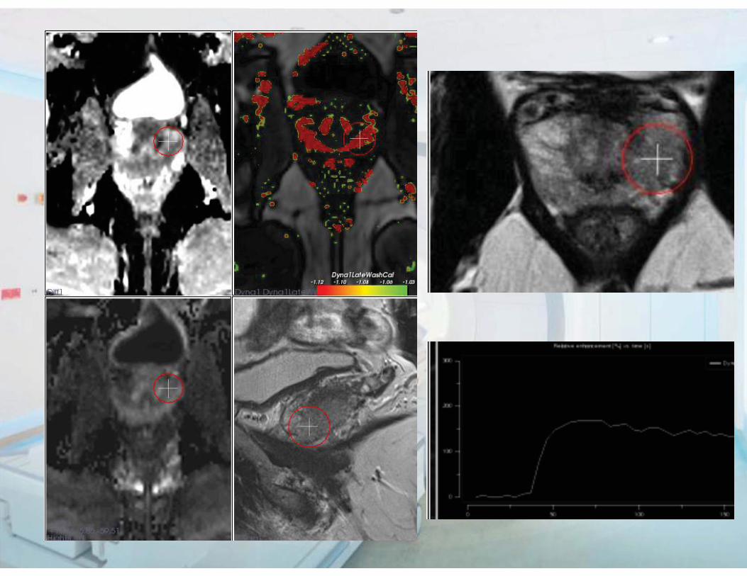
PI-RADS: DCE
Score Criteria
1 Type 1 enhancement curve
2 Type 2 enhancement curve
3 Type 3 enhancement curve
+1 For focal enhancing lesion with curve type 2 or 3
+1 For asymmetric lesion or lesion at an unusual place
with curve shape 2 or 3
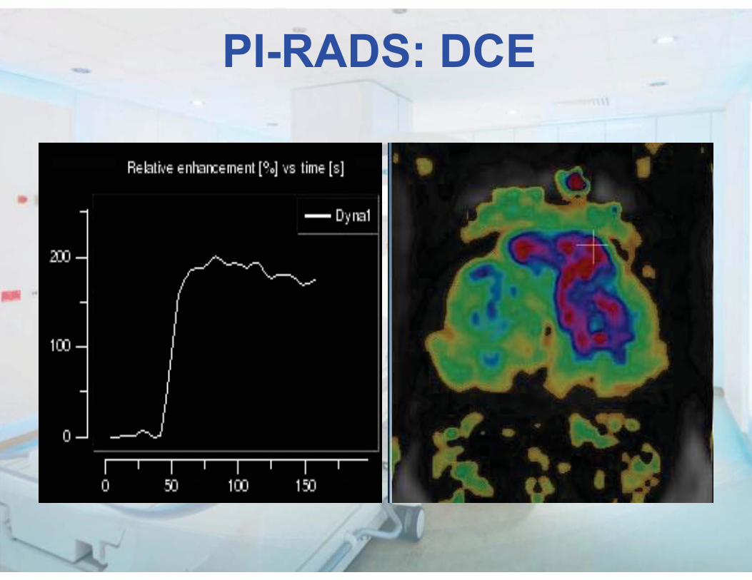
PI-RADS: DCE
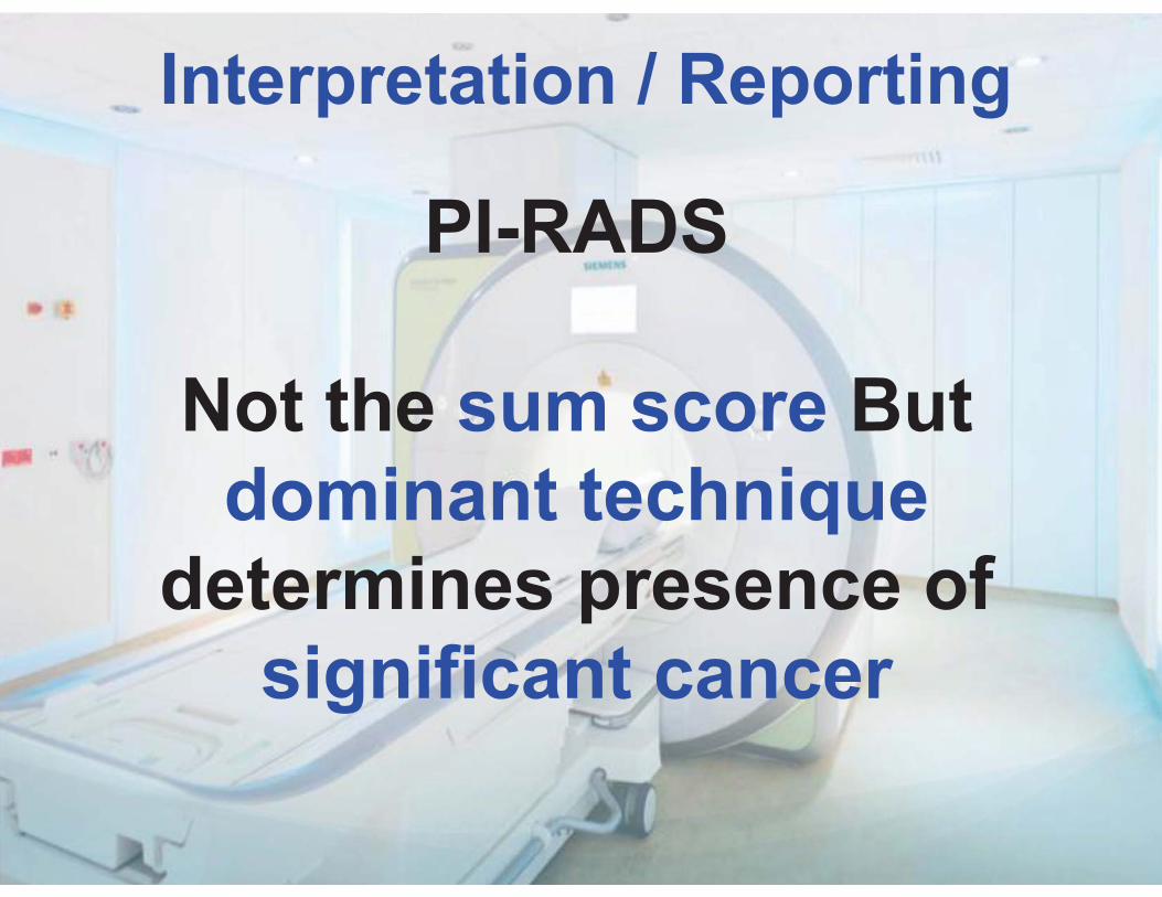
Interpretation / Reporting
PI-RADS
Not the sum score But dominant technique
determines presence of significant cancer
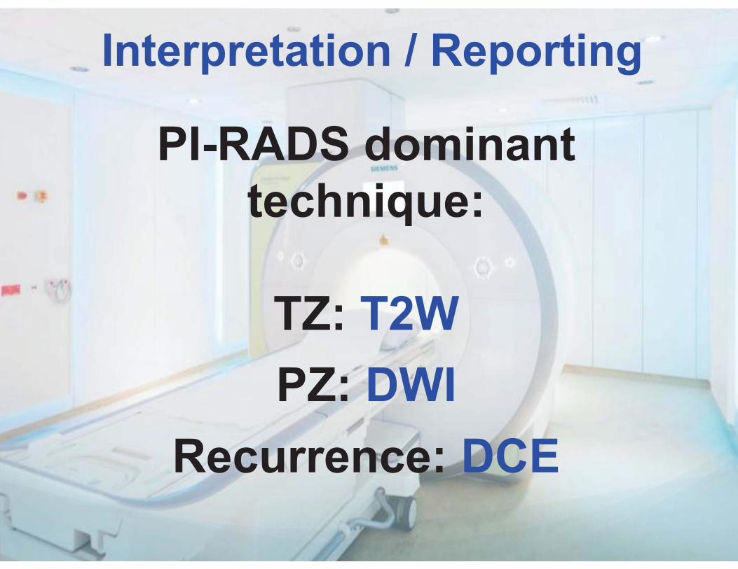
Interpretation / Reporting
PI-RADS dominant technique:
TZ: T2W PZ: DWI
Recurrence: DCE
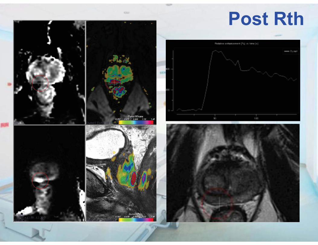
Post Rth
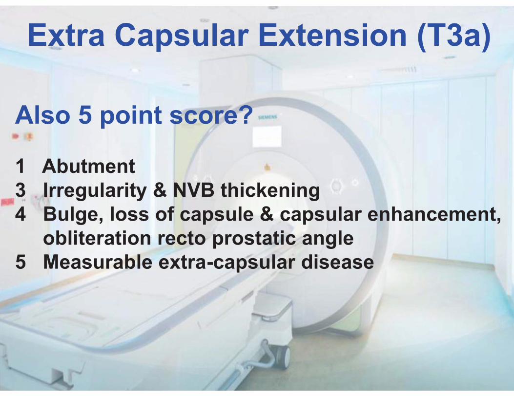
Extra Capsular Extension (T3a)
Also 5 point score? 1 Abutment 3 Irregularity & NVB thickening 4 Bulge, loss of capsule & capsular enhancement, obliteration recto prostatic angle 5 Measurable extra-capsular disease
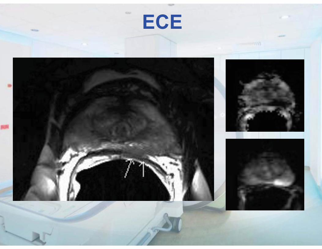
ECE
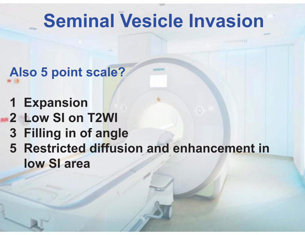
Seminal Vesicle Invasion
Also 5 point scale? 1 Expansion 2 Low SI on T2WI 3 Filling in of angle 5 Restricted diffusion and enhancement in low SI area
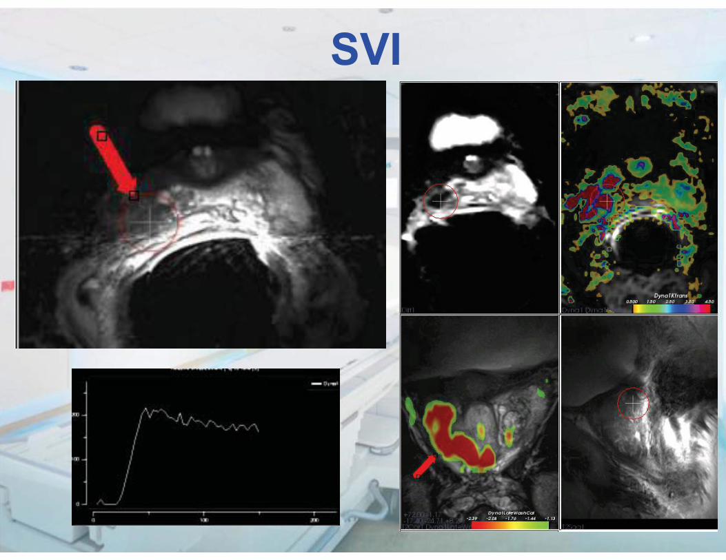
SVI

Prostate MRI Workshop
1. Introduction
2. Acquisition
3. Technical issues
4. Interpretation/Reporting
5. Indications/Clinical relevance
6. Future perspectives
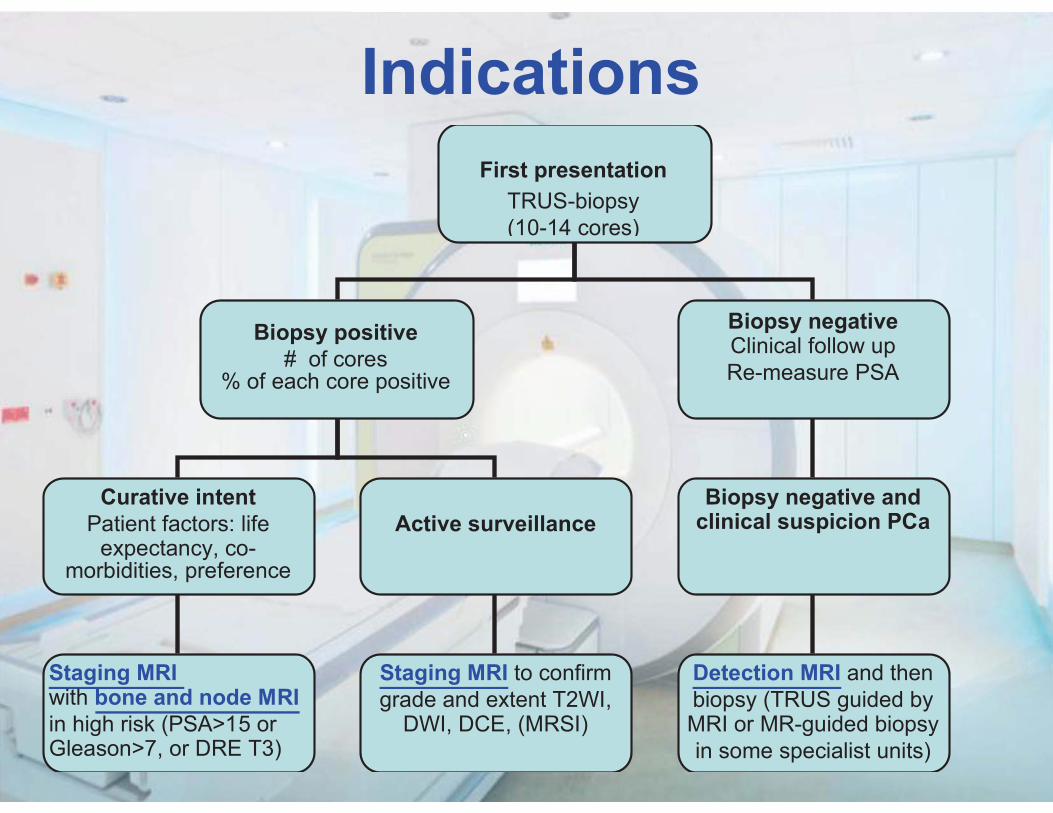
IndicationsFirst presentation
TRUS-biopsy (10-14 cores)
Biopsy positive # of cores
% of each core positive
Biopsy negative Clinical follow up Re-measure PSA
Curative intent Patient factors: life
expectancy, co-morbidities, preference
Active surveillance
Staging MRI with bone and node MRI in high risk (PSA>15 or Gleason>7, or DRE T3)
Biopsy negative and clinical suspicion PCa
Staging MRI to confirm grade and extent T2WI,
DWI, DCE, (MRSI)
Detection MRI and then biopsy (TRUS guided by MRI or MR-guided biopsy in some specialist units)
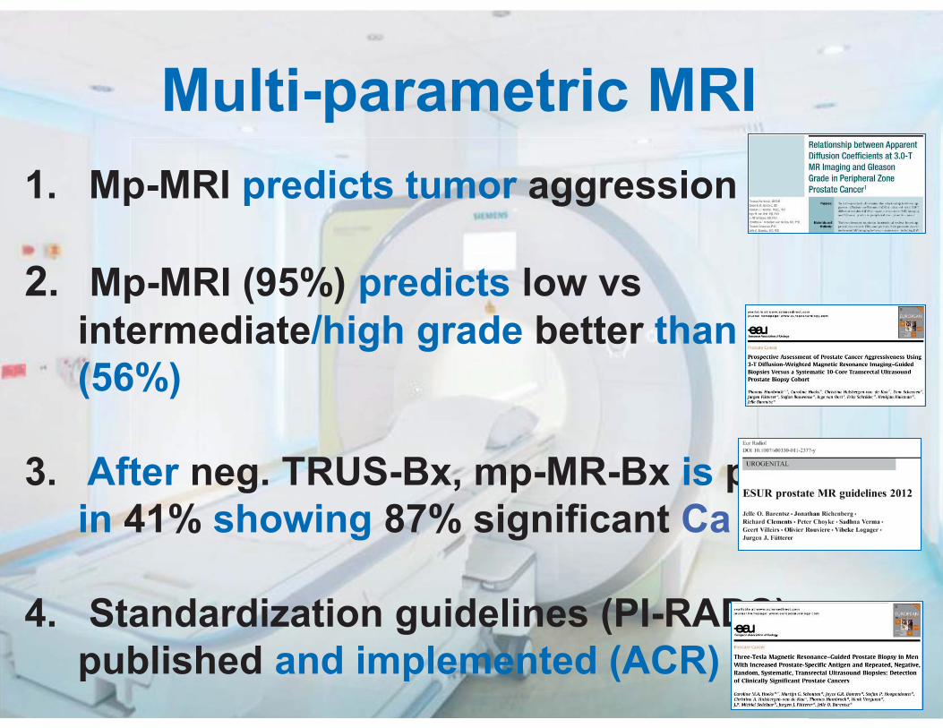
1. Mp-MRI predicts tumor aggression
2. Mp-MRI (95%) predicts low vs intermediate/high grade better than TRUS (56%)
3. After neg. TRUS-Bx, mp-MR-Bx is positive in 41% showing 87% significant Ca
4. Standardization guidelines (PI-RADS) are published and implemented (ACR)
Multi-parametric MRI

Take an “easy case”, in which you will certainly make a success: - many neg. TRUS-Bx and high PSA (>25), than look for anterior or apex tumor.
How to be a “winner”
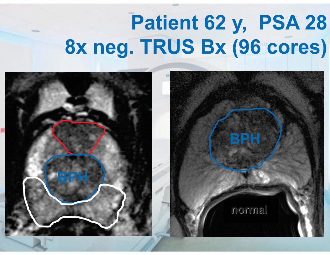
Patient 62 y, PSA 28 8x neg. TRUS Bx (96 cores)
normal
BPH
BPH
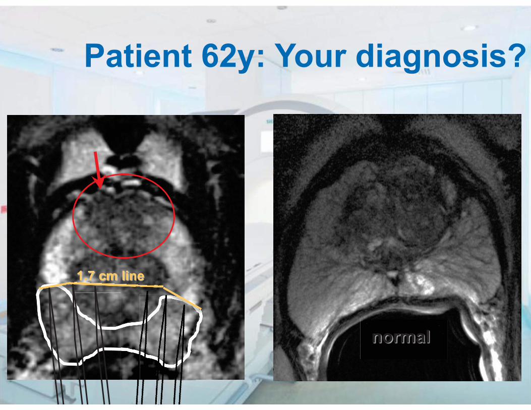
Patient 62y: Your diagnosis?
normal
1.7 cm line
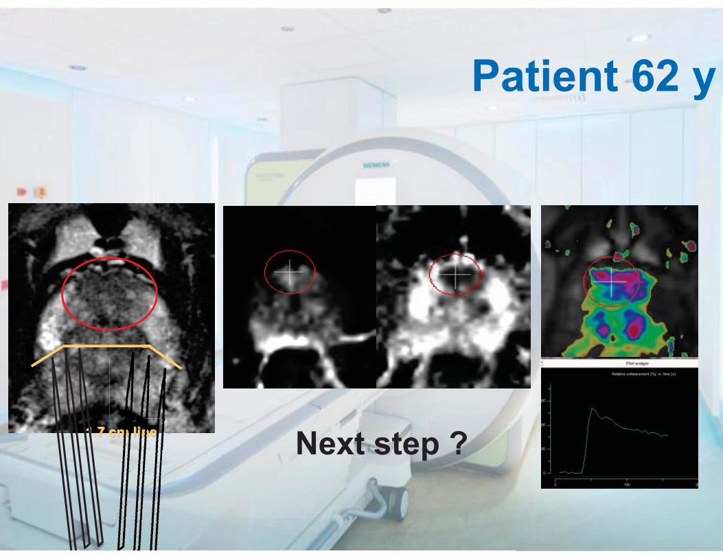
Patient 62 y
Next step ? 1.7 cm line cmmmmmmmmmmmmmmmmmmmmmmmcmmmmmmmmmmmmmmmmmmmmmmmmmmmmmmmmmmmmmmmmmmmmmmmmmmmmmmm liiiiiiiiimmmmmmmmmmmmmmmmmmmmmmmmmmm linnnnnnnnnnnnnnnnnnnnnnnnnnnnnnnnnnnnnnnnnnnnnnnnnnnnnnnnnnnnnnnnnnnnneeeeeeeeeeeeeeeeeeeneeeeeennnnnnnnnnnnnnnnnnnnnnnnnnnnnnnnnnnnnnnnnnnnnnnnnnnnnnnnnnnn
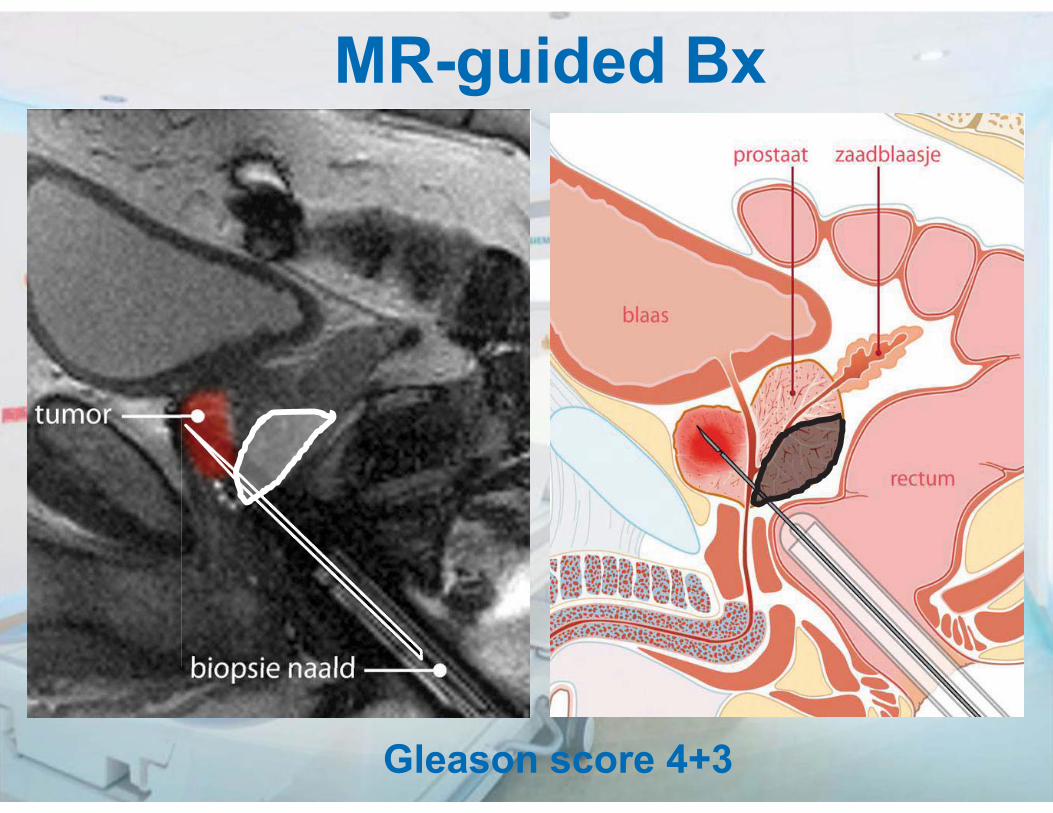
MR-guided Bx
Gleason score 4+3
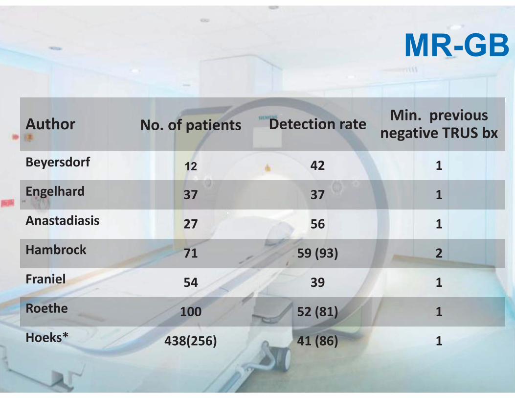
MR-GB
Author No. of patients Detection rate Min. previous negative TRUS bx
Beyersdorf 12 42 1
Engelhard 37 37 1
Anastadiasis 27 56 1
Hambrock 71 59 (93) 2
Franiel 54 39 1
Roethe 100 52 (81) 1
Hoeks* 438(256) 41 (86) 1
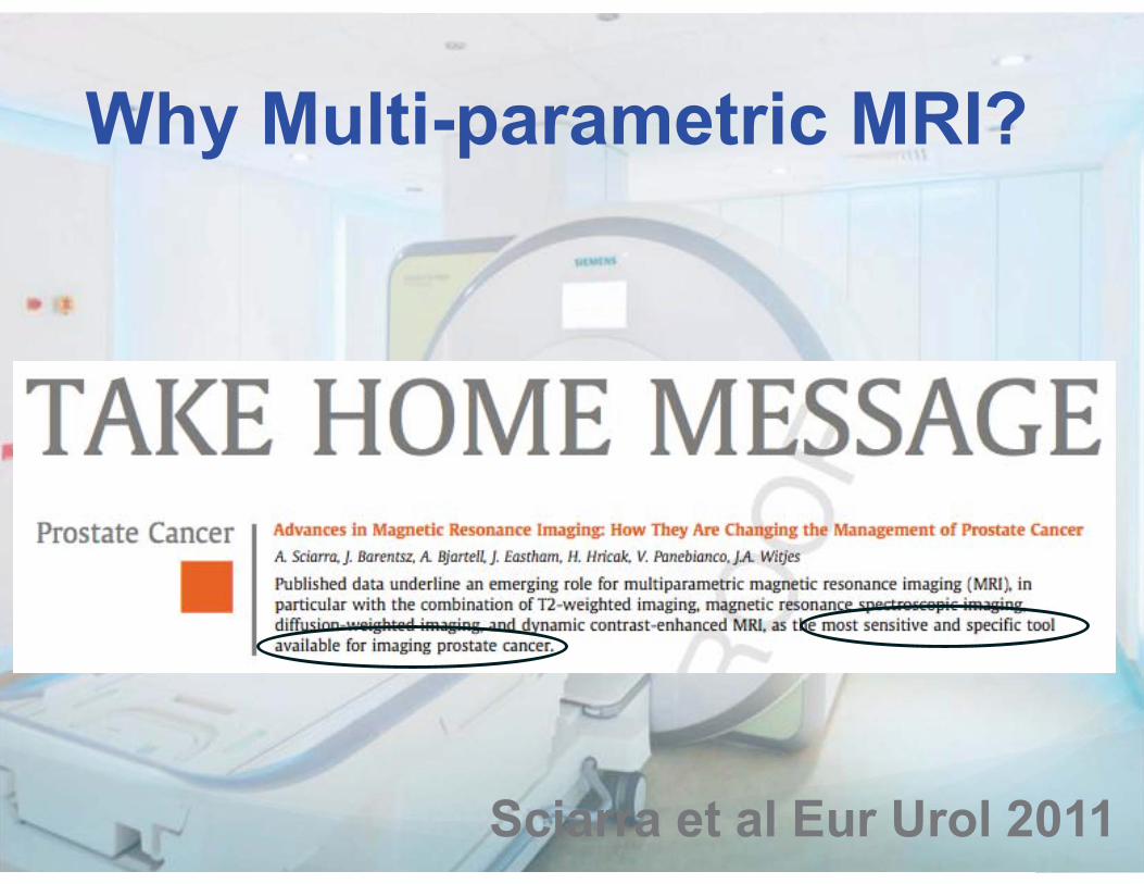
Sciarra et al Eur Urol 2011
Why Multi-parametric MRI?
Claire Allan, Michel Claudon, Francois Cornud, Ferdinand Frauscher, Nicolas Grenier, Alex Kirkham, Frederic Lefevre, Gareth Lewis, Ulrich Muller-Lisse, Anwar Padhani, Valeria Panebianco, Pietro Pavlica, Phillipe Puech, Jarle Rorvik, Andrea Rockall, Catherine Roy,Tom Scheenen, Harriet Thoeny, Baris Turkbey, Ahmet Turgut.
ESUR Prostate MR Guidelines 2011

Jelle Barentsz Prostate MR Center of Excellence
Department of Radiology Radboud University Nijmegen Medical Center [email protected]
Questions?