prognostic value of multidetector coronary computed tomography angiography in relation to exercise...
TRANSCRIPT
Journal of the American College of Cardiology Vol. 60, No. 21, 2012© 2012 by the American College of Cardiology Foundation ISSN 0735-1097/$36.00
Cardiac Imaging
Prognostic Value of Multidetector CoronaryComputed Tomography Angiography in Relation toExercise Electrocardiogram in Patients WithSuspected Coronary Artery Disease
Iksung Cho, MD,* Jaemin Shim, MD,* Hyuk-Jae Chang, MD,*† Ji Min Sung, PHD,‡Youngtaek Hong, BS,† Hackjoon Shim, PHD,† Young Jin Kim, MD,§ Byoung Wook Choi, MD,§James K. Min, MD,� Ji-Ye Kim, MD,* Chi-Young Shim, MD,* Geu-Ru Hong, MD,*Namsik Chung, MD*
Seoul, South Korea; and Los Angeles, California
Objectives This study was designed to determine the prognostic value of multidetector coronary computed tomography an-giography (CTA) in relation to exercise electrocardiography (XECG) findings.
Background The prognostic usefulness of coronary CTA findings of coronary artery disease in relation to XECG findings hasnot been explored systematically.
Methods Patients with suspected coronary artery disease who had undergone both coronary CTA and XECG (�90 daysbetween tests) from 2003 through 2009 were enrolled retrospectively. Coronary CTA results were classified ac-cording to the severity of maximal stenosis (normal, mild: �40% of luminal stenosis, moderate: 40% to 69%,severe: �70%), XECG results were categorized as positive and negative, and Duke XECG score was calculated.Clinical follow-up data were collected for major adverse cardiac events (MACE): cardiac death, nonfatal myocar-dial infarction, unstable angina requiring hospitalization, and revascularization after 90 days from index coronaryCTA. C-statistics were calculated to compare discriminatory values of each test.
Results Among the 2,977 (58 � 10 years) study patients, 12% demonstrated positive XECG results. By coronary CTA,patients were categorized as normal (56%) or having mild (26%), moderate (13%), or severe (5%) disease. Dur-ing a median follow-up of 3.3 years (interquartile range: 2.3 to 4.6), 97 MACE were observed and the 5-year cu-mulative event rate was 3.6% (95% confidence interval: 3.0 to 4.3). Although both XECG (C-statistic: 0.790) andcoronary CTA (C-statistic: 0.908) improved risk stratification beyond clinical risk factors (C-statistic: 0.746, p �
0.05 for all), XECG in addition to coronary CTA (C-statistic: 0.907) did not provide better discrimination than coro-nary CTA alone (p � 0.389). In subgroup analyses, coronary CTA stratified risk of MACE in groups with both posi-tive and negative XECG results (all p � 0.001 for trend). However, positive XECG results predicted risk of MACEon coronary CTA only in the moderate stenosis group (hazard ratio: 2.58, 95% confidence interval: 1.29 to 5.19,p � 0.008) and severe stenosis group (hazard ratio: 2.28, 95% confidence interval: 1.19 to 4.38, p � 0.013).
Conclusions In patients with suspected coronary artery disease, coronary CTA discriminates future risk of MACE in patientsindependent of XECG results. Compared with coronary CTA, XECG has an additive prognostic value only in pa-tients with moderate to severe stenosis on coronary CTA. (J Am Coll Cardiol 2012;60:2205–15) © 2012 by theAmerican College of Cardiology Foundation
Published by Elsevier Inc. http://dx.doi.org/10.1016/j.jacc.2012.08.981
research was supported by the Leading Foreign Research Institute RecruitmentProgram through the National Research Foundation of Korea, funded by the Ministryof Education, Science and Technology (2012027176). Dr. Min has received researchsupport from GE Healthcare. All other authors have reported that they have norelationships relevant to the contents of this paper to disclose. The first two authorscontributed equally to this work.
From the *Division of Cardiology Severance Cardiovascular Hospital, Yonsei Uni-versity College of Medicine, Yonsei University Health System, Seoul, South Korea;†Severance Biomedical Science Institute, Yonsei University Health System, Seoul,South Korea; ‡Department of Research Affairs, Yonsei University College ofMedicine, Yonsei University Health System, Seoul, South Korea; §Division ofRadiology Severance Cardiovascular Hospital, Yonsei University College of Medi-
cine, Seoul, South Korea; and the �Department of Cardiology, Cedar-Sinai HeartInstitute, University of California Los Angeles, Los Angeles, California. ThisManuscript received May 27, 2012; revised manuscript received August 4, 2012,accepted August 13, 2012.
2206 Cho et al. JACC Vol. 60, No. 21, 2012Prognostic Value of Coronary CTA in Relation to XECG November 20/27, 2012:2205–15
Exercise electrocardiography(XECG) has been a widely usedtest in diagnosing and prognos-ticating individuals with sus-pected coronary artery disease(CAD) (1–3). Current guidelinesrecommend XECG as the firstdiagnostic step of suspectedCAD in patients who are able toexercise (1). However, its useful-ness is limited by a modest sen-sitivity and specificity of 68% and77%, respectively, across a widerange of patient subsets (2).
See page 2216
Recently, coronary computedtomography angiography (CTA)
was introduced as a novel, noninvasive approach for theevaluation of CAD. Because coronary CTA demonstrateshigh specificity and negative predictive value in the exclu-sion of CAD (3–5), it has been suggested as a potentialnoninvasive method to rule in or rule out obstructive CAD.Although previous studies revealed the superior diagnosticaccuracy of coronary CTA compared with XECG (6,7), todate, the prognostic value of coronary CTA has not beencompared adequately with that of stress tests (8,9). Inaddition, the usefulness of coronary CTA as an alternativeor an adjunct to stress tests (including XECG) in thediagnostic work-up as well as risk stratification of patientswith chest pain remains to be studied. Because the value ofany noninvasive diagnostic strategy is determined in largepart by its prognostic benefit, we thus sought to assess theprognostic value of coronary CTA in relation to XECG inpatients with suspected CAD.
Methods
Design overview, setting, and participants. The initialstudy sample included 3,944 consecutive patients who hadundergone both coronary CTA and XECG within 90 daysfor evaluation of suspected CAD at Severance Cardiovas-cular Hospital from May 2003 through April 2009 withoutany other cardiovascular testing. Patients were excluded who1) were younger than 30 years (n � 63); 2) had a history ofprior myocardial infarction (MI), coronary revasculariza-tion, or cardiac transplantation (n � 21); 3) had inadequateXECG (137 patients); 4) had insufficient medical recordsor uninterpretable coronary CTA results (n � 26); and 5)were without at least 1 of following symptoms or signs:angina, angina equivalent symptoms, or abnormal restingelectrocardiography (ECG) results (n � 720). After exclu-sion according to the study criteria, a total of 2,977 patientsremained for final analysis. The median number of days
Abbreviationsand Acronyms
CAD � coronary arterydisease
CI � confidence interval
CT � computedtomography
CTA � computedtomography angiography
ECG � electrocardiography
HR � hazard ratio
MACE � major adversecardiac event(s)
MI � myocardial infarction
RF � risk factor
XECG � exerciseelectrocardiography
between coronary CTA and XECG was 8 days (interquar-
tile range: 2 to 14 days). Clinical indications of coronaryCTA and XECG are listed in Online Table 1. Pretestlikelihood of CAD was determined based on AmericanCollege of Cardiology/American Heart Association guide-lines, which were modified from the literature review ofDiamond and Forrester (10,11) (Online Table 2).
Clinical data were collected at the time of the index visit.Hypertension was defined by current use of antihypertensivemedications or a blood pressure of 140/90 mm Hg or more.Diabetes mellitus was defined as receiving antidiabetictreatments or a fasting plasma glucose of 126 mg/dl ormore. Current cigarette smoking was defined as any ciga-rette smoking in the past month. Dyslipidemia wasdefined as use of cholesterol-lowering medications orhaving a total serum cholesterol of 200 mg/dl or more.Institutional review committee approval and informedconsent were obtained.Coronary CTA protocol and image analysis. Data acqui-sition and image analysis were carried out as describedpreviously (12). Briefly, patients without a contraindicationto beta-adrenergic blocking agents (bronchial asthma, overtheart failure, and atrioventricular conduction abnormalities)and with initial heart rates higher than 65 beats/minreceived a single oral dose of 40 mg propranolol hydrochlo-ride (Pranol; Dae Woong, Seoul, Korea) 1 h before coronaryCTA. The patients’ mean heart rate during the CT exam-ination was 58 � 7 beats/min (range: 34 to 110 beats/min).Two types of CT system configurations were used: 1) a64-slice CT scanner (Sensation 64, Siemens Medical Solu-tions, Forchheim, Germany) using retrospective ECG gat-ing with tube current modulation from 2003 through 2009with the following parameters: rotation time: 330 ms, tubevoltage: 100 to 120 KeV, tube current: 400 to 800 mA, andpitch factor: 0.2; and 2) a 64-row CT scanner (LightSpeedVCT XT, GE Healthcare, Milwaukee, Wisconsin) usingprospectively an ECG-gated axial technique from 2008through 2009 with the following parameters: rotation time:350 ms, tube voltage: 100 to 120 KeV, and tube current: 300to 900 mA. A real-time bolus-tracking technique wasapplied to trigger the initiation of the scan. Contrastenhancement was achieved with 75 ml iopamidol (370 mgiodine per milliliter, Iopamiro, Bracco, Milan, Italy) in-jected at 5 ml/s, followed by an injection of 50 ml of salineat 5 ml/s by using a power injector (Envision CT, Medrad,Indianola, Pennsylvania) via an antecubital vein.
Image reconstruction was performed on the scanner’sworkstation using commercially available software (Wizard,Siemens Medical Solutions, or GEAW, GE healthcare).Axial images were reconstructed retrospectively at 65% ofthe RR interval for each cardiac cycle. If artifacts appeared,additional data sets were obtained for various points of thecardiac cycle, and the data set with the minimum artifactwas selected for further analysis. The reconstructed image
data sets were transferred to an off-line workstation (Aquar-
5RcbuN
R
Csophip0tbCCTXT1BCIh(
p
2207JACC Vol. 60, No. 21, 2012 Cho et al.November 20/27, 2012:2205–15 Prognostic Value of Coronary CTA in Relation to XECG
ius Workstation, TeraRecon, Inc., San Mateo, California)for postprocessing and analysis. Each lesion identified wasexamined using maximum intensity projection and multi-planar reconstruction techniques on a short axis and alongmultiple longitudinal axes. Lesions were classified by themaximal luminal diameter stenosis seen on any plane.Coronary CTA was evaluated by 2 experienced cardiacradiologists (Y.J.K. and B.W.C., with 6 and 9 years expe-rience in coronary CTA, respectively), who were blinded toXECG results of each patient. In case of disagreement, ajoint reading was performed to reach a consensus.
We used 3 different coronary CTA models to comparewith prognostic values of XECG: 1) a binary obstructiveCAD model; 2) an extent of CAD model; and 3) a severityof CAD model. Obstructive CAD was defined whencoronary artery segments exhibited plaque with a luminaldiameter stenosis of 50% or more. Extent of CAD wasclassified as the number of obstructive vessels (�50%): noobstructive CAD (absence of obstructive CAD), 1-vesseldisease (VD), 2-VD, and 3-VD. In addition, severity ofCAD was classified into 4 categories according the degree ofstenosis (13): normal (absence of CAD), mild (1% to 39%luminal narrowing in DS), moderate (40% to 69% luminalnarrowing in DS); and severe (�70% luminal narrowing inDS) stenosis.XECG protocol. A symptom-limited exercise treadmilltest was performed according to the Bruce protocol (14).During exercise stress test, heart rhythm and blood pressurewere recorded at rest, at the end of each stage of exercise, atpeak stress, and during recovery. A 12-lead ECG wasobtained every minute, and a 3-lead ECG for heart rhythmwas monitored continuously. Indications for terminatingthe exercise test were as previously described (11). In thesummary of the XECG data set (summary XECG), resultsof the stress test were classified as positive when the STsegment appeared horizontal or had a down-sloping depres-sion of 1 mm or more for 60 to 80 ms after the end of the QRScomplex (11). Inadequate stress tests of patients who had notreached the reference standards established for age, sex, andweight were excluded from the analysis. The detailed methodsto record XECG parameters and to calculate Duke treadmillscore are described in the Online Methods.Follow-up. Clinical follow-up data were obtained via re-view of electronic medical records and telephone contact bya dedicated physician, research nurse, or both, who wereblinded to coronary CTA and XECG results. The primaryendpoint was the occurrence of major adverse cardiac events(MACE), defined as cardiac death, nonfatal MI, unstableangina requiring hospitalization, and revascularization ei-ther by percutaneous coronary intervention or coronaryartery bypass graft after 90 days of the index test. Coronaryrevascularizations occurring within 90 days after the indextest were not included in the analyses to exclude anytest-driven procedure from being considered as a MACE
(15–17). wStatistical analysis. Discrete variables were presented asnumbers (percentages), and continuous variables were ex-pressed as mean � SD or median with interquartile range,as appropriate. Differences between continuous variableswere analyzed by analysis of variance tests, and thosebetween categorical variables were analyzed by the chi-square test or Fisher exact test, as appropriate. Cumulativeevent rates as a function of time were calculated usingKaplan-Meier survival analysis for XECG results andcoronary CTA-diagnosed CAD and were compared us-ing the log-rank statistic. Five-year estimated incidentMACE risk was calculated for each participant using Coxproportional hazards model. Univariate and multivariatemodels were calculated to identify XECG and coronaryCTA predictors of outcome. From the Cox hazardmodels, hazard ratios (HRs) and 95% confidence inter-vals (CIs) were calculated.
To evaluate the discriminatory function of each model,C-statistics for the following models were calculated(18,19): model 1, clinical risk factors (RF); model 2, RF �summary XECG; model 3, RF � summary XECG � Duketreadmill test score; model 4, RF � coronary CTA; model, RF � summary XECG � coronary CTA; model 6,F � summary XECG � Duke treadmill test score �
oronary CTA. p Values less than 0.05 were considered toe statistically significant. Statistical analysis was performedsing SAS software version 9.2 (SAS Institute Inc., Cary,orth Carolina) and R software version 2.13.1.
esults
linical characteristics of study population. Overall, thetudy population consisted of 2,977 patients. The mean agef patients was 58 � 10 years and 50% were male. Therevalences of clinical RF were as follows: diabetes, 16%;ypertension, 48%; dyslipidemia, 44%; and current smok-
ng, 13%. In subset groups of the more severe CAD,atients were male, older, diabetic, and hypertensive (all p �.001) (Table 1). More than one-half of the study popula-ion (1,825 patients, 61%) had intermediate pretest proba-ility of CAD, and 823 (28%) had low pretest probability ofAD. Ninety patients (3%) had high pretest probability ofAD. Detailed patient characteristics are summarized inable 1.ECG and coronary CTA results. As demonstrated inable 2, 358 (12%) patients had positive XECG results, and30 (4%) had equivocal XECG results among study cohort.y coronary CTA, 409 (13%) patients had obstructiveAD: 1-VD (274, 9%), 2-VD (92, 3%), or 3-VD (43, 1%).
n terms of severity of CAD, patients were classified asaving mild (782, 26%), moderate (372, 13%), and severe155, 5%) CAD.
Prevalence of obstructive CAD (1-, 2-, or 3-VD) inatients with positive XECG results was higher than those
ith negative XECG results (28% vs. 11%, respectively, p �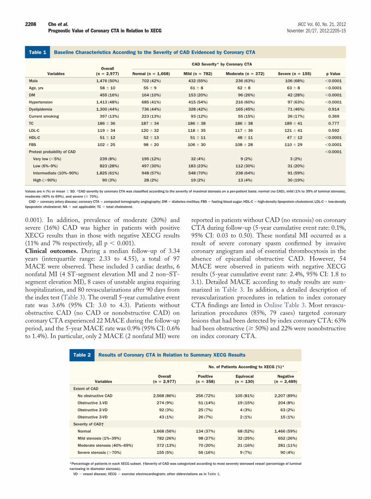
tes me
2208 Cho et al. JACC Vol. 60, No. 21, 2012Prognostic Value of Coronary CTA in Relation to XECG November 20/27, 2012:2205–15
0.001). In addition, prevalence of moderate (20%) andsevere (16%) CAD was higher in patients with positiveXECG results than in those with negative XECG results(11% and 7% respectively, all p � 0.001).Clinical outcomes. During a median follow-up of 3.34years (interquartile range: 2.33 to 4.55), a total of 97MACE were observed. These included 3 cardiac deaths, 6nonfatal MI (4 ST-segment elevation MI and 2 non–ST-segment elevation MI), 8 cases of unstable angina requiringhospitalization, and 80 revascularizations after 90 days fromthe index test (Table 3). The overall 5-year cumulative eventrate was 3.6% (95% CI: 3.0 to 4.3). Patients withoutobstructive CAD (no CAD or nonobstructive CAD) oncoronary CTA experienced 22 MACE during the follow-upperiod, and the 5-year MACE rate was 0.9% (95% CI: 0.6%to 1.4%). In particular, only 2 MACE (2 nonfatal MI) were
Baseline Characteristics According to the Severity of CAD EvidencTable 1 Baseline Characteristics According to the Severity of C
VariablesOverall
(n � 2,977) Normal (n � 1,668)
Male 1,476 (50%) 702 (42%)
Age, yrs 58 � 10 55 � 9
DM 455 (16%) 164 (10%)
Hypertension 1,413 (48%) 685 (41%)
Dyslipidemia 1,300 (44%) 736 (44%)
Current smoking 397 (13%) 223 (13%)
TC 186 � 36 187 � 34
LDL-C 119 � 34 120 � 32
HDL-C 51 � 12 52 � 13
FBS 102 � 25 98 � 20
Pretest probability of CAD
Very low (�5%) 239 (8%) 195 (12%)
Low (6%–9%) 823 (28%) 497 (30%)
Intermediate (10%–90%) 1,825 (61%) 948 (57%)
High (�90%) 90 (3%) 28 (2%)
Values are n (%) or mean � SD. *CAD severity by coronary CTA was classified according to the sevmoderate (40% to 69%), and severe (� 70%).
CAD � coronary artery disease; coronary CTA � computed tomography angiography; DM � diabelipoprotein cholesterol; NA � not applicable; TC � total cholesterol.
Results of Coronary CTA in Relation to SummarTable 2 Results of Coronary CTA in Relation
VariablesOverall
(n � 2,977)
Extent of CAD
No obstructive CAD 2,568 (86%)
Obstructive 1-VD 274 (9%)
Obstructive 2-VD 92 (3%)
Obstructive 3-VD 43 (1%)
Severity of CAD†
Normal 1,668 (56%)
Mild stenosis (1%–39%) 782 (26%)
Moderate stenosis (40%–69%) 372 (13%)
Severe stenosis (�70%) 155 (5%)
*Percentage of patients in each XECG subset. †Severity of CAD was ca
narrowing in diameter stenosis).VD � vessel disease; XECG � exercise electrocardiogram; other abbreviati
reported in patients without CAD (no stenosis) on coronaryCTA during follow-up (5-year cumulative event rate: 0.1%,95% CI: 0.03 to 0.50). These nonfatal MI occurred as aresult of severe coronary spasm confirmed by invasivecoronary angiogram and of essential thrombocytosis in theabsence of epicardial obstructive CAD. However, 54MACE were observed in patients with negative XECGresults (5-year cumulative event rate: 2.4%, 95% CI: 1.8 to3.1). Detailed MACE according to study results are sum-marized in Table 3. In addition, a detailed description ofrevascularization procedures in relation to index coronaryCTA findings are listed in Online Table 3. Most revascu-larization procedures (85%, 79 cases) targeted coronarylesions that had been detected by index coronary CTA: 63%had been obstructive (� 50%) and 22% were nonobstructiveon index coronary CTA.
Coronary CTAEvidenced by Coronary CTA
CAD Severity* by Coronary CTA
p Value(n � 782) Moderate (n � 372) Severe (n � 155)
2 (55%) 236 (63%) 106 (68%) �0.0001
1 � 8 62 � 8 63 � 8 �0.0001
3 (20%) 96 (26%) 42 (28%) �0.0001
5 (54%) 216 (60%) 97 (63%) �0.0001
8 (42%) 165 (45%) 71 (46%) 0.914
3 (12%) 55 (15%) 26 (17%) 0.369
6 � 38 186 � 38 189 � 41 0.777
8 � 35 117 � 36 121 � 41 0.592
1 � 11 48 � 11 47 � 12 �0.0001
6 � 30 108 � 28 110 � 29 �0.0001
�0.0001
2 (4%) 9 (2%) 3 (2%)
3 (23%) 112 (30%) 31 (20%)
8 (70%) 238 (64%) 91 (59%)
9 (2%) 13 (4%) 30 (19%)
maximal stenosis on a per-patient basis: normal (no CAD), mild (1% to 39% of luminal stenosis),
llitus; FBS � fasting blood sugar; HDL-C � high-density lipoprotein cholesterol; LDL-C � low-density
G Resultsummary XECG Results
No. of Patients According to XECG (%)*
Positive(n � 358)
Equivocal(n � 130)
Negative(n � 2,489)
256 (72%) 105 (81%) 2,207 (89%)
51 (14%) 19 (15%) 204 (8%)
25 (7%) 4 (3%) 63 (2%)
26 (7%) 2 (1%) 15 (1%)
134 (37%) 68 (52%) 1,466 (59%)
98 (27%) 32 (25%) 652 (26%)
70 (20%) 21 (16%) 281 (11%)
56 (16%) 9 (7%) 90 (4%)
ed according to most severely stenosed vessel (percentage of luminal
ed byAD
Mild
43
6
15
41
32
9
18
11
5
10
3
18
54
1
erity of
y XECto S
tegoriz
ons as in Table 1.

perdw
pC
0ip
nt(s); NFina; ot
2209JACC Vol. 60, No. 21, 2012 Cho et al.November 20/27, 2012:2205–15 Prognostic Value of Coronary CTA in Relation to XECG
Univariate XECG model and coronary CTA modelsestimating MACE. In a univariate Cox regression analysis,
ositive XECG results, detailed XECG subsets includingxercise time (minutes), percentage of age-predicted heartate, exercise capacity (metabolic equivalent), and interme-iate (�10 to 4) and high (��11) Duke treadmill scoresere predictors of MACE (all p � 0.05). Compared with
patients without CAD, those with obstructive CAD includ-ing 1-VD, 2-VD, and 3-VD had significantly higher risk ofMACE (all p � 0.001). For CAD severity model bycoronary CTA, we used a combined category of normal andmild stenosis as a reference subset for Cox regression andC-statistics analysis, because the MACE rate was extremelylow in patients in the normal category. Patients withmoderate and severe CAD experienced more MACE com-pared with patients with normal and mild stenosis (all p �0.001) (Table 4).
We also explored the prognostic value of coronary CTAin relation to XECG. Patients with normal or mild stenosisdetermined by coronary CTA had an excellent prognosisindependent of the XECG results. The Kaplan-Meier5-year event rate of patients with negative XECG resultsand normal or mild stenosis and patients with positiveXECG results and normal to mild CAD were 0.8% (95%
Major Adverse Cardiac Events According to Summary XECG and SeTable 3 Major Adverse Cardiac Events According to Summary
Variables n MACE*5-Year C
Overall† 2,977 97 3
Summary XECG subset
Negative 2,489 54 2
Equivocal 130 3 2
Positive 358 40 12
Duke treadmill score (2,879) (95)
Low risk (�5) 1,958 48 2
Intermediate risk (–10 to 4) 906 44 5
High risk (�–11) 15 3 20
Coronary CTA: presence ofobstructive CAD (�50%)
Absence of obstructive CAD 2,568 22 0
Presence of obstructive CAD 409 75 2
Coronary CTA: CAD extent‡
Absence of obstructive CAD 2,568 22 0
Obstructive 1-VD 274 39 15
Obstructive 2-VD 92 21 27
Obstructive 3-VD 43 15 32
Coronary CTA: CAD severity§
Normal (no CAD) 1,668 2 0
Mild stenosis (1%–39%) 782 15 2
Moderate stenosis (40%–69%) 372 36 12
Severe stenosis (� 70%) 155 44 28
*Number of patients with MACE or each subset of MACE. †Summary XECG and all coronary CTA mavailable for only 2,879 patients, and 95 MACE were reported among them. ‡Number of obstruarrowing in diameter stenosis).CABG � coronary artery bypass graft; CD � cardiac death; MACE � major adverse cardiac even
revascularization including PCI and CABG after 90 days of index coronary CTA; UA � unstable ang
CI: 0.4 to 1.2) and 1.0% (95% CI: 0.2% to 3.6%), respec-
tively. Compared with patients with negative XECG resultsand normal to mild stenosis, patients with all other combi-nations of XECG and coronary CTA models had a signif-icantly increased risk of MACE (all p � 0.001), except for
atients with positive XECG results and normal to mildAD (p � 0.616).
Multivariate XECG model and coronary CTA modelsestimating MACE. In multivariate Cox regression analysisadjusted by age, sex, hypertension, diabetes, current smok-ing, and dyslipidemia, positive XECG results (HR: 4.53,95% CI: 2.96 to 6.94, p � 0.001) and detailed XECGsubsets, including exercise time (HR: 0.86, 95% CI: 0.77 to0.95, p � 0.003), percentage of age-predicted maximalheart rate (HR: 0.98, 95% CI: 0.97 to 0.99, p � 0.042), andexercise capacity (HR: 0.85, 95% CI: 0.77 to 0.94, p �.001) were associated with future MACE. In addition, thentermediate-risk group (HR: 1.72, 95% CI: 1.13 to 2.63,� 0.012) to high-risk group (HR: 10.51, 95% CI: 3.20 to
34.49, p � 0.001), based on Duke treadmill score, had ahigher risk of MACE compared with the low-risk group.
By coronary CTA, as compared with patients withoutCAD, the relative HR for MACE increased proportionallyto CAD extent for obstructive 1-VD (HR: 16.46, 95% CI:9.30 to 29.12, p � 0.001), 2-VD (HR: 24.52, 95% CI:
y of CAD by Coronary CTA Subsetand Severity of CAD by Coronary CTA Subset
tive Event Rate% CI)
Subset of MACE*
CD NFMI UA
REV
PCI CABG
.0–4.3) 3 6 8 71 9
.8–3.1) 1 3 2 46 2
.6–7.2) 0 0 0 3 0
.3–16.5) 2 3 6 22 7
(3) (6) (8) (69) (9)
.1–3.6) 0 3 4 40 1
.0–7.0) 3 2 4 28 7
.3–48.6) 0 1 0 1 1
.6–1.4) 1 2 3 16 0
6.6–24.7) 2 4 5 55 9
.6–1.4) 1 2 3 16 0
1.7–20.6) 1 3 2 33 0
8.7–37.7) 0 1 2 15 3
9.8–49.0) 1 0 1 7 6
.03–0.5) 0 1 0 1 0
.3–3.5) 1 1 1 12 0
.0–15.9) 0 0 4 30 2
1.7–36.5) 2 4 3 28 7
re available for 2,977 patients, and 97 MACE were reported. However, Duke treadmill score was50%) vessels. §Categorized according to most severely stenosed vessel (percentage of luminal
MI � nonfatal myocardial infarction; PCI � percutaneous coronary intervention; REV � coronaryher abbreviations as in Tables 1 and 2.
veritXECG
umula(95
.6% (3
.4% (1
.4% (0
.5% (9
.7% (2
.3% (4
.0% (5
.9% (0
0.4 (1
.9% (0
.6% (1
.3% (1
.9% (1
.1% (0
.1% (1
.1% (9
.6% (2
odels active (�
12.55 to 47.90, p � 0.001), and 3-VD (HR: 38.20, 95%
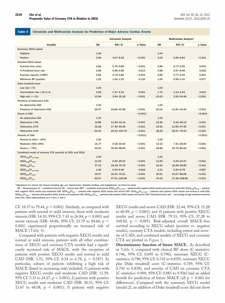
X ith pos
2210 Cho et al. JACC Vol. 60, No. 21, 2012Prognostic Value of Coronary CTA in Relation to XECG November 20/27, 2012:2205–15
CI: 18.37 to 79.44, p � 0.001). Similarly, as compared withpatients with normal to mild stenosis, those with moderatestenosis (HR: 14.10, 95% CI: 7.41 to 26.84, p � 0.001) andsevere stenosis (HR: 44.86, 95% CI: 23.79 to 84.60, p �0.001) experienced proportionally an increased risk ofMACE (Table 4).
Compared with patients with negative XECG results andnormal or mild stenosis, patients with all other combina-tions of XECG and coronary CTA models had a signifi-cantly increased risk of MACE, with the exception ofpatients with positive XECG results and normal to mildCAD (HR: 1.51, 95% CI: 0.34 to 6.78, p � 0.587). Inparticular, subsets of patients exhibiting a high risk ofMACE (listed in increasing risk) included: 1) patients withnegative XECG results and moderate CAD (HR: 11.59,95% CI: 5.33 to 24.27, p � 0.001); 2) patients with positiveXECG results and moderate CAD (HR: 30.51, 95% CI:
Univariate and Multivariate Analysis for Prediction of Major AdversTable 4 Univariate and Multivariate Analysis for Prediction of M
Variable HR
Summary XECG subset
Negative 1.00
Positive 5.56
Detailed XECG subset
Exercise time (min) 0.82
% Predicted heart rate 0.98
Exercise capacity (1-MET) 0.82
Maximum BP (systolic) 1.01
Duke treadmill score
Low risk (�5) 1.00
Intermediate risk (–10 to 4) 2.08
High risk (�–11) 10.94
Presence of obstructive CAD
No obstructive CAD 1.00
Presence of obstructive CAD 25.97
Extent of CAD
No obstructive CAD 1.00
Obstructive 1-VD 19.88
Obstructive 2-VD 32.28
Obstructive 3-VD 52.34
Severity of CAD
Normal to mild (�40%) 1.00
Moderate (40%–69%) 16.77
Severe (�70%) 54.91
Combined model of coronary CTA (severity of CAD) and XECG
XECGnegCTno/mild 1.00
XECGnegCTmod 13.33
XECGnegCTsev 37.12
XECGposCTno/mild 1.46
XECGposCTmod 34.07
XECGposCTsev 92.97
*Adjustment for clinical risk factors including age, sex, hypertension, diabetes mellitus, and dysliBP � blood pressure; CI � confidence interval; HR � hazard ratio; MET � metabolic equivalents; X
with negative XECG results and moderate CAD; XECGnegCTsev � patients with negative XECG resECGposCTmod � patients with positive XECG results and moderate CAD; XECGposCTsev � patients w
heart rate. Other abbreviations as in Tables 1 and 2.
13.67 to 68.08, p � 0.001); 3) patients with negative
XECG results and severe CAD (HR: 32.44, 95% CI: 15.28to 68.89, p � 0.001); and 4) patients with positive XECGresults and severe CAD (HR: 79.33, 95% CI: 37.28 to168.82, p � 0.001). Risk-adjusted overall MACE-freesurvival according to XECG subset (positive vs. negativeresults), coronary CTA models, including extent and sever-ity of CAD, and combined models of XECG and coronaryCTA are plotted in Figure 1.Discriminatory function of future MACE. As describedin Table 5, compared with clinical RF alone (C-statistics:0.746, 95% CI: 0.695 to 0.796), summary XECG (C-statistics: 0.790, 95% CI: 0.742 to 0.839), summary XECGplus Duke treadmill score (C-statistics: 0.789, 95% CI:0.741 to 0.838), and severity of CAD on coronary CTA(C-statistics: 0.909, 95% CI: 0.883 to 0.936) had an addedbenefit for prediction of future MACE (all p � 0.001 fordifferences). Compared with the summary XECG model
diac EventsAdverse Cardiac Events
iate Analysis Multivariate Analysis*
5% CI p Value HR 95% CI p Value
1.00
7–8.43 �0.001 4.53 2.96–6.94 �0.001
5–0.89 �0.001 0.86 0.77–0.95 0.003
6–0.99 0.013 0.98 0.97–0.99 0.042
5–0.89 �0.001 0.85 0.77–0.94 0.001
0–1.02 0.135 1.00 0.99–1.01 0.977
1.00
7–3.15 0.001 1.72 1.13–2.63 0.012
0–35.18 �0.001 10.51 3.20–34.49 �0.001
1.00
4–42.58 �0.001 20.14 11.81–34.35 �0.001
�0.001† �0.001†
1.00
9–34.10 �0.001 16.46 9.30–29.12 �0.001
9–59.56 �0.001 24.52 12.55–47.90 �0.001
2–103.70 �0.001 38.20 18.37–79.44 �0.001
�0.001† �0.001†
1.00
8–30.64 �0.001 14.10 7.41–26.84 �0.001
9–98.90 �0.001 44.86 23.79–84.60 �0.001
1.00
2–26.03 �0.001 11.59 5.33–24.27 �0.001
8–75.78 �0.001 32.44 15.28–68.89 �0.001
3–6.49 0.616 1.51 0.34–6.78 0.587
1–72.51 �0.001 30.51 13.67–68.08 �0.001
1–183.88 �0.001 79.33 37.28–168.82 �0.001
. †p Value for trend.CTno/mild � patients with negative XECG results and normal to mild CAD; XECGnegCTmod � patientsd severe CAD; XECGposCTno/mild � patients with positive XECG results and normal to mild CAD;itive XECG results and severe CAD; %predicted heart rate � percentage of age predicted maximal
e Carajor
Univar
9
3.6
0.7
0.9
0.7
1.0
1.3
3.4
15.8
11.5
17.4
26.4
9.1
30.4
6.5
18.1
0.3
16.0
47.0
pidemiaECGneg
ults an
(model 2), an addition of Duke treadmill score did not show
2211JACC Vol. 60, No. 21, 2012 Cho et al.November 20/27, 2012:2205–15 Prognostic Value of Coronary CTA in Relation to XECG
increment in C-statistics (p � 0.707 for difference). Incontrast, the addition of coronary CTA findings to thesummary XECG model (model 5, C-statistics: 0.908, 95%CI: 0.890 to 0.937) and the summary XECG results plusDuke treadmill score (model 6, C-statistics: 0.909, 95% CI:0.880 to 0.938) showed significant increment in C-statisticscompared with their baseline models without coronary
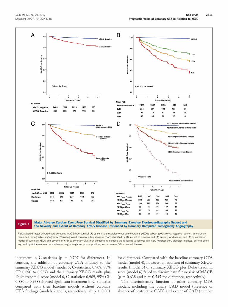
Figure 1 Major Adverse Cardiac Event-Free Survival Stratified bthe Severity and Extent of Coronary Artery Disease Ev
Risk-adjusted major adverse cardiac event (MACE)-free survival (A) by summary excomputed tomographic angiography (CTA)-diagnosed coronary artery disease (CAD)model of summary XECG and severity of CAD by coronary CTA. Risk adjustment ining, and dyslipidemia. mod � moderate; neg � negative; pos � positive; sev � s
CTA findings (models 2 and 3, respectively, all p � 0.001
for difference). Compared with the baseline coronary CTAmodel (model 4), however, an addition of summary XECGresults (model 5) or summary XECG plus Duke treadmillscore (model 6) failed to discriminate future risk of MACE(p � 0.638 and p � 0.545 for difference, respectively).
The discriminatory function of other coronary CTAmodels, including the binary CAD model (presence or
mmary Exercise Electrocardiography Subset andced by Coronary Computed Tomography Angiography
electrocardiography (XECG) subset (positive vs. negative results), by coronaryfied by (B) extent of disease and (C) severity of disease, and (D) by combinedthe following variables: age, sex, hypertension, diabetes mellitus, current smok-VD � vessel disease.
y Suiden
ercisestrati
cludedevere;
absence of obstructive CAD) and extent of CAD (number

al or m
2212 Cho et al. JACC Vol. 60, No. 21, 2012Prognostic Value of Coronary CTA in Relation to XECG November 20/27, 2012:2205–15
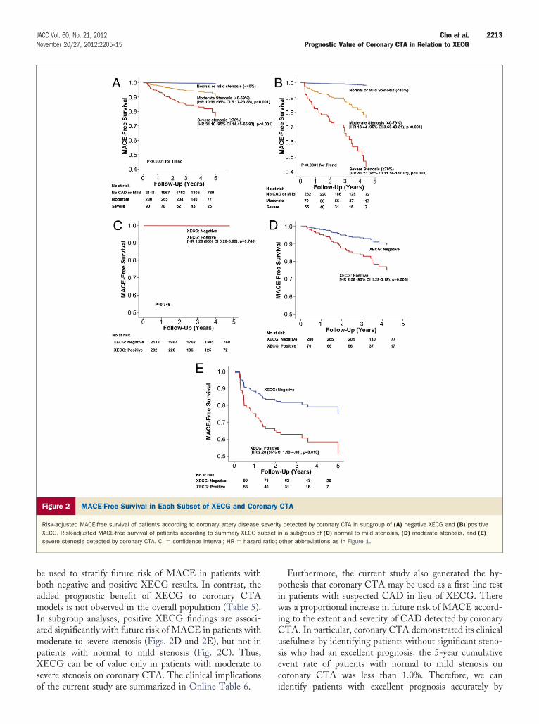
of obstructive stenosis vessels), also were analyzed (OnlineTables 4 and 5). The C-statistics of binary CAD model(0.885, 95% CI: 0.851 to 0.920) and extent of CAD(C-statistics: 0.888, 95% CI: 0.853 to 0.922) were slightlylower than those of the severity of CAD model, butdiscriminatory function compared with clinical RF andXECG results was similar.Patient subset analyses. To determine predicted power ofcoronary CTA in relation to XECG, we also examined thepredictive value of coronary CTA in a subgroup categorizedby XECG and the predictive value of XECG in a subgroupcategorized by severity of CAD determined by coronaryCTA (Fig. 2). Severity of CAD determined by coronaryCTA successfully stratified future risk of MACE in thegroup with positive XECG results as well as the group withnegative XECG results (all p � 0.001 for trend).
However, XECG failed to stratify future risk ofMACE in the group with normal to mild stenosis oncoronary CTA (compared with negative XECG results,HR: 1.28, 95% CI: 0.28 to 5.82, p � 0.746). Abnormal-ity of XECG results predicted future risk of MACE onlyin the subgroups of patients with moderate stenosis(compared with negative XECG results, HR: 2.58, 95%CI: 1.29 to 5.19, p � 0.008) and severe stenosis (com-pared with negative XECG results, HR: 2.28, 95% CI:1.19 to 4.38, p � 0.013).
Discussion
Our study aimed to investigate the roles of coronary CTAand XECG in the risk stratification of patients undergoinginitial evaluation with suspected CAD. The main finding ofthis study is that, in a large number of patients withsuspected CAD, both XECG and coronary CTA areindependently predictive of MACE, but there is no overallincremental benefit for predicting future MACE whenXECG is added to coronary CTA. In subgroup analyses,coronary CTA stratifies risk of future MACE in bothXECG-negative and XECG-positive groups. However,XECG stratifies future risk of MACE only in coronary
C-Statistics for Prediction of 5-Year Risk of Major Adverse CardiacUsing XECG, Coronary CTA (Severity of CAD), and Combined ModeTable 5 C-Statistics for Prediction of 5-Year Risk of Major AdveUsing XECG, Coronary CTA (Severity of CAD), and Com
Model C-Statistics
Model 1: clinical risk factors* 0.746 (0.695–0.796)
Model 2: model 1 � summary XECG‡ 0.790 (0.742–0.839)
Model 3: model 2 � Duke treadmill score 0.789 (0.741–0.838)
Model 4: model 1 � coronary CTA§ 0.909 (0.883–0.936)
Model 5: model 2 � coronary CTA§ 0.908 (0.890–0.937)
Model 6: model 3 � coronary CTA§ 0.909 (0.880–0.938)
*Only the comparisons between baseline models and their nested models were presented. †Clin‡Summary XECG model: positive vs. negative. §Severity of CAD diagnosed by coronary CTA: norm
NA � not applicable; other abbreviations as in Tables 1, 2, 3, and 4.
CTA subgroups of moderate or severe stenoses.
XECG as an initial evaluation of patients with suspectedCAD. Because of its widespread availability, lower cost, andsimplicity of its operation and interpretation, XECG has beenrecommended as the initial diagnostic test for suspected CADpatients with a normal resting ECG results who are able toexercise (14). However, considerable evidence exists that dem-onstrates the limitations of XECG as a diagnostic methodbecause of its relatively low sensitivity and specificity (20,21).Furthermore, it has been reported that there are some limita-tions regarding the use of XECG to identify high-risk patientsin the evaluation of suspected CAD (22).Prognostic value of coronary CTA. Coronary CTA hasemerged as a novel diagnostic tool with high sensitivity andspecificity that demonstrated superiority to XECG in de-tecting CAD in previous head-to-head comparisons (23).In recent 64-slice multidetector coronary CTA studies, thenegative predictive value reached 99%, which is higher thanthat of any other noninvasive imaging techniques (24).Accordingly, it has been suggested that coronary CTAcould be used as a first-line imaging technique to excludeCAD and to replace invasive coronary angiography in somepatients (25). Prognosis prediction of patients suspected ofhaving CAD is as important as detecting disease, because itdetermines the subsequent treatment plan. Recent studieshave shown the potential prognostic value of coronary CTAin suspected CAD patients (26,27). However, to date, itremains unclear whether coronary CTA adds prognosticusefulness beyond standard exercise test findings in patientswho are classified according to XECG results, as well aswhether XECG adds incremental prognostic value to cor-onary CTA.Prognostic value of coronary CTA in relation to XECG.To our knowledge, this is the first registry analysis tosuggest that coronary CTA interpretations of CAD im-prove classification above and beyond XECG findings in alarge cohort with suspected CAD. The current study demon-strated an incremental prognostic value of coronary CTA onXECG findings in the overall population (Table 5). Insubgroup analyses, coronary CTA stratifies future risk ofMACE in groups with both negative and positive XECG
tardiac Eventd Model
p Value for Difference* of C-Statistics Compared With Models
Model 1 Model 2 Model 3 Model 4
NA NA NA NA
0.015 NA NA NA
0.020 0.707 NA NA
�0.001 NA NA NA
�0.001 �0.001 NA 0.638
�0.001 NA �0.001 0.545
k factors included age, sex, hypertension, dyslipidemia, diabetes mellitus, and current smoking.ild (�40%), moderate (40%–69%), and severe CAD (�70%).
Evenlrse Cbine
ical ris
results (Figs. 2A and 2B). Therefore, coronary CTA could
2213JACC Vol. 60, No. 21, 2012 Cho et al.November 20/27, 2012:2205–15 Prognostic Value of Coronary CTA in Relation to XECG
be used to stratify future risk of MACE in patients withboth negative and positive XECG results. In contrast, theadded prognostic benefit of XECG to coronary CTAmodels is not observed in the overall population (Table 5).In subgroup analyses, positive XECG findings are associ-ated significantly with future risk of MACE in patients withmoderate to severe stenosis (Figs. 2D and 2E), but not inpatients with normal to mild stenosis (Fig. 2C). Thus,XECG can be of value only in patients with moderate tosevere stenosis on coronary CTA. The clinical implications
Figure 2 MACE-Free Survival in Each Subset of XECG and Coro
Risk-adjusted MACE-free survival of patients according to coronary artery disease sXECG. Risk-adjusted MACE-free survival of patients according to summary XECG susevere stenosis detected by coronary CTA. CI � confidence interval; HR � hazard
of the current study are summarized in Online Table 6.
Furthermore, the current study also generated the hy-pothesis that coronary CTA may be used as a first-line testin patients with suspected CAD in lieu of XECG. Therewas a proportional increase in future risk of MACE accord-ing to the extent and severity of CAD detected by coronaryCTA. In particular, coronary CTA demonstrated its clinicalusefulness by identifying patients without significant steno-sis who had an excellent prognosis: the 5-year cumulativeevent rate of patients with normal to mild stenosis oncoronary CTA was less than 1.0%. Therefore, we can
CTA
y detected by coronary CTA in subgroup of (A) negative XECG and (B) positiven a subgroup of (C) normal to mild stenosis, (D) moderate stenosis, and (E)other abbreviations as in Figure 1.
nary
everitbset iratio;
identify patients with excellent prognosis accurately by
2214 Cho et al. JACC Vol. 60, No. 21, 2012Prognostic Value of Coronary CTA in Relation to XECG November 20/27, 2012:2205–15
coronary CTA, so that unnecessary additional testing can beavoided in this population. In our present cohort, among2,977 symptomatic patients, coronary CTA identified 2,568(86%) patients without obstructive CAD (�50%) and 2,450(82%) patients without moderate to severe stenosis (�40%)to be at low risk (Table 3). On the contrary, a non-negligible proportion of patients with negative XECGresults are found to have moderate to severe stenosis oncoronary CTA (15%) and to experience higher 5-yearcumulative event rates of 2.4% (95% CI, 1.8% to 3.1%)compared with those patients without obstructive CAD oncoronary CTA. These findings emphasize an importantlimitation of nonimaging exercise-induced ST-segment de-pression for risk stratification, as has been reported in priorstudies (28).
However, despite the powerful prognostic value, coronaryCTA has potential limitations, including radiation hazard,use of iodinated contrast agents, and higher cost comparedwith that of XECG. In addition, most cardiac events in thecurrent study were revascularization procedures, so theprognostic value of coronary CTA to predict a so-calledhard event was not evaluated fully. Moreover, the cumula-tive events rate of 2.4% in patients with negative XECGresults is a fairly good prognosis, although it is higher thanthe cumulative events rate in patients without obstructiveCAD detected by coronary CTA. Therefore, the decision touse coronary CTA as a first-line diagnostic method inpatients with suspected CAD should be deferred until thepotential risk-to-benefit ratio, cost effectiveness, and clinicalefficiency are weighed by prospective randomized trials.Further, future comprehensive studies also are warranted todetermine the most cost-effective, safe, and clinically effi-cient strategy to diagnose CAD and to predict future risk ofcardiac events in low- to intermediate-risk symptomaticpatients without known CAD covering various diagnosticmethods, such as coronary CTA, XECG, myocardial per-fusion imaging, and stress echocardiography.Study limitations. The present study was retrospective andmay have been influenced by unobserved confounders andselection or referral biases, or both. In addition, the effect ofpost-test medical treatments or risk factor control was notconsidered. Especially given the potential advantage ofcoronary CTA to identify nonobstructive CAD for predic-tion of future cardiac events (29), further studies arewarranted to assess the impact of medical therapies inpatients with nonobstructive CAD on coronary CTA.
Although we considered only revascularizations morethan 90 days after coronary CTA as outcome events,revascularizations from the index test may have been in-cluded. Moreover, the pretest probability of the studypopulation was relatively low, which limits the number ofclinical events at follow-up. However, to our knowledge, thepopulation size of this study is the largest to date reportingconcurrent XECG and coronary CTA findings in relation
to downstream clinical outcomes, and we plan to continueto follow through with our investigation to understand thelong-term nature of these study findings.
Conclusions
In patients with suspected but without known CAD,coronary CTA demonstrates added prognostic benefit inpatients with both positive and negative XECG results. Inparticular, the clinical usefulness of coronary CTA is real-ized by identifying patients with normal or mild stenosis(�40%) and accurately predicting the very low risk of futurecardiac events. Conversely, XECG has additive value forrisk stratification on coronary CTA only in patients withmoderate to severe stenosis.
Reprints requests and correspondence: Dr. Hyuk-Jae Chang,Severance Cardiovascular Hospital, Yonsei University Health Sys-tem, 250 Seongsanno Seodaemungu Seoul 120–752, Republic ofKorea. E-mail: [email protected].
REFERENCES
1. Gibbons RJ, Abrams J, Chatterjee K, et al. ACC/AHA 2002 guidelineupdate for the management of patients with chronic stable angina—summary article: a report of the American College of Cardiology/American Heart Association Task Force on Practice Guidelines(Committee on the Management of Patients With Chronic StableAngina). J Am Coll Cardiol 2003;41:159–68.
2. Gianrossi R, Detrano R, Mulvihill D, et al. Exercise-induced STdepression in the diagnosis of coronary artery disease. A meta-analysis.Circulation 1989;80:87–98.
3. Budoff MJ, Dowe D, Jollis JG, et al. Diagnostic performance of64-multidetector row coronary computed tomographic angiographyfor evaluation of coronary artery stenosis in individuals without knowncoronary artery disease: results from the prospective multicenterACCURACY (Assessment by Coronary Computed TomographicAngiography of Individuals Undergoing Invasive Coronary Angiogra-phy) trial. J Am Coll Cardiol 2008;52:1724–32.
4. Meijboom WB, Meijs MF, Schuijf JD, et al. Diagnostic accuracy of64-slice computed tomography coronary angiography: a prospective,multicenter, multivendor study. J Am Coll Cardiol 2008;52:2135–44.
5. Miller JM, Rochitte CE, Dewey M, et al. Diagnostic performanceof coronary angiography by 64-row CT. N Engl J Med 2008;359:2324 –36.
6. Mollet NR, Cademartiri F, Van Mieghem C, et al. Adjunctive valueof CT coronary angiography in the diagnostic work-up of patientswith typical angina pectoris. Eur Heart J 2007;28:1872–8.
7. Nieman K, Galema T, Weustink A, et al. Computed tomographyversus exercise electrocardiography in patients with stable chest com-plaints: real-world experiences from a fast-track chest pain clinic.Heart 2009;95:1669–75.
8. van Werkhoven JM, Schuijf JD, Gaemperli O, et al. Prognostic valueof multislice computed tomography and gated single-photon emissioncomputed tomography in patients with suspected coronary arterydisease. J Am Coll Cardiol 2009;53:623–32.
9. Sato A, Nozato T, Hikita H, et al. Incremental value of combining64-slice computed tomography angiography with stress nuclear myo-cardial perfusion imaging to improve noninvasive detection of coronaryartery disease. J Nucl Cardiol 2010;17:19–26.
10. Diamond GA, Forrester JS. Analysis of probability as an aid in theclinical diagnosis of coronary-artery disease. N Engl J Med 1979;300:1350–8.
11. Gibbons RJ, Balady GJ, Beasley JW, et al. ACC/AHA guidelines forexercise testing. A report of the American College of Cardiology/American Heart Association Task Force on Practice Guidelines(Committee on Exercise Testing). J Am Coll Cardiol 1997;30:
260 –311.
1
1
1
1
2
2
2
2
2
2
2
2
2
2
2215JACC Vol. 60, No. 21, 2012 Cho et al.November 20/27, 2012:2205–15 Prognostic Value of Coronary CTA in Relation to XECG
12. Kwon SW, Kim YJ, Shim J, et al. Coronary artery calcium scoringdoes not add prognostic value to standard 64-section CT angiographyprotocol in low-risk patients suspected of having coronary arterydisease. Radiology 2011;259:92–9.
13. Raff GL, Abidov A, Achenbach S, et al. SCCT guidelines for theinterpretation and reporting of coronary computed tomographic an-giography. J Cardiovasc Comput Tomogr 2009;3:122–36.
14. Gibbons RJ, Balady GJ, Bricker JT, et al. ACC/AHA 2002 guidelineupdate for exercise testing: summary article. A report of the AmericanCollege of Cardiology/American Heart Association Task Force onPractice Guidelines (Committee to Update the 1997 Exercise TestingGuidelines). J Am Coll Cardiol 2002;40:1531–40.
15. Hadamitzky M, Frei�muth B, Meyer T et al. Prognostic value ofcoronary computed tomographic angiography for prediction of cardiacevents in patients with suspected coronary artery disease. J Am CollCardiol Img 2009;2:404–11.
6. Hadamitzky M, Distler R, Meyer T, et al. Prognostic value ofcoronary computed tomographic angiography in comparison withcalcium scoring and clinical risk scores. Clinical perspective. CircCardiovasc Imaging 2011;4:16–23.
7. Hachamovitch R, Nutter B, Hlatky MA, et al. Patient managementafter noninvasive cardiac imaging: results from SPARC (Study ofMyocardial Perfusion and Coronary Anatomy Imaging Roles inCoronary Artery Disease). J Am Coll Cardiol 2012;59:462–74.
8. Pencina MJ, D’Agostino RB. Overall C as a measure of discriminationin survival analysis: model specific population value and confidenceinterval estimation. Stat Med 2004;23:2109–23.
9. Harrell FE, Lee KL, Mark DB. Multivariable prognostic models:issues in developing models, evaluating assumptions and adequacy, andmeasuring and reducing errors. Stat Med 1996;15:361–87.
0. Gibson RS, Beller GA. Should exercise electrocardiographic testing bereplaced by radioisotope methods? Cardiovasc Clin 1983;13:1–31.
1. Podrid PJ, Graboys TB, Lown B. Prognosis of medically treated
patients with coronary-artery disease with profound ST-segmentdepression during exercise testing. N Engl J Med 1981;305:1111–6.2. Epstein SE. Value and limitations of the electrocardiographic responseto exercise in the assessment of patients with coronary artery disease.Controversies in cardiology—II. Am J Cardiol 1978;42:667–74.
3. Dewey M, Dubel HP, Schink T, Baumann G, Hamm B. Head-to-head comparison of multislice computed tomography and exerciseelectrocardiography for diagnosis of coronary artery disease. EurHeart J 2007;28:2485–90.
4. Bastarrika G, Lee YS, Huda W, Ruzsics B, Costello P, Schoepf UJ.CT of coronary artery disease. Radiology 2009;253:317–38.
5. Mollet NR, Cademartiri F, van Mieghem CA, et al. High-resolutionspiral computed tomography coronary angiography in patients referredfor diagnostic conventional coronary angiography. Circulation 2005;112:2318–23.
6. Carrigan TP, Nair D, Schoenhagen P, et al. Prognostic utility of64-slice computed tomography in patients with suspected but nodocumented coronary artery disease. Eur Heart J 2009;30:362–71.
7. Hadamitzky M, Freissmuth B, Meyer T, et al. Prognostic value ofcoronary computed tomographic angiography for prediction of cardiacevents in patients with suspected coronary artery disease. J Am CollCardiol 2009;2:404–11.
8. Sekhri N, Feder GS, Junghans C, et al. Incremental prognosticvalue of the exercise electrocardiogram in the initial assessment ofpatients with suspected angina: cohort study. BMJ 2008;337:a2240.
9. Lin FY, Shaw LJ, Dunning AM, et al. Mortality risk in symptomaticpatients with nonobstructive coronary artery disease: a prospective2-center study of 2,583 patients undergoing 64-detector rowcoronary computed tomographic angiography. J Am Coll Cardiol2011;58:510 –9.
Key Words: coronary computed tomographic angiography yexercise electrocardiography y prognosis.
APPENDIX
For supplemental Methods and tables,
please see the online version of this article.