professional disclosures corneal workshop: keratitis ... · nhelp heal refractory bacterial mk and...
TRANSCRIPT

5/3/2013
1
Corneal Workshop:Keratitis Management
Elizabeth Yeu, MDCornea, Cataract, Anterior Segment,
Refractive SurgeryVirginia Eye Consultants
May 1, 2013
Elizabeth Yeu, MDCornea, Cataract, Anterior Segment,
Refractive SurgeryVirginia Eye Consultants
May 1, 2013
Professional Disclosures
n Allergan: Advisory Board, Speakers’ Bureau
n Bausch + Lomb: Advisory Boardn Rhein Medical: Advisory Board
Keratitis
n Bacterialn Herpeticn Blepharokeratitis
Keratitisn Infectious: Microbial keratitis-- “MK”
– Bacterial– Viral– Parasitic: acanthamoeba– Fungal
n Non-infectious– Inflammatory: blepharokeratitis, PUK – Interstitial– Others

5/3/2013
2
Bacterial Keratitis
n Gram-positive cocci: Staph spp., Strep pneumoniae
n Aerobic gram-negative bacilli: Pseudomonas aerugenosa, Haemophilus influenzae, Moraxella catarrhalis
n Enteric gram-negative bacilli or colonization of normal skin flora: S. aureus, S. epidermides, Serratia spp., Strep viridans
General guidelines
n Ulcer: Epithelial defect + WBC recruitment within stroma– Infectious or sterile
n Infiltrate: grey/white opacities from coalescence of WBCs n Localized or diffusenUsually infectiousn Peripheral (PUK, Mooren’s, marginal keratitis)
more commonly inflammatory and not infectious
General Guidelines
n Appearance of infiltrate– Sharply dilineated, ovoid: gm +– Irregular, indistinct borders: gm –– “Feathery” borders: fungal, strep pneumo– Crystalline: strep pneumoniae– Ring infiltrate:
n Pseudomonas, HSV, acanthamoeba, Neisseria, Corynebacterium, Nocardia, anesthetic abuse

5/3/2013
3
General Guidelines
n Appearance of infiltrate– Aggressive suppuration (“soupy”): gm (–)– Infiltrate with intact epithelium: sterile,
fungus, H. aegyptius, Neisseria gonorrhoeae, Listeria monocytogenes
– Satellite lesions: fungal, atypical mycobacteria, nocardia
– Raised, “clumpy” borders of gray-white epithelium: neurotrophic or toxic
General guidelines
n Duration– Indolent: acanthamoeba, gm +, fungal– Fulminant: gm (–)
n Amount of tissue destruction and thinning
n Gm (–) spp., particularly pseudomonas aeruginosa, cause great necrosis very quickly
Usual suspects
n CL wear à p. aeruginosan “Spontaneous” MKà enteric/ skin flora n Scleral buckle, canalicular tubes à
atypical mycobacteriumn Vegetation, trauma à fungusn Water-related à acanthamoeba

5/3/2013
4
Usual suspects
n PKP: gm + organism– Often strep
pneumoniae (steroids)
n Tx: start broad spectrum fluoro
n S. pneumo– Vancomycin 2.5%
n Culture, inc. sutures!
When to refer?
n Culture - Rules of 1-2-3– Within 1 mm of visual axis– Ulcers with 2 or more infiltrates– 3 mm or more in diameter
Indications for Referral
n Anything central, necrosing or thinningn Poor response to single treatmentn Poorly healing epithliumn Extended durationn Post-surgical n Trauma-related: vegetation, FBn Inflammatory melt
Indications for Referral
n If considering immediate referral, prior to starting meds, consider not starting any treatment in order to obtain highest yield on culture
Culturing the cornea
n Chemistry lab set up, alcohol lamp
n Sterile Kimura spatulasn Slidesn Culture plates and tubes
Procedure
n Anesthetize the cornea – Preservative-free tetracaine
n Scrape ulcer base / leading edge of infiltraten Place specimen on slide, then culture media
– Smears – fixing organisms to be stained / observed
– Culture – microbial growth
n Sterilize spatula over flame between slides / cultures
5/3/2013
5
Culturing the cornea
n Diagnostic
n Commonly, can be THERAPEAUTIC!
Treatment
n D/C CL wearn Primary goal – eliminate the pathogensn Secondary goal – prevent host destructionn Treated as bacterial initiallyn Small infiltrates – empirically (<1.0mm)n Cycloplegics bid-qid
– Cyclopentolate 1%, homatropine 5%, scopolamine 0.25%, atropine 1%
Treatment
n Fluoroquinolones – common standardn Besifloxacin: 3rd generation fluoro, excellent
broad spectrum, inc MRSA and pseudomonasnMoxifloxacin: 4th generation, ? Fungal coverage
n Vision threatening – Fortified antibiotics– Tobramycin/gentamycin (15mg/mL) aggressively
ATC– Cefazolin (50mg/mL), ceftazadime (50mg/ml) or
vancomycin (25mg/mL)
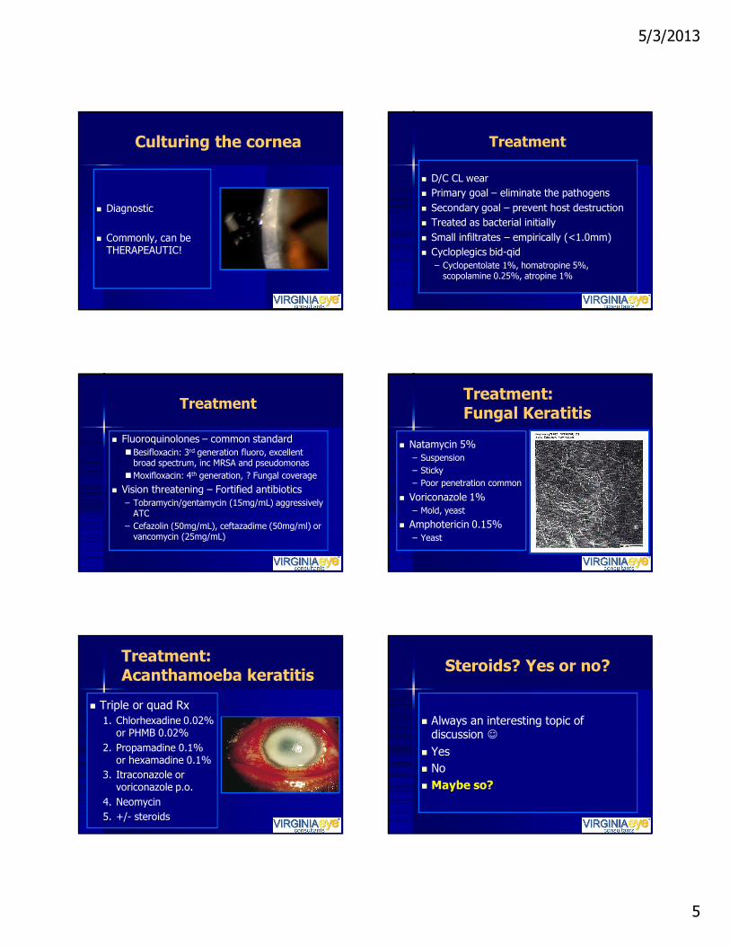
Treatment: Fungal Keratitis
n Natamycin 5%– Suspension– Sticky– Poor penetration common
n Voriconazole 1% – Mold, yeast
n Amphotericin 0.15%– Yeast
Treatment:Acanthamoeba keratitis
n Triple or quad Rx1. Chlorhexadine 0.02%
or PHMB 0.02%2. Propamadine 0.1%
or hexamadine 0.1%3. Itraconazole or
voriconazole p.o.4. Neomycin5. +/- steroids
Steroids? Yes or no?
n Always an interesting topic of discussion ☺
n Yesn No n Maybe so?

5/3/2013
6
Steroids for Corneal Ulcer Trial
n Objective: To determine whether there is a benefit in clinical outcomes with the use of topical corticosteroids as adjunctive therapy in the treatment of bacterial corneal ulcers
n Results: No significant difference was observed – 3-month BSCVA (P =.82)– Infiltrate/scar size (P = .40)– Time to reepithelialization (P = .44)– Corneal perforation (P > .99)
Srinivasan M, Mascarenhas J, Rajaraman R, Ravindran M, Lalitha P, Glidden DV, Ray KJ, Hong KC, Oldenburg CE, Lee SM, Zegans ME, McLeod SD, Lietman TM, Acharya NR; Steroids for Corneal Ulcers Trial Group.
Steroids?
n Steroids– Aggressive suppuration to â necrosis– Healing bacterial MK– PKP patients, not fungal– “Steroid stress test”
n Exacerbates fungal MK
Potential Treatment Option Collagen cross-linking àRiboflavin phototherapyn In vitro studies: Ribloflavin
phototherapy can eradicate S. aureus, MRSA, P.aeruginosa(Martins SA, et al. IOCS. 2008; 49:3402-2408)
n Help heal refractory bacterial MK and halt thinning
(Panda A, et al. Cornea. 2012 Oct; 31(10):1210-3)
Patient Presentation
n 62 yo WMn h/o hyperopic LASIKn + cataracts à uneventful cataract
surgeryn Loose epithelium near edge of prior
LASIK flap à BCL
Patient Presentation
n POD 1, looks greatn POW 1: “BCL fell out yesterday when I
was poolside, so my wife picked it up, rinsed it off with her solution and put it back in”

5/3/2013
7
Patient Presentation
n Minimal injection, superotemporal 1x2 mm ant stromal infiltrate, no thinning
n Plan:– D/C BCL– Start moxifloxacin q2 hours ATC
Patient: Clinical course
n Over the next 2 days, infiltrate shrinking in size, but epithelium not quite healing over
à Add FML qid, decrease moxi to qid
Patient Presentation
n Continued therapy for 2 more daysn On return, infiltrate returning slightly
largern ?? Wrong diagnosis or superinfection
Confocal microscopy performed

5/3/2013
8
Patient Presentation
n Gm stain and CW à hyphael elementsn Voriconazole 1% q1 h ATC,
Voriconazole 200 mg bid
àCulture: Paecilomyces spp.
Clinical scenario
n36 yo male, 2nd opinion of persistent “geographic ulcer”nRed eye OD began ~5w ago
–3rd episode in 5 y
nTrifluridine x4w, at qidnZymar qid
Update: Herpes Keratitis
“Persistent HSV geographic ulcer” OD
Herpetic keratitis
n Regarding herpetic keratitis….n Not too much has changed in the
treatment of HSV keratitis over the years

5/3/2013
9
HSV: Background
n HSV 1: oral, nasal, ocular, throat soresn HSV 2: genital soresn Neurotrophic and neuroinvasive virusesn HSV-1 and -2 persist in the body
– Become latent and hide from the immune system in the cell bodies of neurons
n After primary infection, reactivation can occur anytime– Ocular HSV infections generally reactivation– HSV blepharitis can be primary infection: more
commonly seen in kids
HSV: Epidemiology
n Worldwide: HSV 65% - 90%n U.S.
– HSV 1: ~ 50% - 80%– HSV 2: ~ 20%
HSV Keratitis
n Epithelialn Stromaln Endotheliitisn Metaherpetic and Neurotrophic
HSV Keratitis
n Epithelialn Stromaln Endotheliitisn Metaherpetic and Neurotrophic
Corneal HSV Disease: Epithelial
n Dendriticn Geographicn Marginal ulcern Infection of epi cellsn Base stains with
fluorescein, infected “balloon” cells stain with rose bengal
HSV Dendritic Keratitis

5/3/2013
10
HSV Epithelial Keratitis
n Disease course < 2 weeks– 95% spontaneously heal in 14 days– Treatment speeds up resolution
n Topical nOral
Question
n What is your treatment of choice for management of HSV epithelial keratitis?1. Debriding the epithelium2. Topical trifluridine 1% gtt3. Topical ganciclovir 0.15% gel4. Oral anti-viral therapy5. Debriding the epithelium + medicine
HSV Epithelial Keratitis
n Treatment trends:– General ophthalmologist: topical– Cornea: oral– Greater trend towards topical treatment
with topical GCV 0.15 % gel
HSV Epi keratitis:Treatment
n Trifluridine 1% (TFT): 8x/day (q2h) until epithelium heals, usually 5-10 days
n Taper off within 2 weeks
Trifluridine 1%
n Very toxic to epithelium: – Delayed healing
n Conjunctival scarringn Punctal stenosisn Do not use > 2 weeks
HSV Epithelial Keratitis:Treatment
n Ganciclovir gel 0.15% (GCV)– 5x/day while awake x 7 days,
then TID for 7 days
n Side effect profile: – Much less epitheliopathy– Eye irritation (20%), punctate keratitis
(5%), conj hyperemia (5%)

5/3/2013
11
HSV Epithelial Keratitis:Treatment
n Ganciclovir gel 0.15% (GCV):n Since 1995, the GCV 0.15% available
in 30 countries within Europe, Asia, Africa and South America
n ACV 3% ung and GCV 0.15% gel standard of care in Europe
n Approved by FDA in 2009
HSV Epithelial Keratitis:GCV 0.15% studies
n All phase II/IIII GCV 0.15% studies have been abroad
n GCV 0.15% gel vs. ACV 0.3% ung– GCV as effective as ACV– Less blurring than ACV– Average healing time 7-8 days– 82% - 88% healing rate
HSV Epithelial Keratitis:GCV 0.15% studies
n May be useful as prophylaxis*– 6 patients: 3 s/p PKP, no
recurrences
*Tabbara Kf, Treatment of herpetic keratitis, Ophthalmology, 2005;112:1640.
HSV Epithelial Keratitis
Oral treatment:n Acyclovir 400mg 5x/day (2g/day)n Valacyclovir (Valtrex®) 500mg-1gm TIDn Famcyclovir 125-250mg BIDn In kids: Acyclovir 200mg/5cc qid
HSV Keratitis
n Epithelialn Stromal (15%): immune, necrotizing n Endotheliitisn Metaherpetic and Neurotrophic
Immune-mediated stromal keratitis
n Not active infectionn Occurs up to years after original
epithelial keratitisn Often chronic, recurrent
inflammation

5/3/2013
12
Stromal keratitis:Clinical appearance
n Epithelium intactn Stromal infiltrationn Edeman Stromal vascularizationà lipid n AC reaction
Herpetic Stromal Keratitis
Corneal HSV Disease: Stromal
nNecrotizing–Necrosis, dense
infiltrate–Epi defect–(+) infected
stromal cells & immune reaction
n Non-necrotizing stromal keratitis– Topical prednisolone 1% 4-8x/day– Topical difludprednate
n NOTE: When steroids > bid, must use anti-viral prophylaxis (topical or oral)
n Necrotizing– Aggressive topical steroids– Anti-viral for ACTIVE HSV disease
Treatment:HSV Stromal Keratitis
HSV Keratitis
n Epithelialn Stromaln Endotheliitis: Disciform, diffuse, linearn Metaherpetic and Neurotrophic
Corneal HSV Disease: Endotheliitis
n Immune reaction involving endothelial cells (? live virus)
n Clinical appearance– KP– Stromal and epithelial edema– Iritis– Minimal to no stromal infiltration
n 3 forms: disciform, diffuse, linear

5/3/2013
13
Disciform endotheliitis
n Most common n Occurs some
time after infectious epithelial keratitis
n Disc-shaped area of edema over KP
Diffuse and linear HSV endotheliitis
Diffuse: dense retrocorneal plaque
HSV endotheliitis: Treatment
n Disciform– Topical steroids– Anti-viral prophylaxis
n Diffuse and linear– Aggressive topical steroids– Anti-viral to for active infection
HSV Keratitis: Recurrence
n Rate of recurrence:– 1 year: 9.6%– 2 years: 22.9%– 10 years: 49.5%
n Can cause severe vision loss, espepithelial and stromal keratitis
Management: Immune diseases
n (Non-necrotizing) stromal, disciform endotheliitis
n Prednisolone 1% qid to q2hn Anti-viral prophylaxisn Once controlled, very slow
taper of steroidn May always require topical steroid,
even TIW
When prophylaxis?
nPrevent recurrencesn HEDS, ACV x1y ↓ all forms by ~41% (19%
vs. 32%) and stromal keratitis recurrence by 50% (14% vs. 28%)
nPrevent reactivation during steroid use

5/3/2013
14
Oral prophylaxis
– Acyclovir 400mg bid– Famciclovir 125-250mg qd-bid– Valacyclovir 500mg qd-bid
Topical prophylaxis
–Trifluridine: variablenTID à drop for drop until steroid down
to QD
– GCV gel: ?? No solid recommendations YET ☺n I use BID - TID
HSV Keratitis
n Epithelialn Stromaln Endotheliitisn Metaherpetic and Neurotrophic
When metaherpetic or neurotrophic disease?
n Epi defect on topical anti-viral >2 w n Geographic versus
metaherpetic/neurotrophic ulcer– HSV-infected “balloon” cells stain– Shape of lesion
Geographic Sterile Metaherpetic/ neurotrophic ulcer
n Poor sensationn Oval, rolled edges,
smooth bordersnWithin IPF
Courtesy of MB Hamill, MD

5/3/2013
15
Management:HSV ulcer> 2 weeks
n Stop topical anti-viraln Change to oral anti-viral prnn No preservativesn Non-preserved ung q2hn BCL n Amniotic membranen Omega-3 FA, Doxycyclinen (+) stromal inflammation: cautious steroids
Neurotrophic Ulcers
n Amniotic membrane transplantn Prokera: Self-retaining,
cryopreserved AMT on 16 mm PMMA ring
Self-retaining AMT
n Fairly easy insertion: exam lanen Can stain cornea with NaFL without
removal n Topical meds penetrate through AMTn AMT soaks up meds
Management:HSV Ulcer > 2 weeks
Surgical optionsn Amniotic membrane, esp if thinn Conjunctival flapn Lateral tarsorrhaphyn Keratoplasty

5/3/2013
16
Back to the patient…
n36 yo male, 2nd opinion of persistent “geographic ulcer”nRed eye OD began ~5w ago
–3rd episode in 5 y
nTrifluridine x4w, at qidnZymar qid
Back to the patient….
• Poor k sensation OD• Dx: Neurotrophic ulcer
Photos courtesy of S. Pflugfelder, MD
“Herpes….”
“Attacks the weak- worse in atopes, humbles the physician-fools the best of us, mocks your treatment- hides only to return, and returns more than the taxman” -Ivan R. Schwab, MD
Blepharokeratitis
“Blepharokeratoconjunctivitis”
n BKC is disease entity of adolescents-“a syndrome usually associated
with anterior or posterior lid margin blepharitis, accompanied by episodes of conjunctivitis, and a keratopathyincluding punctate erosions, punctate keratitis, phlyctenules, marginal keratitis, and ulceration”
Blepharokeratitis
n Ocular Rosacean Phlyctenulosisn Marginal keratitis

5/3/2013
17
Ocular Rosacea Ocular Rosacea
Ocular Rosacea
c/o Parag Majmudar, MD
Ocular Rosacea
n Most common in middle-aged femalen Flushed cheeks and nose à
telangiectasis– Vasomotor lability aggravated by coffee,
tea, alcohol, spicy foods, anxiety, hormonal changes
n Rhinophyma (bulbous nose) àsebaceous gland hypertrophy
Ocular Rosacean Etiology: unknownn Colonization of lid margins with flora
(S. epidermides, P. acnes) produce lipases which may alter MG secretions à inflammation
n S. epidermides found only in pustules, not in unaffected skin; may be transported by Demodex mites(Jarmuda S, et al. J Med Microbiol August 2012)
Ocular Rosacea
n Ocular findings ~50%– Blepharitis, MGD, lid thickening,
telangiectasis– Chronic, diffuse conjunctival injection, esp
within IPF
n Corneal involvement ~5-30%

5/3/2013
18
Ocular Rosacea: Corneal Findings
n Punctate epithelial erosionsn Marginal infiltratesn Corneal pannusn Stromal thinning, perforationn Corneal scars
Phlyctenulosis
Phlyctenulosis
c/o Parag Majmudar, MD
Phlyctenulosis
n Inflammatory raised gelatinous nodules in cornea or conjunctiva
n Hypersensitivity reaction
Phlyctenulosis: etiologyn Most commonly associated with
staphylococcal blepharitisn Others
– TB– P. acnes– N. gonorrheae, Chlamydia – HSV– C. albicans– Endemic parasites
Phlyctenulosis
n More common in children and adolescents
n W > M, 2:1 n Up to half of patients w/ bilateral
presentation

5/3/2013
19
Phlyctenulosis
n Phlyctenule can migrate towards center of cornea
n + Feeder vesselsn May ulcerate w/ stromal WBC infiltraten Heal with scarringn May lead to significant visual impairment
Marginal Keratitis
c/o EyeRounds.org
Marginal Keratitis Marginal Keratitis
n AKA catarrhal infiltraten Hypersensitivity reaction to
Staphylococcusn ? May be related to Demodex
blepharitis, esp with recurrent disease
Marginal Keratitis
n Creamy white infiltrate(s) in peripheral cornea
n Infiltrate smooth, distinct bordersn Single or multiplen Overlying epi defect smaller than infiltraten 1-2 mm clear zone from limbusn Infiltrate à then epi ulceration à KNV
Marginal Keratitis
n Most commonly occur where cornea crosses lid margin

5/3/2013
20
Blepharokeratitis: Treatment
n Treat inflammation and blepharitis– Combo steroid/ antibiotic work well
(Tobramycin/dexamethasone)– FML or Pred acetate 1% – Antibiotic ointment to lids
Blepharokeratitis: Treatment
n Oral antibiotic– Doxycycline 20 mg to 200 mg qd/bid(I prefer Doxy 50 mg bid, then taper to qd)
– Minocycline 20 to 40 mg qd– Azithromycin 250 to 500 mg qd x 3 days,
for 3-5 weeksn Azithromycin works wonders in children and
adolescents!
Blepharokeratitis
n Nutritional supplements: O3FA/O6FAn Lid hygienen If recurrent, may consider Demodex txn In-office MG expression
– Probing– IPL– Lipiflow
Device in action
c/o Preeya Gupta, MD
Device in action
Wow…..
Suture Removal

5/3/2013
21
Suture Removal Conclusion: MK
n Broad spectrum fluoroquinolone– Besifloxacin or moxifloxacin
n Withhold steroids until clinical improvement observed
n 1-2-3 Rule for MK referralsn Do not initiate tx if planning to refer
same day à better yield
Conclusion: HSV
n HSV epithelial keratitis: active infection– No steroid with epithelial defect– Topical tx option less toxic– Reconsider tx or diagnosis if >10-14 days
n Stromal or disciform keratitis à aggressive steroid initially, very slow taper
n Always use oral or topical anti-viral prophylaxis with steroid use
n Geographic ulcer? Be aware of possible neurotrophic/ metaherpetic keratopathy!
Blepharokeratitis
n “Triple therapy”:– Topical steroid– Topical antibiotic ointment or gtt– Oral antibiotic
n O3FA/O6FA
Blepharokeratitis
n Lid hygienen Think Demodex for recurrent disease
or chronic blepharitisn In-office MG expression treatment
THANK YOU ☺