0. corneal ulcer€¦ · 0. corneal ulcer . 1. infections :-bacterial keratitis -viral keratitis...
TRANSCRIPT
* Diseases of the cornea : 0. Corneal ulcer .
1. Infections :
-Bacterial keratitis
-Viral keratitis
-Fungal keratitis
-Acanthameba keratitis
2. Keratoconus
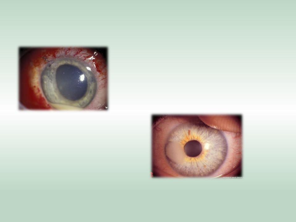
PAINFULL ACUTE LOSS OF VISION
1- CORNEAL ULCERS
Risk Factors :
Contact lens wear
Recent trauma
Poor lid apposition
History of ocular surgery
Chronic topical steroid use
SYMPTOMS
Depends on whether the ulcers are sterile or infectious
Pain, usually severe
Redness
Tearing
Discharge
Photophobia
SIGNS
Dense corneal infiltrate with overlying epithelial defect
Hypopyon ( leukocytic exudate seen in the anterior
chamber )
Corneal destruction
Ocular perforation
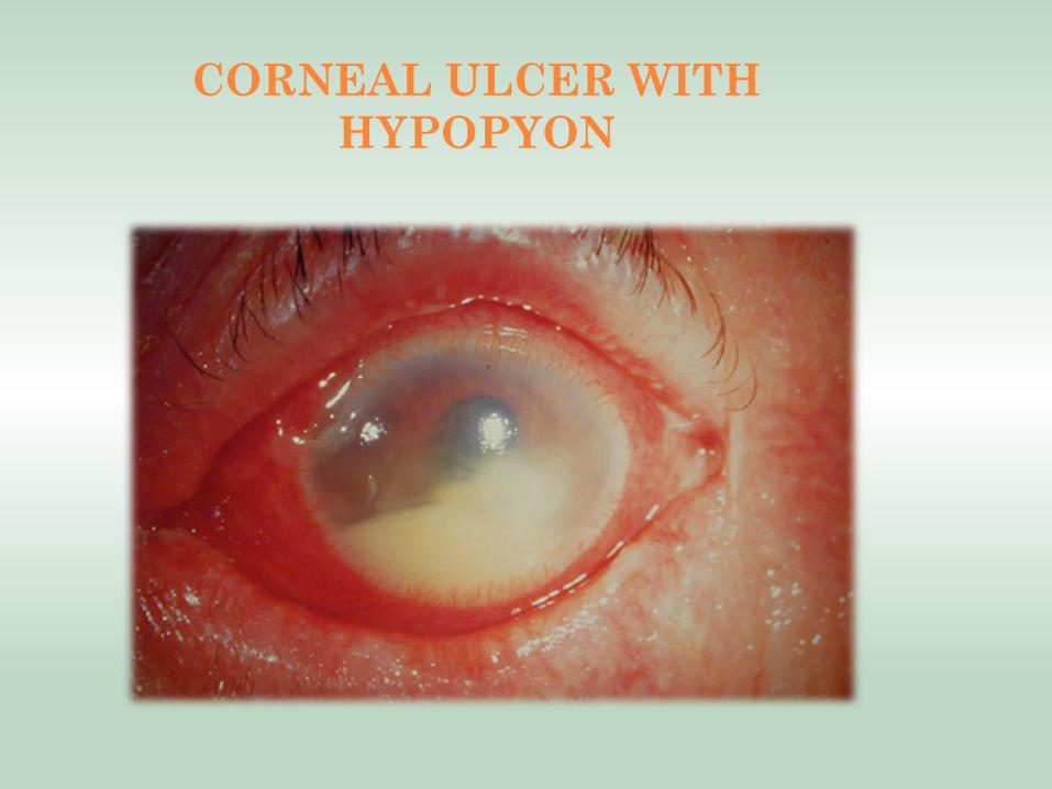
CORNEAL ULCER WITH
HYPOPYON
* Bacterial keratitis :
- Some of the bacteria responsible for the infection :
*Staphylococcus epidermidis
*Staphylococcus aureus
*Streptococcus pneumonia
*Coliforms
*Pseudomonas
*Haemophilus
* Predisposing factors:
- Contact lens wear
- Keratoconjunctivitis sicca (dry eye).
- Prolonged use of topical steroids.
- A breach in the corneal epithelium
e.g. following surgery .
- Decrease immunologic defense.
* Symptoms:
•Rapid onset of pain
•Light sensitivity (photophobia).
•Decreased vision
•Purulent discharge.
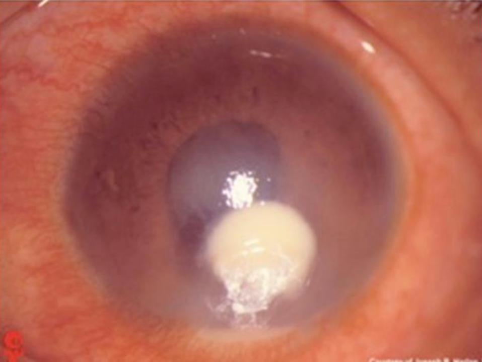
* Signs:
•Hypopyon (accumulation of white cells in the Ant.chamber).
•Ulceration of the epithelium.
•Conjunctival hyperemia (ciliary injection).
•White cornea opacity .
•Dense stromal infiltrate(subepithelial infeltrates).
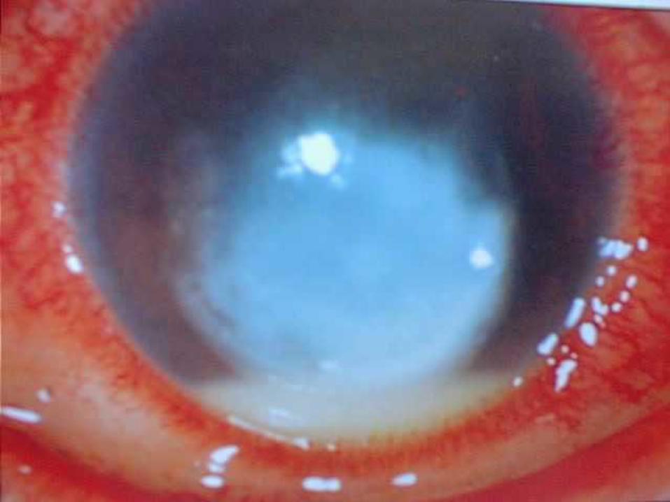
* Complications:
•Thinning of cornea.
•Sloughing of infected stroma.
• Irregular astigmatism{uneven healing ulcer}.
•Corneal ulcer.
•Corneal perforation*; cause secondary
endophthalmitis & loss of the eyes.
-Vision lose
-Corneal leukoma (scar formation&corneal vascularization)
* Treatment:
•Initiate topical broad-spectrum antibiotics often with dual therapy to cover most organisms :
(vancomycin for g+ve , 4th generation fluoroqinalon )
•If the corneal ulcer is small, peripheral and no impending perforation is present, intensive monotherapy with fluoroquinolones ( ciprofloxacin ) is an alternative treatment.
•Corneal graft ( in severe cases) .
* Viral keratitis :
•Herpes simplex keratitis
•Herpes zoster ophthalmicus
* Herpes simplex virus keratitis :
•HSV 1: common viral cause of ocular diseases.
•HSV 2: Rarely .
•Primary infection is usually early in life.
•Enters a latent period in the trigeminal ganglion.
•When activated it moves along the sensory part of the N. toward the target epith. causing damage & ulceration.
•Factors leading to activation : psychiatric diseases ,
systemic illnesses, immunocompromised patients .
* Symptoms:
•Typically unilateral red eye
•Variable degree of pain
•Ocular irritation
•Tearing
•Vision may or may not be affected
•Vesicular skin rash and follicular conjunctivitis
•Fever
* Signs: •A dendritic corneal ulcer (hallmark sign of HSV infection in the active phace).
•Ulcer may heal without scar but may progress to stromal keratitis.
•Associated with inflammatory infiltration and edema.
•Loss of corneal transparency in more severe presentations.
•Uveitis and glaucoma may accompany disease.
* Diagnosis :
•By a slit lamp examination
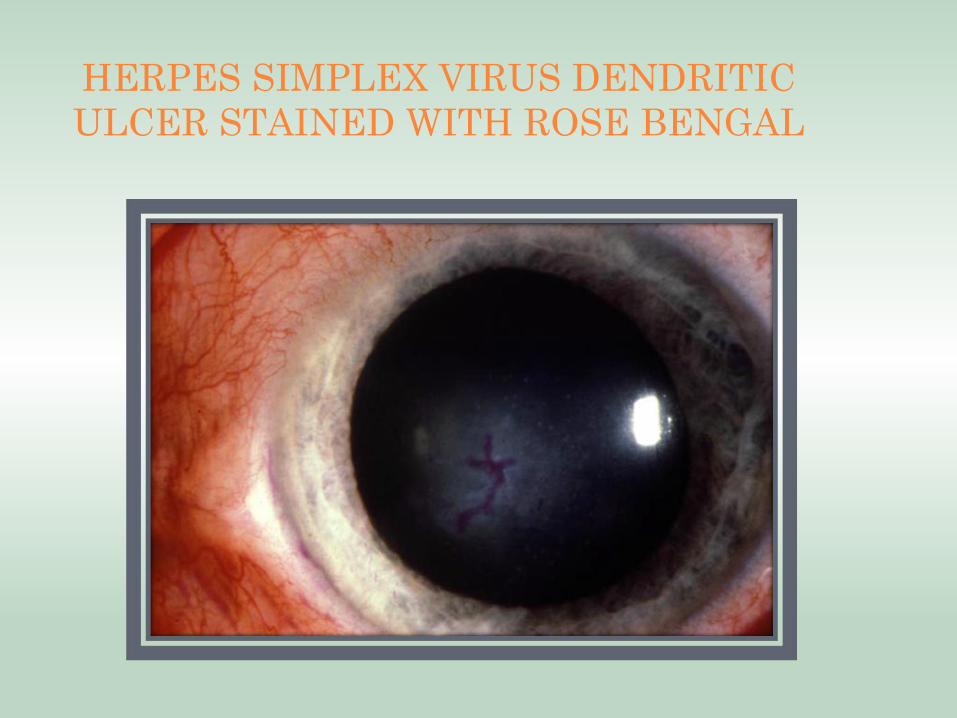
HERPES SIMPLEX VIRUS DENDRITIC
ULCER STAINED WITH ROSE BENGAL
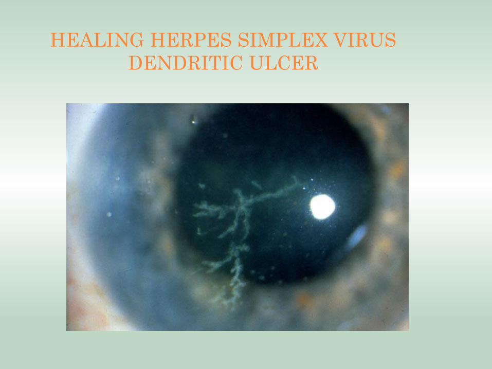
HEALING HERPES SIMPLEX VIRUS
DENDRITIC ULCER
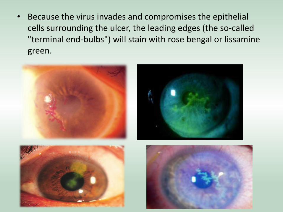
• Because the virus invades and compromises the epithelial cells surrounding the ulcer, the leading edges (the so-called "terminal end-bulbs") will stain with rose bengal or lissamine green.
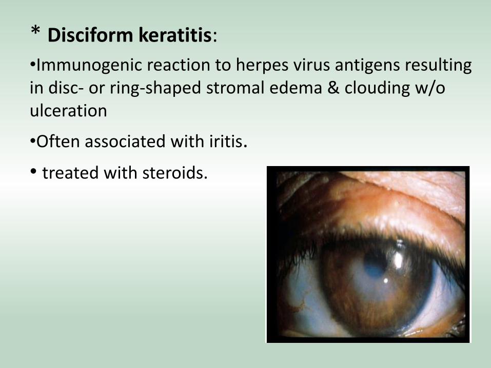
* Disciform keratitis:
•Immunogenic reaction to herpes virus antigens resulting in disc- or ring-shaped stromal edema & clouding w/o ulceration
•Often associated with iritis.
• treated with steroids.
COMPLICATIONS
o Corneal scarring ( can lead to loss of vision) o Chronic interstitial keratitis o Secondary iritis
Treatment :
o Most cases of HSV keratitis resolve spontaneously within 3 weeks, but the treatment is to minimize stromal damage and scarring
o Topical antiviral (Trifluridine or acyclovir oinment) o Dendritic debridement
o DON’T USE TOPICAL STEROIDS as they worsen the
ulcer to geographic ulcer.
o If recur more than twice a year give oral acyclovir.
* Herpes zoster ophthalmicus :
•Varicela zoster virus affects the ophthalmic division of the trigeminal N. (15% )
•Increases with age (6th-7th decades).
•The ocular manifestations are more likely if the nasocillary N is involved >> lid swelling (maybe bilateral), keratitis ,iritis, secondary glaucoma.
•Other ocular manifest : ptosis, mucus secreting conjunctivitis, neuralgia & scleritis which may lead to scleral atrophy.
* Symptoms:
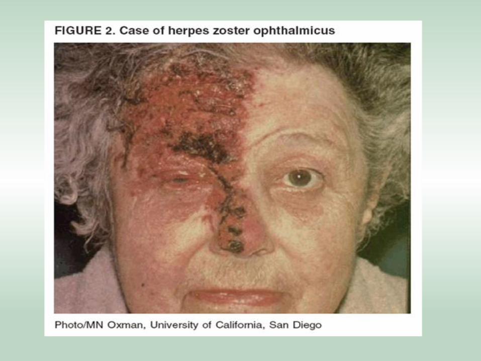
•Has prodromal period, typically presents with nondescript facial pain, fever and general malaise.
•About four days after onset, a unilateral vesicular skin rash over forehead, upper eyelid, nose , (1st
div of 5th CN), characteristically respecting the vertical midline.
•The vesicles will discharge fluid and begin to scab over after about one week. The pain is extreme during the inflammatory stage, and patients are tremendously symptomatic.
• Signs:
– Cornea: Punctate epithelial keratitis (swollen epithelium, 1-2 d); dendritic keratitis (tree branchlike epithelial defects, 4-6 d); stromal keratitis (fine infiltrates beneath the surface, 1-2 wk); deep stromal keratitis (lipid infiltrates and corneal neovascularization, 1 month to years); neurotrophic keratopathy (erosions, persistent defects, corneal ulcers, months to years)
– Ocular involvement may include follicular conjunctivitis, epithelial and/or interstitial keratitis, dendritic keratitis, ant chamber uveitis, scleritis or episcleritis, chorioretinitis, optic neuropathy, and even neurogenic motility disorders (especially fourth cranial nerve palsy).
• Prognostic indicator: Hutchinson’s Sign
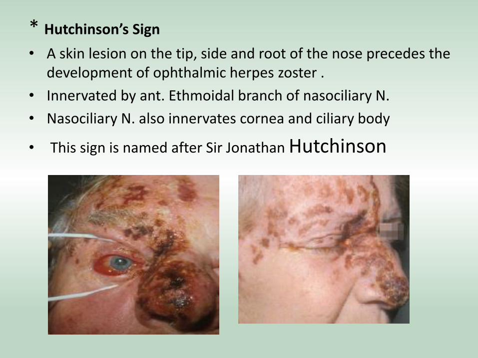
* Hutchinson’s Sign
• A skin lesion on the tip, side and root of the nose precedes the development of ophthalmic herpes zoster .
• Innervated by ant. Ethmoidal branch of nasociliary N.
• Nasociliary N. also innervates cornea and ciliary body
• This sign is named after Sir Jonathan Hutchinson
* Treatment:
•Oral and topical antiviral : acyclovir ( prevent post-infective neuralgia-severe chronic pain over the rash)
•Steroid can be given bcz disease is due to immune rxn not virus itself.
•+/- a cycloplegic agent >> used to relaxe the cilliary
muscles >> like : cyclopentolate , atropine.
* Fungal keratitis : - rare , but they are very severe & devastating as they cause stromal necrosis.
•They are capable of penetrating the descemet’s membrane reaching the ant. chamber where we cannot do anything because of the poor penetration of antimycotic agents to the ant. Chamber.
- Most common causative pathogens:
•Filamentous (aspergillus & fusarium) fungi.
•Candida albicans.
- Progression is much slower & less painful than in bacterial.
• Keratomycosis is in consideration when we find lack of response to antibacterial therapy of corneal ulceration.
* Signs include :
- Filamentous infection : grayish infiltrate with indistinct margins.
• Candidal infection : yellow to white ulcer with suppuration similar to bacterial keratitis.
• Treatment: topical antifungals “pimaricin 5%”
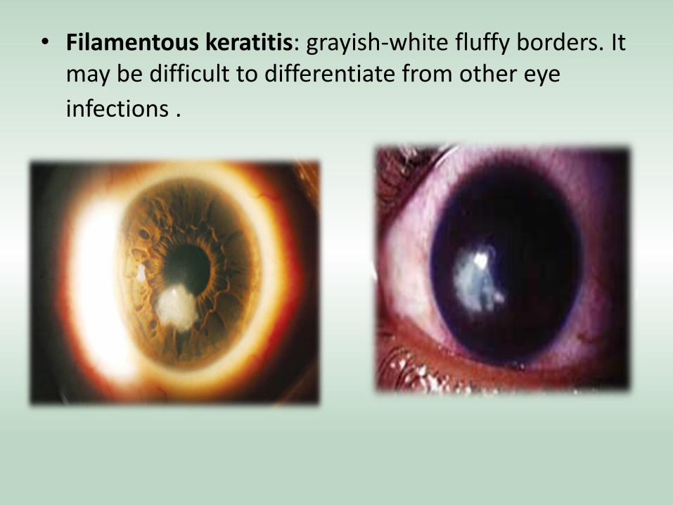
• Filamentous keratitis: grayish-white fluffy borders. It may be difficult to differentiate from other eye
infections .

• Candidal keratitis- Typical yellowish-white base with
feathery borders ulcer w hypopyon.
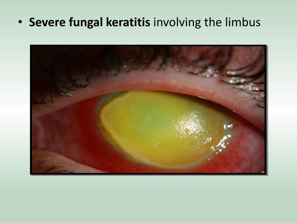
• Severe fungal keratitis involving the limbus
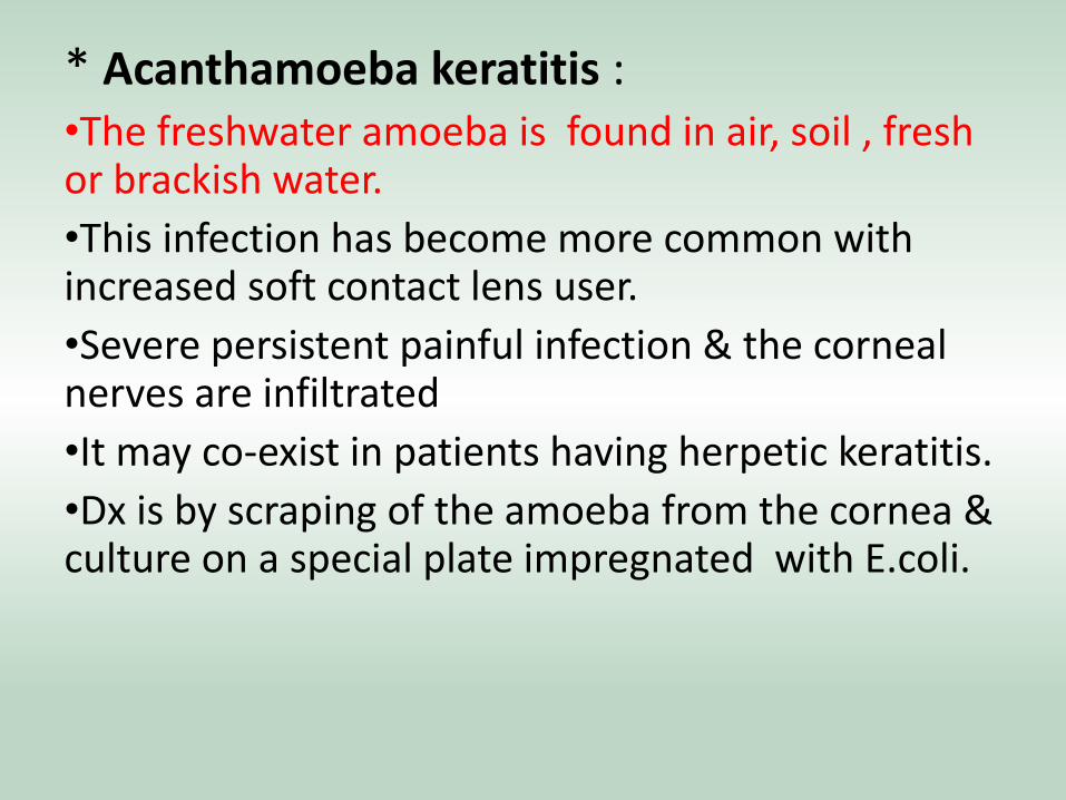
* Acanthamoeba keratitis : •The freshwater amoeba is found in air, soil , fresh or brackish water.
•This infection has become more common with increased soft contact lens user.
•Severe persistent painful infection & the corneal nerves are infiltrated
•It may co-exist in patients having herpetic keratitis.
•Dx is by scraping of the amoeba from the cornea & culture on a special plate impregnated with E.coli.
* Treatment : •Is long, involves toxic medications, and may be unsuccessful in curing the infection if involves the posterior cornea.
•A combination of topical anti-amoebic agents.
•The use of topical steroids is controversial. It clearly improves patient comfort, but may potentiate the infection by conversion of the cyst to trophozoites
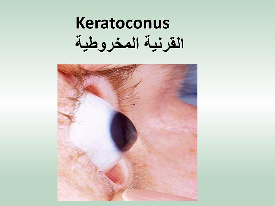
Keratoconus القرنية المخروطية
**Greek words: kerato = cornea
**conus = cone-shaped
**Is a non-inflammatory condition of the cornea in which there is progressive central thinning of the cornea changing it from dome-shaped to cone-shaped. Causing vision to become blurred and distorted.
** Classification based on:
• severity of curvature
• shape
** Based on severity of curvature
• Mild
• Moderate
• Advanced
• Severe
** Based on shape:
• Nipple cones (Small size 5mm )
• Oval cones (larger (5-6mm) ellipsoid)
• Globus cones (Largest >6mm ,may involve over 75% of cornea. )
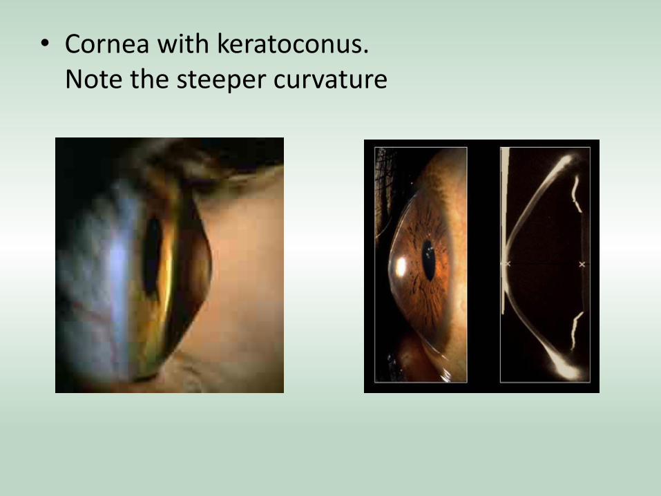
• Cornea with keratoconus. Note the steeper curvature
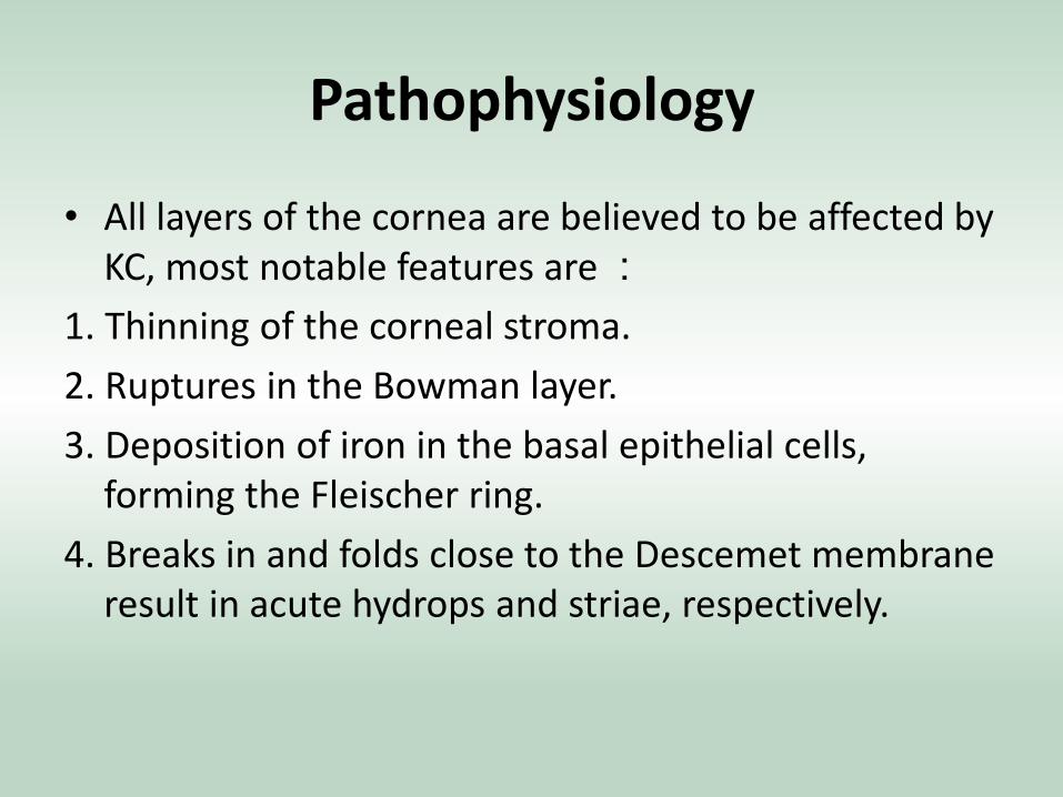
Pathophysiology
• All layers of the cornea are believed to be affected by KC, most notable features are :
1. Thinning of the corneal stroma.
2. Ruptures in the Bowman layer.
3. Deposition of iron in the basal epithelial cells, forming the Fleischer ring.
4. Breaks in and folds close to the Descemet membrane result in acute hydrops and striae, respectively.
** Etiology : • Sporadic 90%: Imbalance of enzymes within the
cornea ; leads to collagen defect . This imbalance makes the cornea more susceptible to oxidative damage from compounds called free radicals, causing it to weaken and bulge forward.
• Heredity
• Eye rubbing as in case of allergic conjunctivitis.
• Contact lenses wear
• Hormonal change
• Collagen systemic disease (down)
** Symptoms : • Start in puberty (in the teens) and may progress for the next
10 to 20 years. Is a progressive disease • Frequent prescription changes in glasses and contact lenses. • Usually bilateral involvement but asymmetrical. • Nearsightedness. • Astigmatism. • Blurred vision and destored vision - even when wearing
glasses and contact lenses. • Glare at night. • Light sensitivity. • Eye rubbing. • Diplopia or polyopia. • No pain. • Maybe family Hx.
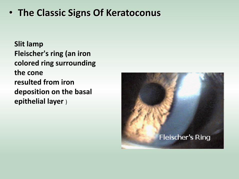
• The Classic Signs Of Keratoconus
Slit lamp Fleischer's ring (an iron colored ring surrounding the cone resulted from iron deposition on the basal epithelial layer )
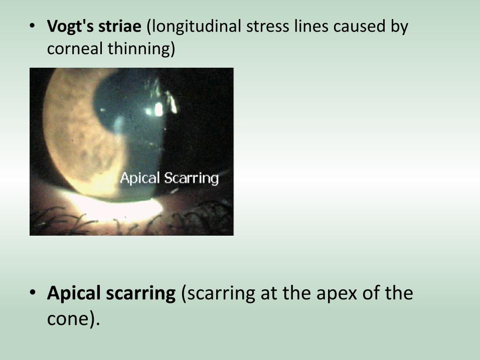
• Vogt's striae (longitudinal stress lines caused by corneal thinning)
• Apical scarring (scarring at the apex of the cone).
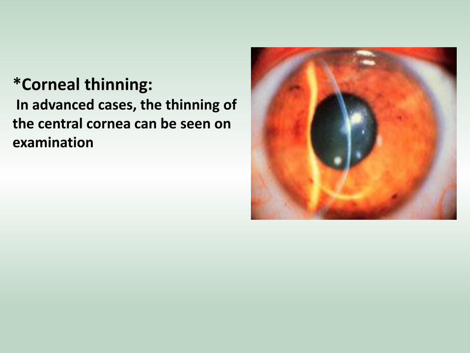
*Corneal thinning: In advanced cases, the thinning of the central cornea can be seen on examination
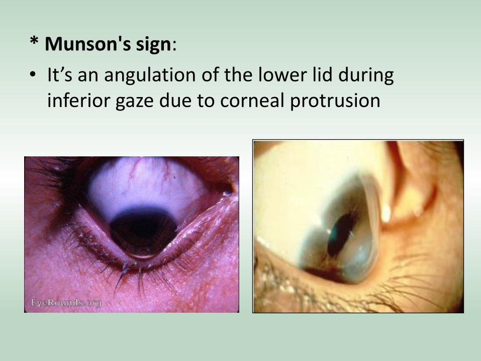
* Munson's sign:
• It’s an angulation of the lower lid during inferior gaze due to corneal protrusion
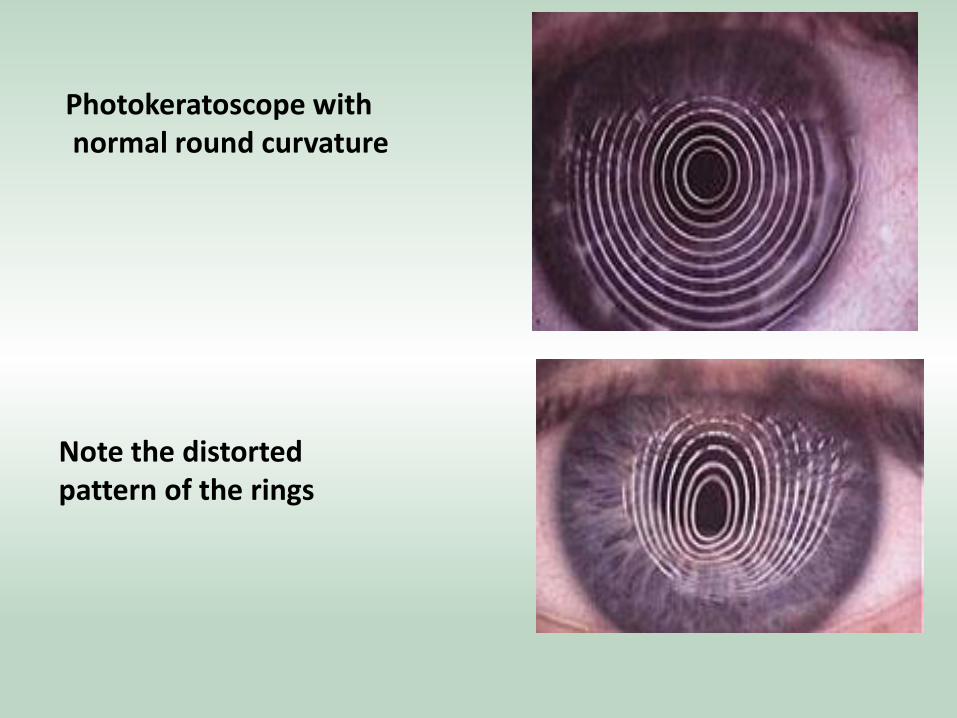
Photokeratoscope with normal round curvature
Note the distorted pattern of the rings
• Hydrops
• Prominent corneal nerve .
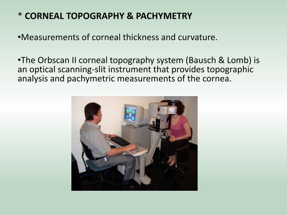
* CORNEAL TOPOGRAPHY & PACHYMETRY •Measurements of corneal thickness and curvature. •The Orbscan II corneal topography system (Bausch & Lomb) is an optical scanning-slit instrument that provides topographic analysis and pachymetric measurements of the cornea.
DX :
• Pentacham is diagnostic
**Stages of Treatment : (To improve vision & to stop progression) 1) Hard contact lens for best vision but has poor tolerance. 2) crosslinking of the stromal collagen by exposing the stroma to UV radiation in the presence of riboflavin to stop the progression. Done for those who aged 25-35 years old . 3)Corneal ring - Intact corneal rings (placing with the corneal stroma in the periphery of the cornea. The result is a flatter cornea and clearer vision). 4) Cornea transplant = done if refractive power reach 0.05-0.1 penetrating keratoplasty. A donor cornea will replace the thinning cornea and can often provide stable vision. Patient will most likely need glasses or contact lenses for clear vision. May be complicated by rejection.