predictive ocular motor control in...
TRANSCRIPT

Brain (1985), 108,925-940
PREDICTIVE OCULAR MOTOR CONTROL INPARKINSON'S DISEASE
by A. M. BRONSTEIN1 and c. KENNARD2
• (From the Department of Neurology, The London Hospital, London El IBB)
SUMMARY
A comparison was made of predictive eye movements of both the saccadic and pursuit ocular motorsystems in parkinsonian patients and aged-matched normal controls. In a predictive task our patients,who were mildly or moderately affected, showed a reduced tendency to make anticipatory saccadescompared with normal controls. Although there was some impairment of pursuit during anonpredictive task as shown by an increased phase lag, a normal amount of improvement took placewith a predictive task. This difference between prediction in the saccadic and pursuit system is possiblyexplained by an increased reliance on a visual input by parkinsonian patients which prevents themmaking use of verbal instruction to generate anticipatory (eye in advance of target) saccades.Improvement of pursuit with a predictive target track is possible in such patients since normally inthese circumstances the target is mainly followed rather than anticipated. The metrics of saccadic andsmooth pursuit eye movements (saccadic velocity and pursuit turnover velocity) were normal.
INTRODUCTION
Tracking experiments, a fruitful approach to the study of movement strategies inParkinson's disease, typically requires the subject to follow, with their eyes or limb, atarget displayed on a screen which either moves in a regular or an irregular pattern.In normal subjects target movement in a regular pattern enables predictive trackingof the eye or limb to take place (Noble et ai, 1955). This requires the formation of amental representation of the target path, followed by the design of a motor programto give a suitable output. As a result of this, the lag and error between the eye or limband the target is reduced and the subject's tracking performance is thereforeimproved. In some circumstances the eye or limb may actually precede the target. Inseveral papers, Flowers (1976, 1978a, b) has shown a number of hand-trackingabnormalities in Parkinson's disease, particularly in those experiments in which apredictive motor strategy was required. In a subsequent paper, however, Flowersand Downing (1978) failed to demonstrate a predictive deficiency in an ocular motor(OCM) tracking experiment and, therefore, concluded that only the limbs showeda predictive abnormality in Parkinson's disease. This seemed to us an unexpected
1 Present Address: M RC Neuro-otology Unit, National Hospital for Nervous Diseases, Queen Square, London,WC1N 3BG.
2 Correspondence to: Dr C. Kennard, Department of Neurology, The London Hospital, Whitechapel, LondonEl IBB.

926 A. M. BRONSTEIN AND C. KENNARD
finding since it would imply that the neural organization of predictive movementsfor eye and limb functions differently in Parkinson's disease, in contrast to otherclinical conditions. For example, patients with prefrontal lesions exhibit defects inhigh level motor programming which are equally present in the OCM and somaticmotor systems (Luria et al., 1966; Guitton et al., 1982). In addition, Flowers andDowning only examined one aspect of ocular motor behaviour, the smooth pursuitsystem. We considered that the saccadic ocular motor system should be assessedseparately because of important functional differences which exist between thesetwo ocular motor subsystems.
In this paper we report the results of tests of both saccadic and smooth pursuit eyemovements in mild to moderately affected parkinsonian patients compared withage-matched normal controls. It will be shown that these patients, although havinga relatively normal basic oculomotor performance for their age (for instance,saccadic velocity and smooth pursuit configuration), displayed abnormalities ofocular motor prediction similar to those which have previously been reported in thelimbs. A preliminary account has already appeared (Bronstein and Kennard, 1984).
MATERIALS AND METHODS
Clinical Material
Eight patients with idiopathic Parkinson's disease were selected for the study. They were mild tomoderately affected (Hoehn and Yahr, grade I—III) and clinically did not have signs or symptoms ofdementia or coexistent cerebrovascular disease {see Table 1). Their mean age was 65 years (range 53-70years) as was that of the control group of 8 age-matched normal subjects (range 50-82 years), who werehospital patients with no neurological abnormality, plus one medical secretary.
Recording Techniques
Eye movements were recorded by the infrared reflection technique (Stark et al., 1962). This systemprovided linear recordings over a range ±20 deg with a bandpass of 300 Hz. Head movements were
TABLE 1. CLINICAL ASSESSMENT OF DISABILITY IN THE PARK.INSONIAN G R O U P
AgeSubject
1234567
8
Sex
MFMMMFM
M
(yrs)69666165636970
53
Tremor
NilMild(B)
NilNil
Mild (B)Severe (U)
Nil
Mild (B)
Rigidity
Moderate (B)Mild(B)Mild(U)
Moderate (B)Moderate (B)
Mild (B)Mild (B)
Mild(B)
Bradykinesia
Mild(B)Mild (B)
Moderate (U)Moderate (U)Moderate (B)Moderate (B)
Mild(B)
Moderate (B)
Drugs
NilSinemetSinemetSinemetSinemetSinemetSinemet
BromocriptineMadopar
Bromocriptine
U — unilateral. B •= bilateral.

EYE MOVEMENTS IN PARKINSON'S DISEASE 927
recorded by means of a light helmet, worn by the subject, which was attached to a low torquepotentiometer via a rigid rod. The visual target was a white spot of light (30 min of arc), reflected onto acurved white screen placed 150 cm away from the subject. Target, eye and head positions were recordedon paper by an ink-jet recorder (Mingograph), and onto magnetic tape for computer analysis. Gazeposition (eye position in space) was derived off-line by adding eye and head position signals in adifferential amplifier.
Subjects performed saccadic and smooth pursuit experiments in one session. The experiments wereperformed with the head motionless and then repeated while the subject was encouraged to move hishead.
Saccadic Experiments
The target moved in a square wave step function across the screen in the horizontal plane in threedifferent conditions.
Random saccades (RS). The target moved randomly between 9 different positions on the screen(1 central and 4 on each side) generating 8 possible target amplitudes, 5, 10, 15, 20, 25, 30, 35 and40 deg. The time interval between target displacements was randomized between 0.6 and 1.2 s. Thesubjects were told that the target would jump randomly on the screen and were asked to follow it asaccurately as possible. The test duration was 50 s generating 55 steps.
Hidden predictive saccades (HPS). This protocol consisted of a sequence of 20 regular targetdisplacements which, without any warning, appeared in the middle of a random sequence. The targetmoved symmetrically 30 deg from 15 deg left to 15 deg right and back, with a fixed time interval of 0.9 s.Test duration and instructions to the subject were identical to RS.
Predictive saccades (PS). This protocol consisted of 33 regular 30 deg target displacements between15 deg left and 15 deg right, with a fixed time interval of 0.9 s (0.55 Hz). Pilot studies and experiments inthe literature (Stark et al., 1962) have shown that this time interval regularly produces predictivesaccades in normal subjects. The subjects were told in advance that the target, in contrast to theprevious test they had undergone, would move regularly on the screen, jumping between two fixedpoints. No instruction as to whether or not they should try to predict or anticipate the target was given.The duration of this test was 30 s.
For eye-head coordination experiments, time intervals between target displacements were 60 percent longer in order to allow complete head stabilization before a new target step took place.
Analysis of results. Eye saccadic velocity was measured with computer software described elsewhere(Smith et al., 1981) and included all saccades from RS, HPS and PS tests, except those produced inadvance of the target displacement (anticipatory saccades). The computer program generates anamplitude/peak velocity plot of saccades and fits an exponential curve to the data. The asymptote ofthe peak velocity (k) is used to express saccadic velocity.
The latency of saccades produced in RS was measured by the computer. The latency of each saccadeoccurring during HPS and PS were determined by hand from the chart recordings since the computerprogram would only measure saccades produced after a target displacement had taken place.
Gaze displacement during head movement was measured by the same computer program asdescribed for eye saccades. In addition, the latencies of head movements occurring during combinedeye-head experiments were measured by computer.
Smooth Pursuit Experiments
The target moved horizontally with a constant peak amplitude of 30 deg, under two differentconditions:
Random smooth pursuit (RSP). The target moved sinusoidally in an unpredictable manner at one offive different frequencies, 0.08, 0.16, 0.32, 0.48 and 0.65 Hz. One frequency was used for a randomperiod of between 1.3 and 2.6 s, after which it randomly changed to a different frequency. Two stimulussequences of 50 s each were presented. The subjects were instructed to follow the target with their eyesas accurately as possible and were informed that it would be moving irregularly and at a variable speed.
928 A. M. BRONSTEIN AND C. KENNARD
Predictive smooth pursuit (PSP). Identical target frequencies were then individually presented. Twosequences of 45 s each were used; the first sequence continued for 22 s at a target frequency of 0.08 Hz(approximately 2 cycles), and then the second for an identical period at 0.16 Hz (approximately 6cycles). In the second sequence three 15 s periods, with target frequencies of 0.32 Hz (5 cycles), 0.48 Hz(8 cycles) and 0.65 Hz (10 cycles), respectively, completed the test. The subjects were informed that thetarget would move regularly and the experimenter told them during the sequences when the targetfrequency was changed. No instruction was given as to whether or not they should anticipate thetarget.
Analysis of results. Phase error of smooth pursuit was measured by hand for two target frequencies,0.32 and 0.65 Hz. Their zero velocity points (maxima and minima) of both smooth pursuit eyemovement and of target movement were determined in saccade-free regions. The difference betweeneye and target position, expressed in degrees, corresponded to the phase error, typically phase lag.Phase measurements were made for the eye, gaze and head in RSP and PSP. Approximately 7 (range 3to 15) half cycles were measured for each condition.
Turnover velocity of the smooth pursuit system was measured by computer as described elsewhere(Bittencourt at ah, 1982). In brief, the program simultaneously measures eye and target velocity and,by comparing the two, is able to identify whether smooth pursuit eye movements or catch-up saccadesare being used to track the visual target. The data are sampled at 256 Hz and a plot is produced of thepercentage of time spent in actual smooth pursuit during tracking as a function of target velocity.Normally, as the speed of the target increases, eye tracking becomes more and more saccadic so thatless time is spent in smooth pursuit. The target velocity at which the percentage of time spent in smoothpursuit drops by 3 dB, is the turnover velocity and provides a measure of the point at which smoothpursuit breaks down into mainly saccadic tracking. Gaze movements were treated in a similar mannerby computer analysis.
A computer program (BMDP2V) for analysis of variance and covariance with repeated measureswas used for statistical analysis. Comparisons between groups (patients and controls) under differenttest conditions (eye-gaze, 0.3-0.6 Hz, random-predictive) were made and interactions between thesevarious groups and conditions were also available. Other statistical tests used are mentioned in theResults.
R E S U L T S
Saccadic Movements
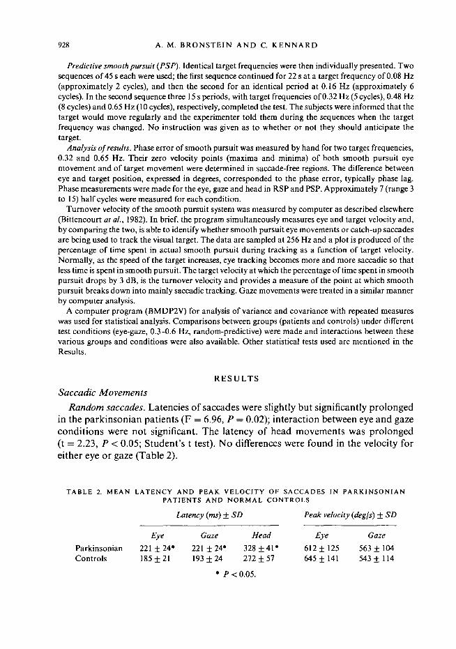
Random saccades. Latencies of saccades were slightly but significantly prolongedin the parkinsonian patients (F = 6.96, P = 0.02); interaction between eye and gazeconditions were not significant. The latency of head movements was prolonged(t = 2.23, P < 0.05; Student's t test). No differences were found in the velocity foreither eye or gaze (Table 2).
TABLE 2. MEAN LATENCY AND PEAK VELOCITY OF SACCADES IN PARKINSONIANPATIENTS AND NORMAL CONTROLS
ParkinsonianControls
Eye
221 ± 24*185 ±21
Latency (ms) ±
Gaze
221 ±24*193 ± 24
* P
SD
Head
328 ± 4 1 *272 ± 57
< 0.05.
Peak velocity {aegis) ± SD
Eye
612 ±125645 ±141
Gaze
563 ± 104543 ±114
EYE MOVEMENTS IN PARKINSON'S DISEASE 929
Target20degIEye20deg
Control
PatientTarget20 deg I
Eye T20 deg 1
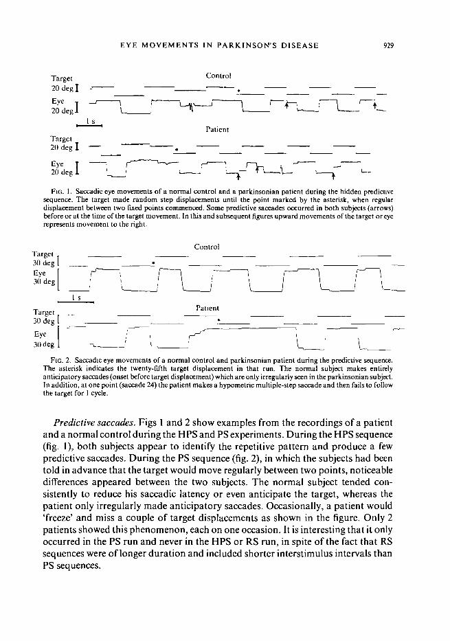
FIG. 1. Saccadic eye movements of a normal control and a parkinsonian patient during the hidden predicUvesequence. The target made random step displacements until the point marked by the asterisk, when regulardisplacement between two fixed points commenced. Some predictive saccades occurred in both subjects (arrows)before or at the time of the target movement. In this and subsequent figures upward movements of the target or eyerepresents movement to the right.
Control
PatientTarget30 deg 1
Eye [30 deg I -T '
FIG. 2. Saccadic eye movements of a normal control and parkinsonian patient during the predictive sequence.The asterisk indicates the twenty-fifth target displacement in that run. The normal subject makes entirelyanticipatory saccades (onset before target displacement) which are only irregularly seen in the parkinsonian subject.In addition, at one point (saccade 24) the patient makes a hypometric multiple-step saccade and then fails to followthe target for 1 cycle.
Predictive saccades. Figs 1 and 2 show examples from the recordings of a patientand a normal control during the HPS and PS experiments. During the HPS sequence(fig. 1), both subjects appear to identify the repetitive pattern and produce a fewpredictive saccades. During the PS sequence (fig. 2), in which the subjects had beentold in advance that the target would move regularly between two points, noticeabledifferences appeared between the two subjects. The normal subject tended con-sistently to reduce his saccadic latency or even anticipate the target, whereas thepatient only irregularly made anticipatory saccades. Occasionally, a patient would'freeze' and miss a couple of target displacements as shown in the figure. Only 2patients showed this phenomenon, each on one occasion. It is interesting that it onlyoccurred in the PS run and never in the HPS or RS run, in spite of the fact that RSsequences were of longer duration and included shorter interstimulus intervals thanPS sequences.
930 A. M. BRONSTEIN AND C. KENNARD
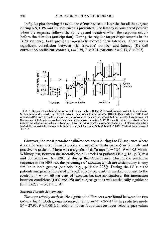
In fig. 3 a plot showing the evolution of mean saccadic latencies for all the subjectsduring RS, HPS and PS sequences is presented. The latency is considered positivewhen the response follows the stimulus and negative when the response occursbefore the stimulus (anticipation). During the regular target displacements in theHPS sequence, both groups progressively reduced their latencies. There was asignificant correlation between trial (saccade) number and latency (Kendallcorrelation coefficient: controls, r = 0.59, P < 0.01; patients, r = 0.33, P < 0.05).
-200-1
-100-
8.+ 100-
+200-
+300-
\ lul1
Random Hidden predictive Predictive
Fio. 3. Sequential analysis of mean saccadic response time (latency) for parkinsonian patients (open circles,broken line) and normal controls (filled circles, continuous line) in random (RS), hidden predictive (HPS) andpredictive (PS) tests. In the RS the mean latency of patients is slightly prolonged, but during HPS it can be seen thatthe latency of both groups gradually shortens with successive cycles. In PS the latency rapidly shortens in bothgroups, but whereas normal controls show a plateau mean response time of approximately — 120 ms (anticipatorysaccades), the patients are unable to improve beyond the response time found in HPS. Vertical bars represent± 1SD.
However, the most prominent differences occur during the PS sequence whereit can be seen that mean latencies are negative (anticipatory) in controls andpositive in patients. There was a significant difference (z = 1.96, P = 0.05 Mann-Whitney test) between the saccadic mean latencies of patients (107 ± 181 (SD) ms)and controls ( — 116 + 220 ms) during the PS sequence. During the predictivesequence in the HPS run the percentage of saccades which are anticipatory is verysimilar in both groups (controls: 23%, patients: 22%). During the PS run thepatients marginally increased this value to 29 per cent, in marked contrast to thecontrols in whom 60 per cent of saccades became anticipatory; this interactionbetween conditions (HPS and PS) and subject groups was statistically significant(F = 5.62, P = 0.03) (fig. 4).
Smooth Pursuit Movements
Turnover velocity analysis. No significant differences were found between the twogroups (fig. 5). Both groups increased their turnover velocity in the predictive mode(F = 27.93, P < 0.001). In addition it was found that turnover velocity gaze values

EYE MOVEMENTS IN PARKINSON'S DISEASE 931
70-|
60-
50-
40-
'M 30-
20-
10-
0 J
Hiddenpredictive
Predictive
FIG. 4. Histograms to show the percentage of saccades which areanticipatory (commenced before target displacement) in normalcontrols (open areas) and parldnsonian patients (hatched areas)during the HPS and PS. No difference is shown in the HPS, butduring PS normal subjects considerably increase the percentage ofanticipatory saccades whereas parkinsonian patients do not.
were higher than eye alone (F = 5.00, P = 0.04), but this was made more evident inthe predictive mode (interaction eye-gaze/random-predictive: F = 5.45, P = 0.03).This improvement in the smooth pursuit turnover velocity during the head-freecondition is thought to be due to an increased efficiency of the catch-up saccadesgenerated by the latter condition and has been discussed elsewhere (Bronstein andKennard, 1984).
Phase lag analysis. Fig. 6 shows a recording from a normal subject and a patientduring random and predictive conditions, in which a few cycles of the target movingat the highest frequency used are shown. Two particular features may be seen; first,
701
60
50-
f- 40-
30-
Eye
li
Gaze
Random Predictive Random Predictive
FIG. 5. Mean turnover velocity (deg/s) of eye and gaze (head free) in random and predictive pursuit for patients(circles) and controls (squares). Vertical bars represent ± 1 SD.
932 A. M. BRONSTEIN AND C. KENNARD
3Odeg
30deg
Control Patient
30deg
3Odeg
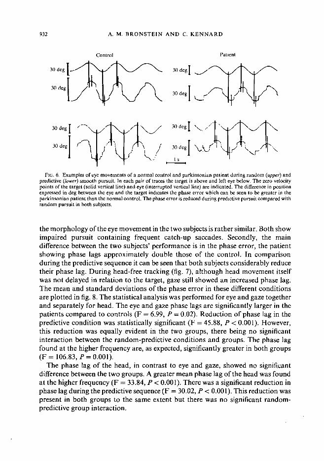
FIG. 6. Examples of eye movements of a normal control and parkinsonian patient during random {upper) andpredictive (lower) smooth pursuit. In each pair of traces the target is above and left eye below. The zero velocitypoints of the target (solid vertical line) and eye (interrupted vertical line) are indicated. The difference in positionexpressed in deg between the eye and the target indicates the phase error which can be seen to be greater in theparkinsonian patient than the normal control. The phase error is reduced during predictive pursuit compared withrandom pursuit in both subjects.
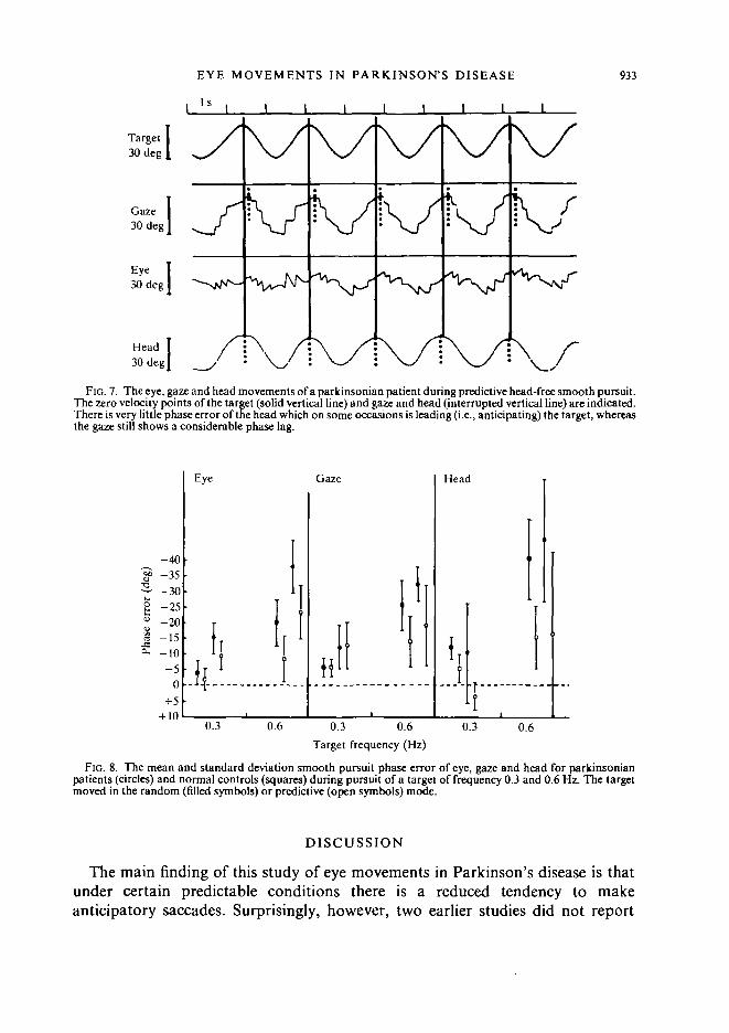
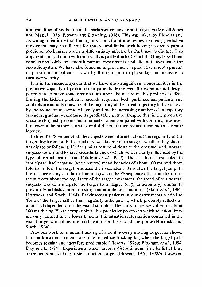
the morphology of the eye movement in the two subjects is rather similar. Both showimpaired pursuit containing frequent catch-up saccades. Secondly, the maindifference between the two subjects' performance is in the phase error, the patientshowing phase lags approximately double those of the control. In comparisonduring the predictive sequence it can be seen that both subjects considerably reducetheir phase lag. During head-free tracking (fig. 7), although head movement itselfwas not delayed in relation to the target, gaze still showed an increased phase lag.The mean and standard deviations of the phase error in these different conditionsare plotted in fig. 8. The statistical analysis was performed for eye and gaze togetherand separately for head. The eye and gaze phase lags are significantly larger in thepatients compared to controls (F = 6.99, P = 0.02). Reduction of phase lag in thepredictive condition was statistically significant (F = 45.88, P < 0.001). However,this reduction was equally evident in the two groups, there being no significantinteraction between the random-predictive conditions and groups. The phase lagfound at the higher frequency are, as expected, significantly greater in both groups(F= 106.83, P = 0.001).
The phase lag of the head, in contrast to eye and gaze, showed no significantdifference between the two groups. A greater mean phase lag of the head was foundat the higher frequency (F = 33.84, P < 0.001). There was a significant reduction inphase lag during the predictive sequence (F = 30.02, P < 0.001). This reduction waspresent in both groups to the same extent but there was no significant random-predictive group interaction.
EYE MOVEMENTS IN PARKINSON'S DISEASE 933
Is
Target3Odeg.
Gaze I30deg|
Eye I3Odeg|
Head30 deg|
FIG. 7. The eye, gaze and head movements of a parkinsonian patient during predictive head-free smooth pursuit.The zero velocity points of the target (solid vertical line) and gaze and head (interrupted vertical line) are indicated.There is very little phase error of the head which on some occasions is leading (i.e., anticipating) the target, whereasthe gaze still shows a considerable phase lag.
-40
leg
1
Pha
se
- 35-30-25
-10- 5
0
+5+ 10
Eye Gaze
H
Head
0.3 0.6 0.3 0.6Target frequency (Hz)
0.3 0.6
FIG. 8. The mean and standard deviation smooth pursuit phase error of eye, gaze and head for parkinsonianpatients (circles) and normal controls (squares) during pursuit of a target of frequency 0.3 and 0.6 Hz. The targetmoved in the random (filled symbols) or predictive (open symbols) mode.
DISCUSSION
The main finding of this study of eye movements in Parkinson's disease is thatunder certain predictable conditions there is a reduced tendency to makeanticipatory saccades. Surprisingly, however, two earlier studies did not report
934 A. M. BRONSTEIN AND C. KENNARD
abnormalities of prediction in the parkinsonian ocular motor system (Melvill Jonesand Mandl, 1976; Flowers and Downing, 1978). This was taken by Flowers andDowning to indicate that the organization of motor activities involving predictivemovements may be different for the eye and limbs, each having its own separatepredictor mechanism which is differentially affected by Parkinson's disease. Thisapparent contradiction with our results is partly due to the fact that they based theirconclusions solely on smooth pursuit experiments and did not investigate thesaccadic system. We have also found an improvement in predictive smooth pursuitin parkinsonian patients shown by the reduction in phase lag and increase inturnover velocity.
It is in the saccadic system that we have shown significant abnormalities in thepredictive capacity of parkinsonian patients. Moreover, the experimental designpermits us to make some observations upon the nature of this predictive defect.During the hidden predictive saccade sequence both parkinsonian patients andcontrols are initially unaware of the regularity of the target trajectory but, as shownby the reduction in saccadic latency and by the increasing number of anticipatorysaccades, gradually recognize its predictable nature. Despite this, in the predictivesaccade (PS) test, parkinsonian patients, when compared with controls, producedfar fewer anticipatory saccades and did not further reduce their mean saccadiclatency.
Before the PS sequence all the subjects were informed about the regularity of thetarget displacement, but special care was taken not to suggest whether they shouldanticipate or follow it. Under similar test conditions to the ones we used, normalsubjects were found to have saccadic latencies which were critically influenced by thetype of verbal instruction (Polidora et al., 1957). Those subjects instructed to'anticipate' had negative (anticipatory) mean latencies of about 100 ms and thosetold to 'follow' the target produced their saccades 100 ms after the target jump. Inthe absence of any specific instruction given in the PS sequence other than to informthe subjects about the regularity of the target movement, the trend of our normalsubjects was to anticipate the target to a degree (60% anticipatory) similar topreviously published studies using comparable test conditions (Stark et al., 1962;Horrocks and Stark, 1964). Parkinsonian patients in our experiments tended to'follow' the target rather than regularly anticipate it, which probably reflects anincreased dependence on the visual stimulus. Their mean latency values of about100 ms during PS are compatible with a predictive process in which reaction timesare only reduced to the lower limit. In this situation information contained in thevisual target can still induce modifications in the saccadic response (Horrocks andStark, 1964).
Previous work on manual tracking of a continuously moving target has shownthat parkinsonian patients are able to reduce tracking lag when the target pathbecomes regular and therefore predictable (Flowers, 1978a; Bloxham et al., 1984;Day et al., 1984). Experiments which involve discontinuous (i.e., ballistic) limbmovements in tracking a step function target (Flowers, 1976, 1978ft), however,

EYE MOVEMENTS IN PARKINSON'S DISEASE 935
showed a failure of prediction. Similarly, Bloxham et al. (1984) have shown thatparkinsonian patients are unable to make use of advance information in performinga reaction time test. These findings, as well as the results in our PS experiment,emphasize the difficulty parkinsonian patients have in making movements of eye orlimb in advance of the visual target. This abnormality reflects an increased relianceon visual input which has previously been described (Cooke et al, 1978; Flowers,19786; Stern et al., 1983), and is reminiscent of the much improved locomotorfunction which occurs clinically when such patients are provided with a powerfulvisual input, for example a striped floor (Martin, 1967).
Two possible pathophysiological explanations for this disturbance of pre-diction are worthy of consideration. The first arises from the similarity betweenparkinsonian patients and patients with frontal lobe lesions who show a difficulty inutilizing verbal instructions in the construction of motor programs (Luria, 1969).An example of this in relation to the ocular motor system is shown in experiments onpatients with well-defined surgical lesions in the prefrontal region who, afterrelevant verbal instructions, were unable to generate saccades in a direction oppositeto a sudden target displacement (Guitton et al., 1982). These patients thereforefollowed the visual stimulus rather than the verbal instruction. Several studiesof cognitive and motor function in Parkinson's disease have already shownabnormalities similar to those found in patients with frontal lobe lesions (Denny-Brown, 1968; Morel-Maroger, 1977; Figini and Bronstein, 1981; Bowen et al,1972; Lees and Smith, 1983). These frontal lobe type abnormalities found inParkinson's disease may be due to dysfunction of the ascending dopaminergicmesocorticolimbic pathway projecting to the frontal cortex (Javoy-Agid andAgid, 1980).
A second possible pathophysiological explanation for the failure of ourparkinsonian patients to make anticipatory saccades during the predictive sequenceis to be found in recent experimental studies of cells in the pars reticulata of thesubstantia nigra in primates. This may also explain the other saccadic abnormalitywe found which was a prolonged latency, previously reported in the ocular(Shibasaki et al, 1979; Teravainen and Calne, 1980a, b; Shimizu et al, 1981) andsomatic motor systems (Kennard et al, 1982; Evarts et al, 1981). These experimentshave shown a clear relationship between the basal ganglia and the major saccadiccontrol centres, the frontal eye fields (area 8) (Kunzle and Akert, 1977) and thesuperior colliculus (Hikosaka and Wurtz, 1983). The dopaminergic cells in the parscompacta of the substantia nigra project to the caudate nucleus and putamen(striatum), which in turn receives a projection from the frontal eye fields,particularly to the posterior caudate nucleus. One of the two major outputs from thestriatum is to the pars reticulata of the substantia nigra, some of whose cells projectto the intermediate and deep layers of the superior colliculus (Jayaraman et al,1977). These cells have been found to reduce their discharge rate both in response toan appropriately located visual target and to a remembered target (Hikosaka andWurtz, 1983). Since they appear to be gabaergic inhibitory cells (Vincent et al,

936 A. M. BRONSTEIN AND C. KENNARD
1978), a decrease in discharge rate would reduce tonic inhibition, and hence increasethe excitability of collicular cells. These cells have been found to exhibit a burst ofdischarge before saccades of a particular direction and amplitude (Wurtz andGoldberg, 1972). A disturbance of the dopaminergic input to the corpus striatum, asoccurs in Parkinson's disease, may lead to an abnormal output to the substantianigra pars reticulata resulting in reduced facilitation of collicular cells leading toprolonged latencies. Since the substantia nigra pars reticulata cells also dischargeprior to saccades to a remembered target, an abnormal input to these cells could leadto an impaired ability to make anticipatory saccades, which are in essence saccadesto a remembered target. It is of interest that DeJong and Melvill Jones (1971)showed that patients with Parkinson's disease took a longer time than controlsto make saccades back and forth between two targets which are continuouslyilluminated. In this case the saccade must be initiated by something other thanthe onset of a visual target, again possibly analogous to saccades to rememberedtargets.
Examination of the smooth pursuit system in our patients showed an increasedphase lag, a feature previously described for hand tracking experiments inParkinson's disease and in monkeys with caudate lesions (Stark, 1968; Bowen, 1969;Bowen et al., 1975; Flowers, 1978a). Since smooth pursuit operates by continuouslydetecting and subsequently correcting retinal error (i.e., a closed-loop system), itcould be argued that a prolonged reaction time at one or more points during thecentral processing for such a tracking task may well produce delay (lag) betweentarget and eye. An interesting finding, however, was that the phase lag of the headduring smooth pursuit was not significantly increased, which indicates its uniqueform of motor control. With the head free to move during visual tracking of a targetthe only important factor in the maintenance of foveation of the target is the retinalor gaze error. Indeed changes in head gain or phase can be enormous and yet gazegain, phase or accuracy will remain unchanged (Lanman et al., 1978; Collewijn etal., 1982). This is due to stabilization of the eyes on target by the vestibulo-ocularreflex despite the variable head error (Lanman et al., 1978). There therefore appearsto be no need for precise detection of head error during head-free smooth pursuitand this is reflected in its large variance for both groups of subjects shown in fig. 8.
Although the absolute value of eye-gaze smooth pursuit phase lag was increasedin the parkinsonian patients the percentage of reduction during regular sine wavetracking was similar in both groups. This is in agreement with previous papers(Flowers and Downing, 1978; Melvill Jones and Mandl, 1976) where preservedpredictive behaviour in the smooth pursuit system of parkinsonian patients wasfound.
This apparent difference in predictive capacity between the saccadic and smoothpursuit ocular motor systems in parkinsonian patients requires some explanation.We believe that the normal marked dependence of smooth pursuit on the presence ofa visible moving target is one of the clues to this problem. Target anticipation insmooth pursuit (i.e., eye in advance of target) occurs only exceptionally, as reflected

EYE MOVEMENTS IN PARKINSON'S DISEASE 937
in the constant delay (lags present between target and eye position), even amongstnormal subjects (fig. 8) (Lisberger et al., 1981). Moreover, it has long been knownthat normal smooth pursuit eye movements are exceedingly difficult to generateexcept in response to a slowly moving target. If, as we have suggested earlier, thepredictive deficit seen in the saccadic experiments is at least partly due to anincreased reliance on visual input, then we would not expect a similar deficit in thesmooth pursuit system since a predictive response does not involve anticipation tothe same degree as in the saccadic system. The target is constantly present and isbeing pursued.
A final point of-interest emerging from our experiments is that, apart from the'high level' abnormalities of the ocular motor system concerned with prediction, themetrics of saccadic and smooth pursuit movements were relatively unimpaired. Inthe patients studied, saccadic peak velocities were normal and the turnover velocityof the smooth pursuit system, although marginally reduced, was not significantlydifferent from the normal controls. This would imply that the peripheral neuro-muscular ocular plant, and the brainstem ocular motor nuclear and prenuclearneurons were functioning normally. This relative normality contrasts with some ofthe earlier literature and is due to at least two factors. First, these studies includedseverely affected patients (Shibasaki et al., 1979; Corin et al., 1972; Teravainen andCalne, 1980a, b), some of whom had other associated brain lesions (White et al.,1983), or patients with undefined disease severity (DeJong and Melvill Jones, 1971;Melvill Jones and Mandl, 1976; Shibasaki et al., 1979). Secondly, there wassometimes a disparity between the ages of patients and controls (Shibasaki et al.,1979; Shimisu et al., 1981). It is now generally agreed that performance of the ocularmotor system, in particular smooth pursuit, is highly dependent upon age (Sharpeand Sylvester, 1978). Spooner et al. (1980) have recently shown that a group ofpatients with vertebrobasilar insufficiency who were administered a battery of eyemovement tests could be considered either as abnormal, if a control group ofunselected age was used, or as normal if compared with an age-matched controlgroup. Consideration of the literature on ocular motor disturbances in Parkinson'sdisease in relation to these points results in the conclusion that mild or moderatelyaffected patients, suffering only from idiopathic Parkinson's disease and comparedwith a properly age-matched control group, show minimally impaired pursuit orslowed saccades and our results would confirm this view. The possibility that moreseverely affected patients could show a more severe degree of impairment obviouslyremains, but in our opinion requires further validation.
In conclusion, this study of eye movements in parkinsonian patients has shownsignificant abnormalities in the predictive behaviour of the saccadic system whichmay relate to dopamine deficiency either in the frontal lobe, or as a result ofabnormal interactions between the basal ganglia and superior colliculus. As similardisturbances have previously been found in hand tracking experiments, our resultsdo not support the hypothesis that the eye and limb predictive mechanisms aredifferentially affected by the disease.

938 A. M. BRONSTEIN AND C. KENNARD
ACKNOWLEDGEMENTS
We are grateful to Dr I. T. Brown for technical expertise, and to Dr L. Henderson for his helpfulcriticism of the manuscript.
This study was partly supported by the Mason Medical Trust and by a fellowship from the WellcomeTrust to Dr Bronstein.
REFERENCES
BITTENCOURT P R M, SMITH A T, LLOYD DSL, RICHENS A (1982) Determination of smooth pursuiteye movement velocity in humans by computer. Electroencephalography and Clinical Neuro-physiology, 54, 399-405.
BLOXHAM C A, MINDEL T A, FRITH C D (1984) Initiation and execution of predictable and
unpredictable movements in Parkinson's disease. Brain, 107, 371-384.BOWEN F P (1969) Visuomotor deficits produced by cryogenic lesions of the caudate. Neuropsychologia,
7, 59-65.BOWEN F P, HOEHN M M, YAHR M D (1972) Cerebral dominance in relation to tracking and tapping
performance in patients with parkinsonism. Neurology, Minneapolis, 22, 32-39.BOWEN F P, KAMTENNY R S, BURNS M M, YAHR M D (1975) Parkinsonism: effects of levodopa on
concept formation. Neurology, Minneapolis, 25, 701-704.BRONSTEIN A M, KENNARD C (1984) Predictive eye movements in normal subjects and in Parkinson's
disease. In: Theoretical and Applied Aspects of Eye Movement Research. Edited by A. G. Gale andF. Johnson. Advances in Psychology, Volume 22. Amsterdam: Elsevier, pp. 463-472.
COLLEWUN H, CONUN P, TAMMINGA E P (1982) Eye-head coordination in man during the pursuit of
moving targets. In: Functional Basis of Ocular Motility Disorders. Edited by G. Lennerstrand,D. S. Zee and E. L. Keller. Oxford: Pergamon, pp. 369-378.
COOKE J D, BROWN J D, BROOKS V B (1978) Increased dependence on visual information for movement
control in patients with Parkinson's disease. Canadian Journal of Neurological Sciences, 5,413-415.
CORIN M S, ELIZAN T S, BENDER M B (1972) Oculomotor function in patients with Parkinson's disease.Journal of the Neurological Sciences, 15, 251 -265.
DAY B L, DICK J P R, MARSDEN C D (1984) Patients with Parkinson's disease can employ a predictivemotor strategy. Journal of Neurology, Neurosurgery and Psychiatry, 47, 1299-1306.
DEJONG J D, MELVILL JONES G (1971) Akinesia, hypokinesia, and bradykinesia in the oculomotor
system of patients with Parkinson's disease. Experimental Neurology, 32, 58-68.DENNY-BROWN D (1968) Clinical symptomatology of diseases of the basal ganglia. In: Handbook of
Clinical Neurology, Volume 6. Edited by P. J. Vinken and G. W. Bruyn. Amsterdam: North-Holland, pp. 133-172.
EVARTS E V, TERAVAINEN H, CALNE D B (1981) Reaction time in Parkinson's disease. Brain, 104,
167-186.FIGINI H A, BRONSTEIN A (1981) Algunos aspectos de la psicomotilidad en los sindromes
parkinsonianos. Revista Neurologica Argentina, 7, 55-56.FLOWERS K A (1976) Visual 'closed-loop' and 'open-loop' characteristics of voluntary movement in
patients with parkinsonism and intention tremor. Brain, 99, 269-310.FLOWERS K (1978a) Some frequency response characteristics of parkinsonism on pursuit tracking.
Brain, 101, 19-34.FLOWERS K (19786) Lack of prediction in thef motor behaviour of parkinsonism. Brain, 101, 35-52.FLOWERS K A, DOWNING A C (1978) Predictive control of eye movements in Parkinson disease. Annals
of Neurology, 4, 63-66.GurrroN D, BUCHTEL H A, DOUGLAS R M (1982) Disturbances of voluntary saccadic eye movement

EYE MOVEMENTS IN PARKINSON'S DISEASE 939
mechanisms following discrete unilateral frontal lobe removals. In: Functional Basis of OcularMolUity Disorders. Edited by G. Lennerstrand, D. S. Zee and E. L. Keller. Oxford: Pergamon,pp. 497-500.
HIKOSAKA O, WURTZ R H (1983) Visual and oculomotor functions of monkey substantia nigra parsreticulata. IV. Relation of substantia nigra to superior colliculus. Journal of Neurophysiology, 49,1285-1301.
HORROCKS A, STARK L (1964) Experiments on error as a function of response time in horizontal eyemovements. MIT Research Laboratory of Electronics Research Report, 72, 267-269.
JAVOY-AGID F, AGID Y (1980) Is the mesocortical dopaminergic system involved in Parkinson disease?Neurology, New York, 30, 1326-1330.
JAYARAMAN A, BATTON R R, CARPENTER M B (1977) Nigrotectal projections in the monkey: anautoradiographic study. Brain Research, Amsterdam, 135, 147-152.
KENNARD C, ZANGEMEISTER W H, MELLORS S, STARK L, HOYT W F (1982) Eye-head coordination in
Parkinson's disease. In: Functional Basis of Ocular Motility Disorders. Edited by G. Lennerstrand,D. S. Zee and E. L. Keller. Oxford: Pergamon, pp. 517-520.
KUNZLE H, AKERT K (1977) Efferent connections of cortical area 8 (frontal eye field) in Macacafascicularis: a reinvestigation using the autoradiographic technique. Journal of ComparativeNeurology, 173, 147-164.
LANMAN J, BIZZI E, ALLUM J (1978) The coordination of eye and head movement during smoothpursuit. Brain Research, Amsterdam, 153, 39-53.
LEES A J, SMITH E (1983) Cognitive deficits in the early stages of Parkinson's disease. Brain, 106,257-270.
LISBERGER S G, EVTNGER C, JOHANSON G W, FUCHS A F (1981) Relation between eye acceleration andretinal image velocity during foveal smooth pursuit in man and monkey. Journal of Neuro-physiology, 46, 229-249.
LURJA A R (1969) Frontal lobe syndromes, In: Handbook of Clinical Neurology, Volume 2. Edited byP. J. Vinken and G. W. Bruyn. Amsterdam: North-Holland, pp. 725-757.
LURIA A R, KARPOV B A, YARBUSS A L (1966) Disturbances of active visual perception with lesions ofthe frontal lobes. Cortex, 2, 202-212.
MARTIN J P (1967) The Basal Ganglia and Posture. London: Pitman Medical, pp. 33-35.MELVILL JONES G, MANDL G (1976) Visual tracking of sinusoidal target movement in Parkinson's
disease. DRB Aviation Medical Research Unit Report, DR 225, 271-287.MOREL-MAROGER A (1977) Effects of levodopa on 'frontal' signs in Parkinsonism. British Medical
Journal, 2, 1543-1544.NOBLE M, FITTS P M, WARREN C E (1955) The frequency response of skilled subjects in a pursuit
tracking task. Journal of Experimental Psychology, 49, 249-256.POLIDORA V J, RATOOSH P, WESTHEIMER G (1957) Precision of rhythmic responses of the oculomotor
system. Perceptual and Motor Skills, 7, 247-250.SHARPE J A, SYLVESTER T O (1978) Effect of aging on horizontal smooth pursuit. Investigative
Ophthalmology and Visual Science, 17, 465-468.SHIBASAKI H, TSUJI S, KUROIWA Y (1979) Oculomotor abnormalities in Parkinson's disease. Archives
of Neurology, Chicago, 36, 360-364.SHIMIZU N, NATTO M, YOSHIDA M (1981) Eye-head co-ordination in patients with Parkinsonism and
cerebellar ataxia. Journal of Neurology, Neurosurgery and Psychiatry, 44, 509-515.SMITH A T, BITTENCOURT P R M, LLOYD D S L , RICHENS A (1981) An efficient technique for
determining characteristics of saccadic eye movements using a minicomputer. Journal ofBiomedical Engineering, 3, 39-43.
SPOONER J W, SAKALA S M, BALOH R W (1980) Effect of aging on eye tracking. Archives of Neurology,Chicago, 37, 575-576.
STARK L (1968) Neurological Control Systems: Studies in Bioengineering. New York: Plenum Press,pp. 338-347.

940 A. M. BRONSTEIN AND C. KENNARD
STARK L, VOSSIUS G, YOUNG L R (1962) Predictive control of eye tracking movements. IRETransactions on Human Factors in Electronics, HFE-2, 52-57.
STERN Y, MAYEUX R, ROSEN J, ILSON J (1983) Perceptual motor dysfunction in Parkinson's disease: adeficit in sequential and predictive voluntary movement. Journal of Neurology, Neurosurgery andPsychiatry, 46, 145-151.
TERAVAINEN H, CALNE D B (1980a) Studies of parkinsonian movement: 1. Programming and executionof eye movements. Ada Neurologica Scandinavica, 62, 137-148.
TERAVAINEN H, CALNE D B (19806) Studies of parkinsonian movement: 2. Initiation of fast voluntaryeye movement during postural disturbance. Ada Neurologica Scandinavica, 62, 149-157.
VINCENT S R, HATTORI T, MCGEER E G (1978) The nigrotectal projection: a biochemical andultrastructural characterization. Brain Research, Amsterdam, 151 159-164.
WHITE O B, SAINT-CYR J A, TOMLINSON R D, SHARPE J A (1983) Ocular motor deficits in Parkinson'sdisease. II. Control of the saccadic and smooth pursuit systems. Brain, 106, 571-587.
WURTZ R H, GOLDBERG M E (1972) Activity of superior colliculus in behaving monkey. III. Cellsdischarging before eye movements. Journal of Neurophysiology, 35, 575-586.
(Received January 2,1985. Revised March 19, 1985. Accepted April 10, 1985)