posterior bulk fill flowable base - dentsply sirona · posterior bulk fill flowable base study and...
TRANSCRIPT
SDR®Posterior Bulk Fill Flowable Base
Study and case compilation
1 Data on file.2 SureFil SDR® flow Preferred Flowable
Survey. Data on file. SureFil SDR® flow = SDR®
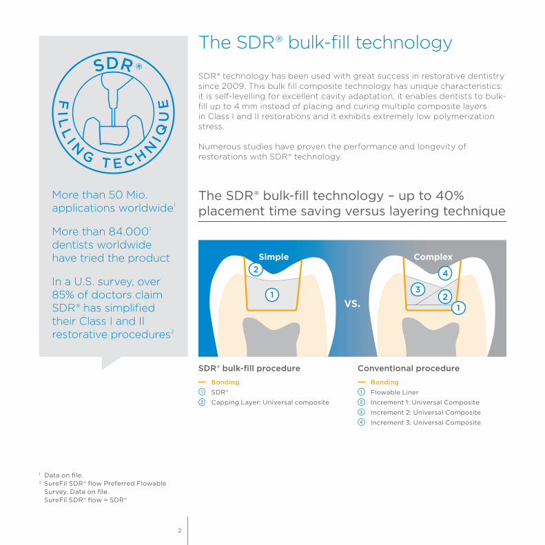
SDR® technology has been used with great success in restorative dentistry since 2009. This bulk fill composite technology has unique characteristics: it is self-levelling for excellent cavity adaptation, it enables dentists to bulk-fill up to 4 mm instead of placing and curing multiple composite layers in Class I and II restorations and it exhibits extremely low polymerization stress.
Numerous studies have proven the performance and longevity of restorations with SDR® technology.
The SDR® bulk-fill technology – up to 40% placement time saving versus layering technique
The SDR® bulk-fill technology
Conventional procedure
Bonding Flowable Liner
Increment 1: Universal Composite
Increment 2: Universal Composite
Increment 3: Universal Composite
SDR® bulk-fill procedure
Bonding SDR®
Capping Layer: Universal composite
ComplexSimple
vs.
More than 50 Mio. applications worldwide1
More than 84.0001 dentists worldwide have tried the product
In a U.S. survey, over 85% of doctors claim SDR® has simplified their Class I and II restorative procedures2
2

Case: Disto-occlusal Composite Resin
A 38 year old male patient presented a failed Class II DO composite restoration on a lower molar. After radiographic and clinical examination, the patient was anesthetized, and the old failed restoration, as well as the caries lesion, was removed. The Class II DO cavity restoration was performed using an optimized approach, using the Palodent® V3 sectional matrix system, the universal adhesive system Prime&Bond elect® in the selective etching mode, the low polymerization stress, self-leveling, bulk-fill composite SDR®, the universal composite TPH Spectra® and Enhance® Finishers.
Clinical case on Class II with SDR® bulk-fill technology
Dr. A. ReisSao Paolo, Brazil
Before After
ConclusionThis case study shows a typical situation where most dentists face three common issues: postoperative sensitivity, composite adaptation and contact point creation. In order to reduce the chance of postoperative sensitivity, the selective etching approach was used with a universal adhesive.For perfect composite adaptation, the low polymerization stress, self-leveling, bulk-fill composite SDR® was used. In addition, for perfect proximal contour and optimal creation of a tight contact point, the sectional matrix system Palodent® V3 was used. Video
3

3. The Palodent® V3 sectional matrix system was placed using the Universal Ni-Ti ring and the 6.5 mm matrix to prevent gaps in gingival-axial corner.
1. Pre-operative appearance with a fractured Class II DO composite restoration.
2. The old composite restoration and caries were removed. Note that the distobuccal cusp was also compromised.
Last, in order to create a nice occlusal anatomy and obtain a perfect shade match, a modern universal composite was applied.The combination of all these dental materials from Dentsply Sirona allows easier, faster and predictable placement of Class II restorations.
References
Kumagai RY, Zeidan LC, Rodrigues JA, Reis AF, Roulet JF. Bond strength of a flowable bulk-fill resin composite in class II MOD cavities. J Adhes Dent. 2015 Aug;17(5):427-32.
Rosatto CM, Bicalho AA, Veríssimo C, Bragança GF, Rodrigues MP, Tantbirojn D, Versluis A, Soares CJ. Mechanical properties, shrinkage stress, cuspal strain and fracture resistance of molars restored with bulk-fill composites and incremental filling technique. J Dent. 2015 Dec;43(12):1519-28.
Van Dijken JW, Pallesen U. Posterior bulk-filled resin composite restorations: A 5-year randomized controlled clinical study. J Dent. 2016 Aug;51:29-35.
Van Ende A, De Munck J, Van Landuyt K, Van Meerbeek B. Effect of bulk-filling on the bonding efficacy in occlusal class I cavities. J Adhes Dent. 2016;18(2):119-24.
4
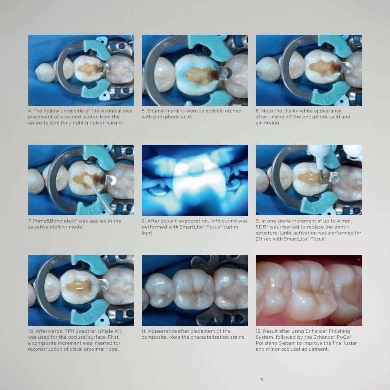
4. The hollow underside of the wedge allows placement of a second wedge from the opposite side for a tight gingival margin.
5. Enamel margins were selectively etched with phosphoric acid.
6. Note the chalky white appearance after rinsing off the phosphoric acid and air-drying.
7. Prime&Bond elect® was applied in the selective etching mode.
8. After solvent evaporation, light curing was performed with SmartLite® Focus® curing light.
9. In one single increment of up to 4 mm, SDR® was inserted to replace the dentin structure. Light activation was performed for 20 sec with SmartLite® Focus®.
10. Afterwards, TPH Spectra® (shade A1), was used for the occlusal surface. First, a composite increment was inserted for reconstruction of distal proximal ridge.
11. Appearance after placement of the composite. Note the characterization stains.
12. Result after using Enhance® Finishing System, followed by the Enhance® PoGo® Polishing System to improve the final luster and minor occlusal adjustment.
5
Technical performance of SDR® bulk-fill technology
Shrinkage stress of resin composites
ObjectiveMonomer development for a reduced shrinkage of composite materials still challenges the modern research. The purpose of this study was to analyze the shrinkage behavior of an innovative composite material for dental restorations based on a resin system that is claimed to control polymerization kinetics having incorporated a photoactive group within the resin.
MethodShrinkage stress development within the first 300 s after photoinitiation was evaluated (n = 10). SDR® was measured in comparison to regular methacrylate-based micro- (Esthet·X® Flow) and nano-hybrid flowable RBCs (Filtek Supreme Plus Flow). Additionally, the high viscosity counterparts of the two regular flowable methacryate-based composites (Esthet·X® HD and Filtek Supreme Plus) as well as a low shrinkage silorane-based micro-hybrid composite (Filtek Silorane) were considered. The curing time was 20 s (LED unit Freelight2, 3M-Espe, 1226 mW/cm2).
ResultsSDR® achieved the significantly lowest contraction stress (1.1 ± .01 MPa) followed by the silorane-based composite (3.6 ± .03 MPa), whereas the highest stress values were induced in the regular methacrylate-based flowable composites Esthet·X® Flow (5.3 ± .3 MPa) and Filtek Supreme Flow (6.5 ± .3 MPa). SDR® achieved also the lowest shrinkage rate (maximum at 0.1 MPa/s). For all analysed materials, no significant difference in the micro-mechanical properties between top and bottom were found when measured on 2 mm thick increments 24 h after polymerization. The categories of flowable materials performed in the measured micro-mechanical properties significantly inferior when compared to the hybrid-composites, showing lower Vickers hardness (HV) and modulus of elasticity (E) and predominantly higher creep and plastic deformation. Within the flowable RBCs, SDR® achieved the lowest Vickers hardness, the highest modulus of elasticity, the highest creep and showed the significantly lowest elastic deformation.
Low shrinkage stress
N. IlieMunich, Germany
6
Source: Investigations on a methacrylate- based flowable composite-based on the SDR® technology (Ilie N, Hickel R, Dental Materials 27 (2011), 348-355)
* Not registered trademarks of Dentsply Sirona, Inc.
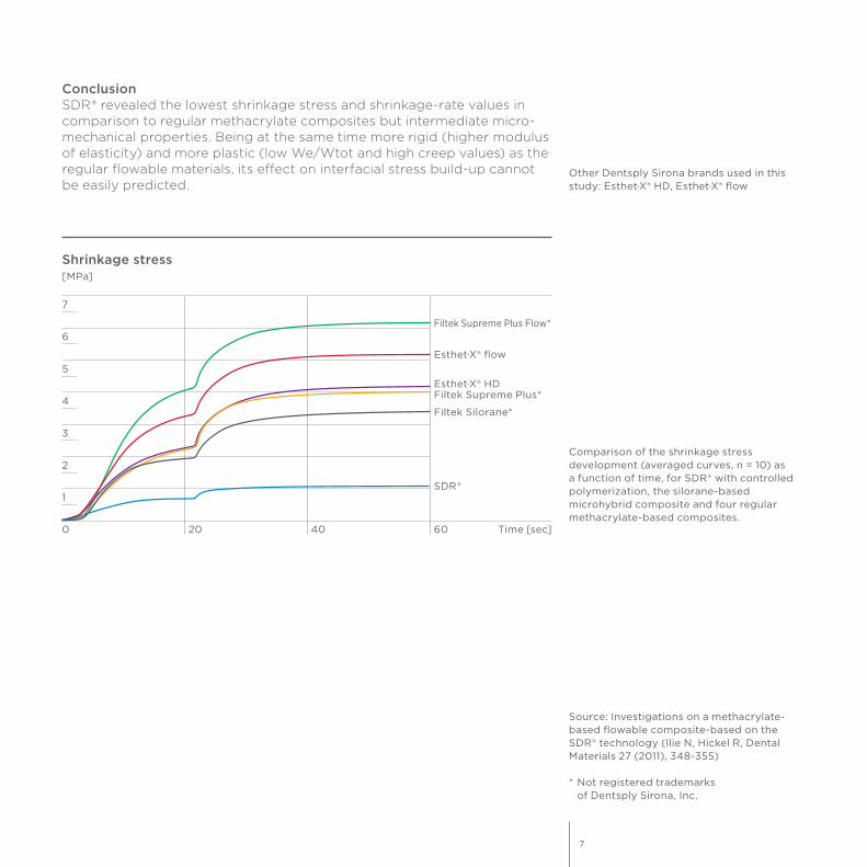
ConclusionSDR® revealed the lowest shrinkage stress and shrinkage-rate values in comparison to regular methacrylate composites but intermediate micro-mechanical properties. Being at the same time more rigid (higher modulus of elasticity) and more plastic (low We/Wtot and high creep values) as the regular flowable materials, its effect on interfacial stress build-up cannot be easily predicted.
Shrinkage stress[MPa]
7
6
3
2
1
0 20 40 60 Time [sec]
5
4
Filtek Supreme Plus Flow*
Esthet·X® flow
Esthet·X® HDFiltek Supreme Plus*
Filtek Silorane*
SDR®
Other Dentsply Sirona brands used in this study: Esthet·X® HD, Esthet·X® flow
Comparison of the shrinkage stress development (averaged curves, n = 10) as a function of time, for SDR® with controlled polymerization, the silorane-based microhybrid composite and four regular methacrylate-based composites.
7
Microleakage at enamel and dentin margins with a bulk-fill flowable resin
ObjectiveThe aim of this in vitro study was to evaluate the marginal sealing ability of a bulk fill flowable resin composite – SureFil SDR®1 – on both enamel and dentin substrates.
MethodIn total, 48 non-carious molars were selected and four Class V cavities were prepared at the CEJ (cemento-enamel junction) of each sample. Cavities were filled with Venus Diamond (Heraeus Kulzer), Venus Diamond Flow (Heraeus Kulzer) and SureFil SDR®1 (Dentsply Sirona). Samples were divided into two groups: First group samples were immersed in a methylene blue solution for 30 min at 25 °C. Second group samples were artificially aged by thermocycling (TC) and then treated with methylene blue. Samples were sectioned in the center of the restoration and observed with a 40x stereomicroscope, and the percentage of cavity infiltration was calculated.
ResultsResults were analyzed statistically by ANOVA (P < 0.05). The amount of infiltration was significantly lower for the enamel substrate compared with dentin (P = 0.0001) and in samples immediately immersed compared with those that were thermocycled aged (P = 0.011). The interaction between the composite material and the marginal substrate significantly affected dye penetration (P = 0.006).
ConclusionBulk fill flowable resins provided significantly better marginal seal in dentin, both before and after thermocycling. Nanohybrid resin composites and bulk fill flowable resins showed similar microleakage values at enamel margins.
Marginal Adaptation
N. ScottiTurin, Italy
1 SureFil SDR® = SDR®
8

40x picture of a sectioned sample with the calculation of the percentage of dye penetration (3-4; 40%) relative to the total length of the restoration interface (1-2; 100%)
40%
100%
SureFil SDR®1
Venus Diamond Flow*
Venus Diamond*
Class V Microleakage vs. Thermocycling (TC)Percentage microleakage [%]
before TC before TCafter TC after TC
60
40
20
0
80
Enamel Dentin100
Source: Microleakage at enamel and dentin margins with a bulk fill flowable resin (Scotti N, Comba A, Gambino A, Paolino DS, Alovisi M, Pasqualini D, Berutti E, Eur J Dent. 2014; 8:1-8)
* Not registered trademarks of Dentsply Sirona, Inc.
1 SureFil SDR® = SDR®
9
Radiopacity of bulk-fill composites
ObjectiveRadiopacity indicates the visibility of composite restorations on X-ray radiographs. The aim was to evaluate the radiopacity of different bulk-fill composites.
MethodThe radiopacity was measured based on ISO 4049. 1.0 mm thick disk composite specimen was cured in disk stainless steel mold, 1 mm thick x 30 mm in diameter, with Triad 2000 for 2 minutes each side. Radiopacity of a restorative material was determined by comparing the optical density of a radiograph of a 1.0 mm thick cured material to that of a 0.5, 1.0, 1.5, 2.0, 2.5, 3.0 mm stepped standard aluminum block.
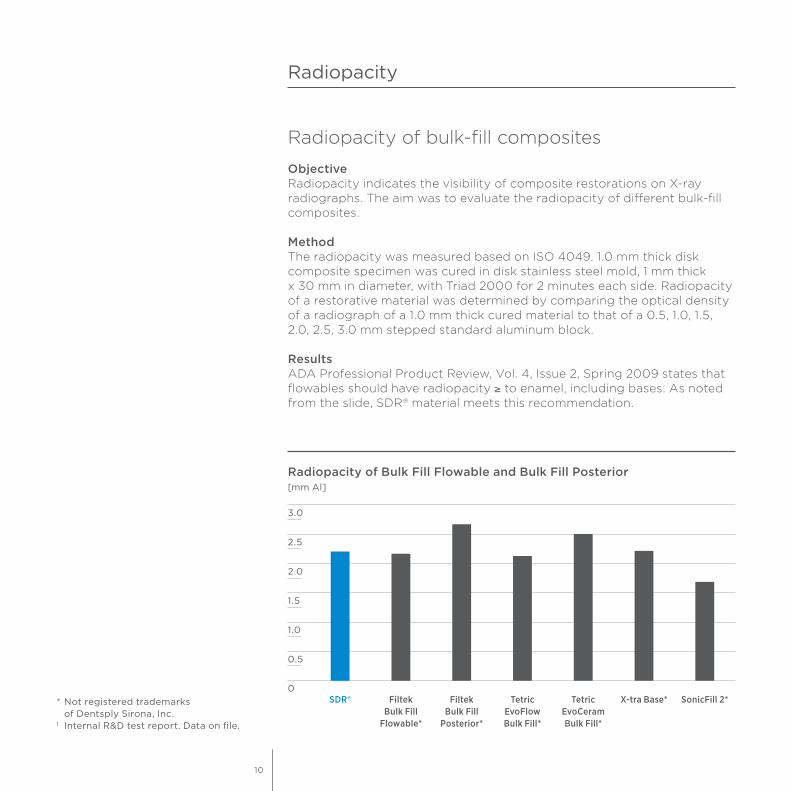
ResultsADA Professional Product Review, Vol. 4, Issue 2, Spring 2009 states that flowables should have radiopacity ≥ to enamel, including bases. As noted from the slide, SDR® material meets this recommendation.
Radiopacity
Radiopacity of Bulk Fill Flowable and Bulk Fill Posterior[mm Al]
SDR® Filtek Bulk Fill
Flowable*
Filtek Bulk Fill
Posterior*
X-tra Base* SonicFill 2*Tetric EvoFlow Bulk Fill*
Tetric EvoCeram Bulk Fill*
1.5
1.0
0.5
0
2.0
2.5
3.0
* Not registered trademarks of Dentsply Sirona, Inc.
1 Internal R&D test report. Data on file.
10
Clinical proof of SDR® bulk-fill technology
Effectiveness of Class I and Class II SDR® bulk filling restorations. Six-year follow-up of posterior bulk-filled resin composite restorations.
ObjectiveClinical evaluation of the bulk placed resin composite SDR® in Class I and Class II cavities, bonded with the single step self-etching primer Xeno V® and covered with the nano-ceramic resin composite Ceram·X® mono.
MethodThirty-eight patients with a total of 53 paired restorations were treated with either conventionally layered (Xeno V® + Ceram·X® mono) or partly bulk filled (SDR®) Class I or II restorations (30 Class I, 76 Class II). After 6 years, 49 paired restorations, 26 Class I and 72 Class II were evaluated.The clinical parameters relevant to the base material evaluated at baseline (within one week of placing restorations) and at each recall evaluation were as follows: Secondary caries, anatomic form, marginal adaptation, marginal discoloration, surface roughness and color match. For marginal adaptation and discoloration, the involvement of marginal excess was noted. Also postoperative sensitivity was evaluated.
ResultSix Class II molar restorations failed, three in each group, which resulted in annual failure rates (AFR) of 1.0% for both groups. The AFR at 6 years for Class I restorations was 0% and for Class II restorations 1.4%.
6 year recall report
J. W. V. van Dijken
11
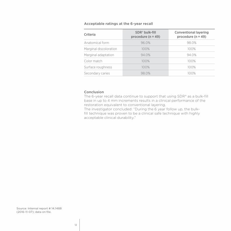
ConclusionThe 6-year recall data continue to support that using SDR® as a bulk-fill base in up to 4 mm increments results in a clinical performance of the restoration equivalent to conventional layering.The investigator concluded: “During the 6 year follow up, the bulk-fill technique was proven to be a clinical safe technique with highly acceptable clinical durability.”
Source: Internal report # 14.1488 (2016-11-07); data on file.
Criteria SDR® bulk-fill procedure (n = 49)
Conventional layering procedure (n = 49)
Anatomical form 96.0% 98.0%
Marginal discoloration 100% 100%
Marginal adaptation 94.0% 94.0%
Color match 100% 100%
Surface roughness 100% 100%
Secondary caries 98.0% 100%
Acceptable ratings at the 6-year recall
12

Posterior bulk-filled resin composite restorations: A 5-year randomized controlled clinical study.
ObjectiveTo evaluate in a randomized controlled study the 5-year clinical durability of a flowable resin composite bulk-fill technique in Class I and Class II restorations.
MethodIn total, 86 patients with one or two pair similar Class I or II cavities received 200 composite restorations by two dentists. The SDR® cavity of each pair was filled in bulks of 4 mm up to 2 mm short of the occlusal surface and covered with the hybrid composite Ceram·X® mono+. The other cavity was conventionally filled with Ceram·X® mono+ in 2 mm layers. The majority of the cavities were deep and had extended size. In all cavities, Xeno® V+ was applied as the adhesive. All restorations were in occlusion. The restorations were evaluated at baseline and then annually during 5 years.
ResultsNo post-operative sensitivity was reported. At 5 years, 183 restorations, 68 Class I and 115 Class II, restorations were evaluated. Ten restorations failed, 4 SDR® and 6 conventionally layered restorations, all of which were Class II. The main reason of failure was tooth fracture and secondary caries resulting in annual failure rates of 1.1% for SDR® and 1.3% for conventionally layered restorations. No significant differences were observed between bulk-filled and conventionally layered composite restorations for the evaluated criteria at the recall (p = 0.12).
5 year recall report
J. W. V. van Dijken
13
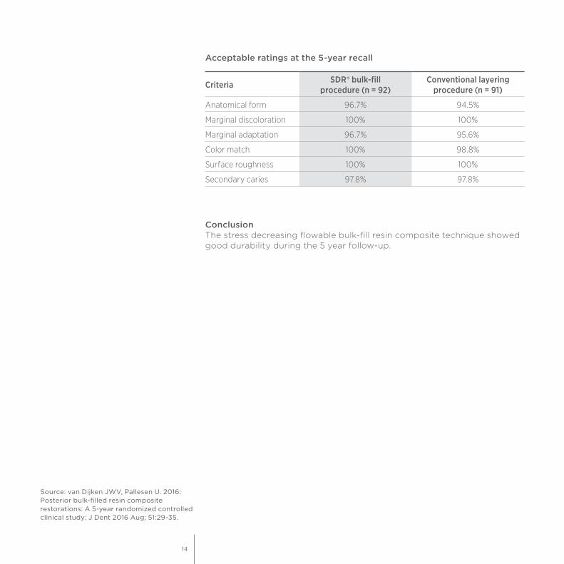
ConclusionThe stress decreasing flowable bulk-fill resin composite technique showed good durability during the 5 year follow-up.
Criteria SDR® bulk-fill procedure (n = 92)
Conventional layering procedure (n = 91)
Anatomical form 96.7% 94.5%
Marginal discoloration 100% 100%
Marginal adaptation 96.7% 95.6%
Color match 100% 98.8%
Surface roughness 100% 100%
Secondary caries 97.8% 97.8%
Source: van Dijken JWV, Pallesen U. 2016: Posterior bulk-filled resin composite restorations: A 5-year randomized controlled clinical study; J Dent 2016 Aug; 51:29-35.
Acceptable ratings at the 5-year recall
14
A Three Year Clinical Evaluation of Class II Composite Resin Restorations using SureFil SDR® and an experimental composite resin or Esthet·X® HD
ObjectiveA three year clinical trial to evaluate the in vivo success of two groups of Class II composite resin restorations both using SureFil SDR®1 (Dentsply Sirona) as the dentin replacement material, one group used an experimental enamel replacement composite and another used Esthet·X® HD (Dentsply Sirona) as the enamel replacement material.
MethodEighty-seven subjects were enrolled into this clinical trial in US dental schools, receiving a total of 170 Class I and II restorations. All cavity preparations were etched for 15 seconds with 37% phosphoric acid, then rinsed and dried but not desiccated. Prime&Bond® NT Bonding Agent (Dentsply Sirona) was applied to all dentin and enamel surfaces and light cured for 10 seconds. SureFil SDR®1 Bulk Fill Flowable (Dentsply Sirona) was then applied in increments up to 4 mm as needed to fill the cavity to the level of the dentin-enamel junction. An experimental low stress micro-hybrid composite resin or Esthet·X® HD High Definition Micro Matrix restorative (Dentsply Sirona) was then layered onto the base to complete the anatomic form of the restoration. Restorations were finished and polished using the Enhance® Finishing System (Dentsply Sirona) and the PoGo® One Step Diamond Micro-Polisher (Dentsply Sirona). Subjects were recalled for evaluation approximately six months, 12 months, 24 months and 36 months following placement of their restoration(s).
The clinical parameters relevant to the base material evaluated at baseline (within one week of placing restorations) and at each recall evaluation were as follows:• Fracture/Surface Defects• Proximal Contact• Recurrent Caries• Postoperative Sensitivity
A gingival index was also noted to measure the inflammatory state of the gingiva adjacent to the restoration.
3 year recall report
J. Burgess, C. Munoz
1 SureFil SDR® = SDR®
15

ResultAfter 3 years, 86 restorations in 49 subjects were available for evaluation.
ConclusionThe results support the conclusion that the low stress resin when used as a bulk fill base in Class I and II restorations with a conventional universal composite resin as occlusal capping layer exhibited acceptable performance after three years. Several restorations showed minor surface defects consistent with three years of intraoral function. In total, six fractures within the capping composite required repair. One restoration was replaced. There was essentially no post-operative sensitivity related to the use of the low stress resin, and the response of the gingiva in contact with the material was within normal limits. There were no observations of recurrent caries associated with the low stress resin and there were no reports of adverse events throughout the duration of the trial.
Source: Internal report # 765-540 (Feb 17, 2010); data on file.
1 SureFil SDR® = SDR®
Acceptable ratings at the 3-year recall
Criteria SureFil SDR®1 – Baseline (n = 170)
SureFil SDR®1 – 3 years (n = 86)
Retention 100% 99%
Proximal Contact 99% 90%
Recurrent caries 100% 97%
Fracture 100% 98%
16
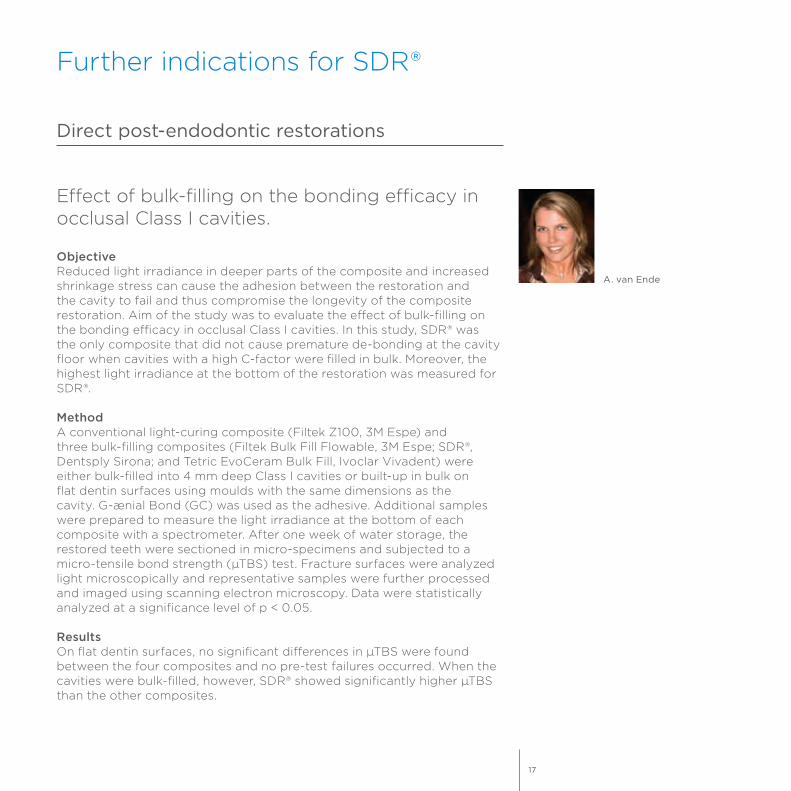
Effect of bulk-filling on the bonding efficacy in occlusal Class I cavities.
ObjectiveReduced light irradiance in deeper parts of the composite and increased shrinkage stress can cause the adhesion between the restoration and the cavity to fail and thus compromise the longevity of the composite restoration. Aim of the study was to evaluate the effect of bulk-filling on the bonding efficacy in occlusal Class I cavities. In this study, SDR® was the only composite that did not cause premature de-bonding at the cavity floor when cavities with a high C-factor were filled in bulk. Moreover, the highest light irradiance at the bottom of the restoration was measured for SDR®.
MethodA conventional light-curing composite (Filtek Z100, 3M Espe) and three bulk-filling composites (Filtek Bulk Fill Flowable, 3M Espe; SDR®, Dentsply Sirona; and Tetric EvoCeram Bulk Fill, Ivoclar Vivadent) were either bulk-filled into 4 mm deep Class I cavities or built-up in bulk on flat dentin surfaces using moulds with the same dimensions as the cavity. G-ænial Bond (GC) was used as the adhesive. Additional samples were prepared to measure the light irradiance at the bottom of each composite with a spectrometer. After one week of water storage, the restored teeth were sectioned in micro-specimens and subjected to a micro-tensile bond strength (µTBS) test. Fracture surfaces were analyzed light microscopically and representative samples were further processed and imaged using scanning electron microscopy. Data were statistically analyzed at a significance level of p < 0.05.
ResultsOn flat dentin surfaces, no significant differences in µTBS were found between the four composites and no pre-test failures occurred. When the cavities were bulk-filled, however, SDR® showed significantly higher µTBS than the other composites.
Further indications for SDR®
Direct post-endodontic restorations
A. van Ende
17

Composite 4 mm bulk on flat surface
4 mm cavity filled in bulk
SDR® 26.7 ± 9.8 (0%) 16.6 ± 7.7 (0%)
Filtek Bulk Fill Flowable 19.7 ± 7.8 (0%) 4.0 ± 7.8 (75%)
Tetric EvoCeram Bulk Fill 21.4 ± 9.0 (0%) 3.9 ± 7.5 (73%)
Filtek Z100 26.0 ± 13.9 (0%) 0.0 ± 0.0 (100%)µTBS results in MPa; pre-test failures in % in brackets.
With Filtek Z100, all the specimens failed prior to µTBS testing, while Filtek Bulk Fill Flowable and Tetric Evo Ceram Bulk Fill resulted in more than 70% pre-test failures. In contrast, SDR® showed no pre-test failures. The highest light irradiance was measured for SDR®, followed by the other bulk-filling composites and, finally, Filtek Z100.
18
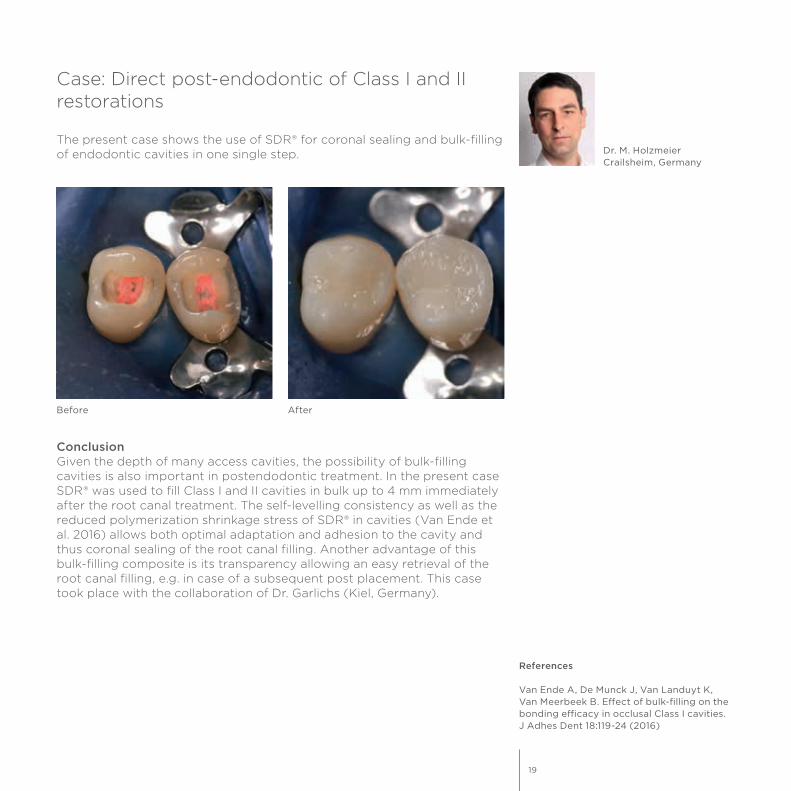
Case: Direct post-endodontic of Class I and II restorations
The present case shows the use of SDR® for coronal sealing and bulk-filling of endodontic cavities in one single step. Dr. M. Holzmeier
Crailsheim, Germany
ConclusionGiven the depth of many access cavities, the possibility of bulk-filling cavities is also important in postendodontic treatment. In the present case SDR® was used to fill Class I and II cavities in bulk up to 4 mm immediately after the root canal treatment. The self-levelling consistency as well as the reduced polymerization shrinkage stress of SDR® in cavities (Van Ende et al. 2016) allows both optimal adaptation and adhesion to the cavity and thus coronal sealing of the root canal filling. Another advantage of this bulk-filling composite is its transparency allowing an easy retrieval of the root canal filling, e.g. in case of a subsequent post placement. This case took place with the collaboration of Dr. Garlichs (Kiel, Germany).
References
Van Ende A, De Munck J, Van Landuyt K, Van Meerbeek B. Effect of bulk-filling on the bonding efficacy in occlusal Class I cavities. J Adhes Dent 18:119-24 (2016)
Before After
19
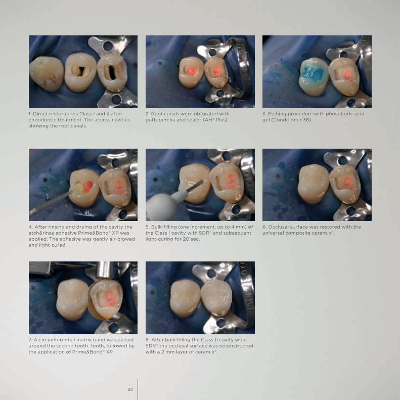
4. After rinsing and drying of the cavity the etch&rinse adhesive Prime&Bond® XP was applied. The adhesive was gently air-blowed and light-cured.
7. A circumferential matrix band was placed around the second tooth. tooth, followed by the application of Prime&Bond® XP.
5. Bulk-filling (one increment, up to 4 mm) of the Class I cavity with SDR® and subsequent light-curing for 20 sec.
8. After bulk-filling the Class II cavity with SDR® the occlusal surface was reconstructed with a 2 mm layer of ceram.x®.
6. Occlusal surface was restored with the universal composite ceram.x®.
3. Etching procedure with phosphoric acid gel (Conditioner 36).
1. Direct restorations Class I and II after endodontic treatment. The access cavities showing the root canals.
2. Root canals were obturated with guttapercha and sealer (AH® Plus).
20
Clinical evaluation of restorative materials for primary teeth
ObjectiveThis randomized control trial evaluated restorations in Class I and Class II cavities in primary molars. 200 fillings were placed using composite materials (Filtek Z250 and SDR®), glass-ionomer cements (Fuji IX GP, ChemFil® Rock), and a resin-modified glass-ionomer cements (Fuji II LC).
MethodClinical evaluation was performed at baseline and at 1 year using modified USPHS criteria. At 1-year recall, 33 SDR®, 28 Fuji II LC, 40 ChemFil® Rock, 28 Fuji IX GP, 24 Filtek Z250 restorations were assessed. Mean scores for materials were compared using Kruskal-Wallis test at a 0.05 probability level.
ResultsNo significant differences between all materials were detected after one year for all evaluated clinical criteria (p > 0.05). The comparison of restoration performance for all five groups showed a significant increase in surface staining in the Fuji IX GP and ChemFil® Rock groups (p = 0.001) and deterioration of marginal integrity in the Fuji IX GP group compared to the SDR® group (p = 0.024). Significant decrease in marginal adaptation (p = 0.002) for ChemFil® was observed. Results for post-operative hypersensitivity, tooth vitality and recurrence of caries, erosion, abfraction showed no significant changes between all groups.
ConclusionIn total, 98.2% SDR®, 95.2% Fuji II LC, 88% ChemFil®, 87.2% Fuji IX and 98.4% Filtek Z250 were assessed as excellent.
SDR® bulk-fill technology in pediatric dentistry
P. JasińskiWarsaw, Poland
Other Dentsply Sirona brand used in this study: ChemFil® Rock
21
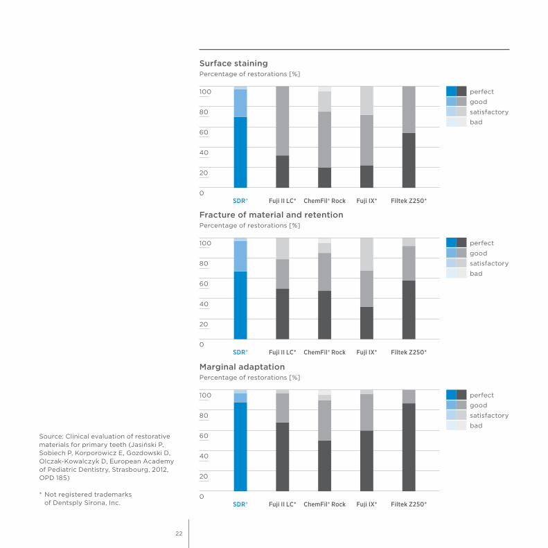
Surface stainingPercentage of restorations [%]
40
20
0
60
80
100
SDR® Fuji II LC* ChemFil® Rock Fuji IX* Filtek Z250*
perfect
satisfactory
good
bad
Fracture of material and retentionPercentage of restorations [%]
40
20
0
60
80
100
SDR® Fuji II LC* ChemFil® Rock Fuji IX* Filtek Z250*
perfect
satisfactory
good
bad
Marginal adaptationPercentage of restorations [%]
40
20
0
60
80
100
SDR® Fuji II LC* ChemFil® Rock Fuji IX* Filtek Z250*
perfect
satisfactory
good
bad
Source: Clinical evaluation of restorative materials for primary teeth (Jasiński P, Sobiech P, Korporowicz E, Gozdowski D, Olczak-Kowalczyk D, European Academy of Pediatric Dentistry, Strasbourg, 2012, OPD 185)
* Not registered trademarks of Dentsply Sirona, Inc.
22
Case: Advantages of using SDR® in Pediatric Dentistry
Bulk-fill materials have been developed to facilitate a filling to be placed quickly and reliably with a single layer of up to 4 mm thickness [1-3]. The flowable bulk-fill material SDR® (Dentsply Sirona), which excels by virtue of its extremely low shrinkage stress, has been available since 2010 [4,5]. Since 2014 it is also approved for Class I and II deciduous tooth fillings in the posterior region without an additional capping layer. This extension of indications is a major benefit for the practitioner – especially in pediatric dentistry.
Bulk-fill materials are ideal for deciduous teeth, where the focus is on rapid application and reliability of the materials used. The time saving in filling placement is a crucial advantage, both in regular treatment of children, as well as in the treatment of children under general anesthesia. The reduced abrasion resistance of flowable bulk-fill materials is compatible with the natural deciduous tooth abrasion and so is not to be viewed as a disadvantage in the wear phase of primary dentition.
Patient casesThree cases treating decidiuous teeth (Class I and II) in the posterior region using SDR® are presented below.
Treatment of a 9-year-old boy with hemophilia AA 9-year-old boy with severe hemophilia A presented a carious lesion on the upper left deciduous molar (Fig. 1). Following excavation and preparation of the cavity margin, the AutoMatrix® system (Dentsply Sirona) was applied (Fig. 2). A self-etch all-in-one adhesive (Xeno® V+, Dentsply Sirona) was applied and light-cured. SDR® was applied directly with its Compula® Tip. Here it is important that the metal cannula is placed on the proximal cavity floor and is extracted while continuously extruding the low-viscosity material. The entire cavity was filled in one single increment and then light-cured for 20 seconds. As it was possible to ensure reliable contamination control using dental rolls and four-handed working, a rubber dam was not used, which was advantageous in view of the boy’s medical history as a hemophiliac. This excluded potential traumatisation of the gingiva from the rubber dam clamp. Finally, the filling was finished with a fine diamond bur (Fig. 3) and the finishing and polishing system Enhance® (Dentsply Sirona).
Dr. V. EhlersMainz, Germany
23
Treatment of a 5-year-old girlThe 5-year-old girl was a former general anesthesia patient who has since allowed herself to be treated in the dental chair. However, the child was restless and not very compliant, so treatment had to be kept short. This is where an all-in-one adhesive and a bulk-fill material is very helpful in therapy, as the treatment steps of conditioning and spraying off the etching gel or multiple layering of the filling are not necessary and the treatment can therefore be performed quickly. The patient presented three carious deciduous molars 85, 75 and 65 (Fig. 4). Following caries excavation on tooth 85 buccal, 75 occlusal and 65 occlusal (Fig. 5) using a round bur and a polymer bur, the all-in-one adhesive (Xeno® V+) was applied and then light-cured. SDR® was applied in a single layer and light-cured. The fillings were finished and polished as described above.
In this case on tooth 85 buccal, the difference in shade to the primary tooth is hardly noticeable (Fig. 6). Similarly, with flat occlusal cavities, e.g. tooth 75 (Fig. 7) and 65 (Fig. 8), the higher translucency does not affect the final outcome. Experience has shown that children and parents are satisfied with the shade. It should certainly also be borne in mind that deciduous teeth are not subject to such high aesthetic requirements as permanent dentition. Tooth 85 was photographed in the course of a follow-up examination and shows the buccal filling after two years and an occlusal compomer filling after five years (Fig. 9). The occlusal SDR® filling on tooth 75 was also photographed after two years (Fig. 10). Fig. 11 shows the occlusal filling on tooth 65 in a follow-up examination after 1 ½ years. The fillings were in a good clinical state in the follow-up examinations.
Treatment of a 4-year old boy under general anesthesiaThe top priority in dental treatment of children under general anesthesia is to keep the anesthesia time as short as possible. That is why the use of dependable and quick-to-apply materials is recommended here, too. In total, the 4-year-old boy had 12 primary teeth to be treated, of which 9 were filled and 3 had to be extracted. The posterior fillings were carried out with SDR®, whereas the front teeth were restored with the compomer Dyract® eXtra, dental shade A2 (Dentsply Sirona). The use of compomers is considered to be the worldwide standard for the restoration of primary tooth defects [6]. In the course of general anesthesia treatment, the carious tooth 64 was excavated (Fig. 12). Excavation was performed as previously described and conditioning also used an all-in-one adhesive. Following finishing and polishing, the filling covered the occlusal surface, as well as the palatal surface of the tooth (Fig. 13).
References
[1] Fleming, G. J., Awan, M., Cooper, P. R., Sloan, A. J.: The potential of a resin-composite to be cured to a 4 mm depth. Dent Mater 24, 522-529 (2008)
[2] Burgess, J., Cakir, D.: Comparative properties of low-shrinkage composite resins. Compend Contin Educ Dent 31, 10-15 (2010)
[3] Roggendorf, M. J., Krämer, N., Appelt, A., Naumann, M., Frankenberger, R.: Marginal quality of flowable 4-mm base vs. conventionally layered resin composite. J Dent 39, 643-647 (2011)
[4] Ilie, N., Hickel, R.: Investigations on a methacrylate-based flowable composite based on the SDR® technology. Dent Mater 27, 348-355 (2011)
[5] Rullmann, I., Schattenberg, A., Marx, M., Willershausen, B., Ernst, C. P.: Photoelastic determination of polymerization shrinkage stress in low-shrinkage resin composites. Schweiz Monatsschr Zahnmed 122, 294-299 (2012)
[6] Krämer, N., Frankenberger, R.: Compomers in restorative therapy of children: a literature review. Int J Paediatr Dent 17, 2-9 (2007)
24
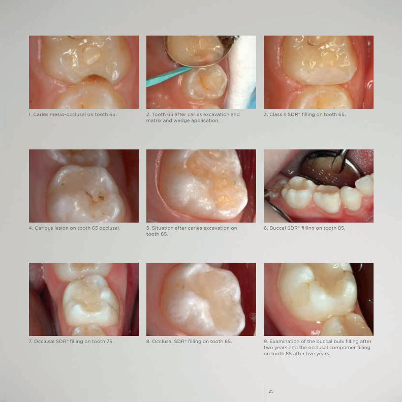
3. Class II SDR® filling on tooth 65.1. Caries mesio-occlusal on tooth 65. 2. Tooth 65 after caries excavation and matrix and wedge application.
4. Carious lesion on tooth 65 occlusal.
7. Occlusal SDR® filling on tooth 75.
5. Situation after caries excavation on tooth 65.
8. Occlusal SDR® filling on tooth 65.
6. Buccal SDR® filling on tooth 85.
9. Examination of the buccal bulk filling after two years and the occlusal compomer filling on tooth 85 after five years.
25
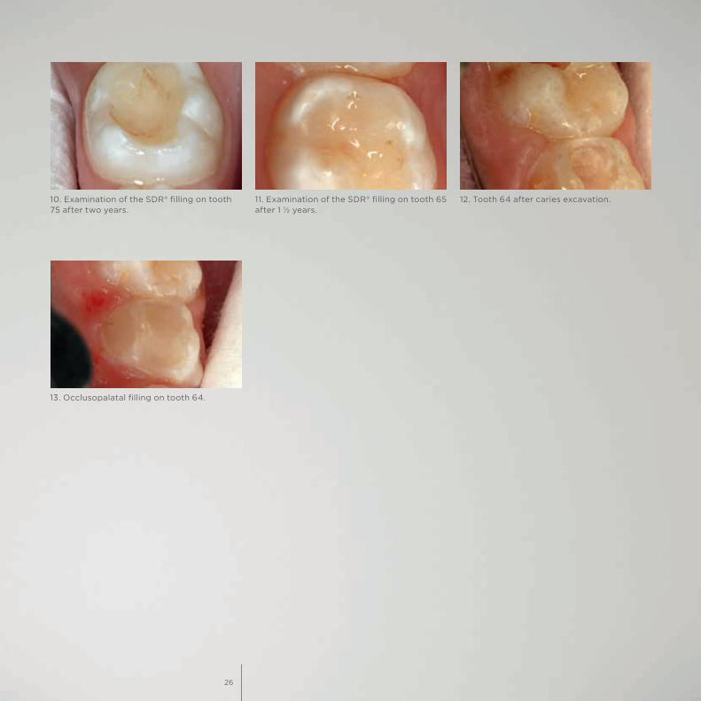
13. Occlusopalatal filling on tooth 64.
10. Examination of the SDR® filling on tooth 75 after two years.
11. Examination of the SDR® filling on tooth 65 after 1 ½ years.
12. Tooth 64 after caries excavation.
26
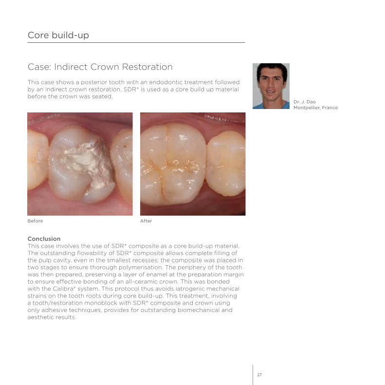
Case: Indirect Crown Restoration
This case shows a posterior tooth with an endodontic treatment followed by an indirect crown restoration. SDR® is used as a core build up material before the crown was seated.
Core build-up
Dr. J. DaoMontpellier, France
ConclusionThis case involves the use of SDR® composite as a core build-up material. The outstanding flowability of SDR® composite allows complete filling of the pulp cavity, even in the smallest recesses; the composite was placed in two stages to ensure thorough polymerisation. The periphery of the tooth was then prepared, preserving a layer of enamel at the preparation margin to ensure effective bonding of an all-ceramic crown. This was bonded with the Calibra® system. This protocol thus avoids iatrogenic mechanical strains on the tooth roots during core build-up. This treatment, involving a tooth/restoration monoblock with SDR® composite and crown using only adhesive techniques, provides for outstanding biomechanical and aesthetic results.
Before After
27
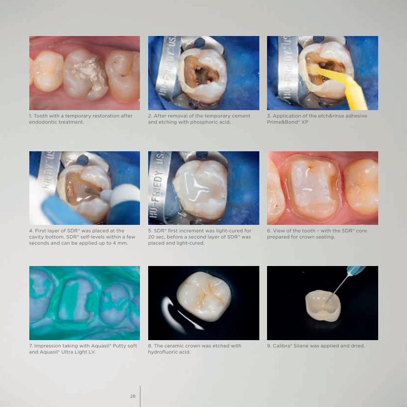
1. Tooth with a temporary restoration after endodontic treatment.
4. First layer of SDR® was placed at the cavity bottom. SDR® self-levels within a few seconds and can be applied up to 4 mm.
2. After removal of the temporary cement and etching with phosphoric acid.
5. SDR® first increment was light-cured for 20 sec, before a second layer of SDR® was placed and light-cured.
3. Application of the etch&rinse adhesive Prime&Bond® XP
6. View of the tooth – with the SDR® core prepared for crown seating.
7. Impression taking with Aquasil® Putty soft and Aquasil® Ultra Light LV.
8. The ceramic crown was etched with hydrofluoric acid.
9. Calibra® Silane was applied and dried.
28
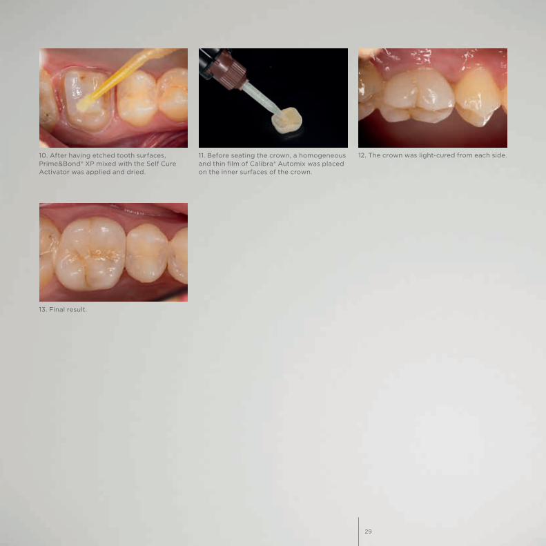
10. After having etched tooth surfaces, Prime&Bond® XP mixed with the Self Cure Activator was applied and dried.
12. The crown was light-cured from each side.
13. Final result.
11. Before seating the crown, a homogeneous and thin film of Calibra® Automix was placed on the inner surfaces of the crown.
29
K79200252-00© Dentsply Sirona 05/2017
For more information visit www.dentsplysirona.com.
www.facebook.com/dentsplysirona.restorative