post partum uterus - obstetrics and gynaecology · post partum uterus imaging of ... by ct or mri...
TRANSCRIPT
POST PARTUM UTERUSIMAGING OF COMPLICATIONS
University of Toronto
Dept Medical Imaging, Obstetrics & Gynecology
University of Chicago: Ultrasound
Symposium

Disclosures
None
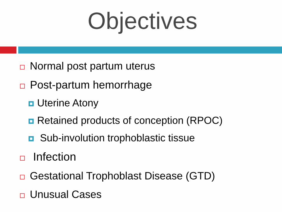
Objectives
Normal post partum uterus
Post-partum hemorrhage
Uterine Atony
Retained products of conception (RPOC)
Sub-involution trophoblastic tissue
Infection
Gestational Trophoblast Disease (GTD)
Unusual Cases
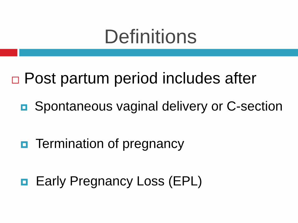
Definitions
Post partum period includes after
Spontaneous vaginal delivery or C-section
Termination of pregnancy
Early Pregnancy Loss (EPL)
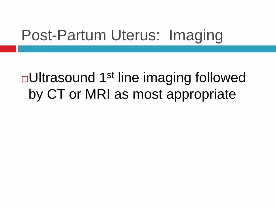
Post-Partum Uterus: Imaging
Ultrasound 1st line imaging followed
by CT or MRI as most appropriate
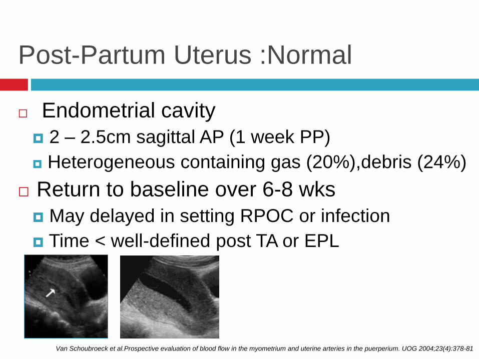
Post-Partum Uterus :Normal
Endometrial cavity
2 – 2.5cm sagittal AP (1 week PP)
Heterogeneous containing gas (20%),debris (24%)
Return to baseline over 6-8 wks
May delayed in setting RPOC or infection
Time < well-defined post TA or EPL
Van Schoubroeck et al.Prospective evaluation of blood flow in the myometrium and uterine arteries in the puerperium. UOG 2004;23(4):378-81
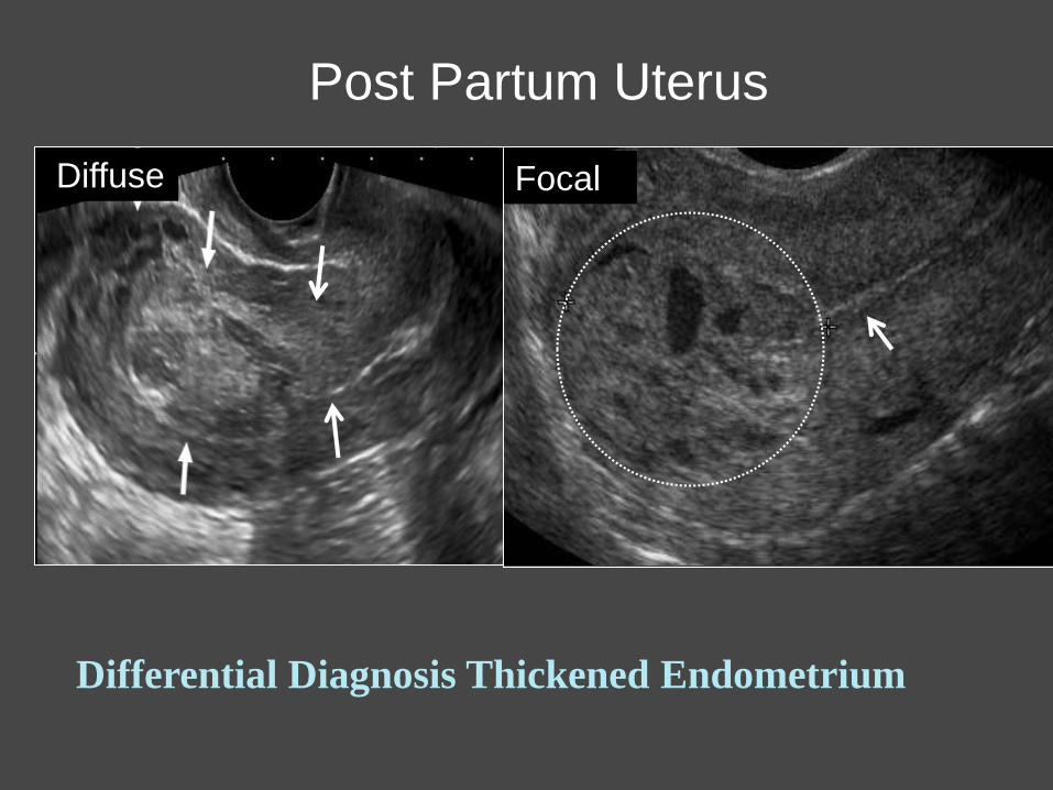
Post Partum Uterus
Differential Diagnosis Thickened Endometrium
Diffuse Focal
Differential Diagnosis Thickened Endometrium
Normal < 2-2.5cm
20% foci gas, 24% debris
Blood clots
RPOC
Subinvolution trophoblastic tissue
Endometritis – (Myometritis)
Gestational trophoblast disease
(GTD)
Differential diagnosis
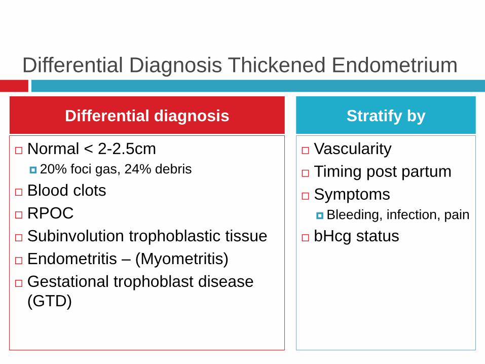
Differential Diagnosis Thickened Endometrium
Normal < 2-2.5cm
20% foci gas, 24% debris
Blood clots
RPOC
Subinvolution trophoblastic tissue
Endometritis – (Myometritis)
Gestational trophoblast disease
(GTD)
Vascularity
Timing post partum
Symptoms
Bleeding, infection, pain
bHcg status
Differential diagnosis Stratify by
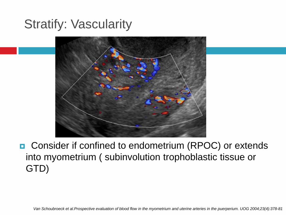
Stratify: Vascularity
Consider if confined to endometrium (RPOC) or extends
into myometrium ( subinvolution trophoblastic tissue or
GTD)
Van Schoubroeck et al.Prospective evaluation of blood flow in the myometrium and uterine arteries in the puerperium. UOG 2004;23(4):378-81
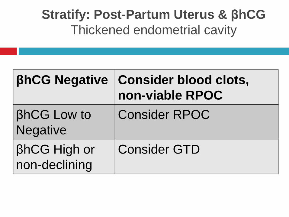
Stratify: Post-Partum Uterus & βhCG
Thickened endometrial cavity
βhCG Negative Consider blood clots,
non-viable RPOC
βhCG Low to
Negative
Consider RPOC
βhCG High or
non-declining
Consider GTD
Retained Products of Conception(RPOC)
RPOC implies incomplete uterine evacuation 2nd commonest etiology PPH after uterine atony
Risk Increased in setting: Late pregnancy termination or loss Uterine atony Placental attachment disorders, succenturiate lobe
Ultrasound can readily exclude retained products when the endometrial cavity is thin (< 2 mm), or contains a small amount of fluid. An echogenic intracavitary mass suggestive of RPOC but can be
misleading in as many as 1/3 cases Calcification favor RPOC
Diagnosis unequivocal: Fetal parts or placenta
Suspicious: Endometrial mass +/- vascularity
Absent CDS not exclude RPOC
Non-viable or necrotic tissue
Natural history of non-vascularized RPOC uncertain but
believe majority pass spontaneously
Specific diagnosis histological proof chorionic vlli
*van den Bosch et al Occurrence and outcome of residual trophoblastic tissue JUM. 2008;27(3):357-61.
Retained Products of Conception (RPOC)
*van den Bosch et al Occurrence and outcome of residual trophoblastic tissue JUM. 2008;27(3):357-61.
Retained Products of Conception (RPOC)
Stratify: Bleeding
Post-Partum Hemorrhage (PPH)
Early (< 24 hours) vs Delayed (> 24 hours)
> 500 ml
1-2% all deliveries
Leading cause maternal mortality
Clinical diagnosis
Knight et al. Trends in PPH in high resource countries: a review and recommendations from the International PPH Collaborative Group. BMC Pregnancy Childbirth. 2009;9:55. Rossen J et al. Is there an increase
PPH, and is severe hemorrhage associated with more frequent use of obstetric interventions? Acta Obstet Gynecol Scand. 2010;89(10):1248-55. Say L et al. WHO systematic review of maternal morbidity and
mortality: the prevalence of severe acute maternal morbidity (near miss). Reprod Health. 2004;1(1):3. Menacker F et al. Recent trends in cesarean delivery in the United States. NCHS Data Brief. 2010(35):1-8.;
Mulic-Lutvica A et al O. US evaluation of the uterus and uterine cavity after normal, vaginal delivery. UOG. 2001;18(5):491-8.
Post-Partum Hemorrhage (PPH)
Etiology
Commonest is uterine atony ( early PPH)
RPOC 2nd commonest etiology
Also increased risk :
Post C-sections (1/3 deliveries are Csect USA)
> rate & variety PPH and infections
Late terminations or pregnancy loss
Placental attachment disorders
Knight et al. Trends in PPH in high resource countries: a review and recommendations from the International PPH Collaborative Group. BMC Pregnancy Childbirth. 2009;9:55. Rossen J et al. Is there an increase
PPH, and is severe hemorrhage associated with more frequent use of obstetric interventions? Acta Obstet Gynecol Scand. 2010;89(10):1248-55. Say L et al. WHO systematic review of maternal morbidity and
mortality: the prevalence of severe acute maternal morbidity (near miss). Reprod Health. 2004;1(1):3. Menacker F et al. Recent trends in cesarean delivery in the United States. NCHS Data Brief. 2010(35):1-8.;
Mulic-Lutvica A et al O. US evaluation of the uterus and uterine cavity after normal, vaginal delivery. UOG. 2001;18(5):491-8.
PPH: Uterine Atony
Commonest etiology early PPH
Clinical diagnosis - suboptimal uterine contraction
Risk factors:
Overdistension uterus ( polyhydramnios, multips)
Uterine relaxants like magnesium sulphate
Abnormal placentation with adherent placenta.
If uterotonic agents ineffective consider non-
permanent embolic agents to gain control over PPH
in short-term
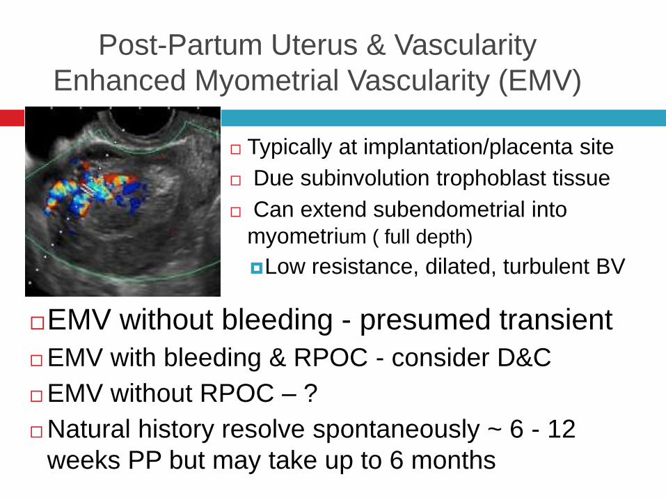
Post-Partum Uterus & Vascularity
Enhanced Myometrial Vascularity (EMV)
Typically at implantation/placenta site
Due subinvolution trophoblast tissue
Can extend subendometrial into
myometrium ( full depth)
Low resistance, dilated, turbulent BV
EMV without bleeding - presumed transient
EMV with bleeding & RPOC - consider D&C
EMV without RPOC – ?
Natural history resolve spontaneously ~ 6 - 12
weeks PP but may take up to 6 months
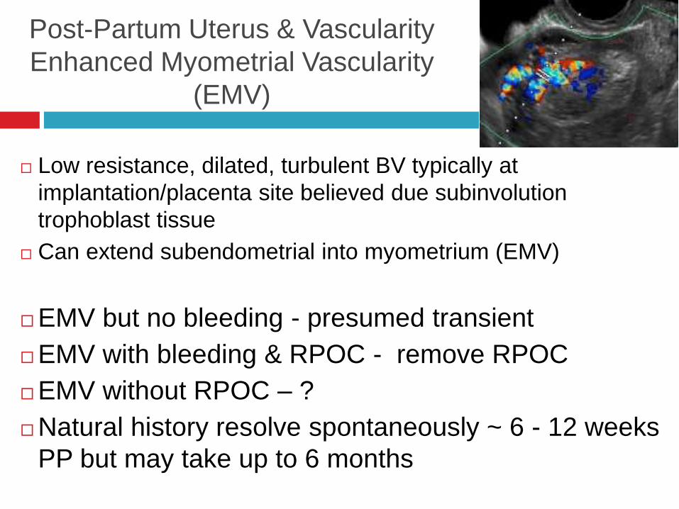
Post-Partum Uterus & Vascularity
Enhanced Myometrial Vascularity
(EMV)
Low resistance, dilated, turbulent BV typically at
implantation/placenta site believed due subinvolution
trophoblast tissue
Can extend subendometrial into myometrium (EMV)
EMV but no bleeding - presumed transient
EMV with bleeding & RPOC - remove RPOC
EMV without RPOC – ?
Natural history resolve spontaneously ~ 6 - 12 weeks
PP but may take up to 6 months
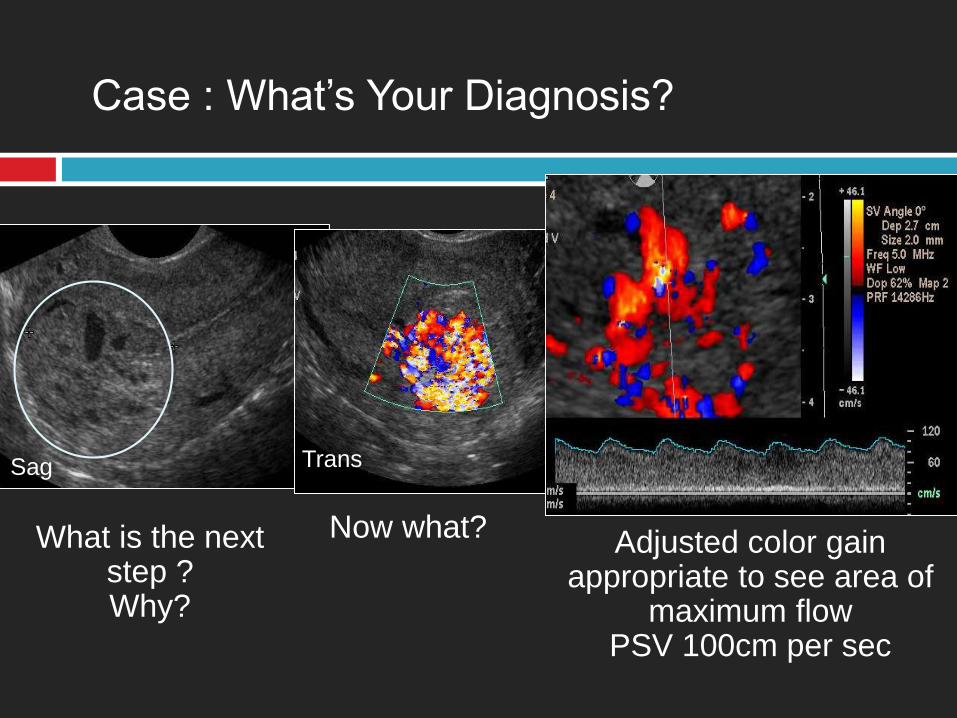
Case : What’s Your Diagnosis?
Adjusted color gain appropriate to see area of
maximum flowPSV 100cm per sec
What is the next step ?Why?
Sag
Now what?
Trans
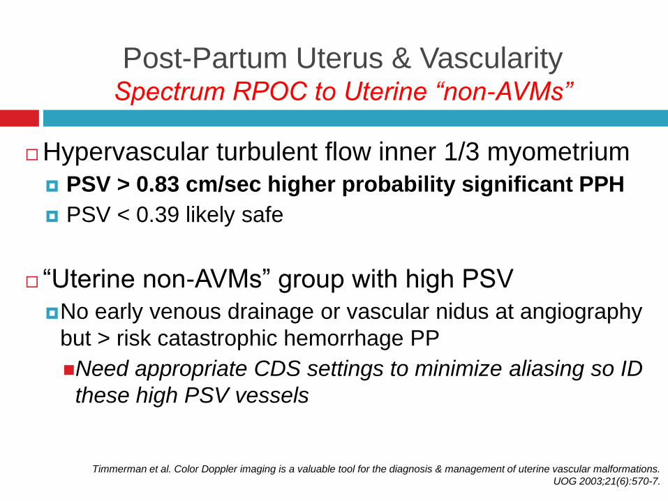
Post-Partum Uterus & VascularitySpectrum RPOC to Uterine “non-AVMs”
Hypervascular turbulent flow inner 1/3 myometrium
PSV > 0.83 cm/sec higher probability significant PPH
PSV < 0.39 likely safe
“Uterine non-AVMs” group with high PSV
No early venous drainage or vascular nidus at angiography
but > risk catastrophic hemorrhage PP
Need appropriate CDS settings to minimize aliasing so ID
these high PSV vessels
Timmerman et al. Color Doppler imaging is a valuable tool for the diagnosis & management of uterine vascular malformations.
UOG 2003;21(6):570-7.
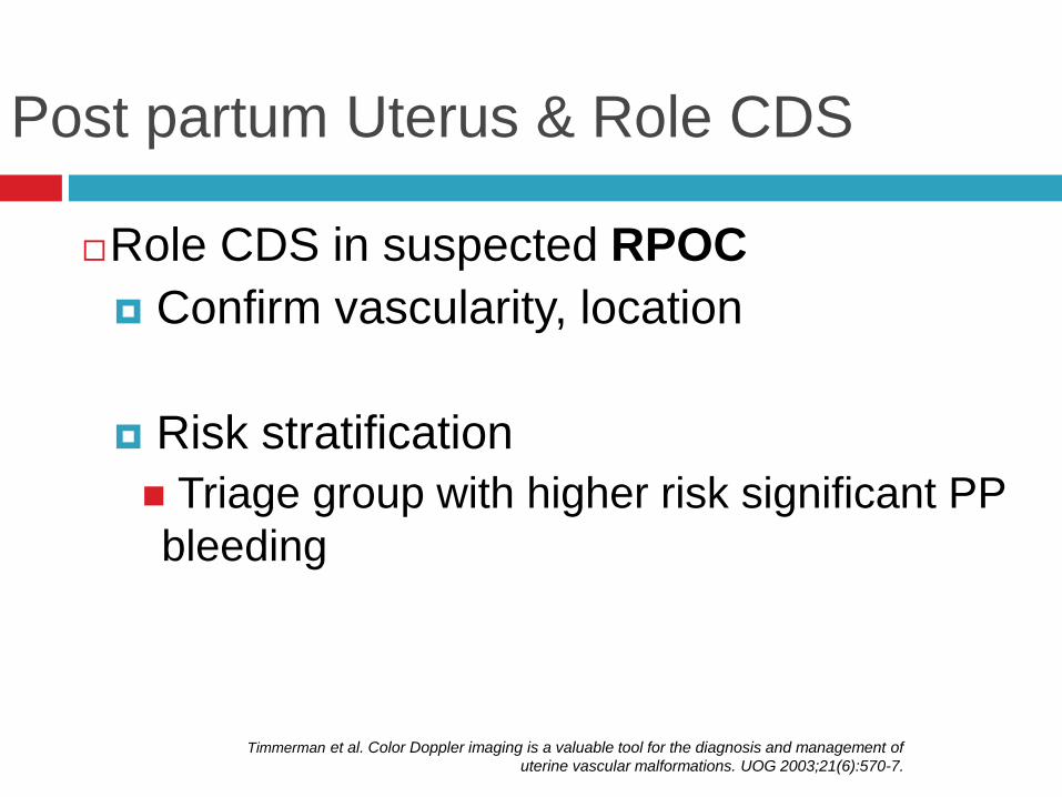
Role CDS in suspected RPOC
Confirm vascularity, location
Risk stratification
Triage group with higher risk significant PP
bleeding
Timmerman et al. Color Doppler imaging is a valuable tool for the diagnosis and management of
uterine vascular malformations. UOG 2003;21(6):570-7.
Post partum Uterus & Role CDS
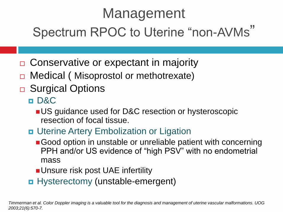
Conservative or expectant in majority
Medical ( Misoprostol or methotrexate)
Surgical Options D&C
US guidance used for D&C resection or hysteroscopic resection of focal tissue.
Uterine Artery Embolization or Ligation
Good option in unstable or unreliable patient with concerning PPH and/or US evidence of “high PSV” with no endometrial mass
Unsure risk post UAE infertility
Hysterectomy (unstable-emergent)
Timmerman et al. Color Doppler imaging is a valuable tool for the diagnosis and management of uterine vascular malformations. UOG
2003;21(6):570-7.
Management
Spectrum RPOC to Uterine “non-AVMs”
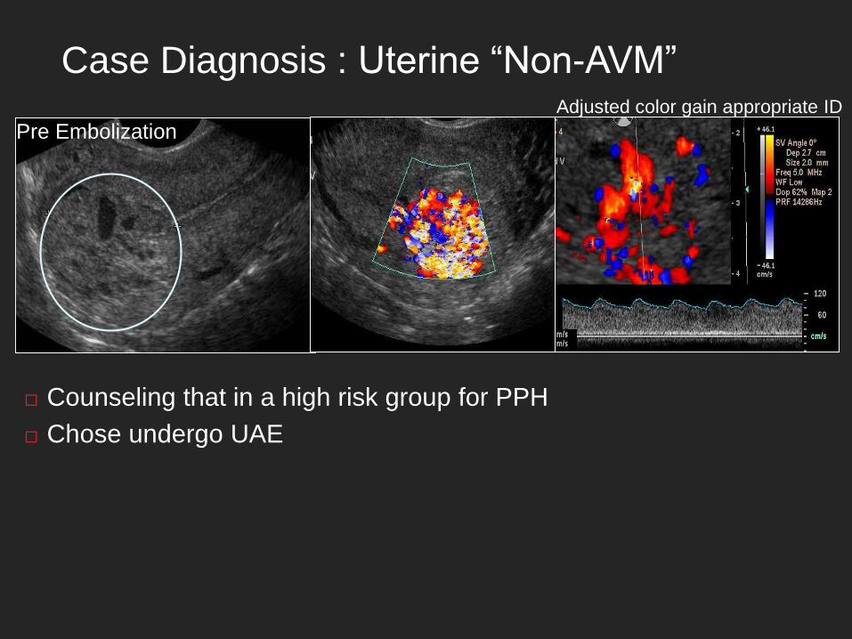
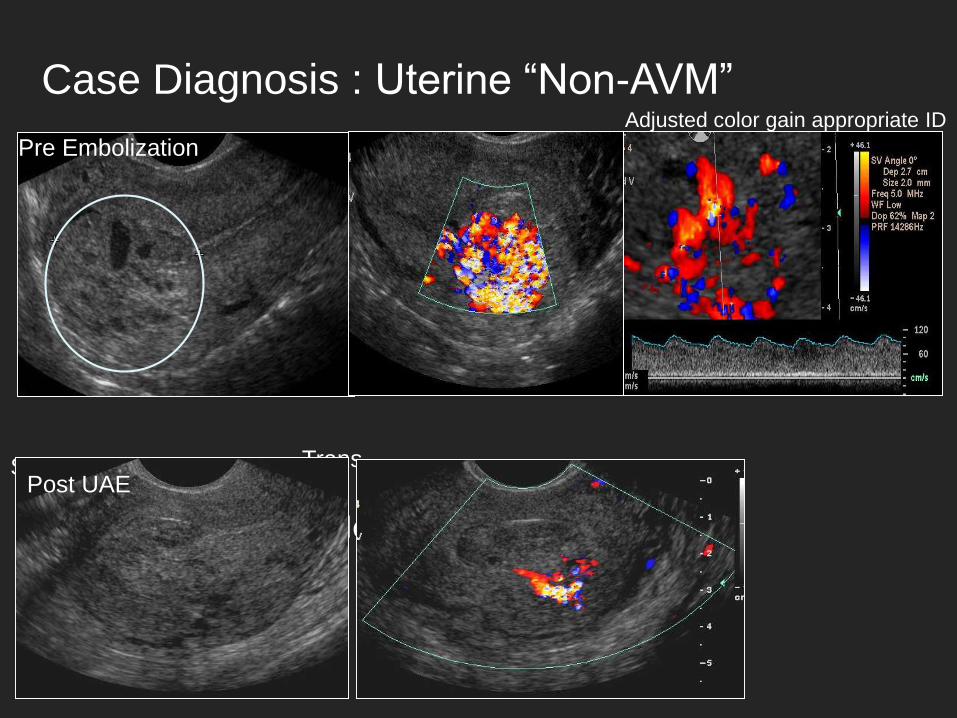
Case Diagnosis : Uterine “Non-AVM”
Counseling that in a high risk group for PPH
Chose undergo UAE
Adjusted color gain appropriate ID maximum PSV 100cm/secPre Embolization
Case Diagnosis : Uterine “Non-AVM”Adjusted color gain appropriate ID
maximum PSV 100cm/sec
What is the next step ?Why?
Sag
Now what?
Trans
Pre Embolization
Post UAE
Case
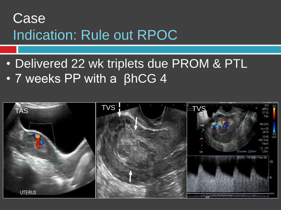
Indication: Rule out RPOC
• Delivered 22 wk triplets due PROM & PTL
• 7 weeks PP with a βhCG 4
TAS TVS TVS
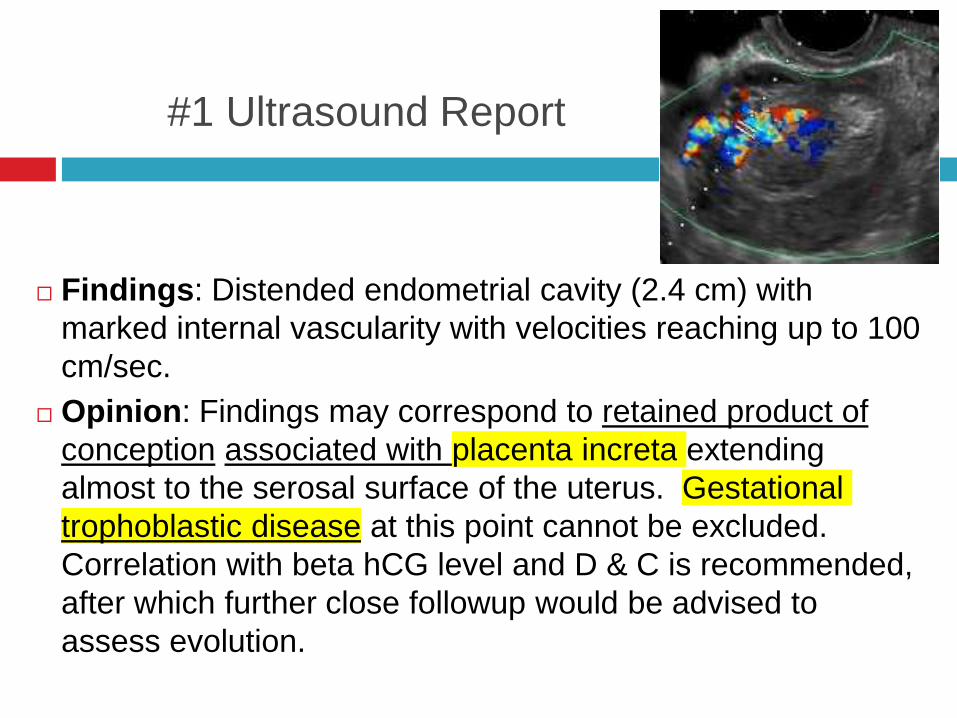
#1 Ultrasound Report
Findings: Distended endometrial cavity (2.4 cm) with
marked internal vascularity with velocities reaching up to 100
cm/sec.
Opinion: Findings may correspond to retained product of
conception associated with placenta increta extending
almost to the serosal surface of the uterus. Gestational
trophoblastic disease at this point cannot be excluded.
Correlation with beta hCG level and D & C is recommended,
after which further close followup would be advised to
assess evolution.
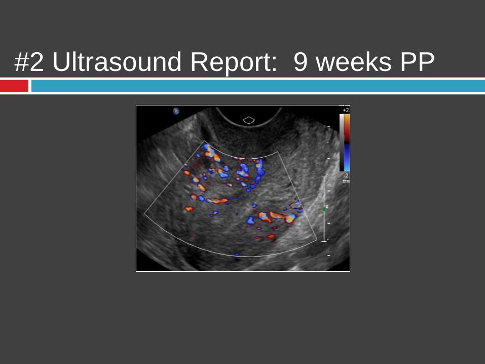
#2 Ultrasound Report: 9 weeks PP
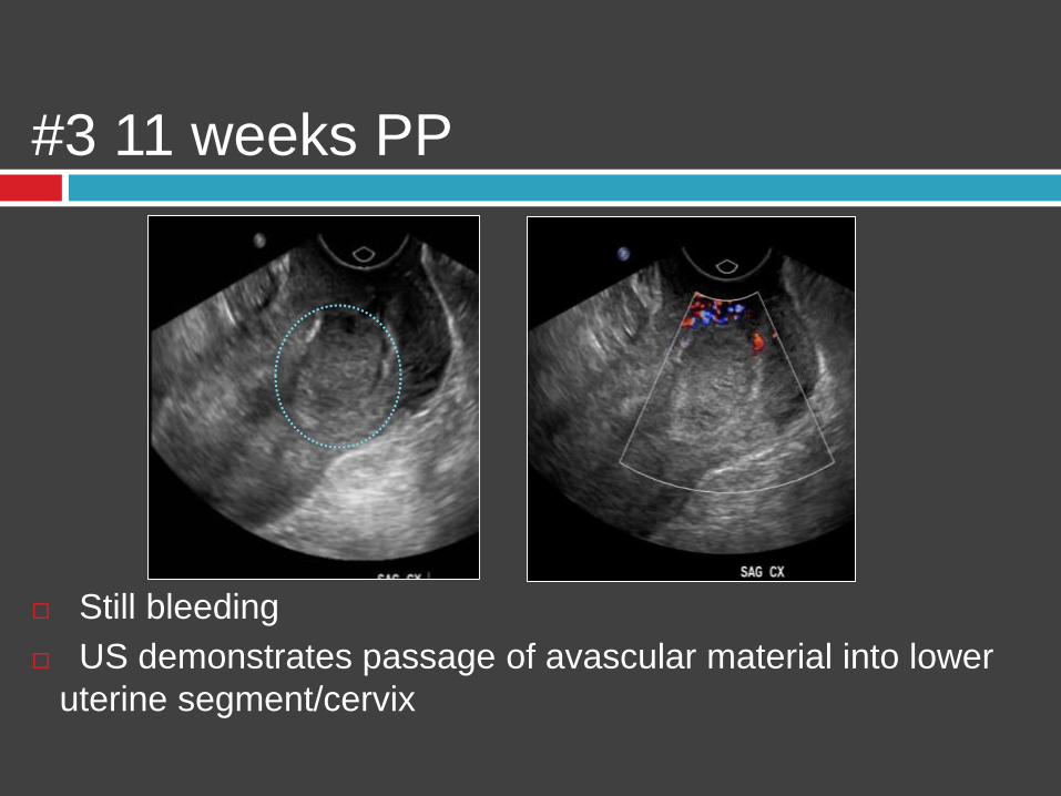
#3 11 weeks PP
Still bleeding
US demonstrates passage of avascular material into lower
uterine segment/cervix
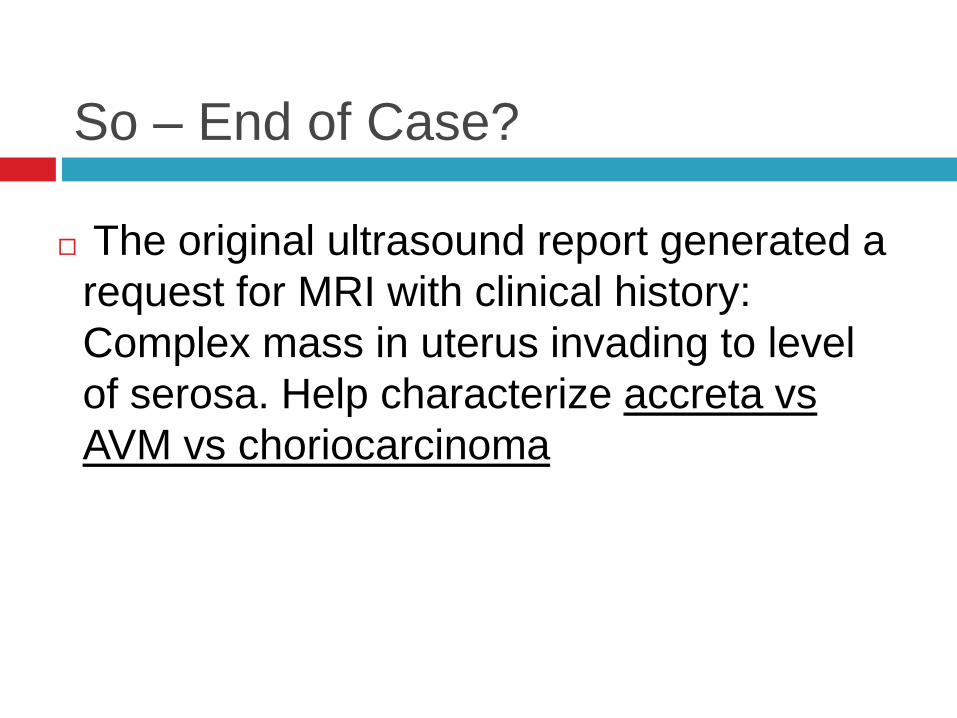
So – End of Case?
The original ultrasound report generated a
request for MRI with clinical history:
Complex mass in uterus invading to level
of serosa. Help characterize accreta vs
AVM vs choriocarcinoma
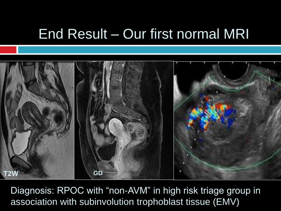
End Result – Our first normal MRI
T2W GD
Diagnosis: RPOC with “non-AVM” in high risk triage group in
association with subinvolution trophoblast tissue (EMV)
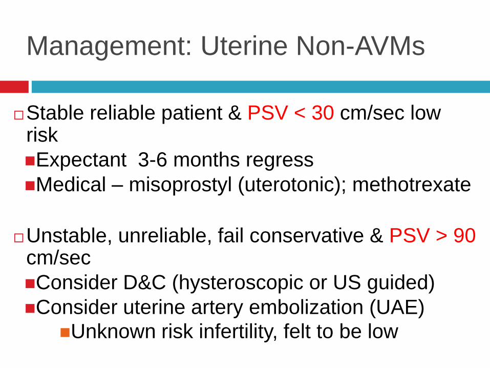
Management: Uterine Non-AVMs
Stable reliable patient & PSV < 30 cm/sec low risk
Expectant 3-6 months regress
Medical – misoprostyl (uterotonic); methotrexate
Unstable, unreliable, fail conservative & PSV > 90 cm/sec
Consider D&C (hysteroscopic or US guided)
Consider uterine artery embolization (UAE)
Unknown risk infertility, felt to be low
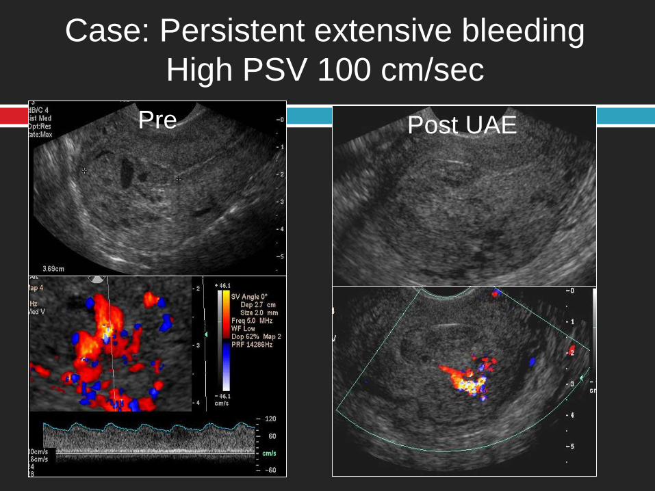
Case: Persistent extensive bleeding
High PSV 100 cm/sec
Post UAEPre
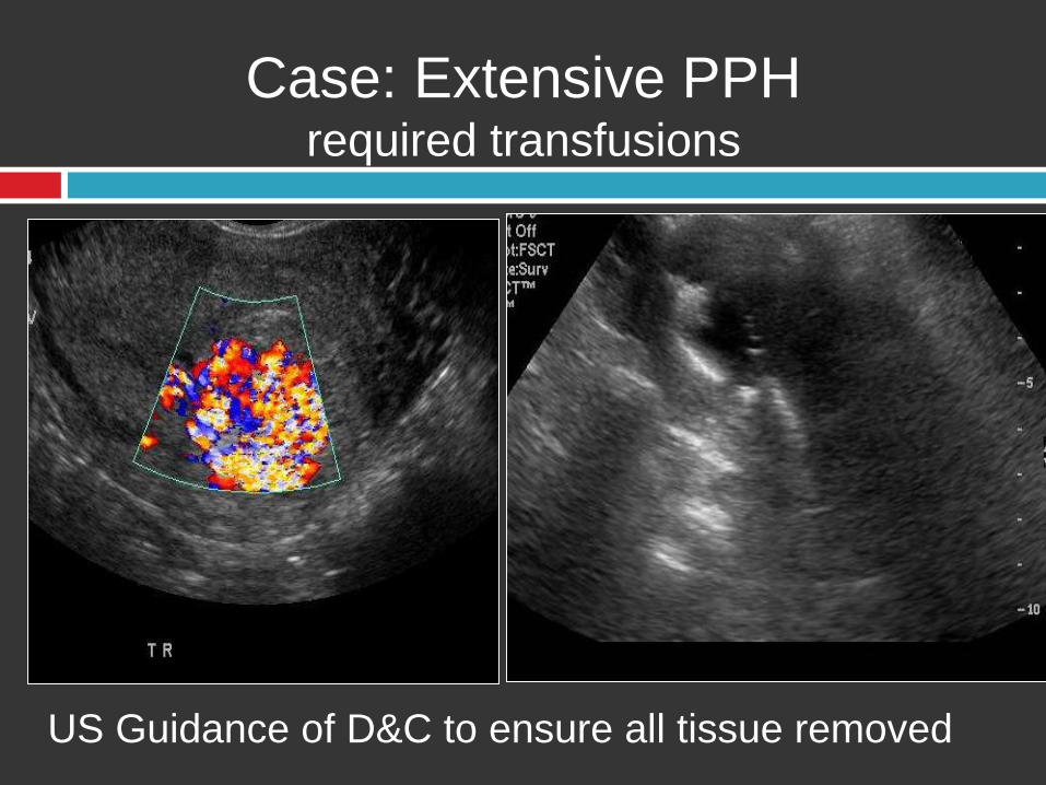
Case: Extensive PPH required transfusions
US Guidance of D&C to ensure all tissue removed
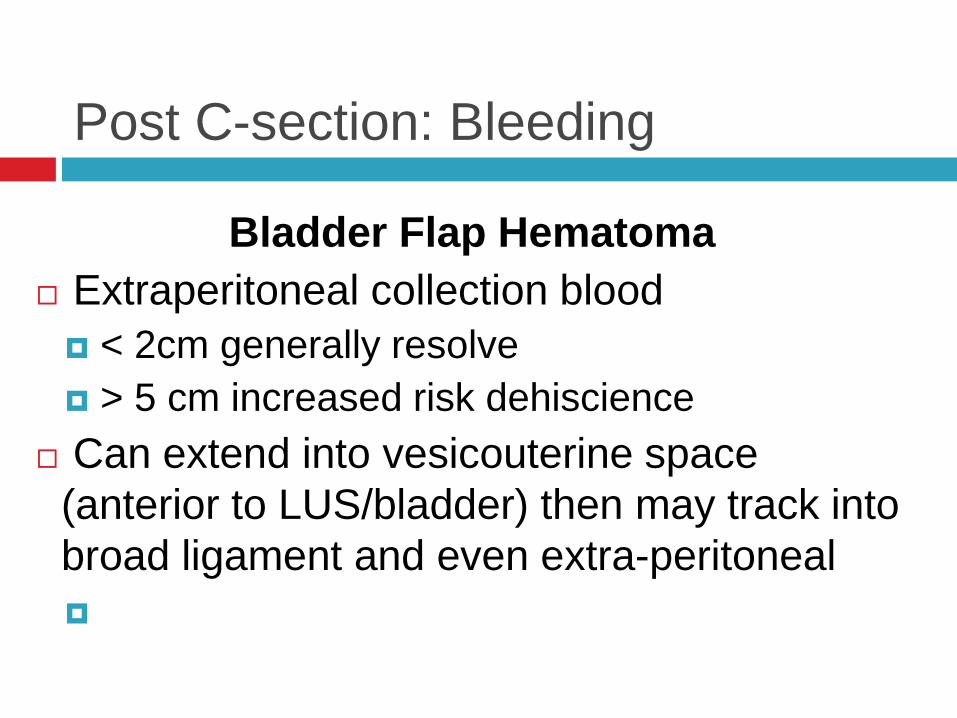
Post C-section: Bleeding
Bladder Flap Hematoma
Extraperitoneal collection blood
< 2cm generally resolve
> 5 cm increased risk dehiscience
Can extend into vesicouterine space
(anterior to LUS/bladder) then may track into
broad ligament and even extra-peritoneal
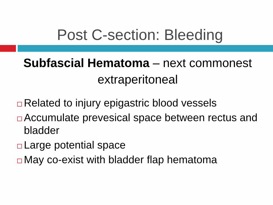
Post C-section: Bleeding
Subfascial Hematoma – next commonest
extraperitoneal
Related to injury epigastric blood vessels
Accumulate prevesical space between rectus and
bladder
Large potential space
May co-exist with bladder flap hematoma
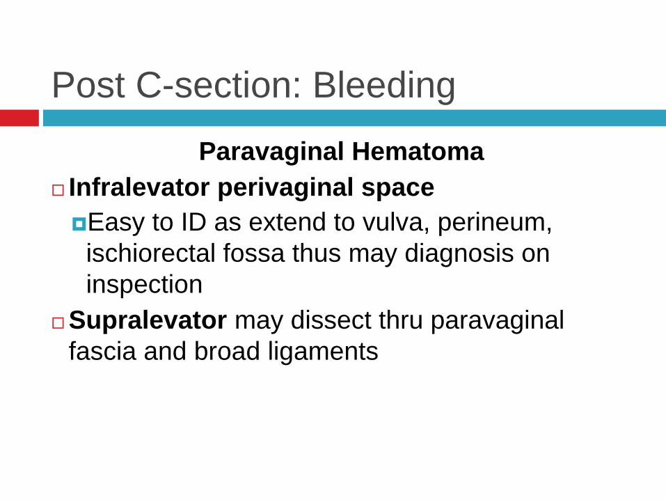
Post C-section: Bleeding
Paravaginal Hematoma
Infralevator perivaginal space
Easy to ID as extend to vulva, perineum,
ischiorectal fossa thus may diagnosis on
inspection
Supralevator may dissect thru paravaginal
fascia and broad ligaments
Post C-section: Bleeding
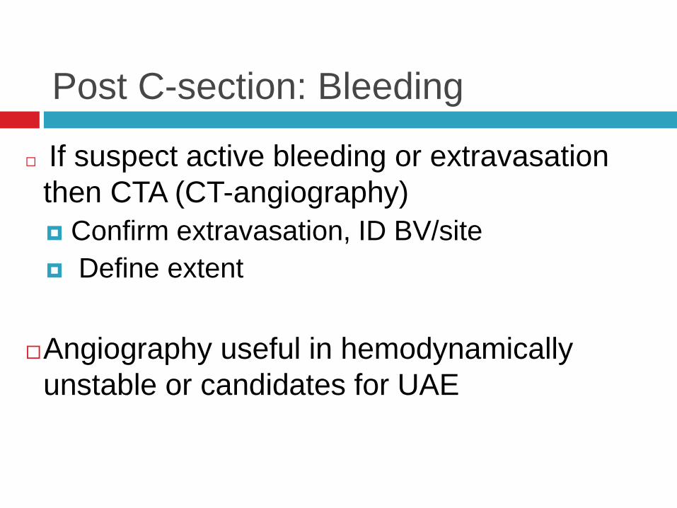
If suspect active bleeding or extravasation
then CTA (CT-angiography)
Confirm extravasation, ID BV/site
Define extent
Angiography useful in hemodynamically
unstable or candidates for UAE
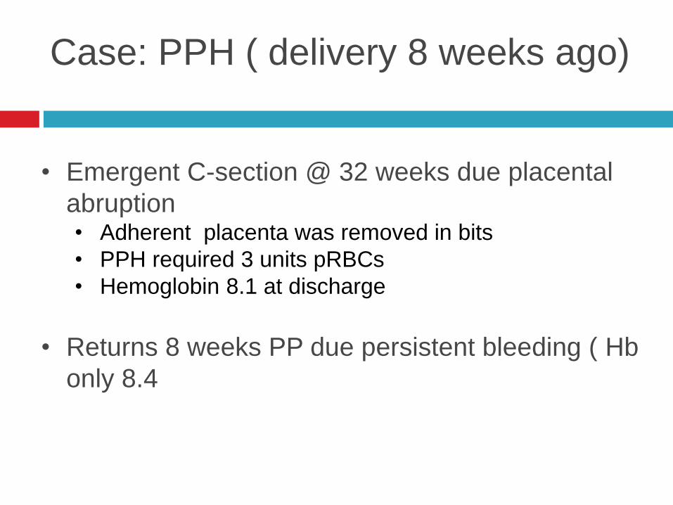
Case: PPH ( delivery 8 weeks ago)
• Emergent C-section @ 32 weeks due placental
abruption• Adherent placenta was removed in bits
• PPH required 3 units pRBCs
• Hemoglobin 8.1 at discharge
• Returns 8 weeks PP due persistent bleeding ( Hb
only 8.4

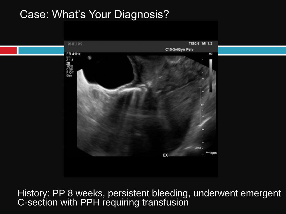
Case: What’s Your Diagnosis?
History: PP 8 weeks, persistent bleeding, underwent emergent C-section with PPH requiring transfusion
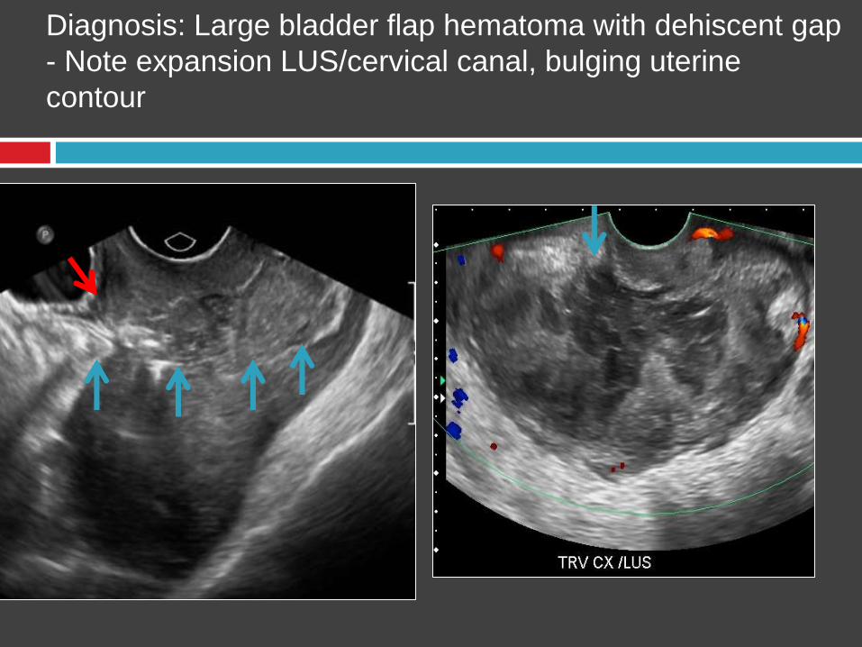
Diagnosis: Large bladder flap hematoma with dehiscent gap
- Note expansion LUS/cervical canal, bulging uterine
contour
Case: What’s Your Diagnosis?
History: PP 8 weeks, persistent bleeding, underwent emergent C-section with PPH requiring transfusion
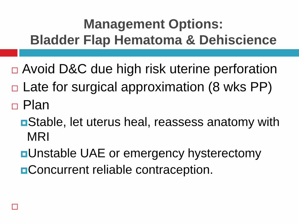
Management Options:
Bladder Flap Hematoma & Dehiscience
Avoid D&C due high risk uterine perforation
Late for surgical approximation (8 wks PP)
Plan
Stable, let uterus heal, reassess anatomy with
MRI
Unstable UAE or emergency hysterectomy
Concurrent reliable contraception.
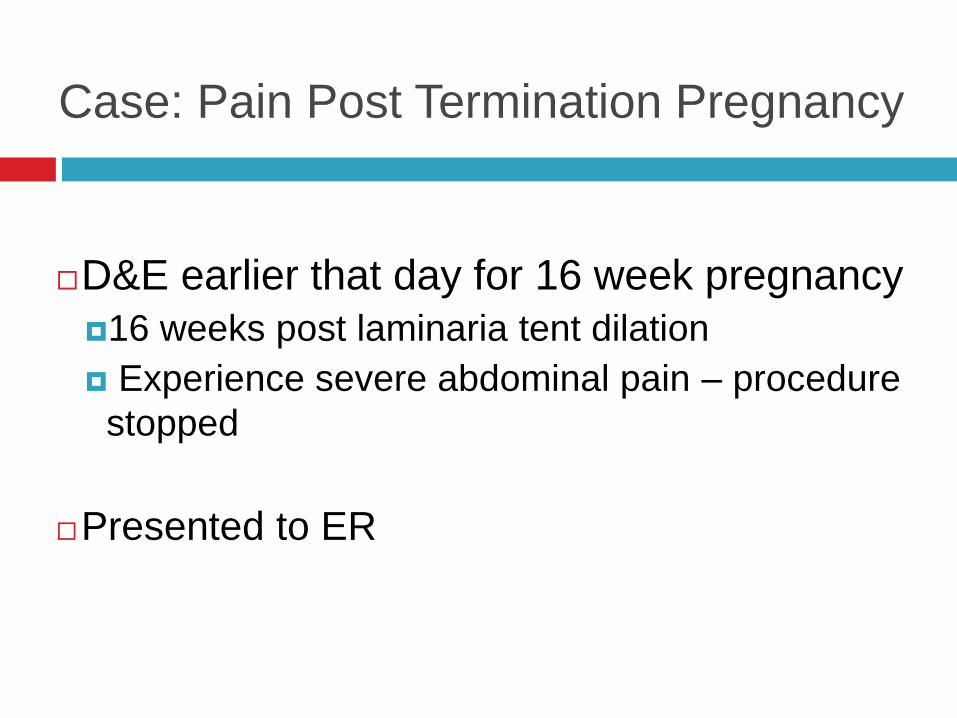
Case: Pain Post Termination Pregnancy
D&E earlier that day for 16 week pregnancy
16 weeks post laminaria tent dilation
Experience severe abdominal pain – procedure
stopped
Presented to ER
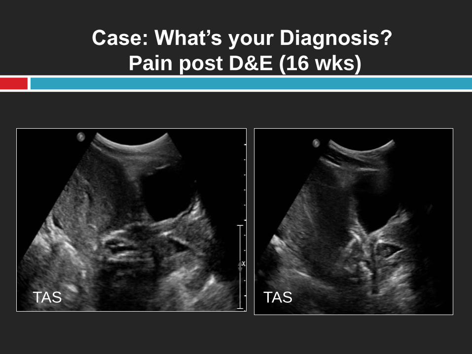
Case: What’s your Diagnosis?
Pain post D&E (16 wks)
TAS TAS
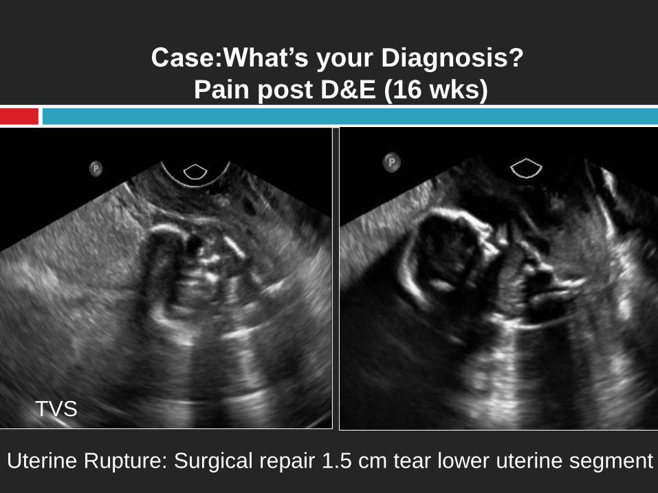
Case:What’s your Diagnosis?
Pain post D&E (16 wks)
TVS
Uterine Rupture: Surgical repair 1.5 cm tear lower uterine segment
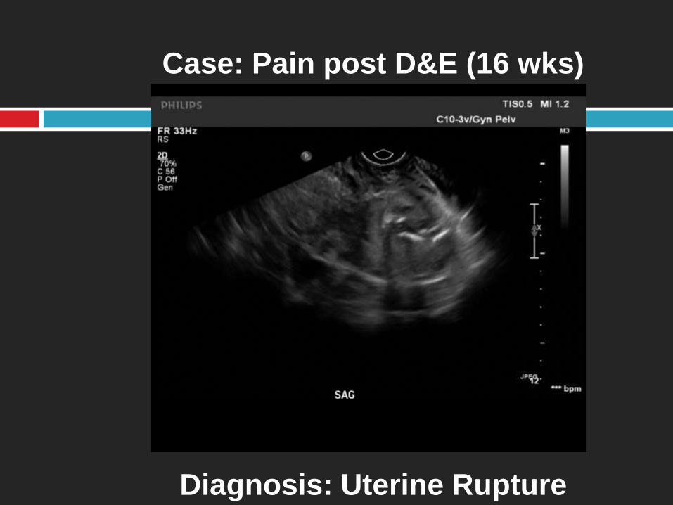
Case: Pain post D&E (16 wks)
Diagnosis: Uterine Rupture

Dehiscience & Rupture
Risk factors
**Prior C-section
Bladder flap hematoma > 5cm
Endometritis
Terminations – late – D&E
PAD (Placental adherence disorder)
GTD
Delayed dehiscience may be related to
inadequate treatment PP endometritis or infected
RPOC
PP/Post C-section: Rupture-Dehiscience
Rare but high morbidity/mortalityPartial implies intact serosa vs complete tear extend
thru serosa
Tends to occur in relatively avascular LUS
Classic signs include severe pain, PPH, hypovolemic shock
Treat vary antibiotics to surgical repair
Concurrent counsel risk future pregnancy and interim use contraceptives
PP/Post C-section: Dehiscience
Thus careful US evaluation for uterine wall integrity
indicated if bladder flap hematoma> 2cm
US, CT, MR all valuable to assess for discontinuity
serosal and/or myometrial layers and blood trackingUS appear normal, subtle thin, frank disruption with extrusion fetal parts
beyond endometrium or bowel loops into myometrium
CT defect enhancement myometrium but phlegmon/defect may appear
similar unless frank disruption.
MR superior demonstrate transmural defect, non-enhancing myometrium
of connection endometrial cavity to serosal surface, lack apposition
endometrium & serosa at incision site
Infection in PP state or Puerperal
Sepsis
1-5% vaginal deliveries vs 5-30% C-sections
Endometritis commonest cause PP fevers
Typically empiric antibiotics without imaging
Small % myometrial involvement
> risk in obese, post Csection, RPOC
Rare uterus rupture - infection & necrosis
US findings generally non-specific
MRI most sensitive distinguish
phlegmon/abscess from true dehiscience
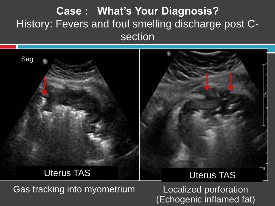
Case : What’s Your Diagnosis?
History: Fevers and foul smelling discharge post C-
section
Uterus TAS
Sag
Localized perforation(Echogenic inflamed fat)
Gas tracking into myometrium
Uterus TAS
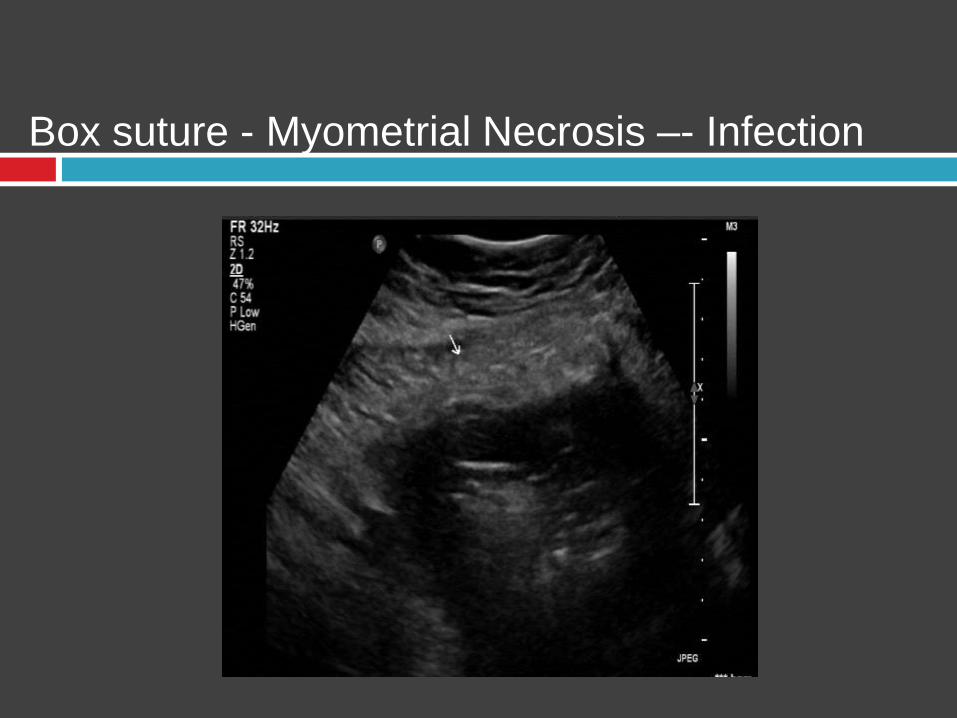
Box suture - Myometrial Necrosis –- Infection
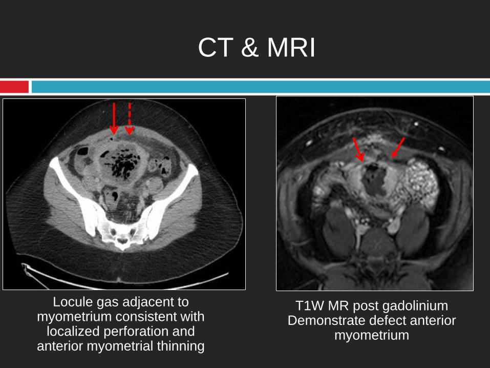
CT & MRI
Locule gas adjacent to myometrium consistent with
localized perforation and anterior myometrial thinning
T1W MR post gadoliniumDemonstrate defect anterior
myometrium
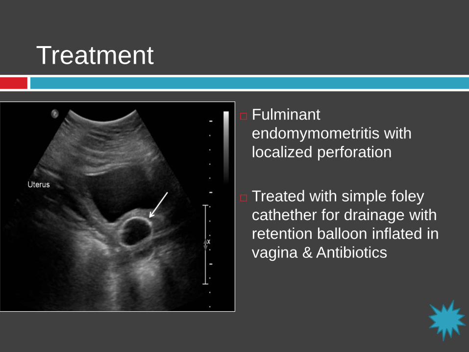
Fulminant
endomymometritis with
localized perforation
Treated with simple foley
cathether for drainage with
retention balloon inflated in
vagina & Antibiotics
Increased risk obese, RPOC, post csection
Treatment

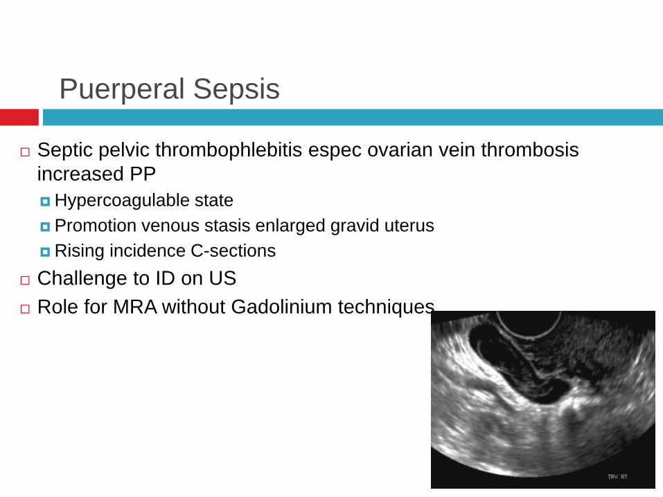
Puerperal Sepsis
Septic pelvic thrombophlebitis espec ovarian vein thrombosis
increased PP
Hypercoagulable state
Promotion venous stasis enlarged gravid uterus
Rising incidence C-sections
Challenge to ID on US
Role for MRA without Gadolinium techniques
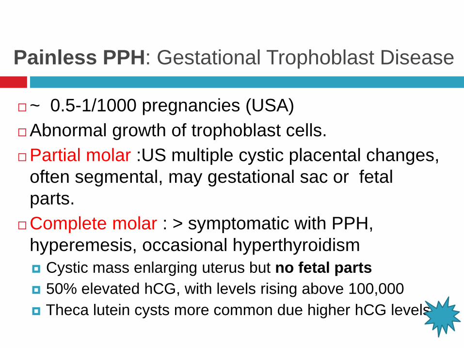
Painless PPH: Gestational Trophoblast Disease
~ 0.5-1/1000 pregnancies (USA)
Abnormal growth of trophoblast cells.
Partial molar :US multiple cystic placental changes,
often segmental, may gestational sac or fetal
parts.
Complete molar : > symptomatic with PPH,
hyperemesis, occasional hyperthyroidism
Cystic mass enlarging uterus but no fetal parts
50% elevated hCG, with levels rising above 100,000
Theca lutein cysts more common due higher hCG levels
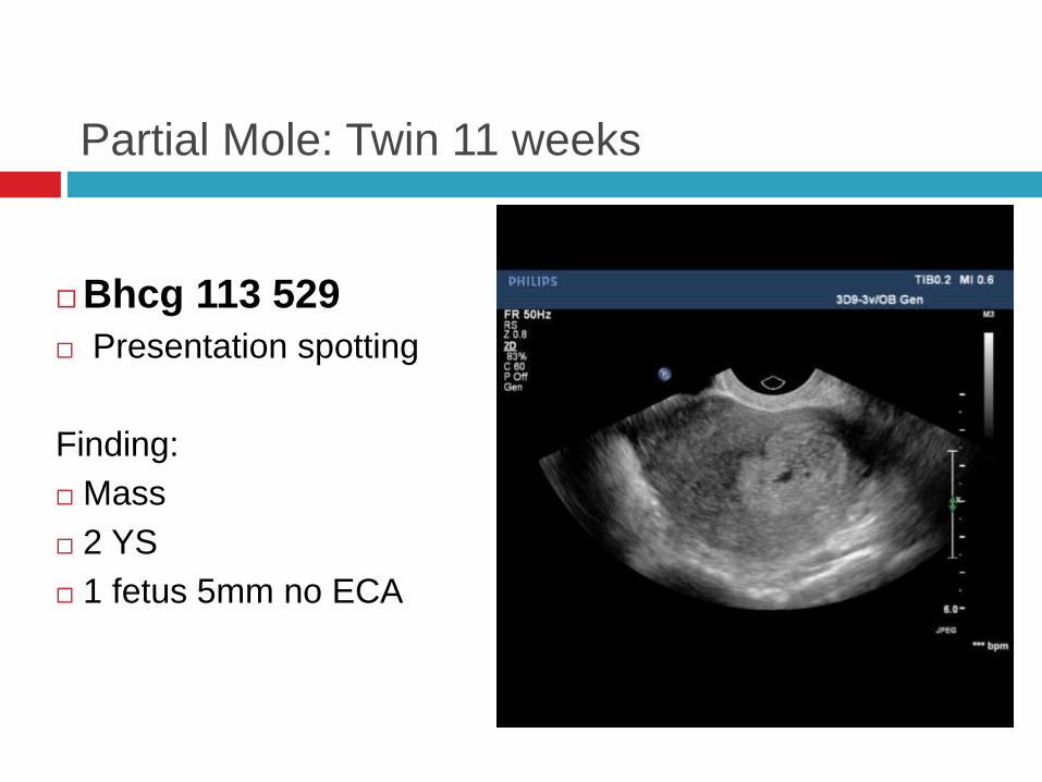
Partial Mole: Twin 11 weeks
Bhcg 113 529
Presentation spotting
Finding:
Mass
2 YS
1 fetus 5mm no ECA
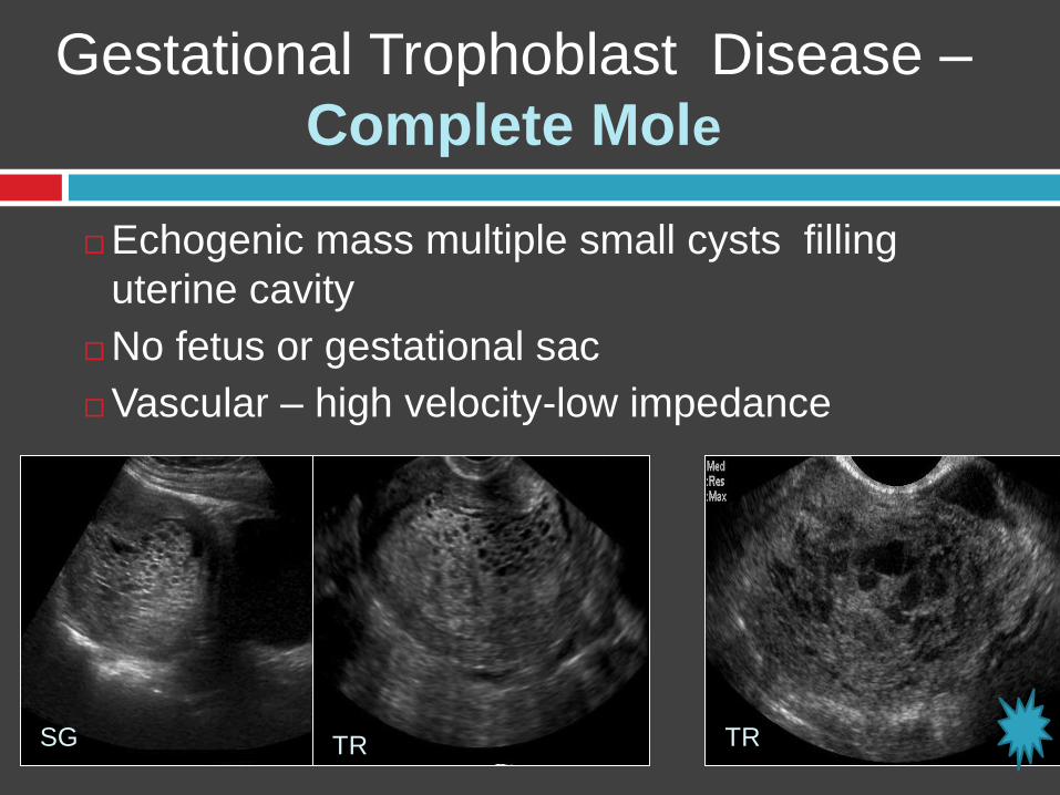
Echogenic mass multiple small cysts filling
uterine cavity
No fetus or gestational sac
Vascular – high velocity-low impedance
Gestational Trophoblast Disease –
Complete Mole
SG TR TR
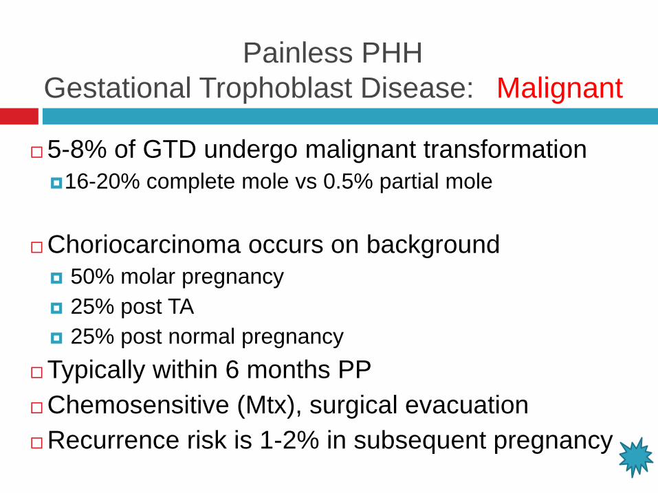
Painless PHH
Gestational Trophoblast Disease: Malignant
5-8% of GTD undergo malignant transformation
16-20% complete mole vs 0.5% partial mole
Choriocarcinoma occurs on background
50% molar pregnancy
25% post TA
25% post normal pregnancy
Typically within 6 months PP
Chemosensitive (Mtx), surgical evacuation
Recurrence risk is 1-2% in subsequent pregnancy

Gestational Trophoblast DiseaseGTN - Malignant
FIGO standard diagnosis:1. hCG level plateau plus or minus 10% of
baseline recorded in 4 measurements over a 3-week duration (days 1, 7, 14, 21)
2. hCG ≥ 10% rise in 3 consecutive measurements recorded over a 2 week duration(days 1,7,14)
3. Persistence of detectable hCG for more than 6 months after molar evacuation.
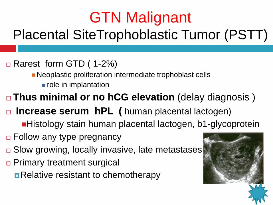
GTN MalignantPlacental SiteTrophoblastic Tumor (PSTT)
Rarest form GTD ( 1-2%)Neoplastic proliferation intermediate trophoblast cells
role in implantation
Thus minimal or no hCG elevation (delay diagnosis )
Increase serum hPL ( human placental lactogen)
Histology stain human placental lactogen, b1-glycoprotein
Follow any type pregnancy
Slow growing, locally invasive, late metastases
Primary treatment surgical
Relative resistant to chemotherapy
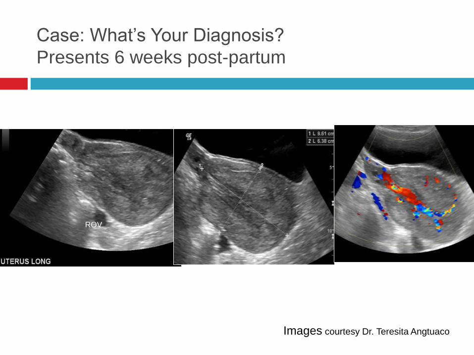
Case: What’s Your Diagnosis?
Presents 6 weeks post-partum
Images courtesy Dr. Teresita Angtuaco
ROV
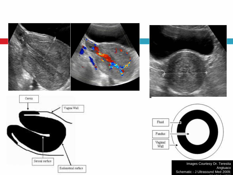
Uterine Inversion
Images Courtesy Dr. Teresita
Angtuaco
Schematic - J Ultrasound Med 2009;
28:1719–1722
Uterine Inversion
Uncommon but important cause PP pain
Attribute excess traction UC in fundal placenta
1/30,000
Easy to miss on US
Uterus projects into vagina thus in sagittal the
transducer abuts cervix rather than fundus
If complete the uterus will protrude thru cervix.
Summary
Knowledge of the early and late complications of
bleeding and pain in the postpartum period can
improve patient care by narrowing or specifying a
diagnosis.
Prompt diagnosis may be life-saving.
Role of CDS in RPOC can be diagnostic and
may potentially stratify patients into low and high
risk categories for potential significant PPH.

Phyllis.Glanc @Sunnybrook.ca
Thank you
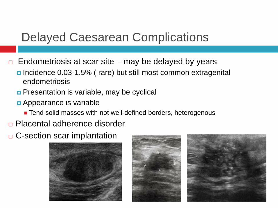
Delayed Caesarean Complications
Endometriosis at scar site – may be delayed by years
Incidence 0.03-1.5% ( rare) but still most common extragenital
endometriosis
Presentation is variable, may be cyclical
Appearance is variable
Tend solid masses with not well-defined borders, heterogenous
Placental adherence disorder
C-section scar implantation
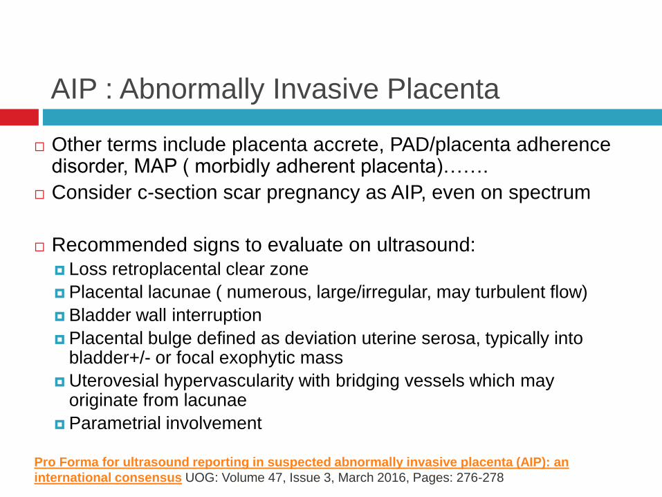
AIP : Abnormally Invasive Placenta
Other terms include placenta accrete, PAD/placenta adherence disorder, MAP ( morbidly adherent placenta)…….
Consider c-section scar pregnancy as AIP, even on spectrum
Recommended signs to evaluate on ultrasound:
Loss retroplacental clear zone
Placental lacunae ( numerous, large/irregular, may turbulent flow)
Bladder wall interruption
Placental bulge defined as deviation uterine serosa, typically into bladder+/- or focal exophytic mass
Uterovesial hypervascularity with bridging vessels which may originate from lacunae
Parametrial involvement
Pro Forma for ultrasound reporting in suspected abnormally invasive placenta (AIP): an
international consensus UOG: Volume 47, Issue 3, March 2016, Pages: 276-278

Placenta Adherence Disorder
Delayed PP Complication post C-section
Sensitivity US vary from 30-90% literature whereas MR typical sensitivity specificity but gen not change management
Recommend dedicated evaluation placenta and LUS at time fetal anatomy study in any patient with prior history repeated C-section or placenta previa
US features: Multiple placental lacunae, thinned/distorted myometrium with loss
retroplacenta clear zone, irreg bladder-placenta interface Hypoechoic foci are abnormal BV clusters < well-defined borders than venous
lakes, may turbulent flow on CDS “Swiss cheese” or “moth-eaten” appearance.
MR features Buging of the placenta, dark intraplacental bands on T2W,
heterogeneous appearance
http://www.ajronline.org/doi/full/10.2214/AJR.12.9637
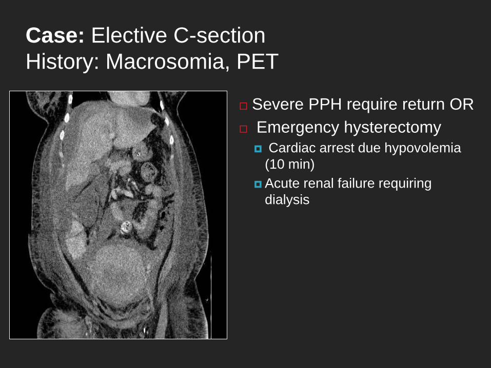
Case: Elective C-section
History: Macrosomia, PET
Severe PPH require return OR
Emergency hysterectomy
Cardiac arrest due hypovolemia
(10 min)
Acute renal failure requiring
dialysis
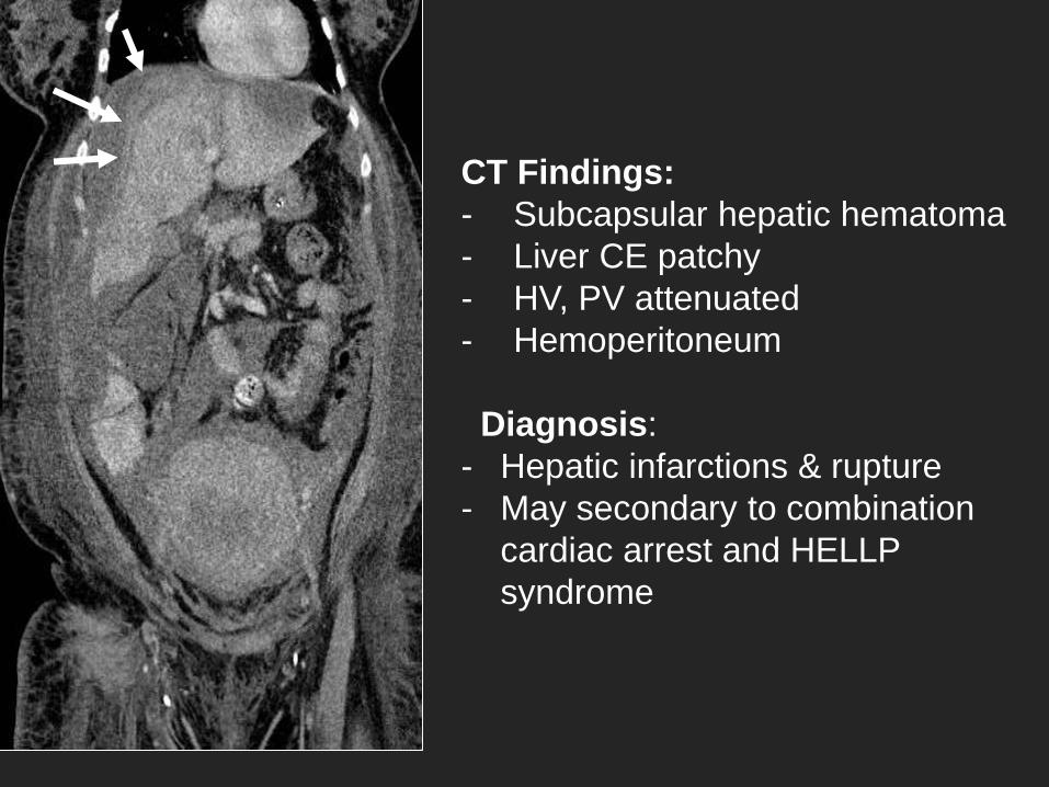
CT Findings:
- Subcapsular hepatic hematoma
- Liver CE patchy
- HV, PV attenuated
- Hemoperitoneum
Diagnosis:
- Hepatic infarctions & rupture
- May secondary to combination
cardiac arrest and HELLP
syndrome
Question 1: The uterus should return to baseline
pre-pregnancy appearance after delivery by?
A. 2-4weeks
B. 4-6 weeks
C. 6-8 weeks
D. 8-10 weeks
Question 1: The uterus should return to baseline
pre-pregnancy appearance after delivery by?
A. 2-4weeks
B. 4-6 weeks
C. 6-8 weeks
D. 8-10 weeks
Solution to Question 1:
The uterus will gradually return to the size and appearance of the
non-pregnant uterus by 6-8 weeks postpartum.Option D is the best
response.
Reference: Mulic-Lutvica A, Bekuretsion M, Bakos O, Axelsson O.
Ultrasonic evaluation of the uterus and uterine cavity after normal,
vaginal delivery. Ultrasound Obstet Gynecol. 2001;18(5):491-8.
Question 2. The most common type of post caesarian
section hematoma is which of the following?
A, Subfascial hematom.
B. Bladder flap hematoma.
C. Paravaginal hematoma
D. Broad ligament hematoma.
Question 2. The most common type of post caesarian
section hematoma is which of the following?
A, Subfascial hematom.
B. Bladder flap hematoma.
C. Paravaginal hematoma
D. Broad ligament hematoma.
Solution to Question 2:
The two most common post caesarian section hematomas are the bladder flap hematoma , followed by subfascial hematoma and then by paravaginal hematomas. The bladder flap hematoma may extend into the broad ligament. The typical size of a bladder flap hematoma is approxmimately2cm or less. When the hematoma is larger than 5 cm there is concern for the occasional occurrence of uterine dehiscence or even rarer rupture. Option B is the best response.
Reference: Baker ME, Bowie JD, Killam AP. Sonography of post-cesarean-section bladder-flap hematoma. AJR Am J Roentgenol. 1985;144(4):757-9.
Question 3: Subinvolution of the placenta
refers to which of the following?
A. Retained products of conception, specifically
placental tissue..
B. Areas of hypervascularity and turbulent flow
in the myomerium.
C. Areas of placental tissue in the
myometrium.
D, Placenta accreta or placental attachment
disorder.
Question 3: Subinvolution of the placenta
refers to which of the following?
A. Retained products of conception, specifically placental tissue..
B. Areas of hypervascularity and turbulent flow in the myomerium.
C. Areas of placental tissue in the myometrium.
D, Placenta accreta or placental attachment disorder.
Solution to question 3:
Areas of hypervascularity and turbulent flow in themyometrium, in particular the inner one-third, are not uncommon and are believed to represent subinvolution of the placenta in the postpartum patient, in the sense that there are persistent enlarged spiral arteries with low resistance and often turbulent flow patterns. These can be in association with retained products of conception or persist despite complete evacuation of products of conception. There is no actual placental tissue in the myometrium, unless there is associated placenta accreta. Option B is the best response.
Reference: Timmerman D, Wauters J, Van Calenbergh S, Van Schoubroeck D, Maleux G, Van Den Bosch T, et al. Color Doppler imaging is a valuable tool for the diagnosis and management of uterine vascular malformations. Ultrasound Obstet Gynecol. 2003;21(6):570-7.
Question 4: Which is the most common form of
malignant gestational trophoblastic disease?
A. Complete molar pregnancy.
B. Partial molar pregnancy.
C Choriocarcinoma.
D. Placental site trophoblast tumor.
Question 4: Which is the most common form of
malignant gestational trophoblastic disease?
A. Complete molar pregnancy.
B. Partial molar pregnancy.
C Choriocarcinoma.
D. Placental site trophoblast tumor.
Solution to question 4
Although molar and partial molar pregnancies are part of thespectrum of gestational trophoblast disease they are not malignant. Placental site trophoblast tumor is an uncommon form of gestationaltrophoblast neoplasia with choriocarcinoma accounting for themajority of gestational trophoblast malignancies. Option C is thebest response.
Reference: :Seckl MJ, Sebire NJ, Berkowitz RS. Gestational trophoblastic disease. Lancet. 2010;376(9742):717-29.
Question 5: In the United States what percentage
of women undergo Caesarian section?
A. 10%
B. 15%
C. 30%
D. 50%
Question 5: In the United States what percentage
of women undergo Caesarian section?
A. 10%
B. 15%
C. 30%
D. 50%
Solution to Question 5:
Up to one-third of birth in the United States are performed by caesarean section. Caesarean section is associated with an increased rate and variety of postpartum complications thus it is important for the clinician and imager to be aware of their incidence and variety of presentation or appearance on imaging. Option C is the best response.
Reference: Menacker F, Hamilton BE. Recent trends in cesarean delivery in the United States. NCHS Data Brief. 2010(35):1-8.
Placental Adherence Disorder
Placenta Accreta