possible contributions of cpg activity to the control of ...dcollins/articles/zehr2004a.pdf ·...
TRANSCRIPT

REVIEW / SYNTHÈSE
Possible contributions of CPG activity to thecontrol of rhythmic human arm movement1
E. Paul Zehr, Timothy J. Carroll, Romeo Chua, David F. Collins, Alain Frigon,Carlos Haridas, Sandra R. Hundza, and Aiko Kido Thompson
Abstract: There is extensive modulation of cutaneous and H-reflexes during rhythmic leg movement in humans. Mech-anisms controlling reflex modulation (e.g., phase- and task-dependent modulation, and reflex reversal) during leg move-ments have been ascribed to the activity of spinal central pattern generating (CPG) networks and peripheral feedback.Our working hypothesis has been that neural mechanisms (i.e., CPGs) controlling rhythmic movement are conservedbetween the human lumbar and cervical spinal cord. Thus reflex modulation during rhythmic arm movement should besimilar to that for rhythmic leg movement. This hypothesis has been tested by studying the regulation of reflexes inarm muscles during rhythmic arm cycling and treadmill walking. This paper reviews recent studies that have revealedthat reflexes in arm muscles show modulation within the movement cycle (e.g., phase-dependency and reflex reversal)and between static and rhythmic motor tasks (e.g., task-dependency). It is concluded that reflexes are modulated simi-larly during rhythmic movement of the upper and lower limbs, suggesting similar motor control mechanisms. One nota-ble exception to this pattern is a failure of contralateral arm movement to modulate reflex amplitude, which contrastsdirectly with observations from the leg. Overall, the data support the hypothesis that CPG activity contributes to theneural control of rhythmic arm movement.
Key words: central pattern generator, locomotion, motor control, neural control. 568
Résumé : La modulation des réflexes cutanés et des réflexes-H est considérable durant le mouvement rythmique desjambes chez les humains. Les mécanismes régulant la modulation des réflexes (p. ex. modulation dépendante de laphase, modulation dépendante de la tâche et inversion des réflexes) durant les mouvements des jambes ont été attribuésà l’activité des réseaux de générateurs centraux de patrons (CPG) spinaux et à la rétroaction périphérique. Notre hypo-thèse de travail a été que les mécanismes neuronaux (c.-à-d. CPG) contrôlant le mouvement rythmique sont situés entrela moelle épinière lombaire et cervicale. Ainsi la modulation des réflexes durant le mouvement rythmique des bras de-vrait être similaire à celle du mouvement rythmique des jambes. Cette hypothèse a été vérifiée en examinant la régula-tion des réflexes dans les muscles des bras durant un cycle rythmique des bras et une marche sur tapis roulant. Cetarticle passe en revue des études récentes qui ont indiqué que les réflexes dans les muscles des bras sont modulés aucours du cycle des mouvements (p. ex. dépendance envers la phase et inversion du réflexe) et entre les tâches motricesstatiques et rythmiques (p. ex. dépendance envers la tâche). On conclut que les réflexes sont modulés de façon simi-laire durant le mouvement rythmique des membres supérieurs et inférieurs, suggérant la présence de mécanismes decontrôle moteur similaires. Une exception notable à ce patron est une incapacité du mouvement du bras controlatéral à
Can. J. Physiol. Pharmacol. 82: 556–568 (2004) doi: 10.1139/Y04-056 © 2004 NRC Canada
556
Received 7 January 2004. Accepted 11 June 2004. Published on the NRC Research Press Web site at http://cjpp.nrc.ca on22 October 2004.
E.P. Zehr.2 Rehabilitation Neuroscience Laboratory, University of Victoria, Victoria, BC V8W 3N4, Canada, and InternationalCollaboration on Repair Discoveries (ICORD), Vancouver, BC V6T 1Z4, Canada.T.J. Carroll. Health and Sports Science, School of Medical Sciences, The University of New South Wales, Sydney, NSW,NSW 2052 Australia.R. Chua. Laboratory for Perceptual-Motor Dynamics, University of British Columbia, Vancouver, BC V6T 1Z1, Canada, andInternational Collaboration on Repair Discoveries (ICORD), Vancouver, BC V6T 1Z4, Canada. .S.R. Hundza. Rehabilitation Neuroscience Laboratory, University of Victoria, Victoria, BC V8W 3N4, CanadaD.F. Collins. Human Neurophysiology Laboratory, Faculty of Physical Education and Recreation, University of Alberta, Edmonton,AB T6G 2H9, Canada, and Centre for Neuroscience, University of Alberta, Edmonton, AB T6G 2S2, Canada.A. Frigon. Centre de recherche en sciences neurologique, Université de Montréal, Montréal, QC H3T 1J4, Canada.C. Haridas and A.K. Thompson. Centre for Neuroscience, University of Alberta, Edmonton, AB T6G 2S2, Canada.
1This paper is one of a selection of papers published in this Special Issue, entitled Nerve, muscle, and beyond: the R.B. SteinSymposium.
2Corresponding author (e-mail: [email protected]).

moduler l’amplitude des réflexes, ce qui est diamétralement opposé à ce qui a été observé pour la jambe. Ces donnéesconfortent l’hypothèse que l’activité des CPG participe au contrôle neuronal du mouvement rythmique des bras.
Mots clés : générateur central de patron, locomotion, contrôle moteur, contrôle neuronal.
[Traduit par la Rédaction] Zehr et al.
Introduction
Without appearing to devote any conscious attention tothe act, we typically move our arms rhythmically during lo-comotion. Locomotor activities like walking, running, andswimming all involve distinct rhythmic patterns of arm mus-cle activity and arm motions. In a considerable number ofinvertebrate and vertebrate preparations, neuronal activity re-lated to central pattern generating (CPG) circuits for rhyth-mic locomotor movement have been identified (Grillner 1975;Grillner and Dubuc 1988; Duysens and Van de Crommert1998; Duysens 1998; Van de Crommert et al. 1998). It wasdemonstrated long ago that the basic “machinery” for gener-ating coordinated patterns of rhythmic movement residelargely in the spinal cord. Brown (Brown 1911; Brown1914) in his experiments in the cat provided compelling evi-dence of neural oscillators (half-centres) resident in thelumbar spinal cord. This “half-centre” model suggests thatdiscrete rhythm or pattern generating (i.e., CPG) networksare responsible for producing the basic locomotor rhythm.Interactions within and between flexor and extensor half-centres of a given limb were presumed to underlie the ob-served locomotor coordination (Grillner 1975). The availableevidence based upon studies of many species indicates thatcentral rhythm generators can produce coordinated motorpatterns in isolation, but that exquisite functional regulationof relevant motor patterns requires afferent and supraspinalinput (Rossignol 1996; Stein and Smith 1997).
Unlike the case of lower animal models such as the lam-prey where direct cellular measurements can be made, wemust rely on indirect evidence and inference to assess thecontributions of CPGs to human movements. The preponder-ance of evidence in spinal cord – injured humans suggeststhat CPG mechanisms contribute to the locomotor patternfor leg muscles during walking (Dietz et al. 1994; Barbeauand Rossignol 1994; Harkema et al. 1997; Dietz 1997;MacKay-Lyons 2002; Steldt and Schmit 2004). Further, ithas been shown that afferent feedback contributes stronglyto the modulation of the putative CPG output during humanwalking (Duysens and Van de Crommert 1998; Duysens1998; Van de Crommert et al. 1998). Research suggests thatthis concept also applies to leg cycling (Ting et al. 1998a,1998b; Brooke et al. 1999; Ting et al. 2000; Zehr et al.2001b). Reflex modulation during rhythmic movement canbe used to infer the activity of CPG circuits (Burke 1999;Burke et al. 2001; Zehr and Duysens 2004). CPG-drivenmodulation of afferent feedback via premotoneuronal gatingcould explain observations of phase- and task-dependency ofreflexes (Duysens and Tax 1994; Duysens and Van deCrommert 1998; MacKay-Lyons 2002; Dietz 2002a, 2002b).Indeed, the modulation of reflexes during rhythmic move-ment as an indirect indicator of CPG regulation of afferentinput provides the background for the studies reviewed inthis paper.
The extent to which CPG mechanisms may contribute tothe control of rhythmic arm movement has remained rela-tively unstudied. Elftman (Elftman 1939) suggested yearsago that rhythmic arm swing during human locomotion wasnot a simple pendular movement due to mechanical interac-tion with leg motion. Rather, muscle activity was necessaryto produce rhythmic arm swing, a suggestion later confirmedby EMG recordings of arm muscles during walking(Fernandez-Ballesteros et al. 1965). It has also been demon-strated that this rhythmic movement is coordinated withlower limb movement during locomotion (Hogue 1969). Asuggested function of arm swing has been to allow forsmooth movement of the centre of mass during walking(Jackson et al. 1983). On the basis of mostly theoretical con-siderations, the idea was advanced that arm movement dur-ing walking is controlled by spinal CPG circuits (Jackson etal. 1978; Jackson 1983; Jackson et al. 1983).
Some time ago, modulation of cutaneous reflexes due tophase in the movement cycle was demonstrated in the cathindlimb during locomotion (Forssberg et al. 1975). Sincethat time the modulation of cutaneous reflexes during rhyth-mic movement has received considerable attention (Duysensand Tax 1994; Brooke et al. 1997). Drew and Rossignol(Drew and Rossignol 1987) extended this work to the catforelimb and showed a similar modulation pattern to thatdocumented in the hindlimb. Thus, even though there aredifferences in functional use of the forelimbs and hindlimbs(e.g., exploratory reaching and manipulation with the fore-limbs vs. locomotion with the hindlimbs), there may be sim-ilar underlying sets of neural circuits for the regulation ofreflexes during rhythmic movement. Accumulating evidencein the feline forelimb preparation has clearly shown evi-dence of CPG activity (Yamaguchi 2004). During fictive lo-comotion, direct recordings from cervical motoneurons andlast order interneurons provide evidence of rhythmic synap-tic activation consistent with reverberating interneuronal cir-cuits (e.g., CPG), as well as phase-modulated post-synapticpotentials evoked by stimulation of a cutaneous nerveinnervating the forelimb (Hishinuma and Yamaguchi 1989;Ichikawa et al. 1991; Yamaguchi 1992a, 1992b; Seki andYamaguchi 1997; Seki et al. 1997; Yamaguchi 2004). Thishas behavioural relevance during quadrupedal locomotionwhere it is necessary to directly couple and regulate rhyth-mic movements of the forelimb with similar mechanisms,like in the hindlimb. During human locomotion, however,there is no obvious need to move the upper limbs in concertwith the lower limbs. Nevertheless, although we can walkquite easily without arm movement, we naturally move ourupper limbs in rhythmic patterns during locomotion.
Our working hypothesis has been that CPG activity is in-volved in the regulation of rhythmic arm movement. This in-cludes generating the basic pattern of muscle activity andcontrolling how afferent feedback helps to shape this basicpattern. Testing the contribution of CPG activity to the basic
© 2004 NRC Canada
Zehr et al. 557

motor pattern for rhythmic movement in humans is difficult.However, investigating possible contributions of CPG activ-ity to the regulation of afferent feedback is possible throughhuman reflex studies. If CPGs contribute to the control ofrhythmic arm movement, it follows that reflex modulation inthese tasks should be similar to what has been shown forrhythmic leg movement. To determine the pattern of reflexmodulation during rhythmic arm movement, it was first re-quired to develop a task that could be easily manipulatedand controlled and in which the arm musculature was rhyth-mically active and independent of leg movement. An armcycling paradigm (see Fig. 1) was developed to examine therole of CPG activity during rhythmic arm movement. Fig. 1ashows a cartoon depicting arm cycling. The lower portion ofthe figure shows the framework in which the movement cy-cle is described for forward cycling. The movement cyclewas broken into 12 phases corresponding to hours on theclock face (e.g., see 12 o’clock at top dead centre). Whenreferenced to the motion of the hand, the movement cyclecan be conceptually divided into 2 functional phases: exten-sion of the arm (primarily elbow extension with shoulderflexion, equivalent to pushing the hand away from the bodyand occurring from 9 to 3 o’clock) and flexion of the arm(equivalent to pulling the hand and which involves shoulderextension and elbow flexion, 3 to 9 o’clock). Using this ap-proach, the working hypothesis that CPG activity contributesto rhythmic arm movement has been tested by examining theextent to which reflex modulation is similar in the arms andlegs. This review will focus on studies of rhythmic humanarm activity that have revealed: (i) rhythmic and stereotypedEMG patterns; (ii) phase-dependent reflex modulation;(iii) task-dependent reflex modulation; (iv) differential ef-fects on reflex amplitude of active vs. passive movement;(v) cutaneous reflex modulation during forward and back-ward cycling; (vi) influence of arm movement on reflexpathways in the legs; and (vii) weak effects on reflex modu-lation of contralateral arm movement. Observations (i)–(vi) all parallel those documented for the leg (Brooke et al.1997; Zehr and Stein 1999; Zehr and Duysens 2004) andtheir existence in the arms, as well, suggests analogous orga-nization of the control of rhythmic arm and leg movement.
EMG patterns in arm muscles during rhythmic armmovement
An important and defining feature of rhythmic limb move-ment is stereotyped rhythmic EMG activity. Rhythmic activ-ity associated with locomotor movement of the legs showsdistinct patterns of bursting between antagonist muscleswithin a limb and out of phase bursting between the limbs(Grillner 1975). During human walking there is rhythmicmovement of the upper limbs due to active contraction ofupper arm muscles that occurs in concert with the leg move-ment (Fernandez-Ballesteros et al. 1965; Hogue 1969).Whether there is a similar neural control mechanism forthese events is uncertain, although comparable rhythmic pat-terns of activity between upper and lower limb muscles havebeen reported during locomotion (Weiss and St. Pierre 1983).
Similar to arm cycling, leg cycling also requires stereo-typed rhythmic EMG activity in leg musculature (Brooke etal. 1997; Ting et al. 1998a, 1998b; Ting et al. 1999; Ting etal. 2000), and also provides for a very controllable paradigm
for the study of neural control. Stereotyped patterns of EMGare also seen in the arms during rhythmic arm cycling.Shown in Fig. 2 are plots of EMG associated with rhythmicarm cycling averaged across 11 subjects (from Zehr andKido 2001). Shown at the bottom left of the figure is the ap-proximate position of the hand cranks and at the bottomright, the approximate position of the arm at the 12, 3, 6,and 9 o’clock positions. As can be seen for muscles of theshoulder, elbow, and wrist, EMG activity is rhythmicallymodulated throughout the movement cycle. The activity offlexor and extensor muscles (e.g., compare Tri with Bic) andipsilateral and contralateral muscles (e.g., compare iAD withcAD) is out of phase and reciprocal. These patterns are simi-lar to those obtained during rhythmic motor activities per-formed with the legs during walking (Winter 1991) or legcycling (Ryan and Gregor 1992; Brown et al. 1996; Brownet al. 1997) as well as EMG patterns in the arms duringwalking (Fernandez-Ballesteros et al. 1965; Hogue 1969;Zehr and Haridas 2003). Note also that there are 3 separatetrials of arm cycling plotted in Fig. 2. There is considerablesimilarity across the 3 trials shown, which suggests stabilityin the stereotyped neural control.
© 2004 NRC Canada
558 Can. J. Physiol. Pharmacol. Vol. 82, 2004
Fig. 1. (A) Illustration of the arm cycling paradigm. For forwardmotion, the direction of motion should be read clockwise fromtop (e.g., as indicated by the arrow in (B)). (B) Illustration of 2portions of movement related to general arm extension or flexionand their relationship to movement phases described by theclocks face (e.g., the 12, 3, 6, and 9 o’clock positions).

Phase-dependent reflex modulation: independence ofmotor output and reflex amplitude
In the lower limb, the general observation regarding re-flexes is that the reflex amplitude is often uncoupled fromthe locomotor EMG and is instead related to the phase of themovement cycle during which the reflex is evoked (“phase-dependent modulation” see Brooke et al. 1997 for review;Komiyama et al. 2000; Duysens and Tax 1994). Such uncou-pling implies a gating of afferent feedback to motoneuronsduring movement. A principle from the lower limb is that
the functional utility of reflexes is due, in large part, to flexi-bility of responses occurring throughout a movement cycle(Zehr and Stein 1999). Consequently, the gain of reflexeschanges according to the behavioural context so that reflexescan contribute to correct or assist the ongoing movement.
Earlier research demonstrated a phase-dependent modula-tion of stretch reflexes in the arm. Stretch reflexes in theelbow flexor muscles biceps brachii and brachialis weremodulated at different positions during the performance ofcyclical reciprocating flexion-stop/extension-stop movements
© 2004 NRC Canada
Zehr et al. 559
Fig. 2. Normalized background EMG during rhythmic arm cycling. Values are averaged across 11 subjects for each trial. Abbreviationsare: AD (anterior deltoid), PD (posterior deltoid), Bic (biceps brachii), Tri (triceps brachii), FCR (flexor carpi radialis), ECR (extensorcarpi radialis), FCR flexor carpi radialis, FDI (first dorsal interosseus). “i” and “c” denote ipsilateral and contralateral (relative to siteof stimulation) muscles. Values are normalized to the peak value obtained during arm cycling for each subject. Shown by the 3 differ-ent lines are the averages for 3 consecutive movement trials (each containing ~100 cycles) for each subject. The approximate positionof the hand and ipsilateral arm in the movement cycle are indicated at the bottom of the figure. At bottom left the numbers correspondto position on the clock face. Adapted from Zehr and Kido (2001).

at the elbow (MacKay et al. 1983). This observation ofmovement-induced modulation of muscle afferent reflexeswas one of the first demonstrated in the upper limb. How-ever it is uncertain whether this modulation was correlatedwith background EMG activity. Further, movement at indi-vidual joints (particularly the wrist) has also been shown toalter forearm H-reflexes (Carson et al. 1999), but this mayhave been because of differences in EMG activity of theforearm. Asymmetrical flexion-extension movements (i.e.,reciprocating displacements of variable amplitude) at thewrist or elbow were also shown to significantly reduce H-reflex amplitude in forearm flexor muscles (Brooke et al.2000). We recently showed that rhythmic whole arm cyclingsignificantly suppresses the forearm H-reflex in a mannerthat is independent of muscle activity level (Zehr et al.2003). The significance of these observations is that H-reflex amplitudes in arm muscles can be modulated duringrhythmic movement in a manner that is independent of EMGactivity. We suggest that a combination of CPG activity andperipheral feedback cause this modulation.
As shown in Fig. 3a, the amplitudes of cutaneous reflexeswere phase-dependently modulated throughout the move-
ment cycle. Reflexes changed in amplitude as a function ofthe phase of the movement. However, the background EMGlevel also changed with position in the cycle (see verticaltraces to the right of each panel). Thus, while there is a pha-sic pattern of cutaneous reflex modulation during rhythmicarm movement, there is also background EMG-dependentmodulation (Zehr and Chua 2000). We evaluated the extentto which early and middle latency cutaneous reflex ampli-tudes were related to background EMG (Zehr and Kido2001). In a larger sample of 10 muscles studied with stimu-lation of 3 cutaneous nerves, reflexes in more than half ofthe muscles failed to demonstrate a significant background-dependency. We also evaluated whether reflexes were mod-ulated by arm position during static contraction. While thebackground EMG level was significantly altered by the posi-tion of the arm for 5 muscles, there were no instances wherearm position during static contraction significantly modu-lated early or middle latency reflexes in any muscle. Thus,there was no modulation of cutaneous reflexes by alterationsin arm posture during static contraction but only during rhyth-mic movement. H-reflexes in wrist flexor muscles were alsoshown to be modulated by phase during rhythmic cycling
© 2004 NRC Canada
560 Can. J. Physiol. Pharmacol. Vol. 82, 2004
Fig. 3. Phase-dependence of reflexes during arm cycling. A. Cutaneous reflexes are modulated in amplitude across the movement cycle(read from top to bottom). Note that the background rhythmic EMG amplitudes are shown at far right for each muscle. Abbrevations:AD (anterior deltoid), PD (posterior deltoid), i (ipsilateral) and c (contralateral) to site of stimulation. Adapted from Zehr and Chua(2000). B. Modulation of forearm H-reflexes during arm cycling (light bars) but not during static contraction (adapted from Zehr et al.2003). Values are mean ± SE. **, denotes significant differences between static and cycling conditions.

but not during static contraction (Zehr et al. 2003) (SeeFig. 3b).
Cutaneous reflexes are also phase-modulated in arm mus-cles during walking (Zehr and Haridas 2003). Eight musclescontrolling the elbow and shoulder were studied bilaterallyduring walking with superficial radial nerve (SR; innervatesdorsolateral portion of the hand) stimulation. Reflexes weresignificantly modulated by position in the step cycle (seeFig. 4). Reflex traces from 16 portions of the walking cycleare shown superimposed for 4 muscles in the figure and themodulation is highlighted by the vertical gray rectangle.While presentation of the data in this way obscures strictcorrespondence with position in the movement cycle, it canbe seen that reflex amplitude varied across the walking cy-cle. In no case was reflex amplitude during walking signifi-cantly correlated with arm muscle EMG activity. The phase-dependent modulation of reflex amplitude (including rever-sal of sign) suggests the premotoneuronal gating of afferentinputs to motoneurons by the activity of neural circuits,which are active during rhythmic movement. These circuitscould be supraspinal and (or) related to spinal CPG net-works. A critical distinction used to infer CPG activity isthat these patterns of reflex control are differentially modu-lated in different tasks where voluntary control of static con-tractions can be separated from more automatic control ofrhythmic muscle activity.
Task-dependent reflex modulationReflex control seen in the human legs during rhythmic
movement has a strict dependency on the motor task per-formed (i.e., task-dependency) (Stein and Capaday 1988).Task-dependency refers to changes in reflex sign and ampli-tude that occur between different motor tasks. For cutaneousreflexes in leg muscles, task-dependency has been shown instanding vs. running (Duysens et al. 1993), standing vs.walking (Komiyama et al. 2000), cyclic vs. static contraction(Brown and Kukulka 1993; Zehr et al. 2001), and stable vs.unstable standing (Burke et al. 1991). The main observationfrom these experiments was that rhythmic movements havedistinctly different reflex patterns from static contractions.Task dependency of reflexes in arm muscles has also beenobserved during walking. During static motor tasks by armmuscles, there was a direct relationship between backgroundEMG amplitude and cutaneous reflex amplitude evoked bystimulation in the hand (Zehr and Haridas 2003) and foot(Dietz et al. 2001; Haridas and Zehr 2003). This relationshipwas absent or very weak during locomotion.
During arm cycling, the amplitude of H-reflexes in fore-arm flexor muscles was reduced significantly compared withstatic contraction (Zehr et al. 2003; see Figs. 3b, 5a, and 6).Bilateral cycling and active and passive movement of thestimulated limb had similar effects on H-reflex amplitude,but movement of the contralateral arm did not have a signifi-
© 2004 NRC Canada
Zehr et al. 561
Fig. 4. Reflex EMG traces (created by subtraction of control EMG from traces with stimulation) from a single subject showing re-flexes evoked by SR nerve stimulation during walking. Reflex traces for each of the 16 parts of the step cycle have been superimposedin each panel. Stimulus artefact has been removed from 0 to ~30 ms post-stimulus and is shown as a flat horizontal line during thisperiod. Muscle abbreviations are AD (anterior deltoid) and PD (posterior deltoid). Ipsilateral and contralateral refer to site of nervestimulation. Calibration bars are 1 and 5 µV, for AD and PD, respectively. Muscles are plotted to the same scales (adapted from Zehrand Haridas 2003).

cant effect (see sections 4 and 7). Thus, H-reflexes are task-dependent during rhythmic movement of the upper limb.This is consistent with results on task-dependent modulationof cutaneous reflexes during arm cycling (Zehr and Kido2001). In that study, task-dependency could be observed as achange in reflex amplitude or sign (see Fig. 5b). Shown inthe figure is an example of task-dependent reflex reversal ofa middle latency cutaneous reflex. Generally, cutaneous re-flexes are of larger amplitude during arm cycling than dur-
ing static contraction. Sometimes the task-dependency canalso be expressed as a switch in the sign of middle latencycutaneous reflexes, from facilitation during static contractionto inhibition during movement. Interestingly, this task-dependency of cutaneous reflexes is very similar to that seenduring leg cycling (Zehr et al. 2001b). In that study it wasshown that cutaneous reflexes in the ankle extensor medialgastrocnemius (MG) was excitatory during static contractionbut switched to inhibition during leg cycling at matched po-sitions (see Fig. 5c). CPG activity could regulate this reflexreversal during leg cycling and a similarity of responses inthe arms during arm cycling suggests similar underlyingneural mechanisms. The implication of these observations isthat neural mechanisms active only during rhythmic move-ment (i.e., CPGs or peripheral feedback) have a specificmodulatory effect on reflex amplitude.
Active vs. passive arm movementComparison of active and passive movement can be used
to dissociate the contributions to reflex modulation that stemfrom central structures (e.g., supraspinal inputs and CPGoutput) from those related to peripheral feedback generatedby the movement itself (e.g., muscle spindle discharge, skinstretch, etc.). Modulation of reflexes during passive move-ment provides evidence that reflex pathways can be gated inpart by peripheral feedback. This has been demonstrated togreat effect for the H-reflex pathway in the human leg,where soleus H-reflexes during leg cycling are strongly in-hibited by both active and passive movement (see Brooke etal. 1997 for review). Passive movement about the hip alsomodulates H-reflex amplitude in neurologically intact andspinal cord injured humans (Knikou and Rymer 2002a,2002b). Cutaneous reflexes in the leg are modulated during
© 2004 NRC Canada
562 Can. J. Physiol. Pharmacol. Vol. 82, 2004
Fig. 5. (A) Task-dependent reflex modulation in H-reflexes and(B and C) cutaneous reflexes when going from static contraction(light trace) to rhythmic cycling (dark trace). (A) Plotted are H-reflexes in FCR muscle from one subject. The large clipped ver-tical deflection at time 0 is the stimulus artifact and the M-waveis seen following it. The H-reflex has been highlighted by thevertical gray rectangle. Note the inhibition of H-reflex amplitudeduring arm cycling. Adapted from Zehr et al. (2003). (B) Plottedare subtracted reflex EMG traces evoked in AD muscle by SRnerve stimulation for one subject during rhythmic arm cycling.The middle latency response has been highlighted by the verticalgrey rectangle. Note the reversal in sign from excitation duringstatic contraction to inhibition during arm cycling. Adapted fromZehr and Kido (2001). (C) Subtracted reflex EMG traces evokedby distal tibial nerve stimulation for MG muscle in one subjectcomparing reflex amplitude during static contraction and activeleg cycling. The reversal in reflex sign is highlighted by the rect-angle. Note the similarity of reversal here and in panel B duringarm cycling. Adapted from Zehr et al. (Zehr et al. 2001b).
Fig. 6. Modulation of the forearm flexor H-reflex by arm move-ment. H-reflexes were significantly attenuated during bilateral,and ipsilateral active (IPSI) and passive (IPSIpassive) arm cyclingcompared with static contraction (horizontal gray line). Move-ment of the contralateral (CONTRA) arm had no significant ef-fect. There were no differences in M-wave amplitudes or FCREMG levels (not plotted). Values are means ± SEM for 9 sub-jects. **, significant (p < 0.01) differences from static controldata (shown by thick, horizontal gray line). Adapted from Zehret al. (2003).

the movement cycle when active leg cycling is performed(Brown and Kukulka 1993). In contrast, such modulation ofcutaneous reflexes is absent during passive movement(Brooke et al. 1999). Thus there is a difference in the controlof cutaneous and H-reflexes in leg muscles during leg move-ment. Cutaneous reflexes are likely gated primarily by CPGmechanisms whereas H-reflexes are gated by a combinationof CPG activity and afferent feedback (Zehr et al. 2001b).
As has been shown in the leg, H-reflexes in the forearmmuscles were shown to be inhibited by passive movement(Zehr et al. 2003)(see Fig. 6) whereas passive movement hadlittle effect on cutaneous reflexes during arm cycling(Carroll et al. 2004). Further, the pattern of cutaneous reflexmodulation depended almost exclusively on the performanceof rhythmic activity with the arm in which the reflex was ex-pressed (Carroll et al. 2004). In comparison to active move-ment, cutaneous reflexes were small during passive armcycling and showed little phase modulation at either early ormiddle latency (such as has been demonstrated during pas-sive leg cycling; Brooke et al. 1999). The general patternsuggests that meaningful modulation of cutaneous reflexesin arm muscles occurs only during active arm cycling.
Cutaneous reflexes during forward and backward armcycling
Experiments in various preparations (e.g., cat and cray-fish) have indicated that the motor program for forwardmovement (e.g., CPG for locomotion) may be run in reverseduring backwards locomotion (Pearson 1993). Studies ofbackward and forward walking in the cat show that when thedirection of motion is reversed, muscles are activated with asimilar temporal pattern but in reverse order (Buford andSmith 1990; Buford and Smith 1993; Perell et al. 1993). Ithas also been demonstrated in humans that the when re-versed, kinematics (most notably at the hip) and EMG ofbackward walking resemble those of forward walking(Thorstensson 1986; Winter et al. 1989; Grasso et al. 1998).Accordingly, an interesting test of the concept of CPG activ-ity in regulating human rhythmic movement was conductedby Duysens and colleagues (Duysens et al. 1996). Theystudied modulation of cutaneous reflexes during forward andbackward treadmill walking. Possibly because of the smalldifferences in the biomechanics of forward vs. backwardwalking (e.g., ankle motion), it was not a completely simplereversal of the pattern of cutaneous reflex modulation forforward and backward directions in all muscles. For exam-ple, during forward motion, the swing-to-stance transitionoccurs with heel strike, whereas during backward motion toecontact occurs at the same functional portion for the cycle.The general pattern of modulation of reflex amplitudethroughout the step cycle was explained in terms of a CPGrunning in “reverse” when going backward and allowing theexpression of a cutaneous reflex at the same functional por-tion of the movement cycle. Recently this experimental ap-proach was applied to arm cycling (Zehr and Hundza, inpress). The approach in cycling avoids some of the bio-mechanical difficulties (e.g., ankle motion) mentioned forbackward walking because it is relatively simple to performa simple reversed movement during backward versus for-ward cycling. Indeed, forward and backward leg cyclinggenerate very similar but phase-reversed EMG patterns
(Eisner et al. 1999; Ting et al. 1999). The patterns of EMGactivity were also quite similar for forward and backwardarm cycling (Zehr and Hundza, in press). However, EMGamplitudes were typically higher during backward cycling.Interestingly, reflex amplitudes were also modulated simi-larly during forward and backward cycling when examinedat the same position in the cycle. That is, maximal ampli-tudes of cutaneous reflexes were seen at similar positions inthe arm cycling motion (e.g., at the same “o’clock”) irre-spective of forward vs. backward movement and independ-ent of EMG level. This suggests that the circuits regulatingrhythmic arm movement may work in a similar but simplyreversed way during backward arm cycling. This observa-tion is thus generally similar to that seen during cutaneousreflex modulation in backward walking (Duysens et al.1996).
Effects of rhythmic arm movement on reflexes in thelegs
Pathways linking the forelimbs and hindlimbs duringmovements requiring coordination of all 4 limbs in quadru-peds have received significant attention (Miller et al. 1973;Schomburg and Behrends 1978). Dietz (2002a, 2000b) hassuggested that humans may also use quadrupedal coordina-tion, yet little is actually known about the effect of armmovement on the excitability of reflexes in the legs of hu-mans. The effects of stimulation of the cutaneous superficialperoneal (SP, innervating the foot dorsum) and SR nerves(innervating the hand) were examined in subjects maintain-ing tonic contractions (Zehr et al. 2001a). Interlimb cutane-ous reflexes were seen in lower limb muscles afterstimulation of the SR nerve in the hand as well as in upperlimb muscles after stimulation of the SP nerve in the foot. Itwas recently shown that during locomotion interlimb cutane-ous reflexes in both the arms and legs evoked by stimulationat the hand (SR) and foot (SP) were phase-modulated duringthe walking cycle and were also task-dependent (Haridasand Zehr 2003). Furthermore, extensive phase- and task-dependent modulation of cutaneous reflexes was seen inmany arm and leg muscles after SR stimulation during walk-ing (Zehr and Haridas 2003). This may be evidence for func-tionally useful connections between the arms and legs thatmay play a role in the coordination of the limbs duringwalking. Reflex pathways linking cutaneous fields in thehand to arm and leg muscles could assist in obstacle avoid-ance and response to perturbations as has been discussed forreflexes within the legs (Zehr and Stein 1999). That is, coor-dination between the limbs requires coordination of CPGsfor the limbs. One way this linkage could be tested is toevaluate the effect of movement of the arms on reflexes inthe legs.
Since both rhythmic arm and leg movements are per-formed simultaneously during walking, it is very difficult toevaluate the location of the mechanisms regulating interlimbreflexes during locomotion. Further, there may be an interac-tion between neural centres regulating rhythmic leg and armmovements during walking. Thus, it is also necessary to ex-amine the effects of rhythmic arm and leg movement in iso-lation. That is, for example, when the arms are rhythmicallyactive but the legs are stationary. It had been suggested thatthe position of the arms could affect soleus H-reflex ampli-
© 2004 NRC Canada
Zehr et al. 563

tude (Delwaide et al. 1973; Delwaide et al. 1977; Eke-Okoro1994). Very recently, the effects of rhythmic arm cycling onH-reflex excitability in the soleus muscle have been studied(Frigon et al. 2004). When subjects performed rhythmic armcycling, soleus H-reflexes were inhibited relative to reflexesevoked when there was no arm movement (see Fig. 7). Theeffect of this movement-induced inhibitory conditioning ofsoleus H-reflex amplitude was shown to interact withsomatosensory conditioning evoked by sural or commonperoneal nerve stimulation. This led to the suggestion thatthe modulation is probably mediated by presynaptic inhibi-tion of IA afferent terminals. Interestingly, presynaptic inhi-bition is a dominant mechanism associated with the controlof rhythmic movement (Gossard et al. 1990)(for a reviewsee Stein 1995) and is known to modulate the H-reflex path-way (Brooke et al. 1997). Furthermore, it is thought thatmechanisms of presynaptic inhibition may help functionallylink CPG-related locomotor activity in the cat forelimbs andhindlimbs (Rossignol et al. 1998). It is possible that rhyth-mic arm cycling may set a background level of reflex excit-ability for reflexes in the legs and that this is related to thelinkage between arm and leg movement during locomotion.That is, the pattern of reflex conditioning observed in thesestudies could represent the functional linkage of CPGs regu-lating arm and leg motions during locomotion.
Ipsilateral vs. contralateral movementA requirement for coordinated locomotor-related rhythmic
movement is coupling between the limbs. It has been pro-posed that humans use “quadrupedal coordination” (Dietz2002a) which includes CPGs for each limb (Zehr andDuysens 2004). One aspect of this coupling is the modula-tion of crossed reflexes (e.g., a reflex recorded in one armafter stimulation of the other arm). The extent to whichmovement of one limb influences reflex modulation in theother, contralateral limb, can be used to infer this type ofCPG-related gating of crossed reflexes. The strength of cou-pling between the limbs is quite strong for the legs.Contralateral leg movement has a general suppressive effect
on the soleus H-reflex with no phase-dependence (Collins etal. 1993; Cheng et al. 1998). This effect is also seen duringpassive movement of the contralateral leg (Cheng et al.1998), suggesting part of this modulation is due to periph-eral feedback only. However, experiments which have in-volved similar modifications during walking suggest thatcentral output also has a very strong effect (Garrett et al.1999; Schneider et al. 2000).
In the arm, Delwaide et al. (Delwaide et al. 1988) showedthat both active and passive rhythmic movement of thecontralateral arm alone had little effect on the size of Hmax inthe forearm muscles, a result which contrasts with the obser-vations for the legs. During arm cycling we also found thatcontralateral movement (whether active or passive) did notsuppress forearm H-reflexes of various sizes (Zehr et al.2003)(see Fig. 6). This lack of an effect of contralateralmovement in the arms suggests a strong specification formodulation in the H-reflex pathway associated with eacharm. Similarly, for cutaneous reflexes, contralateral arm cy-cling had relatively little effect on reflex amplitude in theopposite arm (Carroll et al. 2004). These observations on cu-taneous reflexes suggest that there is relatively weak cou-pling between the arms during rhythmic arm movements.Taken together with the observations on H-reflex modula-tion, it appears that the neural mechanisms regulating rhyth-mic arm movement are more powerfully specified for eachindividual arm than is the case for the legs. CPGs for thearms seem to be less involved in gating crossed reflexes thanthat observed for the legs. This may arise because of weakercoupling between CPGs for each arm than is the case for thelegs. We have speculated that this could be because of thedifference in how often isolated arm and leg movements areperformed habitually (Zehr et al. 2003).
Summary and conclusion
In the human, patterns of cutaneous and H-reflex modula-tion in leg muscles likely represent activity of CPG networksassociated with generating rhythmic locomotor drive for legmovement (Duysens and Tax 1994; Duysens and Van deCrommert 1998; Brooke et al. 1999; Zehr et al. 2001b). Theextensive task- and phase-dependency of reflexes observedin arm muscles during rhythmic arm cycling and walkingsuggests that there may be CPG networks contributing to thecontrol of rhythmic arm movement. As described above,much of what has been documented for the legs duringrhythmic leg movement was also seen in the arms duringrhythmic arm movement. It is proposed that the rhythmicEMG activity and patterns of reflex modulation observed inthe arms during rhythmic movements are consistent with theexistence of a separate CPG for the control of each arm.This is similar to what could be predicted based upon a dis-tributed segmental model for CPG networks (Kiehn et al.1997; Stein and Smith 1997;Pearson and Ramirez 1997;Duysens and Van de Crommert 1998; Duysens 1998;Pearson 2000). However, there is relatively weak couplingbetween the arms when compared with that observed for legmuscles. The weaker coupling between the arms is reflectedprimarily by the observation that movement of the contra-lateral arm has little effect on reflex modulation, an observa-tion which contrasts with what has been described for leg
© 2004 NRC Canada
564 Can. J. Physiol. Pharmacol. Vol. 82, 2004
Fig. 7. Inhibition of soleus H-reflex amplitude by arm cycling.Amplitudes during cycling (dark line) and during static trials(light line) are shown for a single subject. The gray rectangle in-dicates the H-reflex. Control data are taken during static contrac-tion at the same position sampled during arm cycling. Adaptedfrom Frigon et al. (Frigon et al. 2004).
muscles. As mentioned, it has been suggested that CPGscontrolling homologous muscles during locomotion aretightly coupled in the quadruped (Hultborn et al. 1998;MacKay-Lyons 2002). It is possible that a tight couplingalso acts between the legs of humans which are habituallyused in reciprocal bilateral movements but that the couplingis weak or absent between the arms which are frequentlyused independently.
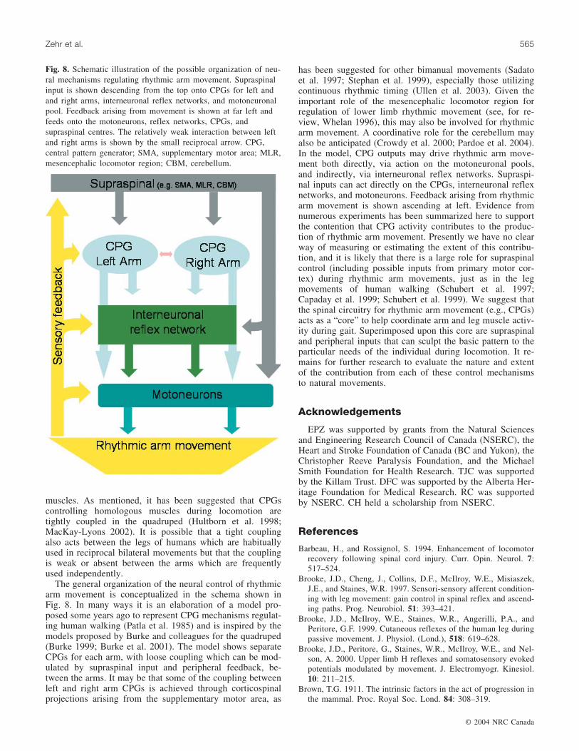
The general organization of the neural control of rhythmicarm movement is conceptualized in the schema shown inFig. 8. In many ways it is an elaboration of a model pro-posed some years ago to represent CPG mechanisms regulat-ing human walking (Patla et al. 1985) and is inspired by themodels proposed by Burke and colleagues for the quadruped(Burke 1999; Burke et al. 2001). The model shows separateCPGs for each arm, with loose coupling which can be mod-ulated by supraspinal input and peripheral feedback, be-tween the arms. It may be that some of the coupling betweenleft and right arm CPGs is achieved through corticospinalprojections arising from the supplementary motor area, as
has been suggested for other bimanual movements (Sadatoet al. 1997; Stephan et al. 1999), especially those utilizingcontinuous rhythmic timing (Ullen et al. 2003). Given theimportant role of the mesencephalic locomotor region forregulation of lower limb rhythmic movement (see, for re-view, Whelan 1996), this may also be involved for rhythmicarm movement. A coordinative role for the cerebellum mayalso be anticipated (Crowdy et al. 2000; Pardoe et al. 2004).In the model, CPG outputs may drive rhythmic arm move-ment both directly, via action on the motoneuronal pools,and indirectly, via interneuronal reflex networks. Supraspi-nal inputs can act directly on the CPGs, interneuronal reflexnetworks, and motoneurons. Feedback arising from rhythmicarm movement is shown ascending at left. Evidence fromnumerous experiments has been summarized here to supportthe contention that CPG activity contributes to the produc-tion of rhythmic arm movement. Presently we have no clearway of measuring or estimating the extent of this contribu-tion, and it is likely that there is a large role for supraspinalcontrol (including possible inputs from primary motor cor-tex) during rhythmic arm movements, just as in the legmovements of human walking (Schubert et al. 1997;Capaday et al. 1999; Schubert et al. 1999). We suggest thatthe spinal circuitry for rhythmic arm movement (e.g., CPGs)acts as a “core” to help coordinate arm and leg muscle activ-ity during gait. Superimposed upon this core are supraspinaland peripheral inputs that can sculpt the basic pattern to theparticular needs of the individual during locomotion. It re-mains for further research to evaluate the nature and extentof the contribution from each of these control mechanismsto natural movements.
Acknowledgements
EPZ was supported by grants from the Natural Sciencesand Engineering Research Council of Canada (NSERC), theHeart and Stroke Foundation of Canada (BC and Yukon), theChristopher Reeve Paralysis Foundation, and the MichaelSmith Foundation for Health Research. TJC was supportedby the Killam Trust. DFC was supported by the Alberta Her-itage Foundation for Medical Research. RC was supportedby NSERC. CH held a scholarship from NSERC.
References
Barbeau, H., and Rossignol, S. 1994. Enhancement of locomotorrecovery following spinal cord injury. Curr. Opin. Neurol. 7:517–524.
Brooke, J.D., Cheng, J., Collins, D.F., McIlroy, W.E., Misiaszek,J.E., and Staines, W.R. 1997. Sensori-sensory afferent condition-ing with leg movement: gain control in spinal reflex and ascend-ing paths. Prog. Neurobiol. 51: 393–421.
Brooke, J.D., McIlroy, W.E., Staines, W.R., Angerilli, P.A., andPeritore, G.F. 1999. Cutaneous reflexes of the human leg duringpassive movement. J. Physiol. (Lond.), 518: 619–628.
Brooke, J.D., Peritore, G., Staines, W.R., McIlroy, W.E., and Nel-son, A. 2000. Upper limb H reflexes and somatosensory evokedpotentials modulated by movement. J. Electromyogr. Kinesiol.10: 211–215.
Brown, T.G. 1911. The intrinsic factors in the act of progression inthe mammal. Proc. Royal Soc. Lond. 84: 308–319.
© 2004 NRC Canada
Zehr et al. 565
Fig. 8. Schematic illustration of the possible organization of neu-ral mechanisms regulating rhythmic arm movement. Supraspinalinput is shown descending from the top onto CPGs for left andand right arms, interneuronal reflex networks, and motoneuronalpool. Feedback arising from movement is shown at far left andfeeds onto the motoneurons, reflex networks, CPGs, andsupraspinal centres. The relatively weak interaction between leftand right arms is shown by the small reciprocal arrow. CPG,central pattern generator; SMA, supplementary motor area; MLR,mesencephalic locomotor region; CBM, cerebellum.

Brown, T.G. 1914. On the nature of the fundamental activity of thenervous centres; together with an analysis of the conditioning ofrhythmic activity in progression, and a theory of the evolution offunction in the nervous system. J. Physiol. 48: 19–46.
Brown, D.A, and Kukulka, C.G. 1993. Human flexor reflex modu-lation during cycling. J. Neurophysiol. 69: 1212–1224.
Brown, D.A., Kautz, S.A., and Dairaghi, C.A. 1996. Muscle activ-ity patterns altered during pedaling at different body orienta-tions. J. Biomech. 29: 1349–1356.
Brown, D.A., Kautz, S.A., and Dairaghi, C.A. 1997. Muscle activ-ity adapts to anti-gravity posture during pedalling in personswith post-stroke hemiplegia. Brain, 120: 825–837.
Buford, J.A., and Smith, J.L. 1990. Adaptive control for backwardquadrupedal walking. II. Hindlimb muscle synergies. J. Neuro-physiol. 64: 756–766.
Buford, J.A., and Smith, J.L. 1993. Adaptive control for backwardquadrupedal walking. III. Stumbling corrective reactions and cu-taneous reflex sensitivity. J. Neurophysiol. 70: 1102–1114.
Burke, R.E. 1999. The use of state-dependent modulation of spinalreflexes as a tool to investigate the organization of spinalinterneurons. Exp. Brain Res. 128: 263–277.
Burke, D., Dickson, H.G., and Skuse, N.F. 1991. Task-dependentchanges in the responses to low-threshold cutaneous afferentvolleys in the human lower limb. J. Physiol. (Lond.), 432: 445–458.
Burke, R.E., Degtyarenko, A.M., and Simon, E.S. 2001. Patterns oflocomotor drive to motoneurons and last-order interneurons:Clues to the structure of the CPG. J. Neurophysiol. 86: 447–462.
Capaday, C., Lavoie, B.A., Barbeau, H., Schneider, C., andBonnard, M. 1999. Studies on the corticospinal control of hu-man walking. I. Responses to focal transcranial magnetic stimu-lation of the motor cortex. J. Neurophysiol. 81: 129–139.
Carroll, T.J., Zehr, E.P., and Collins, D.F. 2004. Modulation of cu-taneous reflexes in human upper limb muscles during arm cy-cling is independent of activity in the contralateral arm. Exp.Brain Res. In press.
Carson, R.G., Riek, S., and Bawa, P. 1999. Electromyographic ac-tivity, H-reflex modulation and corticospinal input to forearmmotoneurones during active and passive rhythmic movements.Hum. Mov. Sci. 18: 307–343.
Cheng, J., Brooke, J.D., Misiaszek, J.E., and Staines, W.R. 1998.Crossed inhibition of the soleus H-reflex during passive pedal-ling movement. Brain Res. 779: 280–284.
Collins, D.F., McIlroy, W.E., and Brooke, J.D. 1993. Contralateralinhibition of soleus H reflexes with different velocities of pas-sive movement of the opposite leg. Brain Res. 603: 96–101.
Crowdy, K.A., Hollands, M.A., Ferguson, I.T., and Marple-Horvat,D.E. 2000. Evidence for interactive locomotor and oculomotordeficits in cerebellar patients during visually guided stepping.Exp. Brain Res. 135: 437–454.
Delwaide, P.J., Figiel, C., and Richelle, C. 1973. Influence de laposition du membre superieur sur l’excitabilite de l’arc soleaire.Electromyogr. Clin. Neurophysiol. 13: 515–523.
Delwaide, P.J., Figiel, C., and Richelle, C. 1977. Effects of posturalchanges of the upper limb on reflex transmission in the lowerlimb. Cervicolumbar reflex interactions in man. J. Neurol.Neurosurg. Psychiatry, 40: 616–621.
Delwaide, P.J., Sabatino, M., Pepin, J.L., and La, G.V. 1988. Rein-forcement of reciprocal inhibition by contralateral movements inman. Exp. Neurol. 99: 10–16.
Dietz, V. 1997. Locomotor recovery after spinal cord injury. TrendsNeurosci. 20: 346–347.
Dietz, V. 2002a. Do human bipeds use quadrupedal coordination?Trends Neurosci. 25: 462–467.
Dietz, V. 2002b. Proprioception and locomotor disorders. Nat. Rev.Neurosci. 3: 781–790.
Dietz, V., Colombo, G., and Jensen, L. 1994. Locomotor activity inspinal man. Lancet, 344: 1260–1263.
Dietz, V., Fouad, K., and Bastiaanse, C.M. 2001. Neuronal coordi-nation of arm and leg movements during human locomotion.Eur. J. Neurosci. 14: 1906–1914.
Drew, T., and Rossignol, S. 1987. A kinematic and electromyo-graphic study of cutaneous reflexes evoked from the forelimb ofunrestrained walking cats. J. Neurophysiol. 57: 1160–1184.
Duysens, J. 1998. From cat to man: basic aspects of locomotionrelevant to motor rehabilitation of SCI. Neurorehabilitation, 10:107–118.
Duysens, J, and Tax, T. 1994. Interlimb reflexes during gait in catand human. In Interlimb coordination: Neural, dynamical, andcognitive constraints. Edited by S.P. Swinnen, H. Heuer, J.Massion, P. Casaer. Academic Press, Inc., pp. 97–126.
Duysens, J., and Van de Crommert, H.W. 1998. Neural control oflocomotion, Part 1: The central pattern generator from cats tohumans. Gait Posture, 7: 131–141.
Duysens, J, Tax, A.A., Trippel, M., and Dietz, V. 1993. Increasedamplitude of cutaneous reflexes during human running as com-pared to standing. Brain Res. 613: 230–238.
Duysens, J., Tax, A.A., Murrer, L., and Dietz, V. 1996. Backwardand forward walking use different patterns of phase-dependentmodulation of cutaneous reflexes in humans. J. Neurophysiol.76: 301–310.
Eisner, W.D., Bode, S.D., Nyland, J., and Caborn, D.N. 1999.Electromyographic timing analysis of forward and backward cy-cling. Med. Sci. Sports Exercise 31: 449–455.
Eke-Okoro, S.T. 1994. Evidence of interaction between humanlumbosacral and cervical neural networks during gait.Electromyogr. Clin. Neurophysiol. 34: 345–349.
Elftman, H. 1939. The function of the arms in walking. Hum. Biol.11: 529–535.
Fernandez-Ballesteros, M.L., Buchtal, F., and Rosenfalck, P. 1965.The pattern of muscular activity during the arm swing of naturalwalking. Acta Physiol. Scand. 63: 296–310.
Forssberg, H., Grillner, S., and Rossignol, S. 1975. Phase depend-ent reflex reversal during walking in chronic spinal cats. BrainRes. 85: 103–107.
Frigon, A., Collins, D.F., and Zehr, E.P. 2004. Effect of rhythmicarm movement on reflexes in the legs: modulation of soleus H-reflexes and somatosensory conditioning. J. Neurophysiol. 91:1516–1523.
Garrett, M., Kerr, T., and Caulfield, B. 1999. Phase-dependent in-hibition of H-reflexes during walking in humans is independentof reduction in knee angular velocity. J. Neurophysiol. 82: 747–753.
Gossard, J.P., Cabelguen, J.M., and Rossignol, S. 1990. Phase-dependent modulation of primary afferent depolarization in sin-gle cutaneous primary afferents evoked by peripheral stimula-tion during fictive locomotion in the cat. Brain Res. 537: 14–23.
Grasso, R., Bianchi, L., and Lacquaniti, F. 1998. Motor patterns forhuman gait: backward versus forward locomotion. J.Neurophysiol. 80: 1868–1885.
Grillner, S. 1975. Locomotion in vertebrates: central mechanismsand reflex interaction. Physiol. Rev. 55: 247–304.
Grillner, S., and Dubuc, R. 1988. Control of locomotion in verte-brates: Spinal and supraspinal mechanisms. In Advances in neu-rology. Functional recovery in neurological disease. Edited byS.G. Waxman. Raven Press, New York, pp. 425–453.
Haridas, C., and Zehr, E.P. 2003. Coordinated interlimb compensa-tory responses to electrical stimulation of cutaneous nerves in
© 2004 NRC Canada
566 Can. J. Physiol. Pharmacol. Vol. 82, 2004

the hand and foot during walking. J. Neurophysiol. 90: 2850–2861.
Harkema, S.J., Hurley, S.L., Patel, U.K., Requejo, P.S., Dobkin,B.H., and Edgerton, V.R. 1997. Human lumbosacral spinal cordinterprets loading during stepping. J. Neurophysiol. 77: 797–811.
Hishinuma, M., and Yamaguchi, T. 1989. Modulation of reflex re-sponses during fictive locomotion in the forelimb of the cat.Brain Res. 482: 184–188.
Hogue, R.E. 1969. Upper-extremity muscular activity at differentcadences and inclines during normal gait. J. Am. Phys. Ther.Assoc. 49: 963–972.
Hultborn, H., Conway, B.A., Gossard, J.P., Brownstone, R.,Fedirchuk, B., Schomburg, E.D., Enriquez-Denton, M., andPerreault, M.C. 1998. How do we approach the locomotor net-work in the mammalian spinal cord? Ann. N.Y. Acad. Sci. 860:70–82.
Ichikawa, Y., Terakado, Y., and Yamaguchi, T. 1991. Last-orderinterneurones controlling activity of elbow extensor moto-neurones during forelimb fictive locomotion in the cat.Neurosci. Lett. 121: 37–39.
Jackson, K.M. 1983. Why the upper limbs move during humanwalking. J. Theor. Biol. 105: 311–315.
Jackson, K.M., Joseph, J., and Wyard, S.J. 1978. A mathematicalmodel of arm swing during human locomotion. J. Biomech. 11:277–289.
Jackson, K.M., Joseph, J., and Wyard, S.J. 1983. The upper limbsduring human walking. Part 2: Function. Electromyogr. Clin.Neurophysiol. 23: 435–446.
Kiehn, O., Hounsgaard, J., and Sillar, K.T. 1997. Basic buildingblocks of vertebrate spinal central pattern generators. In Neu-rons, networks, and motor behavior. Edited by P.S.G. Stein, S.Grillner, A.I. Selverston, D.G. Stuart. MIT Press, London,pp. 47–59.
Knikou, M., and Rymer, W.Z. 2002a. Hip angle induced modula-tion of H reflex amplitude, latency and duration in spinal cordinjured humans. Clin. Neurophysiol. 113: 1698–1708.
Knikou, M., and Rymer, Z. 2002b. Effects of changes in hip jointangle on H-reflex excitability in humans. Exp. Brain Res. 143:149–159.
Komiyama, T., Zehr, E.P., and Stein, R.B. 2000. Absence of nerve-specificity in human cutaneous reflexes during standing. Exp.Brain Res. 133: 267–272.
MacKay, W.A., Kwan, H.C., Murphy, J.T., and Wong, Y.C. 1983.Stretch reflex modulation during a cyclic elbow movement.Electroencephalogr. Clin. Neurophysiol. 55: 687–698.
MacKay-Lyons, M. 2002. Central pattern generation of locomo-tion: a review of the evidence. Phys. Ther. 82: 69–83.
Miller, S., Reitsma, D.J., and Meche, F.G. 1973. Functional organi-zation of long ascending propriospinal pathways linking lumbo-sacral and cervical segments in the cat. Brain Res. 62: 169–188.
Pardoe, J., Edgley, S.A., Drew, T., and Apps, R. 2004. Changes inexcitability of ascending and descending inputs to cerebellarclimbing fibers during locomotion. J. Neurosci. 24: 2656–2666.
Patla, A.E., Calvert, T.W., and Stein, R.B. 1985. Model of a patterngenerator for locomotion in mammals. Am. J. Physiol. Regul.Integr. Comp. Physiol. 248: R484-R494.
Pearson, K.G. 1993. Common principles of motor control in verte-brates and invertebrates. Annu. Rev. Neurosci. 16: 265–297.
Pearson, K.G. 2000. Neural adaptation in the generation of rhyth-mic behavior. Annu. Rev. Physiol. 62: 723–753.
Pearson, K.G., and Ramirez, J.M. 1997. Sensory modulation ofpattern-generating circuits. In Neurons, networks, and motor be-havior. Edited by P.S.G. Stein, S. Grillner, A.I. Selverston, D.G.Stuart. MIT Press, London, pp. 226–235.
Perell, K.L., Gregor, R.J., Buford, J.A., and Smith, J.L. 1993.Adaptive control for backward quadrupedal walking. IV.Hindlimb kinetics during stance and swing. J. Neurophysiol. 70:2226–2240.
Rossignol, S. 1996. Neural control of stereotypic limb movements.In Section 12: Exercise: Regulation and integration of multiplesystems. Edited by L.B. Rowell and J.T. Shepherd. Oxford Uni-versity Press, New York, pp. 173–216.
Rossignol, S., Beloozerova, I.N., Gossard, J.-P., and Dubuc, R.1998. Presynaptic mechanisms during locomotion. InPresynaptic inhibition and neural control. Edited by P. Rudomin,R. Romo, and L.M. Mendell. Oxford University Press, NewYork, pp. 385–397.
Ryan, M.M., and Gregor, R.J. 1992. EMG profiles of lower ex-tremity muscles during cycling at constant workload and ca-dence. J. Electromyogr. Kinesiol. 2: 69–80.
Sadato, N., Yonekura, Y., Waki, A., Yamada, H., Ishii, and Y. 1997.Role of the supplementary motor area and the right premotorcortex in the coordination of bimanual finger movements. J.Neurosci. 17: 9667–9674.
Schneider, C., Lavoie, B.A., and Capaday, C. 2000. On the originof the soleus H-reflex modulation pattern during human walkingand its task-dependent differences. J. Neurophysiol. 83: 2881–2890.
Schomburg, E.D., and Behrends, H.B. 1978. Phasic control of thetransmission in the excitatory and inhibitory reflex pathwaysfrom cutaneous afferents to alpha-motoneurones during fictivelocomotion in cats. Neurosci. Lett. 8: 277–282.
Schubert, M., Curt, A., Jensen, L., and Dietz, V. 1997.Corticospinal input in human gait: modulation of magneticallyevoked motor responses. Exp. Brain Res. 115: 234–246.
Schubert, M., Curt, A., Colombo, G., Berger, W., and Dietz, V.1999. Voluntary control of human gait: conditioning of magneti-cally evoked motor responses in a precision stepping task. Exp.Brain Res. 126: 583–588.
Seki, K., and Yamaguchi, T. 1997. Cutaneous reflex activity of thecat forelimb during fictive locomotion. Brain Res. 753: 56–62.
Seki, K., Kudo, N., Kolb, F., and Yamaguchi, T. 1997. Effects ofpyramidal tract stimulation on forelimb flexor motoneurons dur-ing fictive locomotion in cats. Neurosci. Lett. 230: 195–198.
Stein, P.S.G., and Smith, J.L. 1997. Neural and biomechanical con-trol strategies for different forms of vertebrate hindlimb motortasks. In Neurons, networks, and motor behavior. Edited byP.S.G. Stein, S. Grillner, A.I. Selverston, D.G. Stuart. MITPress, London, pp. 61–73.
Stein, R.B., and Capaday, C. 1988. The modulation of human re-flexes during functional motor tasks. Trends Neurosci. 11: 328–332.
Stein, R.B. (1995) Presynaptic inhibition in humans. Prog.Neurobiol. 47(6): 533–544.
Steldt, R.E., and Schmit, B.D. 2004. Modulation of coordinatedmuscle activity during imposed sinusoidal hip movements in hu-man spinal cord injury. J. Neurophysiol. 92: 673-685.
Stephan, K.M., Binkofski, F., Halsband, U., Dohle, C., Wunderlich,G., Schnitzler, A., Tass, P., Posse, S., Herzog, H., Sturm, V.,Zilles, K., Seitz, R.J., and Freund, H.J. 1999. The role of ventralmedial wall motor areas in bimanual co-ordination. A combinedlesion and activation study. Brain, 122: 351–368.
Thorstensson, A. 1986. How is the normal locomotor programmodified to produce backward walking? Exp. Brain Res. 61:664–668.
Ting, L.H., Kautz, S.A., Brown, D.A., Van der Loos, H.F., andZajac, F.E. 1998a Bilateral integration of sensorimotor signalsduring pedaling. Ann. N.Y. Acad. Sci. 860: 513–516.
© 2004 NRC Canada
Zehr et al. 567

Ting, L.H., Raasch, C.C., Brown, D.A., Kautz, S.A,. and Zajac,F.E. 1998b. Sensorimotor state of the contralateral leg affectsipsilateral muscle coordination of pedaling. J. Neurophysiol. 80:1341–1351.
Ting, L.H., Kautz, S.A., Brown, D.A., and Zajac, F.E. 1999. Phasereversal of biomechanical functions and muscle activity in back-ward pedaling. J. Neurophysiol. 81: 544–551.
Ting, L.H., Kautz, S.A, Brown, D.A., and Zajac, F.E. 2000. Con-tralateral movement and extensor force generation alter flexionphase muscle coordination in pedaling. J. Neurophysiol. 83:3351–3365.
Ullen, F., Forssberg, H., and Ehrsson, H.H. 2003. Neural networksfor the coordination of the hands in time. J. Neurophysiol. 89:1126–1135.
Van de Crommert, H.W., Mulder, T., and Duysens, J. 1998. Neuralcontrol of locomotion: sensory control of the central patterngenerator and its relation to treadmill training. Gait Posture, 7:251–263.
Weiss, P.L., and St. Pierre, D. 1983. Upper and lower extremityEMG correlations during normal human gait. Arch. Phys. Med.Rehabil. 64: 11–15.
Whelan, P.J. 1996. Control of locomotion in the decerebrate cat.Prog. Neurobiol. 49: 481–515
Winter, D.A. 1991. The biomechanics and motor control of humangait. University of Waterloo Press, Waterloo, Ont.
Winter, D.A., Pluck, N., and Yang, J.F. 1989. Backward walking:A simple reversal of forward walking? J. Mot. Behav. 21: 291–305.
Yamaguchi, T. 1992a. Activity of cervical neurons during forelimbfictive locomotion in decerebrate cats. Jpn. J. Physiol. 42: 501–514.
Yamaguchi, T. 1992b. Muscle activity during forelimb stepping indecerebrate cats. Jpn. J. Physiol.1992. 42: 489–499.
Yamaguchi, T. 2004. The central pattern generator for forelimb lo-comotion in the cat. Prog. Brain Res. 143: 115–122.
Zehr, E.P., and Chua, R. 2000. Modulation of human cutaneous re-flexes during rhythmic cyclical arm movement. Exp. Brain Res.135: 241–250.
Zehr, E.P., and Duysens, J. 2004. Regulation of arm and leg move-ment during human locomotion. Neuroscientist. 10: 347-361.
Zehr, E.P., and Haridas, C. 2003. Modulation of cutaneous reflexesin arm muscles during walking: further evidence of similar con-trol mechanisms for rhythmic human arm and leg movements.Exp. Brain Res. 149: 260–266.
Zehr, E.P., and Hundza, S.R. Forward and backward arm cyclingare regulated by equivalent neural mechanisms. J. Neurophysiol.In press.
Zehr, E.P., and Kido, A. 2001. Neural control of rhythmic, cyclicalhuman arm movement: task dependency, nerve specificity andphase modulation of cutaneous reflexes. J. Physiol. (Lond.),537: 1033–1045.
Zehr, E.P., and Stein, R.B. 1999. What functions do reflexes serveduring human locomotion? Prog. Neurobiol. 58: 185–205.
Zehr, E.P., Collins, D.F., and Chua, R. 2001a. Human interlimb re-flexes evoked by electrical stimulation of cutaneous nervesinnervating the hand and foot. Exp. Brain Res. 140: 495–504.
Zehr, E.P., Hesketh, K.L., and Chua, R. 2001b. Differential regula-tion of cutaneous and H-reflexes during leg cycling in humans.J. Neurophysiol. 85: 1178–1185.
Zehr, E.P., Collins, D.F., Frigon, A., and Hoogenboom, N. 2003.Neural control of rhythmic human arm movement: Phase de-pendence and task modulation of Hoffmann reflexes in forearmmuscles. J. Neurophysiol. 89: 12–21.
© 2004 NRC Canada
568 Can. J. Physiol. Pharmacol. Vol. 82, 2004