original article interferon epsilon mrna expression could … · proteins, involved in the first...
TRANSCRIPT

Int J Clin Exp Pathol 2018;11(4):1979-1988www.ijcep.com /ISSN:1936-2625/IJCEP0070901
Original ArticleInterferon epsilon mRNA expression could represent a potential molecular marker in cervical cancer
Daniel Marrero-Rodríguez1,5*, Victor Baeza-Xochihua1*, Keiko Taniguchi-Ponciano1, Victor Huerta-Padilla1, Gustavo Ponce-Navarrete1, Alejandra Mantilla2, Daniel Hernandez3, Angeles Hernandez3, Guillermo Gomez-Gutierrez4, Luis Serna-Reyna4, Ma del Pilar Figueroa-Corona5, Laura Gomez-Virgilio5, Miriam Rodriguez-Esquivel1, Pablo Romero-Morelos1, Miguel A Vazquez-Moreno6, Sarahi Loza-Medrano6, Jorge Ortiz-Leon4, Edgar Hernandez-Rico7, Marco Meraz-Rios5, Mauricio Salcedo1
1Laboratorio de Oncología Genómica, Unidad de Investigación Médica en Enfermedades Oncológicas, Hospital de Oncología, CMN-SXXI, IMSS, México D.F. 06720, México; 2Servicio de Patología, Hospital de Oncología CMN-SXXI, IMSS, Mexico; 3División de Laboratorios de Vigilancia e Investigación Epidemiológica, IMSS, México, D.F., México; 4Clínica de Displasias, Hospital General de México, México, D.F., México; 5Departamento de Biomedicina Molecu-lar, CINVESTAV, Mexico D.F. 07360, México; 6Unidad de Investigación Medica en Bioquimica, Hospital de Espe-cialidades, CMN-SXXI, IMSS, México, D.F., México; 7University of Texas at El Paso, El Paso, Texas, United States of America. *Equal contributors.
Received December 13, 2017; Accepted January 30, 2018; Epub April 1, 2018; Published April 15, 2018
Abstract: The effects of the immune system response in the malignant transformation process have been de-scribed. Molecules such as interferons are involved in such process. Interferons are small single-chained glyco-proteins, involved in the first line of defense against pathogens such as viruses, bacteria, and parasites. Interferon epsilon (IFNε) is located in the 9p21.3 cytogenetic region, transcribes into a single exon mRNA. Contrary to other family members, IFNε exerts low antiviral activity. In the present work molecular alterations such as copy number variation (CNV) and expression were analyzed by available microarrays and fifty-nine cervical tissues ranging from normal to cancer and three cell lines were assessed for IFNε expression by RT-PCR, immunohistochemistry, and im-munocytofluorescence. No significant CNV alterations were observed. Positive immunosignal was primarily present in the proliferative basal strata cells in the normal tissue, whereas in cervical cancer, all epithelial transformed cells were positive. The cell lines analyzed were HPV16, -18, and negative, all three cell-lines were positive for cytoplas-mic protein presence. Interestingly, at the mRNA level, increased band intensity was observed, as the lesions were higher, and IFNε up-regulation in CC (P=0.0001) is reported here. Our results suggest that up-regulation is present as an independent event from single or multiple HPV infection (P=0.90). In conclusion, we suggest that IFNε mRNA up-regulation could represent a potential molecular marker in CC. Expression of IFNε might not be related to HPV infection or CNV, which could have an important role in cellular homeostasis and could influence immune related events in cervical carcinogenesis.
Keywords: Cervical cancer, cytokines, interferon epsilon, molecular marker
Introduction
Cervical cancer (CC) is the final stage of pre-defined precursor lesions, as the Low Grade-Squamous Intraepithelial Lesion (LG- SIL) followed by the High Grade-SIL (HGSIL) and finally CC. It is the second place in in- cidence and mortality among female neop- lasias worldwide [1]. These neoplasias cou- ld have several risk factors, among them, pr- egnancy number, alcohol and tobacco con-sumption, number of sexual partners, and,
the most related etiological factor, high risk-Human papillomavirus (hr-HPV) infection [2].
The transformation step is not a common oc- currence of an HPV infection, and only a small number of cervical lesions infected by hr-HPV types evolve into CC. The immune response plays an important role in clearing most of these infections, but some infections cannot be eliminated and persist for several years, becom-ing an additional risk factor [3].

IFNε cytokine expression in cervical cancer
1980 Int J Clin Exp Pathol 2018;11(4):1979-1988
Two of the hallmarks of cancer are related to the immune system response, one of them related to the avoidance of the immune system recognition and another related to inflamma-tion prompted by the tumor. These are mediat-ed by a plethora of molecules such as cytokines like interferons (IFN) and chemokines among others [4].
Interferons are small single-chained glycopro-teins, involved in the first line of defense against pathogens such as viruses, bacteria, and para-sites. Interferons are classified in Type I (IFNα, -β, -ε, -κ, -τ and -ω) and Type II (IFNγ) upon rece- ptor binding. Type I IFN binds to IFNAR1 and IFNAR2, while type II IFN binds to IFNGR1 and IFNGR2 [5].
Specifically, IFNε gene is located in chromo-some 9p21, gives arise to a single exon mRNA, and is translated to a 192 amino acid protein [5, 6]. IFNε has found to be constitutively expressed in lung, brain, small intestine, and reproductive tissues. It exerts lower anti-viral, anti-proliferative, and natural cell killer enhanc-ing activities compared to IFNα family mem-bers [5].
Immunological mechanisms play a key role in the etiology and progression of many and per-haps all cancers [7], elevated type I IFN is a component of the signature associated with chronic immune activation [8]. Although most women will be infected by HPV at some point, very few will progress to invasive disease [1]. The identification of more robust markers of the disease progression requires a more com-plete molecular and cellular characterization. At present reports about IFNε expression in CC are scarce.
Therefore, we decided to evaluate the potential molecular alterations at DNA and RNA levels by in silico methods, and by means of RT-PCR at mRNA level and at protein level by immunohis-tochemistry and immunocytofluorescence in cervical tissue with the different lesions of the cervix.
Materials and methods
Chromosome 9 CNV in silico analysis
As previously reported (Marrero-Rodriguez 2017 data submitted) for copy number varia-tion analysis, a total of 115 cancer libraries and 32 control libraries corresponding to
GSE10092 and GSE52904 were downloaded. Partek v6.6 (Partek Incorporated, Saint Louis, MO, USA) were used. Stringent parameters were set as follows: each segment must con-tain a minimum of 10 consecutive filtered probe sets, a p-value threshold of 0.001 when compared to the neighboring adjacent regions and a signal-to-noise threshold of 0.5. The cut-off value for the gain was set at above 2.3, while loss was set at below 1.7. CNV was called for the gains or losses that occurred in at least 10% of the total samples. The 22 somatic chro-mosomes and sexual chromosomes were ana-lyzed. Hierarchical clustering was done by the following parameters, sample dissimilarity: Euc- lidian and cluster method: average linking.
Cervical cancer transcriptome in silico analysis
As previously reported (Marrero-Rodriguez 2017 submitted) a total of 8 normal (N) and 57 cervical cancer (CC) experiments were down-loaded and analyzed. The data used for these analyses were downloaded from European Bioinformatics Institute (EMBL-EBL) and GEO. These data correspond to GSE7307, GSE5787, GSE3526 and 1 Gene Expression Atlas (GSE- 2109) corresponding to Affymetrix Human Gene Chip U133 platform.
Data sets analyses were achieved by means of CEL files with the Expression Console, Partek Genomics Suite 6.6v software (Partek Incor- porated, Saint Louis, MO, USA) and Transcri- ptome Analysis Console (Affymetrix, Santa Clara, CA, USA). Pearson and Spearman corre-lation was performed and probe sets were summarized by means of Median Polish and normalized by quantiles with no probe sets excluded from analysis. Background noise cor-rection was achieved by means of Robust Mul- ti-chip Average (RMA) and data were log2- transformed. Data grouping and categorization was achieved by principal component analysis (PCA). Differentially expressed genes were de- tected by means of ANOVA. Genes were consid-ered altered with +1.5 or -1.5 fold change, P≤0.05 and FDR>0.05 parameters.
Cervical tissue collection
Fifty nine cervical tissue samples (taken from patients aged between 18 and 74 years) in- cluding: three histological and colposcopical normal tissues, twenty LGSIL, sixteen HGSIL, and twenty carcinomas (16 squamous cell car-
IFNε cytokine expression in cervical cancer
1981 Int J Clin Exp Pathol 2018;11(4):1979-1988
ously reported. Sequences were as followed: IFNεF 5’-CCA AAA AGC ACA CAC TCT GGC-3’, IFNεR 5’-CCG TGT GGT TTT CCT CCC AA-3’, RPS18F 5’-AATCCACGCCAGTACAAGATCCCA-3’ and RPS18R 5’-TTTCTTCTTGGACACACCCACG- GT-3’. For end point PCR, the conditions were as follows: 12.5 μl of GoTaq® Green Master Mix (Promega, WI, USA), 1 μl of cDNA template, and 20 pmol of each primer, in a 25 μl total volume reaction, with the following program: 45 sec-onds at 95°C, 30 seconds at 60°C and 30 sec-onds at 72°C for 30 cycles. Endpoint PCR prod-ucts were resolved in 2% ethidium bromide stained agarose gel. The intensity of each band was analyzed employing Kodak 200 Molecular imaging analysis software and densitometry assay was carried out.
DNA extraction and HPV detection and geno-typing
DNA from cervical tissue samples was extract-ed from the phenol phase of the RNA extrac- tion procedure, incubated for 5 min with 300 μl 100% ethanol at room temperature, centri-fuged at 10,000 rpm for 5 min. Resulting pellet was washed with 1 ml sodium citrate (0.1 M sodium citrate in 10% ethanol, pH 8.5) for 30 min at room temperature and then centrifug- ed at 10,000 rpm for 5 min (3×). DNA was washed with 75% ethanol and centrifuged at 10,000 rpm for 5 min, pellet was air- dried, and DNA resuspended in nuclease free water. HPV detection was accomplished using oligos GP5+/GP6+ as described [10], conditions were as follows: 12.5 μl GoTaq Green Master Mix, 20 pmol of each primer, and 100 ng DNA in a 25 μl final volume. PCR program used was 40 cycles of 94°C for 30 sec, 55°C for 1.5 min, followed by 72°C for 1.5 min. PCR products were resolved in 2% ethidium bromide stained agarose gel. The intensity of each band was analyzed employing Kodak 200 molecular imaging analysis software. HPV genotyping was carried out using Linear Array® Genotyping Test (Roche, IN, USA) and HPV 2 CLART® (Genomica, Madrid, Spain) according to manufacturer’s instruction.
IFNε immunohistochemistry assay
A Tissue microarray (TMA) of the 59 cases was constructed as follow. Sections from paraffin-embedded, formalin-fixed tissue blocks were stained with hematoxylin-eosin and reviewed
cinomas and 4 cervical adenocarcinomas), were collected from the Hospital General de Mexico in Mexico City, from May 2012 to May 2013. Detailed clinic-pathological information was obtained from the patient’s records. Patients were recruited with signed informed consent and ethical approval from the local institutional board in accordance with the Helsinki declaration. Tissue samples were col-lected prior to the administration of any chemo-therapy and/or radiotherapy treatments. These same cervical tissues were previously reported [9].
RNA extraction and reverse transcription reac-tion
Total RNA was extracted with the RNAeasy tis-sue Mini Kit (Qiagen Inc, USA). The cervical tis-sue samples were disrupted and homogenized in 1 ml of Qiazol Lysis Reagent. Samples were then incubated at room temperature for 5 min. Next, 200 μl of chloroform was added, and samples were incubated at room temperature for 3 min. The mixture was centrifuged at 12,500 rpm for 15 min at 4°C. The aqueous phase was transferred to a new tube and mixed with an equal volume of 70% ethanol. Samples were then transferred to an RNAeasy Column in a 2 ml tube, and centrifuged at 10,000 rpm for 15 sec. After centrifugation, 700 μl of RW1 buf-fer was added and mixture was centrifuged at 10,000 rpm for 15 sec. Flow-through was dis-carded and 500 μl of RPE buffer was added to the membrane and then centrifuged at 10,000 rpm for 15 sec (2×). The column was trans-ferred to a new collection tube then 30 μl of RNAse free water was added and centrifuged for 1 min at 10,000 rpm. RNA elution was mea-sured in a Nanodrop-ND-1000 (Thermo Scien- tific, DE, USA) and RNA integrity was checked in 1.5% agarose gel. After purification, 1 μg of total RNA was retro transcribed in a 20 μl final volume reaction with the SuperScript VILO Master Mix (Applied Biosystems, CA, USA) 4 μl of Master Mix were added, and the reaction mixture was incubated at 25°C for 10 min., 42°C for 60 min., and 85°C for 5 min., accord-ing to manufacturer protocols.
IFN epsilon transcripts detection
Specific IFNε primers were in-house designed using primer BLAST software found on the NCBI website, with the NCBI reported sequences and RPS18 were used as internal control, previ-

IFNε cytokine expression in cervical cancer
1982 Int J Clin Exp Pathol 2018;11(4):1979-1988
by a pathologist to select areas of invasive tumor. Core samples were taken using 0.6 mm2 blunt-tip needles and were placed on the recipient microarray block using a Tissue Microarrayer (Chemicon Co., MA, USA). Tumors were represented with 2-fold redundancy, which has been shown to provide a sufficiently representative sample. Sections (3 μm) were cut and placed onto coated slides. TMA slides were deparaffinized with xylene followed by ethanol and rehydrated. Immunostaining was performed using a streptavidin-biotin complex peroxidase method (Dako, Glostrup, Denmark).
Briefly, after de-waxing the sections, endoge-nous peroxidase activity was inhibited with freshly prepared 0.5% H2O2 in distilled water for 20 min. Next, the sections were processed in a 600 W microwave oven at maximum power, three times for 5 min each in Tris-EDTA buffer (pH 9.0). Incubation with polyclonal rabbit anti-IFNε (NBP1-92018; Novus Biologicals, CO, USA), was performed overnight at 4°C in a humidity chamber, at 1:100 dilution, in 1% Bovine serum albumin (BSA). Sections were developed with a peroxidase substrate solution (0.05% 3,3-diaminobenzidine tetrahydrochlo-
ride, 0.01% H2O2 in PBS), counterstained with haematoxylin, dehydrated, and mounted. Testis and placenta tissues were used as positive bio-logical controls, and negative controls consist-ed of the replacement of the primary antibody with 1% BSA. Three independent observers performed assessment of IFNε expression at independent times by light microscopy at 20× magnification. Immunostaining was evaluated as negative and positive staining.
IFNε Immunocytofluorescence in CC cell lines
SiHa, HeLa and C33A cells were grown on 5 mm coverslips (previously treated with poly-D-Lys). Cells at 70% of confluence were fixed with 4% paraformaldehyde in PBS 1× during 30 min. Fixed cells were washed (3 times) with PBS 1×. Cells were permeabilized with PBS1x-Triton 0.2% (200 μl of Triton X-100 in 100 ml of PBS 1× pH 7.4) during 30 min. To block unspecific binding of the primary antibody BSA in PBS 1× was used (10 mg/ml BSA in PBS 1× pH 7.4-Triton 0.2%) at room temperature during 1 hr. Overnight incubation at 4°C of the primary anti-body anti-IFNε (NBP1-92018; Novus Biolo- gicals, CO, USA), 1:200 dilution was used. After
Figure 1. Heatmap of the chromosome 9 in cervical cancer. Hierarchical clustering of one hundred and fifteen cervi-cal cancer copy number variation results of the chromosome 9. The 9p21 cytogenetic region where IFNε is coded shows no significant alterations.

IFNε cytokine expression in cervical cancer
1983 Int J Clin Exp Pathol 2018;11(4):1979-1988
IFNε antibody incubation, three PBS 1x-Triton 0.2% washes was done. Incubation of 1 hr with the secondary antibody goat anti-rabbit Alexa488 (A11034, Abcam, MA, USA) at 1:500 dilution was done protected from light, fol- lowed by three PBS 1x-Triton 0.2% washes. Finally, the nucleus was stained with 4’6-dia- midino-2-phenylindole (DAPI) at 10 μg/ml during 10 min and three PBS 1x-Triton 0.2% washes. Finally coverslips were mounted using Fluorogel (17985-10, Electron Microscopy Sciences).
Statistical analysis
Clinical and pathological relation analysis was performed by means of the ANOVA and X2 tests for IFNε expression. All p values represent two-tailed tests and were considered significant at 0.05. Statistical analysis was performed using SPSS v15 statistical software.
Results
Interferon epsilon copy number variation and expression in silico molecular analyses
Our first approach was to perform copy number variation and expression analysis using the available cervical cancer microarray libraries.
The copy number variation results showed that ~10% (11/115) gained the 9p21.3, IFNε gene coding region, whereas ~3% (4/115) lost it and the remaining 87% (100/115) showed no alteration (Figure 1). Meaning that there were no significant CNV alterations at DNA level of the IFNε gene.
On the other hand, in silico expression analysis showed that there is an up-regulation of the IFNε mRNA (Figure 2). Following these results we decided to validate the in silico findings by end point-PCR.
Interestingly these results potentially tells us that up-regulation of the IFNε gene could not be related to copy number, but instead by epigen-etic mechanisms, which must be validated by proper experiments.
IFNε mRNA expression in cervical tissue
First, we observed a positive faint band in the normal tissues, and this was considered as basal expression (value 1) and then com-pared against LGSIL, HGSIL and CC samples. In the LGSIL group, 16/20 present amplifica-tion with similar densitometric values as nor-mal tissue, near one, while the remaining 4/20 did not show any amplification. Most of the
Figure 2. Interferon epsilon mRNA expression in cervical tissue. A. Shows dot blot from the IFNε gene expression pattern in cervical cancer and normal cervical tissues, with red dots representing CC and blue dots representing the normal cervix. B. Consists in four panels corresponding to normal, LGSIL, HGSIL and CC; each panel present two agarose gel electrophoresis, upper gel corresponds to IFNε (104 bp) PCR product and lower gel corresponds to internal control, the constitutive expressed RPS18 (240 bp). IFNε expression in normal cervical tissue was consid-ered as basal with presence of faint band. LGSIL present the same band intensity as normal cervix in 16/20 tissues analyzed, representation of five of these are presented, and two of the 4/20 negative to amplification. The HGSIL panel shows the five with marked band intensity, while the remaining presented a similar band intensity or nega-tive amplification. Increased band intensity was observed in 16/20 CC samples, representation is showed in seven samples analyzed and one showing same band intensity as the normal cervix.

IFNε cytokine expression in cervical cancer
1984 Int J Clin Exp Pathol 2018;11(4):1979-1988
HGSIL biological samples analyzed resulted positive on IFNε expression. Meaning that, only 2/16 did not show any amplification, and simi-lar band intensity to LGSIL was observed in 9/16, whilst 5/16 from this group showed marked band intensity, densitometric assays (data not shown) corroborate this observation. Interestingly, in CC 2/20 did not show any amplification, 2/20 showed similar band inten-sity to normal tissue, and the remaining 16/20 showed a marked augmented presence of the IFNε mRNA band (Figure 2). Our results sug-gest that IFNε up-regulation in cancer (P= 0.0001) is an event associated primarily to cel-lular transformation rather than an HPV-indu- ced event. Apparently up-regulation is present as an independent event from single or multi-ple HPV infection (P=0.90) and as for the pres-ence of HPV specific genotype infection (P=0.29) (Table 1).
essarily represent an overexpression of the gene, but rather, an increased number of cells expressing the gene.
IFNε in cervical cancer cell lines
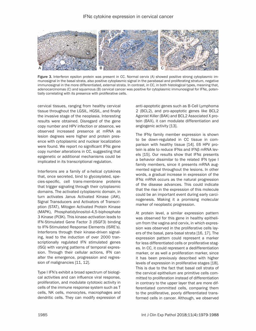
To corroborate the IFNε expression regardless of the HPV genotype and/or infection observed in cervical tissue, immunocytofluorescence in HPV16 (SiHa), HPV18 (HeLa) and HPV- (C33A) cell lines was carried out. We observed positive immunostaining of IFNε protein primarily in cytoplasm in the three cell lines in disregard of the HPV status (Figure 4).
Discussion
IFNε is a relatively new described gene [6]. In the present work, we analyzed the mRNA and protein expression of this molecule in different
Table 1. Statistical correlation between IFNε expression and clinical featuresClinical Variables Average ± SD IFNε (p value)Age 40.44±15.11 0.07 ≥50 14 <50 45 Pregnancies 3.31 ± 2.6 0.05*
≥3 29 <3 30 HPV single or multiple infection 0.90 1 21 2 10 3 or more 5 Negative or not identified 13Menopause 45.78 ± 4.94 0.06 ≥45 13 <45 46 Tobacco consumption 0.03*
Positive 16 Negative 43 Alcohol consumption 0.86 Positive 13 Negative 46 Contraceptives 0.03*
Positive 30 Negative 26 Diagnosis 0.0001*
CC 20 Non CC 36 *Statistical significance.
IFNε protein in cervical tissue by IHC
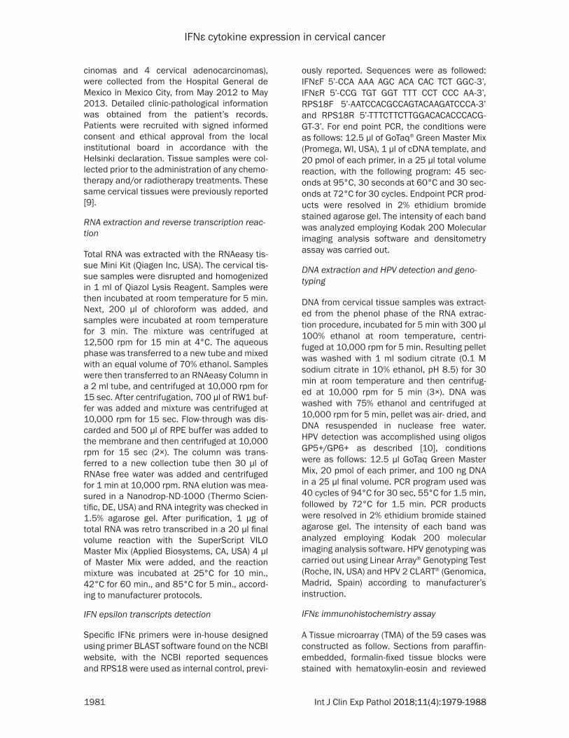
In order to corroborate if the increased mRNA level was due to an up-regula-tion in gene expression or if it was related to the number of cells ex- pressing the gene we carried out immunohistochemistry of the IFNε protein. We observed that IFNε protein immunostaining was primarily present in the epithelial basal cells. Also, less intense signal was present in supra-basal and granular strata and expres-sion was primarily in the proliferative non-differentiated strata cervical cells in the normal cervical tissue. The same IFNε protein expression pattern was observed in the LGSIL, in which the immunostaining was primarily in the basal, suprabasal and less differ-entiated layers of the cervical epitheli-um. Interestingly, samples from the CC group, displayed a positive immunos-taining in all transformed epithelial cells (Figure 3). IFNε expression was not limited to the epithelial cells, but also in the vascular endothelium. In other words, most of the tissues cor-responding to CC showed positive expression. Interestingly, the tissues analyzed for IFNε protein expression showed a similar intensity in expres-sion at the protein level. This could possibly mean that the augmented presence of the mRNA might not nec-
IFNε cytokine expression in cervical cancer
1985 Int J Clin Exp Pathol 2018;11(4):1979-1988
cervical tissues, ranging from healthy cervical tissue throughout the LGSIL, HGSIL, and finally the invasive stage of the neoplasia. Interesting results were obtained. Disregard of the gene copy number and HPV infection or absence, we observed increased presence at mRNA as lesion degrees were higher and protein pres-ence with cytoplasmic and nuclear localization were found. We report no significant IFNε gene copy number alterations in CC, suggesting that epigenetic or additional mechanisms could be implicated in its transcriptional regulation.
Interferons are a family of α-helical cytokines that, once secreted, bind to glycosylated, spe-cies-specific, cell trans-membrane proteins that trigger signaling through their cytoplasmic domains. The activated cytoplasmic domain, in turn activates Janus Activated Kinase (JAK), Signal Transducers and Activators of Transcri- ption (STAT), Mitogen Activated Protein Kinase (MAPK), Phosphatidylinositol-4,5-biphosphate 3 Kinase (PI3K). This kinase-activation leads to IFN-Stimulated Gene Factor 3 (ISGF3) binding to IFN-Stimulated Response Elements (ISRE’s). Interferons through their kinase-driven signal-ing, lead to the induction of over 2000 tran-scriptionally regulated IFN stimulated genes (ISG) with varying patterns of temporal expres-sion. Through their cellular actions, IFN can alter the emergence, progression and regres-sion of malignancies [11, 12].
Type I IFN’s exhibit a broad spectrum of biologi-cal activities and can influence viral response, proliferation, and modulate cytotoxic activity in cells of the immune response system such as T cells, NK cells, monocytes, macrophages and dendritic cells. They can modify expression of
anti-apoptotic genes such as B-Cell Lymphoma 2 (BCL2), and pro-apoptotic genes like BCL2 Agonist Killer (BAK) and BCL2 Associated X pro-tein (BAX), it can modulate differentiation and angiogenic activity [13].
The IFNγ family member expression is shown to be down-regulated in CC tissue in com- parison with healthy tissue [14]. E6 HPV pro- tein is able to reduce IFNα and IFNβ mRNA lev-els [15]. Our results show that IFNε presents a behavior dissimilar to the related IFN type I family members, since it presents mRNA aug-mented signal throughout the lesions. In other words, a gradual increase in expression of the IFNε mRNA occurs as the natural progression of the disease advances. This could indicate that the rise in the expression of this molecule could be an important event during early carci-nogenesis. Making it a promising molecular marker of neoplastic progression.
At protein level, a similar expression pattern was observed for this gene in healthy epitheli-um from the vagina and cervix, in which expres-sion was observed in the proliferative cells lay-ers of the basal, para-basal strata [16, 17]. The expression pattern could represent a marker for less differentiated cells or proliferative stag-es. In CC, it could represent a dedifferentiation marker, or as well a proliferation marker, since it has been previously described with higher levels of expression in proliferative stages [18]. This is due to the fact that basal cell strata of the cervical epithelium are primitive cells com-mitted to proliferation instead of differentiation in contrary to the upper layer that are more dif-ferentiated committed cells, comparing them to the proliferative, poorly differentiated trans-formed cells in cancer. Although, we observed
Figure 3. Interferon epsilon protein was present in CC. Normal cervix (A) showed positive strong cytoplasmic im-munosignal in the basal strata, also positive cytoplasmic signal in the parabasal and proliferating stratum, negative immunosignal in the more differentiated, external strata. In contrast, in CC, in both histological types, meaning that, adenocarcinomas (C) and squamous (B) cervical cancer was positive for cytoplasmic immunosignal for IFNε, poten-tially correlating with its presence with proliferative cells.

IFNε cytokine expression in cervical cancer
1986 Int J Clin Exp Pathol 2018;11(4):1979-1988
positive immune signal in endothelial and fibro-blast cells.
Although the IFNε pathway and biological func-tion are not fully understood, further research is needed. A report showed that CC cell line HeLa express constitutively IFNε and no other IFN family member. In addition, IFNε shows diminished expression leading to attenuated expression and phosphorylation of STAT1 and down regulation of the Retinoic Acid-Inducible Gene 1 (RIG1) expression [19].
Although IFN may be important for cellular homeostasis and resistance to cancer growth, production of endogenous IFN can also contrib-ute to pathology. Increased expression of ISG has been identified in cancer cells compared to corresponding normal primary cells or normal tissues with correlation to the degree of tumor invasion [11]. This could potentially correlate with our results, where low IFNε expression lev-els in healthy tissue may be produced continu-ously and remain localized with no systemic effect [11], while in CC up-regulation of IFNε
Figure 4. Protein expression in distinct HPV-status cervical cancer cell lines. Three cervical cancer cell lines were used. First, SiHa correspond to a HPV16 squamous cell carcinoma, HeLa correspond to an HPV18 cervical ad-enocarcinoma and C33A which correspond to a HPV- squamous cell carcinoma. (A-C) Show HeLa, SiHa and C33A (respectively) DAPI nuclear staining. (D-F) Show the positive, cytoplasmic IFNε immunosignal in the three cell lines. (G-I) Show the merged image from panels (A-F). Positive immunosignal was observed in the three cell lines regard-less HPV status.

IFNε cytokine expression in cervical cancer
1987 Int J Clin Exp Pathol 2018;11(4):1979-1988
gene expression could play an important role. Up-regulation of IFNε expression has been associated to proliferation, and regulated by sex hormones, particularly by estrogen recep-tor [18], which is expressed in CC [20].
In conclusion, an increase in IFNε mRNA could represent a promising molecular marker in CC cells that could have an important role in cellu-lar homeostasis and could influence immune related events in cervical carcinogenesis. Additionally, IFNε expression was not related to HPV infection or genotype, and potentially CNV does not affect IFNε gene expression in CC, suggesting that it could be regulated by epigen-etic mechanisms. Further research is needed to fully elucidate the role of IFNε role in CC. We are currently approaching this issue by more thorough experiments, and the possible valida-tion of molecular marker as diagnostic or prog-nostic is also being addressed by increasing sample number as well as improved patient fol-low up.
Acknowledgements
During this work, the authors VBX, KTP, GPN, VHP, PRM, MRE, and LGV were recipients of a Consejo Nacional de Ciencia y Tecnologia fel-lowship. MS is recipient of a Fundacion IMSS fellowship. The author DMR would like to thank to the Pleyades laboratory members for their support, comments and brilliant discussions. This work was partially supported by grant 202222 from Fondos Sectoriales Consejo Nacional de Ciencia y Tecnologia-Mexico.
Disclosure of conflict of interest
None.
Address correspondence to: Dr. Mauricio Salcedo, Laboratorio de Oncología Genómica, Unidad de Investigacion Medica en Enfermedades Oncologi- cas, Hospital de Oncologia, CMN-SXXI, IMSS, Av. Cuauhtémoc 330, Col. Doctores, México D.F. 06720, México. Tel: +5556276900 Ext. 22706; E-mail: [email protected]; Dr. Marco Meraz-Rios, Departamento de Biomedicina Molecular, Centro de Investigacion y Estudios Avanzados del IPN, Av. I.P.N. 2508, San Pedro Zacatenco, Mexico D.F. 07360, México. E-mail: [email protected]
References
[1] Woodman C, Collins S and Young L. The natu-ral history of cervical HPV infection: unresolved issues. Nat Rev Cancer 2007; 7: 11-22.
[2] Janicek M and Averette M. Cervical cancer: prevention, diagnosis, and therapeutics. CA-Cancer J Clin 2001; 51: 92-114.
[3] Amador-Molina A, Hernandez-Valencia F, Lam-oyi E, Contreras-Paredes A and Lizano M. Role of innate immunity against human papilloma-virus (HPV) infections and effect of adjuvants in promoting specific immune response. Virus-es 2013; 5: 2624-2642.
[4] Hanahan D and Weinberg R. Hallmarks of can-cer: the next generation. Cell 2011; 144: 646-674.
[5] Xi Y, Day S, Jackson R and Ranasinghe C. Role of novel type I interferon epsilon in viral infec-tion and mucosal immunity. Mucosal Immunol 2012; 5: 610-622.
[6] Hardy M, Owczarek C, Jermiin L, Ejdebäck M and Hertzog P. Characterization of the type I interferon locus and identification of novel genes. Genomics 2004; 84: 331-345.
[7] Feng Q, Wei H, Morihara J, Stern J, Yu M, Kiviat N, Hellstrom I and Hellstrom K. Th2 type in-flammation promotes the gradual progression of HPV-infected cervical cells to cervical carci-noma. Gynecol Oncol 2012; 127: 412-419.
[8] Nguyen P, Kafka J, Ferreira V, Roth K and Kaushic C. Innate and adaptive immune re-sponses in male and female reproductive tracts in homeostasis and following HIV infec-tion. Cell Mol Immunol 2014; 11: 410-427.
[9] Marrero-Rodríguez D, Taniguchi-Ponciano K, Jimenez-Vega F, Romero-Morelos P, Mendoza-Rodríguez M, Mantilla A, Rodriguez-Esquivel M, Hernandez D, Hernandez A, Gomez-Gutier-rez G, Munoz-Hernandez N, Arreola-De la Cruz H, Vargas-Requena C, Diaz-Hernandez C, Ser-na-Reyna L, Meraz-Rios M, Bandala C, Ortiz-Leon J and Salcedo M. Krüppel-like factor 5 as potential molecular marker in cervical cancer and the KLF family profile expression. Tumor Biol 2014; 35: 11399-11407.
[10] Husman A, Walboomers J, van den Brule A, Meijer C and Snijders P. The use of general primers GP5 and GP6 elongated at their 3’ ends with adjacent highly conserved sequenc-es improves human papillomavirus detection by PCR. J Gen Virol 1995; 76: 1057-1062.
[11] Cheon H, Borden E and Stark G. Interferons and their stimulated genes in the tumor micro-environment. Semin Oncol 2014; 41: 156-173.
[12] Kalliolias GD and Ivashkiv LB. Overview of the biology of type I interferons. Arthritis Res Ther 2010; 12: S1.
[13] Pestka S, Krause C and Walter M. Interferons, interferon-like cytokines, and their receptors. Immunol Rev 2004; 202: 8-32.
[14] Hazelbag S, Fleuren G, Baelde J, Schuuring E, Kenter G and Gorter A. Cytokine profile of cervi-cal cancer cells. Gynecol Oncol 2001; 83: 235-243.

IFNε cytokine expression in cervical cancer
1988 Int J Clin Exp Pathol 2018;11(4):1979-1988
[15] Nees M, Geogehgan J, Hyman T, Frank S, Miller L and Woodworth C. Papillomavirus type 16 oncogenes downregulate expression of inter-feron-responsive genes and upregulate prolif-eration- associated and NF-KB-Responsive genes in cervical keratinocytes. J Virol 2001; 75: 4283-4296.
[16] Demers A, Kang G, Ma F, Lu W, Yuan Z, Li Y, Lewis M, Edmundo N, Montaner L and Li Q. The mucosal expression pattern of interferon e in rhesus macaques. J Leukocyte Biol 2014; 96: 1101-1107.
[17] Fung K, Mangan N, Cumming H, Horvat J, May-all J, Stifter S, De Weerd N, Roisman LC, Ross-john J, Robertson SA, Schjenken JE, Parker B, Gargett CE, Nguyen HP, Carr DJ, Hansbro PM and Hertzog PJ. Interferon-ε protects the fe-male reproductive tract from viral and bacteri-al infection. Science 2013; 339: 1088-1092.
[18] Eid SG, Mangan NE, Hertzog PJ and Mak J. Blocking HIV-1 transmission in the female re-productive tract: from microbicide develop-ment to exploring local antiviral responses. Clin Transl Immunology 2015; 4: e43.
[19] Matsumiya T, Prescott SM and Stafforini DM. IFN-epsilon mediates TNF-alpha-induced ST- AT1 phosphorylation and induction of retinoic acid-inducible gene-I in human cervical cancer cells. J Immunol 2007; 179: 4542-4549.
[20] López-Romero R, Garrido-Guerrero E, Rangel-López A, Manuel-Apolinar L, Piña-Sánchez P, Lazos-Ochoa M, Mantilla-Morales A, Bandala C and Salcedo M. The cervical malignant cells display a down regulation of ER-α but retain the ER-β expression. Int J Clin Exp Pathol 2013; 6: 1594-1602.