interferon e macrofagos
TRANSCRIPT

8/2/2019 Interferon e Macrofagos
http://slidepdf.com/reader/full/interferon-e-macrofagos 1/9
The EMBO Journal Vol.17 No.13 pp.3660–3668, 1998
Stat1 combines signals derived from IFN- γ and LPSreceptors during macrophage activation
Pavel Kovarik, Dagmar Stoiber,Michael Novy and Thomas Decker1
Vienna Biocenter, Institute of Microbiology and Genetics,Dr Bohr-Gasse 9, A-1030 Vienna, Austria
1Corresponding authore-mail: [email protected]
Complete activation of macrophages during immuneresponses results from stimulation with the activatingcytokine interferon- γ (IFN- γ ) and a second stimulus,usually a microbial product. Bacterial infection of macrophages, or treatment with bacterial lipopoly-saccharide (LPS), resulted in rapid Stat1 phosphoryl-
ation on Ser727 (S727) independently of concomitanttyrosine phosphorylation. IFN- γ also caused rapidphosphorylation of S727. In both situations, S727phosphorylation was reduced by pre-treatment of cellswith the serine kinase inhibitor H7. When macrophageswere treated sequentially or simultaneously with LPSand IFN- γ , the pool of molecules phosphorylated onboth Tyr701 (Y701) and S727 was strongly increased.Consistently, Stat1-dependent transcription in responseto IFN- γ was significantly enhanced if the cells werepre-treated with bacterial LPS. The relative amountof S727-phosphorylated Stat1 in the non-tyrosine phos-phorylated fraction was considerably smaller than thatin the tyrosine-phosphorylated fraction. No evidence
was found for an effect of S727 phosphorylation onthe phosphorylation of Y701 by IFN- γ . Thus, serineand tyrosine phosphorylation of Stat1 are caused inde-pendently of each other, but the serine kinase mayrecognize tyrosine-phosphorylated Stat1 preferentiallyin the course of an IFN- γ response. The data suggestStat1 to be a convergence point for immunologicalstimuli in a macrophage proinflammatory response.Keywords: IFN/LPS/serine phosphorylation/Stat/ transcription
Introduction
Innate immunity provides an immediate host responseagainst bacterial infection. The antibacterial mechanismsrecruited by the innate immune system usually are trig-gered by the invading microorganism itself. The adaptiveimmune response, ultimately orchestrated by lymphocytes,arises as a consequence of antigen presentation andenhances the antibacterial response through antigen-specific mechanisms. However, cells of the adaptiveimmune system also provide a positive feed-back to theinnate immune system through the synthesis of cytokinesthat either increase effector cell numbers or activate thesecells for an increased antibacterial performance. This has
3660 © Oxford University Press
been well documented in the case of macrophages. Theserespond to the initial bacterial infection with an immediatesynthesis of proinflammatory mediators and contribute inmany ways to natural immunity, but also to the onset of a specific immune response (Kielian and Blecha, 1995;Viriyakosol and Kirkland, 1995). Once activated Th1 cellsare present, they provide the macrophage-activating factorinterferon-γ (IFN-γ ) which acts in concert with productsof the microbe, for example the cell wall componentlipopolysaccharide (LPS), to generate macrophages witha maximal capacity to ingest and kill microbes, as wellas to synthesize membrane receptors and secreted molec-ules that function in a proinflammatory immune response(Nacy and Meltzer, 1991).
The intracellular events leading to macrophage activa-tion have been uncovered to some extent. IFN-γ binds toa hetero-oligomeric, class II cytokine receptor and activatesa Jak–Stat signaling pathway consisting of the Jak1 andJak2 kinases as well as the transcription factor signaltransducer and activator of transcription 1 (Stat1). Stat1binds to the cytoplasmic portion of the ligand-activatedIFN-γ receptor and is phosphorylated by Jaks on a singletyrosine residue (Y701). Phosphorylation on Y701 causesSH2 domain-mediated dimerization, nuclear translocationand binding of Stat1 to its cognate promoter DNA, thegamma interferon-activated site (GAS; reviewed in Darnellet al., 1994; Muller et al., 1994; Schindler, 1995; Leamanet al., 1996; Darnell, 1997; Decker, 1997; Decker et al.,1997). To act with maximal efficiency as a transcriptionfactor, Stat1 must also be phosphorylated on S727 whichlies within a potential MAPK consensus motif at theC-terminus, a putative transactivation domain (Wen et al.,1995). At present, the kinase-phosphorylating S727 is notclearly defined. The MAPK ERK2 was suggested to exertStat1 serine kinase activity (David et al., 1995), but theextent to which this applies is currently a matter of debate(Chung et al., 1997; Nishiya et al., 1997; Zhu et al.,1997). Zhu and colleagues recently provided preliminaryevidence for a Stat1 serine kinase distinct from ERKs(Zhu et al., 1997).
Early post-receptor events in LPS-induced signal trans-
duction involve tyrosine phosphorylation, but it is unclearwhich tyrosine kinases are critically involved in signaltransduction downstream of the CD14 or other LPSreceptors (reviewed in Sweet and Hume, 1996). In additionto the tyrosine kinases, a number of serine kinases wereshown to be activated by LPS, including c-Raf, the MAPkinases Erk1 and Erk2, the p38 kinase, c-Jun kinase andceramide-activated kinase (CAK; Joseph et al., 1994;Reimann et al., 1994; Buscher et al., 1995; Wright andKolesnick, 1995; Hambleton et al., 1996; Sanghera et al.,1996; Meng and Lowell, 1997). Importantly, LPS alsoactivates Iκ B kinase. NF-κ B, and to some extent AP1,plays a central role in the transcriptional activation of

8/2/2019 Interferon e Macrofagos
http://slidepdf.com/reader/full/interferon-e-macrofagos 2/9
Stat1 serine phosphorylation by LPS
Fig. 1. Specificity of the anti-Stat1 pS727 serum. 293 cells weretransfected with an S727A mutant of Stat1 which was tagged at theN-terminus with myc epitopes and thus migrated more slowly inSDS–PAGE. Half of the transfected cells were treated with IFN-γ .Extracts from IFN-γ -treated and untreated cells were subjected toimmunoprecipitation with anti-Stat1α serum and the precipitates werestained with anti-pS727 serum or normal anti-Stat1α serum. Theadditional, lower molecular weight bands in the Stat1 blot stem fromdegradation products derived from transfected Stat1S727A.
numerous genes encoding immunologically importantcytokines or other mediators (reviewed in Baeuerle andHenkel, 1994; Baeuerle and Baltimore, 1996). Therefore,the functional cooperation between IFN-γ and LPS canbe explained in terms of each inducing a set of proinflam-matory gene products. However, some genes, such asthose encoding iNOS, transcription factor IRF-1 or thecell adhesion molecule ICAM-1, were also shown to beinduced cooperatively by NF-κ B and Stat1 (Sims et al.,1993; Caldenhoven et al., 1994; Look et al., 1994; Xieet al., 1994; Ledebur and Parks, 1995; Gao et al., 1997;Pine, 1997).
In the present study, we investigated whether bacteriaand their products directly influence the output of the Jak–Stat signal transduction pathway. Our results suggest thatbacterial infections augment Stat1-mediated transcriptionby activating a kinase capable of phosphorylating S727.
Results
An antiserum specifically recognizing Stat1phosphorylated on Ser727 We immunized rabbits with a peptide corresponding topart of the Stat1 C-terminus and including phosphorylatedS727. The resulting serum (hereafter designated anti-pS727) was tested by enzyme-linked immunosorbent assay(ELISA) and in Western blots of gels in which immunopre-cipitates of cellular Stat1 were compared with recombinant(unphosphorylated) Stat1 C-terminus or Stat1S727Amutant C-terminus. These experiments revealed an ~1000-
fold preference of the antibodies for the phosphorylatedC-terminus of Stat1 (data not shown). The specificity of anti-pS727 was demonstrated further in an experimentshown in Figure 1. 293 kidney fibroblasts were transfectedwith a plasmid expressing high levels of myc epitope-tagged Stat1S727A. The transfected cells expressedapproximately equal amounts of endogenous wtStat1 andexogenous Stat1S727A. Following stimulation with IFN-γ and immunoprecipitation of cellular extracts, an increasedsignal of wtStat1 was observed in Western blots with anti-pS727. In contrast, Stat1S727A did not react with theantibody in either unstimulated or IFN-γ -treated cells. Theexperiment thus indicates that some wtStat1 phosphoryl-
3661
Fig. 2. Induction of Stat1 S727 phosphorylation in U937 cells andhuman PBMC. (A) Cells from a proliferating culture (lane 1) or afterovernight starvation were stimulated for 20 min with the indicatedagents. (B) Human PBMC were prepared under serum-free conditionsand stimulated with IFNs for 20 min. Determination of Stat1 S727phosphorylation was with anti-pS727 as described for Figure 1.
ated on S727 is present in 293 cells which, in accordancewith published results (Wen et al., 1995), is stronglyincreased by treatment with IFN-γ . The lack of reactivitywith our antiserum of the S727A mutant under all condi-tions suggested the suitability of our antibodies to revealthe activity of the Stat1 S727 kinase and thus to study itsregulation by stimuli of cell-surface receptors. To obtainfurther evidence for this assumption, we tested a numberof stimuli previously implicated in causing Stat1 phospho-rylation on S727 (Figure 2A; Wen et al., 1995). U937promonocytes grown in the presence of 10% fetal calf serum (FCS) produced a Stat1 signal with anti-pS727.Starvation from serum for 20 h completely abolished Stat1reactivity with anti-pS727, and a brief stimulation with10% re-established it. Both types of IFN and phorbol estercaused a Stat1 signal in Western blots stained with anti-pS727. These results show beyond doubt that anti-pS727correctly reveals the regulation of the Stat1 S727 kinasein intact cells.
The presence of factors stimulating S727 kinase activity
in serum raised the question of whether such factors arepresent in blood plasma, thus causing constitutive S727phosphorylation in blood leukocytes. Peripheral bloodmononuclear cells (PBMC) isolated from whole blooddemonstrated a low constitutive S727 phosphorylation of Stat1 which could be stimulated significantly by bothIFN-α and IFN-γ (Figure 2B). This result confirms theimportance of regulating S727 phosphorylation in vivo.
Bacterial infection and LPS activate the Stat1 S727 kinase The experiment shown in Figure 2A demonstrates rapidphosphorylation of Stat1 S727 in response to the phorbol

8/2/2019 Interferon e Macrofagos
http://slidepdf.com/reader/full/interferon-e-macrofagos 3/9
P.Kovarik et al .
Fig. 3. The effect of bacteria, LPS or CSF-1 on Stat1 S727phosphorylation in mouse macrophages. (A) Bac1.2F5 macrophages
were either infected with S.typhimurium or treated with LPS for20 min. Stimulation with human recombinant CSF-1 was for 10 min.Determination of Stat1 S727 phosphorylation was with anti-pS727 asdescribed for Figure 1. (B) Extracts from Bac1.2F5 cells after LPStreatment or Salmonella infection were assayed for tyrosinephosphorylation of Stats in the EMSA with a β-casein GAS sequenceas a labeled probe.
Fig. 4. The kinetics of Stat1 S727 phosphorylation by LPS. Bac1.2F5macrophages were stimulated with LPS for the indicated periods.Determination of Stat1 S727 phosphorylation in cellular extracts waswith anti-pS727 as described for Figure 1.
ester TPA. Since TPA does not cause Stat1 phosphorylationon tyrosine (Eilers et al., 1994), the drug exploits thepossibility of independently regulating Y701 and S727phosphorylation. We investigated the possibility that thismight also occur in response to (patho-)physiologicalstimuli that cooperate with IFN-γ in causing macrophageactivation. Macrophages were infected with Salmonellatyphimurium for 20 min. Alternatively, macrophages were
treated with LPS. In either case, a strong stimulation of Stat1 phosphorylation on S727 was observed which wassimilar, or even superior, to that caused by IFN-γ (Figure 3A). By contrast, no evidence for tyrosine-phos-phorylated Stats was observed in the EMSA upon brief treatment of macrophages with LPS or after brief infectionwith S.typhimurium, suggesting that LPS did not activatea Jak–Stat pathway (Figure 3B). The kinetics of S727kinase activation by LPS are shown in Figure 4. Import-antly, LPS rapidly activated S727 kinase, thus ruling outthe possibility of activation by indirect mechanisms (seealso Figure 9C). The kinetics of LPS- and IFN-γ -inducedS727 phosphorylation were found to be similarly rapid
3662
Fig. 5. The effect of serine kinase inhibitor H7 on Stat1 S727phosphorylation. (A) Bac1.2F5 macrophages were pre-treated with50 µM H7 for 10 min and subsequently stimulated with IFN-γ for25 min. (B) Macrophages were pre-treated similarly with H7 andsubsequently stimulated with LPS for 25 min. Determination of Stat1S727 phosphorylation was with anti-pS727 as described for Figure 1.
Fig. 6. Activation of MAP kinases ERK1 and ERK2 by IFNs andLPS. Bac1.2F5 cells were stimulated with CSF-1 for 10 min, or withIFN-α, IFN-γ or LPS for 20 min. Simultaneous treatment with IFNsand LPS was for 25 min. Following stimulation, cellular extracts wereprepared and ERK activation was measured by in-gel phosphorylationof co-polymerized myelin basic protein.
(Figures 4 and 9C). Consistent with previous data (Zhu
et al., 1997), maximal levels of tyrosine phosphorylationin response to IFN-γ were reached earlier than maximallevels of S727 (data not shown).
LPS- and IFN-γ -induced activation of the Stat1 S727kinase were sensitive to the serine kinase inhibitor H7(Figure 5). Moreover, H7 also suppressed basal S727phosphorylation caused by serum factors (Figure 5A). Insix independent experiments, the inhibition of IFN-γ -mediated S727 phosphorylation ranged between 50 and80%. On average, the drug was less efficient in inhibitingkinase activation by LPS (between 20 and 70% inhibitionin five independent experiments). It is unclear at presentwhether incomplete inhibition by H7, particularly of LPS-

8/2/2019 Interferon e Macrofagos
http://slidepdf.com/reader/full/interferon-e-macrofagos 4/9
Stat1 serine phosphorylation by LPS
induced S727 phosphorylation, indicates the presence of H7-sensitive and -insensitive pathways targeting this Stat1phosphorylation site.
We tested whether the MAP kinases ERK1 and ERK2might be a common downstream target of LPS and IFN-γ to cause S727 phosphorylation in macrophages. As apositive control, colony-stimulating factor 1 (CSF-1),which is known to cause robust MAPK activation inBac1.2F5 macrophages, was used. Our results (Figure 6)demonstrate strong activation of both ERKs by CSF-1and LPS. By contrast, IFN-γ caused very little ERK2activation and no activation of ERK1. IFN-α activated
3663
ERKs, but to a fairly small extent compared with CSF-1or LPS. Combined treatment with IFNs and LPS slightlyincreased activation by LPS alone, but this effect was notvery pronounced. Thus, MAPK activity can be uncoupledfrom S727 kinase because IFN-γ causes robust S727phosphorylation whereas CSF-1 causes no detectable S727kinase activation (Figure 3). To assess further the possibil-ity of Stat1 phosphorylation by ERKs, for example in thecase of LPS, we performed in-gel kinase assays or ERK2immune complex kinase assays using recombinant Stat1C-terminus as a substrate. These experiments revealed nodetectable ability of CSF-1 or Salmonella-activated ERKsto phosphorylate S727 (data not shown).
LPS pre-treatment increases the transcriptional response to IFN- γ To be functionally meaningful, LPS must cooperate withIFN-γ to increase the pool of Stat1 molecules phosphoryl-ated on both Y701 and S727, and this pool of moleculesmust cause an increased target gene transcription rate. Wefirst tested the effect of LPS pre-treatment on IFN-γ activation of Stat1. Macrophage extracts after treatmentwith IFN-γ alone, or after LPS pre-treatment and sub-sequent IFN-γ stimulation, were immunoprecipitated witheither anti-pS727 antibodies or with antibodies to theStat1 C-terminus. Western blots of these precipitates weredeveloped with either pS727 serum (in the case of Stat1immunoprecipitation only), with anti-phosphotyrosinemonoclonal antibody or with Stat1 antibodies. This experi-ment (Figure 7A) showed that the pS727-specific antibod-ies precipitate much more tyrosine-phosphorylated Stat1from LPS-pre-treated cells pulsed for a brief period(10 min) with IFN-γ . In fact, precipitation of tyrosine-phosphorylated Stat1 with anti-pS727 serum from cellstreated for 10 min with IFN-γ alone was below the levelof detection. The control Stat1 antibody precipitated equalamounts of tyrosine-phosphorylated protein from IFN-γ -treated and pre-LPS/IFN-γ -treated cells (note that theantibody to the Stat1 C-terminus precipitates more effici-ently compared with the pS727 antiserum; therefore signalintensities in Figure 7A and B cannot be compareddirectly). This result indicates a larger amount of tyrosine-phosphorylated molecules in the S727-phosphorylatedStat1 pool after LPS pre-treatment and allows the conclu-
Fig. 7. The effect of LPS pre-treatment on the S727 phosphorylationstatus of tyrosine-phosphorylated Stat1 isolated from IFN-γ - or LPS/ IFN-γ -treated macrophages. Bac1.2F5 macrophages were pre-treatedwith LPS for 20 min, and untreated or pre-treated cells werestimulated with IFN-γ and LPS for 10 min. Pre-treated control cellswere stimulated further with LPS alone for another 10 min. Extracts
were subjected to immunoprecipitation with either anti-pS727 serum(A) or serum against the Stat1 C-terminus (B) as indicated. Westernblots of the immunoprecipitates were developed with anti-pS727antiserum [only Stat1Ab immunoprecipitate in (B)], anti-phosphotyrosine or anti-Stat1 antibodies. Note that the antibody to theStat1 C-terminus precipitates more efficiently compared with thepS727 antiserum; therefore, signal intensities in (A) and (B) cannot becompared directly. Levels of Stat1 precipitated by the anti-pS727serum from cells treated for 10 min with IFN-γ alone were below thelevel of detection. (C) Extracts derived from macrophages after10 min of treatment with IFN-γ alone, or with IFN-γ for 10 min afterpre-treatment with LPS for 20 min, were subjected to EMSA with aβ-casein probe. Where indicated, supershift analysis was performedwith anti-pS727 antibodies. The supershifted Stat1 band is marked byan asterisk.
8/2/2019 Interferon e Macrofagos
http://slidepdf.com/reader/full/interferon-e-macrofagos 5/9
P.Kovarik et al .
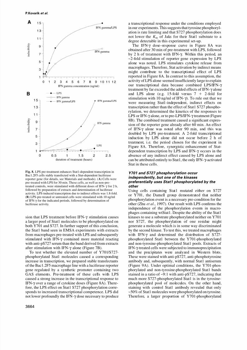
Fig. 8. LPS pre-treatment enhances Stat1-dependent transcription inBac1.2F5 cells stably transfected with a Stat-dependent luciferasereporter gene (for details, see Materials and methods). (A) Cells were
pre-treated with LPS for 30 min. These cells, as well as non-pre-treated controls, were stimulated with different doses of IFN-γ for 2 h,followed by preparation of extracts and determination of luciferaseactivity. LPS-induced transcription due to indirect effects was 1.8-fold.(B) LPS-pre-treated or untreated cells were stimulated with 10 ng/mlof IFN-γ for the indicated periods, followed by determination of luciferase activity.
sion that LPS treatment before IFN-γ stimulation causesa larger pool of Stat1 molecules to be phosphorylated onboth Y701 and S727. In further support of this conclusion,the Stat1 band seen in EMSA experiments with extractsfrom macrophages pre-treated with LPS and subsequentlystimulated with IFN-γ contained more material reacting
with anti-pS727 serum than the band derived from extractsafter stimulation with IFN-γ alone (Figure 7B).To test whether the elevated number of Y701/S727-
phosphorylated Stat1 molecules caused a correspondingincrease in transcription, we prepared stable transfectantsof the Bac1.2F5 macrophage line with a luciferase reportergene regulated by a synthetic promoter containing twoGAS elements. Pre-treatment of these cells with LPScaused a strong increase in the transcriptional response toIFN-γ over a range of cytokine doses (Figure 8A). There-fore, the LPS effect on Stat1 S727 phosphorylation corre-sponds to increased transcriptional competence. LPS didnot lower profoundly the IFN-γ dose necessary to produce
3664
a transcriptional response under the conditions employedin our experiments. This suggests that tyrosine phosphoryl-ation is rate limiting and that S727 phosphorylation doesnot lower the K m of Jaks for their Stat1 substrate to adegree detectable in this experimental set-up.
The IFN-γ dose–response curve in Figure 8A wasobtained after 30 min of pre-treatment with LPS, followedby 2 h of treatment with IFN-γ . Within this period, an~2-fold stimulation of reporter gene expression by LPSalone was noted. LPS stimulates cytokine release frommacrophages. Therefore, Stat activation by indirect meansmight contribute to the transcriptional effect of LPSreported in Figure 8A. In contrast to this assumption, theactivity of LPS alone seemed insufficiently large to explainour transcriptional data because combined LPS/IFN-γ treatment by far exceeded the added effects of IFN-γ aloneand LPS alone (e.g. 15-fold versus 7 2-fold forstimulation with 10 ng/ml of IFN-γ ). To rule out that wewere measuring Stat1-independent, indirect effects ontranscription rather than the effect of Stat1 S727 phospho-rylation, we determined the kinetics of the responses toLPS or IFN-γ alone, or to pre-LPS/IFN-γ treatment (Figure8B). The combined treatment caused a significant expres-sion of the reporter gene already after 60 min. An effectof IFN-γ alone was noted after 90 min, and this wasdoubled by LPS pre-treatment. A 2-fold transcriptionalinduction by LPS alone did not occur before 2 h of treatment, i.e. the period chosen for the experiment inFigure 8A. Therefore, synergistic enhancement of Stat-dependent transcription by LPS and IFN-γ occurs in theabsence of any indirect effect caused by LPS alone andcan be attributed entirely to Stat1, the only IFN-γ -activatedStat in these cells.
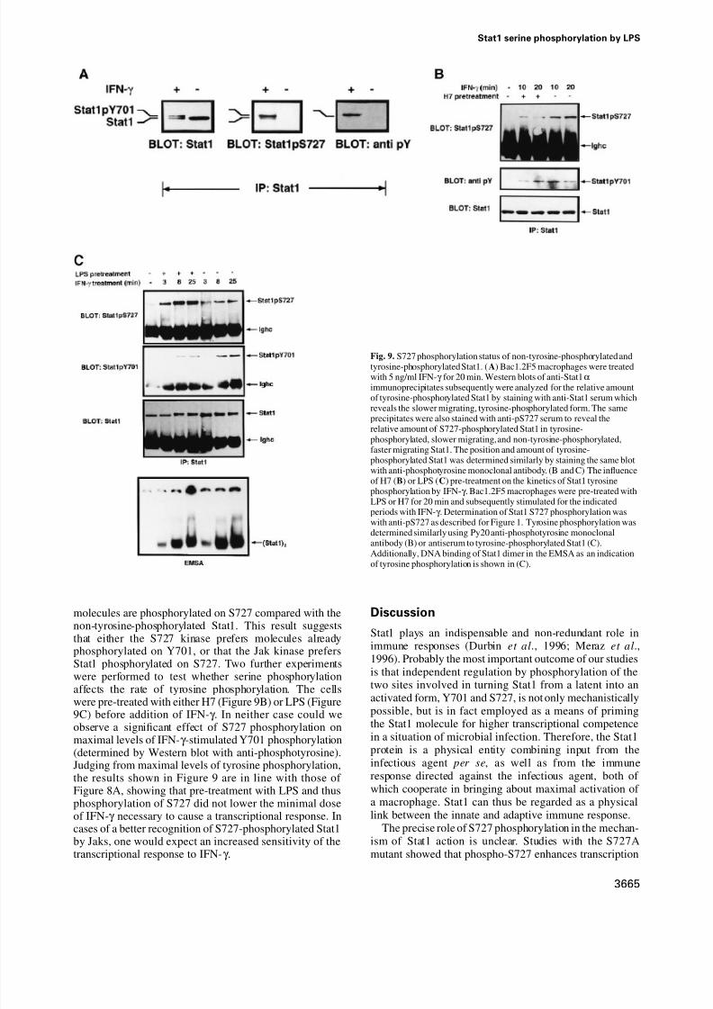
Y701 and S727 phosphorylation occur independently, but one of the kinases preferentially uses Stat1 phosphorylated by the other Using cells containing Stat1 mutated either on S727or Y701, the Darnell group demonstrated that neitherphosphorylation event is a necessary pre-condition for theother (Zhu et al., 1997). Our result with LPS confirms theindependence of the phosphorylation events in macro-phages containing wtStat1. Despite the ability of the Stat1kinases to use a substrate phosphorylated neither on Y701nor S727, the phosphorylation of one residue mightgenerate a molecule which is in some way discriminatedby the second kinase. To test this, we treated macrophageswith IFN-γ and determined the distribution of S727-phosphorylated Stat1 between the Y701-phosphorylated
and non-tyrosine-phosphorylated Stat1 pools. Extracts of IFN-γ -treated cells were subjected to immunoprecipitationand the precipitates were analyzed in Western blots.These were stained with anti-pS727, anti-phosphotyrosineantibody and, subsequently, with normal Stat1 antiserum(Figure 9A). Under optimal conditions, the Y701-phos-phorylated and non-tyrosine-phosphorylated Stat1 bandsstained in a ratio of ~9:1 with anti-pS727, indicating thatmuch more S727-phosphorylated Stat1 is in the tyrosine-phosphorylated pool of molecules. On the other hand,staining with control Stat1 antibody revealed that only~50% of Stat1 molecules were phosphorylated on tyrosine.Therefore, a larger proportion of Y701-phosphorylated
8/2/2019 Interferon e Macrofagos
http://slidepdf.com/reader/full/interferon-e-macrofagos 6/9
Stat1 serine phosphorylation by LPS
Fig. 9. S727 phosphorylation status of non-tyrosine-phosphorylated andtyrosine-phosphorylated Stat1. (A) Bac1.2F5 macrophages were treatedwith 5 ng/ml IFN-γ for 20 min. Western blots of anti-Stat1αimmunoprecipitates subsequently were analyzed for the relative amountof tyrosine-phosphorylated Stat1 by staining with anti-Stat1 serum whichreveals the slower migrating, tyrosine-phosphorylated form. The sameprecipitates were also stained with anti-pS727 serum to reveal therelative amount of S727-phosphorylated Stat1 in tyrosine-phosphorylated, slower migrating, and non-tyrosine-phosphorylated,faster migrating Stat1. The position and amount of tyrosine-phosphorylated Stat1 was determined similarly by staining the same blotwith anti-phosphotyrosine monoclonal antibody. (B and C) The influenceof H7 (B) or LPS (C) pre-treatment on the kinetics of Stat1 tyrosinephosphorylation by IFN-γ . Bac1.2F5 macrophages were pre-treated withLPS or H7 for 20 min and subsequently stimulated for the indicatedperiods with IFN-γ . Determination of Stat1 S727 phosphorylation waswith anti-pS727 as described for Figure 1. Tyrosine phosphorylation was
determined similarly using Py20 anti-phosphotyrosine monoclonalantibody (B) or antiserum to tyrosine-phosphorylated Stat1 (C).Additionally, DNA binding of Stat1 dimer in the EMSA as an indicationof tyrosine phosphorylation is shown in (C).
molecules are phosphorylated on S727 compared with thenon-tyrosine-phosphorylated Stat1. This result suggeststhat either the S727 kinase prefers molecules alreadyphosphorylated on Y701, or that the Jak kinase prefersStat1 phosphorylated on S727. Two further experimentswere performed to test whether serine phosphorylationaffects the rate of tyrosine phosphorylation. The cells
were pre-treated with either H7 (Figure 9B) or LPS (Figure9C) before addition of IFN-γ . In neither case could weobserve a significant effect of S727 phosphorylation onmaximal levels of IFN-γ -stimulated Y701 phosphorylation(determined by Western blot with anti-phosphotyrosine).Judging from maximal levels of tyrosine phosphorylation,the results shown in Figure 9 are in line with those of Figure 8A, showing that pre-treatment with LPS and thusphosphorylation of S727 did not lower the minimal doseof IFN-γ necessary to cause a transcriptional response. Incases of a better recognition of S727-phosphorylated Stat1by Jaks, one would expect an increased sensitivity of thetranscriptional response to IFN-γ .
3665
Discussion
Stat1 plays an indispensable and non-redundant role inimmune responses (Durbin et al., 1996; Meraz et al.,1996). Probably the most important outcome of our studiesis that independent regulation by phosphorylation of thetwo sites involved in turning Stat1 from a latent into anactivated form, Y701 and S727, is not only mechanistically
possible, but is in fact employed as a means of primingthe Stat1 molecule for higher transcriptional competencein a situation of microbial infection. Therefore, the Stat1protein is a physical entity combining input from theinfectious agent per se, as well as from the immuneresponse directed against the infectious agent, both of which cooperate in bringing about maximal activation of a macrophage. Stat1 can thus be regarded as a physicallink between the innate and adaptive immune response.
The precise role of S727 phosphorylation in the mechan-ism of Stat1 action is unclear. Studies with the S727Amutant showed that phospho-S727 enhances transcription

8/2/2019 Interferon e Macrofagos
http://slidepdf.com/reader/full/interferon-e-macrofagos 7/9
P.Kovarik et al .
factor activity, and it is clear from the data provided herethat LPS-induced S727 phosphorylation increases theStat1-dependent transcriptional response. A likely explana-tion for this effect is the phosphorylation-dependent associ-ation of a transcriptional Stat1 cooperation partner orcoactivator. CBP/p300 proteins, coactivators recentlyimplicated in Stat transcriptional responses, appear to bindto Stat1 independently of S727 phosphorylation (Zhanget al., 1996). Therefore, the relevant target of the phos-phorylated C-terminus currently is elusive.
The occurrence of regulated serine phosphorylationrecently has been recognized for most Stats (Boultonet al., 1995; Eilers et al., 1995; Lutticken et al., 1995;Wen et al., 1995; Zhang et al., 1995; Beadling et al.,1996; Cho et al., 1996; Kirken et al., 1997a,b). However,only in the case of Stat1 and Stat3 has a target sitefor proline-directed serine kinase activity been definedprecisely (Wen et al., 1995; Wen and Darnell, 1997). Thedata provided in our study, as well as recent experimentalevidence published by other laboratories (Chung et al.,1997; Nishiya et al., 1997; Zhu et al., 1997), while notruling out specific situations in which Erks may act asStat1 kinases, disprove the notion that the Stat1 C-terminusmight generally couple ERK activity to Stat activation.Firstly, ERK activation by ligands that activate Stats doesnot correspond to S727 phosphorylation in a number of situations, and this includes CSF-1 or IFN-γ stimulationof macrophages as shown in our report. Secondly, inaccordance with a previously published report, we findthat the Stat1 C-terminus is not phosphorylated readily byERKs in situ or in vitro (Chung et al., 1997). Finally,Darnell and co-workers recently have presented prelimin-ary evidence for a Stat1 serine kinase distinct from ERKswith the ability to phosphorylate S727 (Zhu et al., 1997).In the case of Stat3, the situation may be different,and evidence for both ERK-dependent and -independentphosphorylation of Stat3 S727 has been published (Chunget al., 1997). Thus, the question remains open as to whatextent Stat1 and Stat3 S727 kinases are identical. For bothStat1 and Stat3, it is clear, however, that either distinctserine kinases exist or that there is more than one signaltransduction pathway that targets them. In the case of Stat3, this was shown by Cantrell’s group using the kinaseinhibitors H7 and PD98059 and stimulation of serinephosphorylation either by CD3 activation or by stimulationwith interleukin-2 (IL-2) (Ng and Cantrell, 1997). Stat1S727 may be targeted similarly by H7-sensitive and-insensitive signaling pathways, as suggested by theincomplete inhibition of S727 phosphorylation, especiallyin the case of LPS treatment. For IFN-γ -stimulated S727
kinase activation, a requirement for Jak2 has been demon-strated. We and others (Deng et al., 1996) have notobserved stimulation of a Jak–Stat signaling pathway inLPS-treated macrophages, and would therefore concludethat Jaks are not needed for LPS-induced S727 phospho-rylation. However, Tsukada and colleagues recentlyreported activation of a tyrosine-phosphorylated, Stat-liketranscription factor by LPS and IL-1 which does not bindto GAS elements (Tsukada et al., 1996). Therefore, thepotential role of Jaks in LPS signal transduction still needsto be clarified.
The experiment shown in Figure 9A suggests that thephosphorylation events on Y701 or S727 influence each
3666
other. Unlike what was recently suggested for Stat3(Chung et al., 1997), we find no evidence for a negativeinterference of serine with tyrosine phosphorylation. Thedata clearly indicate a relatively larger proportion of S727phosphorylated in the tyrosine-phosphorylated fraction of molecules. Our further experiments (Figure 9B and C)argue against a positive effect of phospho-S727 on Y701phosphorylation, at least in the sense that the maximallevels of tyrosine phosphorylation that can be caused byIFN-γ are the same with or without priming Stat1 onS727. Therefore, we are led to conclude that the S727kinase may phosphorylate tyrosine-phosphorylated Stat1preferentially. The Y701 phosphogroup might increasethe affinity of association, or the S727 kinase mightpreferentially recognize Stat1 dimers. Alternatively, thealtered subcellular localization of tyrosine-phosphorylatedStat1 might increase the accessibility for the serine kinase.
In conclusion, our study provides evidence for theimportance of Stat serine phosphorylation during theimmune response to bacterial infections. Issues connectedto signal transduction and the function of the S727 kinasewill be a further subject of future studies.
Materials and methods
Cells, cytokines and drugs U937 promonocytes were cultured in RPMI medium containing 10%FCS. Bac1.2F5 cells, or Bac1.2F5-derived clones carrying a Stat-dependent reporter plasmid (see below), represent an early stage of mature macrophages and grow in a CSF-1-dependent manner (Morganet al., 1987). They were grown in Dulbecco’s modified Eagle’s medium(DMEM) with 10% FCS and 20% L-cell conditioned medium as asource of CSF-1. For stimulation with CSF-1, 10 000 U/ml of humanrecombinant CSF-1 (a kind gift from J.Schreurs, Chiron Corp.,Emeryville, CA) were used. Primary mouse bone macrophages wereobtained from femurs as described (Baccarini et al., 1985). HumanPBMC were obtained from whole blood of healthy donors (P.K. andT.D.) by Ficoll gradient centrifugation. The cells contained ~20%monocytes. Recombinant human IFN-α and IFN-γ were kind gifts of
P.von Wussow (Hannover Medical School, Hannover, Germany). Murinerecombinant IFN-γ was kindly provided by G.Adolf (Bender, Vienna).Murine recombinant IFN-α was purchased from Hycult biotechnology(Uden, The Netherlands). For stimulation of cells, IFN-α was used at aconcentration of 1000 U/ml; IFN-γ was used at a concentration of 5 ng/ ml, unless otherwise indicated. LPS (Sigma, St Louis, MO) was eitherfrom Salmonella or Escherichia coli and was used at a concentration of 1 µg/ml, unless stated otherwise. The phorbol ester TPA (Sigma, St Louis,MO) was used at a concentration of 50 nM. The serine kinase inhibitorH7 (Calbiochem, La Jolla, CA) was used at 50 µM.
Plasmids A Stat-dependent reporter plasmid (IFP53GAS2-Luc) was constructedby inserting two copies of an oligonucleotide comprising the IFP53-GAS sequence (Strehlow et al., 1993) into vector pADneo2Bgluci (akind gift of C.Stratowa, Bender and Co., Vienna). This sets the GASsequences in front of a β-globin minimal promoter and a luciferase
reporter gene. Additionally, the vector contains a Rous sarcoma virus(RSV) promoter-driven neo gene for selection.
Antibodies Rabbit antiserum to phospho-S727-Stat1 (anti-pS727) was obtained byimmunization with keyhole limpet hemocyanin (KLH)-coupled phospho-peptide with the sequence DNLLPMpSPEEFDE (synthesized byResearch Genetics, Huntsville, AL). The resulting serum was used inWestern blots at a dilution of 1:5000, in immunoprecipitation at adilution of 1:50 and in ‘supershift’ EMSA experiments at 1:125. Rabbitantiserum to the Stat1α C-terminus was obtained by immunizing with aGST fusion protein comprising the 39 C-terminal amino acids of Stat1α(plasmid kindly provided by Chris Schindler, Columbia University, NY).The serum was used for immunoprecipitation at a dilution of 1:100 andin Western blots at a dilution of 1:5000. Rabbit antiserum specific for
8/2/2019 Interferon e Macrofagos
http://slidepdf.com/reader/full/interferon-e-macrofagos 8/9
Stat1 serine phosphorylation by LPS
the tyrosine-phosphorylated Stat1 was a kind gift of David Frank andMichael Greenberg (Harvard Medical School). The antiserum was usedfor Western blots at a dilution of 1:500. Mouse monoclonal antibodyPy20, directed against phosphotyrosine, was purchased from Transduc-tion Laboratories (Lexington, KY) and used for Western blots at 2 µg/ml.
Cellular extracts Methods to generate nuclear or whole-cell extracts for EMSA have beendescribed (Eilers et al., 1994 and references therein). For immunoprecipi-tation and Western blot analysis, the cells were lysed in lysis buffercontaining 10 mM Tris–HCl (pH 7.05), 50 mM NaCl, 30 mM NaPPi,50 mM NaF, 2 mM EDTA, 1%TritonX-100, 1 mM phenylmethylsulfonylfluoride, 0.1 mM sodium vanadate, pepstatin 1 µg/ml, leupeptin0.5 µg/ml, aprotinin 3 µg/ml. Extracts were cleared by centrifugation at15 000 r.p.m., 4°C, 8 min.
Electrophoretic mobility shift assay (EMSA) We recently have described this assay using an oligonucleotide corres-ponding to the β-casein GAS sequence as a probe (Gouilleux et al.,1995; Woldman et al., 1997).
Immunoprecipitation and Western blot analysis Cellular extracts were normalized on the basis of protein determinations.Normalized volumes of extracts were incubated for 2–4 h at 4°Cwith specific antibodies. Immune complexes were collected followingincubation (2–12 h at 4°C) with protein A–Sepharose beads. Immunopre-cipitates were washed four times with lysis buffer. The beads wereeluted by boiling in Laemmli sample buffer. Proteins were resolved by10% SDS–PAGE and electrotransferred to nitrocellulose. Membraneswere blocked with 2% bovine serum albumin (BSA) in phosphate-buffered saline including 0.1% Tween-20 (PBST) and incubated withthe appropriate concentration of specific antibody, diluted in PBST 2% BSA. After washing, the blots were incubated with horseradishperoxidase-conjugated second antibody and developed using an ECLsystem (Amersham, UK) according to the manufacturer’s instructions.
In-gel assay for MAP kinase activationDenatured protein samples (15 µg/lane) were fractionated over a 10%SDS–acrylamide gel containing 0.2 mg/ml co-polymerized substrate(myelin basic protein, Sigma). After the run, the gel was washed twicefor 10 min in 50 mM HEPES, pH 7.4, 5 mM 2-mercaptoethanol and20% isopropanol. The gel was then washed twice for 10 min in 50 mMHEPES, pH 7.4, 5 mM 2-mercaptoethanol and denatured twice for30 min in 50 mM HEPES, pH 7.4, 5 mM 2-mercaptoethanol containing6 M guanidine–HCl. The gel was renatured with four changes of 50 mMHEPES, pH 7.4, 5 mM 2-mercaptoethanol, 0.04% Tween-20 for 16 htotal at 4°C. At 30 min before the assay, the gel was equilibrated in25 mM HEPES, pH 7.4, 5 mM 2-mercaptoethanol, 10 mM MgCl2,90 mM Na3VO4 at 30°C. The assay was performed by incubating thegel in 25 mM HEPES, pH 7.4, 5 mM 2-mercaptoethanol, 10 mM MgCl 2,90 mM Na3VO4 containing 10 µCi of [γ -32P]ATP at 30°C for 60 min.The reaction was terminated by four washes in 5% trichloroacetic acid(TCA),10 mM Na pyrophosphate at room temperature for 4 h.
Transient and stable transfection293 human kidney embryonic fibroblasts were transfected with Superfectreagent (Quiagen) according to the manufacturer’s instructions. Expres-sion of protein from the transfected gene was determined 24 h aftertransfection. Stable transfectants of Bac1.2F5 cells were generated byelectroporating log-phase cells with IFP53GAS2-Luc plasmid. After2–3 weeks of selection in G418, individual clones were isolated,
expanded and tested for IFN-γ -inducible luciferase activity. Severalclones were analyzed and gave very similar or identical results withrespect to Stat-dependent luciferase induction. One of these (C11) wasused for the experiments shown in this study.
Luciferase assays Extraction of cells and determination of luciferase activity were per-formed according to standard protocols (Sambrook et al., 1989).
Acknowledgements
We thank Manuela Baccarini for helpful suggestions and critical readingof the manuscript. We are grateful for generous support received fromAlexander von Gabain. We also thank Martin Willheim (Institute of General and Experimental Pathology, University Hospital, Vienna) for
3667
preparation of mononuclear cells from our blood. This work was fundedby the austrian Fonds zur Forderung der wissenschaftlichen Forschung,grant No. P-11530-MED to T.D.
References
Baccarini,M., Bistoni,F. and Lohmann Matthes,M.L. (1985) In vitronatural cell-mediated cytotoxicity against Candida albicans:macrophage precursors as effector cells. J. Immunol., 134, 2658–2665.
Baeuerle,P.A. and Baltimore,D. (1996) NF-κ B: ten years after. Cell, 87,13–20.
Baeuerle,P.A. and Henkel,T. (1994) Function and activation of NF-κ Bin the immune system. Annu. Rev. Immunol., 12, 141–179.
Beadling,C., Ng,J., Babbage,J.W. and Cantrell,D.A. (1996) Interleukin-2 activation of STAT5 requires the convergent action of tyrosinekinases and serine/threonine kinase pathway distinct from the Raf1/ ERK2 MAP kinase pathway. EMBO J ., 15, 1902–1913.
Boulton,T.G., Zhong,Z., Wen,Z., Darnell,J.E.,Jr, Stahl,N. andYancopoulos,G.D. (1995) STAT3 activation by cytokines utilizinggp130 and related transducers involves a secondary modificationrequiring an H7-sensitive kinase. Proc. Natl Acad. Sci. USA, 92,6915–6919.
Buscher,D., Hipskind,R.A., Krautwald,S., Reimann,T. and Baccarini,M.(1995) Ras-dependent and -independent pathways target the mitogen-activated protein kinase network in macrophages. Mol. Cell. Biol., 15,466–475.
Caldenhoven,E., Coffer,P., Yuan,J., Van de Stolpe,A., Horn,F., Kruijer,W.and Van der Saag,P.T. (1994) Stimulation of the human intercellular
adhesion molecule-1 promoter by interleukin-6 and interferon-γ involves binding of distinct factors to a palindromic response element.
J. Biol. Chem., 269, 21146–21154.Cho,S.S., Bacon,C.M., Sudarshan,C., Rees,R.C., Finbloom,D., Pine,R.
and O’Shea,J.J. (1996) Activation of STAT4 by IL-12 and IFN-α:evidence for the involvement of ligand-induced tyrosine and serinephosphorylation. J. Immunol., 157, 4781–4789.
Chung,J., Uchida,E., Grammer,T.C. and Blenis,J. (1997) Stat3 serinephosphorylation by ERK-dependent and -independent pathwaysnegatively modulates its tyrosine phosphorylation. Mol. Cell. Biol.,17, 6508–6516.
Darnell,J.E.,Jr (1997) STATs and gene regulation. Science, 277, 1630–1635.
Darnell,J.E.,Jr, Kerr,I.M. and Stark,G.R. (1994) Jak–STAT pathways andtranscriptional activation in response to IFNs and other extracellularsignaling proteins. Science, 264, 1415–1421.
David,M., Petricoin,E.R., Benjamin,C., Pine,R., Weber,M.J. and
Larner,A.C. (1995) Requirement for MAP kinase (ERK2) activity ininterferon α- and interferon β-stimulated gene expression throughSTAT proteins. Science, 269, 1721–1723.
Decker,T. (1997) The molecular biology of type I interferons (interferon-α / β) (gene activation, promoters, proteins induced). In Reder,A.T.(ed.), Interferon Therapy of Multiple Sclerosis. Marcel Dekker, Inc.,New York, pp. 41–77.
Decker,T., Kovarik,P. and Meinke,A. (1997) Gas elements: a fewnucleotides with a major impact on cytokine-induced gene expression.
J. Interferon Cytokine Res., 17, 121–134.Deng,W., Ohmori,Y. and Hamilton,T.A. (1996) LPS does not directly
induce STAT activity in mouse macrophages. Cell. Immunol., 170,20–24.
Durbin,J.E., Hackenmiller,R., Simon,M.C. and Levy,D.E. (1996)Targeted disruption of the mouse Stat1 gene results in compromisedinnate immunity to viral disease. Cell, 84, 443–450.
Eilers,A., Baccarini,M., Horn,F., Hipskind,R.A., Schindler,C. and
Decker,T. (1994) A factor induced by differentiation signals in cellsof the macrophage lineage binds to the gamma interferon activationsite. Mol. Cell. Biol., 14, 1364–1373.
Eilers,A., Georgellis,D., Klose,B., Schindler,C., Ziemiecki,A.,Harpur,A.G., Wilks,A.F. and Decker,T. (1995) Differentiation-regulated serine phosphorylation of STAT1 promotes GAF activationin macrophages. Mol. Cell. Biol., 15, 3579–3586.
Gao,J.J., Morrison,D.C., Parmely,T.J., Russell,S.W. and Murphy,W.J.(1997) An interferon γ activated site (gas) is necessary for fullexpression of the mouse iNos gene in response to interferon gammaand lipopolysaccharide. J. Biol. Chem., 272, 1226–1230.
Gouilleux,F., Pallard,C., Dusanter Fourt,I., Wakao,H., Haldosen,L.A.,Norstedt,G., Levy,D. and Groner,B. (1995) Prolactin, growth hormone,erythropoietin and granulocyte–macrophage colony stimulating factorinduce MGF-Stat5 DNA binding activity. EMBO J ., 14, 2005–2013.

8/2/2019 Interferon e Macrofagos
http://slidepdf.com/reader/full/interferon-e-macrofagos 9/9
P.Kovarik et al .
Hambleton,J., Weinstein,S.L., Lem,L. and DeFranco,A.L. (1996)Activation of c-Jun N-terminal kinase in bacterial lipopolysaccharide-stimulated macrophages. Proc. Natl Acad. Sci. USA, 93, 2774–2778.
Joseph,C.K., Wright,S.D., Bornmann,W.G., Randolph,J.T., Kumar,E.R.,Bittman,R., Liu,J. and Kolesnick,R.N. (1994) Bacteriallipopolysaccharide has structural similarity to ceramide and stimulatesceramide-activated protein kinase in myeloid cells. J. Biol. Chem.,269, 17606–17610.
Kielian,T.L. and Blecha,F. (1995) CD14 and other recognition moleculesfor lipopolysaccharide: a review. Immunopharmacology, 29, 187–205.
Kirken,R.A., Malabarba,M.G., Xu,J., Dasilva,L., Erwin,R.A., Liu,X.W.,Hennighausen,L., Rui,H. and Farrar,W.L. (1997a) Two discrete regionsof interleukin 2 (Il2) receptor β independently mediate Il2 activationof a PD98059/rapamycin/wortmannin insensitive Stat5a/b serinekinase. J. Biol. Chem., 272, 15459–15465.
Kirken,R.A., Malabarba,M.G., Xu,J., Liu,X.W., Farrar,W.L.,Hennighausen,L., Larner,A.C., Grimley,P.M. and Rui,H. (1997b)Prolactin stimulates serine/tyrosine phosphorylation and formation of heterocomplexes of multiple Stat5 isoforms in Nb2 lymphocytes.
J. Biol. Chem., 272, 14098–14103.Leaman,D.W., Leung,S., Li,X. and Stark,G.R. (1996) Regulation of
STAT-dependent pathways by growth factors and cytokines. FASEB J ., 10, 1578–1588.
Ledebur,H.C. a nd Parks,T.P. (1995) Transcriptional regulation of theintercellular adhesion molecule-1 gene by inflammatory cytokines inhuman endothelial cells. Essential roles of a variant NF-κ B site andp65 homodimers. J. Biol. Chem., 270, 933–943.
Look,D.C., Pelletier,M.R. and Holtzman,M.J. (1994) Selectiveinteraction
of a subset of interferon-γ response element-binding proteins with theintercellular adhesion molecule-1 (ICAM-1) gene promoter controlsthe pattern of expression on epithelial cells. J. Biol. Chem., 269,8952–8958.
Lutticken,C., Coffer,P., Yuan,J., Schwartz,C., Caldenhoven,E.,Schindler,C., Kruijer,W., Heinrich,P.C. and Horn,F. (1995) Interleukin-6-induced serine phosphorylation of transcription factor APRF:evidence for a role in interleukin-6 target gene induction. FEBS Lett .,360, 137–143.
Meng,F. and Lowell,C.A. (1997) Lipopolysaccharide (LPS)-inducedmacrophage activation and signal transduction in the absence of Src-family kinases Hck, Fgr and Lyn. J. Exp. Med ., 185, 1661–1670.
Meraz,M.A. et al. (1996) Targeted disruption of the Stat1 gene in micereveals unexpected physiologic specificity in the JAK–STAT signalingpathway. Cell, 84, 431–442.
Morgan,C., Pollard,J.W. and Stanley,E.R. (1987) Isolation andcharacterization of a cloned growth factor dependent macrophage cell
line, BAC1.2F5. J. Cell. Physiol., 130, 420–427.Muller,M., Ibelgaufts,H. and Kerr,I.M. (1994) Interferon response
pathways—a paradigm for cytokine signalling? J. Viral. Hepatitis, 1,87–103.
Nacy,C.A. and Meltzer,M.S. (1991) T-cell-mediated activation of macrophages. Curr. Opin. Immunol., 3, 330–335.
Ng,J. and Cantrell,D. (1997) STAT3 is a serine kinase target in Tlymphocytes. Interleukin 2 and T cell antigen receptor signals convergeupon serine 727. J. Biol. Chem., 272, 24542–24549.
Nishiya,T., Uehara,T., Edamatsu,H., Kaziro,Y., Itoh,H. and Nomura,Y.(1997) Activation of Stat1 and subsequent transcription of induciblenitric oxide synthase gene in C6 glioma cells is independent of interferon γ induced Mapk activation that is mediated by P21 (Ras).FEBS Lett ., 408, 33–38.
Pine,R. (1997) Convergence of TNFα and IFNγ signalling pathwaysthrough synergistic induction of IRF-1/ISGF-2 is mediated by acomposite GAS/ κ B element. Nucleic Acids Res., 25, 4346–4354.
Reimann,T., Buscher,D., Hipskind,R.A., Krautwald,S., LohmannMatthes,M.L. and Baccarini,M. (1994) Lipopolysaccharide inducesactivation of the Raf-1/MAP kinase pathway. A putative role for Raf-1 in the induction of the IL-1 β and the TNF-α genes. J. Immunol.,153, 5740–5749.
Sambrook,J., Fritsch,E.F. and Maniatis,T. (1989) Molecular Cloning: A Laboratory Manual. Cold Spring Harbor Laboratory Press, ColdSpring Harbor, NY.
Sanghera,J.S., Weinstein,S.L., Aluwalia,M., Girn,J. and Pelech,S.L.(1996) Activation of multiple proline-directed kinases by bacteriallipopolysaccharide in murine macrophages. J. Immunol., 156, 4457–4465.
Schindler,C. (1995) Cytokine signal transduction. Receptor , 5, 51–62.Sims,S.H., Cha,Y., Romine,M.F., Gao,P.Q., Gottlieb,K. and
Deisseroth,A.B. (1993) A novel interferon-inducible domain: structural
3668
and functional analysis of the human interferon regulatory factor 1gene promoter. Mol. Cell. Biol., 13, 690–702.
Strehlow,I., Seegert,D., Frick,C., Bange,F.C., Schindler,C., Bottger,E.C.and Decker,T. (1993) The gene encoding IFP 53/tryptophanyl-tRNAsynthetase is regulated by the γ -interferon activation factor. J. Biol.Chem., 268, 16590–16595.
Sweet,M.J. and Hume,D.A. (1996) Endotoxin signal transduction inmacrophages. J. Leukocyte Biol., 60, 8–26.
Tsukada,J., Waterman,W.R., Koyama,Y., Webb,A.C. and Auron,P.E.(1996) A novel STAT-like factor mediates lipopolysaccharide,interleukin 1 (IL-1) and IL-6 signaling and recognizes a γ interferonactivation site-like element in the IL1B gene. Mol. Cell. Biol., 16,2183–2194.
Viriyakosol,S. and Kirkland,T.N. (1995) A region of human CD14required for lipopolysaccharide binding. J. Biol. Chem., 270, 361–368.
Wen,Z.L. and Darnell,J.E. (1997) Mapping of Stat3 serinephosphorylation to a single residue (727) and evidence that serinephosphorylation has no influence on DNA binding of Stat1 and Stat3.
Nucleic Acids Res., 25, 2062–2067.Wen,Z., Zhong,Z. and Darnell,J.E.,Jr (1995) Maximal activation of
transcription by Stat1 and Stat3 requires both tyrosine and serinephosphorylation. Cell, 82, 241–250.
Woldman,I., Mellitzer,G., Kieslinger,M., Buchhart,D., Meinke,A.,Beug,H. and Decker,T. (1997) Stat5 involvement in the differentiationresponse of primary chicken myeloid progenitor cells to chickenmyelomonocytic growth factor. J. Immunol., 159, 877–886.
Wright,S.D. and Kolesnick,R.N. (1995) Does endotoxin stimulate cellsby mimicking ceramide? Immunol. Today, 16, 297–302.
Xie,Q.W., Kashiwabara,Y. and Nathan,C. (1994) Role of transcriptionfactor NF-κ B/Rel in induction of nitric oxide synthase. J. Biol. Chem.,269, 4705–4708.
Zhang,J.J., Vinkemeyer,U., Gu,W., Chakravarti,D., Horvath,C.M. andDarnell,J.E.,Jr (1996) Two contact regions between Stat1 and CBP/ p300 in interferon γ signaling. Proc. Natl Acad. Sci. USA, 93,15092–15096.
Zhang,X., Blenis,J., Li,H.C., Schindler,C. and Chen Kiang,S. (1995)Requirement of serine phosphorylation for formation of STAT–promoter complexes. Science, 267, 1990–1994.
Zhu,X.J., Wen,Z.L., Xu,L.Z. and Darnell,J.E. (1997) Stat1 serinephosphorylation occurs independently of tyrosine phosphorylation andrequires an activated Jak2 kinase. Mol. Cell. Biol., 17, 6618–6623.
Received January 29, 1998; revised April 17, 1998;accepted April 30, 1998