oncology news march/april 2015
DESCRIPTION
Oncology News is a unique, free publication for cancer professionals.TRANSCRIPT
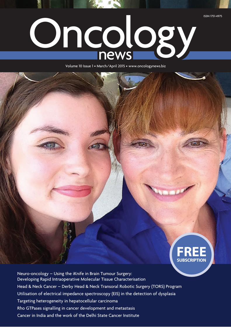
OncologyISSN 1751-4975
Volume 10 Issue 1 • March/April 2015 • www.oncologynews.biz
news
Neuro-oncology – Using the iKnife in Brain Tumour Surgery: Developing Rapid Intraoperative Molecular Tissue Characterisation
Head & Neck Cancer – Derby Head & Neck Transoral Robotic Surgery (TORS) Program
Utilisation of electrical impedance spectroscopy (EIS) in the detection of dysplasia
Targeting heterogeneity in hepatocellular carcinoma
Rho GTPases signalling in cancer development and metastasis
Cancer in India and the work of the Delhi State Cancer Institute
FREE SUBSCRIPTION
XGEVA® IS INDICATED FOR THE PREVENTION OF SREs* IN ADULT PATIENTS WITH BONE METASTASES FROM SOLID TUMOURS1
AVOIDING SREs* AND ASSOCIATED PAIN FOR AS LONG AS POSSIBLE IS CENTRAL TO PRESERVING QUALITY OF LIFE FOR PATIENTS WITH BONE METASTASES 2-3
THE FIRST AND ONLY TREATMENT FOR SRE* PREVENTION IN BREAST CANCER AND OTHER SOLID TUMOURS (EXCLUDING PROSTATE CANCER) WITH POSITIVE NICE GUIDANCE†4
XGEVA®q (denosumab) Brief Prescribing Information
Please refer to the Summary of Product Characteristics (SmPC) before prescribing XGEVA®. Pharmaceutical Form: 1.7 ml solution for injection presented as a single use vial containing 120 mg of denosumab. Contains sorbitol (E420). Indication: Prevention of skeletal related events (pathological fracture, radiation to bone, spinal cord compression or surgery to bone) in adults with bone metastases from solid tumours. Dosage and Administration: Single subcutaneous injection of XGEVA® 120 mg given once every 4 weeks. No dosage adjustment required in patients with renal impairment or in elderly patients (age ≥ 65). Patients must be supplemented daily with at least 500 mg calcium and 400 IU vitamin D unless hypercalcaemia is present. Not recommended in paediatric patients (under 18 years of age). Contraindications: Severe, untreated hypocalcaemia or hypersensitivity to the active substance or to any of the excipients. Special Warnings and Precautions: Pre-existing hypocalcaemia must be corrected prior to initiating therapy with XGEVA®. Hypocalcaemia can occur at any time during therapy. Monitoring of calcium should be conducted prior to initial dose, within two weeks of initial dose and if suspected symptoms of hypocalcaemia occur. Severe symptomatic hypocalcaemia has been reported. Consider additional monitoring of calcium level in patients with risk factors for hypocalcaemia or if otherwise indicated based on clinical condition of the patient. Patients with severe renal impairment (creatinine clearance < 30 ml/min) or receiving dialysis are at greater risk of developing hypocalcaemia; this risk and accompanying elevations in parathyroid hormone increases with increasing degree of renal impairment. Regular monitoring of calcium levels in these patients is especially important. If hypocalcaemia occurs while receiving XGEVA®, additional calcium supplementation and additional monitoring may be necessary. Osteonecrosis of the jaw (ONJ) has occurred commonly in patients treated with XGEVA®. In clinical trials, the incidence of ONJ was higher with longer duration of exposure. For information on known risk factors for ONJ, please refer to the SmPC. In patients with risk factors for
ONJ, an individual benefi t:risk assessment should be performed before initiating therapy with XGEVA®. A dental examination with appropriate preventive dentistry is recommended prior to treatment. XGEVA® should not be initiated in patients with an active dental or jaw condition requiring surgery or in patients who have not recovered following oral surgery. Patients should be encouraged to maintain good oral hygiene practices and receive routine dental check-ups during treatment with XGEVA®. Patients should avoid invasive dental procedures if possible while on treatment. For patients who develop ONJ while on XGEVA® therapy, dental surgery may exacerbate the condition. The management plan of individual patients who develop ONJ should be set up in close collaboration between the treating physician and a dentist or oral surgeon with expertise in ONJ. Atypical femoral fracture (AFF) has been reported in patients receiving XGEVA®. Discontinuation of XGEVA® therapy in patients suspected to have AFF should be considered pending evaluation of the patient based on an individual benefi t risk assessment. Patients being treated with XGEVA® should not be treated concomitantly with other denosumab containing medicinal products (for osteoporosis indications) or with bisphosphonates. Patients with rare hereditary problems of fructose intolerance should not use XGEVA®. Interactions: No interaction studies have been performed. Pregnancy and lactation: There are no adequate data on the use of XGEVA® in pregnant women. Not recommended for use in pregnant women or women of childbearing potential not using contraception. It is unknown whether XGEVA® is excreted in human milk. A risk/benefi t decision should be made in patients who are breast-feeding. No data are available on the effect of XGEVA® on human fertility. Undesirable Effects: Adverse reactions in patients receiving XGEVA® to prevent the occurrence of skeletal related events: very common (≥ 1/10) dyspnoea, diarrhoea and musculoskeletal pain; common (≥ 1/100 to < 1/10) hypocalcaemia, hypophosphataemia, tooth extraction, hyperhidrosis and osteonecrosis of the jaw; rare (≥ 1/10,000 to < 1/1000) drug hypersensitivity, anaphylactic reaction, atypical femoral fracture. In 3 phase III clinical trials, ONJ was confi rmed in 1.8% of patients treated with XGEVA® and
1.3% of patients treated with zoledronic acid (primary treatment phase). Among subjects with confi rmed ONJ, most (81% in both treatment groups) had a history of tooth extraction, poor oral hygiene, and/or use of a dental appliance. Hypocalcaemia was reported in 9.6% of patients treated with XGEVA® and 5.0% of patients treated with zoledronic acid. Neutralizing antibodies have not been observed in clinical studies. In the postmarketing setting, severe symptomatic hypocalcaemia (including fatal cases), hypersensitivity (including rare events of anaphylactic reaction) and musculoskeletal pain (including severe cases) have been reported. Please consult the SmPC for a full description of undesirable effects. Pharmaceutical Precautions: Do not mix with other medicinal products. Store at 2°C to 8°C (in a refrigerator). XGEVA® may be stored at room temperature (up to 25°C) for a maximum single period of up to 30 days in its original container. Once removed from the refrigerator, XGEVA® must be used within this 30 day period. Do not freeze. Keep vial in outer carton to protect from light. XGEVA® solution should be inspected visually before administration. Do not inject the solution if it is cloudy or discoloured. Legal Category: POM. Presentation, Basic Costs and Marketing Authorisation Number: XGEVA® 120 mg: Pack of 1: £309.86; EU/1/11/703/001. Marketing Authorisation Holder: Amgen Europe B.V., Minervum 7061, NL-4817 ZK Breda, The Netherlands. Further information is available from Amgen Limited, 240 Cambridge Science Park, Milton Road, Cambridge, CB4 0WD. XGEVA® is a registered trademark of Amgen Inc. Date of PI preparation: August 2014 (Ref: DMO-GBR-AMG-315-2014-P)
This medicinal product is subject to additional monitoring. Adverse events should be reported.
Reporting forms and information can be found at www.mhra.gov.uk/yellowcard. Adverse events should also be
reported to Amgen Limited on +44 (0) 1223 436712
REFERENCE:1. XGEVA® (denosumab), Summary of Product Characteristics.2. von Moos, R et al. Pain and health-related quality of life in patients with advanced solid tumours and bone metastases: integrated results from three
randomised, double-blind studies of denosumab and zoledronic acid. Support Care Cancer. 2013; 21(12): 3497-3507.3. Coleman, R. Clinical Features of Metastatic Bone Disease and Risk of Skeletal Morbidity. Clinical Cancer Research. 2013; 21(12): 3497-3507. 4. NICE guidance. Denosumab for the treatment of bone metastases from solid tumours.http://guidance.nice.org.uk/TA265/guidance/pdf/english. Last
accessed August 2014* SREs = skeletal related events. These are defi ned as pathological fracture, radiation to bone, spinal cord compression and surgery to bone† NICE Technology Appraisal guidance 265
©2014 Amgen Inc. All rights reserved. Amgen Oncology UKIE-P-162x-0215-102167 Date of preparation: Febuary 2015

Volume 10 Issue 1 • March/April 2015 3
FROM THE EDITOR
Volatolomics – a diagnostic aid in many diseases, including cancer
Although it has become increasingly apparent using mass spectroscopy and other modern techniques to detect and often quantify
molecules at very low density, the question is not whether it is useful in diagnosis of diseases, but whether it can have a broader spectrum of use in the future. This claim has been gathering momentum since the early 1990s [1] and has now reached a level of sophistication when there is genuine belief that it will be a useful aid in diagnosis [2]. Pauling was the first to use gas-liquid partition chromatography, but newer techniques involve specifically capped gold or platinum nanoparticles coated with receptors [3]
Odours from body fluids can be characteristic and certainly can change considerably with the onset of stress and disease. Our sweat has chemicals that others can detect at extremely low concentrations (including pheromones), and some animals have receptors in their nasal epithelia* that can outdo the performance and sensitivity of some of the most modern and elaborate spectrometric and meterometric techniques. Apart from sweat, urine has long been tested by tasting for the presence of sweetness for its connection with diabetes. Feces can also liberate some tell-tale odours. But the breath may be one of the best sources of volatile biomarkers to explore. Examining the breath odour of patients was used long before the time of Hippocrates [2]. In all probability the Chinese and perhaps many races from time immemorial have smelt the breath of sick patients (part of the skill of the “medicine man”, e.g. the renowned Zhang Zhongjing). The breath of a bipolar patient will be distinctly different in depression than hypomania. Not only might it be used in mental health and many physical disorders, it is possible that one might distinguish cancerous and non-cancerous growth in the body. Three factors seem to be important in taking this aid to diagnosis further: first, far more information is needed from many independent studies; second there is the question of the practicality relative to other procedures, such as blood sampling; and third there remains the problem that the flora and fauna of the body might be responsible for different volatile biomarkers in the breath under many changing circumstances in the individual, which will almost certainly contribute substantially to the difficulty in making a clear diagnosis of some underlying
condition. These issues make the problem of setting any base-line a headache.
Taking a patient over a short-term of examination, changes might well reflect disturbances in the metabolomics of the body, discussed in one of our previous articles in relation to cancer [4]. This will make interpretation of changes in the volatile biomarker “signatures” or “profiles” for the individual patient very difficult, but at this stage, Haick’s group [2] are more concerned with the patterns in subpopulations where a profile has some common features in a group of patients suffering from similar conditions. In their work, clustering analysis seemed to give greater similarities between cancerous conditions, with sub-clustering being apparent in diseases that have a high inflammatory activity (e.g. inflammatory bowel disease). Some diseases did have distinctive profiles, including chronic kidney disease and pre-eclampsia. They have explored a range of other conditions; Parkinsonism, pulmonary artery hypertension, and six different types of cancer. It seems there is a very long way to go before more disorders and diseases might show truly distinctive patterns. There is also the possibility that there are volatile biomarkers not yet perceived as being important in diagnosis, such as a product from an altered enzyme function in the body due to a gene mutation.
Volatolomics can have its place in medicine if it builds on past experience, however anecdotal this may seem at present. Regarding cancer in particular, inputs from regulatory agencies indicates that it would have great promises for screening in the far future, but its first application might be in monitoring aspects of the cancer or as complementary approach for existing methods, such as low –dose CT. As Dr Haick says - “Although it has, however, been under extensive research in the last 40 years, it has failed to pave its way towards daily clinical application. This is mainly due to the lack of comprehensive evidence that different pathophysiological processes result in distinct breath volatolomes. We have made good advances in providing part of this evidence – something that will soon be shared with the scientific committee” [2].
* I am reminded of a surgeon with whom I once worked,
who was convinced that just before death dying patients
gave off a characteristic odour to which most (if not all) of
us are insensitive, but this resonates with a news-item about
a dog in a care-home that would lead nurses to a room in
which a patient was in extremis or had just died.
Denys WheatleyEditor
1. Breteton R. Chemometrics, application of mathematics and statistics to laboratory systems Ellis Horwood, Chichester, UK, 1990.
2. Haick H, Broza YY, Mochalski P, Ruzsanyi V, Amann A. Assessment, origin, and implementation of breath volatile cancer markers. Chem Soc Rev. 2014;Mar 7;43:1423-49. [PubMed PMID: 24305596. Epub 2013/12/07]
3. Dovgolevsky E, Konvalina G, Tisch U, Haick H. Monolayer-Capped Cubic Platinum Nanoparticles for Sensing Nonpolar Analytes in Highly Humid Atmospheres. J Phys Chem C. 2010;114:14042-9.
4. Wheatley DN. Arginine deprivation and metabolomics: important aspects of intermediary metabolism in relation to the differential sensitivity of normal and tumour cells. Semin.Cancer Biol. 15, 247-253.
REFERENCES
4 Volume 10 Issue 1 • March/April 2015
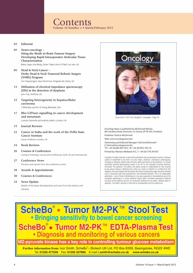
ContentsVolume 10 Number 1 • March/February 2015
Oncology News is published by McDonnell Mackie, 88 Camderry Road, Dromore, Co Tyrone, BT78 3AT, N Ireland.
Publisher: Patricia McDonnell
Web: www.oncologynews.biz
Advertising and Editorial Manager: Patricia McDonnell • E: [email protected] T/F: +44 (0)288 289 7023 • M: +44 (0)7833 185116
Printed by: Warners Midlands PLC, T: +44 (0)1778 391057
Copyright: All rights reserved; no part of this publication may be reproduced, stored in a retrieval system or transmitted in any form or by any means, electronic, mechanical, photocopying, recording or otherwise without either the prior written permission of the publisher or a license permitting restricted photocopying issued in the UK by the Copyright Licensing Authority. Disclaimer: The publisher, the authors and editors accept no responsibility for loss incurred by any person acting or refraining from action as a result of material in or omitted from this magazine. Any new methods and techniques described involving drug usage should be followed only in conjunction with drug manufacturers’ own published literature. This is an independent publication - none of those contributing are in any way supported or remunerated by any of the companies advertising in it, unless otherwise clearly stated. Comments expressed in editorial are those of the author(s) and are not necessarily endorsed by the editor, editorial board or publisher. The editor’s decision is final and no correspondence will be entered into.
03 Editorial
05 Neuro-oncology – Using the iKnife in Brain Tumour Surgery: Developing Rapid Intraoperative Molecular Tissue Characterisation Babar Vaqas, Julia Balog, Zoltan Takats, Kevin O’Neill, Lon don, UK
09 Head & Neck Cancer – Derby Head & Neck Transoral Robotic Surgery (TORS) Program Giri Wijayasingam, Sean Mortimore, Mriganka De, Derby, UK
13 Utilisation of electrical impedance spectroscopy (EIS) in the detection of dysplasia John Tidy, Sheffield, UK
15 Targeting heterogeneity in hepatocellular carcinoma C Billie Bian and Xin W Wang, Bethesda, USA
17 Rho GTPases signalling in cancer development and metastasis Lorenza Giannella and Anatoliy Markiv, London, UK
19 Journal Reviews
22 Cancer in India and the work of the Delhi State Cancer Institute Lauren Anderson, London, UK
24 Book Reviews
26 Courses & Conferences Listing of meetings, courses and conferences, both UK and international.
27 Conference News Previews and reports from the conference scene.
28 Awards & Appointments
30 Courses & Conferences
34 News Update Details of the latest developments and news from the industry and
charities.
Ovacome’s ‘Tell Your Daughter’ campaign - Page 34.
Further information from: Ivor Smith, ScheBo • Biotech UK Ltd, PO Box 6359, Basingstoke, RG22 4WETel: 01256 477259 Fax: 01256 327889 E-mail: [email protected] www.schebo.co.uk
®
• Bringing sensitivity to bowel cancer screening
• Diagnosis and monitoring of various cancers
®
•
•®
M2-pyruvate kinase has a key role in controlling tumour glucose metabolism

Volume 10 Issue 1 • March/April 2015 5
Surgery remains a critical step in the management of most of the 9,400 brain tumours diagnosed in the UK each year. The role of surgery is to obtain tissue for
diagnosis and often also to immediately improve symptoms of mass effect. Obstacles to successful surgery include difficulty in identifying a suitable target for biopsy, resulting in a non-diagnostic procedure and difficulty in identifying the tumour itself which results in residual tumour left behind after surgery and thus early recurrence of disease. Both of these difficulties may lead to the need for a second operation. Difficulty in identifying the difference between tumour tissue and normal brain tissue can also result in neurological damage if functioning brain tissue is damaged; this may mean that crucial further treatment such as radiotherapy cannot be offered resulting in decreased survival and a poor quality of life for the patient. Accurate tissue identification is also required for the maximal safe resection of intrinsic tumours.
Rapid tissue characterisation is a potential solution to these problems which Neurosurgeons confront every time they operate on brain tumours, particularly on intrinsic tumours with less well defined margins and tissue characteristics.
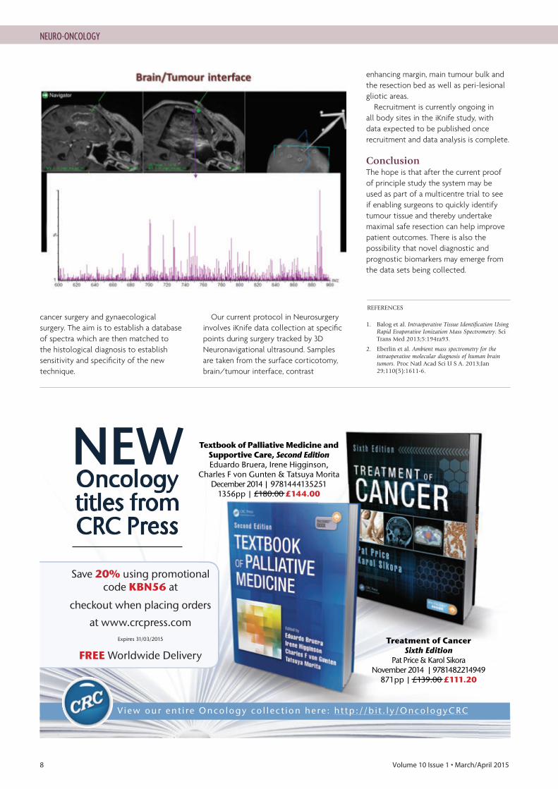
Neuro-Oncological Surgery Neurosurgeons currently use a multi-modality approach to help determine resection margins when operating on tumours. Although defining brain metastatic disease can be relatively easy as there is a clearer delineation between tumour and brain, intrinsic tumours pose more of a challenge as they often have less well defined brain/tumour interface. Preoperative MRI scans can be used during surgery as part of a neuronavigation system and can help delineate tumour characteristics during surgery as long as there is no significant brain shift which renders the preoperative scans inaccurate. Intraoperative MRI (available only in a small number of centres in the UK due to cost) or intraoperative ultrasound can help maintain navigational accuracy during surgery. Optical methods such as an operating microscope and 5-ALA fluorescence further help the surgeon in identifying the tumour and its margin. 5- ALA is however not as useful due to poor fluorescence in low grade gliomas. Texture and feel are also important features which help the surgeon further define the tumour.
The current gold standard for tissue diagnosis during Neurosurgery is frozen section. This can take up to an hour to process, during which time
Using the iKnife in Brain Tumour Surgery: Developing Rapid Intraoperative Molecular Tissue Characterisation
NEURO-ONCOLOGY
Babar Vaqas, FRCS (SN), George Pickard Fellow in Neurosurgery, Imperial College Healthcare NHS Trust.
Julia Balog, PhD, Imperial College London.
Zoltan Takats, PhD, Professor of Analytical Chemistry, Imperial College London.
Kevin O’Neill, FRCS, (SN), Head of Neurosurgery and Neuro-Oncology Lead, Imperial College Healthcare NHS Trust.
Correspondence address: E: [email protected] Figure 1 Mass Spectral Analysis on a Principle Component Analysis plot showing separation of different tissue types.

6 Volume 10 Issue 1 • March/April 2015
the surgeon continues to operate without knowing the type of tumour on which they are operating. Frozen sections also have poor spatial resolution, normally relying on the surgeon labelling the general area from which the sample is taken.
The limitations of current technology have led to the need for new modalities to help better define intrinsic brain tumours and immediately characterise the tissue type being sampled.
Medical Mass SpectrometryMass spectrometry is an analytical chemistry technique that can identify the amount and types of chemicals present in a sample by calculating the mass to charge ratio of gas-phase ions. Currently it is widely used in industry.
Mass spectrometry also holds great promise as a method for fast and accurate tissue classification during surgery. The technique involves utilising the aerosolisation effect of bipolar electrocautery used by Neurosurgeons in almost all brain tumour operations. Tumour tissue particles form the smoke produced by bipolar forceps which has
for long been considered a nuisance and even carcinogenic in its own right. The extracted smoke consists of gas-phase ionic species amenable to mass spectrometric analysis. The iKnife is the name given to the coupling of a mobile mass spectrometry machine with electrosurgical instruments in the operating theatre so that the smoke generated from surgery is instantly analysed to produce a mass spectrum. The resulting mass spectrometric profiles are highly tissue specific allowing tissue characterisation and identification. This process is extremely fast, taking place within seconds, allowing immediate feedback to the operating surgeon. The elegance of this solution lies in that no sample preparation is required as is the case with other mass spectrometry systems and the system requires no new surgical instrumentation; it simply analyses a byproduct of surgery and is easily integrated into the operating theatre workflow. Two recent studies have shown that intraoperative mass spectrometry can be undertaken and can yield accurate results.
Balog et al [1] published their ex-vivo analysis of 37 brain tumours and in-vivo analysis of 11 tumours reaching 100%
sensitivity and specificity. The tumour types successfully diagnosed included glioblastoma multiforme (GBM) and WHO grade 2 oligoastrocytoma as well as lung and colon metastases.
A second study by Eberlin et al. [2] has shown the ability of Desorption Electron Spray Ionisation Mass Spectrometry (DESI-MS) to classify stereotactically registered surgical specimens with excellent correlation with histopathology. The system could also determine tumour cell concentration in peripheral regions of the tumour. This system is different from the iKnife as sample pre-treatment is required prior to mass spectrometry readings.
This has led to the development and use of the iKnife as a new intraoperative tool for surgeons to help maximise the safe and more complete resection of tumours.
Current Use at Imperial College LondonThe iKnife is currently being trialled at Imperial College London on several body sites during brain tumour surgery, gastrointestinal tumour surgery, breast
NEURO-ONCOLOGY
Figure 2: Overview of iKnife data collection and analysis.

ONJA14_ILR SO04 29/06/2014 14:32 Page 85
8 Volume 10 Issue 1 • March/April 2015
NEURO-ONCOLOGY
cancer surgery and gynaecological surgery. The aim is to establish a database of spectra which are then matched to the histological diagnosis to establish sensitivity and specificity of the new technique.
Our current protocol in Neurosurgery involves iKnife data collection at specific points during surgery tracked by 3D Neuronavigational ultrasound. Samples are taken from the surface corticotomy, brain/tumour interface, contrast
enhancing margin, main tumour bulk and the resection bed as well as peri-lesional gliotic areas.
Recruitment is currently ongoing in all body sites in the iKnife study, with data expected to be published once recruitment and data analysis is complete.
ConclusionThe hope is that after the current proof of principle study the system may be used as part of a multicentre trial to see if enabling surgeons to quickly identify tumour tissue and thereby undertake maximal safe resection can help improve patient outcomes. There is also the possibility that novel diagnostic and prognostic biomarkers may emerge from the data sets being collected.
1. Balog et al. Intraoperative Tissue Identification Using Rapid Evaporative Ionization Mass Spectrometry. Sci Trans Med 2013;5:194ra93.
2. Eberlin et al. Ambient mass spectrometry for the intraoperative molecular diagnosis of human brain tumors. Proc Natl Acad Sci U S A. 2013;Jan 29;110(5):1611-6.
REFERENCES
Volume 10 Issue 1 • March/April 2015 9
Derby Robotic Head and Neck programme started following a generous donation by a local entrepreneur. He wanted to
contribute to Derbyshire community and hospi-tal. We will be grateful for his help. Currently Urology, Head and Neck surgery and Colorectal surgery are the specialities using robotic assisted surgery. Gynaecology are planning to start within next month. This is a multi disciplinary robotic surgery programme. The robotic surgery is a novel surgical technique. It is a significant step to help with the patient care pathway in Derby and its surrounding areas.
Derby Head and and neck trans oral robotic assisted surgery programme is lead by two Head and Neck surgeons. There are few centres in the UK where robotic assisted head and neck surgery is performed. We are proud of our achievement and the advantage it will bring to our patients. Transoral robotic surgery was approved by FDA (USA) in 2009. The trust management with the help of a core robotic multi disciplinary surgical group after thorough examination of the business case and safety precautions agreed to start the programme.
The advantages of robotic surgery (TORS) compared to the conventional open surgery are precise removal of cancerous tissue, low complication rate, minimal blood loss, minimal need of tracheostomy tube, ability to swallow early and minimal hospital stay [1]. 1.5 million robotic surgery has been performed worldwide.
Transoral Robotic surgery is performed, via transoral route similar to other minimal invasive surgery with some similarity to Transoral Laser Surgery. However, the da Vinci system features a magnified 3Dimensional high definition vision system which is better than a conventional view through an endoscope.
The special wrested forceps which also functions as a bipolar diathermy has the ability to move on many planes and is steady without any tremors or minor movement. Hence, has the ability to flex and rotate far grater than a human hand through a small space. We can therefore operate with an enhanced vision, precision, dexterity and control. Since the robotic arm/forceps can move on many planes a selected group of oropharyngeal and supra
glottic tumours which otherwise would need a mandibular split or mandibular dislocation for access and would need tracheostomy can be per-formed. Open resections with reconstructions using free flaps can be avoided [2,3].
With conventional open surgery recovery is longer. There is high risk of infection, pain and swallowing difficulties [2] . There are certain disadvantages of laser where the access is difficult and not in a straight line. Neck dissection can be performed with the primary tumour resection or separately. Human Papilloma viral infection (HPV 16) has been recognised as an aetiology of oropharyngeal carcinoma in younger patients. In these group of patients, radiotherapy can give unwanted long term side effects, including a small chance of radiation induced sarcoma. Benign and malignant lesions affecting the oropharynx, supra glottis, glottis, and hypo pharynx can be treated by TORS [2].Other indication of TORS are tongue base varicosities, lingual tonsillar hypertrophy, tongue base reduction for obstructive sleep apnoea and thyroid/parathyroid surgery. Limited mouth opening and advanced tumours are contra indications for TORS surgery.
Components of the system are:
Surgeon console• Using the da Vinci Surgical System, the
surgeon operates using the console while viewing a high definition, 3D image inside the patient’s body.
• The surgeon’s fingers grasp the master controls below the display with hands and wrists naturally positioned relative to his or her eyes.
• The system seamlessly translates the surgeon’s hand, wrist and finger movements into precise, real-time movements of surgical instruments.
Patient side cart• The patient side cart is where the patient is
positioned during surgery. It includes either three or four robotic arms that carry out the surgeon’s commands.
• The robotic arms move around fixed pivot points.
• The system requires that every surgical manoeuver be under the direct control of
Derby Head & Neck Transoral Robotic Surgery (TORS) Program
HEAD & NECK CANCER
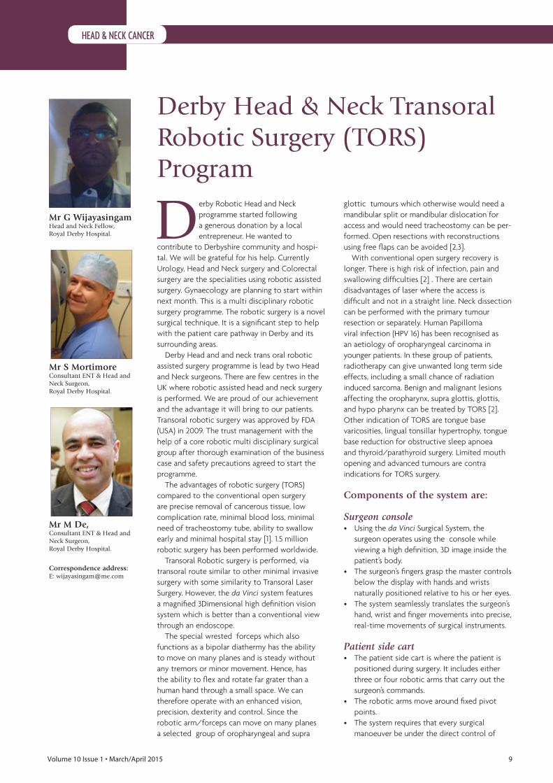
Mr G Wijayasingam Head and Neck Fellow, Royal Derby Hospital.
Mr S Mortimore Consultant ENT & Head and Neck Surgeon, Royal Derby Hospital.
Mr M De, Consultant ENT & Head and Neck Surgeon, Royal Derby Hospital.
Correspondence address:E: [email protected]

10 Volume 10 Issue 1 • March/April 2015
the surgeon. Repeated safety checks prevent any independent movement of the instruments or robotic arms.
Endowrist instrument• A full range of Endowrist instruments
is available to the surgeon while operating.
• The instruments are designed with seven degrees of motion – a range of motion even greater than the human wrist
• Each instrument has a specific surgical mission such as clamping, suturing and tis-sue manipulation.
• Quick-release levers speed instrument changes during surgery.
Vision system• The vision system is equipped with a
high-definition, 3D endoscope (flexible tube with a camera and light at the tip) and image processing equipment that provides true-to-life images of the patient’s anatomy.
• A view of the operating field is available to the entire OR team on a large viewing monitor (vision cart). This widescreen view provides the surgical assistant at the pa-tient’s side with a broad perspective and visualisation of the procedure [4].
There is a training programme for the surgeons to under go before starting to operate on patients.
Firstly the surgeons will practice on simulation for at least 20-30 hours. This is to train the hand eye co ordination and familiarise with console. The surgeons will
visit an international recognised centre to observe TORS procedure. Next step in the training programme is cadaveric dissection course recognise by da Vinci surgical system to enable surgeons to obtain console certificate.
The theatre scrub nurses undergoes a rigours training with the help of Intuitive surgical. This includes case observation and on line certification and a dry run prior to to live surgery.
Once the training was completed and certified by the Intuitive surgical the initial 4 to 5 surgical resection needs be done under the guidance of a mentor. Our mentor was a TORS surgeon from Hamburg, Germany. The mentor was helpful with first couple of resections. The plan is to reassess both the surgeons at Derby in three months time.
We at Derby were fortunate to have dual consoles. The operating surgeon will sit on one console while the mentor or a junior doctor would watch in the second console.
The second surgeon will sit on the head side of the patient and will guide the console surgeon. He would keep the operative field clear of secretions. The patient will be draped in as per protocol for a standard robotic surgery. The scrub nurse will assist the second surgeon.The anaesthesist is also well informed about the surgery. He/she will insert and place the endotracheal tube without obstructing the operative area. At Derby both the head and neck surgeons are experienced Trans oral Laser surgeons. The experience makes it easier for adapting and learning the new technique.
HEAD & NECK CANCER
REFERENCES
1. Iseli TA1, Kulbersh BD, Iseli CE, Carroll WR, Rosenthal EL, Magnuson JS. Functional outcomes after transoral robotic surgery for head and neck cancer. Head Neck Surg. 2009 Aug;141(2):166-71.
2. Vergez S, Lallemant B, Ceruse P, Moriniere S, Aubry K, De Mones E, Benlyazid A, Mallet Y. Initial multi-institutional experience with transoral robotic surgery. Head Neck Surg. 2012; Sep;147(3):475-81.
3 . Gorphe P, Brasnu D. Robotic surgery for head and neck carcinomas. Eur Arch Otorhinolaryngol. 2012; Aug;269(8):1979-84.
4. www.davincisurgery.com
Images show intraoperative photos of TORS oropharyngeal surgery.

Volume 10 Issue 1 • March/April 2015 11
Professor Denys Wheatley is Editor, and is Director of BioMedES. He has strong research ties in Albany, Davis, Auckland, Valencia, Detroit, Budapest, St Petersburg, Heidelberg, Zürich and Hong Kong. He is eager to establish strong interaction with cancer and cell biology teams worldwide, and initiate programmes in the areas in which his expertise lies. His work in cancer research, other scientific fields, with IFCB, and in publishing and scientific communication has led to his receiving awards in recent years.
Dr Richard J Ablin (Associate Editor), is Professor, Pathology, University of Arizona College of Medicine and a Member of the Arizona Cancer Center, Tucson, Arizona. He received the First Award for scientific excellence from The Haakon Ragde Foundation for Advanced Cancer Studies. Dr Ablin discovered prostate-specific antigen (PSA) in 1970. A pioneer of cryosurgery and cryoimmunotherapy, he has extensive experience in cancer research.
Mr Richard Novell is Assistant Co-Editor – Gastrointestinal Section, and is a Consultant Colorectal Surgeon at the Royal Free Hospital. He was a member of the Court of Examiners of the Royal College of Surgeons for eight years and has been an advisor to NICE, NCEPOD and CORESS, the Confidential Reporting System in Surgery.
Dr Miriam Dwek is Assistant Co-Editor – Breast Cancer, she is a Senior Lecturer in Biochemistry at the Department of Molecular and Applied Biosciences, School of Life Sciences, University of Westminster in London.
Farrokh Pakzad is Assistant Editor – Skin Cancer, and is currently Consultant Oncoplastic Breast and Melanoma Surgeon at Royal Surrey County Hospital. His main areas of specialist interest are in the management of breast disease, oncoplastic and reconstructive breast surgery and the management of skin cancers, in particular, melanoma. Farrokh completed his higher surgical training in London, during which he was selected onto the highly competitive National Oncoplastic Fellowship program.
Dr Constantino Carlos Reyes-Aldasoro is Assistant Editor – Image Analysis. He is a Lecturer in Biomedical Image Analysis at the School of Engineering and Mathematical Sciences, City University London. He has developed a unique portfolio of interdisciplinary skills that span from the acquisition of microscopical images to the analysis of biomedical datasets such as magnetic resonance, computed tomography and microscopy to advanced computer programming and website development.
Mriganka De is Assistant Editor – Head & Neck Oncology. Mr De is a Consultant ENT/Head and Neck surgeon at Royal Derby Hospital, Derby. His interest is head and neck cancer with particular focus on management of early laryngeal cancers.
International Liaison Committee
Mikhail Yu Reutovich, Abdominal Oncology Department, NN Alexandrov National Cancer Center of Belarus, Minsk, Belarus.
Alan Cooper is Assistant Co-Editor – Urology, and is Lead Scientist with the urology research group in Southampton University Hospitals and senior lecturer (albeit with virtually no lecturing burden) in the Department of Biomedical Sciences at Portsmouth University.
Meet the Editorial Team
Prof Mohammed RS Keshtgar BSc, FRCSI, FRCS (Gen), PhD is Assistant Co-Editor – Breast Cancer, and is a Professor of Cancer Surgery and Surgical Oncology, Royal Free London Foundation Trust. His main area of interest is minimally invasive approaches in diagnosis and treatment of breast cancer. His research interest is in sentinel node biopsy, intra-operative radiotherapy, quantum dot nanotechnology in breast cancer.
Professor Geoffrey J Pilkington is Assistant Editor Neuro-Oncology, is a Professor of Cellular and Molecular Neuro-oncology at the Institute of Biomedical and Biomolecular Sciences, Portsmouth. His research focuses on the development of models for the study of intrinsic brain tumours, elucidation of their metabolism and mechanisms underlying diffuse local invasive behaviour.

12 Volume 10 Issue 1 • March/April 2015
BioMedES LtdProducing the very best definitive versions of
your biomedical reports and papers www.biomedes.co.uk
• BioMedESreworkssoundscientificpapers,technicalreports,andother(biomedical)documents,puttingthemintothemostidiomaticEnglishandpassesthemoststringentpeerreviewandqualitycontrolassessments
• Itcopyeditsforanumberofbigbiomedicalpublishinghouses
• Runsfourjournalsinthelifesciences
• ItprovidestheSecretariatforaninternationalorganisationforcellbiology(www.ifcbiol.org)andanenterprisecompany
• Itpreparesandpublishese-booksinbiomedicine
• Itdesignslogosforbiomedicalandmanyotherorganisations
• Itcollatesandpreparesabstractsforscientificandothermeetings
• Thecompanyisinvolvedinarrangingbothnationalandinternationalconferences
• Italsorunscoursesonscientificandmedicalwriting,andonelectronicpublishingathomeandabroad
Why not contact us at:BioMedES Ltd
LeggatHouse,Keithhall,Inverurie,AB510LX,UK.T:+44-1467-6702809,F:+44-1467-629123,
What does BioMedES do?
Never heard of us?Notsurprising,sinceouroperationsaremostly“behindthescenes”.Butwemaybeabletohelpyouwithyourpublicationproblems,fromtechnicalnotestoe-books.
Volume 10 Issue 1 • March/April 2015 13
T here has been interest for many years in the use of electrical impedance to identify and image biological tissues; however, identifying a reliable method
to detect changes has been challenging and hence has restricted this technique from wider application. Underpinning the technology is the observation that all biological tissues have electrical impedance, which is a function of frequency. The reason for this dependence is that tissues contain components that have both resistive and capacitive properties. Both the size of the impedance, as well as the dependence of impedance on frequency, are related to tissue composition [1,2].
Different tissues have different frequency bands within an impedance spectrum. At high frequencies (>1 GHz) molecular structure is the determining factor whereas at low frequencies (<100 Hz) charge accumulation at large membrane interfaces dominate. At frequencies of a few kHz to 1 MHz, sometimes referred to as the β dispersion region, cell structures are the main determinant of tissue impedance.
Within the β dispersion region, low frequency current can be considered as passing through the extracellular space; the current has to pass around the cells and the resistance to flow will depend upon cell spacing and arrangement. However, at higher frequencies current can penetrate the cell membranes and hence passes through both intracellular and extracellular spaces. Current will pass through the cells and will be determined by intracellular volume and, possibly, the size of the nucleus. The diameter of the electrodes and the distance between the electrodes will determine the depth of tissue measured by EIS. If the diameter of the electrodes and the distance between the electrodes is too large the current will flow into the stroma. This will have an
adverse effect on the ability of EIS to detect any changes within the epithelium.
EIS in the detection of dysplasiaThe alterations in architecture of an epithelial surface associated with the development of dysplasia can be measured by EIS. Dysplastic epithelia have increased extracellular space, lowering resistance to the flow of an electrical current at low frequency. Increase in the ratio between nucleus and cytoplasm may influence revistivity at higher frequencies. EIS technology has been used on epithelia to assess whether it can detect changes associated with dysplasia.
Detection of skin melanoma and basal cell carcinoma Åberg et al. [3] used a multiple electrode device to take skin impedance measurements over the range 1-1000 kHz in 252 patients that had a spectrum of skin lesions [3]. There were significant differences between normal skin, benign pigmented nevi, dysplastic nevi and basal cell cancers, with the biggest being between normal skin and basal cell carcinoma. A range of parameters were used to describe the impedance spectra, which showed that the mean was higher in the basal cell carcinoma group than normal skin, and the changes with frequency were also reduced. Malignant melanoma could be distinguished from benign nevi. Nevisense, the product developed by Scibase, has recently been evaluated in a multicentre trial to assess if malignant melanoma can be separated from benign lesions; this device had a sensitivity of 97% for detection of melanoma and a specificity of 34% with lesions thought to be melanomas [4].
Detection of cervical intra-epithelial neoplasiaCervical epithelium is a highly structured, stratified tissue. Preceding the development of cervical cancer the epithelia surface under goes dysplastic change usually as a consequence of infection by high risk human papillomavirus types. This dysplastic change is known as cervical intra-epithelial neoplasia (CIN). Cervical screening programmes use exfoliative cytology to detect the changes associated with the development of CIN and women with abnormal cytology are referred for colposcopy to examine the
Utilisation of electrical impedance spectroscopy (EIS) in the detection of dysplasia
FEATURE ARTICLE
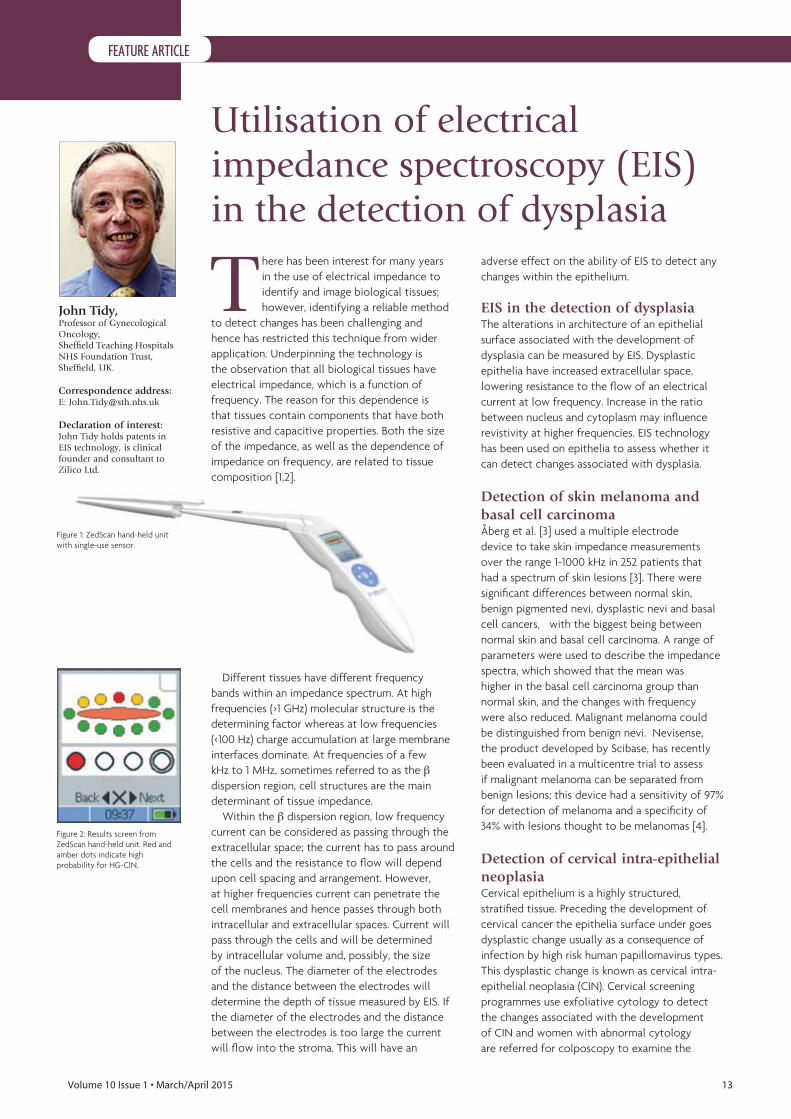
John Tidy, Professor of Gynecological Oncology,Sheffield Teaching Hospitals NHS Foundation Trust,Sheffield, UK.
Correspondence address:E: [email protected]
Declaration of interest:John Tidy holds patents in EIS technology, is clinical founder and consultant to Zilico Ltd.
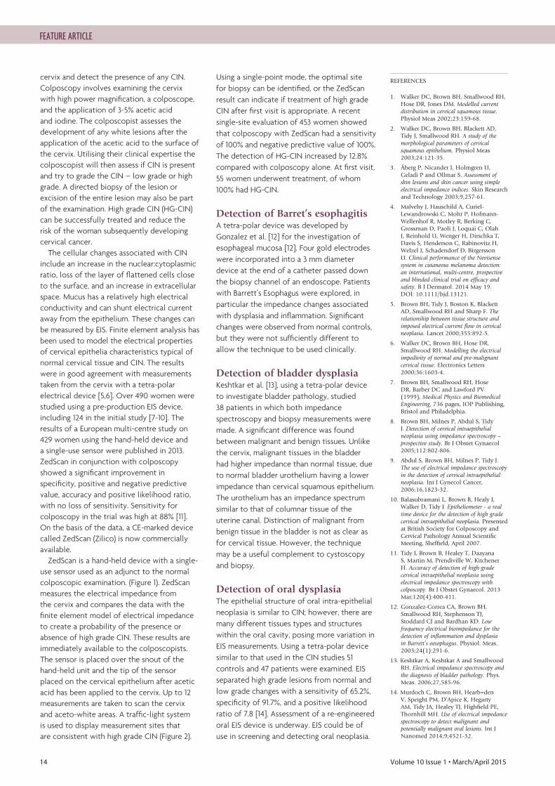
Figure 2: Results screen from ZedScan hand-held unit. Red and amber dots indicate high probability for HG-CIN.
Figure 1: ZedScan hand-held unit with single-use sensor.
14 Volume 10 Issue 1 • March/April 2015
cervix and detect the presence of any CIN. Colposcopy involves examining the cervix with high power magnification, a colposcope, and the application of 3-5% acetic acid and iodine. The colposcopist assesses the development of any white lesions after the application of the acetic acid to the surface of the cervix. Utilising their clinical expertise the colposcopist will then assess if CIN is present and try to grade the CIN – low grade or high grade. A directed biopsy of the lesion or excision of the entire lesion may also be part of the examination. High grade CIN (HG-CIN) can be successfully treated and reduce the risk of the woman subsequently developing cervical cancer.
The cellular changes associated with CIN include an increase in the nuclear:cytoplasmic ratio, loss of the layer of flattened cells close to the surface, and an increase in extracellular space. Mucus has a relatively high electrical conductivity and can shunt electrical current away from the epithelium. These changes can be measured by EIS. Finite element analysis has been used to model the electrical properties of cervical epithelia characteristics typical of normal cervical tissue and CIN. The results were in good agreement with measurements taken from the cervix with a tetra-polar electrical device [5,6]. Over 490 women were studied using a pre-production EIS device, including 124 in the initial study [7-10]. The results of a European multi-centre study on 429 women using the hand-held device and a single-use sensor were published in 2013. ZedScan in conjunction with colposcopy showed a significant improvement in specificity, positive and negative predictive value, accuracy and positive likelihood ratio, with no loss of sensitivity. Sensitivity for colposcopy in the trial was high at 88% [11]. On the basis of the data, a CE-marked device called ZedScan (Zilico) is now commercially available.
ZedScan is a hand-held device with a single-use sensor used as an adjunct to the normal colposcopic examination. (Figure 1). ZedScan measures the electrical impedance from the cervix and compares the data with the finite element model of electrical impedance to create a probability of the presence or absence of high grade CIN. These results are immediately available to the colposcopists. The sensor is placed over the snout of the hand-held unit and the tip of the sensor placed on the cervical epithelium after acetic acid has been applied to the cervix. Up to 12 measurements are taken to scan the cervix and aceto-white areas. A traffic-light system is used to display measurement sites that are consistent with high grade CIN (Figure 2).
Using a single-point mode, the optimal site for biopsy can be identified, or the ZedScan result can indicate if treatment of high grade CIN after first visit is appropriate. A recent single-site evaluation of 453 women showed that colposcopy with ZedScan had a sensitivity of 100% and negative predictive value of 100%. The detection of HG-CIN increased by 12.8% compared with colposcopy alone. At first visit, 55 women underwent treatment, of whom 100% had HG-CIN.
Detection of Barret’s esophagitisA tetra-polar device was developed by Gonzalez et al. [12] for the investigation of esophageal mucosa [12]. Four gold electrodes were incorporated into a 3 mm diameter device at the end of a catheter passed down the biopsy channel of an endoscope. Patients with Barrett’s Esophagus were explored, in particular the impedance changes associated with dysplasia and inflammation. Significant changes were observed from normal controls, but they were not sufficiently different to allow the technique to be used clinically.
Detection of bladder dysplasiaKeshtkar et al. [13], using a tetra-polar device to investigate bladder pathology, studied 38 patients in which both impedance spectroscopy and biopsy measurements were made. A significant difference was found between malignant and benign tissues. Unlike the cervix, malignant tissues in the bladder had higher impedance than normal tissue, due to normal bladder urothelium having a lower impedance than cervical squamous epithelium. The urothelium has an impedance spectrum similar to that of columnar tissue of the uterine canal. Distinction of malignant from benign tissue in the bladder is not as clear as for cervical tissue. However, the technique may be a useful complement to cystoscopy and biopsy.
Detection of oral dysplasiaThe epithelial structure of oral intra-epithelial neoplasia is similar to CIN; however, there are many different tissues types and structures within the oral cavity, posing more variation in EIS measurements. Using a tetra-polar device similar to that used in the CIN studies 51 controls and 47 patients were examined. EIS separated high grade lesions from normal and low grade changes with a sensitivity of 65.2%, specificity of 91.7%, and a positive likelihood ratio of 7.8 [14]. Assessment of a re-engineered oral EIS device is underway. EIS could be of use in screening and detecting oral neoplasia.
FEATURE ARTICLE
1. Walker DC, Brown BH, Smallwood RH, Hose DR, Jones DM. Modelled current distribution in cervical squamous tissue. Physiol Meas 2002;23:159-68.
2. Walker DC, Brown BH, Blackett AD, Tidy J, Smallwood RH. A study of the morphological parameters of cervical squamous epithelium. Physiol Meas 2003;24:121-35.
3. Åberg P, Nicander I, Holmgren U, Geladi P and Ollmar S. Assessment of skin lesions and skin cancer using simple electrical impedance indices. Skin Research and Technology 2003;9,257-61.
4. Malvehy J, Hauschild A, Curiel-Lewandrowski C, Mohr P, Hofmann-Wellenhof R, Motley R, Berking C, Grossman D, Paoli J, Loquai C, Olah J, Reinhold U, Wenger H, Dirschka T, Davis S, Henderson C, Rabinovitz H, Welzel J, Schadendorf D, Birgersson U. Clinical performance of the Nevisense system in cutaneous melanoma detection: an international, multi-centre, prospective and blinded clinical trial on efficacy and safety. B J Dermatol. 2014 May 19. DOI: 10.1111/bjd.13121.
5. Brown BH, Tidy J, Boston K, Blackett AD, Smallwood RH and Sharp F. The relationship between tissue structure and imposed electrical current flow in cervical neoplasia. Lancet 2000;355:892-5.
6. Walker DC, Brown BH, Hose DR, Smallwood RH. Modelling the electrical impedivity of normal and pre-malignant cervical tissue. Electronics Letters 2000;36:1603-4.
7. Brown BH, Smallwood RH, Hose DR, Barber DC and Lawford PV. (1999), Medical Physics and Biomedical Engineering, 736 pages, IOP Publishing, Bristol and Philadelphia.
8. Brown BH, Milnes P, Abdul S, Tidy J. Detection of cervical intraepithelial neoplasia using impedance spectroscopy – prospective study. Br J Obstet Gynaecol 2005;112:802-806.
9. Abdul S, Brown BH, Milnes P, Tidy J. The use of electrical impedance spectroscopy in the detection of cervical intraepithelial neoplasia. Int J Gynecol Cancer, 2006;16,1823-32.
10. Balasubramani L, Brown B, Healy J, Walker D, Tidy J. Epitheliometer - a real time device for the detection of high grade cervical intraepithelial neoplasia. Presented at British Society for Colposcopy and Cervical Pathology Annual Scientific Meeting, Sheffield, April 2007.
11. Tidy J, Brown B, Healey T, Daayana S, Martin M, Prendiville W, Kitchener H. Accuracy of detection of high-grade cervical intraepithelial neoplasia using electrical impedance spectroscopy with colposcopy. Br J Obstet Gynaecol. 2013 Mar;120(4):400-411.
12. Gonzalez-Correa CA, Brown BH, Smallwood RH, Stephenson TJ, Stoddard CJ and Bardhan KD. Low frequency electrical bioimpedance for the detection of inflammation and dysplasia in Barrett’s oesophagus. Physiol. Meas. 2003;24(1):291-6.
13. Keshtkar A, Keshtkar A and Smallwood RH. Electrical impedance spectroscopy and the diagnosis of bladder pathology. Phys. Meas. 2006;27,585-96.
14. Murdoch C, Brown BH, Hearb=den V, Speight PM, D’Apice K, Hegarty AM, Tidy JA, Healey TJ, Highfield PE, Thornhill MH. Use of electrical impedance spectroscopy to detect malignant and potentially malignant oral lesions. Int J Nanomed 2014;9;4521-32.
REFERENCES
Volume 10 Issue 1 • March/April 2015 15
Hepatocellular carcinoma (HCC) is the second most lethal cancer in the world among men. Given the difficulty of early detection, most
patients are diagnosed at stages too late for potentially curative treatments. As a result, HCC, a clinically and biologically heterogeneous disease, is highly resistant to treatment, making it one of the most difficult malignancies to manage, the 5-year survival rate being <16% in the United States. Why has there been so little progress in finding therapeutics against this disease, despite the plethora of research occurring within fields of HCC and all other cancers? Why have so many recent clinical trials of small molecules failed [1]? Perhaps some drugs have been quite successful in a specific subpopulation of patients, but the results are overshadowed by the general (apparent) ineffectiveness. Perhaps there should be a shift in research dogma of how HCC is examined and diagnosed. Rather than assuming all HCC cases to be a single disease, we need to consider HCC as a group of related but different cancers, each with its own unique tumour biology and treatment protocol correlating to a particular phenotypic, genetic or aetiologic moiety.
The diverse aetiology of HCC, including viral infections from Hepatitis B (HBV) and C (HCV), environmental carcinogens such as aflatoxin B1, excessive alcohol consumption and obesity is among the factors contributing to a high incidence of HCC. The endemic nature of HBV and HCV in some parts of Asia has exacerbated the disease burden. These disparate origins of the disease, compounded by multiple environmental factors that both aggravate and mask disease progression, are seen in the biological and clinical heterogeneity of tumours, making early HCC detection and curative treatment both very ineffective.
Tumour resection and liver transplantation remain the only forms of targeted therapy for HCC
with a curative potential, though both procedures are often restricted to patients in early stages of the disease, and postoperative recurrence is common [2]. Other locoregional therapies, such as radiofrequency ablation (RFA) and transarterial chemoembolisation (TACE), are also commonly used, but tumour relapse is usually inevitable. In systemic therapy, little progress has been made in discovering effective therapeutic targets against HCC, especially compared to the plethora of options for other carcinomas. The inherent high expression of multidrug resistance proteins (MDRP) in hepatocytes, a facet aimed at filtering and expelling toxic foreign molecules from the liver, makes HCC innately drug resistant. The only approved drug on the market, sorafenib, prolongs average survival by 2.8 months [3]. The approved use of sorafenib, a multikinase inhibitor targeting the vascular endothelial growth factor receptor (VEGFR) and Raf, is based on the success of the initial Sorafenib HCC Assessment Randomised Protocol (SHARP) trial [3]. While this landmark study offers promising hope to HCC patients, the treatment is limited to Child-Pugh class A, indicating functional liver and minimal cirrhosis. Success of sorafenib in patients of greater disease severity remains uncertain. Nevertheless, the success of SHARP — both in drug effectiveness and trial design — led to a substantial increase in phase two and three trials for small molecules similar to sorafenib. These trials imitated the completely blinded model of SHARP, randomising all eligible patients and disregarding aetiologic background, environmental factors, patient phenotype and even known tumour mutations. Nearly all subsequent trials were unsuccessful [1].
In contrast, some evidence indicates that patient subpopulations with similar tumour biology and genetic background may respond favorably to specific treatments. For example, while initial results of a phase II study with tivantinib as second-line
Targeting heterogeneity in hepatocellular carcinoma
FEATURE ARTICLE
C Billie Bian,Laboratory of Human Carcinogenesis, Center for Cancer Research, National Cancer Institute, National Institutes of Health, Bethesda, MD 20892, USA.
Xin W Wang,Senior InvestigatorChief, Liver Carcinogenesis Section, Deputy ChiefLaboratory of Human Carcinogenesis, Center for Cancer Research, National Cancer Institute, National Institutes of Health, Bethesda, MD 20892, USA.
Acknowledgements:The authors disclose no conflicts. This work was supported by grants from the Intramural Research Program of the Center for Cancer Research, the National Cancer Institute.
Correspondence address:E: [email protected]
Blue Faery: The Adrienne Wilson Liver Cancer Association is proud to announce the annual Blue Faery Award (BFA) for Excellence in Liver Cancer Research. Primary liver cancer, also known as hepatocellular carcinoma (HCC), is the third leading cause of cancer deaths worldwide. Blue Faery created the award to recognise medical professionals who develop innovative research in the fight against HCC, which currently has no cure. This year the
BFA the award winner is, Dr Xin W Wang, Senior Investigator, Chief, Liver Carcinogenesis Section, Deputy Chief Laboratory of Human Carcinogenesis, Center for Cancer Research, National Cancer Institute, National Institutes of Health, Bethesda, USA.
For further information on the Blue Faery Award visit: www.bluefaery.org

16 Volume 10 Issue 1 • March/April 2015
treatment for advanced HCC were negative, subsequent analyses indicated effectiveness in a subgroup of MET-high patients [4]. Tivantinib is an inhibitor of MET, a high affinity tyrosine kinase receptor for human growth factor (HGF); secretion by stromal cells aids tumourigenesis. Tivantinib efficacy in MET-high patients has subsequently been confirmed in an ongoing phase III METIV-HCC trial, perhaps the first successful trial using a biologically-selected cohort [5]. HCC patients with low miR-26 levels have a shorter survival time, but a better response to interferon therapy [6], indicating miR-26 as a useful biomarker to enrich patients for this treatment [7]. A recent study of a Chinese cohort treated with TACE showed statistically significant cumulative correlation between a number of known single nucleotide polymorphisms (SNPs) in the gene expressing isocitrate dehydrogenase (IDH) and decreased overall survival. IDH, an enzyme of the citric acid cycle, greatly impacts tumour metabolism when differentially expressed [8]. These encouraging results emphasise the importance that we become cognisant of the heterogenic background of the patient population and their respective tumours. Studies should be designed in a manner so that such heterogenic differences can be analysed and used to improve diagnosis and treatment.
As well as inter-tumour heterogeneity, much evidence already exists regarding intra-tumour genetic heterogeneity, the most pivotal involving repeated observation of mutually exclusive nucleotide level heterogeneity in different regions of the same tumour, regardless of tumour type [9]. This revelation counters the current practice of single point biopsies, suggesting an incomplete and biased examination of the tumour genome. This discovery also seemingly counters the accepted clonal theory of cancer biology, modeled on the assumption that all cancer cells originate from a single ancestor, with each generation adaptively acquiring additional mutations. This intra-tumour heterogeneity poses additional challenges in accurately defining druggable targets in HCC. A potential solution to these challenges is to implement mandatory guidelines to bank high quality tumour specimens in clinical trials, allowing better understanding of their individual tumour biology and more precise selection of effective treatment. New methods are needed to assess more accurately molecular changes in formadehyde-fixed paraffin-embedded (FFPE) tumours that are easily
stored and transported, compared to flash-frozen tumour specimens. But, increased efforts are also needed to comprehensively gather flash-frozen samples, which fully preserve nucleotide integrity. Another important approach is to study the genome of circulating tumour cells (CTC) and circulating tumour DNA (ctDNA), suspected of being shedded from primary tumours. CTCs, the assumed culprit of metastasis, can now be easily enriched and isolated. Because CTCs are associated with metastasis and tumour relapse following curative treatments, it is imperative to understand the biology of CTCs. Recent technological developments will allow for genetic examination of tumours without the need for invasive tissue sampling. More importantly, such tumours can be examined at the single-cell level, which has not been developed to its full potential in cancer biology, especially in a paraclinical setting.
Clinically, interrogating CTCs and ctDNA can be used to monitor tumour progression and phenotype; any genetic changes in the primary or metastatic tumour – including mutations that may predict resistance or susceptibility to certain chemotherapeutics – would be known, information that might be vital for the attending physician. Yet despite these promising avenues, CTCs and ctDNA may themselves present inherent bias, as the collected information is completely dependent on what is detectable (and is capable of being detected). The potential inherent bias also induced by the hospitality — or lack thereof — of the peripheral blood must also be taken into account. It is possible that any detected ctDNA may in fact be biased towards those cells that are easily engulfed by the host immune system. It is also possible that the genetic mutations in singularly detected CTCs may actually be mutations that are unfavorable to metastasis. Yet even with these persistent questions, these possibilities need to be probed in light of the inevitable microscopic and macroscopic heterogeneity of tumours.
HCC seems to be a small field of lower profile, yet the disease is ironically among the most fatal of diagnoses, accompanied by one of the lowest survival rates. Progress in the field is limited in part to the lack of available biopsy samples to allow active monitoring of disease progression. Small sample sizes, especially when coming from diverse origins, have often led to pooling of samples, which can potentially mask distinct aetiologically-based tumour characteristics. Priority needs to be placed on recognising aetiologic,
phenotypic and genetic differences amongst HCC cases, and the need for targeted/individualised (customised) treatment. The rapid pace of clinical trial development and growth that lacks a biomarker-enriched patient selection strategy can negatively impact the overall goal of finding curative drugs against HCC; promising drugs that can only target a subpopulation of HCC patients can potentially be halted indefinitely due to poor results seen in analysis of larger heterogenetic HCC populations. Recent technological developments will help direct attention towards macroscopic, and especially microscopic, heterogeneity of tumours — primary, metastatic and circulatory.
Medicine is evolving towards a future of precision and customised medicine. Perhaps that is the key to curing cancer – not just HCC – in the future, i.e. recognising that all cancers are heterogenic and requiring unique treatment. This recognition, combined with the invaluable information available through retrospective analysis of clinical trials and the rapidly growing health technology industry, might lead to exponential increase in the field of HCC research in the future.
FEATURE ARTICLE
1. Llovet JM, Hernandez-Gea V. Hepatocellular carcinoma: reasons for phase III failure and novel perspectives on trial design. Clin Cancer Res 2014;20:2072-9.
2. Zhou XD, Tang ZY, Yang BH, Lin ZY, Ma ZC, Ye SL, Wu ZQ, et al. Experience of 1000 patients who underwent hepatectomy for small hepatocellular carcinoma. Cancer 2001;91:1479-86.
3. Llovet JM, Ricci S, Mazzaferro V, Hilgard P, Gane E, Blanc JF, de Oliveira AC, et al. Sorafenib in advanced hepatocellular carcinoma. N.Engl.J.Med. 2008;359:378-90.
4. Santoro A, Rimassa L, Borbath I, Daniele B, Salvagni S, Van Laethem JL, Van VH, et al. Tivantinib for second-line treatment of advanced hepatocellular carcinoma: a randomised, placebo-controlled phase 2 study. Lancet Oncol. 2013;14:55-63.
5. Rimassa L, Porta C, Borbath I, Daniele B, Finn RS, Raoul JL, Schwartz LH, et al. Tivantinib in MET-high hepatocellular carcinoma patients and the ongoing Phase III clinical trial. Hepat Oncol. 2014;1:181-8.
6. Ji J, Shi J, Budhu A, Yu Z, Forgues M, Roessler S, Ambs S, et al. MicroRNA expression, survival, and response to interferon in liver cancer. N.Engl.J.Med. 2009;361:1437-47.
7. Ji J, Yu L, Yu Z, Forgues M, Uenishi T, Kubo S, Wakasa K, et al. Development of a miR-26 companion diagnostic test for adjuvant interferon-alpha therapy in hepatocellular carcino ma. Int.J.Biol.Sci. 2013;9:303-12.
8. Zhang H, Guo X, Dai J, Wu Y, Ge N, Yang Y, Ji J, et al. Genetic variations in IDH gene as prognosis predictors in TACE-treated hepatocellular carcinoma patients. Med Oncol 2014;31:278.
9. Gerlinger M, Rowan AJ, Horswell S, Larkin J, Endesfelder D, Gronroos E, Martinez P, et al. Intratumor heterogeneity and branched evolution revealed by multiregion sequencing. N.Engl.J.Med. 2012;366:883-92.
REFERENCES

Volume 10 Issue 1 • March/April 2015 17
R ho GTPases are a family of small signalling G proteins, belonging to the Ras superfamily. They function as GDP/GTP-related molecular switches
cycling between active, GTP- bound, and inactive, GDP bound, states. The activation of GTPases is stimulated by guanine nucleotide exchange factors (GEFs), while GTPases-activating proteins (GAPs) promote their inactivation (Figure 1) [1]. Rho family members can be activated by various extracellular stimuli and, once activated, interact with cellular target proteins and effectors, triggering a wide number of cellular responses. The main function of Rho proteins is to control the actin cytoskeleton, thus, they drive many essential cellular processes, such as morphogenesis, endocytosis, migration and cytokinesis [2]. Rac (1, 2, 3), Cdc42 and Rho (A, B, C) are the best characterised members of this family. This review will focus on the biological role of Rho GTPases in the cell and their role in human cancer, before considering their potential as drug targets.
Biological role of Rho GTPases in cell signallingThe main function of Rho GTPases is to control the assembly and the disassembly of actin filament and the reorganisation of actin cytoskeleton, processes that must be coordinated for a cell to migrate. Cell migration occurs in different steps. Firstly, a protusion is generated
from the cell, an adhesion site is established at the front, the cell contracts and, finally, detaches from the adhesion point at its rear [3]. It is assumed that each Rho GTPase is central to different signalling pathways controlling migration, but currently just Rac (1, 2, 3), Rho (A, B, C) and Cdc42 have been studied in detail [4]. The three Rho isoforms are responsible for the cross-link of myosin and actin filaments and the generation of contractile force, resulted by Rho interaction with mDia and p160 Rho kinase. Rac proteins and Cdc42 both activate the Actin Related Protein 2/3 complex (Arp 2/3), but through different mediators. Rac (1, 2, 3) interact with the specifically Rac1-associated protein 1 (Sra-1), whereas Cdc42 effector protein is Wiskott-Aldrich syndrome protein (WASP). As a result, Rac pathway leads to the formation of lamellipodia and the Cdc42 cascade causes the generation of filopodia [4]. Cdc42 is also significant in the establishment and the maintenance of anterior-posterior and apical-basal cell polarity [4], through its interaction with partitioning-defective protein 6 (Par6), Par3 and atypical protein kinase C (aPKC) [5]. Thus, in the migration process, Cdc42 is essential for the direction of movement, Rac (1, 2, 3) for the protusions and Rho for contraction [3]. These proteins have also been reported to play a role in cell adhesion. Rho (A, B, C) regulate integrin-mediated focal adhesion interaction with Rho-kinase, while Cdc42 and the three Rac isoforms influence cadherin-mediated cell-cell adhesion, inhibiting IQGAP1 [6]. The signalling cascades in which the other Rho GTPases are involved are not fully understood and further research could provide a complete explanation of cell migration and other processes driven by actin organisation, such as morphogenesis, endocytosis and cytokinesis. Because of their essential role in migration and polarity, Rho GTPases are particularly significant in organ development and embryogenesis [4].
Role of Rho GTPases in human cancer Since they influence many cellular processes which may affect cancer progression, such as cell cycle, gene transcription, cell survival, cell migration and vesicle transport, it is not surprising that deregulation of Rho GTPases
Rho GTPases signalling in cancer development and metastasis
FEATURE ARTICLE
Lorenza Giannella,PhD Student at University of Westminster.
Dr Anatoliy Markiv,Senior Lecturer in Biomedical Sciences at University of Westminster.
Correspondence address:E: l.giannella@ my.westminster.ac.ukw
Figure 1. Rho GTPases cycle between active states, promoted by GEFs, and inactive states, occurring after GAP binding. When active, Rho proteins bind and activate their effectors, initiating a signalling cascade.

18 Volume 10 Issue 1 • March/April 2015
promotes abnormal cell proliferation (Fig. 2) and have a significant role in cancer development and metastasis [2]. However, the functional role of GTPases in human cancer has been elucidated in just a few scenarios. For instance, high proteins levels of RhoA have been observed in hepatocellular, melanoma, colorectal, ovarian and bladder cancers and this protein has been reported to be involved in cancer proliferation and survival, but also invasion and migration [2]. Similarly, high proteins levels of Rac1 have been suggested to drive cancer proliferation in testicular, gastric and breast cancer [7]. While these proteins drive cells growth, high protein levels of RhoC and overexpression of Cdc42 have been reported to induce cell invasion and migration resulting in cancer metastasis in various tumours [7, 8]. The role of other Rho family members in human cancer is not fully understood yet, but each of these proteins has been observed to have an altered expression and/or to be mutated in human cancer [2]. Further investigation is needed into the molecular signalling that occurs during
tumour development and progression, in order to identify the proteins involved in this process and their function in physiology and disease. There is potential to use GTPases as molecular markers in clinical practice and as alternative therapeutic targets for cancer.
Current knowledge and future perspectives of GTPases as a cancer drug targetsWhilst the current knowledge surrounding the function of Rho proteins is fairly limited, a recent article illustrated the potential for the clinical use of an anti-GTPase. Zins and his group demonstrated the effect of a small molecule drug, AZA197, in human colon cancer cells, first in vitro, and then in vivo, using a xenograft mouse model of human colon cancer. AZA197 has been demonstrated to be specific for Cdc42, to suppress cell proliferation, migration and invasion and to increase apoptosis [9]. The data showed the potential of a cancer therapy that is directed at the Rho GTPases. Moreover, since Rho
proteins can endow cancer cells with elevated metastatic ability and since the development of metastasis are the cause of 90% of cancer-related mortality [10], further investigation in the intracellular pathways and design of new therapeutic compounds could be of great importance for the treatment of cancer. In conclusion, continued investigation into the function of Rho proteins is necessary in order to develop new drugs to further impact on patient survival.
FEATURE ARTICLE
Figure 2. Confocal image of p185HER-2 -overexpressing SK-BR-3 cells. Cells incubated with nuclear stain TO-PRO3 (blue) and actin label Phalloidin-FITC conjugate (green). Image shows formation of very large multinucleated cells and deregulation of an actin cytoskeleton.
REFERENCES
1. Hall A. Rho family GTPases. Biochemical Society Transactions. 2012;40(6):1378-82.
2. Vega FM, Ridley AJ. Rho GTPases in cancer cell biology. FEBS Letters. 2008; 582: 2091-2101
3. Raftopoulou M, Hall A. Cell migration: Rho GTPases lead the way. Developmental Biology. 2004;265:23-32.
4. Hall A. Rho GTPases and the control of cell behaviour. Biochemical Society Transactions. 2005;33(5):892-5.
5. Melendez J, Grogg M, Zheng Y. Signaling role of cdc42 in regulating mammalian physiology. The journal of biological chemistry. 2011;286:2375-81.
6. Fukata M, Nakagawa M, Kuroda S, Kaibuchi K. Cell adhesion and Rho small GTPases. Journal of cell science. 1999;112:4491-500.
7. Parri M, Chiarugi P. Rac and Rho GTPases in cancer cell motility control. Cell communication and signalling. 2010;8:23.
8. Zuo Y, Wu Y, Wehrli B, Chakrabarti S, Chakraborty C. Modulation of ERK5 is a novel mechanisms by which cdc42 regulates migration of breast cancer cells. Journal of cellular biochemistry. 2014
9. Zins K, Gunawardhana S, Lucas T, Abraham D, Seyedhossein A. Targeting cdc42 with the small molecule drug AZA197 suppresses primary colon cancer growth and prolongs survival in a preclinical mouse xenograft model bt deregulation of PAK1 activity. Journal of translational medicine. 2013;11(295).
10. Spano D, Heck C, De Antonellis P, Christofori G, Zollo M. Molecular networks that regulate cancer metastasis. Seminar in cancer biology. 2012;22(3):234-49.

Volume 10 Issue 1 • March/April 2015 19
JOURNAL REVIEWS
Journal of Clinical OncologyImportance of Surveillance and Success of Salvage Strategies after Definitive Chemoradiation in Patients With Esophageal Cancer
Sudo K, Xiao L, Wadhwa R, et al. Journal of Clinical Oncology, 2014; 20 Oct;32(30):3400-405.
PURPOSE: Patients with esophageal carcinoma (EC) treated with definitive chemoradiotherapy (bimodality therapy [BMT]) frequently relapse. In a large cohort, we assessed the timing, frequency and types of relapses during an aggressive surveillance program, and the value of the salvage strategies.PATIENTS AND METHODS: Patients with EC (N = 276) who received BMT were analysed, but not those who had surgery within 6 months of chemoradiotherapy. We focused on local relapse (LR) and distant metastases (DM), and the salvage treatment of patients with LR only. Standard statistical methods were applied.RESULTS: The median follow-up time was 54.3 months (95% CI, 48.4 to 62.4). First relapses included LR only in 23.2% (n=64), DM with or without LR in 43.5% (n=120), and no relapses in 33.3% (n=92) of patients. Final relapses included no relapses in 33.3%, LR only in 14.5%, DM only in 15.9%, and DM plus LR in 36.2% of patients. Ninety-one percent of LRs occurred within 2 years and 98% occurred within 3 years of BMT. Twenty-three (36%) of 64 patients with LR only underwent salvage surgery, their median overall survival beings 58.6 months (95% CI, 28.8 to - not reached) compared with those patients with LR only unable to undergo surgery (9.5 months; 95% CI, 7.8 to 13.3).CONCLUSION: Unlike patients undergoing trimodal therapy for whom surveillance/salvage treatment is less important, (1) in the BMT population, ~8% of all patients (or 36% of patients with LR only) with LRs occurring >6 months after chemoradiotherapy can undergo salvage treatment, their survival being excellent. Our data support vigilant surveillance, at least in the first 24 months after chemotherapy, in these patients.REVIEWERS OPINION: Treatment of clinically localised oesopha-geal and gastroesophageal junction cancers remains controversial, particularly in the use of trimodal versus bimodal treatment, and the salvage therapy of isolated loco-regional recurrence. Although most studies found that post-operative adjuvant chemoradiation therapy was difficult to deliver and had a high morbidity, pre-operative trearment remains a feasible option. In this study, the majority of patients had adenocarcinoma histology, were male with an average age of 67 years, had tumours of the gastroesophageal junction, about half had poorly differentiated cancers and the majority had AJCC Stage III disease. The surveillance approach was intensive with regular upper GI endoscopic evaluations from multiple biopsies and CT (or preferably CT-PET) imaging. Definitive chemoradiation comprised 50.4 Gy using intensity-modulated radiotherapy or proton beam therapy with concurrent fluoropyrimidine chemo-therapy plus either taxanes or platinum agents, although a third of the patients also received induction chemotherapy. Notably, this approach, without primary surgical resection, was associated with long-term disease-free survival in about one third of patients, which compares favourably with outcomes using peri-operative chemotherapy and primary surgery. This study also showed that nearly one-third of patients had persistent locoregional disease, and highlighted the inadequacies of functional imaging and multiple endoscopic biopsies to detect local recurrence early. Although the majority of recurrences were combined distant metastases and loco-regional failure, isolated loco-regional recurrence was not
uncommon. The key finding was that in those patients, about one-third could undergo salvage surgery with the vast majority achieving an R0 (microscopically complete) resection and a median survival approaching five years without evidence of increased surgical morbidity or mortality. As 98% of local recurrences occurred within three years, intensive surveillance could reasonably be restricted to this period. Surveillance is, however, costly and anxiety-provoking for patients, making cost-effectiveness and qual-ity-of-life studies relevant. – AR
Incorporation of Pazopanib in Maintenance Therapy of Ovarian CancerAndreas du Bois, Anne Floquet, Jae-Weon Kim, et al. Journal of Clinical Oncology. 2014; 20 Oct; 32(30):3374-82.
Purpose: Pazopanib is an oral multikinase inhibitor of vascular endothelial growth factor receptor (VEGFR) -1/-2/-3, platelet-de-rived growth factor receptor (PDGFR) -α/-α, and c-Kit. Preclinical and clinical studies support VEGFR and PDGFR as targets for advanced ovarian cancer treatment. This study assessed pazopanib mainten-ance therapy in patients with ovarian cancer whose disease did not progress during first-line chemotherapy. Patients and Methods: Nine hundred and forty patients with histologically confirmed cancer of the ovary, fallopian tube, or peri-toneum, at International Federation Gynecology Obstetrics (FIGO) stages II-IV, with no evidence of progression after primary therapy consisting of surgery, and having had least five cycles of platin-um-taxane chemotherapy were randomised 1:1 to receive pazo-
@Paxmancoldcap
paxman-coolers.com
The leadingglobal expertin scalp cooling.

20 Volume 10 Issue 1 • March/April 2015
JOURNAL REVIEWS
panib 800 mg once per day or placebo for up to 24 months. The primary end-point was progression-free survival by RECIST 1.0. Results: Maintenance pazopanib prolonged progression-free survival compared with placebo (hazard ratio [HR], 0.77; 95% CI, 0.64 to 0.91; P = 0.0021; median, 17.9 v 12.3 months, respectively). Interim survival analysis based on events in 35.6% of the population were not significantly different. Grade 3 or 4 adverse events of hypertension (30.8%), neutropenia (9.9%), liver-related toxicity (9.4%), diarrhoea (8.2%), fatigue (2.7%), thrombocytopenia (2.5%), and palmar-plantar erythrodysesthesia (1.9%) were signifi-cantly higher in the pazopanib arm. Treatment discontinuation resulted on more adverse events ing patients treated with pazo-panib (33.3%) (placebo 5.6%). Conclusion: Pazopanib maintenance therapy provided a median improvement of 5.6 months (HR, 0.77) in progression-free survival in patients with advanced ovarian cancer who have not progressed after first-line chemotherapy. Overall survival data to this point did not suggest any benefit. Additional analysis should help to identify subgroups of patients in whom improved efficacy may balance toxicity REVIEWERS OPINION: Optimal debulking surgery and peri-opera-tive platinum and taxane based chemotherapy remain the corner-stones of treatment of advanced ovarian cancer, achieving long-term survival in perhaps one in 3 patients. Treatment of platinum resistant disease remains very challenging, with modest palliative benefits of further chemotherap, such as liposomal doxorubicin, weekly paclitaxel, or topotecan. Anti-angiogenic therapies have begun to be effective in ovarian cancer. In the ICON-7 study, there was a 5-6 month improvement in median progression-free and overall survival with concurrent and maintenance bevacizumab (anti-VEGF-A monoclonal antibody) in the first line setting in patients with Stage IV disease and Stage III with over 1cm residual lesions post-operatively. The oral VEGFR tyrosine kinase inhibitor, cediranib, given concurrently with chemotherapy and as main-tenance treatment increased median overall survival by almost 3 months in women with platinum sensitive relapsed ovarian cancer in the ICON-6 study, and encouraging findings on bevacizumab are also emerging in the platinum resistant context from the AURELIA study. This well designed randomised study compared the predominantly anti-angiogenic tyrosine kinase inhibitor, pazopanib, already in use in metastatic renal cell carcinoma and soft-tissue sarcoma, with placebo after non-progression on completion of first line therapy in advanced ovarian cancer. Treatment continued until progression. About 85% of patients had no evidence of disease radiologically or biochemically after surgery and chemotherapy, and almost 60% of patients achieved complete macroscopic resection whether with primary or delayed debulking surgery. Trial treatment generally started within 2 months of the final dose of chemo-therapy. The key findings were that pazopanib maintenance therapy increased median progression-free survival by 5.6 months with no effect on median overall survival. Moreover, the PFS benefit was restricted to patients of non-East Asian ethnicity, with a detrimental effect in East Asian patients possibly due to higher rates of drug discontinuation and dose reduction, which may reflect inter-ethnic pharmacogenetic differences. The toxicity profile was that expected from other trials, with fatigue, hypertension, abnormal transamin-ases, diarrhoea and hand-foot syndrome, although there were higher than expected rates of neutropenia perhaps related to recent cytotoxic chemotherapy (which would not have been the case in the renal cell carcinoma trials). This study showed the biological activity of pazopanib in this setting; treatment did delay the time to 2nd line chemotherapy (reflecting perhaps the time to symptomatic
progression), although it will be interesting to see the effects on quality of life and whether concurrent and maintenance treatment is needed to increase overall survival - AR
Combined BRAF (Dabrafenib) and MEK Inhibition (Trametinib) in Patients with BRAFV600-Mutant Melanoma Experiencing Progression with Single-Agent BRAF inhibitorJohnson DB, Flaherty KT, Weber JS, et al. Journal of Clinical Oncology 2014; 20 Nov; 32(33):3697-704.
PURPOSE: Preclinical and early clinical studies have demonstrated that initial therapy with combined BRAF and MEK inhibition is more effective in BRAF(V600)-mutant melanoma than single-agent BRAF inhibitors. This study assessed the safety and efficacy of dabrafenib and trametinib in patients who had received prior BRAF inhibitor treatment.PATIENTS AND METHODS: In this open-label phase I/II study, we evaluated the pharmacology, safety and efficacy of dabrafenib and trametinib. Patients treated with combination therapy after disease progression with BRAF inhibitor treatment administered before study enrollment (part B; n = 26) or after cross-over at progression with dabrafenib monotherapy (part C; n = 45).RESULTS: In parts B and C, confirmed objective response rates (ORR) were 15% (95% CI, 4 to 35%) and 13% (95% CI, 5 to 27%), respectively; an additional 50% and 44% experienced stable disease ≥8 weeks, respectively. In part C, median progression-free survival (PFS) was 3.6 months (95% CI, 2 to 4), and median overall survival was 11.8 months (95% CI, 8 to 25) from cross-over. Patients who previously received dabrafenib ≥6 months had better outcomes with the combination compared with those treated <6 months; median PFS was 3.9 (95% CI, 3 to 7%) versus 1.8 months (95% CI, 2 to 4%; hazard ratio, 0.49; P = 0.02), and ORR was 26% (95% CI, 10 to 48%) versus 0% (95% CI, 0 to 15%).CONCLUSION: Dabrafenib plus trametinib has modest clinical efficacy in patients with BRAF inhibitor-resistant melanoma. This regimen may be a therapeutic strategy for patients who previ-ously benefited from BRAF inhibitor monotherapy ≥6 months, but demonstrates minimal efficacy after rapid progression with BRAF inhibitor therapy.
The range of therapeutic options for patients with metastatic melanoma has expanded greatly in recent times. Previously, the two approved treatments were dacarbazine chemotherapy - with a low objective response rate and no evidence of survival benefit, and high-dose interleukin 2 that can lead to durable regressions in the face of significant toxicity. Over the last decade, however, an improved understanding of the molecular pathogenesis of the disease and also tumour immunology has led to the develop-ment and approval of ipilimumab (anti-CTLA-4), dabrafenib and vemurafenib (RAF inhibitors), trametinib (MEK inhibitor) and latterly pembrolizumab (anti-PD-1). RAF and MEK inhibitors target the MAP kinase pathway at different points, but the role of MEK inhibitors remains unclear in the clinical context. In the first line setting, trametinib proved superior to chemotherapy (dacarbazine/paclitaxel) in the METRIC study in patients with codon 600 BRAF mutation, and evidence is emerging that first-line treatment with combined RAF/MEK blockade may be better than single-agent RAF inhibition, although Phase III evidence is awaited. This study addressed the important question of treatment after failure of first-line single-agent RAF inhibitor therapy which typically occurs within seven months of treatment initiation. The results confirmed that trametinib and dabrafenib could be safely delivered together with
Volume 10 Issue 1 • March/April 2015 21
PANEL OF JOURNAL REVIEWERS
Dr Qian An, PhD MD, Senior Research Fellow, Portsmouth University, UK.
Mr Mriganka De, FRCS (ORL-HNS), Consultant ENT Head & Neck/Thyroid Surgeon, Derby Royal Hospital, UK.
Ms Helen Evans, Senior Lecturer in Cancer Nursing, Institute of Nursing and Midwifery, University of Brighton, UK.
Mr Tasadooq Hussain, BA(Edu.) (MD) MRCS a Clinical Research Fellow Breast Surgery at Castle Hill Hospital, Hull and Eat Yorkshire Hospitals NHS, UK.
Richard Novell, MChir FRCS, Consultant Coloproctologist, The Royal Free Hospital, London, UK.
Xinchao Pan, postdoctoral fellow, Department of Internal Medicine, Division of Nephrology in UT Southwestern Medical Center, Dallas, TX , USA.
Dr Ankit Rao, ST5 in Medical Oncology, West Midlands Deanery, Birmignham, UK.
Dr Sunil Upadhyay, Consultant Clinical Oncologist, Queen’s Centre for Oncology, Castle Hill Hospital, Hull, UK.
JOURNAL REVIEWS
a reduced risk of squamous cell carcinoma and keratoacanthoma, but fever and cardiac dysfunction were noted toxicities. In terms of efficacy, approximately half of patients had stable disease for at least 8 weeks, although the response rate was low. The key finding was in patients with delayed resistance (>6 months) to single-agent RAF inhibitor, the response rate being 26%, with one complete response, and median progression-free survival was ~4 months. These clin-ical findings are consistent with pre-clinical data that, in terms of resistance to RAF inhibitor therapy, secondary N-RAS mutation is uncommon and typically develops late after >6 months of therapy, and patients with N-RAS mutations can respond to MEK inhibition. Investigating the mechanisms of acquired resistance to targeted therapy in melanoma remains very high priority, which will help design strategies that delay or prevent it. We also must get a better understanding of the interaction between small molecular inhibitors and immunotherapy, which might allow rational design of combina-torial approaches. – AR
Neuro OncologyThe combination of IDH1 mutations and MGMT methylation status predicts survival in glioblas-toma better than either IDH1 or MGMT alone.Molenaar RJ, Verbaan D, Lamba S, et al. Neuro Oncology 2014;16(9):1263-73.
Glioblastoma, the most common malignant brain tumour, has a poor prognosis. Most glioblastomas are primary, ie they manifest rapidly de novo without recognisable precursor lesions. Approximately 5% of glioblastomas are diagnosed in patients with a preceding low-grade glioma that has progressed to secondary glioblastoma over a period of years. Both genotypes are considered to be histopatho-logically indistinguishable, but differences in molecular alterations are apparent. Genetic and epigenetic profiling of glioblastomas has provided a comprehensive list of altered cancer genes of which only O6-methylguanine-methyltransferase (MGMT) methylation is used so far as a predictive marker in a clinical setting. This study investigated the prognostic significance of genetic and epigenetic alterations in glioblastoma patients by screening 98 human glioblastoma samples for alterations in 10 genes and chromosomal loci by PCR and multi-
plex ligation-dependent probe amplification (MLPA). Data analyses showed that mutations in isocitrate dehydrogenase 1 (IDH1), promoter methylation of MGMT, irradiation dosage, and Karnofsky Performance Status (KFS) were independent prognostic factors. A 2-gene predictor for glioblastoma survival was generated. Based on the genetic and epigenetic status of IDH1 and MGMT, glioblastoma patients were stratified into 3 clinically different genotypes: glioblastoma patients with IDH1mt/MGMTmet had the longest survival, followed by patients with IDH1mt/MGMTunmet or IDH1wt/MGMTmet, and patients with IDH1wt/MGMTunmet had the shortest survival. This 2-gene predictor was an independent prognostic factor and was significantly better at predicting survival than either IDH1 mutations or MGMT methylation alone. The predictor was also validated in 3 external datasets.
Reviewer’s opinion: MGMT methylation is a predictive factor in the response of glioblastoma patients to temozolomide and radiotherapy, and hence their survival. However, conflicting results have been reported on the methylation status of MGMT as a positive prognostic marker independent of therapy, new and more effective prognostic biomarkers being needed. Recently, mutations of the IDH1 and IDH2 genes have been identified in a subset of glioblastoma. Notably, IDH1/2 mutations occur predominantly in younger patients and secondary glioblastomas. IDH1, but not IDH2, mutations are independent posi-tive prognostic markers for glioblastoma patient survival. The study indicates that the combination of IDH1 mutations and MGMT methyl-ation status predicts survival in glioblastoma patients better than either IDH1 or MGMT alone. The finding is interesting and might provide certain guideline in clinical practice. – QA

22 Volume 10 Issue 1 • March/April 2015
It is well recognised that cancer is one of the primary causes of mortality and morbidity in the world, in 2012 alone 8.2 million people died from the disease, with a staggering 14.1
million adults being diagnosed [1]. These figures are predicted to rise considerably year upon year.
The most common types of cancer in India vary depending on the state. However in females across the country, the most prevalent types are breast cancer and cervical cancer. Cancer is high in prostate, as is lung cancer. Oral cancer is also a major concern for Indian doctors due to the popularity of addictive habits, such as chewing tobacco and betel, being in the top 3 most common types of cancer in the country as a whole [2,3]. Logically, this has led to India focussing a lot of its research on oral cancers. India is part of the International Cancer Genome Consortium, under the aegis of the Council of Scientific and Industrial Research [4]. This project is looking for genetic changes in 50 different cancer types to aid researchers in finding their causes and how to control them [4]. Within the project, India will be concentrating on oral gingivobuccal in trying to identify the genes that may lead to this disease [4].
Cancer has been a major health concern in the UK for many years; there has been emphasis on raising awareness and educating the public to allow earlier diagnosis, and therefore quicker treatment. The public is being regularly informed of the research that is taking place to produce new treatments and therapies, and encouraged to donate to charities that help fund these projects. However, in developing countries such as India, cancer awareness is not as widespread. Many Indians believe that cancer is incurable and therefore they should not tell their neighbours or family [5], which can have a significant effect on whether these individuals seek treatment. Moreover, due to this lack of awareness, some individuals know little or nothing about the signs and symptoms of cancer, and therefore are not aware that they need to be checked out. With cancer cases in India predicted to almost double in the next 20 years, it is rapidly becoming one of the country’s biggest health problems [6].
The need for action against cancer in India has been recognised by many experts and specialists, not least by Professor Rajesh K Grover, Director and CEO at Delhi State Cancer Institute (DSCI). The DSCI opened in 2006 with the main objective
of providing “diagnosis and treatment of all types of cancers”. The hospital treats approximately 1000 patients daily and has a very clear policy of treating anyone.
The institute has the latest facilities, including digital X-Ray with fluoroscopy, digital mammography, 3 linear accelerators, CT scanner with RT simulation, Low-Dose Rate manual brachytherapy and chemotherapy [7]. Test results are typically processed in the onsite laboratories with a 24 hour turnaround time. With the achievements thus far, the institute is now about to launch further development programmes. One objective is the installation of a Heavy Ion (Particle) Therapy facility used for treating difficult tumours with a greater degree of precision. Compared to other types of radiotherapy, the use of heavy ions means a higher dose conformation with a lower percentage of the normal tissue surrounding the target being affected [8].
However the biggest programme launched by DSCI is the new onsite research centre which is currently being developed. Another of the key objectives set up by Professor Grover at the institute is for it to become “an advanced institute for dedicated research” and to “establish a core facility for research on cancer” [9]. This new centre will have specialised fully-equipped laboratories to enable several research projects to take place concurrently. DSCI aims to provide and maintain strong links between a wide range of scientific fields within India, such as virology, molecular biology, biotechnology, pharmacology and immunology [9]. By connecting scientists within these areas of expertise and by interacting with specialists within the industry, the aim is to work towards the development and manufacture of new treatments and technologies to prevent cancer arising, diagnose it more quickly, and
Cancer in India and the work of the Delhi State Cancer Institute
Lauren Anderson BSc, Laboratory Technician, UCL Institute of Child Health, London, UK.
Correspondence address:E: laurenanderson292@ gmail.com
FEATURE ARTICLE

Volume 10 Issue 1 • March/April 2015 23
FEATURE ARTICLE
manage it more effectively. Gall bladder cancer rates are very high in Delhi in comparison with other Indian states [10]; indeed, Northern India has the highest incidence of gall bladder cancer worldwide, and there is plenty of scope for research in this area.
Perhaps the most exciting prospect for the institute, however, is its recent collaboration with MD Anderson Cancer Centre in Houston, Texas. The centre, designated by the National Cancer Act of 1971, produces ground-breaking research and “is considered one of the most productive in the world aimed solely at cancer” [11]. Currently the centre has approximately 1,065 active clinical research protocols with ~7,600 patient registrants on clinical trials [11].
Robert Gorter, the Medical Director of Medical Centre, Cologne, has also visited DSCI to meet with representatives of the MD Anderson Cancer Centre to talk about collaboration and share ideas about establishing DSCI as a leading centre in India for treating cancer and conducting cancer research. After his visit in April 2013, Robert Gorter has given his commitment to Professor Grover to develop and assist in the running of a cell laboratory, as well as providing his expertise on the production of adult stem cells, activated natural killer cells and dendritic cells [12]. This is excellent news for DSCI and will hopefully allow them to carry out cutting-edge cancer research, particularly as the world is moving towards targeted therapies and the personalised medicine model.
To have both MD Anderson Cancer Centre and Robert Gorter on board for the new research centre plans and development will certainly be extremely valuable to DSCI and its future. DSCI is already in the process of organising an international workshop in New Delhi in September 2015, in collaboration with MD Anderson Cancer Centre, covering cancer awareness, prevention, screening and early detection for the countries of the South Asian
Association for Regional Cooperation (SAARC). This workshop could be a vital step forward in implementing programmes to engage and educate people about cancer in South Asia.
However, there is one major concern that may hinder the progress of the research centre at DSCI, namely funding. In the UK, many charities raise millions of pounds every year to help fund new specialist centres, treatment, technology and research projects. There is also substantial government funding for cancer research. In India, however, things are very different. It is frequently suggested that the lack of governmental action and aid is one of the main factors leading to India’s growing cancer concern [6]. Due to this lack of funding, if an individual is diagnosed with cancer in India, the costs fall to their families as the money has to come from their own pockets. This understandably has a detrimental effect on the welfare and education of the family for years to come [6]. Furthermore, the majority of Indian hospitals do not have the resources and facilities required to give high quality treatment and care for people with cancer [6].
It is clear that as the cancer burden grows in India, the need for investments in public awareness, facilities, cancer research and clinical trials is becoming increasingly necessary. This is why the actions that are being taken by Professor Grover and the DSCI are exciting and encouraging. The development of a fully-equipped research centre and international collaboration with experts in the field gives India new hope to alleviate the huge cost that cancer is currently inflicting on the national economy, especially if it brings India in line with Western countries as an international leader in cancer research. Since India has been successful in producing lower cost cancer treatment and has established a research foundation, it could be moving towards making a bigger contribution to the global battle against cancer than some more developed countries.
REFERENCES
1. Cancer Research UK (2014) Worldwide Cancer Statistics [Online]. Available at http://www.cancerresearchuk.org/cancer-info/cancerstats/world/cancer-worldwide-the-global-picture.
2. Coelho KR. Challenges of the Oral Cancer Burden in India. J Cancer Epidemiol 2012; 2012: 701932.
3. Byakodi R, Byakodi S, Hiremath S, Byakodi J, Adaki S, Marathe K, Mahind P. Oral cancer in India: an epidemiologic and clinical review. J Community Health 2012 37(2):316-9.
4. Pai SA. India initiates new cancer genome research project. The Lancet Oncology 2001; 2:463.
5. Kings College London (2015) India: Political leaders must act on ‘devastating’ costs of cancer [Online]. Available at http://www.kcl.ac.uk/newsevents/news/newsrecords/2014/April/Political-leaders-urged-to-act-on-devastating-economic-and-human-costs-of-cancer-in-India.aspx.
6. Nelson R. India: Cancer Incidence Increasing, Mortality High. Medscape 2014.
7. MD Anderson (2015) [Online]. Available at http://www.mdanderson.org/publicationsgapconnect/issues/2011-november/22-november-2011.html.
8. Gunma University Heavy Ion Medical Centre (2013) Advantages of Heavy Ion Therapy [Online]. Available at http://heavy-ion.showa.gunma-u.ac.jp/en/therapy04.html.
9. Delhi State Cancer Institute (2014) Primary Aims and Objectives [Online]. Available at http://dsci.nic.in/primary-aims-objectives.html.
10. Indian Council of Medial Research (2015) Cancer Research in ICMR [Online]. Available at http://icmr.nic.in/cancer.pdf.
11. MD Anderson (2015) [Online]. Available at http://www.mdanderson.org/careers/working-at-md-anderson/index.html.
12. Robert Gorter (2015). Robert Gorter collaborates with the MD Anderson Medical Center to support Delhi State Cancer Institute [Online]. Available at http://robert-gorter.info/visit-by-the-md-anderson-team-to-delhi-state-cancer-institute-janakpuri-india-march-2013/.
13. Mallath MK, Taylor DG, Badwe RA et al. The growing burden of cancer in India: epidemiology and social context. The Lancet Oncology 2014;15:205-212.
24 Volume 10 Issue 1 • March/April 2015
BOOK REVIEWS
Communicating with Cancer Patients John F Smyth. Published by: CRC Press. Price: £18.99. ISBN: ISBN 9781482226782.
AO Spine Masters Series: Volume 2 primary Spinal TumoursEditor: Luiz Roberto Vialle, Guest editors: Ziya L Gokaslan, Charles G Fisher, Stefano Boriani, Published by: Thieme. Price: £70.60. ISBN: 978-1-62623-047-7.
T his is a super little book! This 91 page book is written by Professor John Smyth and is endorsed by ESMO (European Society of
Medical Oncology). The book is aimed mainly at oncology trainee doctors though is very useful for doctors in all specialities dealing with cancer patients, clinical nurse specialists and indeed all staff treating cancer patients and their families.
The author draws on his extensive experience of speaking to patients and their relatives at all points along the cancer journey, when anxiety levels and feelings maybe running very high. This book does not aim to discuss the scientific knowl-edge of oncology, but rather the soft skills required for effective communication at a difficult time. The book is divided into 8 chapters which take the reader from an Introduction in chapter 1 to diagnosis and staging; chapter 2. This chapter provides a thorough guide of how to conduct a consultation in an ideal setting which will be invaluable to the junior oncologist. Bullet points are given at the end of the chapter to highlight the essential components of the first consultation. Chapter 3: Primary Treatment discusses the uses of the various treatment modalities with the exception of radiotherapy and helps the reader to understand purpose of treatments and how to
explain this to the patient. I found chapter 5; Explaining follow-up to be helpful in that it discusses the purpose of follow up, the rationale for investigations and how to avoid creating needless anxiety by follow up and over investigation. Progression of disease and terminal care are discussed well in chapter 6. The challenges of symptom control of advanced cancer are explored, with the emphasis on communication, verbal and non-verbal leading the reader in to the realms of palliative care.
The epilogue discusses the vital though often over-looked effect of the impact of caring on the doctor. This short section highlights the phenomenon of “burn out “on the oncologist and the importance of maintaining support networks within and outside the hospital setting.
I feel that more could be written about the challenges one faces when having to deal with poor information provided to the patient, either from ill- informed colleagues or the public, as well as having to deal with un- realistic patient demands and complaints. On the whole this is a well written and easy to read text, one that I would recommend to the trainee oncologist priced at £18.99.
Dr Karin Baria, Retired Consultant Oncologist
Primary Spinal Tumours is the second volume of a 10 volume series on spinal conditions. This 198 page hardback book
delivers pathology focussed expert opinions on procedures, diagnosis, clinical wisdom and pitfalls and highlights the essential research papers, on primary spinal tumours.
Thirty three worldwide experts have contributed to this edition. This book is aimed at neurosur-geons, orthopaedic surgeons, neuro-oncologists, orthopaedic oncologists and their trainees. There are 16 chapters. They are clearly presented and make good use of 86 figures; diagrams and colour photographs. Pearls of information are highlighted in bullet form with pitfalls of practice at the end of each chapter. A comprehensive reference list is also provided with the “5 must read” references highlighted.
The first three chapters are devoted to the evaluation and deci-sion making, e.g. Obtaining a biopsy and staging (Enneking and WBB). The risk of complications and recurrence are discussed whilst chapter 3 evaluates the interventional options for diagnosis and treatment.
Chapter 4: Radiation Therapy for Primary Bone Tumours. This chapter focuses on delivering increased radiation doses to the tumour whilst sparing normal tissues by using particle beams (protons), brachytherapy and high dose conformal photon therapy, such as image-guided intensity modulated radiation therapy (IMRT)
or stereotactic radiosurgery (SRS). Unfortunately, particle beam therapy, and stereotactic radiation are not widely available in the UK, however the use of IMRT is discussed along with brachytherapy using Iodine 125, iridium 192 and yttrium 90.
Chapter 5: Medical Oncology Principles for the Spine Oncology Surgeon. This chapter provides insight for the surgeon of the principles of medical oncology involved in treating the patient with adjuvant or neoadjuvant chemotherapy. However chemotherapy regimens are not mentioned nor does one get the sense of the ordeal of undergoing such treatment. Chapters 6-10 discuss specific histological types: Spinal Osteoid Osteoma and Osteoblastoma, Aneurysmal Bone Cyst and Giant Cell Tumour, Chordoma,
Chondrosarcoma, Ostoeogenic Sarcoma and Ewing’s Sarcoma of the spine. These are well written chapters with lots of informative photographs. The histology, staging, imaging, and treatment are discussed with clarity. The remaining 6 chapters are concerned with surgical matters such as surgical resection margins, the surgical approach, spinopelvic reconstruction/ fixation and fusion and wound closure techniques. Very detailed specialist information is discussed in an understandable way.
In summary this is an excellent specialist text using evidence based material which will assist the multidisciplinary team in the management of primary spinal tumours.
Dr Karin Baria, Retired Consultant Oncologist

Volume 10 Issue 1 • March/April 2015 25
BOOK REVIEWS
Integrative Oncology: Second Edition Authors: Donald I Abrams and Andrew T Weil. Publisher: Oxford University Press. ISBN: 978-0-19-932972-4. Price: £42.00.
Renal Cell CarcinomaEditor: Nizar M Tannir. Published by: Oxford University Press. ISBN: 978-0-19-998813-6. Price: £24.00.
T he incidence of cancer is rising but so is the success of its management outcome. In the modern era, cancer management has
become multidisciplinary and the approach holistic. Professionals and patients alike are exploring unconventional avenues to improve management outcome and enhance quality of life. Therefore, in parallel with conventional therapies, lifestyle and complimentary medicine has gradually established its well deserved position to an extent that a new sub-specialty called “Integrative Oncology” is emerging. Integrative Oncology can be defined as healing oriented medicine which takes account of the whole person (body, mind and spirit) as well as all aspects of lifestyle. It emphasises the therapeutic relation-ship and makes use of appropriate therapies, both conventional and alternative. Unfortunately, the majority of professionals and cancer patients lack the knowledge of the actual role and suitability of integrative oncology for the specific management of specific patients and their needs. Recent publication of the first clinical practice guidelines on integrative therapy in breast cancer by the Society for Integrative Oncology in Journal of the National Cancer Institute Monographs is a welcome step in this direction.
The book is a brave attempt to provide comprehensive informa-tion for those seeking holistic information. The authors successfully achieve their goal “to combine the best ideas and practices of conventional and alternative medicine in to cost effective treatments without embracing alternative practices uncritically.” Building on the
success and popularity of their first edition, this revised and extended volume emphasises how the science of integrative oncology has developed to its current level of height and the great potential it has to help cancer victims. The book provides in-depth updates on the role of mind, spirituality, and alternative medicine like natu-ropathy, Ayurveda, Yoga, Chinese medicine and many other options for patients and their carers. With chapters by internationally recognised professionals in specialties concerned with the management of cancer, the book offers more options to professionals and the cancer patients in their arduous journey through the active treat-ment to survivorship. Each chapter of this book contains an extensive list of references, with many diagrams, tables
and a useful key summary. Chapters covering the role of diet and nutrition, botanical and mycological products, cannabis, massage, exercise and spirituality are extensive but well balanced. Some physicians would find the information on the possible interactions between the conventional drugs and dietary supplements of prac-tical value. Most of the physicians are not trained in the science of uncertainty and probability but the patients confronted with serious illness try to get help from all avenues. The book provides knowl-edge to eliminate the gambling element from the science of care. The index is comprehensive and hence the readers will find it an important resource of holistic medicine.
Dr Sunil Upadhyay, Consultant Clinical OncologyCastle Hill Hospital, Hull, UK.
T his 194 page pocket sized handbook is written for clinicians and trainees who treat renal cell cancer (RCC) patients. It brings
together the experience of thirty nine contributors mainly from the USA and the book is edited by Nizar Tannir, Professor of Genitourinary Medical Oncology, Texas, USA.
The book is well structured and the chapters encompass all stages of disease from diagnosis and treatment to palliative care.
The initial chapters are concerned with the epide-miology, pathology and biology of RCC. The impor-tance of genetic susceptibility is stressed. Chapter 4: Inherited Renal Cell Cancer provides insight into the molecular biology of RCC and is very infor-mative. I found the chapters to be well written and relevant to clinical practice from both a surgical and oncological perspective. For example chapter 5: Interventional Radiology Procedures in RCC discusses the role of diagnostic and therapeutic procedures and chapter 6: Management of Small Renal Masses and Early Stage RCC high-lights the management of such lesions and the dilemmas of investigation and treatment of these lesions in the high risk patient. Chapters 8, 9 and 10 discuss the use of targeted
and immunotherapeutic approaches for conventional type RCC. A lot of clinical trial data is drawn together to provide a comprehensive, evidence based body of infor-mation on the first, second and third line therapies of metastatic RCC. Chapter 11 discusses systemic therapies for metastatic RCC of variant histology, whilst prognostic and biomarkers in RCC are reviewed in chapter 12. The aim is to enable the oncologist to provide a more effica-cious and personalised therapy for the individual patient. The oncologist will find chapter 13: Management of Complications of Targeted Therapies to be helpful in the clinic. The adverse effects and the management of the particular class effects are discussed fully.
In summary this is a useful textbook containing a lot of up to date information especially concerning clinical
trial data. I found that the layout was quite compact and the font size small. It contains a lot of tables, black and white diagrams and reproductions of CTscans. The chapters are very well referenced throughout. It is a handy resource for the clinician treating patients with RCC and is good value for money priced about £24.00.
Dr Karin Baria, Retired Consultant Oncologist

26 Volume 10 Issue 1 • March/April 2015
COURSES AND CONFERENCES
2015
MarchHead & Neck Cancer 6 March 2015; Manchester, UK W: www.christie.nhs.uk/school-of-oncology/ education-events T: +44 (0)161 446 3773 or E: [email protected]
Cancer Care in China 9-23 March 2015 W: www.jonbainestours.co.uk
Psycho-Social Impact of Cancer 9, 10, 11, 19 & 2- March 2015; London UK W: www.royalmarsden.nhs.uk/school T: +44 (0)20 7808 2900 E: [email protected]
Neuro-oncology Meeting 10 March 2015; Leeds, UK W: www.rcr.ac.uk/oncologyevents E: [email protected]
Paediatric Palliative Care Study Day 10 March 2015; London, UK W: www.royalmarsden.nhs.uk E: [email protected] T: +44 (0)20 7808 2921
ESMO Symposium on Signalling Pathways in Cancer 13-14 March 2015; Barcelona, Spain W: esmo.org
Lung Cancer Care 16–20 March 2015; London UK W: www.royalmarsden.nhs.uk/school T: +44 (0)20 7808 2900 E: [email protected]
Burn injury: adults and paediatrics 16–18 March 2015; Glasgow, UK E: [email protected] W: www.gla.ac.uk/schools/ medicine/nursing/
Palliative Care in South Africa 17-29 March 2015 W: www.jonbainestours.co.uk
Lymphoma management for specialist trainees 23-24 March 2015; Oxford, UK E: healthprofessionals@ lymphomas.org.uk W: www.lymphomas.org.uk/health-professionals
Edinburgh Oncology Course 2015 23–27 March 2015; Edinburgh, UK Felicity Garvie E: [email protected] T: +44 (0)131 247 3607
2nd Immunotherapy of Cancer Conference (ITOC-2) 25-27 March 2015; Munich, Germany W: www.cddf.com
National Pain Management Study Day 25 March 2015; London, UK W: www.royalmarsden.nhs.uk E: [email protected] T: +44 (0)20 7808 2921
Lymphoedema Advanced Practice 25, 26 March 2015; Glasgow, UK E: [email protected] W: www.gla.ac.uk/schools/ medicine/nursing/
10th London Head and Neck Dissection Course 25-27 March 2015; London, UK Samantha Womack E:samantha.womack@ aesculap-academy.com T: +44 (0)114 225 9035
NEW Annual Marie Curie Research Conference 27 March 2015; London, UK W: www.rsm.ac.uk/
EORTC-EANO-ESMO Trends in Central Nervous System Malignancies 27-28 March 2015; Istanbul, Turkey W: www.esmo.org/Conferences/Trends-in-Central-Nervous- System-Malignancies-2015 T: +41 (0)91 973 19 00
The Future of Palliative Care 27 March 2015; London, UK E: [email protected]
Wear a Hat Day 27 March 2015 W: wearahatday.org
April Motivating for Self Management 9, 23 April and 20 May 2015 W: www.royalmarsden.nhs.uk/school T: +44 (0)20 7808 2900 E: [email protected]
Communication in Cancer Care 14, 15, 28, 29 April & 12 May 2015 W: www.royalmarsden.nhs.uk/school T: +44 (0) 20 7808 2900 E: [email protected]
5th ELCC – European Lung Cancer Conference 15-18 April 2015; Geneva, Switzerland W: esmo.org
Stem Cell Transplantation in Cancer Care 16, 17, 24, 30 April & 1 May 2015; London, UK W: www.royalmarsden.nhs.uk/school T: +44 (0) 20 7808 2900 E: [email protected]
NEW Irish Association for Nurses in Oncology (IANO) Annual Conference 17-18 April 2015; Salthill Hotel, Galway, Ireland E: [email protected] W: www.iano.ie
Evolution vs. Revolution The APM’s 3rd Biennial Conference and AGM 2015 23–24 April 2015; London, UK T: +44 (0)1489 565475 E: [email protected] W: www.apmonline.org
Connected – National Advanced Communication Skills Training 23–24 April 2015; London, UK W: www.royalmarsden.nhs.uk/school T: +44 (0)20 7808 2900 E: [email protected]
BAHNO Annual Conference 24 April 2015; London, UK W: www.bahno.org.uk
Clinical Trials in Cancer Care (on-line) 29 April to 14 July 2015 W: www.royalmarsden.nhs.uk/school T: +44 (0)20 7808 2900 E: [email protected]
MayTrainee in Difficulty 6 May 2015; London, UK W: www.rcr.ac.uk/radiologyevents E: [email protected]
Acute Cancer Care 7, 14, 21, 28 May, 4 June & 23 July 2015; London UK W: www.royalmarsden.nhs.uk/school T: +44 (0) 20 7808 2900 E: [email protected]
Practical Image Guided Gynaecological Brachytherapy 7 May 2015; Manchester, UK W: www.christie.nhs.uk/school-of-oncology/ education-events T: +44(0)161 446 3773 or E: [email protected]
IMPAKT Breast Cancer Conference 7-9 May 2015; Brussels, Belgium W: esmo.org
Annual trainee oncologists meeting 9-10 May 2015; Glasgow, UK W: www.rcr.ac.uk/oncologyevents E: [email protected]
NEW All Ireland Cancer Consortium Conference 10-13 May 2015; Belfast, UK http://www.qub.ac.uk/research-centres/CentreforCancer ResearchCellBiology/Opportunities/Events/AICC2015/
Trainee in Difficulty 11 May 2015; Edinburgh, UK W: www.rcr.ac.uk/radiologyevents E: [email protected]
NEW The 2015 Controlling Cancer Summit 12-14 May 2015; London, UK W: www.regonline.co.uk/cancer2015
Chemotherapy in Cancer Care 13, 14, 15 May, 1 & 2 June 2015; London UK W: www.royalmarsden.nhs.uk/school T: +44 (0)20 7808 2900 E: [email protected]
Global Health Day 20 May 2015; London, UK W: www.rcr.ac.uk/radiologyevents E: [email protected]
Second National Oncology Update for Palliative Care Physicians 22-25 May 2015; Preston, UK W: www.ehospice.com/uk/Events
NEW Orthognathic Surgery: Principles, Planning and Practice 28-29 May 2015; London, UK W: www.aesculap-academia.co.uk
JuneTraining the Trainer 2-3 June 2015; Birmingham, UK W: www.rcr.ac.uk/radiologyevents E: [email protected]
NEW 8th UK Radiation Oncology Conference (UKRO) 8-10 June 2015; Coventry, UK W: www.ukro.org.uk
Supervisor skills workshop 11 June 2015; Manchester, UK W: www.rcr.ac.uk/oncologyevents E: [email protected]
BACR Cancer Evolution and Tumour Heterogeneity 11 June 2015; London, UK E: [email protected]
2nd London International Thyroid Forum 12-13 June 2015; London, UK Samantha Womack E: samantha.womack@ aesculap-academy.com T: +44 (0)114 225 9035
To have your event listed in the Oncology News diary, E: [email protected] by April 5th 2015.

Volume 10 Issue 1 • March/April 2015 27
Are you organising an annual meeting or conference which you would like to tell our readers about? Or would you like to write a report on a meeting or conference of particular interest?If so, contact Patricia McDonnell at Oncology News on T/F: +44 (0)288 289 7023, E: [email protected]
CONFERENCE NEWS
BAHNO Annual Scientific Meeting 2015Date: 24 April 2015. Venue: London, UK. By: Ricard Simo.
Rhe British Association of Head & Neck Oncologists (BAHNO) meeting for 2015 will be back at its usual place at the Royal College of Physicians of London on Friday 24th of
April 2015. The theme of this year’s meeting will be based around Outcomes and Data in Head and Neck Surgery. The programme will follow the usual format with two sessions of free papers, one in the morning and one in the afternoon together with the DAHNO and NCRI reports.
Professor Ben Bridgewater will address the meeting first thing in the morning to set the scene with a lecture on ‘Surgery, Transparency and the NHS’. The BAHNO memorial lecture will given by Professor Vincent Gregoire from Belgium on the “Optimal management of recurrent head and neck cancer”. This will be followed by a round table on the management of recurrence in head and neck cancer. This will be chaired by Professor Vincent Gregoire and the panellists will be Dr Mehmet Sen, Ms Sarah Orr, Professor Rob George and Professor Hisham Mehanna which will
be no doubt very stimulating.In the afternoon, one of the most popular sections of the
meeting “The debate” will take place at it usual time. This year’s debate will be titled “This house believes that the publication of clinician level data outcome data is of proven benefit in improving the quality of patient care in Head and Neck Cancer”. For the motion will present Mr Ian Martin and Professor Tony Narula and against the motion Ms Anita Hazari and Mr David Chadwick and we hope it would be as entertaining as it has been in the previous years. As usual, we would remind all presenters of oral presentations and posters, the need to register.
We would encourage everyone on the Multidisciplinary teams to attend from Consultants, trainees and Allied Health Professionals. We look forward to seeing you all on the 24th of April in London.
For further information visit: www.bahno.org.uk
Preview
Recently a tide has turned in favour of the use of immunotherapies in Oncology. In particular the licencing of ipilimumab and emerging antibody therapies demonstrate
proof of principal that enhancement of immune responses will be an important tool for Oncologists. But where does standard chemo- and radiotherapy fit in with these emerging treatments?
Traditionally immunotherapeutic intervention was considered to be incompatible with chemotherapy, principally because of concerns about the suppression of immune responses. Indeed research over the last decade indicates that virtually all chemotherapies have some influence on the immune system: Surprisingly many of these drugs have useful immunomodulatory effects. In 2013 the Cancer Vaccine Institute ran a meeting at the Royal Society to discuss this emerging paradigm. Three key effects areas were explored1. Standard treatments in oncology alter the way the immune
system sees tumours. There is a body of in vitro evidence to show that tumours upregulate receptors like MHC class I (the key molecule recognised by T cells) in response to radiotherapy and some drugs e.g. gemcitabine. Receptors like Fas, through which apoptosis is induced, are upregulated by a range of chemotherapies.
2. Cancer patients experience immune suppression due to tumour-derived factors that promote the regulatory arm of the immune system. Typically cancer patients have higher
levels of both circulating and intratumoural T-regulatory cells and Myeloid suppressor cells. It is clear that a range of chemotherapies (including sunitinib, cyclophosphamide and gemcitabine) reduce levels of suppressor cells in patients and so, by implication, reduce immune suppression.
3. Positive effects of chemotherapy have been demonstrated on dendritic cells (the key regulators of T-cell responses) and on other lymphocyte populations with inherent anti-tumour properties.
Despite the problems of translating this work to the clinic there are clear indications that combinations of chemotherapy/radiotherapy and immunotherapy do induce significant immune responses in patients and that these translate to clinical benefit across a range of cancers.
To recognise the importance of this field of research the Cancer Vaccine Institute will run another symposium in May 2015 to discuss the current status of “chemo-immunotherapy” research. This is a field with huge unexplored potential which can only be translated to the clinic with proper understanding of the immunomodulatory effects of drugs leading to the rational design of chemotherapy-immunotherapy combination treatments.
For further information visit: www.cvi.org.uk
Cancer Vaccine Institute’s 2nd International Symposium on Immunotherapy Date: 15-16 May 2015. Venue: London, UK Preview

CONFERENCE NEWS
1st Prize 132 A proposal for minimum quality standards for EBUS-TBNA outcomesM Evison1,2 *, H Al-Najjar1, P Crosbie1,2, J Martin1, P Barber1, R Booton1,2. 1North West Lung Centre, University Hospital, South Manchester, Manchester, UK, 2The Institute of Inflammation and Repair, The University of Manchester, Manchester, UK.
1st Prize 294 Prospective audit of smoking cessation and lung cancer nurse specialist intervention within the Papworth Thoracic Oncology ServiceM King*, E Nicol, LRA Magee. Thoracic Oncology, PapworthHospital, Cambridge, UK.
1st Prize 3160 Review of the national lung cancer audit of SCLC chemotherapy rates in Northern IrelandR Johnston1 *, C Watson2, P Scullin3, J McAleese1. 1Clinical Oncology, Northern Ireland Cancer Centre, Belfast, UK, 2Oncology, Craigavon Area Hospital, Craigavon, UK, 3Medical Oncology, Northern Ireland Cancer Centre, Belfast, UK.
10 x Runner-Up
Runner-Up 11 Circulating tumour cells from small cell lung cancer patients are tumourigenicC Hodgkinson1, C Morrow1, J Tugwood1 *, Y Li2, R Metcalf1,D Rothwell1, F Trapani1, D Burt1, K Simpson1, K Morris1,S Pepper3, D Nonaka4, A Greystoke4, P Kelly1, B Bola1,R Polanski1, M Krebs1, J Antonello1, M Ayub1, S Faulkner1,L Priest1, L Carter1, C Tate1, C Miller2, F Blackhall4, G Brady1,C Dive1. 1Clinical & Experimental Pharmacology, Cancer Research UK Manchester Institute, Manchester, UK, 2Computational Biology Support Group, Cancer Research UK, Manchester Institute, Manchester, UK, 3Molecular Biology Core Facility, Cancer Research UK Manchester Institute, Manchester, UK, 4Christie Hospital, The Christie NHS Foundation Trust, Manchester, UK.
Runner-Up 255 Contrast-enhanced magnetic resonance imaging as a marker of tumour angiogenesis in malignant pleural mesotheliomaS Tsim1 *, CA Humphreys2, D Stobo3, JE Foster4, R Woodward5, C Dick2, KG Blyth1. 1Respiratory Medicine, Southern General Hospital, Glasgow, UK, 2Pathology, Southern General Hospital, Glasgow, UK, 3Radiology, Victoria Infirmary, Glasgow, UK, 4Mr Physics, NHS Greater Glasgow & Clyde, Glasgow, UK, 5Glasgow Clinical Research Imaging Facility, NHS Greater Glasgow & Clyde, Glasgow, UK.
Runner-Up 367 A retrospective audit of adjuvant chemotherapy in stage Ib IIIb non small cell lung cancer the Northern Ireland experience 2004 2012L Campbell1, C Davidson1 *, M Devlin1, P Scullin2.
1Belfast City Hospital, Northern Ireland Cancer Centre, Belfast, UK, 2Medical Oncology, Northern Ireland Cancer Centre, Belfast, UK.
Runner-Up 470 Molecular analysis in advanced NSCLC are we doing the best we can?RS Davies1 *, C Smith2, R Butler3, D Parry4, JF Lester5.1Clinical Oncology, South West Wales Cancer Centre, Swansea, UK, 2Oncology, Velindre Cancer Centre, Cardiff, UK, 3Medical Genetics, Cardiff and Vale UHB, Cardiff, UK, 4Respiratory Medicine, University Hospital Llandough, Cardiff, UK, 5Clinical Oncology, Velindre Cancer Centre, Cardiff, UK.
Runner-Up 597 New approaches to lung cancer careH Ball *, R Trehy, C Brimacombe, D Cowee. Oxford Centre for Respiratory Medicine, Oxford University Hospitals NHS Trust, UK.
Runner-Up 6122 Lung cancer treated using radiofrequency ablation: two year outcome dataJ Beeson1 *, S Kaul2, V Anikin3, P Dalal2. 1Research, Harefield Hospital, Harefield, UK, 2Respiratory Medicine, Royal Brompton & Harefield NHS Foundation Trust, UK, 3Thoracic Surgery, Harefield Hospital, London, UK.
Runner-Up 7136 Review of CT imaging following stereotactic ablative radiotherapy for stage 1 non small cell lung cancerAC Pascoe*, K Foweraker, C Esler, S Morgan. Clinical Oncology,Nottingham City Hospital, Nottingham, UK.
Runner-Up 8152 25 Gy in 5 fractions in one week as palliative radiotherapy in NSCLCS Jones1*, A Pope1, V Kelly2, J Maguire2. 1Radiotherapy, Clatterbridge Cancer Centre, Liverpool, UK, 2Research Department, Liverpool Heart and Chest Hospital,Liverpool, UK.
Runner-Up 9158 Radical radiotherapy for non-small cell lung cancer (NSCLC) using two different modalities: single centre audit outcomeD Tripathi*, DP Srinivasan, N Hatton, SG Garikipati, PM Fisher, CE Lee, J Mohanamurali, MQ Hatton. Clinical Oncology, Weston Park Hospital, Sheffield NHS trust, Sheffield, UK.
Runner-Up 10174 Validation of a surgical predictive score for 90 day mortality in lung cancer and comparison with ThoracoscoreEL O’Dowd1*, T McKeever1, DR Baldwin2, S Anwar2, R Hubbard3. 1Epidemiology and Public Health, University of Nottingham, Nottingham, UK, 2David Evans Research Centre, Nottingham City Hospital, Nottingham, UK, 3Division of Epidemiology and Public Health, University of Nottingham, University of Nottingham, Nottingham, UK.
BTOG 2015 POSTER WINNERS
28 Volume 10 Issue 1 • March/April 2015

Volume 10 Issue 1 • March/April 2015 29
Oncology Tools for Results
E: [email protected] • T: +44 (0) 1638 782600 • F: +44 (0) 1638 782606
Key ProductsAntibodies Assays Biochemicals
Proteins Reagents Vectors
Over 800,000 Products Online!Stratech supports your specialist product needs byproviding a cost effective, convenient & reliable sourceof life science products. Browse our Oncology range at:
www.stratech.co.uk/cancer
StratechAd_Oncology180x65 10/09/2014 13:27 Page 1
CONFERENCE NEWS
The International Society of Nurses in Cancer Care (ISNCC) is pleased to host the International Conference on Cancer Nursing (ICCN) 2015 at the Westin Bayshore, Vancouver, Canada from July 8-11, 2015. ICCN is the longest running international conference for our profession and offers a unique opportunity to meet with international cancer nursing leaders from all over the world, in one place, at one
time. The theme for ICCN 2015 is “Cancer Nursing Research: Global Strategies and Implications for Evidence Based Practice.”
Register online and join us at this exciting event! Early registration ends May 15, 2015. Visit the ICCN 2015 webpage for more conference information. We look forward to seeing you in Vancouver!
International Conference on Cancer Nursing (ICCN) 2015Date: 8-11 July 2015. Venue: Vancouver, Canada.
The All Ireland Cancer Consortium (AICC) was formed in 1999 by government representatives of Ireland, Northern Ireland, and the United States with the aim reducing cancer
incidence and mortality across the whole of the island of Ireland; which, at that point in time was among the highest rates of cancer in Europe. The AICC in conjunction with the National Cancer Institute (NCI) hoped to reduce cancer incidence and mortality through cross-border and transatlantic collaborations in cancer research and education. These aims have reaffirmed most recently in 2012 are now focused on, but not exclusively: Prevention and early detection; Palliative care and survivorship; Research; Education and Training; Epidemiology and Cancer Policy and Economics.
Since 1999, the AICC has organised several conferences demonstrating the tri-partite interactions and achievements. The latest of these conferences, AICC2015, will be held at Riddel Hall in Belfast in May. The theme of “New Horizons in cancer – removing barriers” will showcase research across several work streams including cancer research, population health and health promotion, survivorship, palliative care, cancer nursing research, future policy and planning, quantitative biology and improving cancer outcomes.
The involvement of patients and charities has been an important aspect of previous AICC conferences and is maintained in the “Reclaiming life after cancer” event on Sunday 10th May which will explore the many issues around living with and beyond cancer.
The meeting will reflect on the achievements of the AICC before keynote speakers will present on new concepts and directions in the main themes. The break-out work-streams feature speakers from Northern Ireland, Ireland, UK, Europe and the US. The
cancer research work stream will include sessions on genetic basis and stratification, the hallmarks of cancer and innovative clinical trials. Population health will focus on epidemiology and surveillance of cancer, health promotion and risk factors and early awareness and diagnosis. The palliative cancer work stream has sessions on symptom management psychosocial communication and information and research methodology. These will be complemented by sessions on cancer nursing research, quantitative cancer research, discussions on policy planning by experts from Ireland and Northern Ireland and on how to “maximise the value of cancer care and research”.
The meeting will enable the momentum developed by the AICC over the past 15 years to be maintained by enabling patients, charities, scientists, clinicians, nurses or anybody involved in improving cancer outcomes to interact through shared knowledge, scientific analysis and challenging debate.
Further information on the conference can be found at www.qub.ac.uk/AICC2015
All Ireland Cancer Consortium Conference 2015Date: 10-13 May 2015. Venue: Belfast, UK. Preview
Preview
For further information www.isncc.org or E. [email protected]
Riddel Hall at Queen’s University Belfast.

30 Volume 10 Issue 1 • March/April 2015
BACR/CRUK Joint Meeting
Evolution and Intratumoural Heterogeneity
11th June 2015Royal Society of Medicine, London
Topics:Mapping tumour evolution in NSCLC
The Evolution of Small Cell Lung Cancer
Intratumoural Heterogeneity and Evolution in Oesophageal Cancer
Heterogeneity and Evolution of Colorectal Cancer
Deciphering Tumour Heterogeneity
Evolution in Haematological Malignancies
Deciphering Cancer Clonal Population Structures
Profiling Cancer Genome Evolution by Bulk and Single Cell Sequencing
Confirmed Speakers:Anton Berns Netherlands Cancer Institute, Amsterdam
Alberto Bardelli University of Torino, Italy
Peter Campbell Welcome Trust Sanger Institute, UK
Rebecca Fitzgerald MRC Cancer Unit, University of Cambridge, UK
Sohrab Shah University of British Columbia, Canada
Charles Swanton CRUK London Research Institute, UK
Peter Van Loo Welcome Trust Sanger Institute, UK
Catherine Wu Dana-Farber Cancer Institute, USA
The day will be fully interactive with invited lectures, a poster session and three proffered papers.
Organisers: Caroline Dive (CRUK Manchester Institute) Charles Swanton (CRUK London Research Institute)
Abstract Deadline: 1st May 2015Early Bird Deadline: 11th May 2015
For more information please go to www.bacr.org.uk
Burrell, Mcgranahan, Bartek and Swanton Nature 2013
BACR Special Conference – Sponsored by Breast Cancer Campaign and Breakthrough Breast Cancer
Breast Cancer – Bridging Gaps in our Knowledge to Improve Patient Outcomes
7th to 9th October 2015SAGE, Newcastle/Gateshead, UK
Confirmed sessions and speakers:Breast Cancer Risk: Doug Easton, David Cameron
Prevention: Jack Cuzick, Mitchell GailMolecular Pathology: Louise Jones, Anne Vincent-Salomon
Endocrine Resistance: Matt Ellis, Jason CarrollMetastasis: Rob Coleman, Yibin KangBiomarkers: Andy Tutt, Jacqui Shaw
Future Directions: Alastair Thompson, Sue EcclesIn addition to the programme of invited talks, there will be 2 poster sessions
and 12 abstracts will be selected for presentation as proffered papers.
Registration opens 9th January (Early bird until 7th June) Abstract Deadline: 7th May
Organising Committee: Robert Clarke, James Flanagan, Ingunn Holen and Val Speirs
www.bacr.org.uk
FURTHER INFORMATION: www.christie.nhs.uk/school-of-oncology or [email protected]
Study Day & Conference ProgrammeEducation Centre (Dept 17), The Christie, Manchester, M20 4BX
Lymphoma Study Day (9 Jun 2015)Educating health professionals about lymphoma, including histopathology, current management of different subtypes including immmunochemotherapy, radiotherapy and stem cells. Fee: £75/£65
Decision Making & Dilemmas in Head & Neck Cancer Care (6 Mar 2015)The course aims to help health professionals with early recognition of potential problems associated with head and neck cancer and its treatment Fee: £90/£75
Practical Image Guided Gynaecological Brachytherapy (7 May 2015)Sharing experience and techniques for 3D image guided gynae brachytherapy treatment and providing a forum for exchange of ideas and development in the area of IGBT Fee: £150/£130/£120
The 3rd Christie Metastatic Spinal Cord Compression Study Day (13 May 2015)Raising awareness of how to manage patients with suspected or confirmed metastatic spinal cord compression throughout the pathway Fee: £100/£80
PRiMa - Palliative Care Research in Manchester (26 Mar 2015)
Understanding key challenges for palliative and supportive care in Manchester and one chance to get it right - key priorities for future research Fee: £30
Acute Oncology – Scenario Based Learning (25 Mar 2015)Aiming to provide a greater understanding of acute oncology services and increase understanding and management of the acute effects of radiotherapy and systemic anti-cancer treatments Fees: Free
Experimental Cancer Therapies (23 Mar 2015)Informing attendees about the changing face of early phase drug development and the impact this has on patient management Fee: £90/£75
This event intends to promote the role of allied health professionals in cancer rehabilitation and to share best practice within the profession and with other professions Fee: £50
AHP’s Have Got Talent! (15 Jun 2015)
Participants will be exposed to the various dilemmas faced (from diagnosis onwards) by health care professionals during the patient’s journey Fee: £90/£75
Liver Cancer: An Extended Case Based Discussion (27 Apr 2015)
Volume 10 Issue 1 • March/April 2015 31
Irish Association for Nurses in Oncology (IANO)
Annual Conference
17th / 18th April 2015, Salthill Hotel, Galway 2015 IANO Conference Agenda
8.30 – 9.00 Registration
9.00 – 9.15 President’s Address – Ms. Eileen O’Donovan, IANO President
9.15- 10.00 ‘Leading the way’ – Ms. Mary Day, CEO Mater Hospital and Lead for Dublin /East Hospital Group
10.00 – 10.30 NCCP Update – Ms. Mary Hynes, Assistant Nursing Director, NCCP
10.30 – 11.15 Coffee & Poster Viewing
11.15- 12.00 Legal Aspects of Documentation – Ms. Rosemary Wilson, Legal, Health & Social Care Education
12.00 – 12.20 Evaluation of the Irish Cancer Society’s Cancer Information Services – Dr. Patricia Fox & Dr. Eileen Furlong, UCD School of Nursing
12.20 – 12.50 Teenagers/Young Adults with Cancer – A lost or new found tribe? – Ms. Maria Cable, Course Director, Teenage Cancer Care Programmes, Coventry University and Teenage Cancer Trust
12.50 – 13.00 Sheila Clarke Bursary Award – Ms. Eileen O’Donovan
13.00 - 14.00 Lunch
Special Interest Groups’ workshops: 2pm – 2.45pm and 3pm – 3.45pm
ILCNG (Lung)
2pm: "Living Beyond Lung Cancer"- A Survivorship Programme – Ms. Collette Grant, Cancer Information Service Nurse & Ms. Maeve O Grady, Senior Physiotherapist, CUH
3pm: Meeting the Mesothelioma Challenge – Ms. Liz Darlison, Macmillan Mesothelioma Nurse Consultant, Mesothelioma UK / University Hospitals of Leicester
IBCNG (Breast)
2pm: Update on breast reconstruction options – Mr. Dhafir Alazawi, Consultant Oncoplastic Surgeon, St. James Hospital
3pm: Psychological support needs following a breast cancer diagnosis – Mr. Eugene Beirne, Clinical Nurse Specialist, Psychological Medicine, St. James Hospital
CRCNG (Colorectal)
2pm: Cytoreductive surgery & HIPEC – Mr. Jurgen Mulsow, Mater Hospital
3pm: Colorectal Cancer screening – Ms. Brid Ni Fhionnagain, NCSS Galway University Hospital
ORNG (Research)
2pm: Overview of Immunotherapies in Oncology – Prof. Christine Loscher, Director of Health Technologies Research and Enterprise Hub, DCU
IGONG (Gynaecological)
2pm: Cervical & HPV Screening – An Update – Ms. Elaine Buckley, Smeartaker Co-ordinator, National Cancer Screening Service
3pm: Robotic Surgery for Endometrial Cancer – What the Future Holds – Mr. Bill Boyd, Consultant Gynaecological Oncologist, MMUH
Free to members – For more information, please contact [email protected] or visit www.iano.ie
32 Volume 10 Issue 1 • March/April 2015

Volume 10 Issue 1 • March/April 2015 33
5th Trends in Head and Neck Oncology
5-7 November 2015Lisbon Marriott Hotel, Lisbon, Portugal
THNO
CONGRESS & EXHIBITION ORGANISERSP.O. Box 440 - 5201 AK ’s-Hertogenbosch - The NetherlandsTel. +31 (0)73 690 14 15 - [email protected] - www.congresscare.com
www.THNO2015.org
SYMPOSIUM2015
NOVEL TARGETS, INNOVATIVE AGENTS, ADVANCED TECHNOLOGIES: A WINNING STRATEGY?
With renowned participation of:
James Doroshow, John Mendelsohn,Andrea Califano, Antoine Yver, Waun Ki Hong,Jean-Charles Soria, Razelle Kurzrock, James Allison, Ton Schumacher, Ralph Weichselbaum, Alexander Eggermont and many others
JUNE 29-30, 2015 PARIS, FRANCEwww.winsymposium.org
DON’T MISS AN EXCEPTIONAL SCIENTIFIC EVENT
4th INTRODUCTION-AL COURSE TOHEAD AND NECK SURGERYDepartment of Head and Neck Surgery Department of Otorhinolaryngology University Medical Center, Utrecht The Netherlands
4-6June2015
INTRODUCTIONWelcome to the 4th introductional course to Head and Neck Surgery of the University Medical Center Utrecht! The course is an introduction to Head and Neck surgery.
We discuss the multidisciplinary work-up and care around the head and neck patient, the anatomy and radiology of the head and neck region. Live endoscopy and surgery is performed, topics like airway surgery as well as surgery of the oral cavity, oro/hypopharynx, larynx and salivary glands are discussed.
We started this course for ENT residents from The Netherlands and Belgium in 2011. In 2013 we opened the course for international participants. That year, part of the participants were young specialists who attended the course with great enthusiasm. For this edition of the course we aim to educate residents, fellows and practicing physicians who are interested in Head and Neck Surgery. The combination of lectures, live surgery and hands-on cadaver dissection gives you the opportunity to practice your skills and engage your knowledge.
Luuk M Janssen Course Director
Board of teachersProf. R.L.A.W. Bleys, MD, PhD W.W. Braunius, MD Prof. R. de Bree, MD, PhDR.J.J. van Es, MD, PhD L.M. Janssen, MD, PhDF.A P. ameijer M, D P, hD A.J. Pothen, MD J. Klomp
Language English
Information and Organization University Medical Center Heidelberglaan 100
3584 CX Utrecht, The Netherlands D.Houtkamp@ umcutrecht.nl

Marking the start of Ovarian Cancer Awareness Month this March, former health minister Edwina Currie, comedian Jenny Éclair, former Liberty X singer Michelle Heaton and TV presenter Lorraine Kelly are pledging to tell their daughters the symptoms of this little known disease.
They are backing the ovarian cancer charity Ovacome’s ‘Tell Your Daughter’ social media campaign in which parents are posting ‘selfies’ of themselves with their daughters, promising to tell them the symptoms of ovarian cancer and donating £2 to the charity (by texting OCAM00,
followed by £2 to 70070), before passing the baton to another parent/daughter pair.
Despite being the fifth most common cancer for women in the UK and around 6,800 new cases being reported every year, there is no screening programme in place and few know the symptoms of the disease. The most common signs are outlined in Ovacome’s BEAT acronym: B is for bloating that does not come and go; E is for eating less and feeling fuller quicker; A is for abdominal pain and T is for telling your GP.
Visit: www.ovacome.org.uk
Latest developments on products and services from the industry. To have your news included contact Patricia McDonnell at Oncology News on T/F: +44 (0)288 289 7023, E: [email protected]
NEWS UPDATE
Varian honoured among world’s 100 most sustainable corporations
Varian Medical Systems, world leader in radiotherapy equipment and software, has been honoured for its commitment to sustainability with inclusion on a prestigious list of the world’s most sustainable companies. Varian is the highest ranked healthcare equipment company among the Corporate Knights Global 100 Most Sustainable Corporations ranking, announced today during the World Economic Forum at Davos, Switzerland.
“We are proud to be recognised for our commitment to sustainability and this will spur us on to continually improve our efforts,” says Dow Wilson, Varian’s chief executive officer. “Our company’s mission is to help save lives around the world and we seek to do this in ways which benefit the communities in which we operate.”
To determine the final list, Corporate Knights analysed over 4,600 companies against global industry peers using twelve quantitative key performance indicators. The full ranking is published in the annual Global 100 issue of Corporate Knights magazine as well as online at http://Global100.org.
Varian operates globally with significant production facilities in the United States, Europe and China. The company makes a yearly submission to the Carbon Disclosure Project and publishes an annual report measuring its performance against defined goals. The 2014 Varian Sustainability Report
can be found here: https://www.varian.com/about-varian/citizenship
For further information contact: Neil Madle, Varian Medical Systems T: +44 7786 526068 E: [email protected] W: www.varian.com
FDA allows Provectus to go forward on Phase 3
Provectus Biopharmaceuticals, Inc. (“Provectus”) held a Type C meeting with the U.S. Food and Drug Administration (the “FDA” or the “Agency”) to review certain operational aspects of the protocol for its planned phase 3 clinical trial of intralesional PV-10, its novel investigational drug for cancer, as a treatment for melanoma. The meeting was held by teleconference on January 29, 2015.
Eric Wachter, PhD, CTO of Provectus, stated, “As noted in our press release of December 22, 2014, when we submitted the protocol to the Agency in November 2014, we included a brief list of questions about certain operational aspects of the protocol. The FDA subsequently indicated that a formal meeting was appropriate to assure that these questions were addressed in a timely and comprehensive manner. As is typical for such meetings, we provided a more extensive list of questions in the formal meeting package. This led to a very thorough and helpful review of the protocol as a result of the meeting.”
Topics formally reviewed included subject eligibility requirements, primary and secondary study end points, and study lesion definitions and conventions for defining disease progression.
For further information visit: www.pvct.com
34 Volume 10 Issue 1 • March/April 2015
Ovacome’s ‘Tell Your Daughter’ campaign
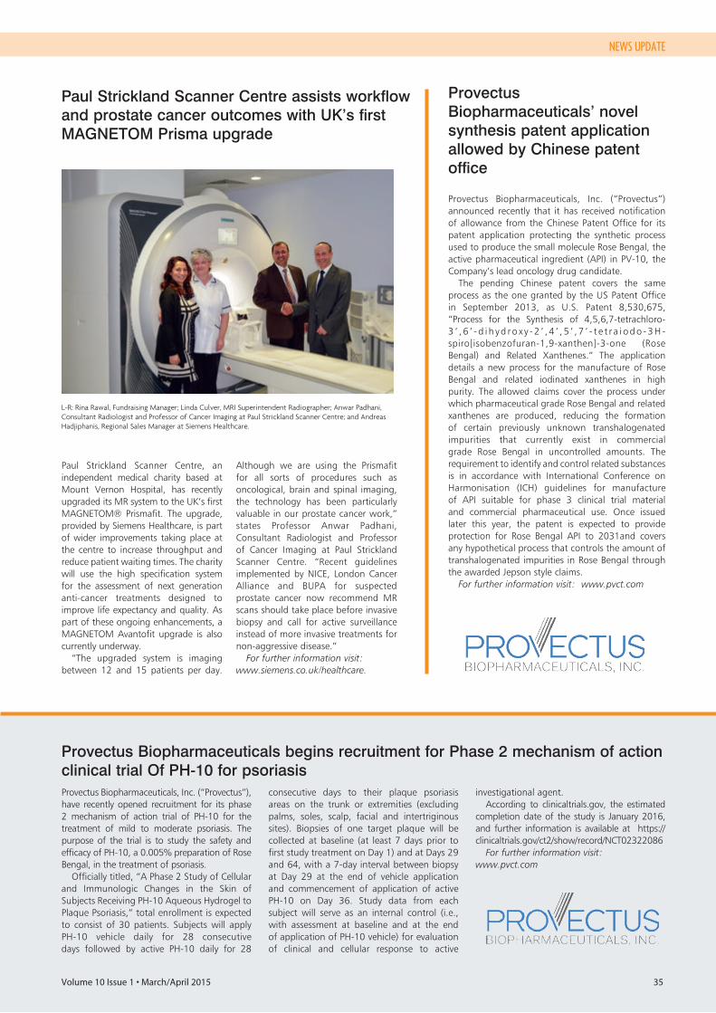
NEWS UPDATE
Provectus Biopharma ceuticals’ novel synthesis patent application allowed by Chinese patent office
Provectus Biopharmaceuticals, Inc. (“Provectus”) announced recently that it has received notification of allowance from the Chinese Patent Office for its patent application protecting the synthetic process used to produce the small molecule Rose Bengal, the active pharmaceutical ingredient (API) in PV-10, the Company’s lead oncology drug candidate.
The pending Chinese patent covers the same process as the one granted by the US Patent Office in September 2013, as U.S. Patent 8,530,675, “Process for the Synthesis of 4,5,6,7-tetrachloro-3 ’ , 6 ’ - d i h yd ro x y -2 ’ , 4 ’ , 5 ’ , 7 ’ - t e t r a i odo -3H-spiro[isobenzofuran-1,9-xanthen]-3-one (Rose Bengal) and Related Xanthenes.” The application details a new process for the manufacture of Rose Bengal and related iodinated xanthenes in high purity. The allowed claims cover the process under which pharmaceutical grade Rose Bengal and related xanthenes are produced, reducing the formation of certain previously unknown transhalogenated impurities that currently exist in commercial grade Rose Bengal in uncontrolled amounts. The requirement to identify and control related substances is in accordance with International Conference on Harmonisation (ICH) guidelines for manufacture of API suitable for phase 3 clinical trial material and commercial pharmaceutical use. Once issued later this year, the patent is expected to provide protection for Rose Bengal API to 2031and covers any hypothetical process that controls the amount of transhalogenated impurities in Rose Bengal through the awarded Jepson style claims.
For further information visit: www.pvct.com
Provectus Biopharmaceuticals begins recruitment for Phase 2 mechanism of action clinical trial Of PH-10 for psoriasis Provectus Biopharmaceuticals, Inc. (“Provectus”), have recently opened recruitment for its phase 2 mechanism of action trial of PH-10 for the treatment of mild to moderate psoriasis. The purpose of the trial is to study the safety and efficacy of PH-10, a 0.005% preparation of Rose Bengal, in the treatment of psoriasis.
Officially titled, “A Phase 2 Study of Cellular and Immunologic Changes in the Skin of Subjects Receiving PH-10 Aqueous Hydrogel to Plaque Psoriasis,” total enrollment is expected to consist of 30 patients. Subjects will apply PH-10 vehicle daily for 28 consecutive days followed by active PH-10 daily for 28
consecutive days to their plaque psoriasis areas on the trunk or extremities (excluding palms, soles, scalp, facial and intertriginous sites). Biopsies of one target plaque will be collected at baseline (at least 7 days prior to first study treatment on Day 1) and at Days 29 and 64, with a 7-day interval between biopsy at Day 29 at the end of vehicle application and commencement of application of active PH-10 on Day 36. Study data from each subject will serve as an internal control (i.e., with assessment at baseline and at the end of application of PH-10 vehicle) for evaluation of clinical and cellular response to active
investigational agent. According to clinicaltrials.gov, the estimated
completion date of the study is January 2016, and further information is available at https://clinicaltrials.gov/ct2/show/record/NCT02322086
For further information visit: www.pvct.com
Paul Strickland Scanner Centre assists workflow and prostate cancer outcomes with UK’s first MAGNETOM Prisma upgrade
Paul Strickland Scanner Centre, an independent medical charity based at Mount Vernon Hospital, has recently upgraded its MR system to the UK’s first MAGNETOM® Prismafit. The upgrade, provided by Siemens Healthcare, is part of wider improvements taking place at the centre to increase throughput and reduce patient waiting times. The charity will use the high specification system for the assessment of next generation anti-cancer treatments designed to improve life expectancy and quality. As part of these ongoing enhancements, a MAGNETOM Avantofit upgrade is also currently underway.
“The upgraded system is imaging between 12 and 15 patients per day.
Although we are using the Prismafit for all sorts of procedures such as oncological, brain and spinal imaging, the technology has been particularly valuable in our prostate cancer work,” states Professor Anwar Padhani, Consultant Radiologist and Professor of Cancer Imaging at Paul Strickland Scanner Centre. “Recent guidelines implemented by NICE, London Cancer Alliance and BUPA for suspected prostate cancer now recommend MR scans should take place before invasive biopsy and call for active surveillance instead of more invasive treatments for non-aggressive disease.”
For further information visit: www.siemens.co.uk/healthcare.
L-R: Rina Rawal, Fundraising Manager; Linda Culver, MRI Superintendent Radiographer; Anwar Padhani, Consultant Radiologist and Professor of Cancer Imaging at Paul Strickland Scanner Centre; and Andreas Hadjiphanis, Regional Sales Manager at Siemens Healthcare.
Volume 10 Issue 1 • March/April 2015 35

NEWS UPDATE
Sunesis Pharmaceuticals selects Quotient Clinical’s real-time adaptive manufacturing to support its vosaroxin clinical mass balance/ADME program
Quotient Clinical, the Translational Pharmaceutics® Company, and Sunesis Pharmaceuticals have announced the initiation of a clinical mass balance/ADME trial for vosaroxin – a candidate therapeutic for acute myelogenous leukemia (AML).
For this program, Quotient will employ its ‘real-time adaptive manufacturing’ approach to supply 14C radiolabeled vosaroxin to a specialist oncology clinic in the Netherlands. The intravenous product will be manufactured on a ‘per patient’ basis, and supplied to the clinic ready for dosing within less than two weeks of patient notification. This approach offers the most efficient use of the radiolabeled
drug, and tailors drug product manufacturing to ongoing patient recruitment.
Mark Egerton, CEO of Quotient Clinical, commented: “Vosaroxin represents an exciting potential therapeutic option for AML patients, and we are delighted to be supporting Sunesis
on its clinical mass balance/ADME program. Our real-time adaptive manufacturing approach offers increased flexibility and enables customisation of drug product supply to the requirements of individual patients and trials.”
Gene Jamieson, Vice President of Technical Operations at Sunesis Pharmaceuticals, added: “Working with Quotient Clinical will help to accelerate the clinical mass balance/ADME program for this promising therapeutic.”
For further information contact: Lisa Clarke-Lens, Quotient Clinical E: [email protected]
Provectus Biopharmaceuticals, Inc (“Provectus”) presented at the 17th Annual BIO CEO and Investor Conference on in February 2015.
The presentation was an update on the business, it took place in the Duke of Windsor Room at the Waldorf Astoria Hotel in New York City. The presentation is available for viewing on the Provectus website, http://pvct.com/presentation/index.html.
About BIO CEO & Investor Conference The BIO CEO & Investor Conference is the largest investor conference focused on established and emerging publicly traded
and select private biotech companies. Each year the BIO CEO & Investor Conference provides a neutral forum where institutional investors, industry analysts, and senior biotechnology executives have the opportunity to shape the future investment landscape of the biotechnology industry. The
conference features issue-oriented plenary sessions, educational sessions focused on hot therapeutic areas and key business issues, company presentations, one-on-one meetings, and networking opportunities. The therapeutic workshops feature MDs, CSOs and industry analysts discussing the latest information on pipeline innovation for breakthrough therapeutic topics in biopharma. Seasoned industry executives and analysts delve into timely and relevant business models, deal-making and investment trends on our business roundtables.
For further information visit: www.pvct.com
Provectus Biopharmaceuticals presented at 17th Annual BIO CEO and Investor Conference February 2015
PV-10 featured in article by Sanjiv Agarwala in Current Opinion in OncologyProvectus Biopharmaceuticals, Inc. (“Provectus”) announced recently that an article, entitled “Intralesional therapy for advanced melanoma: promise and limitation,” authored by Sanjiv S Agarwala, MD, has been published in the March issue of Current Opinion in Oncology, now available online.
The entire article may be found at: http://journals.lww.com/co-ncology/Fulltext/2015/03000/Intralesional_therapy_for_advanced_melanoma_.12.aspx
Dr Agarwala reviews the research history of the first intralesional treatment Bacille Calmette-Guerin (BCG), as well as the newer treatments Allovectin-7 (velimogene aliplasmid), plasmid IL-12, talimogene laherparepvec (T-VEC) and Provectus Biopharmaceutical’s PV-10. Key points of the article include: • Risk for recurrence, progression and
metastasis is high among patients with unresectable, multiple or advanced locally/
regionally metastatic stage IIIB/C or stage IV M1a melanoma.
• Most recent clinical trials of intralesional therapies show promise for their response rates, low toxicity and likely systemic immunological effects.
• Ongoing and planned clinical trials will test T-VEC in combination with systemic immunological therapies and PV-10 as monotherapy versus chemotherapy in patients who have failed or are ineligible for systemic immunological therapy.
Dr Agarwala’s findings stated, “After promising phase 2 results with Allovectin-7 (velimogene aliplasmid), overall survival in a phase 3 study was shorter for Allovectin-7 than for dacarbazine/temozolomide (median 18.8 versus 24.1 months). In a phase 2 trial of intratumoural electroporation of plasmid interleukin-12 among 28 patients with advanced melanoma, the primary endpoint of best overall response
rate within 24 weeks of first treatment was 32.2% for objective response and 10.7% for complete response. In the phase 3 OPTiM trial of talimogene laherparepvec, the intralesional agent that is furthest along in clinical testing, the primary endpoint of durable response rate was 16% for talimogene laherparepvec and 2% for granulocyte macrophage colony-stimulating factor. In the PV-10 phase 2 trial among 80 patients with stage III–IV melanoma, the overall response rate was 51%, with a 26% complete response rate.”
For further information visit: www.pvct.com
36 Volume 10 Issue 1 • March/April 2015
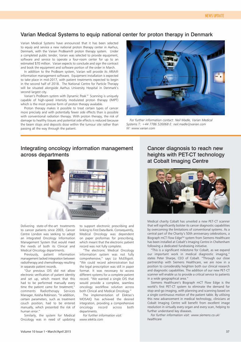
NEWS UPDATE
Varian Medical Systems to equip national center for proton therapy in Denmark
Integrating oncology information management across departments
Varian Medical Systems have announced that it has been selected to equip and service a new national proton therapy center in Aarhus, Denmark, with the Varian ProBeam® proton therapy system. Under a completed public tender, Varian was selected to provide equipment, software and service to operate a four-room center for up to an estimated $70 million. Varian expects to conclude and sign the contract and book the equipment and software portion of the order in March.
In addition to the ProBeam system, Varian will provide its ARIA® information management software. Equipment installation is expected to take place in mid-2017, with patient treatments expected to begin in the second half of 2018. The National Centre for Particle Therapy will be situated alongside Aarhus University Hospital in Denmark’s second largest city.
Varian’s ProBeam system with Dynamic Peak™ Scanning is uniquely capable of high-speed intensity modulated proton therapy (IMPT) which is the most precise form of proton therapy available.
Proton therapy makes it possible to treat certain types of cancer more precisely and with potentially fewer side effects than is possible with conventional radiation therapy. With proton therapy, the risk of damage to healthy tissues and potential side effects is reduced because the beam stops and deposits dose within the tumour site rather than passing all the way through the patient.
For further information contact: Neil Madle, Varian Medical Systems T: +44 7786 526068 E: [email protected] W: www.varian.com
Delivering state-of-the-art treatments to cancer patients since 2003, Cancer Centre London was seeking to adopt an integrated Oncology Information Management System that would meet the needs of both its Clinical and Medical Oncology departments.
Previously, patient information management lacked integration between radiotherapy and chemotherapy resulting in separate patient records.
“Our previous OIS did not allow electronic verification of patient identity and set up, which meant that this had to be performed manually every time the patient came for treatment,” comments Radiotherapy Services Manager, Keisha Robinson. “In addition, certain parameters, such as treatment couch position, had to be entered manually, which presented the risk of human error.”
Similarly, the system for Medical Oncology was in need of updating
to support electronic prescribing and linking to First Data Bank. Consequently, Medical Oncology was dependent on paper proformas for prescribing, which meant that the electronic patient record was not fully complete.
“The electronic Medical Oncology information system was not fully comprehensive,” says Liz McElligott. “We could record administration but the legal prescription was still in paper format. It was necessary to access different systems for a complete patient record. “We wanted a single OIS that would provide a complete, seamless oncology workflow solution across both Clinical and Medical Oncology.”
The implementation of Elektas’s MOSAIQ has achieved the desired integration, providing a comprehensive oncology record across both departments.
For further information visit: www.elekta.com
Cancer diagnosis to reach new heights with PET-CT technology at Cobalt Imaging Centre
Medical charity Cobalt has unveiled a new PET-CT scanner that will significantly bolster its cancer diagnostic capabilities by overcoming the limitations of conventional systems. As a central part of the Charity’s 50th anniversary celebrations, a Biograph mCT Flow EdgeTM system from Siemens Healthcare has been installed at Cobalt’s Imaging Centre in Cheltenham following a dedicated fundraising initiative.
“This is a significant milestone for Cobalt, as we expand our important work in medical diagnostic imaging,” states Peter Sharpe, CEO of Cobalt. “Through our close partnership with Siemens Healthcare, we are now in a position to considerably heighten both our clinical research and diagnostic capabilities. The addition of our new PET-CT scanner will enable us to provide a critical service to patients in a wide geographical area.”
Siemens Healthcare’s Biograph mCT Flow Edge is the world’s first PET-CT system to eliminate the demand for stop-and-go imaging, with planning and scanning based on a single continuous motion of the patient table. By utilising this new advancement in medical technology, clinicians at Cobalt Imaging Centre will benefit from excellent image resolution in virtually every organ and every scan, helping to further understand key diseases.
For further information visit: www.siemens.co.uk/healthcare
Volume 10 Issue 1 • March/April 2015 37

NEWS UPDATE
Clinical research confirms how ‘scalp cooling works’
Clinical research has confirmed how scalp cooling works; data which will be formally presented at the 14th St.Gallen International Breast Cancer Conference being held in Vienna, Austria, on March 18-21, 2015.
The studies showed that although chemotherapy drugs are highly toxic to cells, cooling markedly reduced or completely stopped hair follicles from dying.
The research also revealed that cooler scalp temperatures used in Paxman systems provided better clinical outcomes for patients, helping to change the lives of thousands of cancer patients worldwide.
Richard Paxman, CEO of Paxman, said: “We are extremely excited that the results of the research are being presented at this year’s BCC conference. Every day we hear personal stories from patients and their families about the positive results of scalp cooling so it is great to see clinical evidence to back this up.”
The research was carried out by Omar Hussain, Research Scientist at Paxman, in conjunction with the University of Huddersfield. The full abstracts will be presented on Thursday, 19 March at BCC 2015.
For more information visit www.paxman-coolers.com
A 76-year-old man with prostate cancer has become the first radiotherapy patient in the world whose treatment was planned using new RapidPlan™ knowledge-based software from Varian Medical Systems. Specialists at Royal Surrey County Hospital in Guildford, England, carried out the advanced IMRT (intensity modulated radiotherapy) treatment using RapidPlan to plan and guide the process.
“We planned the patient’s treatment both conventionally and using RapidPlan and we were comfortable that the RapidPlan treatment plan produced better dose delivery and beam modulation,” says Tom Jordan, head of radiotherapy physics. “With RapidPlan, the mean target dose achieved was slightly higher and the beam-shaping efficiency was greater, giving reduced
dose to the critical normal tissues as compared to a conventional plan. It also greatly speeded up the planning process by automating the selection of planning parameters.”
Tom Jordan explained that once a model had been created for a particular disease site by inputting a library of historical plans into the system, the RapidPlan system then typically suggests a better plan. “There is some work involved in inputting the data but once you have between 20 and 40 previous relevant cases in the system and you have optimised the planning priorities, it becomes a lot faster to produce plans for future treatments in RapidPlan,” he said.
For further information contact: Neil Madle, Varian Medical Systems T: +44 7786 526068 E: [email protected] W: www.varian.com
First radiotherapy treatment planned using Varian’s RapidPlan takes place at leading UK cancer centre
Terminally ill Sadie, 23 bravely shares her cancer story
38 Volume 10 Issue 1 • March/April 2015
Aged just 22, Sadie Rance, Bromley was told she had one to two years to live having being diagnosed with terminal, ovarian cancer.
Sadie first started to feel unwell when travelling through Australia with her partner Jason. She was suffering from stomach pains and constipation and her symptoms were first diagnosed by doctors as IBS. However, despite taking medically prescribed laxatives, Sadie’s condition didn’t improve and she took to the internet to look into her symptoms further, which she realised were the same as those associated with ovarian cancer.
After further tests, a tumour the size of a melon was discovered on her ovary and she was diagnosed with stage 4 ovarian cancer. Despite attempting to operate, the tumour had attached itself to her internal organs and the cancer had spread to her small bowel, liver, diaphragm, heart and lungs.
Despite the devastating news, Sadie remains positive and has an inspirational outlook on life and she has been fundraising for the Royal Marsden Hospital and a charity that has supported her every step of the way during her difficult journey; Teens Unite.
To show your support for Sadie, you can kindly make a donation here – www.justgiving.com/sadiefrance Website: www.teensunitefightingcancer.org Blog: www.teens-unite.blogspot.co.uk

NEWS UPDATE
The end of January brought about a fresh challenge for Oncology Systems Limited (OSL) as they exhibited the MacroMedics patient positioning range for the first time at the Annual Radiotherapy Conference.
MacroMedics and OSL have recently joined forces in the UK & Ireland to bring the very latest innovations in patient positioning and fixation to the radiotherapy community, from double shell systems to all-in-one platforms for every disease site.
“OSL has a long track record of introducing leading-edge technology to the local radiotherapy community and we are very proud to be working with such a fresh-thinking, dynamic and innovative manufacturer, in the form of MacroMedics”, said Stuart Baldwin, Managing Director at OSL.
One of the innovative products available in the MacroMedics range is the Double Shell Positioning System (DSPS) which, in 2013 was nominated for and reached the finals of the Dutch Design Award.
According to the selection commission of the Dutch Design Award, this medical system
improves the precision of the radiation treatment. It allows the head to remain completely motionless in a relatively comfortable position. It is an essential product that improves radiation treatment for head & neck.
Contact: Sarina Gloster E: [email protected] T: +44 1743 462694 W: www.osl.uk.com
Varian Medical Systems reported is progress towards its goal of making advanced treatments systems more available for cancer patients across Africa. The company is presenting to government and healthcare leaders at the 2nd Africa Healthcare Summit.
“We are pleased at the opportunity to engage with governments and other stakeholders at the Africa Healthcare Summit, to further explore ways of extending access to advanced cancer care,” says Burt Lang, Varian’s managing director in Africa. “Cancer is growing rapidly in Africa and has become one of the continent’s top healthcare concerns. Radiotherapy plays a vital and cost effective role in treating cancer
and we are committed to making it available to more patients across the continent.”
According to a study published in Lancet Oncology, only 23 out of 52 African countries have radiotherapy available for patients. The World Health Organization reports that by 2030 there will be some 1.6 million new cancer cases in Africa each year, resulting in 1.2 million
deaths. The most common cancers in Africa are cancers of the cervix, breast, lung, liver and prostate.
Varian has installed more than 100 radiotherapy treatment systems in Africa over the last 25 years. The company recently announced major projects in Algeria, Egypt and South Africa. Varian has also installed equipment in several sub-Saharan nations including Ghana, Angola, Kenya, and Madagascar.
For further information contact: Neil Madle, Varian Medical Systems T: +44 7786 526068 E: [email protected] W: www.varian.com
Design award for new head and neck solution in radiotherapy
Varian Medical Systems to make radiotherapy available for more cancer patients in Africa
A father who lost his life to a brain tumour and a Portsmouth man who is fighting one were the inspiration behind an ambitious cycling challenge in which their friends set out to cover 400 miles in just four days.
Carol Robertson, Head of Community Fundraising for Brain Tumour Research and champion fundraiser Simon Tier, along with others, are hoping the “Tour de Labs” will raise £43,840 to fund four days of research at each of the four Brain Tumour Research Centres of Excellence they are visiting along the route.
In their thoughts were Ian Meek, from Bristol, who lost his life to a brain tumour in 2012 and a friend of Simon’s, from Portsmouth, who is currently being treated for a glioblastoma multiforme (GBM) tumour.
The route started at the Buckinghamshire headquarters of the charity Brain Tumour Research, taking them to Imperial College and Queen Mary, University London, then heading for the University of Portsmouth before finishing at Plymouth University during March, national Brain Tumour Awareness Month.
More details can be found at www.braintumourresearch.org/tour-de-labs
Tour de Labs funds Brain Tumour Research
Provectus Biopharmaceuticals will hold its 2014 year-end quarterly business update conference call at 4pm Eastern, Thursday, March 12, 2015
Call to be webcast, available for digital replay
Management will provide a business update on PV-10 and PH-10 to the investment community and answer questions from investors.
Those who wish to participate in the conference call may telephone 877-407-4019 from the US International callers may telephone 201-689-8337, approximately 15 minutes before the call. A webcast will also be available at: www.pvct.com.
A digital replay will be available by telephone
approximately two hours after the completion of the call until March 31, 2015, and may be accessed by dialing 877-660-6853 from the US or 201-612-7415 for international callers, and using the Conference ID# 13601930.
For further information visit: www.pvct.com
Volume 10 Issue 1 • March/April 2015 39