of - dm5migu4zj3pb.cloudfront.netdm5migu4zj3pb.cloudfront.net/manuscripts/100000/... · the skin...
TRANSCRIPT

STUDIES OF AN URTICARIAL RESPONSETO BLUE ANDVIOLETLIGHT IN MAN
By H. F. BLUM1AND R. J. WEST(From the Division of Physiology, University of California Medical School, Berkeley)
(Received for publication October 23, 1936)
A case of urticaria solare being available forstudy, it has seemed wise to determine as manyof the characteristics of the photo-response asfeasible, in the hope that these might ultimatelyshed light on the obscure etiology of this rare butdistressing disease. Preliminary studies of thiscase by Blum, Allington and West (1) haveshown that the individual is sensitive to wave-lengths included in the spectral region between3900 and 5300 A, i.e., in the blue and violet partsof the visible spectrum, radiation from whichregion of the spectrum elicits no response in nor-mal individuals. The urticaria solare individualresponds to short periods of this kind of radiationwith an erythema restricted to the irradiated area,followed by an edematous wheal over the samearea with spreading erythemal flare when the ir-radiation is sufficiently intense or prolonged; thisresponse has thus the characteristics of the " tripleresponse " as described by Lewis (2), and we maymake the tentative assumption that it occurs as theresult of the elaboration of a histamine-like " H"substance, further evidence for which will be pre-sented in the course of this paper. The responsedisappears in a few hours, leaving no trace. Ul-traviolet radiation shorter than 3200 A bringsforth the same delayed erythema followed by pig-mentation that is produced in the normal indi-vidual, but no traces of the urticarial reactioncharacteristic of the response to blue and violetlight.
CLINICAL ACCOUNT
As a clinical report of this case has not been published,the following brief account is included.
The patient, a white male, age 21, associates the firstappearance of abnormal sensitivity to light with a beesting on the left malar area which occurred May 18,1934. Severe pain and swelling of the face which sub-sided in a day or two followed the sting. The sensitivityto light was noticed shortly after.
Previous history reveals no allergic or urticarial dis-
'Assisted by a grant from the research funds of theUniversity of California.
orders. Prior to the onset of the present difficulty hisskin had always responded normally to exposure to lightThere is no family history of allergy or light sensitivity.
Physical examination shows a well developed male ofaverage height and weight. He is a brunette, but theskin is pale from avoidance of light. He is sufferingfrom a moderately severe indolent papular type of acnevulgaris, and presents scattered areas of tinea versicolorover the upper trunk. There are no other abnormalities.The skin reacts normally to heat and cold. There is nodermatographism.
Laboratory findingsBlood. Numerous counts have shown an average of
about 5,000,000 erythrocytes and 7,000 leukocytes per cu.mm. There is an increase in small lymphocytes up toabout 50 per cent, otherwise the differential count is nor-mal. Hemoglobin determination, made with a photelome-ter, based on the Newcomer standard, was 85 per cent.Spectroscopic examination of the serum for abnormalpigments negative.
Urine. Routine examination and examination for por-phyrins were negative on several occasions.
Fasting blood sugar-100 mgm. per 100 cc.Fasting blood uric acid-3.3 mgm. per 100 cc.Wassermann-negative.Gastric analysis. Alcohol test meal.
Time (minutes): fating 15 30 45 75 90 105 120FreeHCI 0 0 0 0 0 0 2.4 8.0Total acidity 4.2 9.6 13.7 14.3 13.3 10.2 14.0 15.0
Scratch tests for a large variety of food, pollen, andepidermal extracts showed no reactions.
Basal metabolic rate-minus 14.
METHODS
General method. The arrangement of the ap-paratus used in the following experiments is dia-grammed in a general way in Figure 1. It con-sists of a 500-watt projection type Mazda lamp Aplaced at a given distance from an opaque screenB, which has a circular opening S about 2 cm. indiameter through which an area of the skin maybe irradiated. The screen is so fixed that the skinof any desired part of the body may be firmlypressed against it without disturbing the arrange-ment of the apparatus; thus the distance betweenB and A is established and reproducible through-
261

H. F. BLUM AND R. J. WEST
outletA
)
FIG. 1. ARRANGEMENTOF APPARATUSA, 500-watt projection type Mazda lamp; B, opaque
screen with circular opening S; C, water filter; D, glasscolor filter; E, concave mirror.
out a given experiment. C is a water filter forremoving infra-red radiation; it consists of apyrex cylinder 10 cm. in length and 5 cm. indiameter with inlet and outlet tubes so that a cur-rent of water may be passed through it to preventexcessive heating of the apparatus. D indicatesthe position at which glass filters may be intro-duced to obtain restricted wavelength regions. Eis a concave mirror used in some experiments toconcentrate the light rays. The distances be-tween the various elements of the apparatus werealtered to meet the requirements for any particu-lar experiment, and in some cases one or more ofthe elements was removed, as will be explained inthe description of the different procedures.
The sensitivity of the skin was measured interms of the time of irradiation required to pro-duce a just observable erythemal response, thethreshold time; the justification of this criterionwill appear as the various experiments are dis-cussed. The measurement was made in the fol.-lowing way. The skin was placed against thescreen B, and irradiated through the opening Sfor a measured number of seconds. The skinwas then moved away from the screen and ob-served for the appearance of erythema on theirradiated area. Most of the experiments werecarried out at room temperature and under theseconditions it was found that if erythema did notappear within fifteen minutes after the irradiationceased, none was ever observed. The procedurewas repeated on other areas of skin using differentperiods of irradiation, the shortest period whichwould just produce an erythemal response being
taken as the threshold time. It was found thatthis threshold time could be determined with anaccuracy better than ten per cent in most casesbut that it was safer to make no attempt to re-duce this error. It is quite easy to make the de-cision as to whether an erythema has appeared ornot, when no greater accuracy is attempted.
The 500-watt projection-type Mazda lampsused as sources burn at a higher color temperaturethan most incandescent lamps, and consequentlyhave a greater proportion of their emission in theblue-violet region in which we are interested.The shape of the emission curve, being verynearly that of a theoretical black body, can bereadily calculated from Wien's equation, if thecolor temperature is known (see Harrison (3)).Figure 2 shows emission curves for one of the
Wave lengths ; AFIG. 2. SPECTRAL DISTRIBUTION OF EMISSION FOR A
BLACK BODYAT DIFFERENT TEMPERATURES.The temperatures are the color temperatures for one
of the Mazda lamps used, when operated at differentvoltages as follows:
Voltage Color temperature1t0ots 0 K.105 .3010110 .3060115 .3110120 .3180
lamps used in the spectral regions with which weare concerned; the four curves are for color tem-peratures corresponding to four different lamp
262
URTICARIAL RESPONSETO BLUE AND VIOLET LIGHT
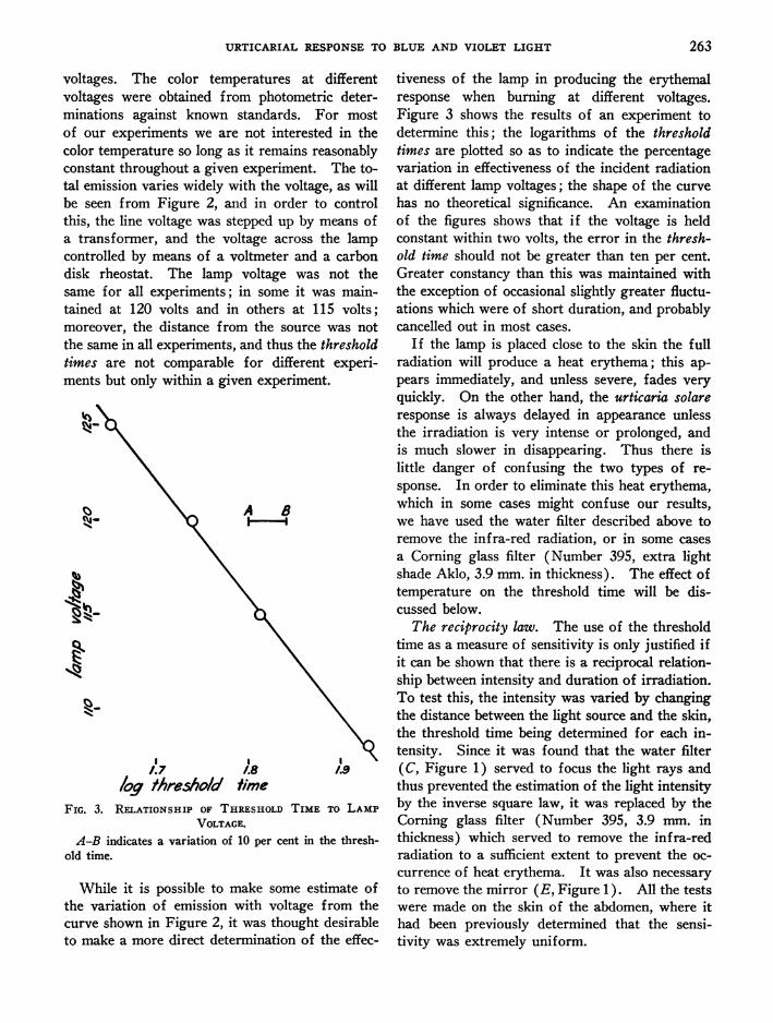
voltages. The color temperatures at differentvoltages were obtained from photometric deter-minations against known standards. For mostof our experiments we are not interested in thecolor temperature so long as it remains reasonablyconstant throughout a given experiment. The to-tal emission varies widely with the voltage, as willbe seen from Figure 2, and in order to controlthis, the line voltage was stepped up by means ofa transformer, and the voltage across the lampcontrolled by means of a voltmeter and a carbondisk rheostat. The lamp voltage was not thesame for all experiments; in some it was main-tained at 120 volts and in others at 115 volts;moreover, the distance from the source was notthe same in all experiments, and thus the thresholdtimes are not comparable for different experi-ments but only within a given experiment.
Nr
A BCrj_
1.7 1.8 1.9
loq /hres/o/d timeFIG. 3. RELATIONSHIP OF THRESHOLDTIME TO LAMP
VOLTAGE.A-B indicates a variation of 10 per cent in the thresh-
old time.
While it is possible to make some estimate ofthe variation of emission with voltage from thecurve shown in Figure 2, it was thought desirableto make a more direct determination of the effec-
tiveness of the lamp in producing the erythemalresponse when burning at different voltages.Figure 3 shows the results of an experiment todetermine this; the logarithms of the thresholdtimes are plotted so as to indicate the percentagevariation in effectiveness of the incident radiationat different lamp voltages; the shape of the curvehas no theoretical significance. An examinationof the figures shows that if the voltage is heldconstant within two volts, the error in the thresh-old time should not be greater than ten per cent.Greater constancy than this was maintained withthe exception of occasional slightly greater fluctu-ations which were of short duration, and probablycancelled out in most cases.
If the lamp is placed close to the skin the fullradiation will produce a heat erythema; this ap-pears immediately, and unless severe, fades veryquickly. On the other hand, the urticaria solareresponse is always delayed in appearance unlessthe irradiation is very intense or prolonged, andis much slower in disappearing. Thus there islittle danger of confusing the two types of re-sponse. In order to eliminate this heat erythema,which in some cases might confuse our results,we have used the water filter described above toremove the infra-red radiation, or in some casesa Corning glass filter (Number 395, extra lightshade Aklo, 3.9 mm. in thickness). The effect oftemperature on the threshold time will be dis-cussed below.
The reciprocity law. The use of the thresholdtime as a measure of sensitivity is only justified ifit can be shown that there is a reciprocal relation-ship between intensity and duration of irradiation.To test this, the intensity was varied by changingthe distance between the light source and the skin,the threshold time being determined for each in-tensity. Since it was found that the water filter(C, Figure 1) served to focus the light rays andthus prevented the estimation of the light intensityby the inverse square law, it was replaced by theCorning glass filter (Number 395, 3.9 mm. inthickness) which served to remove the infra-redradiation to a sufficient extent to prevent the oc-currence of heat erythema. It was also necessaryto remove the mirror (E, Figure 1). All the testswere made on the skin of the abdomen, where ithad been previously determined that the sensi-tivity was extremely uniform.
263

H. F. BLUM1 AND R. J. WEST
TABLE I
The reciprocity lawd=distance from lamp in cm.; t=time in seconds;
IXt=k= I Xt, where I= intensity.
Per centperit lDte |d | k Avemge t devia-
men D tion fromment ~~~~~~~~~averageI August 29 29.8 95 .103
21.9 65 .13615.5 30 .12510.0 12 .120
.1214.018 15.0
2 August 31 10.0 14 .14021.6 65 .13950.3 300 .11915.3 30 .12833.2 130 .118
.1294.011 8.5
3 September 5 10.0 14 .14050.0 315 .126
.1334.700 5.2
4 September 19 15.0 31 .13830.0 100 .11110.0 13 .13050.0 300 .12020.0 55 .137
.127 .160 12.5
The results of four experiments on four dif-ferent days are summarized in Table I. Of these,Experiment 2 was the most carefully conductedand may be taken as a fair index of the accuracy
of adherence to the reciprocity law under our
experimental conditions. It will be seen that forthis experiment, the values for k in the equation:Intensity X time k vary from the average byabout 8.5 per cent, indicating an outside error ofless than + 10 per cent. The results for theother experiments, which cover a period of threeweeks, show average values for k which agree
within + 5 per cent, showing that there was lit-tle or no fluctuation in the sensitivity over thistime; the variations within these experiments are
somewhat greater than in Experiment 2. Fig-ure 4 shows graphically the range over which thereciprocity law holds and the deviation of our
experimental measurements. It would seem fromthese data that we may safely assume that thereciprocity law holds for the urticaria response,and that the threshold time may be used as a
' I
/00 200Exposure (t, seconds)
FIG. 4. THE RECIPROCITY LAW.
The curve is drawn from the equation: I X t = 0.129.
measure of the photosensitivity with an error ofnot more than + 10 per cent if the conditionsare properly controlled.
A point of practical importance to the patientas regards artificial illumination may be men-tioned here. From the above data one may cal-culate that at a distance from the lamp of 3meters, 3 hours of irradiation would be requiredto produce a response. Since the lamps ordi-narily used for illuminating purposes burn atlower temperatures, their emission is less, andparticularly in the shorter wavelengths to whichthis individual is sensitive. Thus, ordinary con-ditions of artificial lighting are not bothersome,although even reflected sunlight in a room withlight colored walls may be a source of considerableannoyance.
Wemay make a rough estimate of the quantityof radiation required to produce the response.From the data given by Luckiesch (4) we find
264

URTICARIAL RESPONSETO BLUE AND VIOLET LIGHT
that a black body at 32000 K. emits 6.12 micro-watts per square centimeter per foot-candle in thespectral region 4000 to 7600 A. The color tem-perature of a tungsten lamp is very close to thatof a black body at this range of temperatures(Holladay (5)) so that this value may be takenas sufficiently close for our purposes. From thecurve for emission of a black body at this tem-perature it may be calculated that between one-sixth and one-seventh of the emission between4000 A and 7600 A lies between 4000 A and5000 A, the spectral region which elicits the urti-caria solare response, so that we may estimate thatabout one microwatt per sq. cm. per foot-candleis emitted in this region. Our lamp emits about1300 horizontal foot-candles, and from our datafor the reciprocity law we see that at a distance ofone foot, the threshold time is about one minute.Thus we may estimate that approximately 1300microwatts of radiant energy of wavelengths 4000to 5000 A must fall on the skin of this individualin order to produce an urticarial response in oneminute.
The effect of temperature on the response. Itwas recognized early in the investigation that tem-perature might have a considerable influence onthe response under certain circumstances. Stud-ies were therefore attempted to determine themagnitude of this effect so that an estimate of theinfluence of temperature on the accuracy of ourmeasurements might be made.
The effect of temperature on the threshold timewas first determined. For these experiments thestop B was replaced by a thin black paper stopwith a 2 cm. hole, placed tightly against the waterfilter C, no color filters (D) being used. Theskin of the abdomen was held firmly against thisblack paper so that the skin area next to the waterfilter would tend to take the temperature of thewater which could be varied by passing differentmixtures of hot and cold tap water through thefilter. The temperature of the filter was deter-mined by means of a thermometer placed in theoutlet stream from the filter, and the surface tem-perature of the skin corresponding to any givenfilter temperature was estimated as follows. Theskin was held against the filter for a given periodof time, then moved away, the time noted, andthe surface skin temperature determined as
quickly as possible by means of a thermopile de-signed for this purpose, the time at which themeasurement was made being carefully noted.Further measurements were made at successiveintervals as the skin temperature changed towardthe normal, and from these successive measure-ments a curve was plotted and an extrapolationmade to zero time which should correspond to thetemperature of the skin when in contact with thefilter. It was found that for periods shorter thanfive minutes in contact with the water filter thevalues obtained for skin temperature varied con-siderably, but that when the skin was allowed toremain in contact for periods as long as twentyminutes, the values were very little different thanthose obtained for the five-minute periods. Inour subsequent experiments we therefore kept theskin in contact with the filter for five minutes be-fore the beginning of each experiment, i.e., beforebeginning the irradiation of the skin. From dataobtained for the skin surface temperature at vari-ous filter temperatures we were able to plot thecurve shown in Figure 5, from which an estimateof the skin surface temperature could be made bymerely measuring the temperature of the water atthe filter outlet. It is doubtful if these estimatesfor the skin surface temperatures can be regardedas within better than one or two degrees of theactual skin surface temperature, and the tempera-ture inside the skin where the response takesplace must have been somewhat different thanthose of the skin surface. It would seem that therange of temperatures inside the skin would besomewhat less than those on the surface, but wehave no way of estimating this difference.
Measurement of the threshold time at differenttemperatures was made as follows. The skin washeld against the filter for a period of five minutesin order to establish the temperature of the skin.The lamp was then turned on for a given irradia-tion period, the skin being maintained in contactwith the filter during this time. The skin wasthen moved away, and allowed to adjust towardnormal temperature while observation for the ap-pearance of a threshold erythemal response wasmade. By repeating this procedure for differentperiods of irradiation, the threshold times for theresponse at given temperatures were determined.Data for a series of such measurements are plotted
265

H. F. BLUM AND R. J. WEST
'3(
1%
b
IkI
_b
I I
20 30
Filfer temt"raire °FIG. 5. RELATIONSHIP OF FILTER TEMPERATURETO SKIN TEMPERATURE.
in Figure 6. From these data it appears that theQ10 for the threshold time is about 1.3 to 1.4 over
the range studied. This is a quite reasonable co-
efficient for a photochemical reaction.Reference to Figure 6 will give some indication
of how differences in temperature may affect theexperimental determination of threshold time in
0
O
I..
* N_~~~~~~~~~W
25 30Thresho/d time
FIG. 6. EFFECT OF TEMPERAI
the rest of our experiments. We may assume
that the normal temperature of the skin surfaceof the abdomen when exposed to ordinary room
temperatures is about 300 C., and in this regionwe see that a change of about 30 C. would resultin a variation of 10 per cent in our measurementsof threshold time. This is a much greater tem-
0~~~
0
\ 940 45
rURE ON THRESHOLDTIME.
40
266

URTICARIAL RESPONSETO BLUE AND VIOLET LIGHT
perature variation than is to be expected if theroom temperature is maintained reasonably con-stant as was the case in our experiments, but wesee that temperature variation is a factor whichcannot be entirely neglected, particularly in com-paring experiments made on different days.
The low temperature coefficient of the thresholdtime would indicate its rather direct relationshipto the primary photochemical mechanism. How-ever, the rate of development of the urticarial re-sponse as indexed by its latent period is not takeninto account in the measurement of threshold time.
using the same period of irradiation, but a shortertotal period in contact with the filter, or if theerythema is not present, the total period is in-creased. By a series of such trials the least timerequired for a minimal erythema to appear is de-termined and may be taken as an index of the rateof development of the erythemal response for thegiven temperature and period of irradiation. Byrepeated experiments using different temperaturesand different periods of irradiation a series ofsuch measurements were obtained which are dis-played in Figure 7. Since the primary photo-
ii
I I I I5 /0 /S 20
71me to aiopearance of minima/ eryfhemnu/ response rmn,res.FIG. 7. EFFECT OF TEMPERATUREON LATENT PERIOD.
The latent period is shortened as the period of ir-radiation is decreased. We have attempted tomeasure the effect of temperature on the rate ofdevelopment of the erythemal reaction after theend of the irradiation period, as follows. Theskin is held against the water filter for five min-utes, the lamp then turned on, and irradiation con-
tinued for a given length of time which is knownfrom the data of Figure 6 to be longer than thethreshold time for the particular temperature.When the lamp is turned off, the skin is kept incontact with the filter for a further period of time,and is then moved away and observed for the min-imal erythemal response. If the erythema is ob-served to be present at the time the skin is movedaway from the filter, the experiment is repeated
chemical reaction has a low temperature coeffi-cient, the period of irradiation may be taken torepresent the production of the same quantity ofreactants at all temperatures used, and the ratesmeasured for different temperatures when thesame period of irradiation was used should besubject to comparison. Unfortunately, thesemeasurements are laborious and trying to the sub-ject, and the measurements which we have madeare scanty and rough for this reason; they seem,however, to be significant.
The data collected in Figure 7 show that the timeof appearance of the threshold erythemal responseis greatly lengthened at low temperatures. Infact, the effect is so great that it is difficult toobtain comparable data over a wide range of
267

H. F. BLUM AND R. J. WEST
temperatures, and no attempt has been made tocalculate the temperature characteristics of thisresponse. Furthermore, when a temperature of350 C. was maintained in the filter and the skinkept in contact with it for a long enough periodof time (over seven minutes), no response ap-peared, so that the temperature coefficient couldnot be determined in this region. Our resultswould seem to be quite comparable to those ob-tained by Lewis (2, Chapter VII), who foundthat either low (12 to 150 C.) or high (45 to470 C.) temperatures inhibit the appearance ofthe triple response following histamine pricks innormal individuals, or stroking in urticarial sub-jects. Lewis has explained this as due, in partat least, to changes in the local circulation pro-duced by changes in temperature, and if we invokethe same explanation it would seem meaninglessto attempt to determine the temperature charac-teristics accurately, since they would not be anindex to the specific reactions of urticaria solare,but of more general reactions.
Lewis has suggested that the triple response inother types of urticaria results from the releaseof a histamine-like H substance from the cells ofthe skin, and it seems reasonable to extend thisconcept to urticaria solare. The similarity of be-havior with respect to temperature is added evi-dence to justify this position. Wemight, then,suggest the following scheme to represent themechanism of urticaria solare:
1. S + hvSr2. Sr + cells H3. H+ vessels -> triple response
In the primary reaction, 1, S is the light ab-sorbing molecule in the skin which is responsiblefor the initiation of the response, hv is a quantumof light absorbed by S (h = Planck's constantand v the frequency of the radiation), and Sr thereactive molecule resulting. By Sr we do notwish to imply an activated molecule in the strictphotochemical sense, but merely to indicate thatthe molecule S has been in some way modified andenabled to take part in a subsequent reaction. Inreaction 2, Sr reacts with skin cells to release thehistamine-like substance H, which then reactswith the small blood vessels to produce the tripleresponse 3. Obviously reaction 2 may be a chainreaction involving many steps. Reaction 1 must
have a very low temperature coefficient since it isa purely photochemical reaction, and its rate isnot dependent on the energy of activation but onthe capture of light quanta. However, reactions2 and 3 are thermal reactions and may have hightemperature coefficients. It seems probable thatreaction 3 is the one which dominates the picturewhen the effect of temperature on the rate of ap-pearance of the urticaria solare response is stud-ied, because of the similarity of behavior to thatobtained when histamine is introduced directlyinto the skin. The temperature coefficient ob-tained for the threshold time is probably deter-mined principally by reaction 1 which would ac-count for its low value; reaction 3 probably playslittle part in the determination of the thresholdtime where the temperature is only maintainedduring the period of irradiation, and the develop-ment of the response takes place at approximatelynormal skin temperature.
TABLE II
Sensitivity on various regions of the body, December 3, 1935
Region Threshold time
Ventral surface of abdomen. . 60 secondsLumbar region of back .... 60 secondsBack over scapula..... 70 secondsMedial surface of thigh
near knee.... 165 secondsOuter surface of forearm
(15 cm. above wrist) ... . 150 secondsInner surface of forearm
(15 cm. above wrist) . 1... 105 secondsInner surface of forearm at
wrist .................. 180 secondsPalm of hand. 180 seconds (itching only, no
observable erythema)Dorsum of hand ........... Longer than 20 minutesCheek .................. Longer than 20 minutes
Topographical distribution of photosensitivtty.Determinations of the threshold time for a num-ber of regions of the body are given in Table II.Aconstant light intensity was maintained through-out, and all the determinations were made withina period of three hours. The sensitivity varieswidely over the body, the abdomen and lumbarregion of the back being at least twenty times assensitive as the face and the back of the hands.The surface of the abdomen is uniformly sensitivewithin the limits of our experimental error, andwe have used this region in all our other experi-ments. It is of interest to note how the sensi-tivity may vary in adjacent regions, e.g., compare
268

URTICARIAL RESPONSETO BLUE AND VIOLET LIGHT
the palm and dorsum of the hand. Although wewere unable to produce any erythemic responseby exposure of the face and dorsum of the handsto our light source for twenty minutes, it mustnot be judged that these parts have become en-tirely insensitive, for the response can still beelicited by exposure to sunlight, and the subject isstill made uncomfortable by such exposure, al-though to a much less extent than two years ago.Unfortunately, no measurements of this kind weremade early in the development of the disease, thefigures in Table II being obtained about ninemonths after its onset. We cannot, therefore,make a definite statement that the decreased sensi-tivity of the exposed parts has developed as a re-sult of exposure to light; but judging from thesubject's reaction to casual exposure to sunlight atthe onset and at present we feel no doubt whatso-ever that this is true. Moreover, Duke (6), Val-lery-Radot (7), and Blum, Allington and West(1) have all found that exposure to light has someeffect in decreasing the sensitivity of the skin tolight.
Numerous possibilities offer themselves for theexplanation of the difference in sensitivity of vari-ous regions of the skin, and the mechanism ofdesensitization of local areas. Among these arisesthe question of the thickness of the skin, and itsrelative transparency to light. Miescher (8) haspointed out that the thickness of the skin is veryimportant in determining the sensitivity of normalskin to the erythema producing ultraviolet radia-tion (principally shorter than 3200 A), and thatthe decrease in sensitivity to such radiation afterexposure to it, may be due to the thickening of theepidermal layers. The data of Bachem and Reed(9) for the absorption of different wavelengths oflight by the various layers of the skin, afford theopportunity to make some estimate of the effect ofthe thickness of the epidermis on the normal ery-themic response and on the urticaria solare re-sponse. The normal erythemic mechanism wouldseem to be set off in the epidermis, probablychiefly in the malpighian layer, since there is verylittle penetration (9 to 16 per cent) of the excitingwavelengths below this layer, since the malpighianlayer is the principal site of pigment depositionwhich follows the erythema (Laurens (10)), andsince the erythema is delayed as though time might
.05 ./0 .15
FIG. 8. EFFECT OF THICKENING OF SKIN ON PENETRA-TION OF RADIATION.
Abscissa-thickening in mm. Ordinates-per cent re-duction in radiation reaching the photosensitive layer.Broken line-3000 A, malpighian layer (normal erythemicresponse). Solid line-4500 A, papillary layer (urticariasolare).
be required for the products resulting from theirradiation to reach the papillary layer where thefirst blood vessels are found. In Figure 8 hasbeen plotted the percentage reduction in radiationof wavelength 3000 A reaching the malpighianlayer which would be caused by thickening of thecorneum, based on the data of Bachem and Reedwhich is for skin from the region of the flexorsurface of the arm and the abdomen. It will beseen that a thickening of 0.03 mm., which amountsto doubling the thickness of the corneum, wouldreduce the radiation reaching the malpighian layerby about 66 per cent, so that an amount of thick-ening which would be difficult to observe wouldcause a considerable difference in the sensitivity ofthe skin to radiation of this wavelength. Thussmall differences in the thickness of the skin atdifferent regions of the body would producemarked variations in sensitivity, and it is con-ceivable that thickening resulting from irritationcaused by irradiation would account for at leastpart of the resistance of the skin to ultravioletradiation subsequent to exposure (Laurens (10) ).
The case of the urticaria solare response issomewhat different, the radiation which evokes itbeing transmitted by the skin to a much greaterextent. The response appears almost immediatelyafter the irradiation, in contrast to the response
269

H. F. BLUM AND R. J. WEST
elicited by ultraviolet light, which suggests thatthe locus of action is close to the small vesselswhich first appear in the papillary layer. Uponreference to the data of Bachem and Reed (9),we find that only about 20 per cent of the totallight of wavelengths 4000 to 5000 A incident uponthe skin is absorbed in the epidermal layers, butthat 50 per cent is absorbed in the papillary layer,so that it is quite possible that the urticaria solareresponse is elicited in the latter layer. In Fig-ure 8 is plotted the percentage decrease in radia-tion of wavelength 4500 A, which results fromincreasing the thickness of the epidermis; thisindicates that the thickness of the epidermisshould have much less effect on the urticaria solareresponse than on the erythemic response to ultra-violet radiation. From Figure 8 it may be seenthat a thickening of 0.1 mm. which amounts totripling the thickness of the epidermis would berequired to reduce the light reaching the papillaryby 60 per cent. While differences in the thicknessof the skin might be an important factor in deter-mining the threshold time for the various regionsof the body, it is improbable that it is a very im-portant factor in the desensitization of the skin.
Blum, Allington and West (1) found that suc-cessive irradiation of an area of the skin with thequartz-mercury arc in quantity sufficient to pro-duce a strong pigmentation reduced the sensitivityof that area to a marked degree. A filter wasinterposed (Corning 986) to remove the radiationabove 4000 A, so that the urticaria solare responsewas not elicited during the building up of the pig-ment. As we have pointed out above, thickeningof the epidermis could hardly account for a largepart of the decrease in sensitivity. The pigment,which is deposited in the basal cells of the mal-pighian layer of the epidermis, may be very effec-tive as a filter if the urticaria solare response orig-inates in the papillary layer provided it absorbsstrongly in the blue-violet region, and may con-ceivably be the cause of the apparent desensitiza-tion in the experiment of Blum, Allington andWest. However, at the time the tests recordedin Table II were made, the face of the subjectshowed very little pigmentation so that the lackof sensitivity of this region could not be creditedto any extent to this factor.
Another possibility is that the exposed areas of
skin in which the urticarial response has been fre-quently produced by the action of light, has de-creased in sensitivity to the products of the photo-chemical reaction; for instance, its sensitivity tohistamine. This was tested by pricking histamineinto various regions of the skin, and comparingthe reaction with that of a series of normal indi-viduals.2 The subject's reactions to histaminewere within normal limits on all parts of the bodyincluding the exposed parts. The sensitivity tohistamine is normally less on the exposed parts,but the difference is not of the order of the differ-ence in sensitivity to blue-violet light shown byour subject. Reactions to adrenalin pricks werealso within normal limits. Thus we have no rea-son to suspect an abnormal vascular reactivity inthe subject.
Finally, we come to the concept that exposureto light exhausts some part of the photochemicalmechanism. This is the suggestion of Duke(11). Wemust admit this as a distinct and rea-sonable possibility, but one that cannot be cate-gorically accepted in face of the other possibilitiescited above, unless further proof can be found.The fact that Blum, Allington and West (1) wereable to produce a decrease in sensitivity by ultra-violet irradiation without eliciting the urticariasolare response demonstrates that some other fac-tor is involved than the wearing out of the photo-chemical mechanism. It is probable that all thefactors mentioned play a part in decreasing thesensitivity of the skin which is exposed intermit-tently to sunlight.
Variations in the general sensitivity with time.An attempt was made to follow the sensitivity ofa region of the skin normally covered by clothing,the abdomen, over the course of ten months.These determinations were subject to a greatererror than our other studies, because it was im-possible to use the same lamp throughout the en-tire series, and because in some cases the voltagewas not carefully controlled. Moreover, at thetime of the earlier measurements we were notaware of some of the possible errors in the detec-tion of the threshold. In general, the later deter-minations are more trustworthy, but it is difficult
2 These tests were made through the courtesy of Dr.Eric Ogden who will publish an account of the methodand results of a series of such tests at a subsequent time.
270
URTICARIAL RESPONSETO BLUE AND VIOLET LIGHT
VIi
9 I OF THE HO5 /0TIM DR20 2A PI 5 /0 MONTS.June>/ Au9.
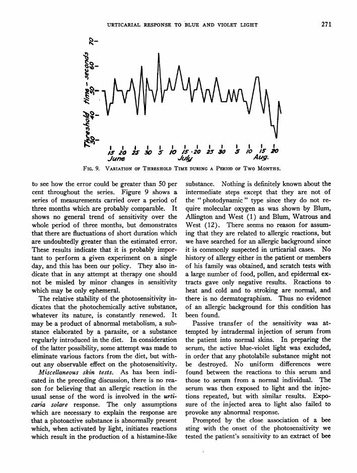
FIG. 9. VARIATION OF THRESHOLDTIME DURtING A PERIOD OF TWOMONTHS.
to see how the error could be greater than 50 per
cent throughout the series. Figure 9 shows a
series of measurements carried over a period ofthree months which are probably comparable. Itshows no general trend of sensitivity over thewhole period of three months, but demonstratesthat there are fluctuations of short duration whichare undoubtedly greater than the estimated error.
These results indicate that it is probably impor-tant to perform a given experiment on a singleday, and this has been our policy. They also in-dicate that in any attempt at therapy one shouldnot be misled by minor changes in sensitivitywhich may be only ephemeral.
The relative stability of the photosensitivity in-
dicates that the photochemically active substance,whatever its nature, is constantly renewed. Itmay be a product of abnormal metabolism, a sub-stance elaborated by a parasite, or a substanceregularly introduced in the diet. In considerationof the latter possibility, some attempt was made toeliminate various factors from the diet, but with-out any observable effect on the photosensitivity.
Miscellaneous skin tests. As has been indi-cated in the preceding discussion, there is no rea-
son for believing that an allergic reaction in theusual sense of the word is involved in the urti-caria solare response. The only assumptionswhich are necessary to explain the response are
that a photoactive substance is abnormally presentwhich, when activated by light, initiates reactionswhich result in the production of a histamine-like
substance. Nothing is definitely known about theintermediate steps except that they are not ofthe " photodynamic " type since they do not re-quire molecular oxygen as was shown by Blum,Allington and West (1) and Blum, Watrous andWest (12). There seems no reason for assum-ing that they are related to allergic reactions, butwe have searched for an allergic background sinceit is commonly suspected in urticarial cases. Nohistory of allergy either in the patient or membersof his family was obtained, and scratch tests witha large number of food, pollen, and epidermal ex-tracts gave only negative results. Reactions toheat and cold and to stroking are normal, andthere is no dermatographism. Thus no evidenceof an allergic background for this condition hasbeen found.
Passive transfer of the sensitivity was at-tempted by intradermal injection of serum fromthe patient into normal skins. In preparing theserum, the active blue-violet light was excluded,in order that any photolabile substance might notbe destroyed. No uniform differences werefound between the reactions to this serum andthose to serum from a normal individual. Theserum was then exposed to light and the injec-tions repeated, but with similar results. Expo-sure of the injected area to light also failed toprovoke any abnormal response.
Prompted by the close association of a beesting with the onset of the photosensitivity wetested the patient's sensitivity to an extract of bee
271

H. F. BLUM AND R. J. WEST
venom, prepared as suggested by Thompson (13)by triturating the poison apparatus in normalsaline and filtering through a Seitz filter. One-tenth cubic centimeter of a solution estimated tocontain 1 to 1500 parts of venom when injectedintradermally provoked a wheal approximatelyone centimeter in diameter showing free pseudo-podia. In normal individuals the usual result isan erythematous papule less than half this size,so that the patient seems somewhat sensitive tobee venom. Suspecting some relationship be-tween this sensitivity to bee venom and to light,attempts were made to desensitize the patient bya series of injections of the dilute bee venomsolution, but without any definite effect on thesensitivity of the patient to light. Simultaneouslyand subsequently the patient was given hydro-chloric acid by mouth to correct a condition ofachlorhydria, but no change in sensitivity to lightresulted.
The active wavelengths. As stated above, thepreliminary studies by Blum, Allington and West(1) delimited the wavelengths which produce thisurticarial response to the region between 3900and 5300 A, which is in very good agreement withthe findings of Duke (6), Vallery-Radot (7), andFrei (14) for their cases. Wemust assume thatwhatever the ultimate chemical reaction whichproduces this urticarial response, the primary re-action is the absorption of a quantum of light bya molecule of some photoactive substance in theskin as represented in reaction 1 above, and what-ever this substance may be, it must have a char-acteristic absorption spectrum, i.e., there must beonly restricted regions of the spectrum which itcan absorb, and consequently only light of thesespectral regions can bring about the urticarial re-sponse. We dwell upon this fundamental pointbecause it has been too frequently disregarded inthe study of photosensitivity in man. The rela-tive sensitivity to different wavelengths shouldcorrespond to a certain degree with the amountof light absorption of the photosensitizing sub-stance, and the curve relating sensitivity andwavelengths should approximate the absorptionspectrum of that substance. There are, of course,a number of factors which would tend to producesome disagreement in these curves, such as theeffect of the solvent on the absorption spectrum of
the photoactive substance, the specific absorptionof the skin, variations in photochemical efficiencywith wavelength, etc.; but if we are able to obtaina sensitivity wavelength curve for the photo-dermal response, we have a basis for a guess as tothe nature of the photosensitizing substance. Forthis reason we have attempted to make some morequantitative determinations than those previouslyreported.
In these studies colored glass filters were usedto isolate relatively narrow bands of wavelengths.The transmissions of the various filter combina-tions were determined for the visible region ofthe spectrum by means of a spectrophotometer.Since we had already determined by means ofglass color filters (Blum, Allington and West(1)) and sunlight or quartz mercury arc thatwavelengths outside the 3900 to 5300 A regionwere ineffective, we could disregard any filtertransmission outside of this restricted region.The spectrophotometric measurements could notbe made for wavelengths shorter than 4300 A,and it was necessary to extrapolate to zero trans-mission beyond this point, but our preliminarydeterminations would indicate that the sensitivitybelow this wavelength is so slight that no sig-nificant error could have been introduced.
The lamp used in these particular experimentswas the one whose emission curves are plotted inFigure 2; it was operated at 120 volts, at whichvoltage the color temperature was 31800 K. Theemission curve was calculated for a black body atthis temperature by means of Wien's equation(see Harrison (3)), and the transmission of each
filter combination for given wavelengths was mul-tiplied by the emission for the correspondingwavelengths; thus curves were obtained for therelative quantities of radiant energy incidentthrough the various filters. A further correctionwas made to give a relative number of quanta bymultiplying by the wavelength of the radiation.8
8 Since the amount of photochemical reaction dependsupon the number of quanta absorbed by the photochemi-cally active substance S, it is proper to compare the rela-tive number of quanta rather than the relative energiescorresponding to the exciting wavelengths. The correc-tion is so small in this case as to be of little significance,however. The subject is discussed by Blum and Scott(15), in which paper Equation 8 should read: Nx' =IxSxTx(- log Tx).
272

URTICARIAL RESPONSETO BLUE AND VIOLET LIGHT
'tuv 1Wa/ve /engJ,sT AFIG. 10. LIGHT PASSING THROUGHFILTERS USED TO ISOLATE
SPECTRAL REGIONS.
The curves for the relative number of quantapassing through the filter are shown in Figure 10.
TABLE III
Photosensitivity to restricted spectral regions(Experiments 1, 2, and 3 were performed on October 12,
October 29, and October 30, respectively)
x Ex-Filter Max- A pex- I k, =A X t
imum ment k.
I secondsBG2 + GG3 4350 .44 1 150 66 .015
2 150 66 .0153 195 86 .012
Average =.014038 + 585 4500 .38 1 90 42 .025
2 90 42 .0253 85 39 .036
Average =.025
BG1 + GG8 4650 1.00 1 90 90 .0112 70 70 .0143 70 70 .014
Average =.013
BG1 + GG7 4850 .32 1 165 53 .0192 195 62 .0163 165 53 .019
Average =.018
352 +554 4900 .19 1 330 64 .0163 300 58 .017
Average -.016
986 300 (no response)
338 100 (heat response)
The relative number of quanta reaching the skinthrough each filter should be proportional to thearea under the curves. These areas, measured bymeans of a planimeter, are expressed in relativeunits as A in Table III.
The threshold time for the erythemal responsewas determined for each filter, the values forthree experiments being given in Table III.Since the reciprocity law holds for light made upof all wavelengths, it may be assumed that it alsoholds for the restricted spectral bands which passthrough the filters. Thus the product of A,which is a measure of the relative number ofquanta passing a given filter, and the thresholdtime t, should give a value kx, which is a measureof the relative number of quanta required to elicita response at that wavelength. The reciprocal ofkx should be a measure of the relative sensitivityat this wavelength, and is the value which may becompared with the absorption of a substance sus-pected of being the photochemically active agent.Assuming the values of 1/kx to represent thesensitivity of the urticaria solare response at thewavelengths corresponding to the maximum trans-missions of the various filters, it would appearthat the sensitivity has two wavelength maxima,one at about 4500 A, and another at about 4900 A,
273

H. F. BLUM AND R. J. WEST
with a distinct minimum at about 4650 A. Thatthere is little sensitivity above 5000 A or below4000 A was shown clearly by the earlier experi-ments of Blum, Allington and West (1). Ob-viously, these values of 1/kx are subject to a con-siderable degree of error in both parameters, inthe one case due to the experimental error in-volved in determining the threshold, and in theother due to the fact that the band of light trans-mitted by the filter is rather wide; the degree oferror in the latter case cannot be accurately esti-mated. Another error is introduced, of course,in the determination of the color temperature ofthe lamp which might somewhat alter the relativevalues, although this could hardly affect the posi-tion of the maxima. At best, these values can-not be considered as very accurate, but they mightbe expected to give an approximate picture of theabsorption spectrum of the photosensitizing sub-stance which is responsible for the urticarialresponse.
Wemay now begin our search for the absorp-tion spectrum of some biological substance whichwill fit this region of absorption with a reasonabledegree of approximation. By reference to thedata on absorption bands accumulated in TabulaeBiologicae (16, 17, 18, 19, 20) we find that onlyone group of pigments there listed has the max-ima of absorption of its members confined to thegeneral spectral region which elicits the urticariasolare response; these are the carotenoids. Aspointed out by Blum, Allington and West (1) , theporphyrins, which are active photosensitizers, ex-hibit a minimum of absorption between 4000 and5000 A (21); the bile pigments show no maximawhich will correspond with the region of sensitiv-ity as is also true for cytochrome and hemochro-mogens in general (22). The flavines, anothergroup of naturally occurring photolabile pigments,absorb in the same general spectral region as thecarotenoids (4000 to 5000 A), but show greaterabsorption in the near ultraviolet (23); so that ifone of these were the sensitizer in the presentinstance, we should expect to find photosensitivityin the corresponding spectral region where theindividual with urticaria solare exhibits little orno sensitivity.
Thus the carotenoids which are definitely pho-tolabile seem to offer the best agreement with our
experimental data, and in Figure 11 we have plot-ted the absorption spectrum of a carotene in alco-
4000 4S00 J0oFIG. 11.
Abscissa-Angstrom units; ordinates-arbitrary values.Circles-relative sensitivity of urticaria solare (1/k).Horizontal lines and crosses-relative sensitivity of pho-totropic bending of the oat seedling. From Johnston(25), corrected to relative number of quanta. Curve-absorption of a carotene in alcohol. From Miller, Mac-kinney and Zscheile (24).
hol (24), together with our experimentally ob-tained values 1/k,\ for the urticaria solare re-
sponse. In the figure the ordinates for both were
chosen so as to bring the absorption curve ofa carotene and the values of 1/kx into relation-ship. It will be seen that at least a rough agree-ment exists.
In Figure 11 we have also plotted spectral sen-
sitivity data for the phototropic response of theoat seedling, as given by Johnston (25), choosingour ordinates so as to bring the data into ac-cordance with the absorption curve of a carotene.The agreement is rather good, but we could notfrom this make a categorical statement that a
carotene or any other specific carotenoid is thephotoactive substance responsible for phototro-pism in the oat seedling, although we must admitthat possibility which has been previously sug-gested by Bachmann and Bergann (26). Wenotethe existence of two distinct maxima in the dataof Johnston corresponding approximately to the
0
0
274
Na_
a
URTICARIAL RESPONSETO BLUE AND VIOLET LIGHT
two maxima in our own data for the urticariasolare response. Other data for the phototropicbending of the oat seedling do not agree quan-titatively with that of Johnston (see Bachmannand Bergann (26); Haig (27), but all the meas-urements agree in the delimitation of the generalspectral region and in general in the display oftwo maxima. In considering the data from sev-eral sets of measurements on the phototropicbending of the same organism, the oat seedling(Avena sativa), we see that the deviations amongthe various sets of measurements are as great asthe deviation of our own measurements from anyone of the above, even though our own data areadmittedly of a relatively low degree of accuracy.Thus, so far as the evidence goes the photo-chemically active substance may be the same inboth. Carotenoid pigments have been suspectedas the photosensitive materials in phototropicbending of plants, and Castle (28) has been ableto extract such a pigment from phycomyces whoseabsorption spectrum fits well with the spectralsensitivity curve which he has determined for thatorganism, and which would also approximate ourown data and that for the oat seedling. Thisdiscussion of the spectral sensitivity of the orient-ing mechanism in plants has been introduced prin-cipally to show the deviation in measurementsobtained on other living systems. Numerousfactors exist which would create differences be-tween the curves of spectral sensitivity of theliving organism, and the absorption curve of theresponsible photochemically active substance whenremoved from the living tissue and in solution insome solvent other than that in which it is dis-solved in the organism. For example, the dif-ference in transmissivity of the epidermis to thevarious active wavelengths might alter the effec-tiveness of the various spectral bands in elicitingthe urticaria solare response; but from the dataof Bachem and Reed (9), we may estimate arather uniform decrease in transmittency of about15 per cent from 5000 to 4000 A, which could notgreatly alter our sensitivity curve. Again, it isunfair to make a comparison of the absorptionspectrum of a carotenoid pigment in solution inalcohol with the spectral sensitivity curve of anorganism in which the absorbing pigment mustbe in solution in some other solvent. The ab-
sorption spectra of the carotenoids are shiftedvery markedly with the solvent employed, so thatit is impossible to select any specific carotenoidas showing better agreement with the data thananother. The choice of a carotene in alcohol forplotting in Figure 11 was made only because itshowed the possibility of agreement, not to indi-cate that this is the actual carotenoid involved.An examination of the absorption spectra in themonograph of Zechmeister (29) will show theextent of the shift of the absorption spectra ofcarotenoids with the solvent, and will also showthat these spectra display two principal maximaseparated by a well defined minimum, no matterwhat the solvent. The apparent existence of twomaxima in our measurements of the spectral sen-sitivity of urticaria solare, and of the phototropismof Avena gives strong support to the hypothesisthat carotenoid pigments are responsible for bothphoto-physiological responses.
Until other evidence is offered, then, we mustsuspect that a carotenoid pigment is the photosen-sitizing agent in the case of urticaria solare nowin hand, and may use this as a working hypothesis.Following this evidence we have attempted toproduce local sensitivity to light by injecting solu-tions of carotene and xanthophyll into rabbits'ears and into human skin, but exposures of theinjected areas to sunlight for periods as long astwenty minutes produced no response which couldbe taken as definite evidence of photosensitivity.The solvents used for injection were cottonseedoil and propylene glycol. In both cases, particu-larly the former, a considerable irritation wasproduced by the injected material which may havemasked any response resulting from exposure tolight.
DISCUSSION
The evidence presented in the preceding pageswould best be explained by the postulation that acarotenoid pigment is present in the skin, whichmay be activated by light to set off a series ofreactions which result in the release of H sub-stance in the region of the small blood vessels.The ultimate result is the appearance of the tripleresponse on the area of skin reached by the light.The only abnormal part in such a mechanism isthe presence of the carotenoid pigment, and itwill be well, therefore, to inquire into the plausi-
275
H. F. BLUM AND R. J. WEST
bility of the presence of such a pigment, and itspossible origin. Carotenoids are taken up in largequantities in the normal diet, a certain fractionbeing changed into vitamin A, and a large partexcreted in the feces. In some cases, after in-gestion of great quantities of food rich in caro-tenoids, enough may accumulate in the skin togive the individual a yellow or reddish color (29).Hess and Myers (30) found that infants fed anexcess of carrots assumed a yellow coloration,and that carotene could be isolated from the bloodand urine in such cases; there seems to be norecord of sensitivity to light, but Klose (31) statesthat the yellow color is most pronounced on theparts normally exposed to light.
It would seem thus that great quantities ofcarotenoid pigments may be present in the skinwithout sensitivity to light, at least in a degreecomparable with that of our subject. Moreover,our patient does not show a general yellow tint,so that there can be no great excess of carotenoids.It seems probable that any carotenoid entering theskin from the blood stream would be depositedprincipally in the fat of the subcutaneous layersbecause the carotenoids are very soluble in fatand insoluble in water, and the observations ofKlose (31) would indicate that this is the pointof deposition. The penetration of blue and violetlight to the subcutaneous layers is not great, andfor reasons discussed above it seems probable thatthe urticaria solare response is set off in the papil-lary layer of the corium. It would seem neces-sary to assume from this that in our subject thecarotenoid is deposited superficially to the subcu-taneous fat, that it is not present in great quanti-ties, and that it owes its effectiveness as a photo-sensitizer to its position in the skin or that it isa specific kind of carotenoid which is not ordi-narily present in the human organism. All thissuggests that the carotenoid may be produced inthe skin through the agency of a parasite. It istherefore worthy of remark that our subject dis-plays a few of the yellow macules of Tenia versi-color which result from infection of the skin bythe fungus Malasezia furfur, and that it may bethis organism which is producing the photosensi-tizing carotenoid. Wehave been unable, however,to show that the yellow macular areas are anymore sensitive than parts of the skin which appear
free from the infection, so that this hypothesisreceives no substantiation for the present.
SUMMARY
Photo-physiological studies of an urticarial re-sponse elicited by blue and violet light demonstratethe following.
1. The response obeys the reciprocity law.2. Studies of the effect of temperature indi-
cate that the mechanism of the response includesa photochemical reaction which is not greatly af-fected by temperature, and a thermal reactionwhich is greatly modified by changes in tempera-ture. The latter is probably the action of a hista-mine-like substance on the small vessels of theskin.
3. All parts of the body are sensitive to lightbut the degree of sensitivity varies from region toregion. The exposed parts are much less sensi-tive than the parts covered by clothing. The rea-sons for these variations are discussed.
4. The sensitivity fluctuated somewhat withtime, but there was no general trend in the courseof ten months.
5. Determination of the spectral sensitivity sug-gests that the photosensitizer is a carotenoid pig-ment. The possible origin of such a carotenoidin the skin is discussed.
Wewish to express our appreciation of the assistancegraciously given by the following: Dr. H. V. Allingtonwho made the clinical studies; Professor L. M. K. Boel-ter who determined color temperatures of lamps; Pro-fessor Eric Ogden for histamine tests; Professor A. P.Krueger for bacteriological control of materials for in-jection; and Doctors G. Mackinney and S. Lepkovskywho provided samples of xanthophyll and carotene.
BIBLIOGRAPHY
1. Blum, H. F., Allington, H., and West, R., On an urti-carial response to light and its photophysiology.J. Clin. Invest., 1935, 14, 435.
2. Lewis, T., The Blood Vessels of the Human Skin andTheir Responses. Shaw and Sons, London, 1927.
3. Harrison, G. R., Instruments and methods used formeasuring spectral light intensities by photography.J. Optic. Soc. America, 1929, 19, 267.
4. Luckiesch, M., Artificial Sunlight Combining Radia-tion for Health with Light for Vision. Van Nos-trand, New York, 1930, p. 165.
5. Holladay, L. L., Proportion of energy radiated byincandescent solids in various spectral regions. J.Opt. Soc. America, 1928, 17, 329.
276

URTICARIAL RESPONSETO BLUE AND VIOLET LIGHT
6. Duke, W. W., Urticaria caused by light. Preliminaryreport. J. A. M. A., 1923, 80, 1835.
7. Vallery-Radot, P., Blamoutier, P., Besangon, J., andSaidman, J., Urticaire solaire. Bull. et mem. Soc.med. d. h6p. de Paris, 1926, 50, 1116.
8. Miescher, G., Das Problem des Lichtschutzes und derLichtgewohnung. Strahlentherapie, 1930, 35, 403.
9. Bachem, A., and Reed, C. I., The penetration of lightthrough human skin. Am. J. Physiol., 1931, 97, 86.
10. Laurens, H., Photochemistry in medicine. A generaloutline. Cold Spring Harbor Symposia on Quan-titative Biology, 1935, 3, 277.
11. Duke, W. W., Allergy, Asthma, Hay Fever, Urti-caria, and Allied Manifestations of Disease. C. V.Mosby Co., St. Louis, 1925.
12. Blum, H. F., Watrous, W. G., and West, R. J., Onthe mechanism of photosensitization in man. Am.J. Physiol., 1935, 113, 350.
13. Thompson, F., About bee venom. Lancet, 1933, 225,446.
14. Frei, W., Lokale urtikarielle Hautreaktion auf Son-nenlicht. Arch. f. Dermat. u. Syph., 1925, 149,124.
15. Blum, H. F., and Scott, K. G., Photodynamically in-duced tropisms in plant roots. Plant Physiol.,1933, 8, 525.
16. Brigl, P., Pflanzliche Farbstoffe. Tabulae Biologicae,1926, 3, 333 and 338.
Fischer, H., and Treibs, Tierische Farbstoffe. Ibid.,pp. 349 to 361.
17. Kirstahler, A., Blut-Farbstoff und Derivate Spektrendes Farbstoff-Anteils des Hamoglobins u. seinerUmwandlungsprodukte. Tabulae Biologicae, 1931,7, pp. 66 to 87.
Chlorophyll a und b, ihre Umwandlungs-bezw.Abbau Produkte II. Ibid., pp. 245 to 257.
18. Karrer, P., Carotinoide. Tabulae Biologicae, 1933, 8,342.
19. Fernholz, E., Vitamin B1, B2, C und D. TabulaeBiologicae, 1934, 9, pp. 196 to 202.
20. Haurowitz, F., Respiratorische Farbstoffe (Chromo-proteide). Tabulae Biologicae, 1935, 10, pp. 25 to28.
21. Clar, E., and Haurowitz, F., Die Konstitution derPorphyrine. Ber. deutsch. chem. Gesell., 1933, 66,331.
22. Roche, J., and Benevent, M., Recherches sur la consti-tution du cytochrome c. Bull. Soc. chim. biol.,1935, 17, 1473.
23. Kuhn, R., Gyorgy, P., and Wagner-Jauregg, T., UberOvoflavin, den Farbstoff des Eiklars. Berl. d.deutsch. chem. Gesell., 1933, 66, 576.
24. Miller, E. S., Mackinney, G., and Zscheile, F. P., Ab-sorption spectra of alpha and beta carotenes andlycopene. Plant Physiol., 1935, 10, 375.
25. Johnston, E. S., Growth Movements in Relation toRadiation. (Chapter XXXIII in Biological Ef-fects of Radiation. Edited by B. M. Duggar.)McGraw-Hill Co., New York, 1936.
26. Bachmann, Fr., and Bergann, Fr., t[ber die Wertig-keit von Strahlen verschiedener Wellenlange furdie phototropische Reizung von Avena sativa.Planta: Archiv. Wiss. Bot., 1930, 10, 744.
27. Haig, C., The spectral sensibility of Avena. Proc.Nat. Acad. Sc., 1934, 20, 476.
28. Castle, E. S., Photic excitation and phototropism insingle plant cells. Cold Spring Harbor Symposiaon Quantitative Biology, 1935, 3, 224.
29. Zechmeister, L., Carotinoide. Springer, Berlin, 1934.30. Hess, A. F., and Myers, V. C., Carotinemia: A new
clinical picture. J. A. M. A., 1919, 73, 1743.31. Klose, E., Hautverfiirbung bei Siiuglingen und Klein-
kindern infolge der Nahrung. Miunchen med.Wchnschr., 1919, 66, 419.
277