notch/delta signalling is not required for segment - development
TRANSCRIPT

5015RESEARCH ARTICLE
INTRODUCTIONSegmented body plans or body regions are characteristic featuresof many animals, including arthropods and vertebrates. Whetherthe last common ancestor of arthropods and vertebrates wassegmented, and how it might have achieved segmentation, are amatter of intense debate. A related question concerns themechanism of segmentation that operated in the last commonancestor of each of these groups. In vertebrates, somites areformed by a mechanism involving oscillatory waves of geneexpression (Dequéant and Pourquie, 2008), and the Notchsignalling pathway is a crucial component of this mechanism. Inarthropods, however, the situation is more complex. Themolecular mechanisms underlying Drosophila segmentation donot involve the Notch pathway; instead, segments are formednear simultaneously (long-germ segmentation) via progressivespatial delimitation by a transcription factor cascade (Peel et al.,2005). However, most arthropods generate segments sequentiallyfrom anterior to posterior (called short-germ segmentation ininsects), by elongation of a subterminal region of the embryotermed the ‘growth zone’. This process morphologicallyresembles vertebrate somite formation, and is considered to be
ancestral in arthropods. In these animals, creating segments thusinvolves the distinct, but linked, processes of posteriorelongation, which creates apparently naïve tissue, and segmentpatterning and morphogenesis, in which groups of cellsdifferentiate into segments (Minelli, 2005; Dequéant andPourquie, 2008; Aulehla and Pourquie, 2009).
The first suggestion that the Notch pathway was involved inarthropod segmentation came from functional studies in thespider Cupiennius salei (Stollewerk et al., 2003; Schoppmeierand Damen, 2005), and then from the cockroach Periplanetaamericana (Pueyo et al., 2008), which both make segmentssequentially. These observations, as well gene expression patterndata from a centipede (Chipman and Akam, 2008), have beeninterpreted as revealing an ancestral Notch-based mechanism ofarthropod segmentation. Further, they have been proposed tosupport the hypothesis of a common origin of segmentationacross the Bilateria (Stollewerk et al., 2003; Schoppmeier andDamen, 2005; De Robertis, 2008; Pueyo et al., 2008). Recentdata from the cricket Gryllus bimaculatus also suggest thatNotch signalling plays some role in segmentation (Mito et al.,2011).
However, the Notch pathway plays multiple roles in metazoandevelopment (Artavanis-Tsakonas et al., 1999), and severalcomplex developmental processes take place simultaneously withsegment patterning in short-germ arthropods, includingneurogenesis, axis elongation and apoptosis. Expression of Notchpathway genes in the short-germ insects Tribolium castaneum(beetle) and Schistocerca gregaria (locust) do not suggest roles inearly segment generation (Dearden and Akam, 2000; Aranda et al.,2008). Moreover, in G. bimaculatus, Notch signalling is maternally
Development 138, 5015-5026 (2011) doi:10.1242/dev.073395© 2011. Published by The Company of Biologists Ltd
1Department of Organismic and Evolutionary Biology, Harvard University, 16 DivinityAvenue, Cambridge, MA 02138, USA. 2Laboratory for Development and Evolution,University Museum of Zoology, Department of Zoology, University of Cambridge,Downing Street, Cambridge CB2 3EJ, UK.
*Author for correspondence ([email protected])
Accepted 13 September 2011
SUMMARYArthropods and vertebrates display a segmental body organisation along all or part of the anterior-posterior axis. Whether thisreflects a shared, ancestral developmental genetic mechanism for segmentation is uncertain. In vertebrates, segments are formedsequentially by a segmentation ‘clock’ of oscillating gene expression involving Notch pathway components. Recent studies inspiders and basal insects have suggested that segmentation in these arthropods also involves Notch-based signalling. Theseobservations have been interpreted as evidence for a shared, ancestral gene network for insect, arthropod and bilateriansegmentation. However, because this pathway can play multiple roles in development, elucidating the specific requirements forNotch signalling is important for understanding the ancestry of segmentation. Here we show that Delta, a ligand of the Notchpathway, is not required for segment formation in the cricket Gryllus bimaculatus, which retains ancestral characteristics ofarthropod embryogenesis. Segment patterning genes are expressed before Delta in abdominal segments, and Delta expressiondoes not oscillate in the pre-segmental region or in formed segments. Instead, Delta is required for neuroectoderm andmesectoderm formation; embryos missing these tissues are developmentally delayed and show defects in segment morphologybut normal segment number. Thus, what initially appear to be ‘segmentation phenotypes’ can in fact be due to developmentaldelays and cell specification errors. Our data do not support an essential or ancestral role of Notch signalling in segmentgeneration across the arthropods, and show that the pleiotropy of the Notch pathway can confound speculation on possiblesegmentation mechanisms in the last common bilaterian ancestor.
KEY WORDS: Arthropod, Segmentation clock, Evolution, Neurogenic phenotype, Gryllus bimaculatus
Notch/Delta signalling is not required for segmentgeneration in the basally branching insect GryllusbimaculatusFranz Kainz1,2, Ben Ewen-Campen1, Michael Akam2 and Cassandra G. Extavour1,*
DEVELO
PMENT

5016
required for growth zone maintenance and posterior elongation(Mito et al., 2011). It is thus unclear whether the zygotic processesof segment patterning and morphogenesis also specifically requireNotch signalling.
We have investigated whether putative ‘segmentationphenotypes’ caused by Notch pathway disruption in short-germarthropods could be due to defects in embryogenesis unconnectedwith a role in segment patterning per se. In orthopterans (crickets,grasshoppers and locusts), the Notch ligand Delta has a complexexpression pattern throughout embryogenesis (Dong and Friedrich,2005), and maternal knockdowns result in apparent segmentationdisruption (Mito et al., 2011). However, the causes of segmentmalformation remain unknown, and zygotic requirements for Deltahave not yet been examined. We therefore investigated the role ofzygotic Notch signalling during segmentation in G. bimaculatus, ashort-germ insect of the orthopteroid insect orders that branch basalto Drosophila and all other higher insects (supplementary materialMovie 1).
MATERIALS AND METHODSGene cloning and phylogenetic analysisA Delta sequence was recovered from G. bimaculatus cDNA by degeneratePCR and RACE. Identity was confirmed through Bayesian analysis usingMrBayes 3.1.2 MPI (Huelsenbeck and Ronquist, 2001; Altekar et al., 2004)with mixed amino acid fixed-rate models, two independent searches, fourchains, 25% burn-in of trees and 1000 generation sample frequency. Thedataset reached convergence within 2�106 generations with standarddeviation of split frequencies below 0.01 for the two independent searches.The final consensus tree was visualised in Dendroscope 2.3 (Huson et al.,2007) and edited in Illustrator CS3 (Adobe). Sequence data from this studyhave been submitted to GenBank under accessions JF339970 andJF339971.
G. bimaculatus cultureG. bimaculatus laboratory culture was established with animals fromLivefoods Direct (Sheffield, UK). Species identity was confirmed byanalysis of Cytochrome B and 16s rRNA sequences (F. Kainz, PhD thesis,University of Cambridge, 2009). Crickets were reared at 28°C with a 12hour light/12 hour dark cycle and fed with dry cat food (Purina KittenChow), whole grain cereals and Cricket Quencher water gel (FlukerFarms).
Embryo collection, dissection and fixationEggs were collected in moist cotton wool or sand, freed in tap water andincubated on filter paper at 28°C. Embryos were hand-dissected in 1� PBSpH 7.4, fixed in 3.7% formaldehyde in 1� PBS at 4°C overnight (in situ)or for 30-120 minutes at room temperature (immunostaining), and storedin 100% methanol at –20°C (in situ) or in 1� PBS (immunostaining). Forsplit germ band experiments, embryos were bisected with a microscalpelafter fixation.
In situ hybridisationWhole-mount in situ hybridisation was carried out according to standard protocols.
Zygotic RNAiDouble-stranded (ds) RNA was prepared for 526 bp of the Gb-Dl codingregion and for 678 bp of the DsRed coding region by amplifying a PCRproduct from cDNA plasmids with the T7 promoter sequence at both ends(supplementary material Table S1), purifying with the Qiagen PCRPurification Kit (28104), and using 300-500 ng as template for in vitrotranscription (Ambion T7 MEGAscript Kit, AM1334). dsRNAconcentration was measured by NanoDrop (Thermo Scientific) and gelelectrophoresis. dsRNA was resuspended in saline solution (5 mM KCl, 10mM NaH2PO4) (Spradling, 1986) containing 5% Alexa 488-coupledDextran (Invitrogen D22910) to 5-6 g/l for injection.
Quantitative PCRInjected (four biological replicates) and uninjected (three biologicalreplicates) eggs from the same collection were aged 48-55 hours post-injection, flash frozen in liquid N2, and stored at –80°C for 3 days prior toRNA isolation. RNA was isolated from whole eggs with Trizol (Invitrogen15596-026) using standard protocols and 2 l 25 mg/ml GenElute LPA(Sigma 56575) for precipitation, resuspended in DEPC-treated H2O, andtreated with TURBO DNase (Ambion AM2238) at 37°C for 30 minutes.After inactivation at 95°C for 15 minutes, RNA was extracted withphenol/chloroform and resuspended in RNase-free H2O. cDNA wassynthesised with qScript cDNA SuperMix (Quanta Biosciences 95048).Quantitative PCR was carried out using a Stratagene MX3005P withPerfeCTa SYBR Green Super Mix, UNG, Low ROX (Quanta Biosciences95070) and the primers shown in supplementary material Table S1. Ctvalues were obtained with MxPro version 4.10 (Agilent). Ct values[normalised and standardised, calculated as described by Livak andSchmittgen (Livak and Schmittgen, 2001)] for dsRed dsRNA-injected anduninjected samples were not significantly different for any genes tested(P<0.05, independent one-sample t-test). Dl dsRNA-injected anduninjected samples showed significant differences (P<0.05, unequalvariance two-tailed t-test) between Dl RNAi embryos and controls for Dlbut not for tubulin.
ImmunostainingImmunohistochemistry was according to standard protocols and used thefollowing primary antibodies: rabbit anti--catenin (Sigma C2206) 1:100;mouse anti-acetylated tubulin (Sigma T6793) 1:100; rabbit anti-cleavedcaspase 3 (Cell Signaling 9661) 1:100; mouse anti-Pax3/7 (DP311; a giftfrom Greg Davis, Bryn Mawr College, PA, USA) 1:20; and anti-HRP (agift from Sam Kunes, Harvard University, MA, USA) 1:50; secondaryantibodies were goat anti-rabbit and goat anti-mouse coupled to Alexa 488,555, 568, 633 or 647 (Invitrogen) at 1:1000.
Imaging and image analysisIn situ hybridisation images were captured with AxioVision version 4.8(Zeiss) driving a Zeiss Stereo Lumar microscope with an AxioCam MRccamera, and a Zeiss AxioImager microscope with an AxioCam MRmcamera and Apotome. Confocal microscopy was performed with a ZeissLSM 710 microscope using comparable gain, offset and averagingparameters for all samples. Image analyses and assembly were performedwith AxioVision version 4.8, Zen 2009 (Zeiss), Helicon Focus Pro version4.1.1 (HeliconSoft), Photoshop, Illustrator or AfterEffects CS4 (Adobe).
RESULTSIdentification of a G. bimaculatus Deltahomologue (Gb-Dl)A G. bimaculatus Delta orthologue was recently identified (Mitoet al., 2011). We independently identified this gene (Gb-Dl), andconfirmed its identity by phylogenetic analysis (supplementarymaterial Fig. S1A). Deep sequencing of a ~1.5�109 bp G.bimaculatus developmental transcriptome representing over 16,000unique protein-coding genes did not reveal alternative Dlhomologues (V. Zeng and C.G.E., unpublished observations), andother annotated arthropod genomes contain a single Deltahomologue (supplementary material Table S2). We thereforebelieve that this gene represents the single G. bimaculatus Deltaorthologue.
Abdominal expression of Gb-Dl does not oscillateA recent description of Gb-Dl expression during abdominalsegmentation focused largely on the very early and very late stagesof segment patterning (Mito et al., 2011). To investigate thepossibility of a Notch-based ‘segmentation clock’ in the cricket, weasked whether Gb-Dl expression oscillated in abdominal segmentsthroughout the duration of segment patterning (supplementarymaterial Movie 1; Fig. 1A). We collected embryos at short intervals
RESEARCH ARTICLE Development 138 (22)
DEVELO
PMENT

(1-6 hours) and determined embryonic stages by comparingmorphology and length of the post-thoracic region. Consistent withwhat has been described for other short-germ insects (Aranda et al.,2008; Pueyo et al., 2008), expression of Gb-Dl in abdominalsegments precedes the formation of the segmental furrows thatdefine segments morphologically [Fig. 1A, stage (st.) 6.0].However, in contrast to reports on a cockroach (Pueyo et al., 2008),Gb-Dl expression in abdominal segments does not precede theformation of mesoderm (supplementary material Movie 2). As inother short-germ insects (Roonwal, 1936; Handel et al., 2005),gastrulation and mesoderm formation take place several hoursbefore the stage at which abdominal Gb-Dl is first expressed (Fig.1A; supplementary material Movie 2).
Gb-Dl expression does not fade from anterior segments asposterior segments are added; once Gb-Dl expression is establishedin a given abdominal stripe, it remains through to the end ofabdominal segment patterning without oscillating (Fig. 1A, st. 6.0).Although Gb-Dl is transiently expressed in the posterior growthzone prior to abdominal segmentation (Mito et al., 2011), incontrast to what was reported for a centipede (Chipman and Akam,2008), no waves of expression appeared to emanate from thatdomain or in the posterior region (Fig. 1A), and this expressiondomain disappears before the appearance of the first abdominal
stripe (Mito et al., 2011). We did not observe variations in theexpression pattern in embryos of the same developmental stage.The posterior expression of Gb-Dl arising 2 days (d) after egg-laying (AEL) corresponds to the cerci primordia (Fig. 1A, st. 5.1),and persists until just before the end of abdominal segmentation(Fig. 1A, st. 7.0). In summary, abdominal Gb-Dl expression did notappear to be dynamic or cyclic, but rather was detected in stabledomains at all stages examined.
Early segment patterning takes place before Gb-Dl is expressed in the abdomenWe then compared Gb-Dl expression with that of known segmentpatterning genes (Fig. 1B,C). wingless (Gb-wg) is expressed in allsegments in G. bimaculatus before the formation of morphologicalsegment boundaries (Niwa et al., 2000), and has a conservedsegment polarity function in both long-germ and short-germ insects(Oppenheimer et al., 1999; Miyawaki et al., 2004). A recent studyperformed colourimetric double in situ hybridisations for Gb-wgand Gb-Dl and concluded that Gb-wg expression preceded andoverlapped with Gb-Dl expression in each abdominal segment(Mito et al., 2011). However, we have found that overlappingexpression patterns make clear interpretation of colourimetricdouble stainings difficult, especially at low levels of expression
5017RESEARCH ARTICLEDelta in cricket segmentation
Fig. 1. Delta expression during abdominal segment patterning in G. bimaculatus. (A)Before abdominal segment patterning begins (st. 4.3),Dl is expressed in the nervous system, but not in the abdomen. During abdominal segmentation (st. 4.9-7.5), Dl expression arises in stripescorresponding to each abdominal segment. (B)In situ hybridisation for Dl (bottom) and wg (top) on different halves of the same embryo. At early(st. 4.9) and late (st. 5.8) stages of abdominal segmentation, Dl expression does not precede wg expression. (C)In situ hybridisation for Dl (bottom)and eve (top) on different halves of the same embryo. At early (st. 4.9) and late (st. 5.8) stages of abdominal segmentation, eve is expressed beforeDl in abdominal segments. The most recently patterned (posteriormost) abdominal segment is labelled in each panel. Anterior is up in A and to theleft in B,C. See also supplementary material Fig. S2, Movie 1. A1, A2, A4, A8, A9, A10, abdominal segments. Scale bar: 500m.
DEVELO
PMENT

5018
(not shown). We therefore analysed the expression of Gb-Dl andGb-wg in the same embryo bisected along the anterior-posterioraxis and subject to in situ hybridisation for different genes on eachhalf (Fig. 1B,C). Gb-Dl was expressed at the same time as, but notbefore, Gb-wg in abdominal stripes at both early (Fig. 1B, left) andlate (Fig. 1B, right) stages of abdominal segmentation (n25).Overstaining embryos did not reveal the presence of Gb-Dl stripesprior (posterior) to Gb-wg stripes (not shown). The strong stainingconsistently seen in the central nervous system (Fig. 1B) anddetection of expression of other genes in the Gb-Dl-negativeabdominal region at these stages [even-skipped (Fig. 1C) andcaudal (F. Kainz, PhD thesis, University of Cambridge, 2009)],confirmed that the lack of staining in the posterior abdomen wasnot due to technical problems with the in situ probe, nor to unusualcharacteristics of that region of the embryo. The absence ofsegmentally patterned Gb-Dl expression that precedes Gb-wgexpression strongly suggests that Gb-Dl does not play a role inearly segmentation.
To further explore whether Gb-Dl might be involved inestablishing segments, we also compared expression of Gb-Dl withthat of even-skipped (eve) using the split embryo approachdescribed above. In all arthropods examined to date (including G.bimaculatus), eve expression is required for, and/or precedes,expression of segment polarity genes including wg (Patel et al.,1992; Mito et al., 2007), and loss of eve function results in loss ofsegments. Gb-eve was expressed before Gb-Dl in all abdominal
segments (n13; Fig. 1C). For example, at st. 4.9 (just before 2dAEL), when only two Gb-Dl stripes are present, the domainposterior to the second Gb-Dl stripe is already patterned by sixstripes of Gb-eve (Fig. 1C, left). Similarly, at st. 5.8 (2.5d AEL),when Gb-Dl expression is present in abdominal segments A1-A8,all eight of these stripes also express Gb-eve, but Gb-eve hasadditionally patterned A9 and A10, which do not yet express Gb-Dl (Fig. 1C, right). We confirmed that Gb-eve segmentalexpression precedes Gb-Dl expression at all stages of abdominalsegmentation (supplementary material Fig. S2). Overall, theseexpression data indicate that early abdominal segment patterningis likely to involve eve and wg, but not Notch/Delta signalling.
Zygotic Gb-Dl function is not required forsegment formationA recent study reported that mothers injected with Gb-Dl dsRNAlaid a high proportion of apparently undeveloped embryos, and aminority of embryos displayed a loss of terminal structures andabdominal segments and exhibited posterior elongation defects(Mito et al., 2011). This suggests that a maternal requirement inposterior growth zone maintenance might be responsible for theapparent defects in abdominal segment patterning. We detected Gb-Dl expression ubiquitously in oocytes (not shown), consistent witha maternal provision of Delta that might participate in early growthzone function. If this is the case, it might obscure a later zygoticrole for Delta specifically in segment patterning, as distinct from
RESEARCH ARTICLE Development 138 (22)
Fig. 2. Zygotic Dl RNAi yields reproducible embryonic phenotypes. (A)Embryos injected with Dl dsRNA show a survival rate of 89.12%,which is comparable to uninjected controls and to embryos injected with nonspecific RNAs [FP, fluorescent protein mRNA: mRNAs for DsRed (Bairdet al., 2000) (n110), Dendra (Gurskaya et al., 2006) (n187) or GFP (Shimomura et al., 1962) (n21)] or dsRNA for G. bimaculatus caudal (cad).(B,C)Zygotic Dl RNAi phenotypic classes. Two examples each are shown of class Az (B,B�) and class Bz (C,C�) phenotypes. Anterior is to the left. H,head; T, thorax; A, abdomen. Scale bars: 500m. (D)Phenotypic class frequencies of embryos injected with Dl dsRNA. Dl phenotypes are specific toinjection of Dl dsRNA. Numbers in parentheses indicate the number of embryos that survived and were scored.
DEVELO
PMENT

posterior elongation. Moreover, although maternal RNAi cansometimes interfere with zygotic gene function in insects (e.g.Panfilio et al., 2006), we have found that for some G. bimaculatusgenes, maternal RNAi achieves effective knockdown of transcriptlevels in early embryos but that normal transcript levels arerecovered between 2d and 4d AEL (B.E.-C. and C.G.E.,unpublished), which are the critical stages for abdominal segmentpatterning. We therefore performed zygotic RNAi (Miyawaki et al.,2004) by injecting embryos at 3-5 hours AEL with dsRNAtargeting Gb-Dl (Fig. 2A).
We first scored embryos by visualisation through thetransparent eggshell shortly before hatching, and observed twoclasses of phenotypes (Fig. 2B,C). Class Az embryos (60.4%)developed abdominal regions that were shortened (Fig. 2B) orreduced in width (Fig. 2B�). Class Bz embryos (21.2%) showedsegments that were morphologically distinct but abnormallyshaped, and the whole embryo was reduced in length (Fig.2C,C�). These phenotypes were reproducible (n311 across sixtechnical replicates), specific to Gb-Dl (Fig. 2A), and correlatedwith absent or severely reduced levels of Gb-Dl transcript at alldevelopmental stages examined (supplementary material Fig.S3), confirming that we had achieved zygotic knockdown of Gb-Dl.
To determine the role of Gb-Dl during the process of abdominalsegmentation, we examined segment morphology and wgexpression in DlRNAi embryos (Fig. 3). At early stages of abdominalsegmentation (2d AEL, one abdominal wg stripe), all segments of
DlRNAi embryos still expressed wg (n57; Fig. 3A,B). However, thegap in wg expression at the ventral midline seen in wild-typeembryos was missing in DlRNAi embryos, suggesting loss of ventraltissue. A maternal RNAi study had also suggested loss of ventraltissue, but did not determine the nature of the tissue lost (Mito etal., 2011). We performed cellular-resolution analysis of DlRNAi
embryos and detected loss of a ventral strip of cells, 3-4 cells wide(Fig. 3B�,B�; supplementary material Movie 2). This tissue isanalogous to the mesectoderm in Drosophila, a ventral midlinetissue that requires Notch signalling for specification (Menne andKlambt, 1994; Martin-Bermudo et al., 1995; Morel andSchweisguth, 2000).
At later stages of abdominal segmentation (4d AEL, tenabdominal wg stripes), DlRNAi embryos had formed all tenabdominal segments (this count of ten excludes the terminalcerci-bearing segment; n110; Fig. 3C-F). Embryos fell into fourDl-specific phenotypic classes (Fig. 3D-G). Class I embryos(75.5%; Fig. 3D) had all ten abdominal segments, but showedthe same loss of ventral tissue observed at earlier stages (Fig.3C). Abdominal segments of wild-type embryos at this stagehave two lateral mesodermal coelomic pouches separated by theventral neuroectoderm (Fig. 3C�,C�). By contrast, DlRNAi class Iembryos have both coelomic pouches fused into a single pouchand lack ventral neuroectoderm (Fig. 3D�,D�). Class II embryos(9.1%; Fig. 3E,E�) had all ten abdominal segments, which weremorphologically distinct but had little or no wg expression.These embryos displayed the same coelomic pouch fusion seen
5019RESEARCH ARTICLEDelta in cricket segmentation
Fig. 3. Zygotic DlRNAi G.bimaculatus embryos develop allabdominal segments. (A-G�) Theeffect of zygotic Dl RNAi onabdominal segmentation assessed byin situ hybridisation for wg (A-G) andexamination of external (A�-G�) andinternal (A�-G�) segment morphologyat 2 days (d) (A,B) and 4d (C-G) afteregg laying (AEL). The three images ineach row show wg expression (left), a3D reconstruction of the thorax (A,B)or abdomen (C-G) (middle), and a 2Dprojection of multiple (A�,B�,F�,G�) or asingle (C�,D�,E�) optical section ofHoechst-stained embryos (right). Allimages in a given row are of the sameembryo, except for rows C and D,where C�,C� and D-D� are images ofthe same embryo, and C and D aredifferent embryos from the samephenotypic class. Black dots in C-Gindicate abdominal segments. am,amnion (extra-embryonic membrane);mx1, first maxillary segment; T1, firstthoracic segment. WT, wild type. Theanteriormost abdominal segment islabelled in C�-G�. Anterior is to the leftin all panels. Scale bars: 50m, except500m in A-G.
DEVELO
PMENT
5020
in class I embryos (Fig. 3E,E�). One out of 110 embryos (classIII, 0.9%) had ten abdominal wg stripes (Fig. 3F), indicating thatabdominal segment patterning had proceeded normally, butlacked the morphological distinction of segments seen in class Iand II embryos (Fig. 3F�,F�). The thoracic and headmorphologies of class III embryos indicated that they weredevelopmentally delayed compared with over 85% of embryosexamined (compare Fig. 3F with 3C-E). Finally, a single embryoout of 110 (class IV, 0.9%) possessed only two abdominal wgstripes (Fig. 3G) and no morphological segment distinction (Fig.3G�,G�). This degree of abdominal segmentation, as well as thehead and thoracic morphologies of this embryo, are equivalentto those of a wild-type embryo at ~2d AEL (compare Fig. 3Gwith 3A), indicating a developmental delay in all embryonicprocesses rather than a defective abdominal segmentationprogramme.
These data indicate that generic developmental delays, ratherthan specific segment patterning roles for Notch/Delta, maycontribute to the reported loss of segments caused by maternal
RNAi for Notch and Delta (Mito et al., 2011). Class III and IVphenotypes appeared in fewer than 2% of embryos, and as such areclearly not representative of the Gb-Dl loss-of-function phenotype.However, we present them here because they illustrate that a lowfrequency of overall developmental delay phenotypes can appearvery similar to the segmentation phenotypes that we sought toinvestigate in this study. In summary, in contrast to embryosdepleted of maternal Gb-Dl, the vast majority of zygotic DlRNAi
embryos do not exhibit loss of terminal structures, abdominal orthoracic segments, and the small minority of embryos with fewerGb-wg stripes than controls are severely developmentally delayed.
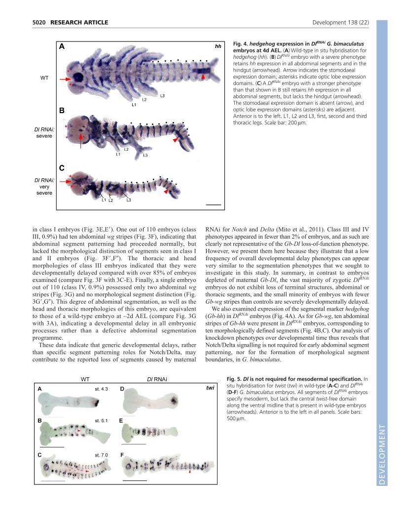
We also examined expression of the segmental marker hedgehog(Gb-hh) in DlRNAi embryos (Fig. 4A). As for Gb-wg, ten abdominalstripes of Gb-hh were present in DlRNAi embryos, corresponding toten morphologically defined segments (Fig. 4B,C). Our analysis ofknockdown phenotypes over developmental time thus reveals thatNotch/Delta signalling is not required for early abdominal segmentpatterning, nor for the formation of morphological segmentboundaries, in G. bimaculatus.
RESEARCH ARTICLE Development 138 (22)
Fig. 4. hedgehog expression in DlRNAi G. bimaculatusembryos at 4d AEL. (A)Wild-type in situ hybridisation forhedgehog (hh). (B)DlRNAi embryo with a severe phenotyperetains hh expression in all abdominal segments and in thehindgut (arrowhead). Arrow indicates the stomodaealexpression domain; asterisks indicate optic lobe expressiondomains. (C)A DlRNAi embryo with a stronger phenotypethan that shown in B still retains hh expression in allabdominal segments, but lacks the hindgut (arrowhead).The stomodaeal expression domain is absent (arrow), andoptic lobe expression domains (asterisks) are adjacent.Anterior is to the left. L1, L2 and L3, first, second and thirdthoracic legs. Scale bar: 200m.
Fig. 5. Dl is not required for mesodermal specification. Insitu hybridisation for twist (twi) in wild-type (A-C) and DlRNAi
(D-F) G. bimaculatus embryos. All segments of DlRNAi embryosspecify mesoderm, but lack the central twist-free domainalong the ventral midline that is present in wild-type embryos(arrowheads). Anterior is to the left in all panels. Scale bars:500m.
DEVELO
PMENT

Gb-Dl knockdown causes loss of mesectoderm andexcess caspase-dependent apoptosis in thenervous systemWe noted that in DlRNAi embryos, although all abdominalsegments were formed, segment morphologies were abnormal(Fig. 3D�-G�; Fig. 4B,C). Specifically, we observed that ventraldomains of the abdomen were more affected by loss of Gb-Dlthan lateral or dorsal domains. Recent maternal RNAiexperiments reported similar phenotypes, but did not determinethe causes of these segment malformations (Mito et al., 2011).To investigate the origin of these abdominal abnormalities anddetermine the role of Dl in wild-type Gryllus development, weexamined cell fate allocation and morphogenesis of specific celltypes in DlRNAi embryos.
As recently reported (Mito et al., 2011), we observed that theposterior region of DlRNAi embryos retained expression of theposterior marker caudal (not shown), consistent with a correctlyestablished posterior growth zone. Formation and identity ofmesoderm was also normal in DlRNAi embryos (Fig. 5). However, asnoted above, we observed that at 2d AEL the ventral mesectodermwas absent (Fig. 6B,B�,F; supplementary material Movie 2). This
absence appeared to be due to a lack of specification rather than celldeath, as apoptosis levels throughout the germ band were onlyslightly higher than in controls at 2d AEL (not shown). By contrast,by 4d AEL, whereas the single fused abdominal coelomic pouchesretained mesodermal identity (Fig. 5F), nearly all nuclei of ventralcells lying between the coelomic pouches and the overlyingectoderm were pycnotic (Fig. 6H) and undergoing caspase-mediatedapoptosis (Fig. 7B�-D�). In DlRNAi embryos, this apoptosis sets inafter the generation and morphological definition of abdominalsegments (Fig. 7). This phenotype is not confined to the abdomen,but occurs throughout the embryo, including the head, trunk andthoracic appendages (supplementary material Figs S4-S6). Thisapoptosis results in massive loss of abdominal ventral tissue andsubsequent collapse of the ventral midline, leading to the observedcoelomic pouch fusion (Fig. 3D,E; Fig. 6H). In severely affectedembryos, the invaginating hindgut (a medial structure) is also absent(Fig. 4C, arrowhead). Also consistent with the loss of midline tissueis the reduction or loss of the stomodaeum (Fig. 4C,D, arrows) andthe spacing of the optic lobe Gb-hh expression domains, which arecloser together in DlRNAi embryos than in controls (Fig. 4C,D,asterisks).
5021RESEARCH ARTICLEDelta in cricket segmentation
Fig. 6. Dl RNAi results in specific loss of midline tissue during abdominal segmentation. Wild-type or DlRNAi G. bimaculatus embryos wereanalysed at 2d or 4d AEL. (A-D)Three-dimensional reconstructions of embryos. (A�-D�) -Catenin immunostaining (green) of embryonic abdominalsegments. Distinct cell shapes of ventral midline central nervous system tissue are missing in DlRNAi embryos (arrowheads). (E-H)Single opticalsections of Hoechst-stained embryos. (E,F)Green and red lines indicate orthogonal section planes of the central image; left and top boxes show theorthogonal section outlined in the corresponding colour. Blue line indicates plane of central image in each orthogonal section image. Arrowheadsindicate that central nervous system tissue along the ventral midline (arrows) is missing in DlRNAi embryos. (G,H)Boxed regions are shown at highermagnification beneath. Neuroectoderm (white inset, arrowheads) visible in orthogonal section (green inset, arrows) has pycnotic nuclei in DlRNAi
embryos. The anteriormost abdominal segment is labelled in each panel. Anterior is to the left in all panels. am, amnion; p, pleuropodium. Scalebars: 50m. D
EVELO
PMENT
5022
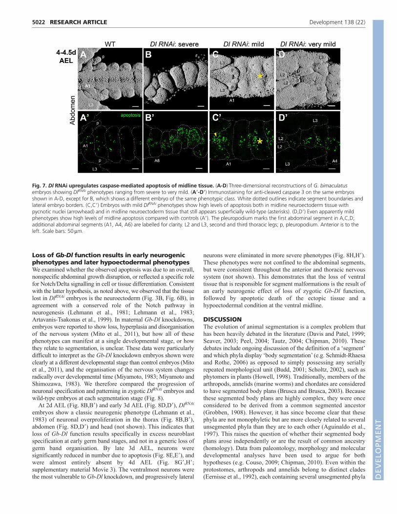
Loss of Gb-Dl function results in early neurogenicphenotypes and later hypoectodermal phenotypesWe examined whether the observed apoptosis was due to an overall,nonspecific abdominal growth disruption, or reflected a specific rolefor Notch/Delta signalling in cell or tissue differentiation. Consistentwith the latter hypothesis, as noted above, we observed that the tissuelost in DlRNAi embryos is the neuroectoderm (Fig. 3B, Fig. 6B), inagreement with a conserved role of the Notch pathway inneurogenesis (Lehmann et al., 1981; Lehmann et al., 1983;Artavanis-Tsakonas et al., 1999). In maternal Gb-Dl knockdowns,embryos were reported to show loss, hyperplasia and disorganisationof the nervous system (Mito et al., 2011), but how all of thesephenotypes can manifest at a single developmental stage, or howthey relate to segmentation, is unclear. These data were particularlydifficult to interpret as the Gb-Dl knockdown embryos shown wereclearly at a different developmental stage than control embryos (Mitoet al., 2011), and the organisation of the nervous system changesradically over developmental time (Miyamoto, 1983; Miyamoto andShimozawa, 1983). We therefore compared the progression ofneuronal specification and patterning in zygotic DlRNAi embryos andwild-type embryos at each segmentation stage (Fig. 8).
At 2d AEL (Fig. 8B,B�) and early 3d AEL (Fig. 8D,D�), DlRNAi
embryos show a classic neurogenic phenotype (Lehmann et al.,1983) of neuronal overproliferation in the thorax (Fig. 8B,B�),abdomen (Fig. 8D,D�) and head (not shown). This indicates thatloss of Gb-Dl function results specifically in excess neuroblastspecification at early germ band stages, and not in a generic loss ofgerm band organisation. By late 3d AEL, neurons weresignificantly reduced in number due to apoptosis (Fig. 8E,E�), andwere almost entirely absent by 4d AEL (Fig. 8G�,H�;supplementary material Movie 3). The ventralmost neurons werethe most vulnerable to Gb-Dl knockdown, and progressively lateral
neurons were eliminated in more severe phenotypes (Fig. 8H,H�).These phenotypes were not confined to the abdominal segments,but were consistent throughout the anterior and thoracic nervoussystem (not shown). This demonstrates that the loss of ventraltissue that is responsible for segment malformations is the result ofan early neurogenic effect of loss of zygotic Gb-Dl function,followed by apoptotic death of the ectopic tissue and ahypoectodermal condition at the ventral midline.
DISCUSSIONThe evolution of animal segmentation is a complex problem thathas been heavily debated in the literature (Davis and Patel, 1999;Seaver, 2003; Peel, 2004; Tautz, 2004; Chipman, 2010). Thesedebates include ongoing discussion of the definition of a ‘segment’and which phyla display ‘body segmentation’ (e.g. Schmidt-Rhaesaand Rothe, 2006) as opposed to simply possessing any seriallyrepeated morphological unit (Budd, 2001; Scholtz, 2002), such asphytomers in plants (Howell, 1998). Traditionally, members of thearthropods, annelids (marine worms) and chordates are consideredto have segmented body plans (Brusca and Brusca, 2003). Becausethese segmented body plans are highly complex, they were onceconsidered to be derived from a common segmented ancestor(Grobben, 1908). However, it has since become clear that thesephyla are not monophyletic but are more closely related to severalunsegmented phyla than they are to each other (Aguinaldo et al.,1997). This raises the question of whether their segmented bodyplans arose independently or are the result of common ancestry(homology). Data from paleontology, morphology and moleculardevelopmental analyses have been used to argue for bothhypotheses (e.g. Couso, 2009; Chipman, 2010). Even within theprotostomes, arthropods and annelids belong to distinct clades(Eernisse et al., 1992), each containing several unsegmented phyla
RESEARCH ARTICLE Development 138 (22)
Fig. 7. Dl RNAi upregulates caspase-mediated apoptosis of midline tissue. (A-D)Three-dimensional reconstructions of G. bimaculatusembryos showing DlRNAi phenotypes ranging from severe to very mild. (A�-D�) Immunostaining for anti-cleaved caspase 3 on the same embryosshown in A-D, except for B, which shows a different embryo of the same phenotypic class. White dotted outlines indicate segment boundaries andlateral embryo borders. (C,C�) Embryos with mild DlRNAi phenotypes show high levels of apoptosis both in midline neuroectoderm tissue withpycnotic nuclei (arrowhead) and in midline neuroectoderm tissue that still appears superficially wild-type (asterisks). (D,D�) Even apparently mildphenotypes show high levels of midline apoptosis compared with controls (A�). The pleuropodium marks the first abdominal segment in A,C,D;additional abdominal segments (A1, A4, A6) are labelled for clarity. L2 and L3, second and third thoracic legs; p, pleuropodium. Anterior is to theleft. Scale bars: 50m.
DEVELO
PMENT
(Brusca and Brusca, 2003). However, anatomical and moleculardevelopmental data from annelids and arthropods (Scholtz, 2002;Prud’homme et al., 2003; Dray et al., 2010; Janssen et al., 2010)suggest that a protostome ancestor might have been segmented.Approaching the even deeper question of whether a bilaterianancestor had a segmented body plan is a challenge that should beexplored at several phenotypic levels, including cellular behaviour,development and anatomy, and gene regulatory networks. BecauseNotch/Delta signalling is crucial for vertebrate segmentation,several studies have focused on this pathway as a major indicatorof the possibility of a segmented bilaterian ancestor. In this contextwe investigated the precise role of this pathway in early segmentpatterning in a basal insect.
We have shown that Gb-Dl is expressed in an iterated pattern inthe abdomen, but find no evidence for cyclic expression of Dlduring cricket segmentation, in contrast to what has been observed
in vertebrates with a Notch-coordinated segmentation clock (Jianget al., 2000; Maruhashi et al., 2005). If Gb-Dl participates in asegmentation mechanism that patterns future segments before theybecome defined morphologically, its expression should precede theexpression of the late segmental marker Gb-wg (Oppenheimer etal., 1999). However, Gb-Dl is expressed in abdominal segmentsonly after expression of Gb-eve has been established, andsimultaneously with the appearance of Gb-wg. Although maternaldepletion of Gb-Dl results in loss of growth zone maintenance andin posterior elongation defects (Mito et al., 2011), our analyses ofzygotic Gb-Dl knockdown show that Notch signalling is notspecifically required for abdominal segment patterning. However,in a small percentage (<2%) of embryos, developmental delays cancreate an initial impression that segmentation is disrupted.Moreover, final segment morphologies are clearly abnormal at laterstages, which can add to the impression of disrupted segmentation.
5023RESEARCH ARTICLEDelta in cricket segmentation
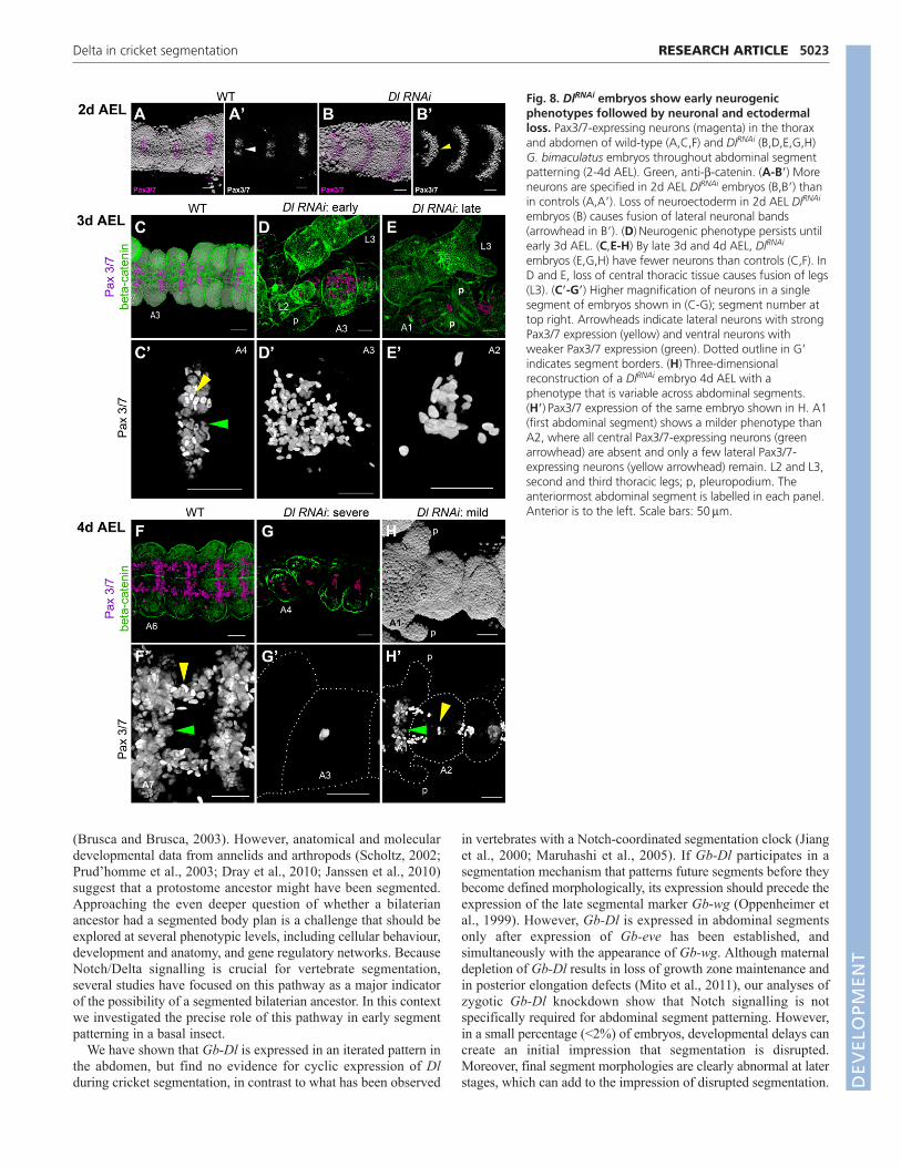
Fig. 8. DlRNAi embryos show early neurogenicphenotypes followed by neuronal and ectodermalloss. Pax3/7-expressing neurons (magenta) in the thoraxand abdomen of wild-type (A,C,F) and DlRNAi (B,D,E,G,H)G. bimaculatus embryos throughout abdominal segmentpatterning (2-4d AEL). Green, anti--catenin. (A-B�) Moreneurons are specified in 2d AEL DlRNAi embryos (B,B�) thanin controls (A,A�). Loss of neuroectoderm in 2d AEL DlRNAi
embryos (B) causes fusion of lateral neuronal bands(arrowhead in B�). (D)Neurogenic phenotype persists untilearly 3d AEL. (C,E-H) By late 3d and 4d AEL, DlRNAi
embryos (E,G,H) have fewer neurons than controls (C,F). InD and E, loss of central thoracic tissue causes fusion of legs(L3). (C�-G�) Higher magnification of neurons in a singlesegment of embryos shown in (C-G); segment number attop right. Arrowheads indicate lateral neurons with strongPax3/7 expression (yellow) and ventral neurons withweaker Pax3/7 expression (green). Dotted outline in G�indicates segment borders. (H)Three-dimensionalreconstruction of a DlRNAi embryo 4d AEL with aphenotype that is variable across abdominal segments.(H�)Pax3/7 expression of the same embryo shown in H. A1(first abdominal segment) shows a milder phenotype thanA2, where all central Pax3/7-expressing neurons (greenarrowhead) are absent and only a few lateral Pax3/7-expressing neurons (yellow arrowhead) remain. L2 and L3,second and third thoracic legs; p, pleuropodium. Theanteriormost abdominal segment is labelled in each panel.Anterior is to the left. Scale bars: 50m.
DEVELO
PMENT
5024
We have elucidated the causes of morphological abnormalitiesin DlRNAi embryos, and show that mesoderm specification is notaffected by Gb-Dl knockdown. However, the mesectoderm is lostin early DlRNAi embryos, and early neurogenic phenotypes arefollowed by caspase-mediated apoptosis of the ectopic neuraltissue. Delta loss of function also enhances caspase-dependentapoptosis in flies and mice (Murata-Ohsawa et al., 2004; Müller etal., 2005). Loss of ventral tissue in DlRNAi embryos is also apparentin previous studies on the function of Notch signalling in spidersegmentation (Stollewerk et al., 2003; Schoppmeier and Damen,2005). This suggests that conserved roles for Notch/Delta signalsin apoptosis and neurogenesis can lead to secondary defects insegment morphologies. In summary, our data show that Notch-based signalling does not oscillate via Delta and is not required forearly segment patterning or morphological formation of segmentboundaries in the cricket G. bimaculatus.
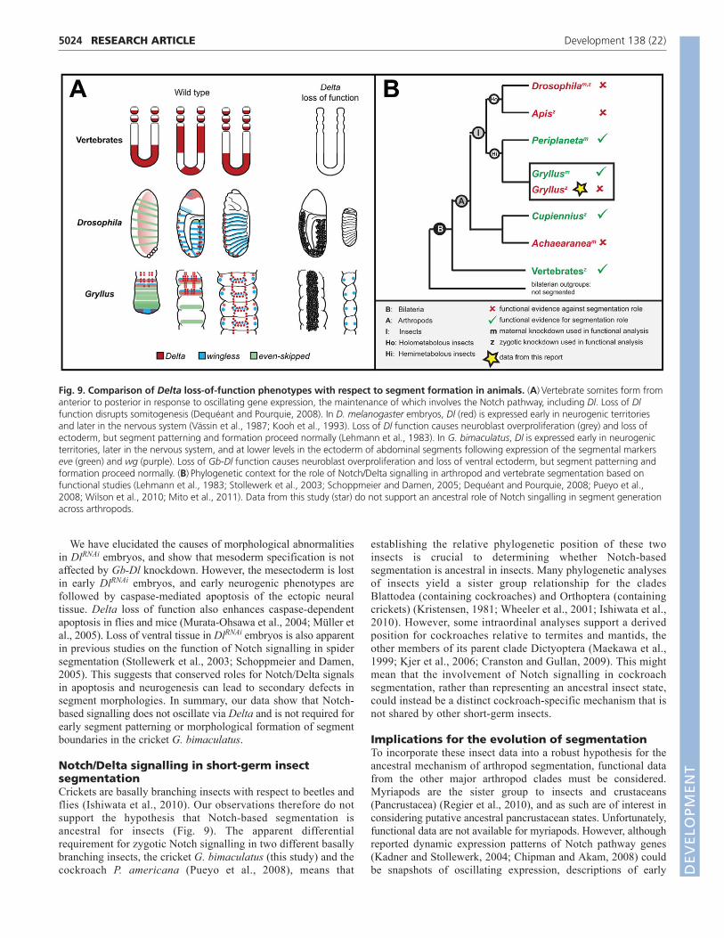
Notch/Delta signalling in short-germ insectsegmentationCrickets are basally branching insects with respect to beetles andflies (Ishiwata et al., 2010). Our observations therefore do notsupport the hypothesis that Notch-based segmentation isancestral for insects (Fig. 9). The apparent differentialrequirement for zygotic Notch signalling in two different basallybranching insects, the cricket G. bimaculatus (this study) and thecockroach P. americana (Pueyo et al., 2008), means that
establishing the relative phylogenetic position of these twoinsects is crucial to determining whether Notch-basedsegmentation is ancestral in insects. Many phylogenetic analysesof insects yield a sister group relationship for the cladesBlattodea (containing cockroaches) and Orthoptera (containingcrickets) (Kristensen, 1981; Wheeler et al., 2001; Ishiwata et al.,2010). However, some intraordinal analyses support a derivedposition for cockroaches relative to termites and mantids, theother members of its parent clade Dictyoptera (Maekawa et al.,1999; Kjer et al., 2006; Cranston and Gullan, 2009). This mightmean that the involvement of Notch signalling in cockroachsegmentation, rather than representing an ancestral insect state,could instead be a distinct cockroach-specific mechanism that isnot shared by other short-germ insects.
Implications for the evolution of segmentationTo incorporate these insect data into a robust hypothesis for theancestral mechanism of arthropod segmentation, functional datafrom the other major arthropod clades must be considered.Myriapods are the sister group to insects and crustaceans(Pancrustacea) (Regier et al., 2010), and as such are of interest inconsidering putative ancestral pancrustacean states. Unfortunately,functional data are not available for myriapods. However, althoughreported dynamic expression patterns of Notch pathway genes(Kadner and Stollewerk, 2004; Chipman and Akam, 2008) couldbe snapshots of oscillating expression, descriptions of early
RESEARCH ARTICLE Development 138 (22)
Fig. 9. Comparison of Delta loss-of-function phenotypes with respect to segment formation in animals. (A)Vertebrate somites form fromanterior to posterior in response to oscillating gene expression, the maintenance of which involves the Notch pathway, including Dl. Loss of Dlfunction disrupts somitogenesis (Dequéant and Pourquie, 2008). In D. melanogaster embryos, Dl (red) is expressed early in neurogenic territoriesand later in the nervous system (Vässin et al., 1987; Kooh et al., 1993). Loss of Dl function causes neuroblast overproliferation (grey) and loss ofectoderm, but segment patterning and formation proceed normally (Lehmann et al., 1983). In G. bimaculatus, Dl is expressed early in neurogenicterritories, later in the nervous system, and at lower levels in the ectoderm of abdominal segments following expression of the segmental markerseve (green) and wg (purple). Loss of Gb-Dl function causes neuroblast overproliferation and loss of ventral ectoderm, but segment patterning andformation proceed normally. (B)Phylogenetic context for the role of Notch/Delta signalling in arthropod and vertebrate segmentation based onfunctional studies (Lehmann et al., 1983; Stollewerk et al., 2003; Schoppmeier and Damen, 2005; Dequéant and Pourquie, 2008; Pueyo et al.,2008; Wilson et al., 2010; Mito et al., 2011). Data from this study (star) do not support an ancestral role of Notch singalling in segment generationacross arthropods.
DEVELO
PMENT

embryogenesis suggest that extensive cellular movements arerequired for shaping the germ band, including the posterior growthzone, during segmentation (Heymons, 1901; Chipman et al., 2004).This makes it likely that cell movements contribute to the observedexpression patterns. Cell rearrangements during axis elongationhave not yet been examined in any short-germ arthropod, butcontribute substantially to elongation during segmentation invertebrates (Zhang et al., 2008). Future work will be necessary todetermine the extent and role of these movements during sequentialarthropod segmentation.
Chelicerates are likely to be the most basally branchingarthropod clade (Regier et al., 2010). In this group, althoughzygotic loss of Notch signalling appears to disrupt segmentationin C. salei (Stollewerk et al., 2003; Schoppmeier and Damen,2005), data from a different spider suggest that embryosdefective in maternal Notch signalling could fail to formsegments because of defective mesoderm-ectoderm cell fatedecisions rather than a defect in segment patterning per se (Odaet al., 2007). These data are reminiscent of some of thedifferences that exist between the results of maternal (Mito et al.,2011) and zygotic (this study) knockdown in G. bimaculatus.However, this also demonstrates the difficulty of consolidatingdata from maternal and zygotic knockdowns performed indifferent species to infer evolutionary scenarios. Future studiesthat combine both types of knockdown experiments could behelpful in addressing the question of ancestral arthropodfunctions for Notch/Delta in segmentation.
This study highlights the importance of detailed phenotypicanalysis for inferring deep homologies and establishing criteria forevolutionary developmental hypotheses. In short-germ arthropods,segmentation takes place simultaneously with several otherdevelopmental events, including mesoderm morphogenesis,neuroectoderm differentiation and neurogenesis. Consequently,disruption of several processes besides those required for segmentpatterning per se can yield what appear to be segmentation disruptionphenotypes. Moreover, we found that examining RNAi phenotypesat several different developmental time points and contrasting theresults of maternal and zygotic knockdown studies were crucial tounderstanding the roles of Delta in cricket embryogenesis. Followingthe progression of the Dl RNAi phenotype over time allowed us todetect tissue-specific effects on neural development, as well asgeneric developmental delays, which are a common feature of loss-of-function mutations in Notch pathway members (Ballard et al.,2010; Julian et al., 2010; Reis et al., 2011). These observationshelped us to distinguish such neural and developmental delayphenotypes from specific effects on segmentation. Given the highdegree of pleiotropy of Notch signalling in metazoan development,it is crucial to examine the complex spatiotemporal integration ofmultiple developmental processes when analysing and interpretingNotch pathway disruption phenotypes. Although whether Notchsignalling was essential for an ancestral arthropod segmentationprogramme remains unknown, our work indicates that the moleculardevelopmental basis for the hypothesis of a common origin ofsegmentation across bilaterians might be weaker than is oftensuggested.
AcknowledgementsWe thank S. Noji and T. Mito for sharing Gryllus protocols and plasmids forwingless and even-skipped; S. Roth and A. Drechsler for sharing twistsequence information; S. Kunes and G. Davis for sharing antibodies; V. Zengfor analysing Delta orthologues in arthropod genomes; and Elena Kramer,Miltos Tsiantis, Carlo Brena and members of the M.A. and C.G.E. laboratoriesfor discussions and comments on the manuscript.
FundingThis work was supported by the National Science Foundation (NSF) [awardIOS-0817678 to C.G.E., predoctoral fellowship to B.E.-C.]; and the Marie CurieResearch and Training Network (MCRTN) ZOONET [MRTN-CT-2004-005624 toM.A., fellowship to F.K.].
Competing interests statementThe authors declare no competing financial interests.
Author contributionsAll authors designed the research; F.K., B.E.-C. and C.G.E. performedexperiments, collected and analysed data; C.G.E. wrote the paper with inputfrom F.K. and M.A.; C.G.E. and M.A. obtained funding for the research.
Supplementary materialSupplementary material available online athttp://dev.biologists.org/lookup/suppl/doi:10.1242/dev.073395/-/DC1
ReferencesAguinaldo, A. M. A., Turbeville, J. M., Linford, L. S., Rivera, M. C., Garey, J.
R., Raff, R. A. and Lake, J. A. (1997). Evidence for a clade of nematodes,arthropods and other moulting animals. Nature 387, 489-493.
Altekar, G., Dwarkadas, S., Huelsenbeck, J. P. and Ronquist, F. (2004). ParallelMetropolis coupled Markov chain Monte Carlo for Bayesian phylogeneticinference. Bioinformatics 20, 407-415.
Aranda, M., Marques-Souza, H., Bayer, T. and Tautz, D. (2008). The role of thesegmentation gene hairy in Tribolium. Dev. Genes Evol. 218, 465-477.
Artavanis-Tsakonas, S., Rand, M. D. and Lake, R. J. (1999). Notch signaling:cell fate control and signal integration in development. Science 284, 770-776.
Aulehla, A. and Pourquie, O. (2009). More than patterning-Hox genes and thecontrol of posterior axial elongation. Dev. Cell 17, 439-440.
Baird, G. S., Zacharias, D. A. and Tsien, R. Y. (2000). Biochemistry, mutagenesis,and oligomerization of DsRed, a red fluorescent protein from coral. Proc. Natl.Acad. Sci. USA 97, 11984-11989.
Ballard, S. L., Jarolimova, J. and Wharton, K. A. (2010). Gbb/BMP signaling isrequired to maintain energy homeostasis in Drosophila. Dev. Biol. 337, 375-385.
Brusca, G. J. and Brusca, R. C. (2003). Invertebrates. Sunderland, MA: SinauerAssociates.
Budd, G. E. (2001). Why are arthropods segmented? Evol. Dev. 3, 332-342.Chipman, A. D. (2010). Parallel evolution of segmentation by co-option of
ancestral gene regulatory networks. BioEssays 32, 60-70.Chipman, A. D. and Akam, M. (2008). The segmentation cascade in the
centipede Strigamia maritima: involvement of the Notch pathway and pair-rulegene homologues. Dev. Biol. 319, 160-169.
Chipman, A. D., Arthur, W. and Akam, M. (2004). Early development andsegment formation in the centipede, Strigamia maritima (Geophilomorpha).Evol. Dev. 6, 78-89.
Couso, J. P. (2009). Segmentation, metamerism and the Cambrian explosion. Int.J. Dev. Biol. 53, 1305-1316.
Cranston, P. S. and Gullan, P. J. (2009). Phylogeny of insects. In Encyclopedia ofInsects (ed. V. H. Resh and R. T. Carde), pp. 780-793. San Diego, CA: Elsevier.
Davis, G. K. and Patel, N. H. (1999). The origin and evolution of segmentation.Trends Genet. 9, M68-M72.
De Robertis, E. M. (2008). The molecular ancestry of segmentation mechanisms.Proc. Natl. Acad. Sci. USA 105, 16411-16412.
Dearden, P. and Akam, M. (2000). A role for Fringe in segment morphogenesisbut not segment formation in the grasshopper, Schistocerca gregaria. Dev.Genes Evol. 210, 329-336.
Dequéant, M. L. and Pourquie, O. (2008). Segmental patterning of thevertebrate embryonic axis. Nat. Rev. Genet. 9, 370-382.
Dong, Y. and Friedrich, M. (2005). Comparative analysis of Wingless patterningin the embryonic grasshopper eye. Dev. Genes Evol. 215, 177-197.
Dray, N., Tessmar-Raible, K., Le Gouar, M., Vibert, L., Christodoulou, F.,Schipany, K., Guillou, A., Zantke, J., Snyman, H., Béhague, J. et al. (2010).Hedgehog signaling regulates segment formation in the annelid Platynereis.Science 329, 339-342.
Eernisse, D., Albert, J. and Anderson, F. (1992). Annelida and Arthropoda arenot sister taxa – a phylogenetic analysis of spiralian metazoan morphology. Syst.Biol. 41, 305-330.
Gerberding, M., Browne, W. E. and Patel, N. H. (2002). Cell lineage analysis ofthe amphipod crustacean Parhyale hawaiensis reveals an early restriction of cellfates. Development 129, 5789-5801.
Grobben, K. (1908). Die systematische Einteilung des Tierreisches. Verhandllungender kaiserlich-kongiglichen Zoologish-Botanischen Gesellschaft in Wien 58, 491-511.
Gurskaya, N. G., Verkhusha, V. V., Shcheglov, A. S., Staroverov, D. B.,Chepurnykh, T. V., Fradkov, A. F., Lukyanov, S. A. and Lukyanov, K. A.(2006). Engineering of a monomeric green-to-red photoactivatable fluorescentprotein induced by blue light. Nat. Biotechnol. 24, 461-465.
5025RESEARCH ARTICLEDelta in cricket segmentation
DEVELO
PMENT

5026
Handel, K., Basal, A., Fan, X. and Roth, S. (2005). Tribolium castaneum twist:gastrulation and mesoderm formation in a short-germ beetle. Dev. Genes Evol.215, 13-31.
Heymons, R. (1901). Entwicklungsgeschichte der Scolopender. Zoologica 33, 1-244.Howell, S. H. (1998). Molecular Genetics of Plant Development. Cambridge:
Cambridge University Press.Huelsenbeck, J. P. and Ronquist, F. (2001). MRBAYES: Bayesian inference of
phylogenetic trees. Bioinformatics 17, 754-755.Huson, D. H., Richter, D. C., Rausch, C., Dezulian, T., Franz, M. and Rupp, R.
(2007). Dendroscope: An interactive viewer for larger phylogenetic trees. BMCBioinformatics 8, 460.
Ishiwata, K., Sasaki, G., Ogawa, J., Miyata, T. and Su, Z.-H. (2010).Phylogenetic relationships among insect orders based on three nuclear protein-coding gene sequences. Mol. Phylogenet. Evol. 158, 169-180.
Janssen, R., Le Gouar, M., Pechmann, M., Poulin, F., Bolognesi, R., Schwager,E. E., Hopfen, C., Colbourne, J. K., Budd, G. E., Brown, S. J. et al. (2010).Conservation, loss, and redeployment of Wnt ligands in protostomes:implications for understanding the evolution of segment formation. BMC Evol.Biol. 10, 374.
Jiang, Y.-J., Aerne, B. L., Smithers, L., Haddon, C., Ish-Horowicz, D. andLewis, J. (2000). Notch signaling and the synchronization of the somitesegmentation clock. Nature 408, 475-479.
Julian, E., Dave, R. K., Robson, J. P., Hallahan, A. R. and Wainwright, B. J.(2010). Canonical Notch signaling is not required for the growth of Hedgehogpathway-induced medulloblastoma. Oncogene 29, 3465-3476.
Kadner, D. and Stollewerk, A. (2004). Neurogenesis in the chilopod Lithobiusforficatus suggests more similarities to chelicerates than to insects. Dev. GenesEvol. 214, 367-379.
Kjer, K. M., Carle, F. L., Litman, J. and Ware, J. (2006). A molecular phylogenyof Hexapoda. Arthropod Systematics and Phylogeny 64, 35-44.
Kooh, P. J., Fehon, R. G. and Muskavitch, M. A. (1993). Implications of dynamicpatterns of Delta and Notch expression for cellular interactions duringDrosophila development. Development 117, 493-507.
Kristensen, N. P. (1981). Phylogeny of insect orders. Ann. Rev. Entomol. 26, 135-157.
Lehmann, R., Dietrich, U., Jiménez, F. and Campos-Ortega, J. A. (1981).Mutations of early neurogenesis in Drosophila. Roux’s Arch. Dev. Biol. 190, 226-229.
Lehmann, R., Jiménez, F., Dietrich, U. and Campos-Ortega, J. A. (1983). Onthe phenotype and development of mutants of early neurogenesis in Drosophilamelanogaster. Roux’s Arch. Dev. Biol. 192, 62-74.
Livak, K. J. and Schmittgen, T. D. (2001). Analysis of relative gene expressiondata using real-time quantitative PCR and the 2(-Delta Delta C(T)) method.Methods 25, 402-408.
Maekawa, K., Kitade, O. and Matsumoto, T. (1999). Molecular phylogeny oforthopteroid insects based on the mitochondrial cytochrome oxidase II gene.Zool. Sci. 16, 175-184.
Martin-Bermudo, M. D., Carmena, A. and Jimenez, F. (1995). Neurogenicgenes control gene expression at the transcriptional level in early neurogenesisand in mesectoderm specification. Development 121, 219-224.
Maruhashi, M., Van De Putte, T., Huylebroeck, D., Kondoh, H. and Higashi,Y. (2005). Involvement of SIP1 in positioning of somite boundaries in the mouseembryo. Dev. Dyn. 234, 332-338.
Menne, T. V. and Klambt, C. (1994). The formation of commissures in theDrosophila CNS depends on the midline cells and on the Notch gene.Development 120, 123-133.
Minelli, A. (2005). A morphologist’s perspective on terminal growth andsegmentation. Evol. Dev. 7, 568-573.
Mito, T., Kobayashi, C., Sarashina, I., Zhang, H., Shinahara, W., Miyawaki,K., Shinmyo, Y., Ohuchi, H. and Noji, S. (2007). even-skipped has gap-like,pair-rule-like, and segmental functions in the cricket Gryllus bimaculatus, abasal, intermediate germ insect (Orthoptera). Dev. Biol. 303, 202-213.
Mito, T., Shinmyo, Y., Kurita, K., Nakamura, T., Ohuchi, H. and Noji, S.(2011). Ancestral functions of Delta/Notch signaling in the formation of bodyand leg segments in the cricket Gryllus bimaculatus. Development 138, 3823-3833.
Miyamoto, T. (1983). Embryonic development of the central nervous system inthe cricket, Gryllus bimaculatus. II. segmental specialization in late neurogenesis.Zoological Magazine 92, 332-341.
Miyamoto, T. and Shimozawa, T. (1983). Embryonic development of the centralnervous system in the cricket, Gryllus bimaculatus. I. segmental homologies inearly neurogenesis. Zoological Magazine 92, 317-331.
Miyawaki, K., Mito, T., Sarashina, I., Zhang, H., Shinmyo, Y., Ohuchi, H. andNoji, S. (2004). Involvement of Wingless/Armadillo signaling in the posteriorsequential segmentation in the cricket, Gryllus bimaculatus (Orthoptera), asrevealed by RNAi analysis. Mech. Dev. 121, 119-130.
Morel, V. and Schweisguth, F. (2000). Repression by suppressor of hairless andactivation by Notch are required to define a single row of single-mindedexpressing cells in the Drosophila embryo. Genes Dev. 14, 377-388.
Müller, D., Kugler, S. J., Preiss, A., Maier, D. and Nagel, A. C. (2005). Geneticmodifier screens on Hairless gain-of-function phenotypes reveal genes involvedin cell differentiation, cell growth and apoptosis in Drosophila melanogaster.Genetics 171, 1137-1152.
Murata-Ohsawa, M., Tohda, S., Kogoshi, H. and Nara, N. (2004). The Notchligand, Delta-1, reduces TNF-alpha-induced growth suppression and apoptosisby decreasing activation of caspases in U937 cells. Int. J. Mol. Med. 14, 861-866.
Niwa, N., Inoue, Y., Nozawa, A., Saito, M., Misumi, Y., Ohuchi, H., Yoshioka,H. and Noji, S. (2000). Correlation of diversity of leg morphology in Gryllusbimaculatus (cricket) with divergence in dpp expression pattern during legdevelopment. Development 127, 4373-4381.
Oda, H., Nishimura, O., Hirao, Y., Tarui, H., Agata, K. and Akiyama-Oda, Y.(2007). Progressive activation of Delta-Notch signaling from around theblastopore is required to set up a functional caudal lobe in the spiderAchaearanea tepidariorum. Development 134, 2195-2205.
Oppenheimer, D. I., MacNicol, A. M. and Patel, N. H. (1999). Functionalconservation of the wingless-engrailed interaction as shown by a widelyapplicable baculovirus misexpression system. Curr. Biol. 9, 1288-1296.
Panfilio, K. A., Liu, P. Z., Akam, M. and Kaufman, T. C. (2006). Oncopeltusfasciatus zen is essential for serosal tissue function in katatrepsis. Dev. Biol. 292,226-243.
Patel, N. H., Ball, E. E. and Goodman, C. S. (1992). Changing role of even-skipped during the evolution of insect pattern formation. Nature 357, 339-342.
Peel, A. (2004). The evolution of arthropod segmentation mechanisms. BioEssays26, 1108-1116.
Peel, A. D., Chipman, A. D. and Akam, M. (2005). Arthropod segmentation:beyond the Drosophila paradigm. Nat. Rev. Genet. 6, 905-916.
Prud’homme, B., de Rosa, R., Arendt, D., Julien, J. F., Pajaziti, R., Dorresteijn,A. W., Adoutte, A., Wittbrodt, J. and Balavoine, G. (2003). Arthropod-likeexpression patterns of engrailed and wingless in the annelid Platynereis dumeriliisuggest a role in segment formation. Curr. Biol. 13, 1876-1881.
Pueyo, J. I., Lanfear, R. and Couso, J. P. (2008). Ancestral Notch-mediatedsegmentation revealed in the cockroach Periplaneta americana. Proc. Natl. Acad.Sci. USA 105, 16614-16619.
Regier, J. C., Shultz, J. W., Zwick, A., Hussey, A., Ball, B., Wetzer, R., Martin,J. W. and Cunningham, C. W. (2010). Arthropod relationships revealed byphylogenomic analysis of nuclear protein-coding sequences. Nature 463, 1079-1083.
Reis, L. M., Tyler, R. C., Schilter, K. F., Abdul-Rahman, O., Innis, J. W., Kozel,B. A., Schneider, A. S., Bardakjian, T. M., Lose, E. J., Martin, D. M. et al.(2011). BMP4 loss-of-function mutations in developmental eye disordersincluding SHORT syndrome. Hum. Genet. 130, 495-504.
Roonwal, M. L. (1936). Studies on the embryology of the african migratory locust,Locusta migratoria migratoides R. and F. I. The early development, with a newtheory of multi-phased gastrulation among insects. Philos. Trans. R. Soc. Lond. BBiol. Sci. 226, 391-421.
Schmidt-Rhaesa, A. and Rothe, B. H. (2006). Postembryonic development ofdorsoventral and longitudinal musculature in Pycnophyes kielensis (Kinorhyncha,Homalorhagida). Integr. Comp. Biol. 46, 144-150.
Scholtz, G. (2002). The Articulata hypothesis – or what is a segment? Org. Divers.Evol. 2, 197-215.
Schoppmeier, M. and Damen, W. G. (2005). Suppressor of Hairless andPresenilin phenotypes imply involvement of canonical Notch-signalling insegmentation of the spider Cupiennius salei. Dev. Biol. 280, 211-224.
Seaver, E. C. (2003). Segmentation: mono- or polyphyletic? Int. J. Dev. Biol. 47,583-595.
Shimomura, O., Johnson, H. F. and Saiga, Y. (1962). Extraction, purification andproperties of aequorin, a bioluminescent protein from the luminoushydromedusan, Aequorea. Weekly Epidemiol. Rec. 59, 223-239.
Spradling, A. (1986). P element-mediated transformation. In Drosophila: APractical Approach (ed. D. B. Roberts), pp. 175-198. Oxford: IRL Press.
Stollewerk, A., Schoppmeier, M. and Damen, W. G. M. (2003). Involvement ofNotch and Delta genes in spider segmentation. Nature 423, 863-865.
Tautz, D. (2004). Segmentation. Dev. Cell 7, 301-312.Vässin, H., Bremer, K. A., Knust, E. and Campos-Ortega, J. A. (1987). The
neurogenic gene Delta of Drosophila melanogaster is expressed in neurogenicterritories and encodes a putative transmembrane protein with EGF-like repeats.EMBO J. 6, 3431-3440.
Wheeler, W. C., Whiting, M., Wheeler, Q. D. and Carpenter, J. M. (2001). Thephylogeny of the extant hexapod orders. Cladistics 17, 113-169.
Wilson, M. J., McKelvey, B. H., van der Heide, S. and Dearden, P. K. (2010).Notch signaling does not regulate segmentation in the honeybee, Apis mellifera.Dev. Genes Evol. 220, 179-190.
Zhang, L., Kendrick, C., Julich, D. and Holley, S. A. (2008). Cell cycleprogression is required for zebrafish somite morphogenesis but notsegmentation clock function. Development 135, 2065-2070.
RESEARCH ARTICLE Development 138 (22)
DEVELO
PMENT