no. 16. section 3 the arteries upon leaving the heart, the blood enters the vascular system, which...
TRANSCRIPT

No. 16No. 16

Section 3 The ArteriesSection 3 The Arteries Upon leaving the heart, the blood enters Upon leaving the heart, the blood enters
the vascular system, which is composed of the vascular system, which is composed of numerous numerous blood vesselsblood vessels. The vessels . The vessels transport the blood to all parts of the transport the blood to all parts of the body, permit the exchange of nutrients, body, permit the exchange of nutrients, metabolic end products, hormones, and metabolic end products, hormones, and other substances between the blood and other substances between the blood and the interstitial fluid, and ultimately return the interstitial fluid, and ultimately return the blood to the heart.the blood to the heart.
The vessels of the circulatory system can The vessels of the circulatory system can be divided into two separate circuits, each be divided into two separate circuits, each of which leaves and returns to the heart.of which leaves and returns to the heart.

The The pulmonary circuitpulmonary circuit carries blood carries blood from the right side of the heart (right from the right side of the heart (right ventricle) to the lungs and back to ventricle) to the lungs and back to the left side of the heart (left atrium).the left side of the heart (left atrium).
The The systemic circuitsystemic circuit carries blood carries blood that has just left the pulmonary that has just left the pulmonary circuit (from the left ventricle) to the circuit (from the left ventricle) to the rest of the body and back to the right rest of the body and back to the right side of the heart (right atrium).side of the heart (right atrium).


Vessels called arteries carry blood Vessels called arteries carry blood away from the heart. Compares to away from the heart. Compares to the other types of blood vessels, the other types of blood vessels, arteries must be able to withstand arteries must be able to withstand the greatest internal pressure. The the greatest internal pressure. The major arteries divide into smaller major arteries divide into smaller arteries, then into still smaller arteries, then into still smaller arterioles, and finally into tiny arterioles, and finally into tiny capillaries.capillaries.

ⅠⅠ. The Arteries of Pulmonary . The Arteries of Pulmonary CirculationCirculation
Ⅰ Ⅰ) The Pulmonary Artery and its ) The Pulmonary Artery and its BranchesBranches
The The pulmonary trunkpulmonary trunk conveys conveys deoxygenated blood from the right deoxygenated blood from the right ventricle of the heart to the lungs. In ventricle of the heart to the lungs. In the concavity of the aortic arch it the concavity of the aortic arch it divides into right and left pulmonary divides into right and left pulmonary arteries.arteries.

The The right pulmonary arteryright pulmonary artery runs to the runs to the hilum of the right lung, where it divides ihilum of the right lung, where it divides into three branches to distribute to the cnto three branches to distribute to the corresponding lobes of the lung.orresponding lobes of the lung.
The The left pulmonary arteryleft pulmonary artery runs to the h runs to the hilum of the left lung, where it divides intilum of the left lung, where it divides into two branches, one for each lobe of the o two branches, one for each lobe of the lung.lung.
Ⅱ Ⅱ) The arterial Ligament) The arterial Ligament The left pulmonary artery is connected to the concavitThe left pulmonary artery is connected to the concavit
y of the aortic arch by a fibrous cord, termed the y of the aortic arch by a fibrous cord, termed the arterarterial ligamential ligament, which is found as the , which is found as the ductus arteriosusductus arteriosus in the fetus. During fetal life, most of the blood in the in the fetus. During fetal life, most of the blood in the pulmonary trunk passes through the ductus arteriosupulmonary trunk passes through the ductus arteriosus into the aorta. After birth, the lumen of the ductus is s into the aorta. After birth, the lumen of the ductus is gradually obliterated and it is transformed into the artgradually obliterated and it is transformed into the arterial ligament.erial ligament.
Occasionally the ductus arteriosus fails to close. In thiOccasionally the ductus arteriosus fails to close. In this acse tying the ductus is often successful in alleviatins acse tying the ductus is often successful in alleviating the condition.g the condition.

ⅡⅡ. The Arteries of Systemic . The Arteries of Systemic CirculationCirculation
Ⅰ Ⅰ) The Aorta) The Aorta It is main trunk of the series of It is main trunk of the series of
arteries which convey the arteries which convey the oxygenated blood to the tissue of the oxygenated blood to the tissue of the body. For convenience, it is body. For convenience, it is described in three portions, the described in three portions, the ascending aorta, the aortic arch and ascending aorta, the aortic arch and the descending aorta.the descending aorta.

1. The 1. The ascending aorta (ascending part of ascending aorta (ascending part of aorta)aorta)
RouteRoute:: It begins at the base of the left ventricle, It begins at the base of the left ventricle, at a at a
level with the lower border of the third costal level with the lower border of the third costal cartilagecartilage, passes obliquely upwards, forwards and , passes obliquely upwards, forwards and to the right. As high as to the right. As high as the upper border of the the upper border of the second right costal cartilagesecond right costal cartilage, it continues with the , it continues with the aortic arch.aortic arch.
BranchesBranches:: The only branches of the ascending aorta are The only branches of the ascending aorta are
coronary arteries.coronary arteries.

2. The 2. The aortic archaortic arch RouteRoute:: It begins It begins at the level of the second right sternocostal jat the level of the second right sternocostal j
ointoint, and runs at first upwards, backwards and to the l, and runs at first upwards, backwards and to the left in front of trachea, it is then directed backwards on eft in front of trachea, it is then directed backwards on the left side of the trachea, and finally passes downwathe left side of the trachea, and finally passes downwards on the left side of rds on the left side of the body of fourth thoracic vertethe body of fourth thoracic vertebrabra, at the lower border of which it is continuous with , at the lower border of which it is continuous with the descending aorta.the descending aorta.
BranchesBranches:: Three branches are given off from the highest part of tThree branches are given off from the highest part of t
he aortic arch.he aortic arch.


From right to left:From right to left: 1) The 1) The brachiocephalic trunk (innominate artbrachiocephalic trunk (innominate art
ery)ery) It passes obliquely upwards, backwards and to It passes obliquely upwards, backwards and to
the right, the right, at the level of the right sternoclaviculat the level of the right sternoclavicular jointar joint, it divides into the right common caroti, it divides into the right common carotid and right subclavian arteries.d and right subclavian arteries.
2) The 2) The left common carotid arteryleft common carotid artery 3) The 3) The left subclavian arteryleft subclavian artery

3. The 3. The descending aorta (descending part of adescending aorta (descending part of aorta)orta)
It is divided into two portions, the thoracic aorIt is divided into two portions, the thoracic aorta and the abdominal aorta.ta and the abdominal aorta.
1) The 1) The thoracic aortathoracic aorta It is contained in the posterior mediastinum. It It is contained in the posterior mediastinum. It
begins at the level of the begins at the level of the lower border of the folower border of the fourth thoracic vertebraurth thoracic vertebra, where it is continuous , where it is continuous with the aortic arch, and ends in front of the with the aortic arch, and ends in front of the lolower border of the twelfth thoracic vertebrawer border of the twelfth thoracic vertebra at t at the aortic hiatus of the diaphragm.he aortic hiatus of the diaphragm.


2) The 2) The abdominal aortaabdominal aorta It begins It begins at the aortic hiatus of the at the aortic hiatus of the
diaphragmdiaphragm, in front of the lower , in front of the lower border of the body of the last border of the body of the last thoracic vertebra, descends in front thoracic vertebra, descends in front of the vertebral column, and ends on of the vertebral column, and ends on the body of the the body of the fourth lumbar fourth lumbar vertebravertebra by dividing into two by dividing into two common iliac arteries.common iliac arteries.

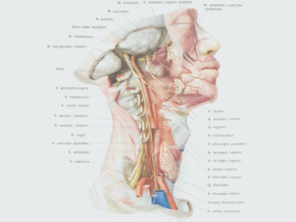
Ⅱ Ⅱ) The Arteries of Head and Neck) The Arteries of Head and Neck The blood supply of the head and neck iThe blood supply of the head and neck i
s chiefly from the branches of the comms chiefly from the branches of the common carotid arteries and partly from those on carotid arteries and partly from those of the subclavian arteries.of the subclavian arteries.

1. The common carotid arteries1. The common carotid arteries The right common carotid artery begins at bifuThe right common carotid artery begins at bifu
rcation of the brachiocephalic trunk behind thrcation of the brachiocephalic trunk behind the right sternoclavicular joint, while the left coe right sternoclavicular joint, while the left common carotid artery springs from the aortic armmon carotid artery springs from the aortic arch.ch.
Each passes obliquely upwards and slightly latEach passes obliquely upwards and slightly laterally, along the side of the trachea and esopherally, along the side of the trachea and esophagus, to the level of the upper border of the thagus, to the level of the upper border of the thyroid cartilage, where it divides into the externyroid cartilage, where it divides into the external and internal carotid arteries.al and internal carotid arteries.

Carotid sinus and carotid bodyCarotid sinus and carotid body A dilatation at the point of the bifurcation of cA dilatation at the point of the bifurcation of c
ommon carotid artery is known as the ommon carotid artery is known as the carotid carotid sinussinus, which usually involves, and may be rest, which usually involves, and may be restricted to the proximal part of the internal carotricted to the proximal part of the internal carotid artery. It acts as a pressure-receptor which iid artery. It acts as a pressure-receptor which is part of the blood pressure regulating mechas part of the blood pressure regulating mechanism. It reacts to changes of the arterial blood nism. It reacts to changes of the arterial blood pressure reflexly.pressure reflexly.

A small, reddish-brown structure behind A small, reddish-brown structure behind the point of division of the common carothe point of division of the common carotid artery is termed the tid artery is termed the carotid bodycarotid body wh which acts as a chemoreceptor. It responds ich acts as a chemoreceptor. It responds to changes in the composition of the bloto changes in the composition of the blood, particularly the oxygen-carbon dioxiod, particularly the oxygen-carbon dioxide ratio.de ratio.

1) The 1) The internal carotid arteryinternal carotid artery It ascends along the pharynx to the It ascends along the pharynx to the
base of the skull, and enters the base of the skull, and enters the cranial cavity through the carotid cranial cavity through the carotid canal without giving off branches in canal without giving off branches in the neck. This is the major artery to the neck. This is the major artery to the brain and other intracranial and the brain and other intracranial and orbital structures.orbital structures.

2) The 2) The external carotid arteryexternal carotid artery It begins oppsite the upper border of the thyroIt begins oppsite the upper border of the thyro
id cartilage, and, gaking a slightly curved courid cartilage, and, gaking a slightly curved course, passes upwards and forwards, and then incse, passes upwards and forwards, and then inclines backwards to a point behind the neck of lines backwards to a point behind the neck of mandible, where, in the substance of the parotmandible, where, in the substance of the parotid gland, it divides into the superficial temporaid gland, it divides into the superficial temporal and maxillary arteries.l and maxillary arteries.

Its main branches are as follows:Its main branches are as follows: ①①The The superior thyroid arterysuperior thyroid artery It supplies the adjacent muscles and the thyroiIt supplies the adjacent muscles and the thyroi
d gland.d gland. BranchBranch:: Superior laryngeal arterySuperior laryngeal artery It accompanies the internal branch of the supeIt accompanies the internal branch of the supe
rior laryngeal nerve and pierces the thyrohyoirior laryngeal nerve and pierces the thyrohyoid membrane to supply the larynx.d membrane to supply the larynx.


② ② The The lingual arterylingual artery It is the principal vessel of the tongue and the It is the principal vessel of the tongue and the
structures on the floor of the mouth.structures on the floor of the mouth. ③ ③ The The facial arteryfacial artery It supplies the submandibular gland, the tonsil It supplies the submandibular gland, the tonsil
and the structures of the face.and the structures of the face. ④ ④ The The occipital arteryoccipital artery It arises from the back of the external carotid aIt arises from the back of the external carotid a
rtery, opposite to the facial artery, and runs uprtery, opposite to the facial artery, and runs upwards and backwards to the posterior part of twards and backwards to the posterior part of the scalp.he scalp.

⑤ ⑤ The The maxillary arterymaxillary artery It is the larger terminal branch of the external It is the larger terminal branch of the external
carotid artery, arises behind the neck of the mcarotid artery, arises behind the neck of the mandible and is at first embedded in the parotid andible and is at first embedded in the parotid gland. It passes forwards to the infratemporal gland. It passes forwards to the infratemporal fossa, and supplies the upper and lower jaws, tfossa, and supplies the upper and lower jaws, teeth and gums, the muscles of mastication, theeth and gums, the muscles of mastication, the palate, the nasal cavity and the paranasal sine palate, the nasal cavity and the paranasal sinuses.uses.

The The middle meningeal arterymiddle meningeal artery is the most imp is the most important branch of the maxillary artery. It ascendortant branch of the maxillary artery. It ascends to the base of the skull to pass into the cranis to the base of the skull to pass into the cranial cavity through the foramen spoinosum. Its bal cavity through the foramen spoinosum. Its branches fan out to supply the cerebral dura mranches fan out to supply the cerebral dura mater. Injury of this artery as in the case of craniater. Injury of this artery as in the case of cranial bone fracture in temporal region may result al bone fracture in temporal region may result in the epidural haematoma.in the epidural haematoma.

⑥ ⑥ The The superficial temporal arterysuperficial temporal artery It is the smaller terminal branch of the externaIt is the smaller terminal branch of the externa
l carotid artery, ascends over the zygomatic arl carotid artery, ascends over the zygomatic arch, and enters the superfiacial fascia of the tech, and enters the superfiacial fascia of the temporal region. Many branches ramify over the mporal region. Many branches ramify over the temporal, auricular, zygomatic and facial regiotemporal, auricular, zygomatic and facial regions from which the arteries receive their names.ns from which the arteries receive their names.

2. The 2. The subclavian arterysubclavian artery The right subclavian artery arises from the braThe right subclavian artery arises from the bra
chiocephalic trunk; the left, from the aortic arcchiocephalic trunk; the left, from the aortic arch. Each artery emerges from the superior aperth. Each artery emerges from the superior aperture of the thorax. It ascends to the root of the ure of the thorax. It ascends to the root of the neck and then arches laterally across the front neck and then arches laterally across the front of the cervical pleura and passes between the of the cervical pleura and passes between the scalenus anterior and the scalenus medius to tscalenus anterior and the scalenus medius to the outer border of the first rib, where it becomhe outer border of the first rib, where it becomes the axillary artery. Hemorrhage of the upper es the axillary artery. Hemorrhage of the upper limb can be controlled by compressing the veslimb can be controlled by compressing the vessel downwards and backwards against the firssel downwards and backwards against the first rib immediately above the mid-point of the clt rib immediately above the mid-point of the clavicle.avicle.


Branches:Branches: 1) The1) The vertebral arteryvertebral artery It arises from the upper and posterior part of tIt arises from the upper and posterior part of t
he proximal end of the subclavian artery. It asche proximal end of the subclavian artery. It ascends through the foramina in the transverse prends through the foramina in the transverse processes of the upper six cervical vertebrae, anocesses of the upper six cervical vertebrae, and enters the skull through the foramen magnud enters the skull through the foramen magnum.m.
It supplies the brain and spinal cord.It supplies the brain and spinal cord.

2) The 2) The internal thoracic (mammary) arteryinternal thoracic (mammary) artery It arises from the inferior surface of the subclaIt arises from the inferior surface of the subcla
vian artery, opposite the vertebral artery. It devian artery, opposite the vertebral artery. It descends behind the cartilage of the upper six ribscends behind the cartilage of the upper six ribs at a distance of 1.2 cm from the lateral bordes at a distance of 1.2 cm from the lateral border of the sternum, and at the level of the sixth inr of the sternum, and at the level of the sixth intercostals space divides into the tercostals space divides into the musculophremusculophrenicnic and and superior epigastric arteriessuperior epigastric arteries..

The superior epigastric artery enters the sheatThe superior epigastric artery enters the sheath of the rectus abdominis, at first lying behind h of the rectus abdominis, at first lying behind the muscle, then perforating and supplying it, the muscle, then perforating and supplying it, and anastomosing with the inferior epigastric and anastomosing with the inferior epigastric artery from the external iliac artery.artery from the external iliac artery.
The branches of the internal thoracic artery arThe branches of the internal thoracic artery are distributed chiefly to the anterior thoracic ane distributed chiefly to the anterior thoracic and abdominal walls, the pleura, the pericardiud abdominal walls, the pleura, the pericardium and the diaphragm.m and the diaphragm.

3) The 3) The thyrocervical trunkthyrocervical trunk It is short, arises from the front of the subclaviIt is short, arises from the front of the subclavi
an artery, close to the medial border of the scaan artery, close to the medial border of the scalenus anterior, and divides almost immediatellenus anterior, and divides almost immediately into three branches. One of them is the inferiy into three branches. One of them is the inferior thyroid artery. Its branches supply the loweor thyroid artery. Its branches supply the lower part of the thyroid and parathyroid glands, lar part of the thyroid and parathyroid glands, larynx, trachea and esophagus.rynx, trachea and esophagus.

4) The 4) The inferior thyroid arteryinferior thyroid artery 5) The 5) The costocervical trunkcostocervical trunk They supply the first two intercostals spThey supply the first two intercostals sp
aces and the muscles of the scapular regaces and the muscles of the scapular region.ion.
Dorsal Dorsal scapular arteryscapular artery::

Ⅲ Ⅲ) The Arteries of Axilla and Upper Limb) The Arteries of Axilla and Upper Limb The subclavian artery, after giving off its The subclavian artery, after giving off its
cervical branches, continues as the greacervical branches, continues as the great arterial stem of the upper limb.t arterial stem of the upper limb.

It is the continuation of the subclavian aIt is the continuation of the subclavian artery and begins at the outer border of trtery and begins at the outer border of the first rib, and ends at the lower border he first rib, and ends at the lower border of the teres major, beyond which the artof the teres major, beyond which the artery takes the name of brachial artery. Its ery takes the name of brachial artery. Its chief branches are:chief branches are:

1) The1) The thoracoacromial arterythoracoacromial artery It is a short branch and divides into several braIt is a short branch and divides into several bra
nches to supply the muscles of thorax and the nches to supply the muscles of thorax and the scapular region.scapular region.
2) The 2) The lateral thoracic arterylateral thoracic artery It follows the lateral border of the pectoralis mIt follows the lateral border of the pectoralis m
inor to the side of the chest, and supplies the sinor to the side of the chest, and supplies the serratus anterior and the muscles of thorax, the erratus anterior and the muscles of thorax, the axillary lymph nodes and the subscapularis.axillary lymph nodes and the subscapularis.


3) The 3) The subscapular arterysubscapular artery It is the largest branch of the axillary artery, usIt is the largest branch of the axillary artery, us
ually it arises at the lower border of the subscaually it arises at the lower border of the subscapularis, runs to the inferior angle of the scapulpularis, runs to the inferior angle of the scapula, where it anastomoses with the lateral thoraa, where it anastomoses with the lateral thoracic and intercostals arteries; finally it ends in tcic and intercostals arteries; finally it ends in the neighbouring muscles and adjacent part of he neighbouring muscles and adjacent part of the chest wall. After a short course it gives off tthe chest wall. After a short course it gives off the circumflex scapular artery and thoracodorshe circumflex scapular artery and thoracodorsal artery.al artery.

The The circumflex scapular arterycircumflex scapular artery is generally larger tha is generally larger than the continuation of the subscapular. It curves round n the continuation of the subscapular. It curves round the lateral border of the scapula, traversing the triangthe lateral border of the scapula, traversing the triangular space; it enters the infraspinous fossa, and anastular space; it enters the infraspinous fossa, and anastomoses with the suprascapular artery and other vesseomoses with the suprascapular artery and other vessels to form an arterial network around the scapula.ls to form an arterial network around the scapula.
The The thoracodorsal arterythoracodorsal artery continues downwards aloncontinues downwards along the posterior axillary wall and is accompanied in its g the posterior axillary wall and is accompanied in its course by the thoracodorsal nerve (to the latissimus dcourse by the thoracodorsal nerve (to the latissimus dorsi). It gives branches to muscles of the posterior wall orsi). It gives branches to muscles of the posterior wall of the axilla.of the axilla.

4) The 4) The anterior humeral circumflex arteanterior humeral circumflex arteryry
It arises from the axillary at the lower boIt arises from the axillary at the lower border of the subscapularis. It runs horizonrder of the subscapularis. It runs horizontally in front of the surgical neck of the htally in front of the surgical neck of the humerus, and anastomoses with the postumerus, and anastomoses with the posterior circumflex humeral artery.erior circumflex humeral artery.

5) The 5) The posterior humeral circumflex arteryposterior humeral circumflex artery It is larger than the anterior one. It arises from It is larger than the anterior one. It arises from
the axillary at the lower border of the subscapthe axillary at the lower border of the subscapularis, and runs backwards through the quadrularis, and runs backwards through the quadrangular space. It winds the neck of the humerangular space. It winds the neck of the humerus and distributes branches to the shoulder joius and distributes branches to the shoulder joint and the adjacent muscles.nt and the adjacent muscles.

2. The 2. The brachial arterybrachial artery It is the continuation of the axillary artery. It bIt is the continuation of the axillary artery. It b
egins at the lower border of the tendon of the tegins at the lower border of the tendon of the teres major, and runs downwards on the mediaeres major, and runs downwards on the medial side of the biceps brachii. It ends in the cubitl side of the biceps brachii. It ends in the cubital fossa, opposite the neck of the radius, by dival fossa, opposite the neck of the radius, by dividing into the radial and ulnar arteries. The braiding into the radial and ulnar arteries. The brachial artery gives off branches to the muscles ochial artery gives off branches to the muscles of the arm and the humerus.f the arm and the humerus.


The The deep brachial arterydeep brachial artery is a larger bra is a larger branch from the brachial artery, just below nch from the brachial artery, just below the lower border of the teres major. It folthe lower border of the teres major. It follows the radial nerve closely, running at lows the radial nerve closely, running at first backwards between the long and mfirst backwards between the long and medial heads of the triceps brachii, then aledial heads of the triceps brachii, then along the groove for the radial nerve. It suong the groove for the radial nerve. It supplies the triceps.pplies the triceps.

3. The 3. The radial arteryradial artery It begins at the division of the brachial artery bIt begins at the division of the brachial artery b
elow the bend of the elbow, and passes along elow the bend of the elbow, and passes along the radial side of the forearm to the wrist, whethe radial side of the forearm to the wrist, where its pulsation can readily be felt and it is usere its pulsation can readily be felt and it is used clinically for taking the pulse.d clinically for taking the pulse.
Its branches are:Its branches are: 1) The muscular branches1) The muscular branches 2) The 2) The superficial palmar branchsuperficial palmar branch 3) The 3) The principal artery of theumbprincipal artery of theumb


4. The 4. The ulnar arteryulnar artery It begins at the level of the neck of the raIt begins at the level of the neck of the ra
dius. Its branches are:dius. Its branches are: 1) The muscular branches1) The muscular branches 2) The common interossous artery2) The common interossous artery 3) The deep palmar branch3) The deep palmar branch

5. The superficial palmar ch and deep palmar a5. The superficial palmar ch and deep palmar arch.rch.
1) The 1) The superficial palmar archsuperficial palmar arch FormationFormation: It is formed mainly by the ulnar art: It is formed mainly by the ulnar art
ery, and is usually completed by the superficiaery, and is usually completed by the superficial palmar branch of the radial artery.l palmar branch of the radial artery.
LocationLocation: It is covered by the palmar aponeuro: It is covered by the palmar aponeurosis, and lies on the flexor tendons of the fingersis, and lies on the flexor tendons of the fingers. Its convexity is placed at the level of a line drs. Its convexity is placed at the level of a line drawn across the hand from the distal border of awn across the hand from the distal border of the root of the extended thumb.the root of the extended thumb.


BranchesBranches: It gives off three : It gives off three common palcommon palmar digital arteriesmar digital arteries. Each then divides i. Each then divides into a pair of vessels, the nto a pair of vessels, the proper palmar proper palmar digital arteriesdigital arteries, which run along the co, which run along the contiguous sides of the index, middle, ring ntiguous sides of the index, middle, ring and little fingers. An additional digital brand little fingers. An additional digital branch of the arch passes to the ulnar side anch of the arch passes to the ulnar side of the fifth finger.of the fifth finger.

2) The 2) The deep palmar archdeep palmar arch FormationFormation: It is formed by the anastomsis of t: It is formed by the anastomsis of t
he terminal part of the radial artery with deep he terminal part of the radial artery with deep palmar branch of the ulnar artery.palmar branch of the ulnar artery.
LocationLocation: It lies upon the proximal ends of the : It lies upon the proximal ends of the metacarpal bones. It is about 2 cm proximal to metacarpal bones. It is about 2 cm proximal to the level of the superficial palmar arch. It covethe level of the superficial palmar arch. It covered by the flexor tendons of the finger.red by the flexor tendons of the finger.
BranchesBranches: The branches of the arch supply the : The branches of the arch supply the bones and muscles of the hand, and join combones and muscles of the hand, and join common digital branches of the superficial palmar mon digital branches of the superficial palmar arch.arch.

Ⅳ Ⅳ) The Arteries of Thorax) The Arteries of Thorax The are chiefly from the thoracic The are chiefly from the thoracic
aorta. For convenience, the arteries aorta. For convenience, the arteries can be divided into the parietal and can be divided into the parietal and visceral branches.visceral branches.

1. The 1. The parietal branchesparietal branches 1) The 1) The posterior intercostals arteriesposterior intercostals arteries There are usually nine pairs of posterior intercThere are usually nine pairs of posterior interc
ostals arteries derived from the thoracic aorta. ostals arteries derived from the thoracic aorta. They are distributed to the lower nine intercosThey are distributed to the lower nine intercostals spaces, being supplied by the supreme instals spaces, being supplied by the supreme insupreme intercostals artery.upreme intercostals artery.


2) The 2) The subcostal arteriessubcostal arteries They are the last pair of arteries arising from tThey are the last pair of arteries arising from t
he thoracic aorta, in series with the posterior ihe thoracic aorta, in series with the posterior intercostals arteries, but are named subcostal ntercostals arteries, but are named subcostal because they are situated below the twelfth ribecause they are situated below the twelfth rib.b.
The posterior intercostals and subcostal arteriThe posterior intercostals and subcostal arteries chiefly supply the wall of thorax, the upper es chiefly supply the wall of thorax, the upper part of the abdominal wall, the spinal cord anpart of the abdominal wall, the spinal cord and its coverings.d its coverings.

2. The 2. The visceral branchesvisceral branches They are the bronchial, esophageal They are the bronchial, esophageal
and pericardial arteries which supply and pericardial arteries which supply arterial blood to the structures for arterial blood to the structures for which they are named.which they are named.

Ⅴ Ⅴ) The Arteries of Abdomen ) The Arteries of Abdomen The abdominal aorta is the main trunk oThe abdominal aorta is the main trunk o
f the abdominal arteries.it also has the pf the abdominal arteries.it also has the parietal and visceral branches.arietal and visceral branches.

1. The 1. The parietal branchesparietal branches 1) The 1) The inferior phrenic arteryinferior phrenic artery Three unpaired visceral arteries (the celiac truThree unpaired visceral arteries (the celiac tru
nk, the superior mesenteric artery and the infenk, the superior mesenteric artery and the inferior mesenteric artery) supply the abdominal orior mesenteric artery) supply the abdominal organs of alimentary system.rgans of alimentary system.
Paired arteries are the middle suprarenal arterPaired arteries are the middle suprarenal arteries, the renal arteries, and the testicular or ovaies, the renal arteries, and the testicular or ovarian arteries.rian arteries.
2) The 2) The lumbar arterieslumbar arteries

2. The 2. The visceral branchesvisceral branches Three unpaired visceral branchesThree unpaired visceral branches:: The celiac trunk,The celiac trunk, The superior mesenteric artery,The superior mesenteric artery, The inferior mesenteric artery,The inferior mesenteric artery, They supply the abdominal organs of alimentaThey supply the abdominal organs of alimenta
ry system.ry system. Four paires of arteries to the paires organs (suFour paires of arteries to the paires organs (su
prarenal gland, kidney) prarenal gland, kidney)


Ⅵ Ⅵ) The Arteries of Pelvis) The Arteries of Pelvis
Ⅶ Ⅶ) The Arteries of Lower Limb) The Arteries of Lower Limb