neurovascular anatomy and physiology -...
TRANSCRIPT
Neurovascular Anatomy and Physiology

Objectives
• Identify normal vascular anatomy and physiology of the brain
• Define Stroke and sub-types
• Recognize clinical presentations of strokes in different vascular
distributions
• Understand differences between imaging and blood flow test
• Understand the NIH Stroke Scale
• Understand the impact of aSAH
• Know the factors associated with aneurysmal growth and rupture
• Recognize the symptoms and presentation of aSAH
• Review the diagnostic work-up of SAH
• Describe the pathophysiology of SAH and its complications
• Understand the basis for SAH management
The Brain

The Brain
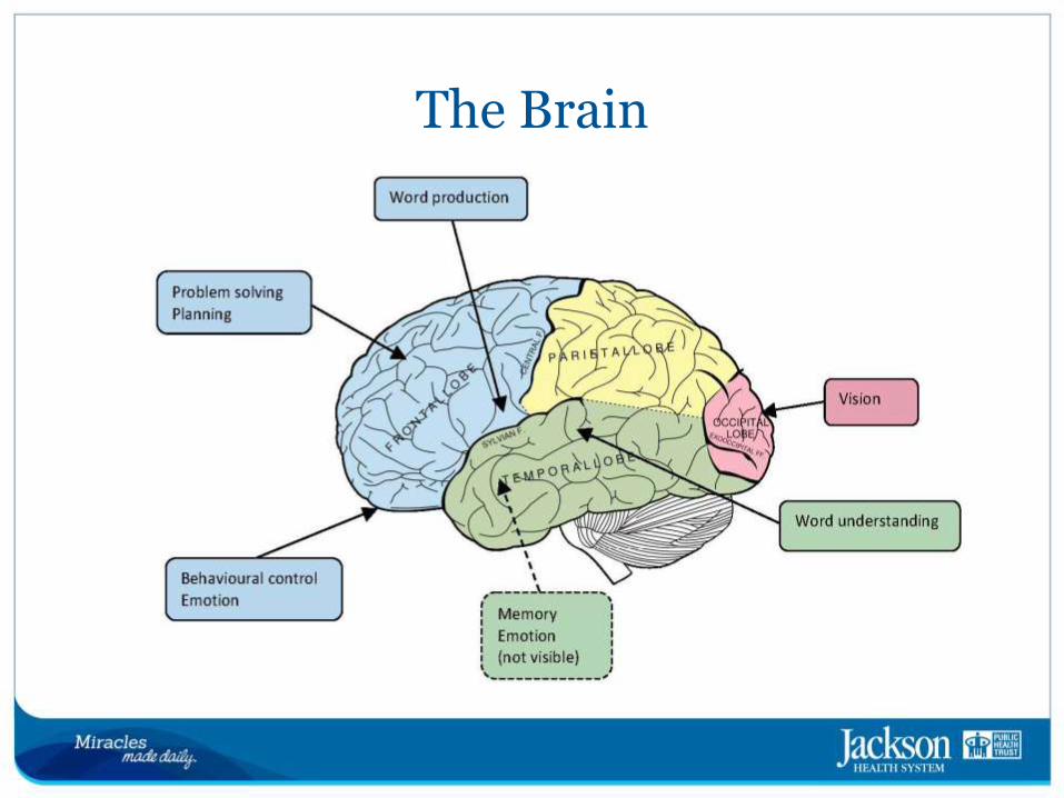
• Brain is divided into lobes based on their location and function. One hemisphere is usually dominant in relation to the other. The majority of right handed individuals are left brain dominant.
• The frontal lobe controls executive function, personality, problem solving, planning skills, language areas and primary motor cortex.
• Parietal lobe for primary sensory cortex, visual spatial orientation, association cortex, and attention to the world around
us.
The Brain
• The temporal lobe is primarily involved in memory.
• Occipital control interpretation of visual stimuli.
• Cerebellum coordinates our movement.
• Brain stem is the major relay area between the brain and the body. Brain stem is responsible for unconscious activity such as breathing and the beating of the heart.
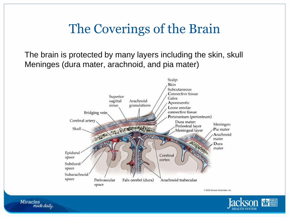
The Coverings of the Brain
The brain is protected by many layers including the skin, skull
Meninges (dura mater, arachnoid, and pia mater)
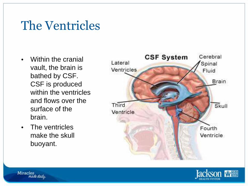
The Ventricles
• Within the cranial vault, the brain is bathed by CSF. CSF is produced within the ventricles and flows over the surface of the
brain.
• The ventricles make the skull buoyant.
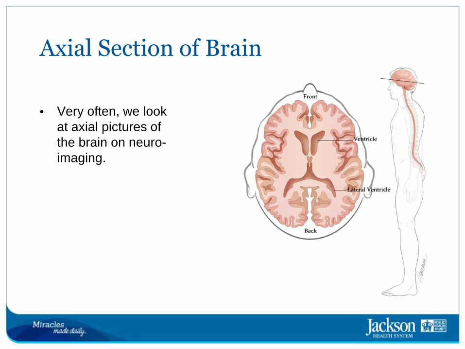
Axial Section of Brain
• Very often, we look at axial pictures of the brain on neuro-imaging.

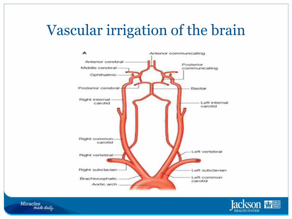
Vascular irrigation of the brain
• The brain is supplied by 4 major vessels; Two internal carotid arteries anteriorly and 2 vertebral arteries posteriorly.
Vascular irrigation of the brain

Diagnostic Tests
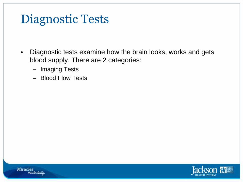
Diagnostic Tests
• Diagnostic tests examine how the brain looks, works and gets blood supply. There are 2 categories:
– Imaging Tests
– Blood Flow Tests
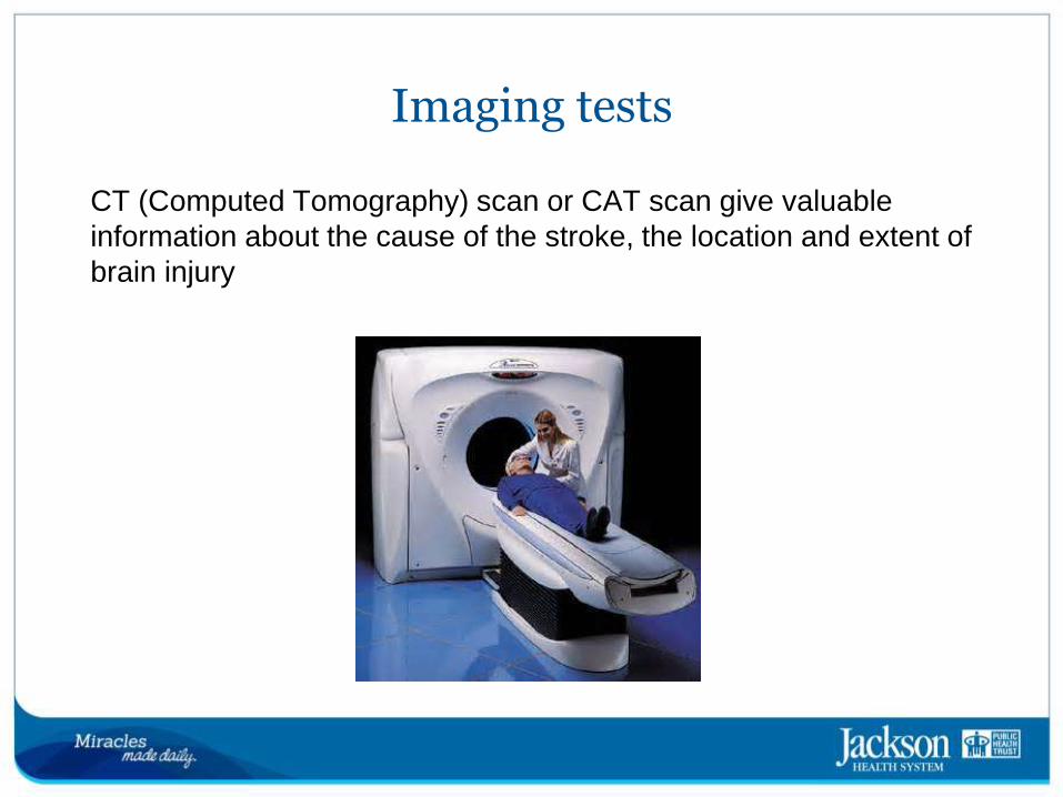
Imaging tests
CT (Computed Tomography) scan or CAT scan give valuable
information about the cause of the stroke, the location and extent of brain injury
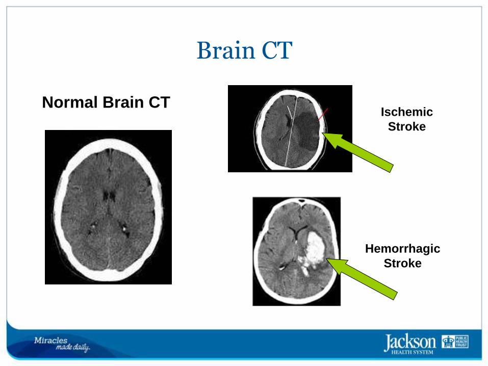
Brain CT
Hemorrhagic
Stroke
Normal Brain CT Ischemic
Stroke

Imaging tests
• CTP Quantitative determination of cerebral blood flow, can be performed quickly, allows to make distinction between the
irreversibly damaged infarct core and the penumbra
Imaging tests
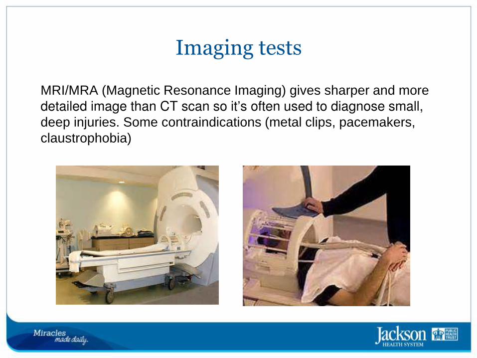
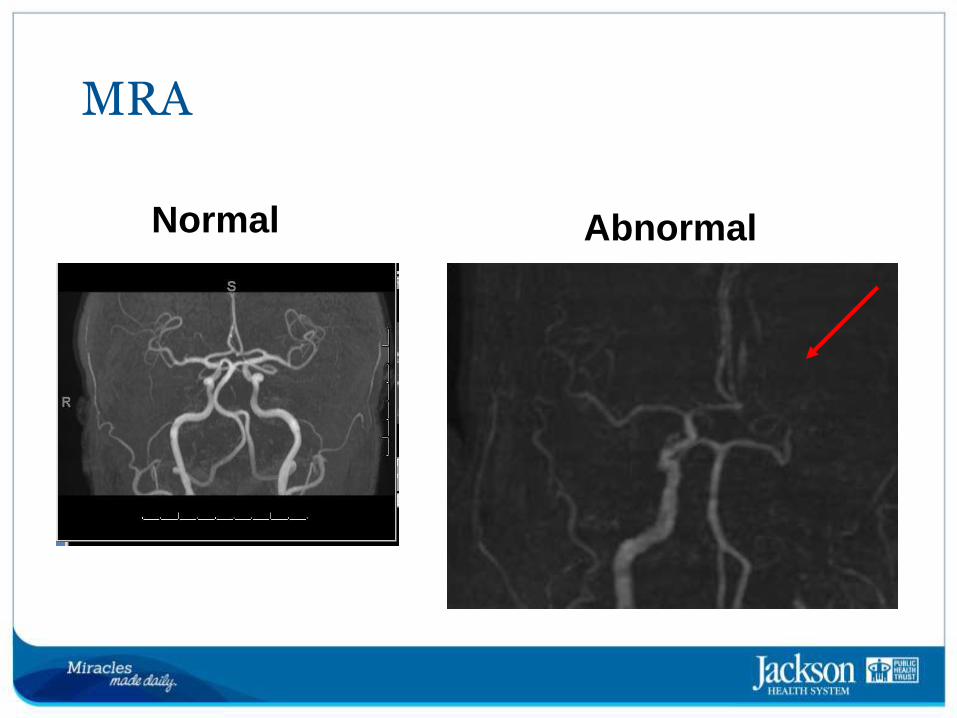
MRI/MRA (Magnetic Resonance Imaging) gives sharper and more
detailed image than CT scan so it’s often used to diagnose small, deep injuries. Some contraindications (metal clips, pacemakers, claustrophobia)
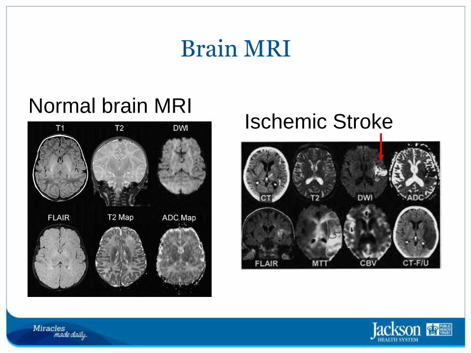
Brain MRI
Normal brain MRI Ischemic Stroke
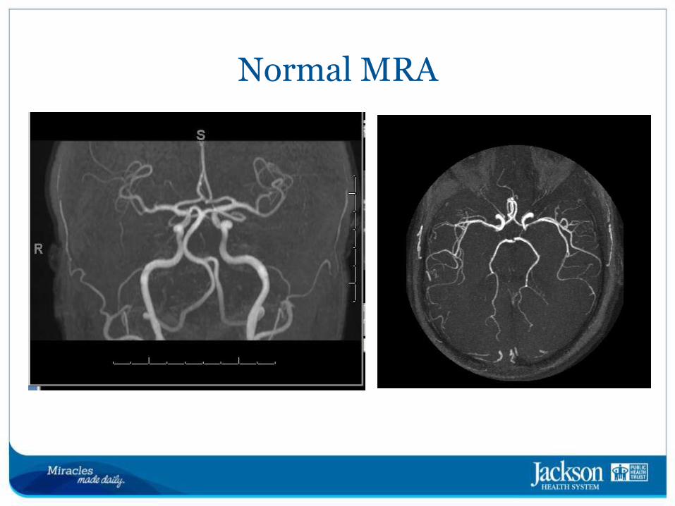
Normal MRA
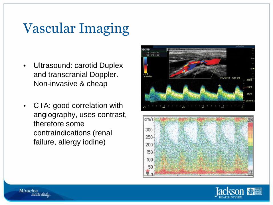
Vascular Imaging
• Ultrasound: carotid Duplex and transcranial Doppler. Non-invasive & cheap
• CTA: good correlation with angiography, uses contrast, therefore some
contraindications (renal failure, allergy iodine)
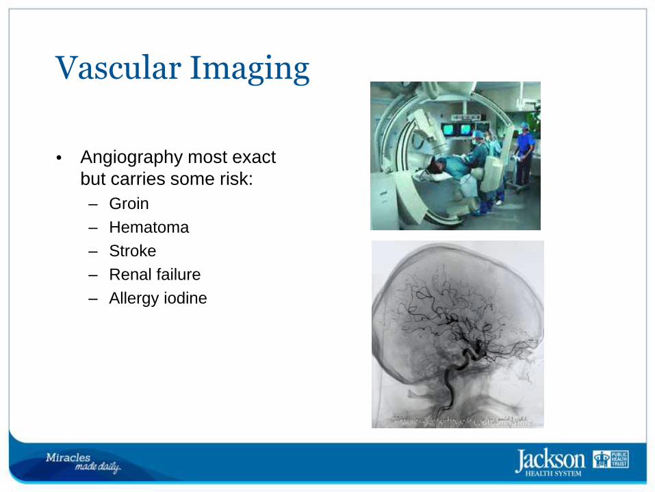
Vascular Imaging
• Angiography most exact but carries some risk:
– Groin
– Hematoma
– Stroke
– Renal failure
– Allergy iodine
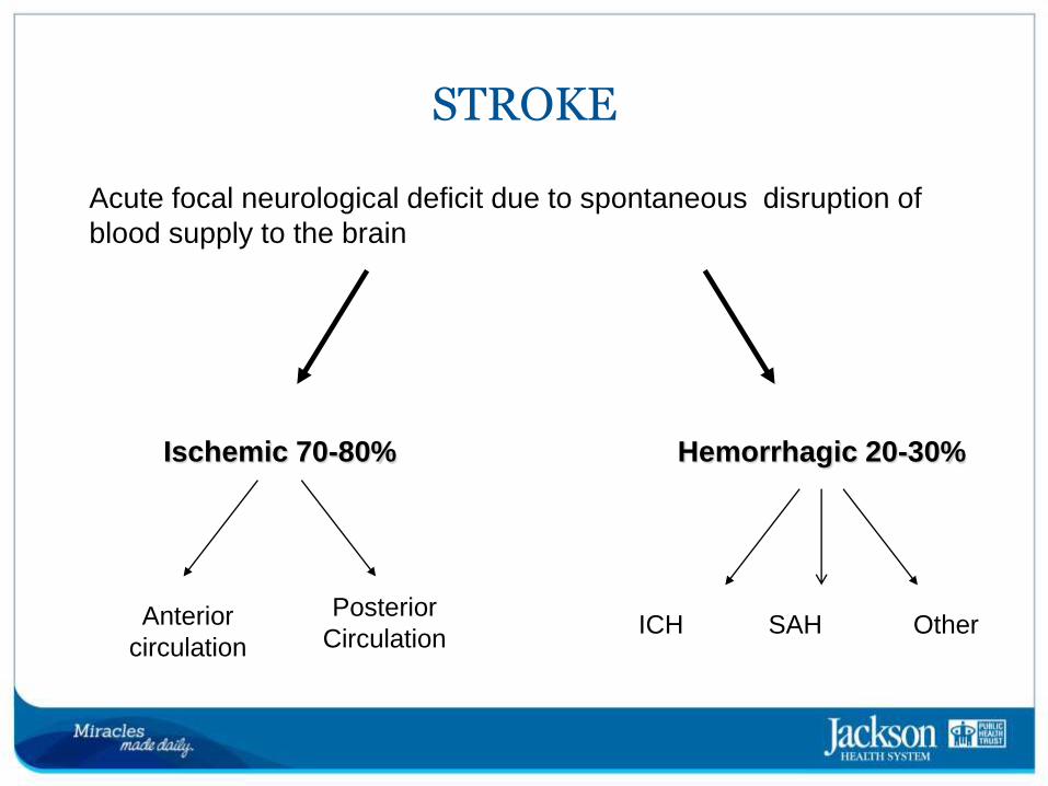
Hemorrhagic 20-30%
ICH SAH Other
Ischemic 70-80%
Anterior
circulation
Posterior
Circulation
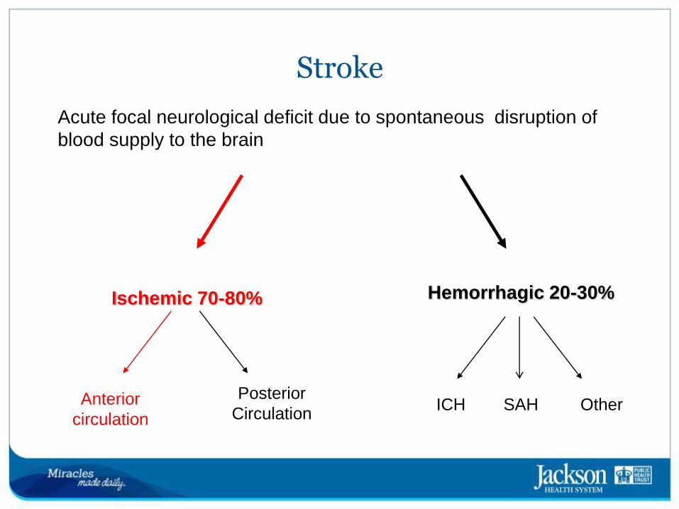
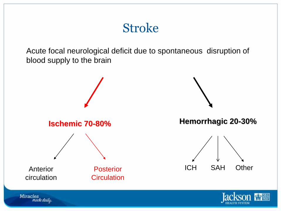
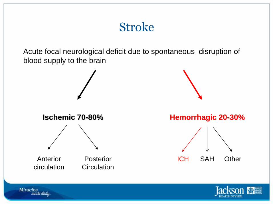
STROKE
Acute focal neurological deficit due to spontaneous disruption of
blood supply to the brain
Ischemic Strokes
• Occurs when blood vessels to the brain become narrowed or clogged thereby cutting off blood flow to brain cells
• 70-80% of all strokes are ischemic
• TIA or “mini stroke” may give some warning of a major ischemic stroke
• High blood pressure is most important modifiable risk factor for ischemic stroke
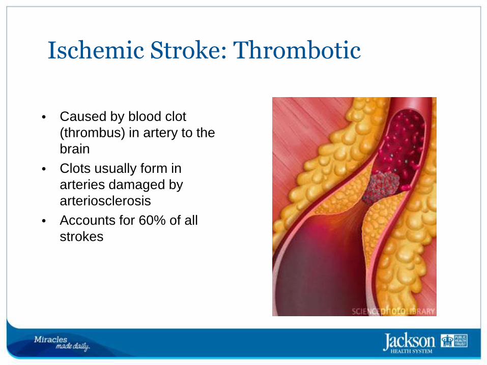
Ischemic Stroke: Thrombotic
• Caused by blood clot (thrombus) in artery to the brain
• Clots usually form in arteries damaged by arteriosclerosis
• Accounts for 60% of all
strokes
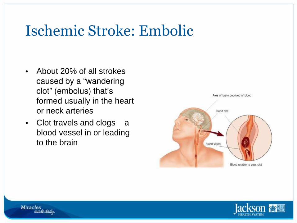
Ischemic Stroke: Embolic
• About 20% of all strokes caused by a “wandering clot” (embolus) that’s formed usually in the heart or neck arteries
• Clot travels and clogs a blood vessel in or leading
to the brain
1. Hillis et al Neuroradiology. 2004
Jan;46(1):31-9.
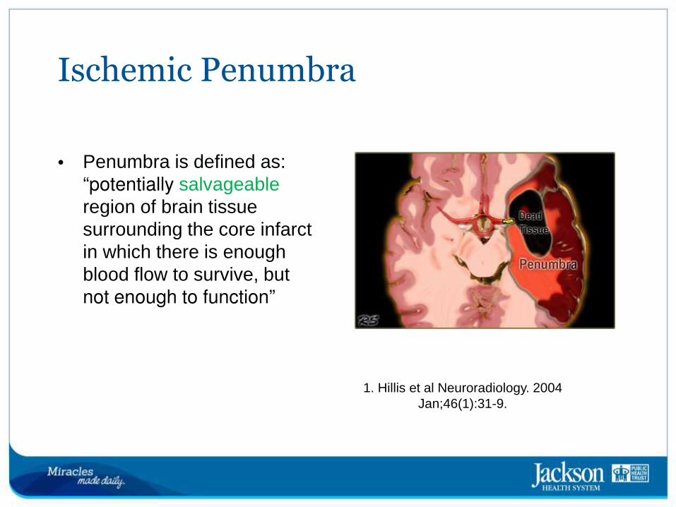
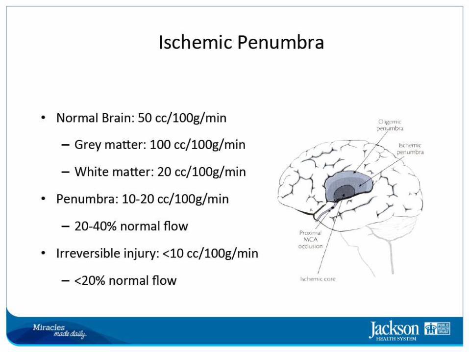
Ischemic Penumbra
• Penumbra is defined as: “potentially salvageable region of brain tissue surrounding the core infarct in which there is enough blood flow to survive, but not enough to function”
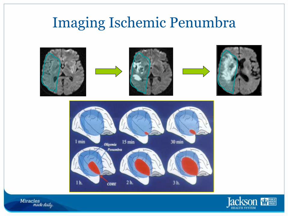
Imaging Ischemic Penumbra
Stroke
Acute focal neurological deficit due to spontaneous disruption of
blood supply to the brain
Hemorrhagic 20-30%
ICH SAH Other
Ischemic 70-80%
Anterior
circulation
Posterior
Circulation
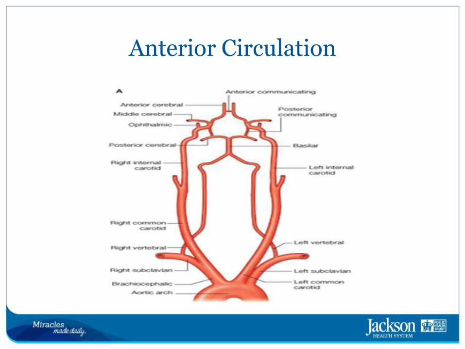
Anterior Circulation
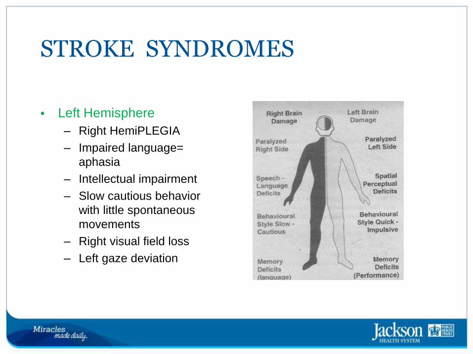
STROKE SYNDROMES
• Left Hemisphere
– Right HemiPLEGIA
– Impaired language=
aphasia
– Intellectual impairment
– Slow cautious behavior
with little spontaneous
movements
– Right visual field loss
– Left gaze deviation
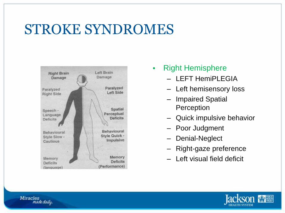
STROKE SYNDROMES
• Right Hemisphere
– LEFT HemiPLEGIA
– Left hemisensory loss
– Impaired Spatial
Perception
– Quick impulsive behavior
– Poor Judgment
– Denial-Neglect
– Right-gaze preference
– Left visual field deficit
Hemorrhagic 20-30%
ICH SAH Other
Ischemic 70-80%
Anterior
circulation
Posterior
Circulation
Stroke
Acute focal neurological deficit due to spontaneous disruption of
blood supply to the brain
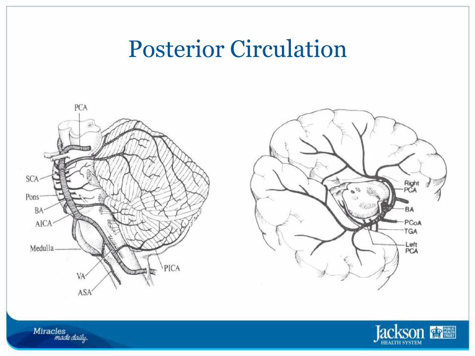
Posterior Circulation
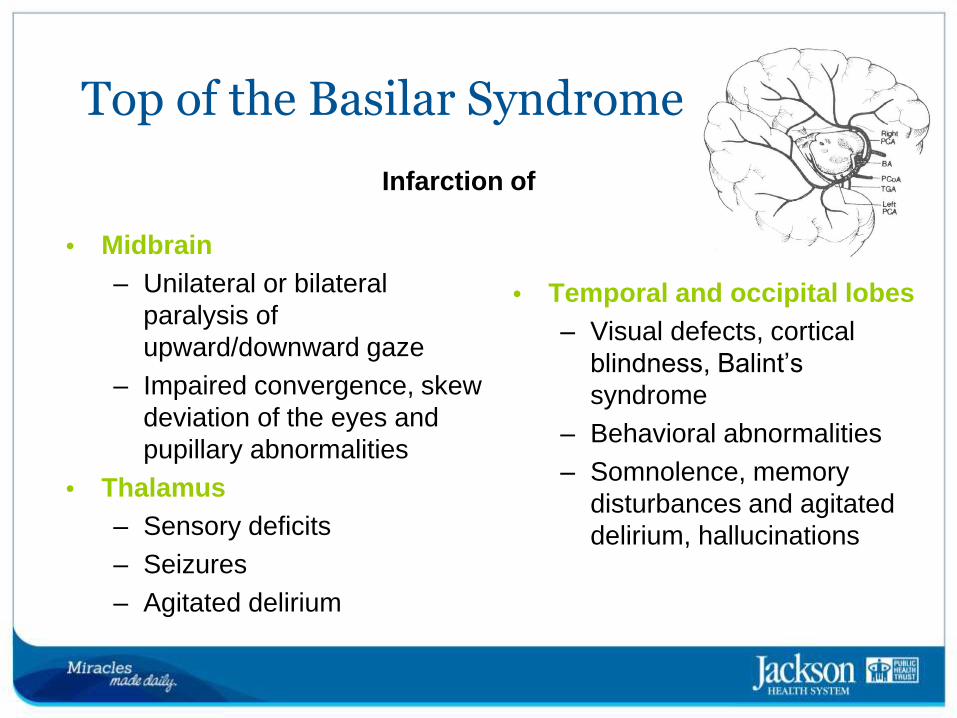
Top of the Basilar Syndrome
• Midbrain
– Unilateral or bilateral
paralysis of
upward/downward gaze
– Impaired convergence, skew
deviation of the eyes and
pupillary abnormalities
• Thalamus
– Sensory deficits
– Seizures
– Agitated delirium
• Temporal and occipital lobes
– Visual defects, cortical
blindness, Balint’s
syndrome
– Behavioral abnormalities
– Somnolence, memory
disturbances and agitated
delirium, hallucinations
Infarction of
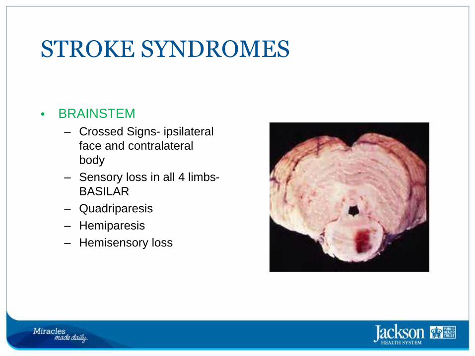
STROKE SYNDROMES
• BRAINSTEM
– Crossed Signs- ipsilateral
face and contralateral
body
– Sensory loss in all 4 limbs-
BASILAR
– Quadriparesis
– Hemiparesis
– Hemisensory loss
STROKE SYNDROMES
Cerebellar Stroke
• Symptoms
– Vertigo, dizziness, nausea, vomiting, gait unsteadiness, limb
clumsiness, headache, dysarthria, diplopia and decreased
alertness
• Signs
– Limb and gait ataxia, dysarthria, nystagmus, altered mental status
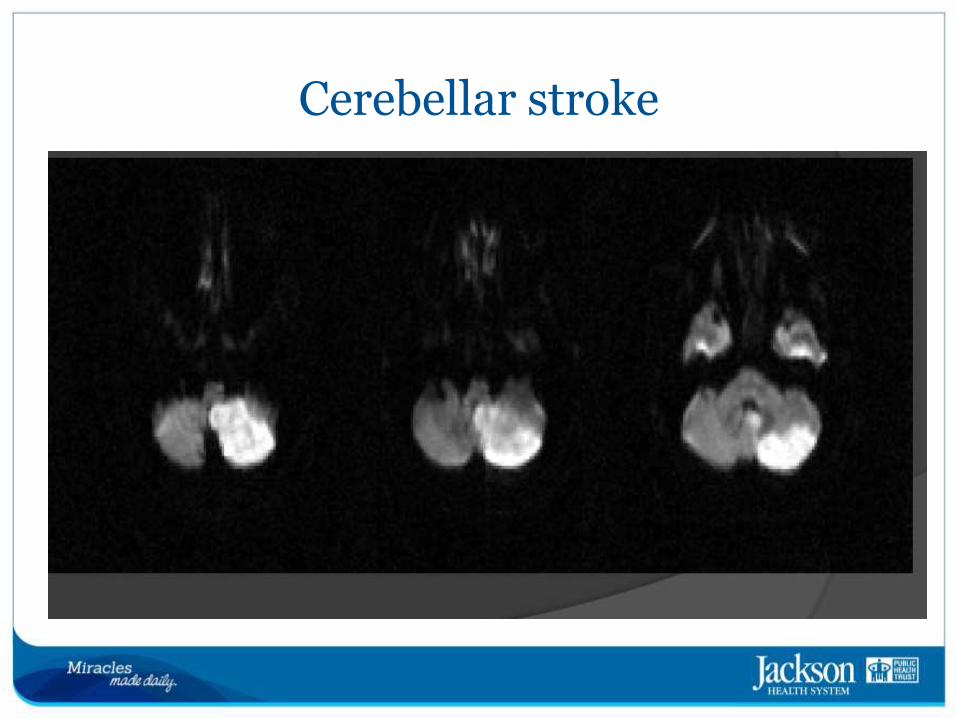
Cerebellar stroke
Hemorrhagic 20-30%
ICH SAH Other
Ischemic 70-80%
Anterior
circulation
Posterior
Circulation
Stroke
Acute focal neurological deficit due to spontaneous disruption of
blood supply to the brain
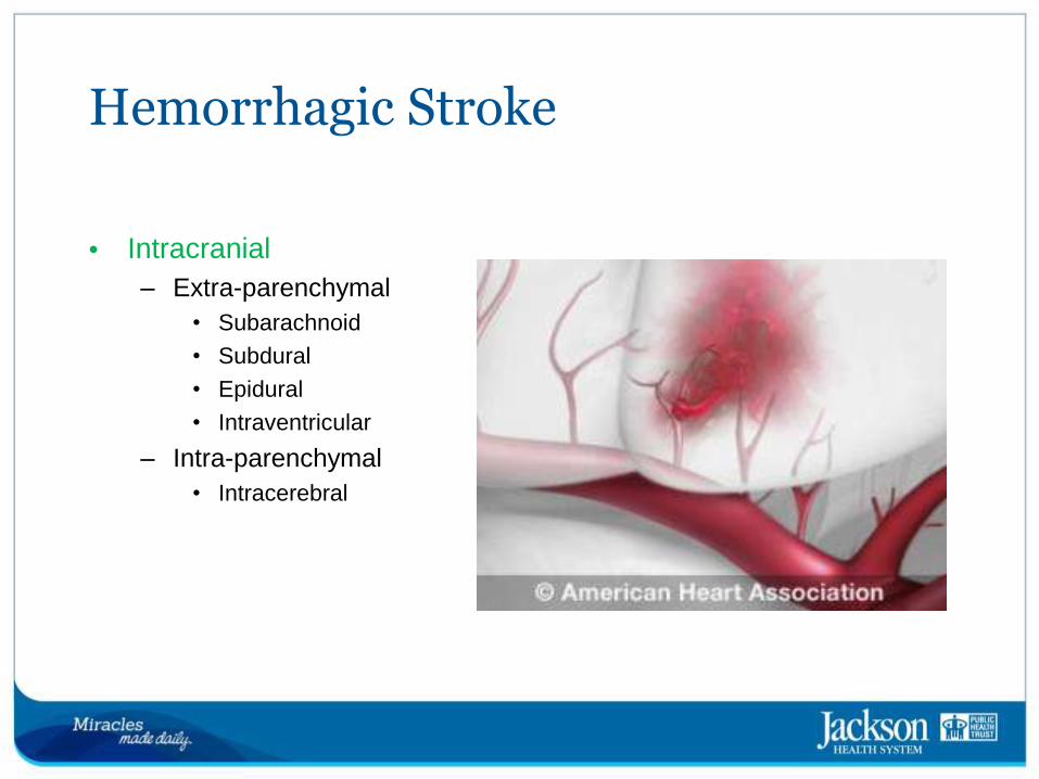
Hemorrhagic Stroke
• Intracranial
– Extra-parenchymal
• Subarachnoid
• Subdural
• Epidural
• Intraventricular
– Intra-parenchymal
• Intracerebral
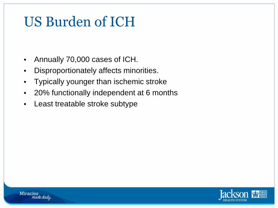
US Burden of ICH
• Annually 70,000 cases of ICH.
• Disproportionately affects minorities.
• Typically younger than ischemic stroke
• 20% functionally independent at 6 months
• Least treatable stroke subtype
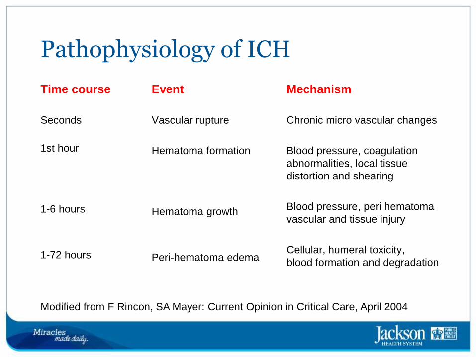
Pathophysiology of ICH
Seconds
1st hour
1-6 hours
1-72 hours
Time course
Chronic micro vascular changes
Blood pressure, coagulation
abnormalities, local tissue
distortion and shearing
Blood pressure, peri hematoma
vascular and tissue injury
Cellular, humeral toxicity,
blood formation and degradation
Mechanism
Vascular rupture
Hematoma formation
Hematoma growth
Peri-hematoma edema
Event
Modified from F Rincon, SA Mayer: Current Opinion in Critical Care, April 2004
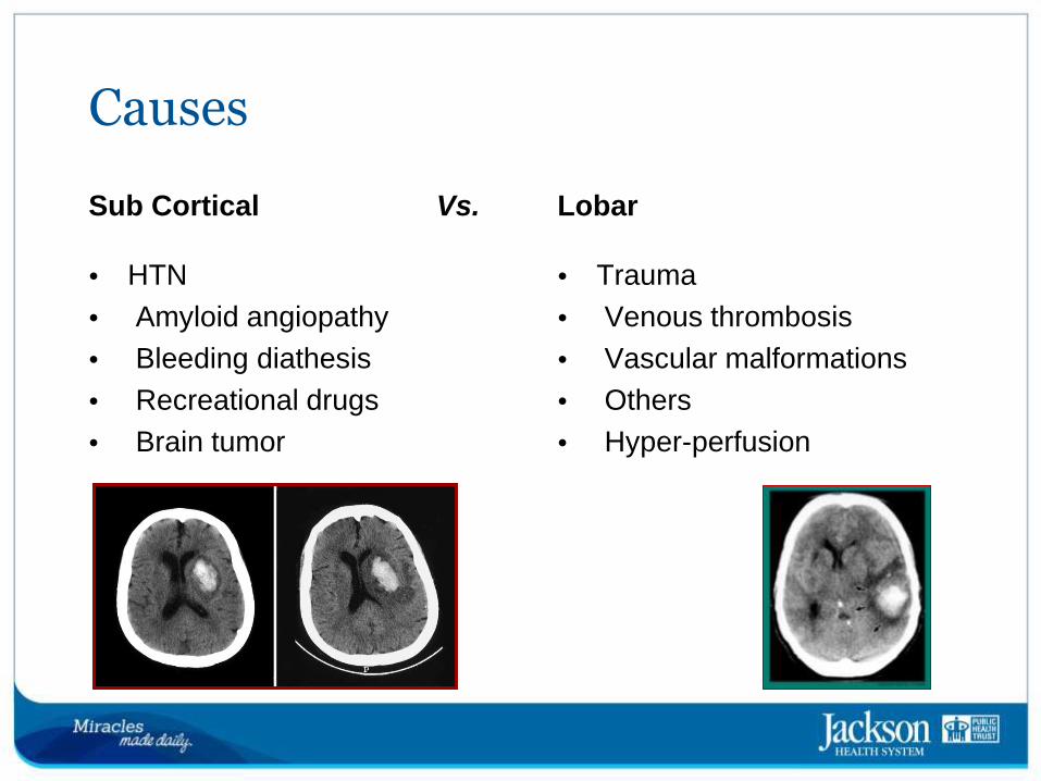
Causes
• HTN
• Amyloid angiopathy
• Bleeding diathesis
• Recreational drugs
• Brain tumor
• Trauma
• Venous thrombosis
• Vascular malformations
• Others
• Hyper-perfusion
Sub Cortical Lobar
Vs.
Hemorrhage
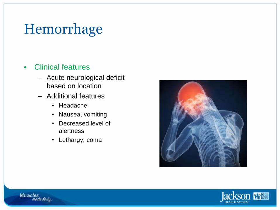
• Clinical features
– Acute neurological deficit
based on location
– Additional features
• Headache
• Nausea, vomiting
• Decreased level of
alertness
• Lethargy, coma
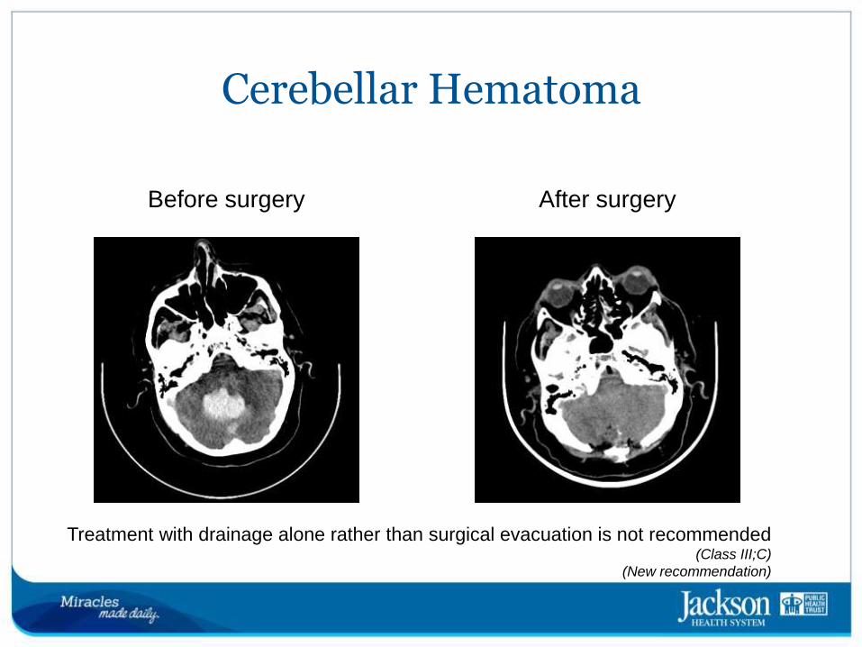
Cerebellar Hematoma
Before surgery After surgery
Treatment with drainage alone rather than surgical evacuation is not recommended (Class III;C)
(New recommendation)

STROKE Acute focal neurological deficit due to spontaneous disruption of
blood supply to the brain
Hemorrhagic 20-30%
ICH SAH Other
Ischemic 70-80%
Anterior
circulation
Posterior
Circulation
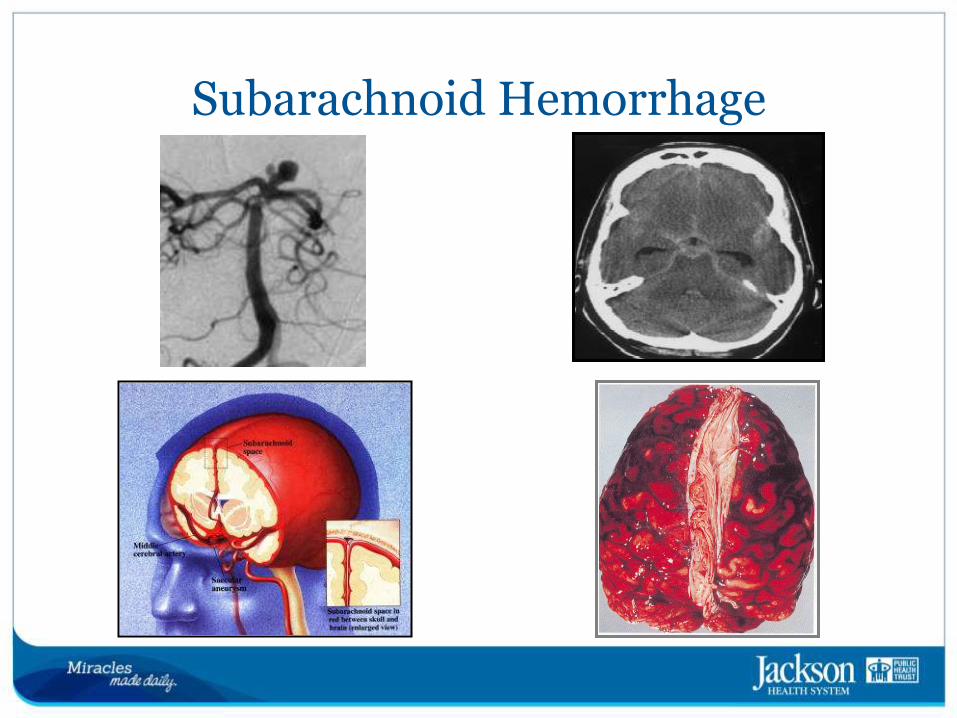
Subarachnoid Hemorrhage
Subarachnoid Hemorrhage
• Subarachnoid hemorrhage (SAH) or subarachnoid hemorrhage, is bleeding into the subarachnoid space surrounding the brain, the area between the arachnoid membrane and the pia mater
• The bleeding may occur spontaneously, usually from a cerebral aneurysm, or may result from trauma. Regardless of the cause, it is considered a medical emergency.
• Symptoms include an intense headache with a rapid onset, vomiting, and an altered level of consciousness.[1]
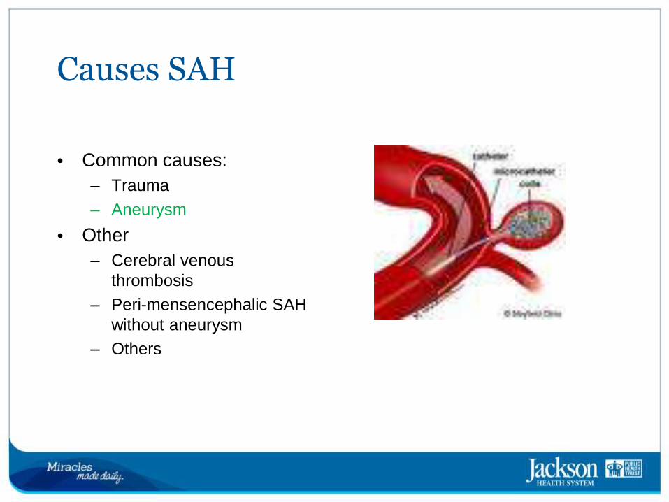
Causes SAH
• Common causes:
– Trauma
– Aneurysm
• Other
– Cerebral venous
thrombosis
– Peri-mensencephalic SAH
without aneurysm
– Others
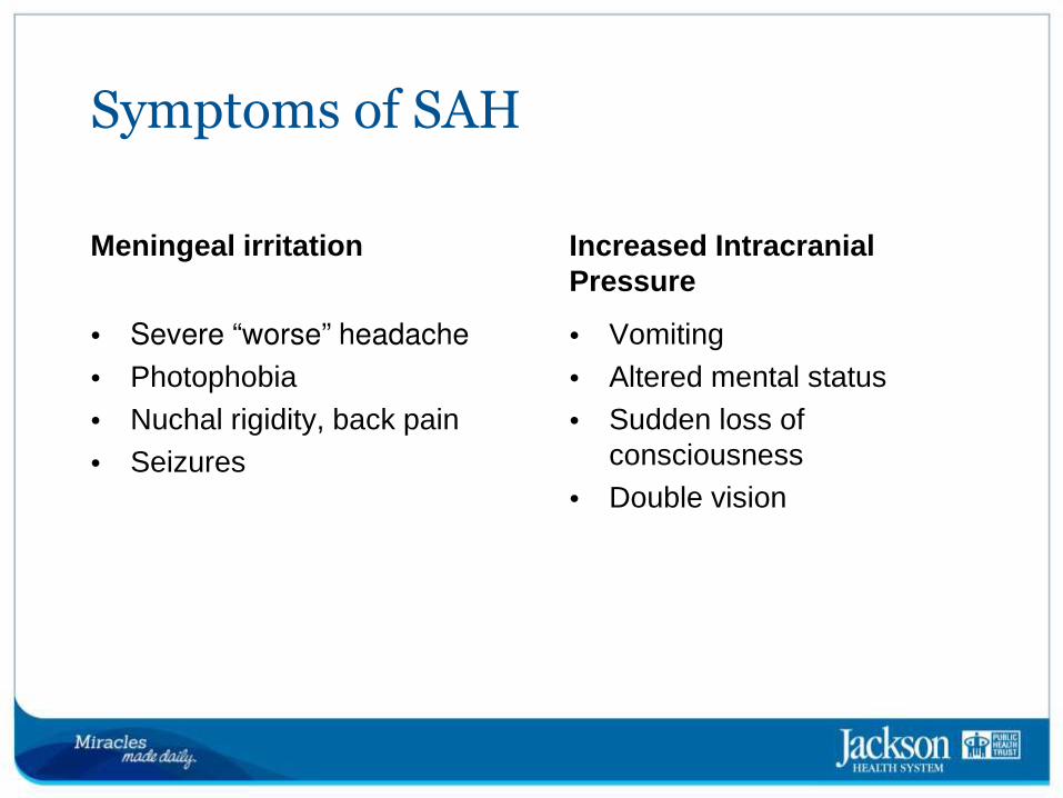
Symptoms of SAH
• Severe “worse” headache
• Photophobia
• Nuchal rigidity, back pain
• Seizures
• Vomiting
• Altered mental status
• Sudden loss of consciousness
• Double vision
Meningeal irritation Increased Intracranial
Pressure
Subarachnoid Hemorrhage
SAH can lead to death or severe disability even if recognized and
treated at an early stage
Treatment is with close observation, medication and early neurosurgical investigations and treatments. Subarachnoid hemorrhage causes between 1 and 7% of all strokes
Of all people with SAH, 10-15% die before arriving in hospital, and
average survival is 50%.[1]
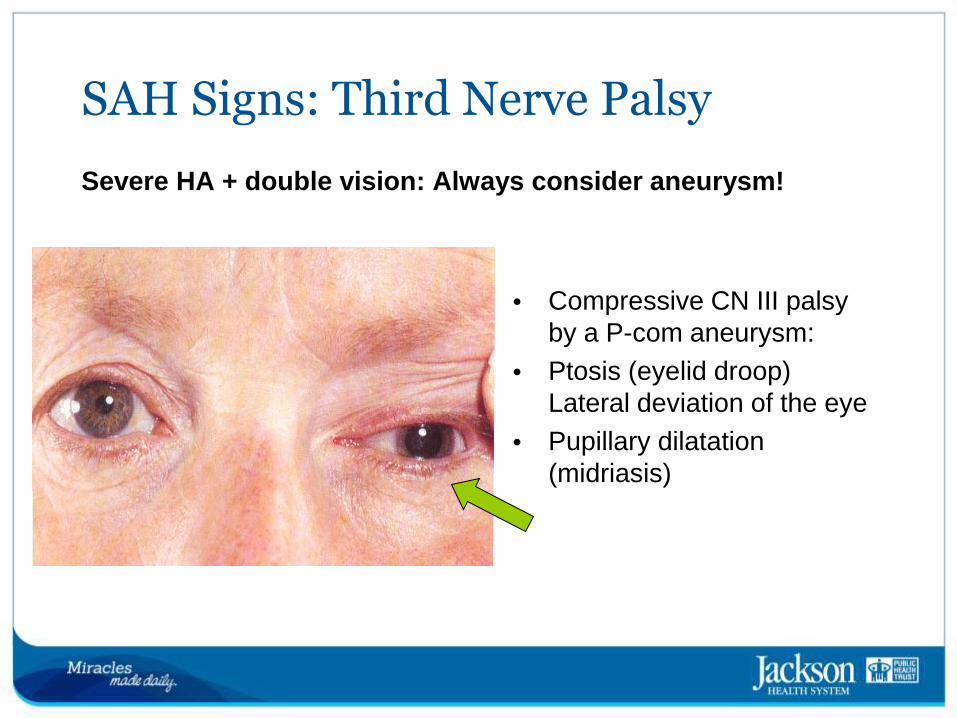
SAH Signs: Third Nerve Palsy
• Compressive CN III palsy by a P-com aneurysm:
• Ptosis (eyelid droop) Lateral deviation of the eye
• Pupillary dilatation
(midriasis)
Severe HA + double vision: Always consider aneurysm!
SAH Assessment: Clinical
• Hunt & Hess scale classifies severity SAH
Grade 1 - Asymptomatic, mild headache, slight nuchal rigidity
Grade 2 - Moderate to severe headache, nuchal rigidity, no
neurological deficit other than cranial nerve palsy
Grade 3 - Drowsiness / confusion, mild focal neurological deficit
Grade 4 - Stupor, moderate-severe hemi paresis
Grade 5 - Coma, decerebrate posturing
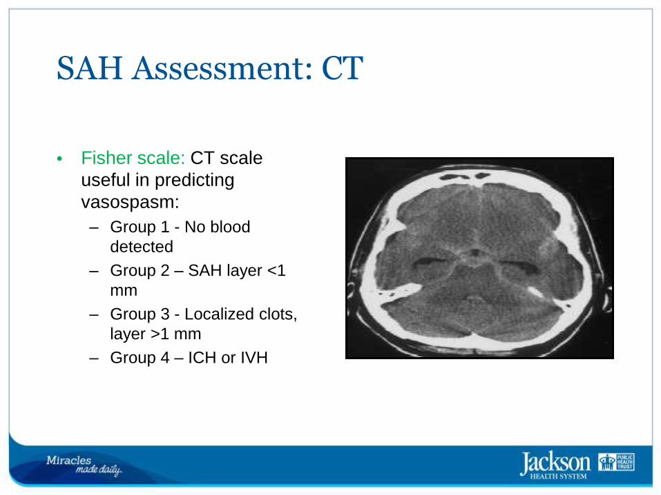
SAH Assessment: CT
• Fisher scale: CT scale useful in predicting vasospasm:
– Group 1 - No blood
detected
– Group 2 – SAH layer <1
mm
– Group 3 - Localized clots,
layer >1 mm
– Group 4 – ICH or IVH

•Cases Studies
Case 1
• 71 year old Right Handed Male
– 20 pack/year history of smoking
– Uncontrolled HTN, DM
– Recurrent episodes of loss of vision in his L eye
– Did not present to medical attention
– Brought to emergency room by family because of R face/arm
weakness, unable to speak
Case 1
• Exam
– T 97 PR 88 BP 218/116 RR 16 Glu 180mmol/L
– CVS: High pitched carotid bruit audible over the L ICA
– Awake, Alert, Grunted only, no words, follows no commands but
will mimic actions.
– Pupils 3mm, blink to threat bilaterally present, R NLF weakness
(sparing of forehead) No R arm movement, He is able to lift the R
leg off the bed.
– NIHSS= 13

NIH Stroke Scale
• Is a 15-item systematic neurologic examination that provides a quantitative measure of stroke-related neurologic deficit
• Ratings for each item are scored with 3 to 5 grades
with 0 as normal, and there is an allowance for un-testable items.
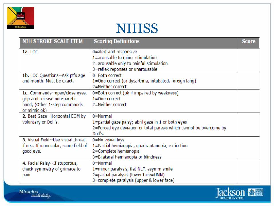
NIHSS
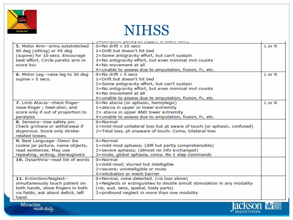
NIHSS
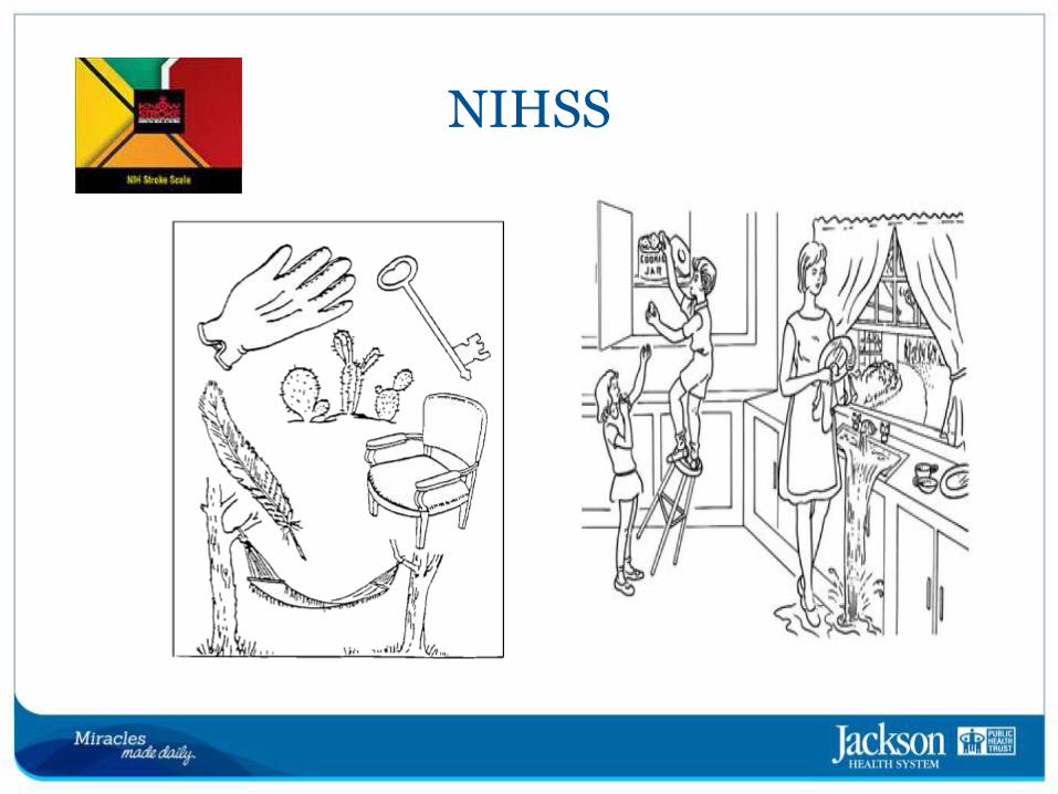
NIHSS
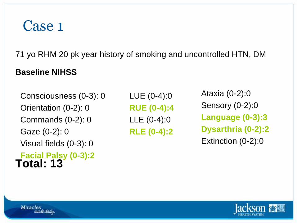
71 yo RHM 20 pk year history of smoking and uncontrolled HTN, DM
Baseline NIHSS
Total: 13
Case 1
Consciousness (0-3): 0
Orientation (0-2): 0
Commands (0-2): 0
Gaze (0-2): 0
Visual fields (0-3): 0
Facial Palsy (0-3):2
LUE (0-4):0
RUE (0-4):4
LLE (0-4):0
RLE (0-4):2
Ataxia (0-2):0
Sensory (0-2):0
Language (0-3):3
Dysarthria (0-2):2
Extinction (0-2):0
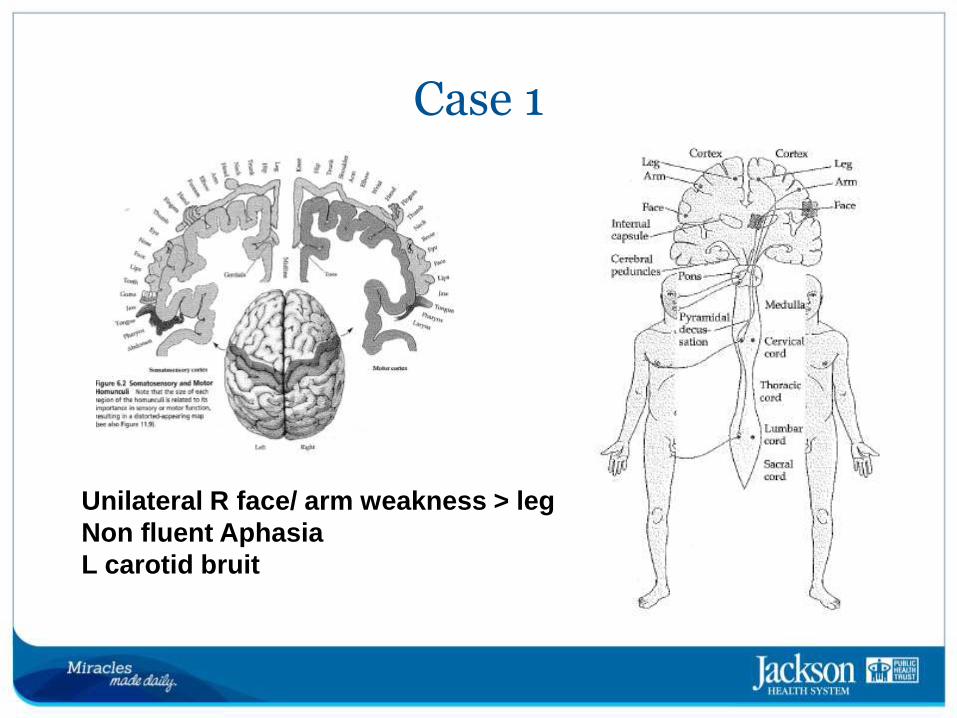
Unilateral R face/ arm weakness > leg
Non fluent Aphasia
L carotid bruit
Case 1
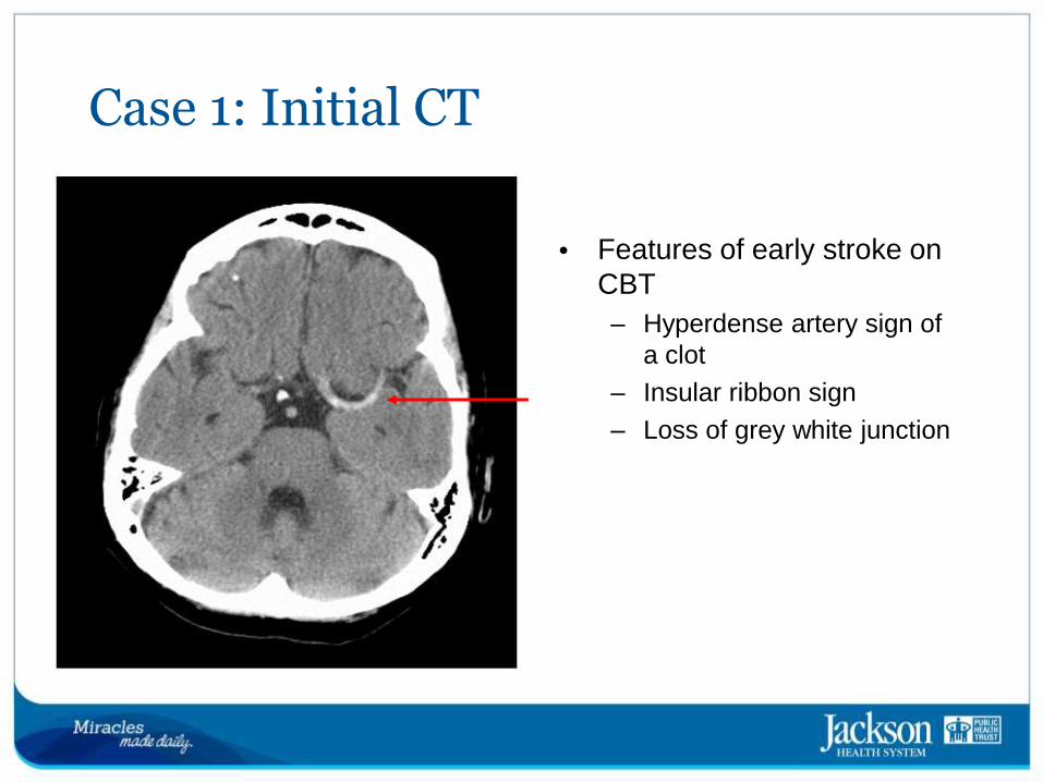
Case 1: Initial CT
• Features of early stroke on CBT
– Hyperdense artery sign of
a clot
– Insular ribbon sign
– Loss of grey white junction
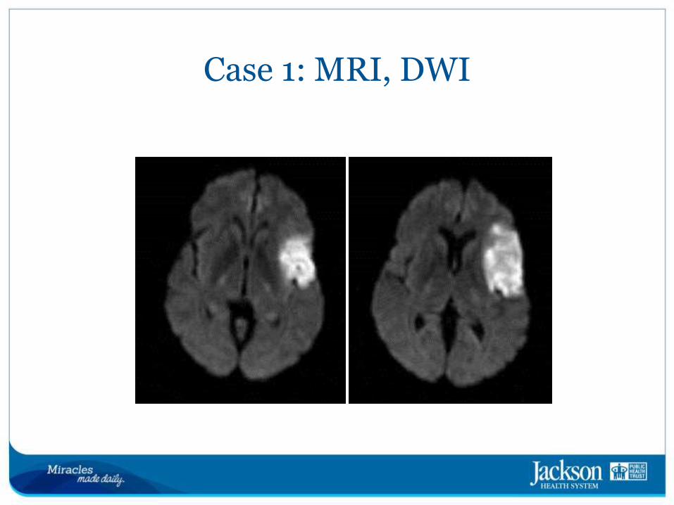
Case 1: MRI, DWI
Case 1 What is the diagnosis ?
L MCA Stroke
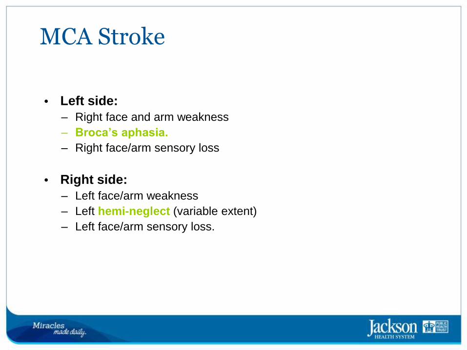
MCA Stroke
• Left side:
– Right face and arm weakness
– Broca’s aphasia.
– Right face/arm sensory loss
• Right side:
– Left face/arm weakness
– Left hemi-neglect (variable extent)
– Left face/arm sensory loss.
Case 2
• 56 year old Hypertensive man with prior history of drug abuse, presents with:
– Vitals signs T 98.6 PR 84 BP 225/130 RR 20
– CVS no murmurs or bruits,
– Drowsy, dysarthric and with major headache
– Pupils 3mm ERL
– Severe nausea and vomiting
– Gait deferred because of drowsiness
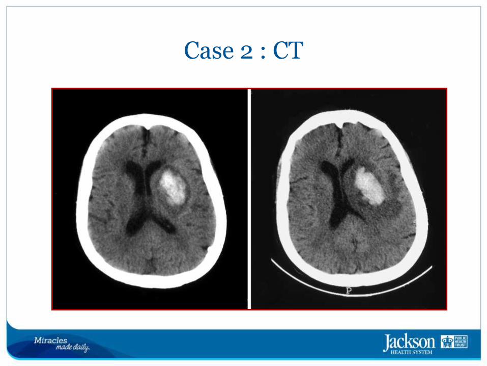
Case 2 : CT
Case 2 What is the diagnosis?
ICH
Medical Management of Acute Cerebrovascular Syndromes
Dr. Gillian L. Gordon Perue
Acute Cerebrovascular Syndromes
• New term introduced by AHA in 2013
– Acute focal neurological deficit due to spontaneous disruption of the
blood supply to the brain.
• Encompasses all subtypes of stroke
– Acute ischemic stroke
– Transient Ischemic attack
– Acute intracerebral hemorrhages
– Acute Subarachnoid hemorrhages
• Emphasizes early recognition to facilitate timely intervention.
• Treatment strategies are as varied as the different cause and depends on establishment of underlying cause.
Stroke Diagnosis
• Critical to determine the type of stroke in progress because treatment is different for ischemic stroke or a hemorrhagic stroke
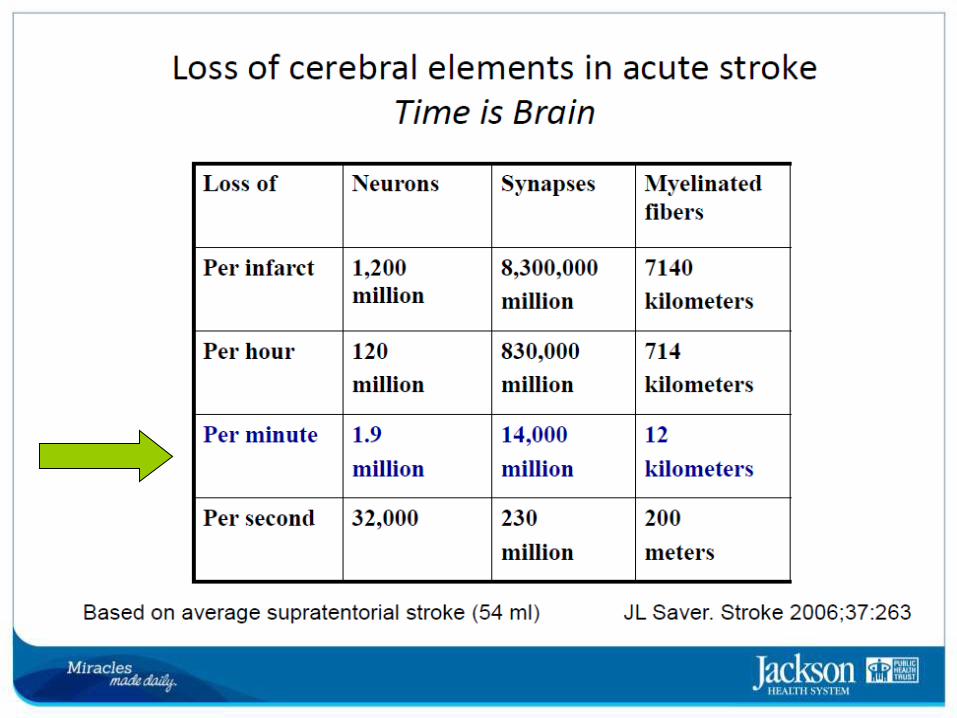
• Time = Brain
• Based on Medical History, physical neurological examination, blood tests, diagnostic tests
• Rule out conditions with similar symptoms like: seizures, fainting, migraine, heart problems

CT Scan is Important in Acute Stroke Care
ACUTE ISCHEMIC STROKE MANAGEMENT
PROTOCOL
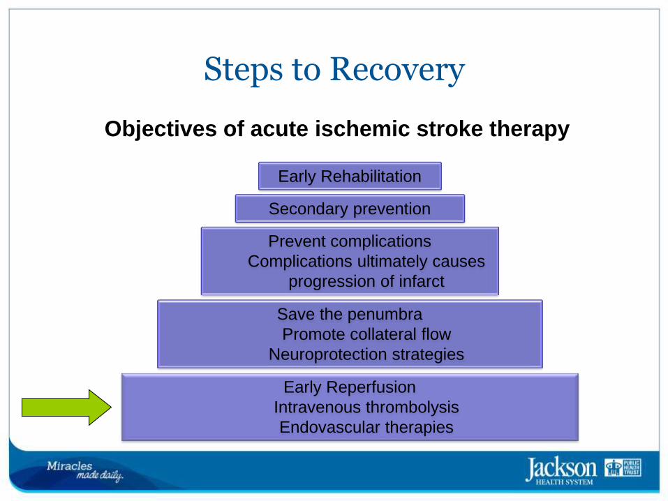
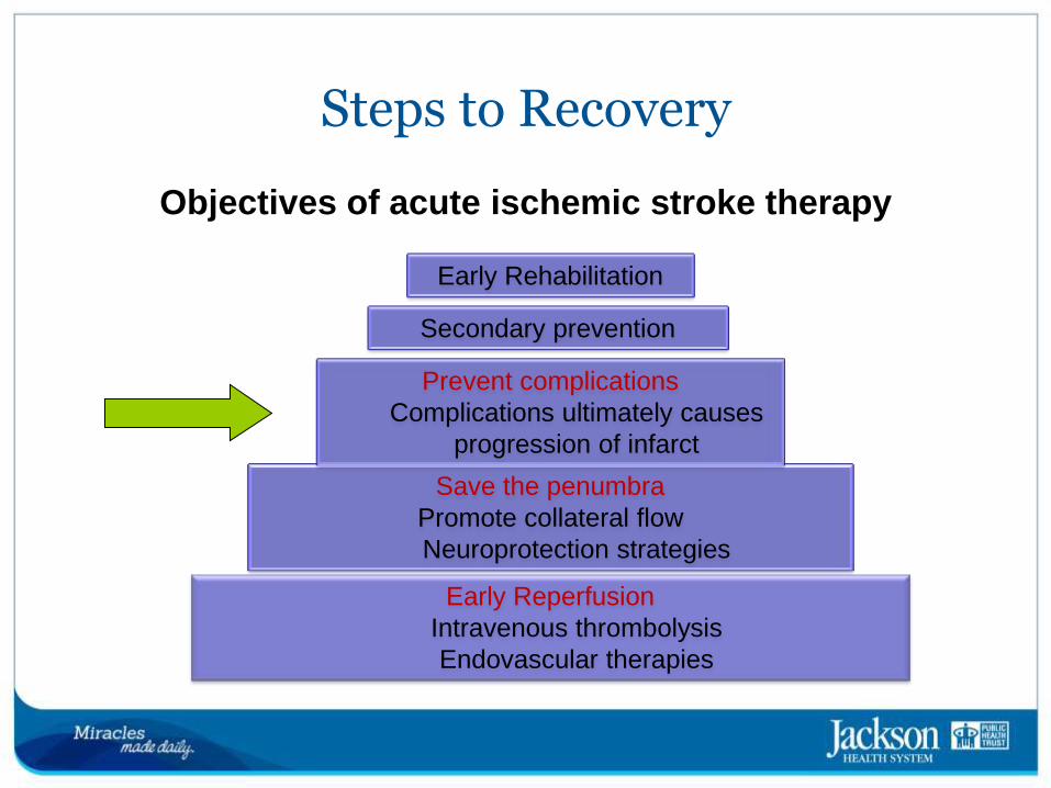
Steps to Recovery
Objectives of acute ischemic stroke therapy
Save the penumbra
Promote collateral flow
Neuroprotection strategies
Early Reperfusion
Intravenous thrombolysis
Endovascular therapies
Prevent complications
Complications ultimately causes
progression of infarct
Secondary prevention
Early Rehabilitation
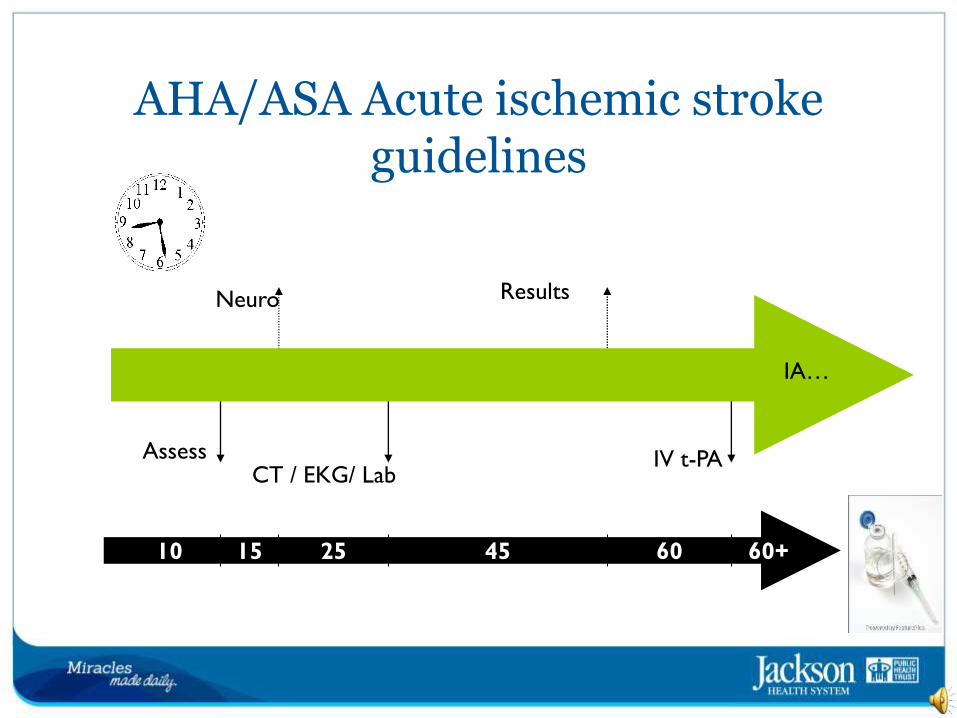
Door to needle / intervention
Assess
Neuro
CT / EKG/ Lab
Results
IV t-PA
10 15 25 45 60+ 60
IA…
AHA/ASA Acute ischemic stroke guidelines
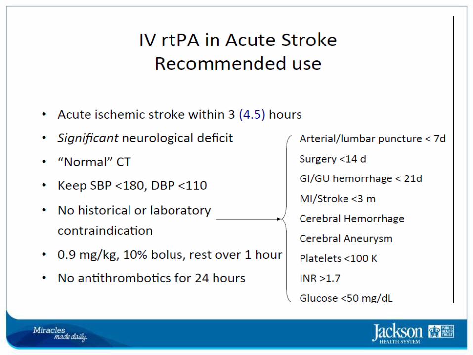
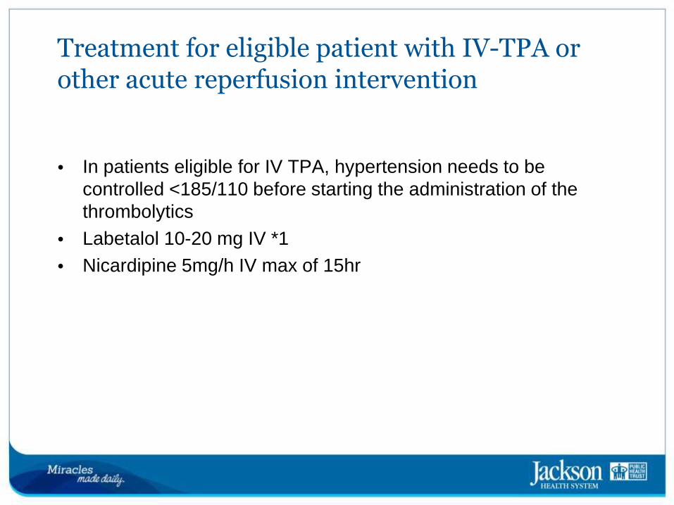
Treatment for eligible patient with IV-TPA or other acute reperfusion intervention
• In patients eligible for IV TPA, hypertension needs to be controlled <185/110 before starting the administration of the thrombolytics
• Labetalol 10-20 mg IV *1
• Nicardipine 5mg/h IV max of 15hr
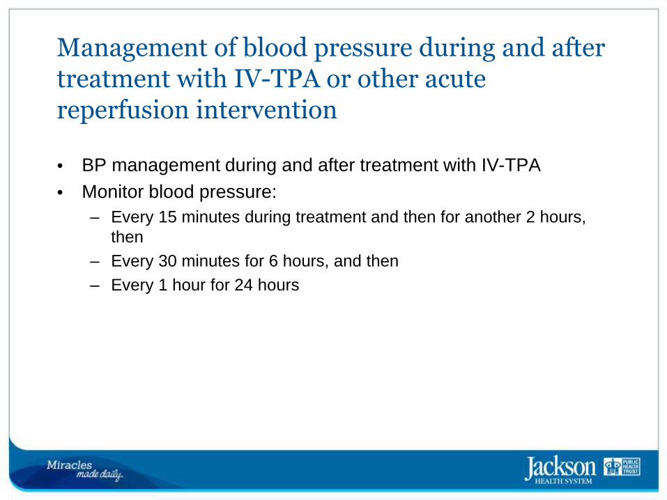
Management of blood pressure during and after treatment with IV-TPA or other acute reperfusion intervention • BP management during and after treatment with IV-TPA
• Monitor blood pressure:
– Every 15 minutes during treatment and then for another 2 hours,
then
– Every 30 minutes for 6 hours, and then
– Every 1 hour for 24 hours

Example of endovascular therapy DWI PWI
MRA
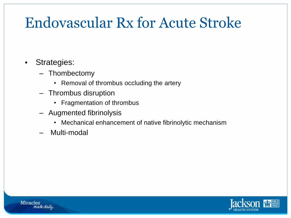
Endovascular Rx for Acute Stroke
• Strategies:
– Thombectomy
• Removal of thrombus occluding the artery
– Thrombus disruption
• Fragmentation of thrombus
– Augmented fibrinolysis
• Mechanical enhancement of native fibrinolytic mechanism
– Multi-modal
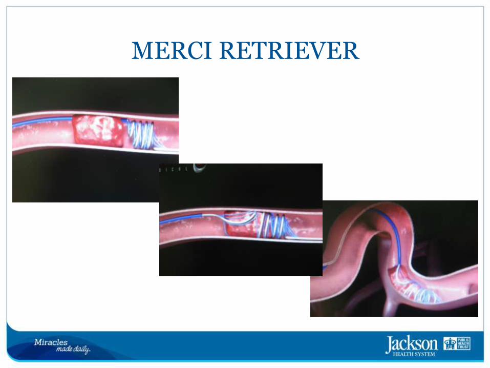
MERCI RETRIEVER
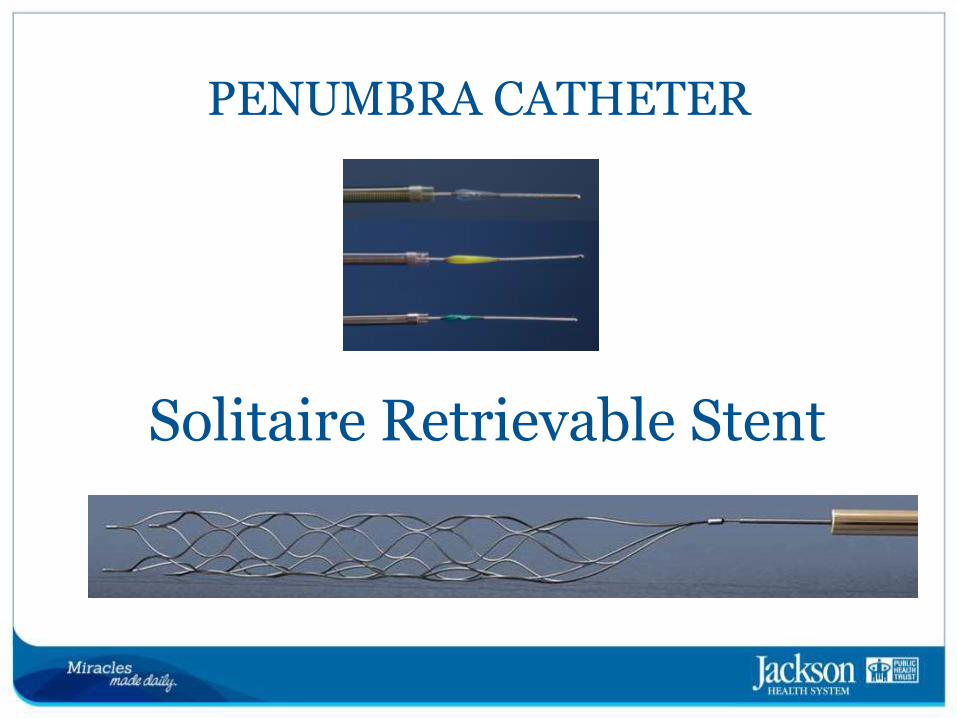
PENUMBRA CATHETER
Solitaire Retrievable Stent
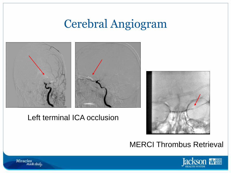
Cerebral Angiogram
Left terminal ICA occlusion
MERCI Thrombus Retrieval
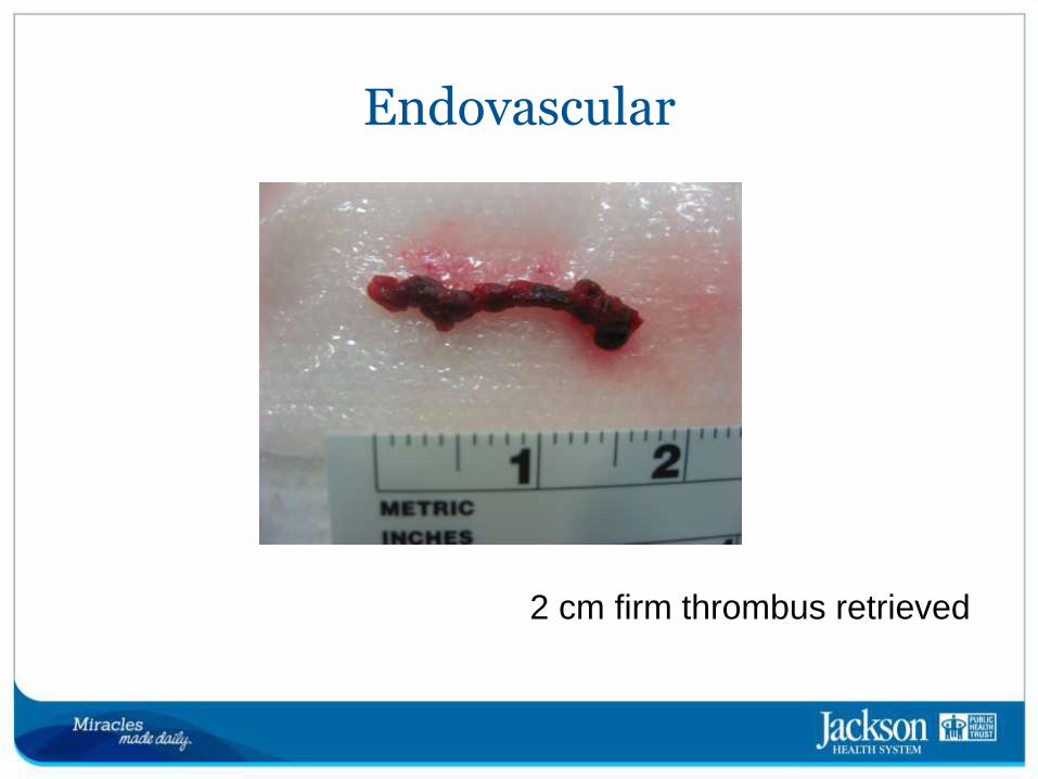
Endovascular
2 cm firm thrombus retrieved
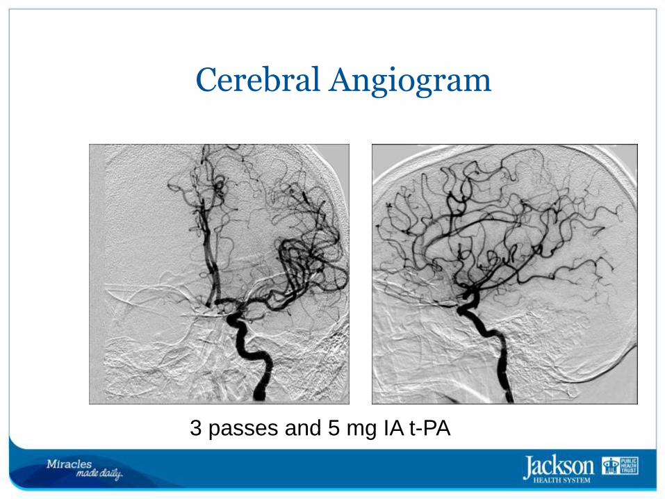
3 passes and 5 mg IA t-PA
Cerebral Angiogram
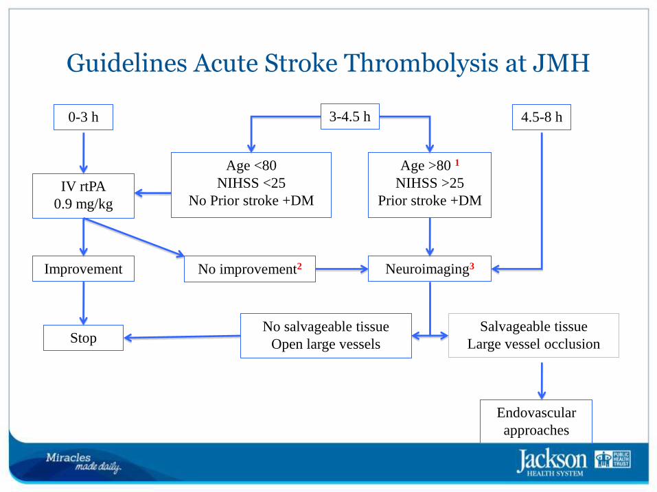
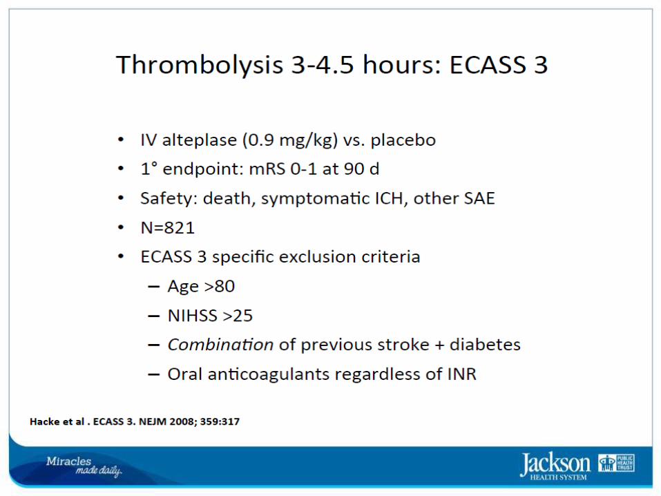
Guidelines Acute Stroke Thrombolysis at JMH
0-3 h 3-4.5 h 4.5-8 h
Age <80
NIHSS <25
No Prior stroke +DM
Age >80 1
NIHSS >25
Prior stroke +DM
Neuroimaging3
Salvageable tissue
Large vessel occlusion
No salvageable tissue
Open large vessels
Endovascular
approaches
IV rtPA
0.9 mg/kg
No improvement2 Improvement
Stop

Save the penumbra
Promote collateral flow
Neuroprotection strategies
Early Reperfusion
Intravenous thrombolysis
Endovascular therapies
Prevent complications
Complications ultimately causes
progression of infarct
Secondary prevention
Early Rehabilitation
Steps to Recovery Objectives of acute ischemic stroke therapy
BLOOD PRESSURE MANAGEMENT IN ACUTE
ISCHEMIC STROKE
(Based on: Guidelines for early management of adults with ischemic stroke. Stroke. 2013;44:870-947)
BLOOD PRESSURE MANAGEMENT IN ACUTE ISCHEMIC STROKE
• If patient is not eligible for treatment with intravenous TPA or other acute reperfusion intervention, then it is not necessary to reduce blood pressure so aggressively
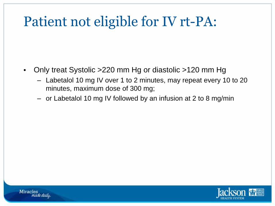
Patient not eligible for IV rt-PA:
• Only treat Systolic >220 mm Hg or diastolic >120 mm Hg
– Labetalol 10 mg IV over 1 to 2 minutes, may repeat every 10 to 20
minutes, maximum dose of 300 mg;
– or Labetalol 10 mg IV followed by an infusion at 2 to 8 mg/min
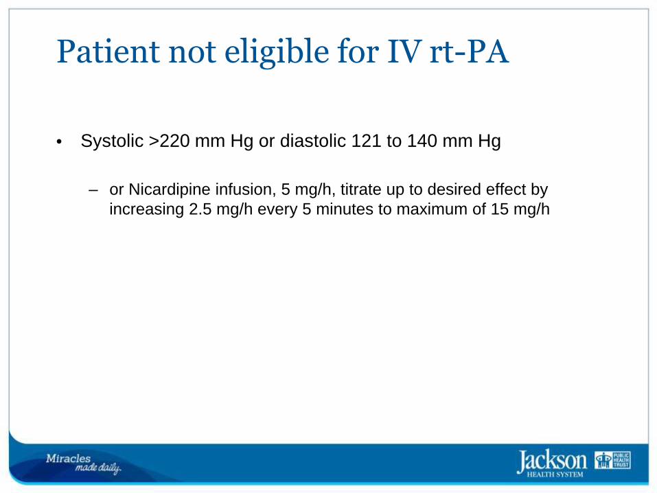
Patient not eligible for IV rt-PA
• Systolic >220 mm Hg or diastolic 121 to 140 mm Hg
– or Nicardipine infusion, 5 mg/h, titrate up to desired effect by
increasing 2.5 mg/h every 5 minutes to maximum of 15 mg/h
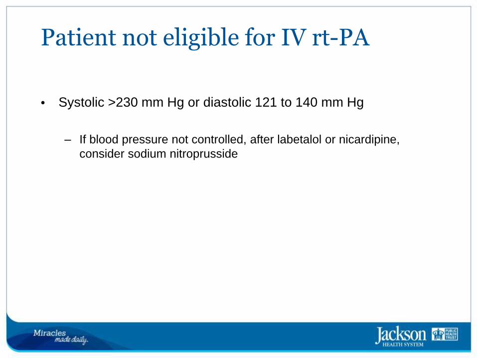
Patient not eligible for IV rt-PA
• Systolic >230 mm Hg or diastolic 121 to 140 mm Hg
– If blood pressure not controlled, after labetalol or nicardipine,
consider sodium nitroprusside
Save the penumbra
Promote collateral flow
Neuroprotection strategies
Early Reperfusion
Intravenous thrombolysis
Endovascular therapies
Prevent complications
Complications ultimately causes
progression of infarct
Secondary prevention
Early Rehabilitation
Steps to Recovery
Objectives of acute ischemic stroke therapy
Prevent Complications
• Aspiration Pneumonia
– Dysphagia screen prior to any PO intake (medications)
• Deep Vein Thrombosis
– No role for full anticoagulation
– Mechanical: SCDs
– Pharmacological
• Heparin subcutaneous, initiate 24 hours after intravenous thrombolytic
therapy
– Early Ambulation
ACUTE ISCHEMIC STROKE MANAGEMENT PROTOCOL
• Temperature management:
– Sources of fever should be treated and antipyretic medications
should be administered to lower temperature in febrile patients.
• Glucose management:
– Regular insulin sliding scale to treat glucose concentrations >121
mg/dL
103
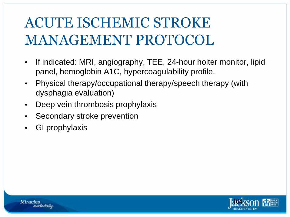
ACUTE ISCHEMIC STROKE MANAGEMENT PROTOCOL • If indicated: MRI, angiography, TEE, 24-hour holter monitor, lipid
panel, hemoglobin A1C, hypercoagulability profile.
• Physical therapy/occupational therapy/speech therapy (with dysphagia evaluation)
• Deep vein thrombosis prophylaxis
• Secondary stroke prevention
• GI prophylaxis
104
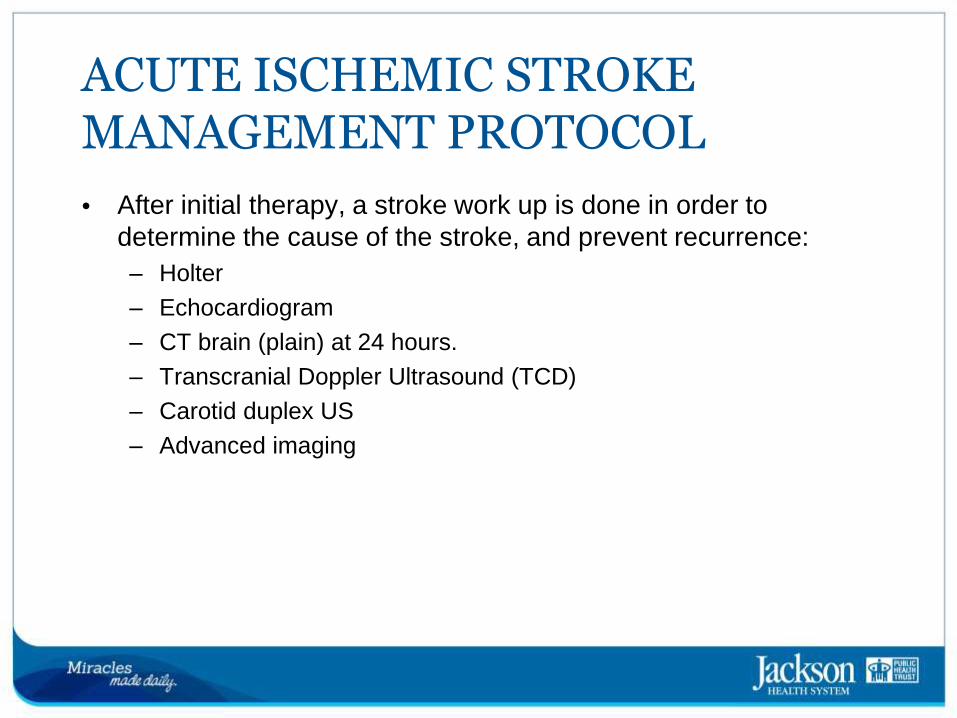
ACUTE ISCHEMIC STROKE MANAGEMENT PROTOCOL • After initial therapy, a stroke work up is done in order to
determine the cause of the stroke, and prevent recurrence:
– Holter
– Echocardiogram
– CT brain (plain) at 24 hours.
– Transcranial Doppler Ultrasound (TCD)
– Carotid duplex US
– Advanced imaging

Acute Stroke Therapy Summary
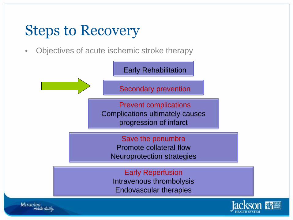
Steps to Recovery • Objectives of acute ischemic stroke therapy
Save the penumbra
Promote collateral flow
Neuroprotection strategies
Early Reperfusion
Intravenous thrombolysis
Endovascular therapies
Prevent complications
Complications ultimately causes
progression of infarct
Secondary prevention
Early Rehabilitation
Secondary prevention
• Blood pressure control
• Blood sugar control
• Antithrombotic therapy
– Starts by day two or earlier (t-PA)
• Lipid lowering treatment = Statin
– Ischemic Stroke
– Stroke + LDL>100mg/dl
– Statin prior to admission
• Contraindications (if any) must be documented
• Anticoagulant in presence of A. fib
• Contraindications (if any) must be documented
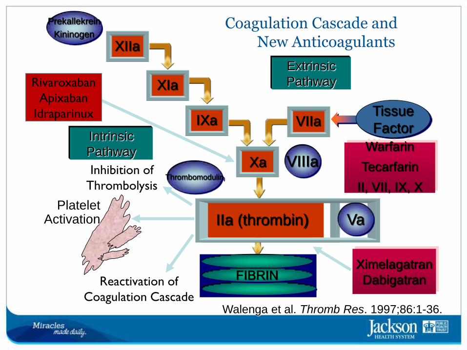
VII Intrinsic
Pathway
Extrinsic
Pathway
IX
II (prothrombin)
XI
XII XIIa
X
VIIa IXa
XIa
IIa (thrombin)
Xa
FIBRINOGEN FIBRIN
Tissue
Factor
VIIIa
Va Platelet
Activation
Inhibition of
Thrombolysis
Reactivation of
Coagulation Cascade Walenga et al. Thromb Res. 1997;86:1-36.
Warfarin
Tecarfarin
II, VII, IX, X
Ximelagatran
Dabigatran
Thrombomodulin
Prekallekrein
Kininogen
Rivaroxaban
Apixaban
Idraparinux
Coagulation Cascade and New Anticoagulants
Acute Cerebrovascular Syndromes
• New term introduced by AHA in 2013
– Acute focal neurological deficit due to spontaneous disruption of the
blood supply to the brain
• Encompasses all subtypes of stroke
– Acute ischemic stroke
– Transient Ischemic attack
– Acute intracerebral hemorrhages
– Acute Subarachnoid hemorrhages
• Emphasizes early recognition to facilitate timely intervention
• Treatment strategies are as varied as the different cause and depends on establishment of underlying cause
INTRACEREBRAL HEMORRHAGE
• Intracerebral hemorrhage may be:
– Primary
– Or secondary due to:
• Coagulopathy:
– Patient anticoagulated (heparin/coumadin)
– Patient treated with thrombolytics (IV TPA)
– Hematological disorder (decreased platelet count, etc)
INTRACEREBRAL HEMORRHAGE MANAGEMENT
• Patients neurological status may deteriorate:
– Mass effect
– Hydrocephalus
– Medical complications
• Infections
• Pulmonary embolism
INTRACEREBRAL HEMORRHAGE MANAGEMENT
• Add to initial labs:
– Toxicology screen in urine.
– Pregnancy test in women of childbearing age.
• Admit to NSICU or Stroke Unit.
INTRACEREBRAL HEMORRHAGE MANAGEMENT
• Elevate head of the bed to 30.
• Head should be midline, and head turning to either side should be avoided.
• Endotracheal tube tape should not compress jugular veins.

Management of HTN in primary ICH
• Clinically reexamine the patient every 15 minutes
• Intravenous medications to be considered for treatment of elevated blood pressure in patients with primary ICH :
Primary ICH Blood pressure treatment
• Treatment goal:
– If no concern for increased ICP the treatment goal is slightly lower
– Systolic blood pressure < 140mmHg
• Blood pressure monitoring every 5 minutes! (if outside parameters)
Primary ICH Blood pressure treatment
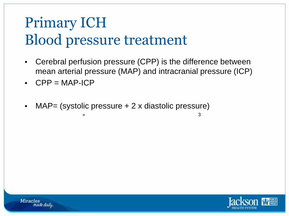
• Cerebral perfusion pressure (CPP) is the difference between mean arterial pressure (MAP) and intracranial pressure (ICP)
• CPP = MAP-ICP
• MAP= (systolic pressure + 2 x diastolic pressure) » 3
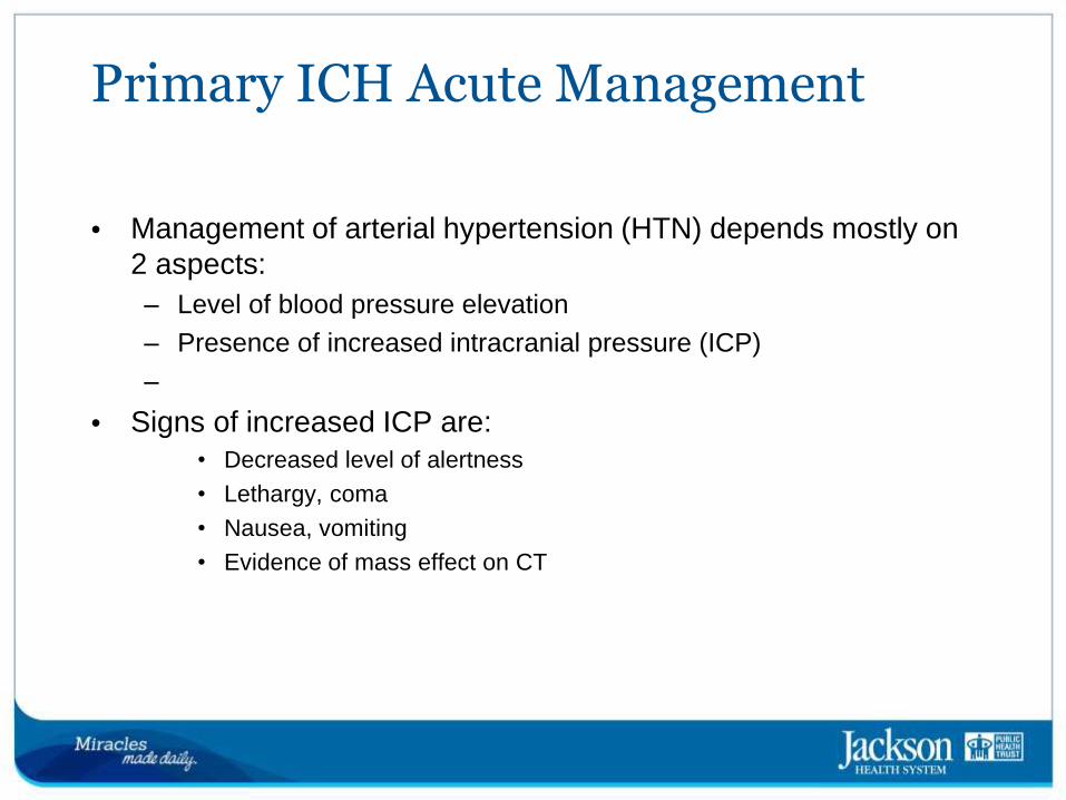
Primary ICH Acute Management
• Management of arterial hypertension (HTN) depends mostly on 2 aspects:
– Level of blood pressure elevation
– Presence of increased intracranial pressure (ICP)
–
• Signs of increased ICP are:
• Decreased level of alertness
• Lethargy, coma
• Nausea, vomiting
• Evidence of mass effect on CT
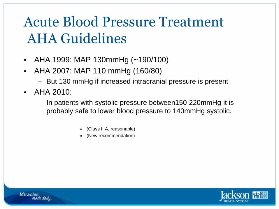
Acute Blood Pressure Treatment AHA Guidelines • AHA 1999: MAP 130mmHg (~190/100)
• AHA 2007: MAP 110 mmHg (160/80)
– But 130 mmHg if increased intracranial pressure is present
• AHA 2010:
– In patients with systolic pressure between150-220mmHg it is
probably safe to lower blood pressure to 140mmHg systolic.
» (Class II A, reasonable)
» (New recommendation)
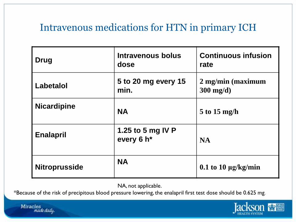
Intravenous medications for HTN in primary ICH
NA, not applicable.
*Because of the risk of precipitous blood pressure lowering, the enalapril first test dose should be 0.625 mg.
Drug Intravenous bolus
dose
Continuous infusion
rate
Labetalol 5 to 20 mg every 15
min.
2 mg/min (maximum
300 mg/d)
Nicardipine
NA 5 to 15 mg/h
Enalapril
1.25 to 5 mg IV P
every 6 h*
NA
Nitroprusside NA
0.1 to 10 μg/kg/min
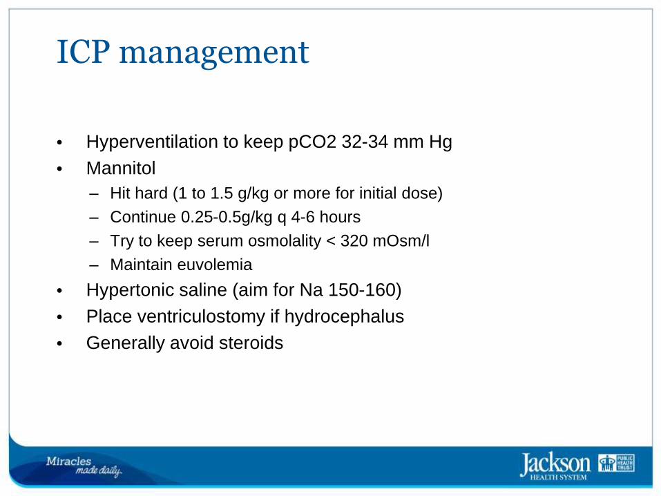
ICP management
• Hyperventilation to keep pCO2 32-34 mm Hg
• Mannitol
– Hit hard (1 to 1.5 g/kg or more for initial dose)
– Continue 0.25-0.5g/kg q 4-6 hours
– Try to keep serum osmolality < 320 mOsm/l
– Maintain euvolemia
• Hypertonic saline (aim for Na 150-160)
• Place ventriculostomy if hydrocephalus
• Generally avoid steroids
Seizures
• Incidence of seizures may be 10-20%
– Usually at the time of onset
• With continuous EEG monitoring 20-30%
• Continuous EEG monitoring for those with unexplained depressed mental status
• Clinical seizures should be treated with antiepileptic drugs
»
(Class I; A)
Seizures
• Prophylactic anticonvulsant medication should not be used.
(Class III, B)
(New recommendation)
• This may be harmful
– Data mostly from patients on phenytoin
INTRACEREBRAL HEMORRHAGE MANAGEMENT
• If patient presents seizures:
– IV diazepam (5-10 mg IV q 10-15 min. as indicated by neurologist)
– IV phosphenytoin (Initial dose: 15-20 mg PE/kg IV x 1; then 4-6 mg
PE/kg IV TID, as indicated by neurologist)
Prevent further Injury
• Fever:
– High incidence of 80-90%.
– May be infectious or due to presence of hematoma itself
– Is independently associated with a poor outcome.

Prevent further Injury
• Hyperglycemia:
– Associated with poor outcome
– Diabetics also have worsened outcome
– May exacerbate peri-hematoma edema
– Avoid glucose containing IV fluids
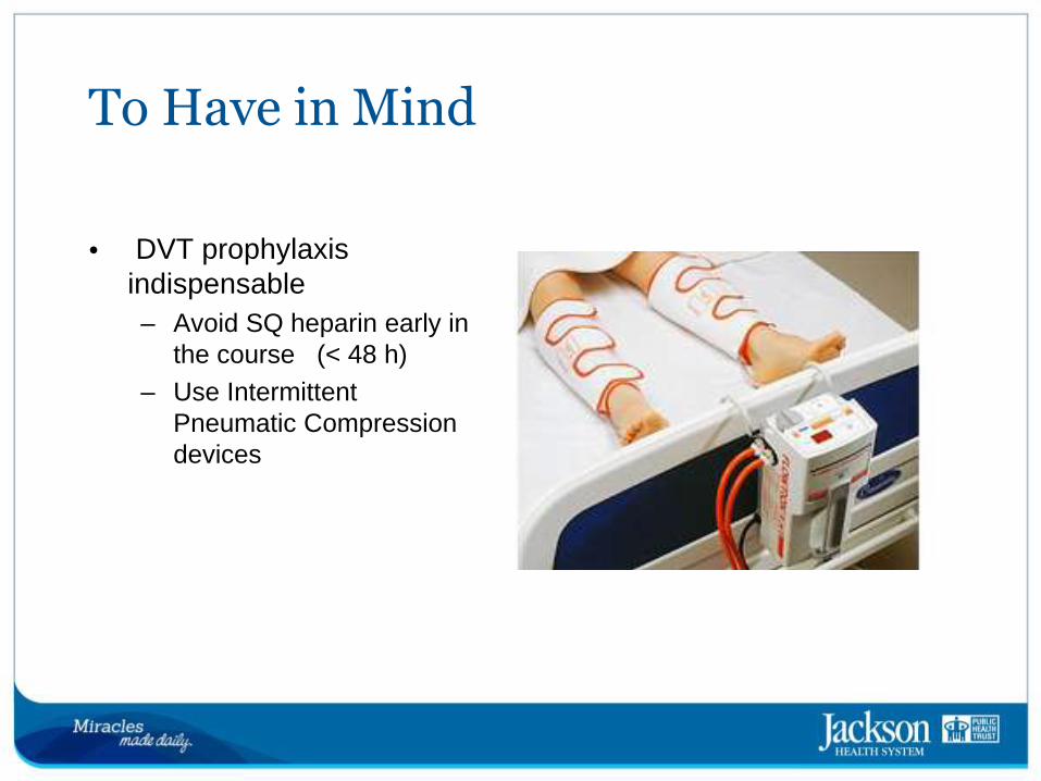
To Have in Mind
• DVT prophylaxis indispensable
– Avoid SQ heparin early in
the course (< 48 h)
– Use Intermittent
Pneumatic Compression
devices
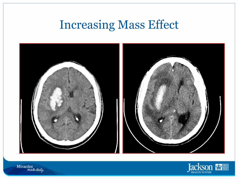
Increasing Mass Effect
Surgical Treatment
• For the majority of ICH no clear surgical indications
• A good number of trials have assessed surgical evacuation with no clear beneficial result
Surgical Treatment
• Few clear indications:
– Cerebellar hematoma > 3cm or 40ml
– Superficial expanding ICH in a deterioration young patient.
• No role in brainstem or deep thalamic ICH
• No role for ultra-early surgery
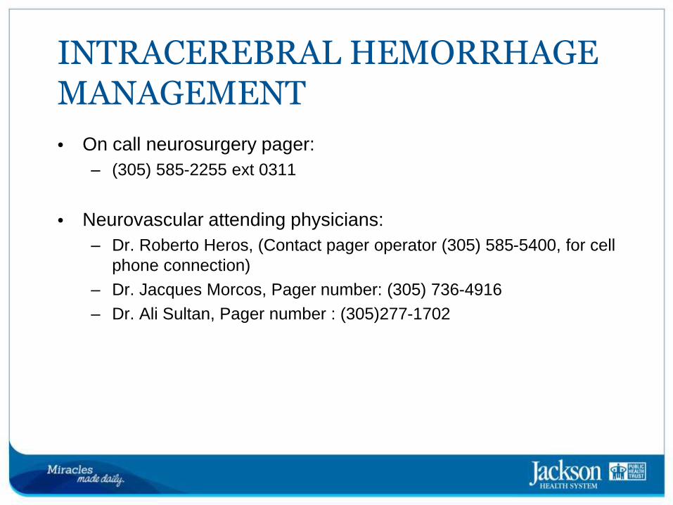
INTRACEREBRAL HEMORRHAGE MANAGEMENT
• On call neurosurgery pager:
– (305) 585-2255 ext 0311
• Neurovascular attending physicians:
– Dr. Roberto Heros, (Contact pager operator (305) 585-5400, for cell
phone connection)
– Dr. Jacques Morcos, Pager number: (305) 736-4916
– Dr. Ali Sultan, Pager number : (305)277-1702
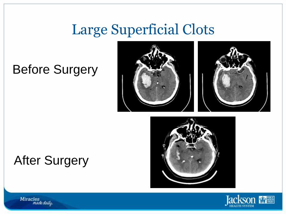
Large Superficial Clots
Before Surgery
After Surgery
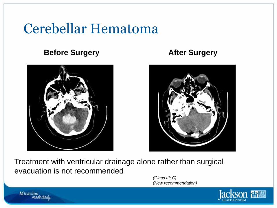
Cerebellar Hematoma
Before Surgery After Surgery
Treatment with ventricular drainage alone rather than surgical
evacuation is not recommended (Class III; C)
(New recommendation)
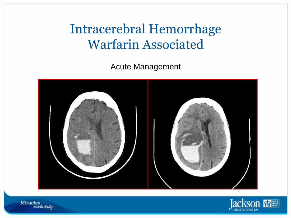
Intracerebral Hemorrhage Warfarin Associated
Acute Management

Warfarin associated ICH
• 10-20% of all Intracranial Cerebral Hemorrhage
• Carries worse prognosis
• Greater initial hemorrhage size
• Greater hematoma growth
• High INR also adversely affects mortality in ICH
– 62% vs. 17%
(CHANT Trial, Cucchiara 2008)
INTRACEREBRAL HEMORRHAGE SECONDARY TO COAGULOPATHY
• If patient is anti-coagulated with heparin:
– Treat with Protamine sulfate (1mg per 100 Units of heparin patient
is receiving) – Note: Protamine sulfate dose needs to be adjusted according to time
elapsed since the last heparin dose
INTRACEREBRAL HEMORRHAGE SECONDARY TO COAGULOPATHY
• If patient is anticoagulant with warfarin (Coumadin):
– Use Vitamin K (10 mg IV x 1 and subsequent doses based on INR)
– Consider activated factor VII, and Pro-thrombin Complex
Concentrates
– Also fresh frozen plasma (FFP) (15-20 mL/Kg)
Prothrombin Complex Concentrates
• Primarily to treat Factor IX deficiency
• Contain II, VII, IX, X
• Increasingly recommended for warfarin reversal
• Reversal within minutes
• Factor VII does not provide all the clotting factors
– Recommendations against routine use of Factor VII only for
reversal of oral anticoagulation associated hemorrhage [American
Society of Hematology and AHA guidelines 2010]
Warfarin Associated ICH
• PCCs have not shown improved outcome compared with FFP but may have fewer complications compared with FFP and are reasonable to consider as an alternative to FFP
(Class IIa; B)
• JMH protocol
• PCC and FFP
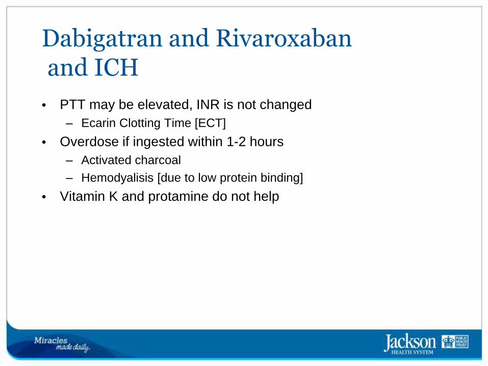
Dabigatran and Rivaroxaban and ICH
• PTT may be elevated, INR is not changed
– Ecarin Clotting Time [ECT]
• Overdose if ingested within 1-2 hours
– Activated charcoal
– Hemodyalisis [due to low protein binding]
• Vitamin K and protamine do not help
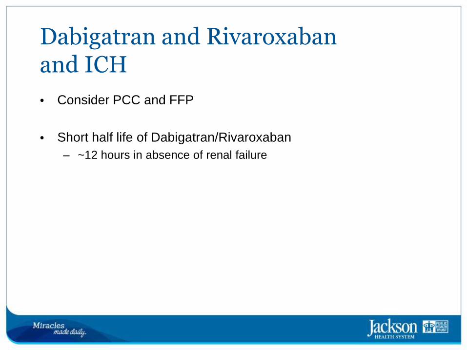
Dabigatran and Rivaroxaban and ICH • Consider PCC and FFP
• Short half life of Dabigatran/Rivaroxaban
– ~12 hours in absence of renal failure
INTRACEREBRAL HEMORRHAGE SECONDARY TO COAGULOPATHY
• If patient was treated with IV thrombolysis (IV TPA) and had a hemorrhagic complication:
– Infuse platelets (6 to 8 Units) and cryoprecipitates that contain
factor VIII
• If there is a hematological disorder
– Treat accordingly depending on the cause (platelet transfusion,
steroids, etc)
Case examples
Case 1
• 72 y/o right-handed woman with h/o HTN
• Presents to ER with left-sided weakness for 2 days.
• Has not been taking her BP medications for 6 months
Case 1
• Vital signs:
– BP: 200/110
– HR: 88/min
– RR: 16/min
• Examination
– Awake and oriented to
person, place and time
– Mild left facial palsy
– Left hemiparesis (arm/leg
drift)
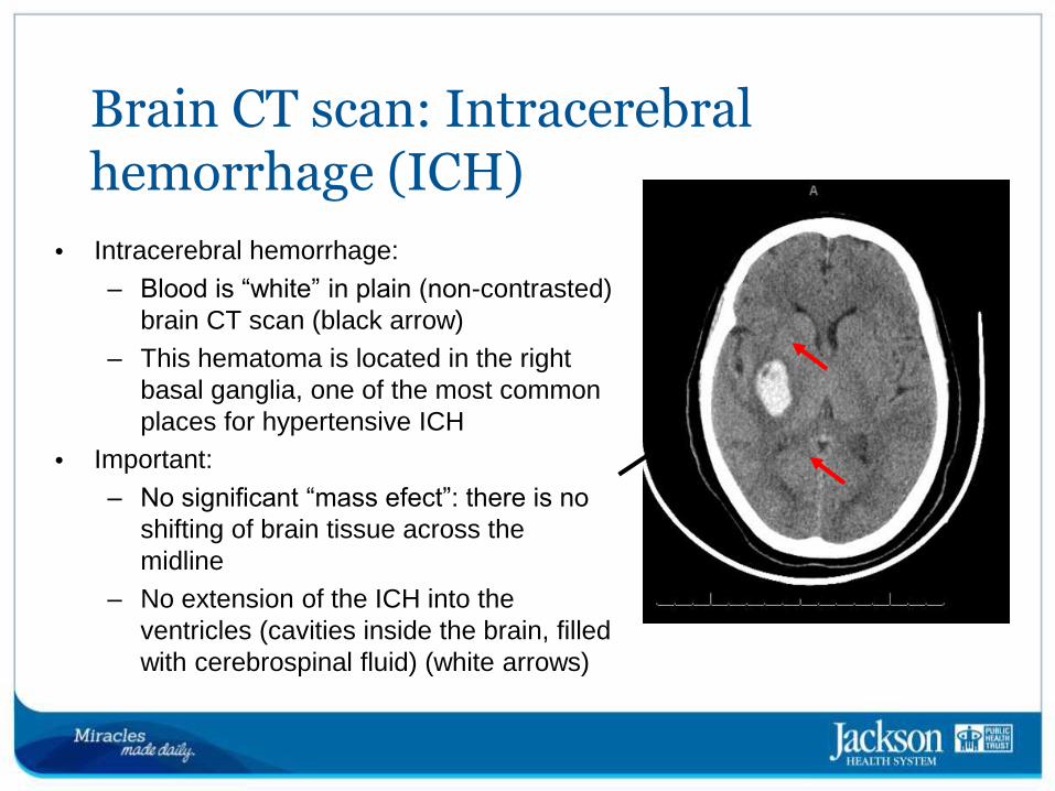
Brain CT scan: Intracerebral hemorrhage (ICH)
• Intracerebral hemorrhage:
– Blood is “white” in plain (non-contrasted)
brain CT scan (black arrow)
– This hematoma is located in the right
basal ganglia, one of the most common
places for hypertensive ICH
• Important:
– No significant “mass efect”: there is no
shifting of brain tissue across the
midline
– No extension of the ICH into the
ventricles (cavities inside the brain, filled
with cerebrospinal fluid) (white arrows)
Case 1
• Patient was treated with Cardene drip, starting at 5 mg/hour, titrated to keep SBP <180 and DBP <110
• After 48 hours, treatment was switched to oral medications (Lisinopril/hydrochlorothiazide)
• Her blood pressure was well controlled and after a week, patient was DC to home
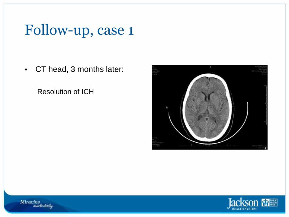
Follow-up, case 1
• CT head, 3 months later:
Resolution of ICH
Case 2 Different outcome
Case 2
• 40-year-old, right-handed man with history of severe hypertension
• Non compliant with medications
• 2 previous episodes of intracerebral hemorrhages in the last 3 years
• Seizure disorder
Case 2
• Found by a neighbor at 11 AM, unresponsive on the ground.
• Last seen well: previous night at 10 PM.
• Brought to ER by EMS. Intubated on the field
• No convulsive movements seen
Case 2
• Vital signs:
– BP: 240/140
– HR: 110/min
– RR: 24/min
– O2 Sat: 95%
• On exam:
– Comatose
– Pupils are equal, round
and sluggishly reactive to
light
– Withdraws to painful
stimuli on right arm/leg
– No movements seen on
left hemibody.
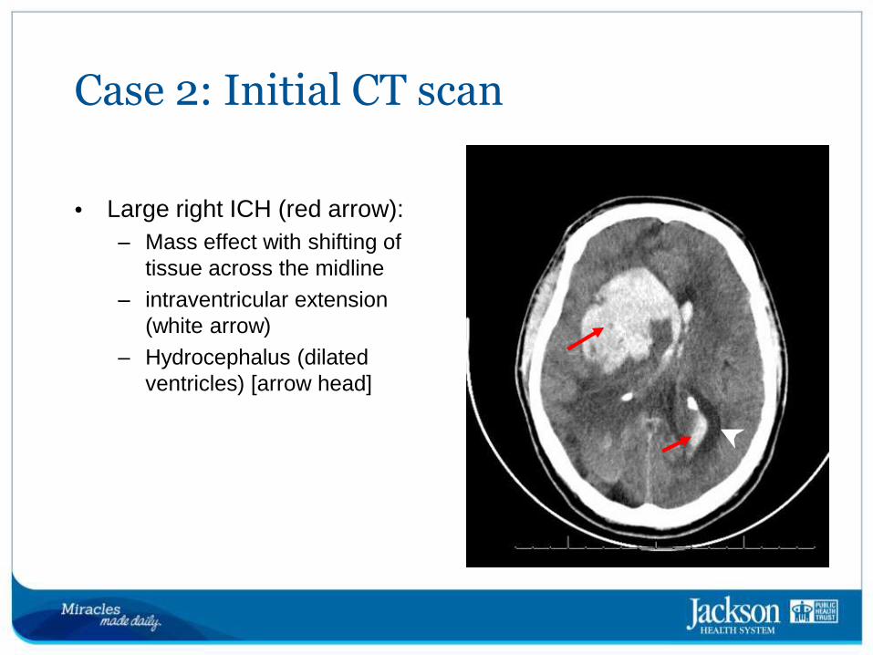
Case 2: Initial CT scan
• Large right ICH (red arrow):
– Mass effect with shifting of
tissue across the midline
– intraventricular extension
(white arrow)
– Hydrocephalus (dilated
ventricles) [arrow head]
Case 2
• Treatment:
– HTN: Cardene drip
– Hydrocephalus: External ventricular drainage (Ventriculostomy)
Case 2
• After prolonged hospitalization, patient required tracheostomy (for mechanical ventilation) and percutaneous gastro-jejunostomy placement (for feeding)
• He remained with severe disability: Left hemiplegia and profound cognitive impairment
• Discharged to a nursing home
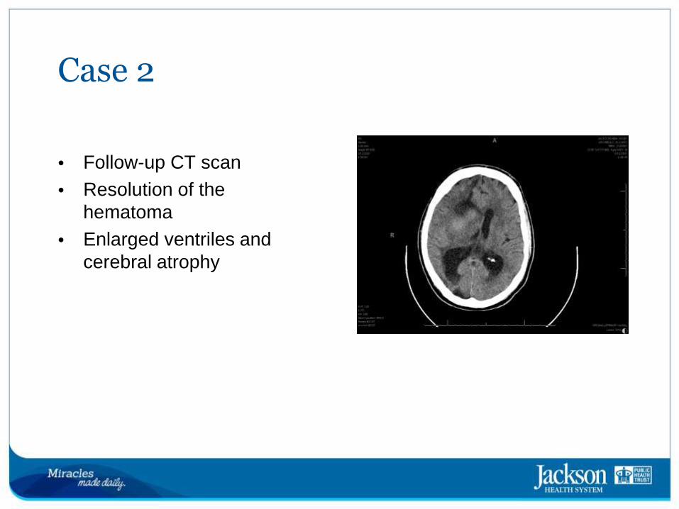
Case 2
• Follow-up CT scan
• Resolution of the hematoma
• Enlarged ventriles and cerebral atrophy

Acute Cerebrovascular Syndromes
• New term introduced by AHA in 2013
– Acute focal neurological deficit due to spontaneous disruption of the
blood supply to the brain
• Encompasses all subtypes of stroke
– Acute ischemic stroke
– Transient Ischemic attack
– Acute intracerebral hemorrhages
– Acute Subarachnoid hemorrhages
• Emphasizes early recognition to facilitate timely intervention.
• Treatment strategies are as varied as the different cause and depends on establishment of underlying cause.
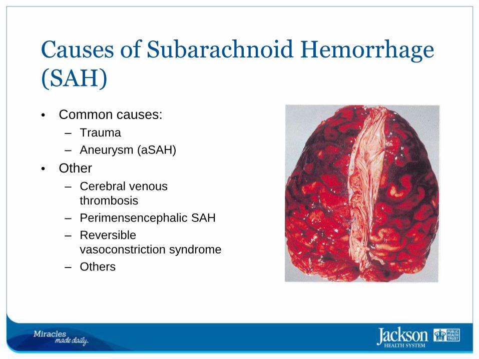
Causes of Subarachnoid Hemorrhage (SAH)
• Common causes:
– Trauma
– Aneurysm (aSAH)
• Other
– Cerebral venous
thrombosis
– Perimensencephalic SAH
– Reversible
vasoconstriction syndrome
– Others
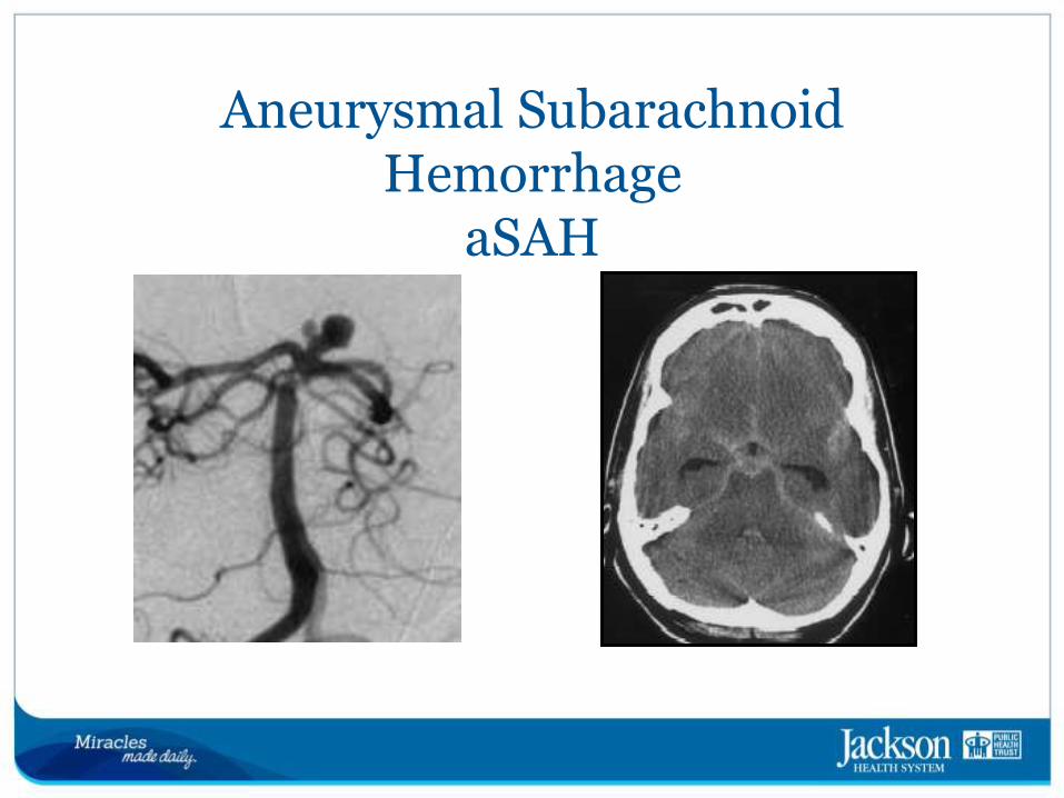
Aneurysmal Subarachnoid Hemorrhage
aSAH
Epidemiology aSAH
• 1/3 of all hemorrhagic strokes
– 12% initially misdiagnosed
• 30,000 aneurysmal SAH each year in US
• Considerable worldwide variation: incidence 2/100,000 China vs. 22.5/100,000 Finland, US 9-14.5/100,000
• Typical age onset in 40-60, mean >50 years
• Women at greater risk: incidence 1.24 times vs. men
Risk Factors for aneurysm formation and rupture
• Hypertension
• Tobacco
• Excessive alcohol
• Cocaine, other
sympathomimetics
• Female sex
• Presence of unruptured aneurysm
• First degree relative with aSAH
• Genetic syndromes: PCKD, ED IV
Modifiable Non-modifiable
Prevention aSAH
• Hypertension should be treated; treatment will prevent ischemic stroke, intracerebral hemorrhage, cardiac and renal disease (I-A), and may reduce risk of aSAH (I-B)
• Tobacco use and alcohol misuse should be avoided to reduce risk of aSAH (I-B)
• Diet rich in vegetables may lower risk of aSAH (IIb-B)
• Reasonable to screen in those with familial aSAH: at least 1 first
degree relative (IIb-B)
Symptoms of SAH
• Increased intracranial pressure
• Severe “worse” headache
– Sentinel HA in 10-43%
• Photophobia
• Nuchal rigidity, back pain
• Seizures
• Vomiting
• Altered mental status
• Sudden loss of consciousness
• Double vision
Meningeal irritation
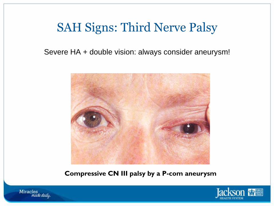
SAH Signs: Third Nerve Palsy
Severe HA + double vision: always consider aneurysm!
Compressive CN III palsy by a P-com aneurysm
Outcomes aSAH
• Mortality 32% US, 44% Europe, 27% Japan
• Does not account for pre-hospital death: 12-15%
• 50% fatality at 30 days
• 20% major disability/dependent
• Another 20% “independent” with significant cognitive impairment 50%
• Lifetime cost $228,000
Outcome after aSAH
• Severity of hemorrhage is the most important predictor of outcome
• Severity graded by clinical status and amount of blood on CT
• Clinical scales: Hunt and Hess, World Federation of Neurological Surgeons
– Initial clinical severity should be determined by validated scales as
an outcome indicator (I-B)
• Radiological scales: Fisher
SAH Assessment: Clinical
• Hunt & Hess scale classifies severity SAH
Grade 1 - Asymptomatic, mild headache, slight nuchal rigidity
Grade 2 - Moderate to severe headache, nuchal rigidity, no
neurological deficit other than cranial nerve palsy
Grade 3 - Drowsiness / confusion, mild focal neurological deficit
Grade 4 - Stupor, moderate-severe hemiparesis
Grade 5 - Coma, decerebrate posturing
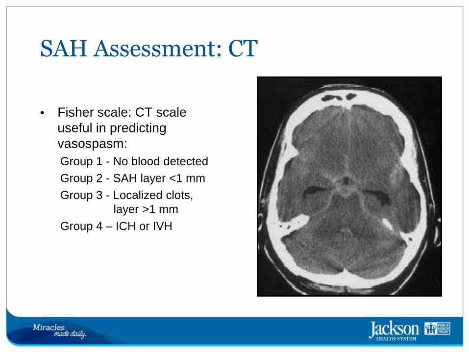
SAH Assessment: CT
• Fisher scale: CT scale useful in predicting vasospasm:
Group 1 - No blood detected
Group 2 - SAH layer <1 mm
Group 3 - Localized clots,
layer >1 mm
Group 4 – ICH or IVH
Clinical Case
• 18 y/o previously healthy man
• Soon after consuming cocaine in a party, he developed sudden-onset, severe headache and then lost consciousness
• Brought to the hospital by EMS
Clinical Case
• Vital signs:
– BP: 190/95
– HR: 65/min
– RR: 12/min
– T: 98◦ F
– O2 Sat: 98%
• On exam:
– Comatose
– Right pupil was dilated and
sluggishly reactive to light
– Stiff neck
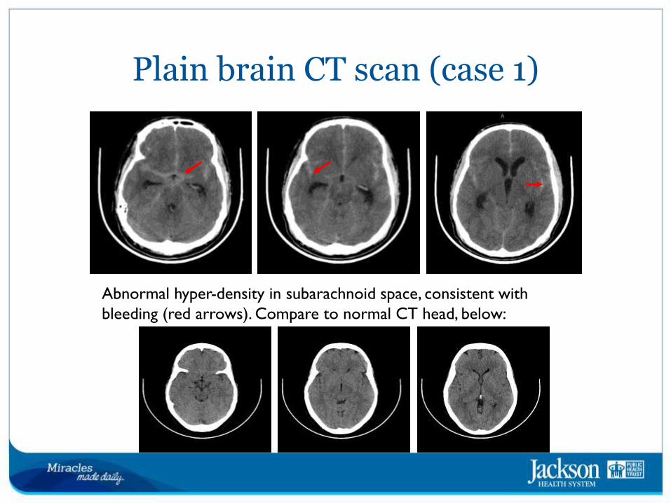
Plain brain CT scan (case 1)
Abnormal hyper-density in subarachnoid space, consistent with
bleeding (red arrows). Compare to normal CT head, below:
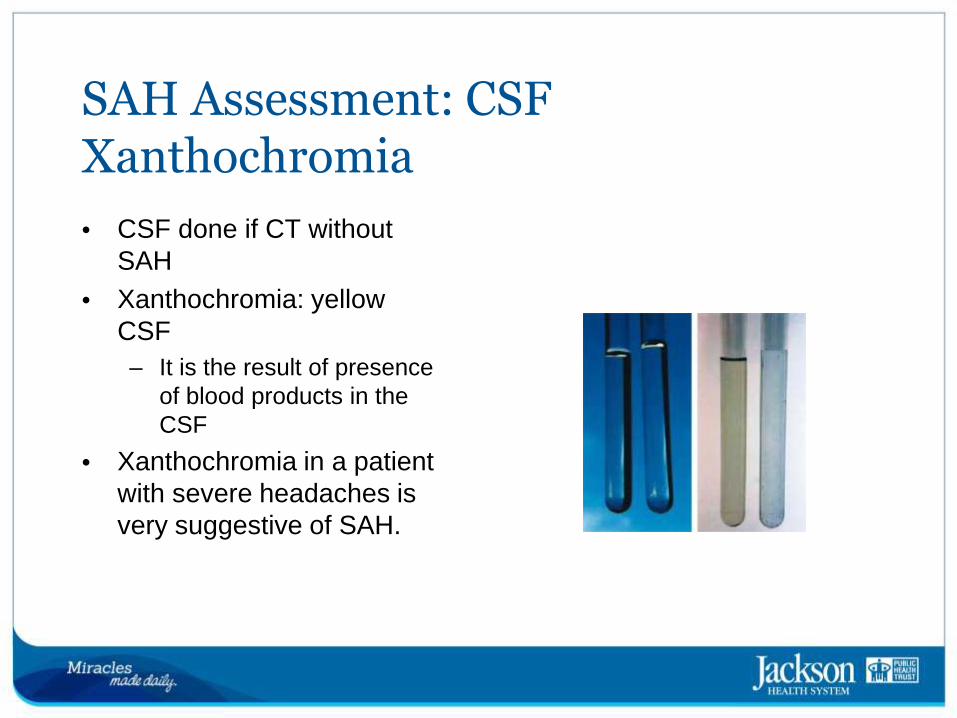
SAH Assessment: CSF Xanthochromia
• CSF done if CT without SAH
• Xanthochromia: yellow CSF
– It is the result of presence
of blood products in the
CSF
• Xanthochromia in a patient with severe headaches is very suggestive of SAH.
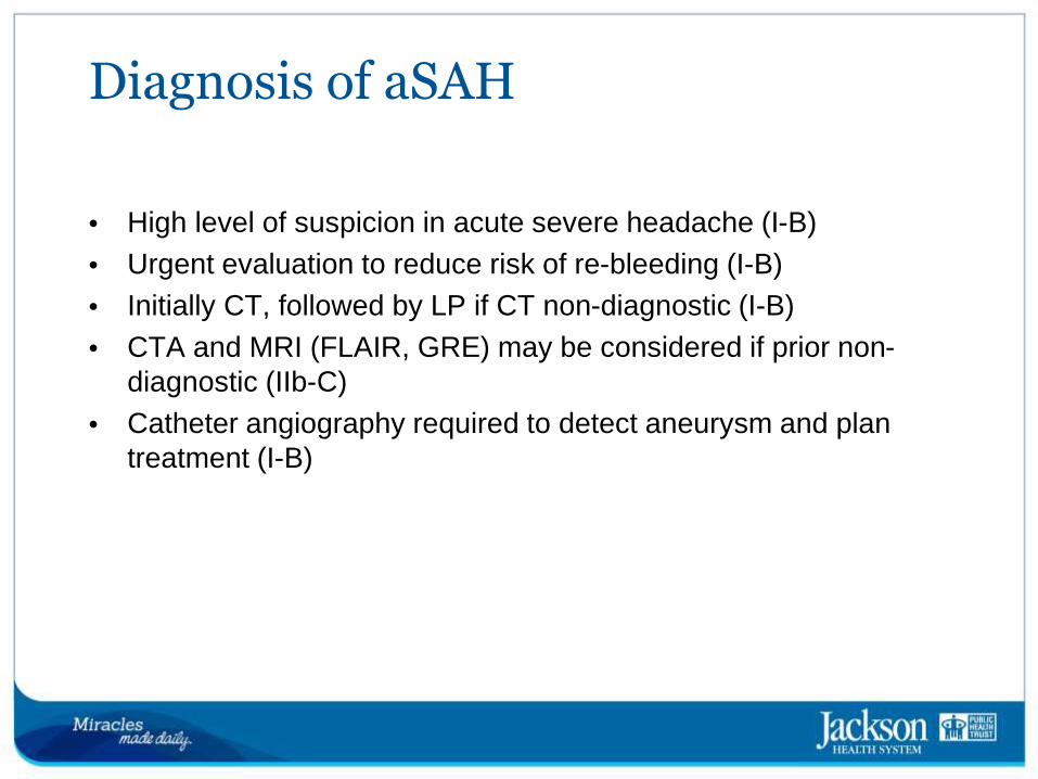
Diagnosis of aSAH
• High level of suspicion in acute severe headache (I-B)
• Urgent evaluation to reduce risk of re-bleeding (I-B)
• Initially CT, followed by LP if CT non-diagnostic (I-B)
• CTA and MRI (FLAIR, GRE) may be considered if prior non-diagnostic (IIb-C)
• Catheter angiography required to detect aneurysm and plan treatment (I-B)
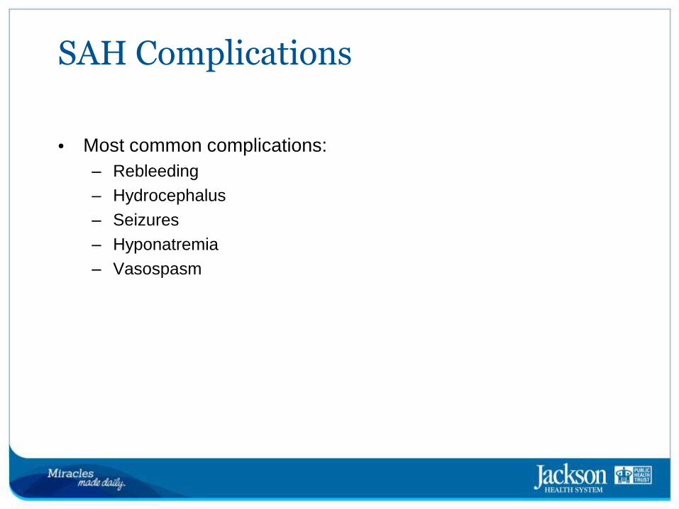
SAH Complications
• Most common complications:
– Rebleeding
– Hydrocephalus
– Seizures
– Hyponatremia
– Vasospasm
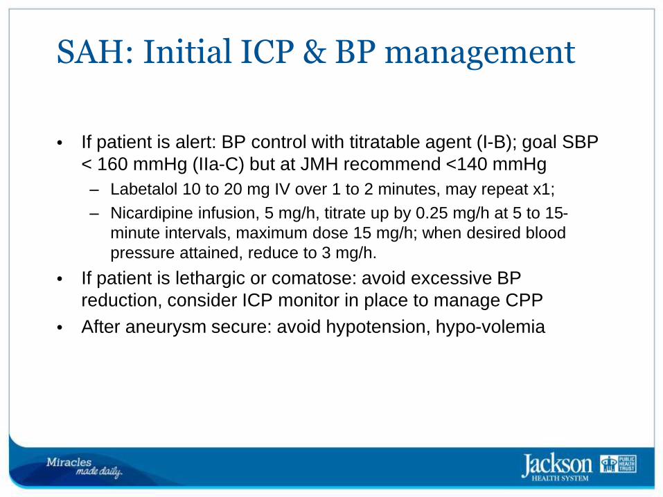
SAH: Initial ICP & BP management
• If patient is alert: BP control with titratable agent (I-B); goal SBP < 160 mmHg (IIa-C) but at JMH recommend <140 mmHg
– Labetalol 10 to 20 mg IV over 1 to 2 minutes, may repeat x1;
– Nicardipine infusion, 5 mg/h, titrate up by 0.25 mg/h at 5 to 15-
minute intervals, maximum dose 15 mg/h; when desired blood
pressure attained, reduce to 3 mg/h.
• If patient is lethargic or comatose: avoid excessive BP reduction, consider ICP monitor in place to manage CPP
• After aneurysm secure: avoid hypotension, hypo-volemia
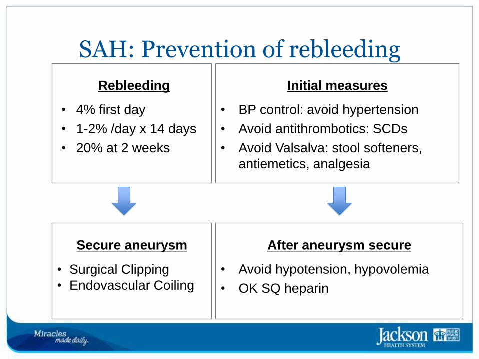
SAH: Prevention of rebleeding
Rebleeding
• 4% first day
• 1-2% /day x 14 days
• 20% at 2 weeks
Secure aneurysm
• Surgical Clipping
• Endovascular Coiling
Initial measures
• BP control: avoid hypertension
• Avoid antithrombotics: SCDs
• Avoid Valsalva: stool softeners,
antiemetics, analgesia
After aneurysm secure
• Avoid hypotension, hypovolemia
• OK SQ heparin
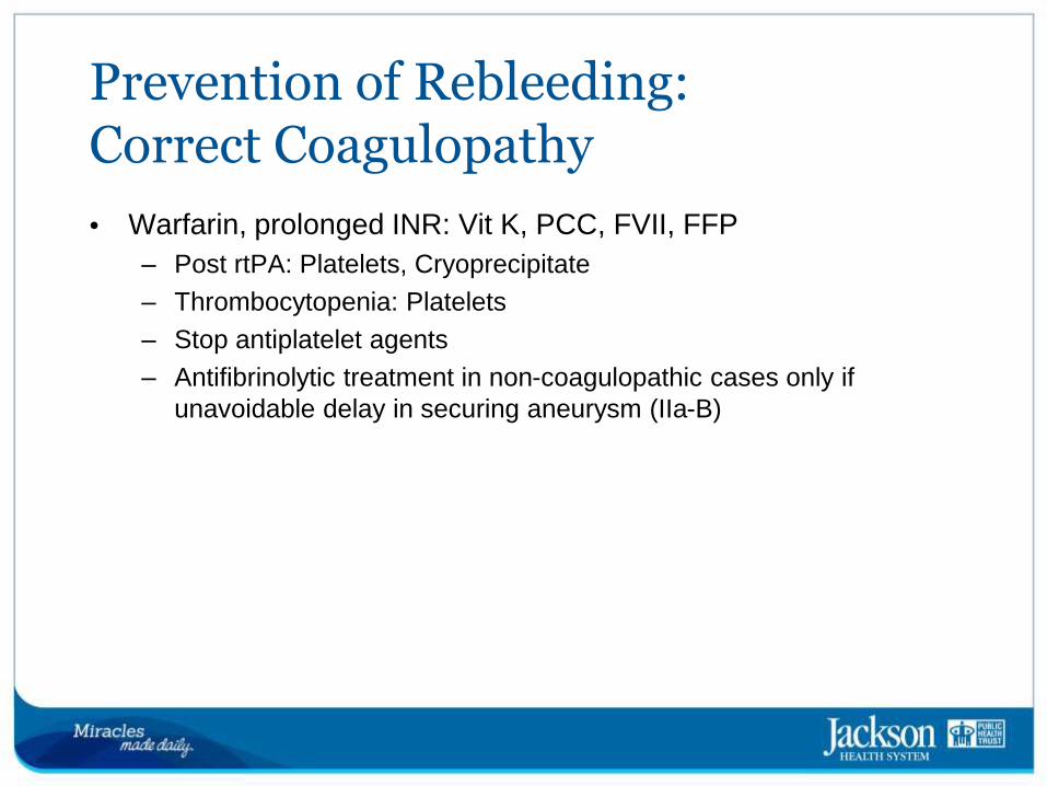
Prevention of Rebleeding: Correct Coagulopathy
• Warfarin, prolonged INR: Vit K, PCC, FVII, FFP
– Post rtPA: Platelets, Cryoprecipitate
– Thrombocytopenia: Platelets
– Stop antiplatelet agents
– Antifibrinolytic treatment in non-coagulopathic cases only if
unavoidable delay in securing aneurysm (IIa-B)
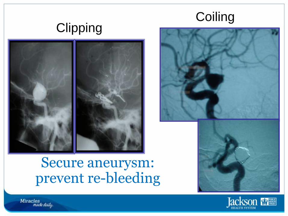
Secure aneurysm: prevent re-bleeding
Clipping Coiling
Clipping and Coiling
• Should be performed as early as possible (I-B).
• Decision by expert team (I-C)
• Clipping: less likely to rebleed, complete occlusion,
but craniotomy, greater disability.
– MCA aneurysms, large ICH (IIb-C)
• Coiling: less disability in 1 study, more likely to have
incomplete obliteration with risk of rebleeding
– Elderly, poor clinical grade, BA aneurysms (Iib-C)
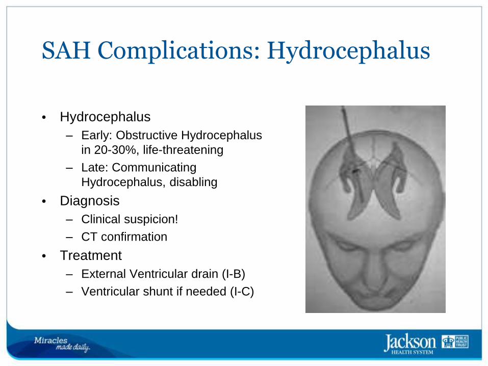
SAH Complications: Hydrocephalus
• Hydrocephalus
– Early: Obstructive Hydrocephalus
in 20-30%, life-threatening
– Late: Communicating
Hydrocephalus, disabling
• Diagnosis
– Clinical suspicion!
– CT confirmation
• Treatment
– External Ventricular drain (I-B)
– Ventricular shunt if needed (I-C)
SAH Complications: Hyponatremia
• Salt wasting syndrome different from syndrome of inappropriate antidiuretic hormone (SIADH): excessive sodium loss vs. water retention
• Water restriction is not recommended
• Replace sodium loss with hypertonic saline or fludrocortisone (IIa-B)
SAH Complications: Seizures
• Seizures increase BP, may lead to rebleeding if aneurysm unsecured
• Status epilepticus very dangerous
• Short-term seizure prophylaxis often indicated (IIb-B)
• Antiepileptic drug may not be necessary in good clinical grade patients (at intensivist’s discretion)
• EEG monitoring considered in poor grade
• If seizures occur: benzodiazepines, phosphenytoin
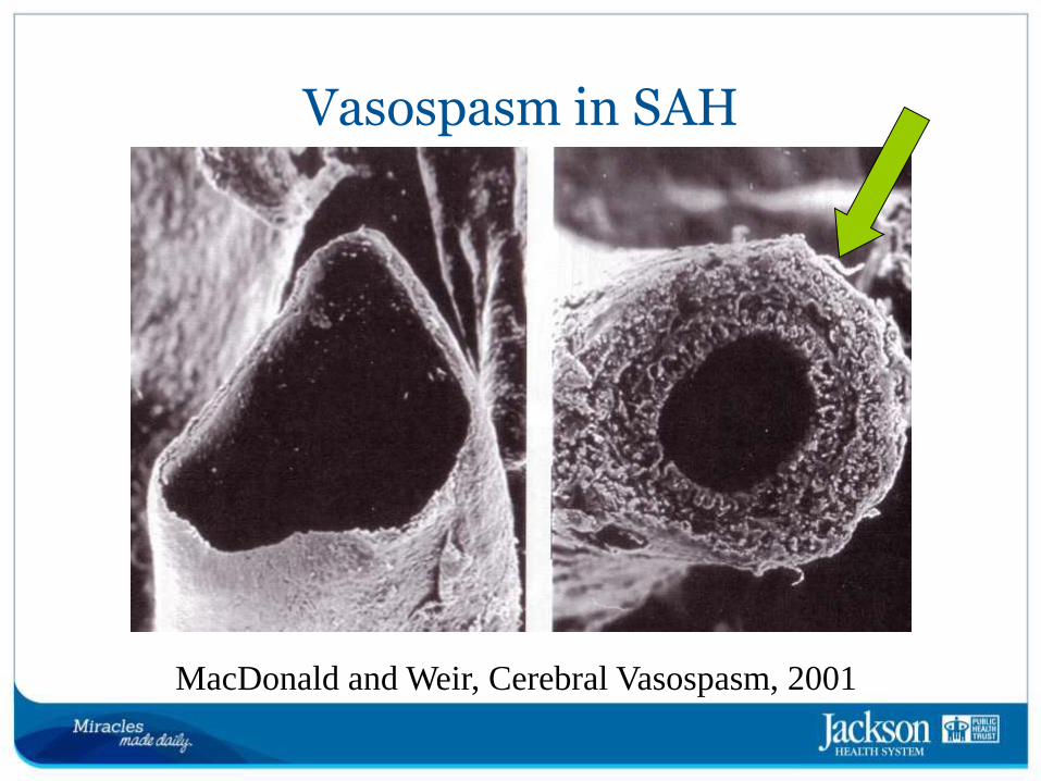
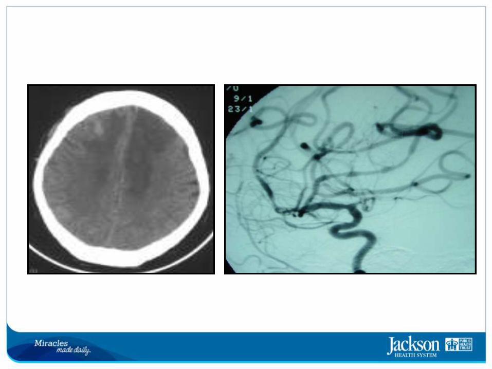
Vasospasm in SAH
MacDonald and Weir, Cerebral Vasospasm, 2001
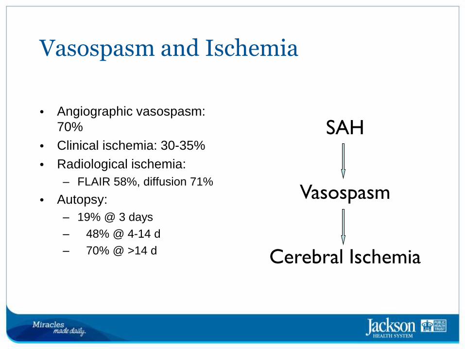
Vasospasm and Ischemia
• Angiographic vasospasm: 70%
• Clinical ischemia: 30-35%
• Radiological ischemia:
– FLAIR 58%, diffusion 71%
• Autopsy:
– 19% @ 3 days
– 48% @ 4-14 d
– 70% @ >14 d
SAH
Vasospasm
Cerebral Ischemia
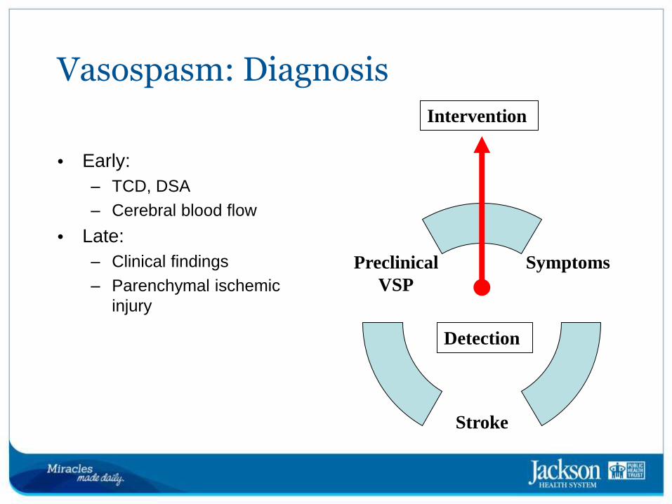
Vasospasm: Diagnosis
• Early:
– TCD, DSA
– Cerebral blood flow
• Late:
– Clinical findings
– Parenchymal ischemic
injury
Symptoms
Stroke
Preclinical
VSP
Detection
Intervention
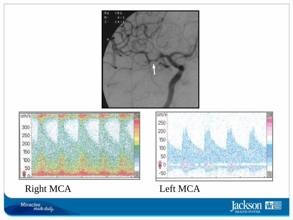
Right MCA Left MCA

Vasospasm Treatment
• Monitor daily clinical exam
• Monitor daily TCD
• Nimodipine
• Avoid hypotension
• Avoid hypovolemia
• Triple H therapy:
– Hypertension
– Hypervolemia
– Hemodilution
• Albumin
• Vasopressors
• Endovascular treatment
– Vasodilators
– Angioplasty
Prevention Treatment
Vasospasm and delayed cerebral ischemia
• Nimodipine to improve outcomes (I-A)
• Eu-volemia to prevent delayed cerebral ischemia (I-B)
• TCD to monitor for vasospasm (IIa-B)
• Induction of hypertension if DCI ensues (I-B)
• Angioplasty or IA vasodilators if no response (IIa-B)
• However, prophylactic HHH or angioplasty not useful (III-B)
Conclusions
• aSAH is an emergency with high fatality and poor outcomes.
• Diagnosis requires high level of suspicion.
• Rapid diagnosis leads to rapid treatment, which prevents re-bleeding.
• Securing the aneurysm should be done early.
• Awareness of pathophysiology leads to recognition and treatment of complications.
• Outcome depends on patient, aneurysm and institutional factors.
• Effective treatment is done by an expert multidisciplinary team.
• There are modifiable risk factors that prevent aneurysmal formation and rupture.
References
• Guidelines for early management of adults with ischemic stroke. Stroke.
2013;44:870-947 Walenga et al. Thromb Res. 1997;86:1-36
• Cucchiara, B.; Messe, S.; at al. Hematoma Growth in Oral Anticoagulant
Related Intracerebral Hemorrhage. American Heart Association. Stroke, 2008;
39:2993-2996; doi: 10.1161/STROKEAHA.108.520668
• American Heart Association. The Cardiac Arrhythmia Suppression Trial. 1995;
91: 245-247 doi: 10.1161/01.CIR.91.1.245
• Fisher, M.; Hachinski, V. European Cooperative Acute Stroke Study III.
American Heart Association. Stroke. 2009; 40: 2262-2263; doi:
10.1161/STROKEAHA.108.544163
• The National Institute of Neurological Disorders and Stroke rt-PA Stroke Study
Group. Tissue Plasminogen Activator for Acute Ischemic Stroke. N Engl J Med
1995; 333:1581-1588. DOI: 10.1056/NEJM199512143332401
• Caplan, L. Caplan’s Stroke. A Clinical Approach. Saunders. 2009
• MacDonald, R.; Weir, B. Cerebral Vasospasm. Thieme. 2001
References
• American Heart & Stroke Foundation web site: http://www.strokeassociation.org
• Caplan’s Stroke. A Clinical Approach by Louis R. Caplan
• Neuroanatomy through clinical cases Hal Blumenfeld
• Localization in clinical neurology by Brazis, Masdeu & Biller
JMH Stroke Education 2013
Gordon-Perue, Gillian; MD
Aranguibel, Ninoska; ARNP