neuro quiz 29 - snacc.org
TRANSCRIPT
Neuro Quiz 29 –
Transcranial
Doppler Monitoring
This quiz is being published
on behalf of the Education
Committee of the SNACC
Quiz Team Shobana Rajan, M.D
Suneeta Gollapudy, M.D
Angele Marie Theard, M.D
Verghese Cherian, MD, FFARCSI
Penn State Hershey Medical Center,
Hershey
START

1.Which of the following statements about
Transcranial Doppler (TCD) is TRUE?
A. Measures the flow through the
cerebral capillaries
B. Measures the cerebral blood flow
C. Provides a direct visualization of the
large cerebral arteries
D. Measures the mean flow velocity
through the cerebral vessels

1.A. Measures the flow through
the cerebral capillaries
TCD measures flow velocity from large
basal arteries of the brain, i.e. branches of
the Circle of Willis
The ultrasound examination using the TCD
is known as ‘INSONATION’
Return to Question Next Question

1.B. Measures the cerebral
blood flow
TCD does not measure flow directly and therefore
cannot provide the absolute Cerebral Blood Flow (CBF)
CBF = 𝐶𝑒𝑟𝑒𝑏𝑟𝑎𝑙 𝑃𝑒𝑟𝑓𝑢𝑠𝑖𝑜𝑛 𝑃𝑟𝑒𝑠𝑠𝑢𝑟𝑒 (𝐶𝑃𝑃)
𝐶𝑒𝑟𝑒𝑏𝑟𝑜−𝑉𝑎𝑠𝑐𝑢𝑙𝑎𝑟 𝑅𝑒𝑠𝑖𝑠𝑡𝑎𝑛𝑐𝑒 (𝐶𝑉𝑅) [Ohm’s: Flow=
𝑃𝑟𝑒𝑠𝑠𝑢𝑟𝑒
𝑅𝑒𝑠𝑖𝑠𝑡𝑎𝑛𝑐𝑒]
Focal and generalized vessel tone determines CVR
Return to Question Next Question

1.C. Provides a direct visualization
of the large cerebral arteries
TCD does not provide a direct visualization of
the insonated artery like a carotid duplex scan
A 2Mz ultrasonic beam reflects off the
erythrocytes within these vessels and is
analyzed
Using the Doppler principle, the velocity and
direction of flow is calculated
Sorry! Try Again!
Return to Question Next Question

1.D. Measures the mean flow velocity
through the cerebral vessels
Using the Doppler principle, the TCD measures the velocity and direction of flow of blood
The ‘Pulsatility Index’ (PI)= Peak systolic velocity – End diastolic velocity
Mean velocity
-Gosling equation
-PI denotes arterial distensibility
The ‘Windkessel’ notch denotes
normal arterial distensibility
Return to Question Next Question

2.Which of the following statements about
Transcranial Doppler (TCD) is FALSE?
A. The TCD probe can be applied to different
sites of the cranium
B. The forehead, just above the root of the
nose is NOT suitable for probe placement
C. The internal carotid artery can be
insonated through the orbit of the eye
D. A burr hole is usually made to get a reliable
TCD waveform

2.A. The TCD probe can be applied
to different sites of the cranium
Natural openings in the cranium and thin bony areas allow passage
of ultrasound waves and these ‘acoustic windows’ are used for TCD
probe placement. Commonly used sites are:
• Temporal
• Orbital
• Foramen magnum
• Submandibular
Return to Question Next Question

2. B. The forehead, just above the
root of the nose is NOT suitable for
probe placement
Areas with thin bone or natural openings form the
‘acoustic windows’
The thick frontal bone would not be a suitable site
and also the presence of air sinus would impede
passage of ultrasound waves
Return to Question Next Question

2.C. The internal carotid artery can
be insonated through the orbit of
the eye
According to the probe position, different arteries
can be insonated
• Trans-temporal – Mid, Ant & Post Cerebral artery,
terminal portion of Internal Carotid (IC)
• Trans-orbital – Ophthalmic & IC(siphon) artery
• Trans-foraminal – vertebral and basilar artery
• Submandibular – extracranial IC artery
Return to Question Next Question

2.D. A burr hole is usually made to get a
reliable TCD waveform
Although, a TCD probe can be applied over
an existing burr hole, it is not needed
The TCD probe can be placed over different
‘acoustic windows’
Return to Question Next Question

3. Well-accepted indications for
TCD include all, EXCEPT
A. Intraoperative monitoring during Carotid
Endarterectomy
B. Intracranial vasospasm following cerebral
aneurysmal bleed
C. Predict stroke in sickle cell disease
D. Measure focal cerebral flow during
intractable seizure activity
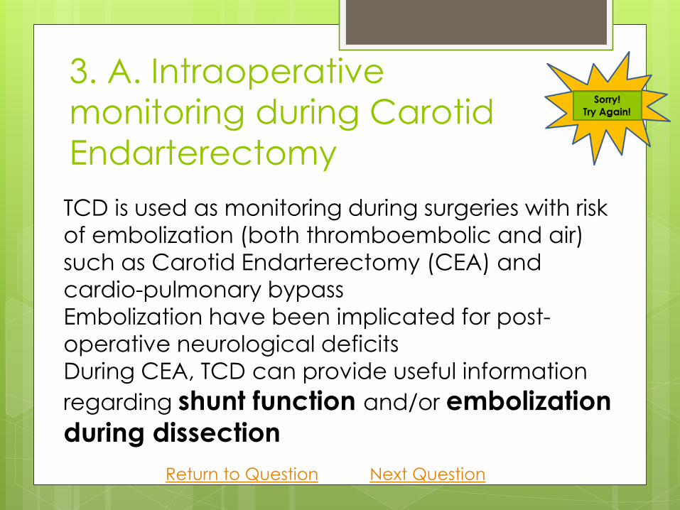
3. A. Intraoperative
monitoring during Carotid
Endarterectomy
TCD is used as monitoring during surgeries with risk
of embolization (both thromboembolic and air)
such as Carotid Endarterectomy (CEA) and
cardio-pulmonary bypass
Embolization have been implicated for post-
operative neurological deficits
During CEA, TCD can provide useful information
regarding shunt function and/or embolization
during dissection
Return to Question Next Question
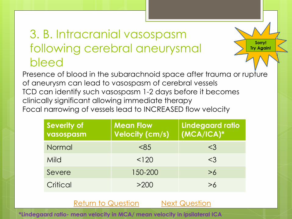
3. B. Intracranial vasospasm
following cerebral aneurysmal
bleed Presence of blood in the subarachnoid space after trauma or rupture
of aneurysm can lead to vasospasm of cerebral vessels
TCD can identify such vasospasm 1-2 days before it becomes
clinically significant allowing immediate therapy
Focal narrowing of vessels lead to INCREASED flow velocity
Severity of
vasospasm
Mean Flow
Velocity (cm/s)
Lindegaard ratio
(MCA/ICA)*
Normal <85 <3
Mild <120 <3
Severe 150-200 >6
Critical >200 >6
Return to Question Next Question
*Lindegaard ratio- mean velocity in MCA/ mean velocity in ipsilateral ICA

3. C. Predict stroke in sickle cell
disease
Sickle cell disease is associated with progressive
occlusion of intracranial (ICA,MCA) vessels
As these arteries are accessible for TCD
insonation, TCD can be used to monitor the flow
velocity in these arteries
Recent guidelines regard TCD as type A level of
evidence Neurology 2004;62:1468
Return to Question Next Question

3. D. Measure focal cerebral flow
during intractable seizure activity
PET or SPECT scans show CBF alteration during seizures
Constant movement would make TCD acquisition difficult
Indications for TCD include Intracranial vasospasm
Arterial stenosis / occlusion
Monitor for emboli during procedures
Brain death – TCD shows ‘reverberating or oscillating’ flow (normal flow during systole and reverse flow during diastole due to high distal resistance, in a ‘brain-dead’ patient)
Testing for cerebrovascular autoregulation
Sickle cell disease – assess stroke risk
Hepatic failure – monitor cerebral edema
Return to Question Next Question

4. The information that can be
obtained from a TCD are all, EXCEPT
A. Blood flow velocity
B. Direction of blood flow
C. The pulsatility index is a reliable marker of
resistance upstream to the insonated site
D. Cerebral vessels are identified depending
on the depth and the ‘acoustic window’

4. A. Blood flow velocity
A 2Mz ultrasonic beam reflects off the
erythrocytes within these vessels and is
analyzed
Using the Doppler principle, the TCD
measures the velocity and direction of
flow of blood
Return to Question Next Question

4. B. Direction of blood flow
A 2Mz ultrasonic beam reflects off the
erythrocytes within these vessels and is
analyzed
Using the Doppler principle, the TCD
measures the velocity and direction of
flow of blood
Return to Question Next Question

4. C. The pulsatility index is a reliable
marker of resistance upstream to the
insonated site
‘Pulsatility Index’ (PI)= Peak systolic velocity – End diastolic velocity
Mean velocity
PI is a reliable marker of resistance distal to the
insonated site and NOT upstream to it Focal narrowing at site of insonation – increase in flow velocity (FV)
Narrowing proximal to insonation site – decrease in FV
Decrease in CVR distal to insonation site (e.g. AV malformation)– ↑ FV, ↓ PI
Increase in CVR distal to insonation site (e.g. stenosis)- ↓ FV, ↑ PI
PI increases in cerebral edema
Return to Question Next Question
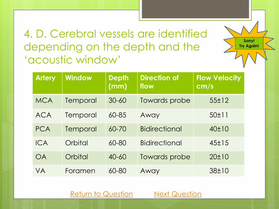
4. D. Cerebral vessels are identified
depending on the depth and the
‘acoustic window’
Artery Window Depth
(mm)
Direction of
flow
Flow Velocity
cm/s
MCA Temporal 30-60 Towards probe 55±12
ACA Temporal 60-85 Away 50±11
PCA Temporal 60-70 Bidirectional 40±10
ICA Orbital 60-80 Bidirectional 45±15
OA Orbital 40-60 Towards probe 20±10
VA Foramen 60-80 Away 38±10
Return to Question Next Question

5. Physiological factors that can decrease
TCD flow velocity include all, EXCEPT
A. Increase age
B. Increase blood viscosity
C. Increase PaCO2
D. Increase cardiac output

5. A. Increase age
Physiological factors that decrease Flow Velocity
• Increase age
• Increase CSF pressure
• Increased CVP
• Increased CO – to maintain normal CBF
• Increased blood viscosity
• Vasoconstrictors
Return to Question References

5. B. Increase blood viscosity
Physiological factors that decrease Flow Velocity
• Increase age
• Increase CSF pressure
• Increased CVP
• Increased CO – to maintain normal CBF
• Increased blood viscosity
• Vasoconstrictors
Return to Question References

5. C. Increase PaCO2
Factors that increase Flow Velocity
Increased PaCO2 – due to vasodilatation
Anemia (low viscosity)
Vasodilators
Return to Question References

5. D. Increase cardiac output
Physiological factors that decrease Flow Velocity
• Increase age
• Increase CSF pressure
• Increased CVP
• Increased CO – to maintain normal CBF
• Increased blood viscosity
• Vasoconstrictors
Return to Question References

References
Kassab etal. Transcranial Doppler: an
introduction for primary care physician.
JAm Board of Family Medicine 2007; 20:65
Moppett IK, Mahajan RP. Transcranial
Doppler ultrasonography in anesthesia
and intensive care. BJA 2004; 93: 710
Clinical Anesthesia, Barash, 7th edition
Q1 Q2 Q3 Q4 Q5