nerve activates contractionimages.pcmac.org/sisfiles/schools/al/coffeecounty/...skull and vertebrae...
TRANSCRIPT

Lecture Presentation by
Patty Bostwick-Taylor
Florence-Darlington Technical College
Chapter 5
The Skeletal System
© 2015 Pearson Education, Inc.

© 2015 Pearson Education, Inc.
The Skeletal System
Two subdivisions of the skeleton
1. Axial skeleton
2. Appendicular skeleton
Parts of the skeletal system
Bones (skeleton)
Joints
Cartilages
Ligaments

© 2015 Pearson Education, Inc.
Functions of Bones
Support the body
Protect soft organs
Skull and vertebrae protect brain and spinal cord
Rib cage protects thoracic cavity organs
Attached skeletal muscles allow movement
Store minerals and fats
Calcium and phosphorus
Fat in the internal marrow cavity
Blood cell formation (hematopoiesis)

© 2015 Pearson Education, Inc.
Bones of the Human Body
The adult skeleton has 206 bones
Two basic types of bone tissue
1. Compact bone
Dense, smooth, and homogeneous
2. Spongy bone
Small needle-like pieces of bone
Many open spaces

© 2015 Pearson Education, Inc.
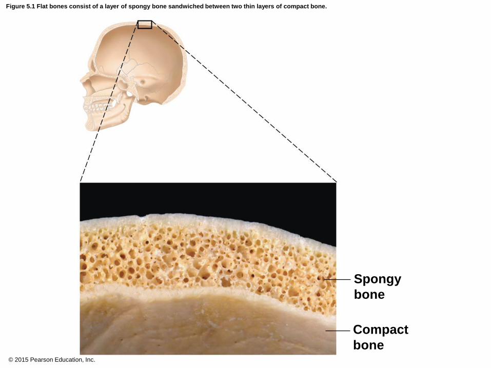
Figure 5.1 Flat bones consist of a layer of spongy bone sandwiched between two thin layers of compact bone.
Spongy
bone
Compact
bone

© 2015 Pearson Education, Inc.
Classification of Bones
Bones are classified on the basis of shape, as:
Long
Short
Flat
Irregular

© 2015 Pearson Education, Inc.
Figure 5.2 Classification of bones on the basis of shape.
(a) Long bone
(humerus)
(b) Irregular bone
(vertebra),
right lateral view
(d) Short bone
(talus)
(c) Flat bone
(sternum)

© 2015 Pearson Education, Inc.
Classification of Bones
Long bones
Typically longer than they are wide
Shaft with heads situated at both ends
Contain mostly compact bone
All of the bones of the limbs (except wrist, ankle, and
kneecap bones) are long bones
Examples:
Femur
Humerus

© 2015 Pearson Education, Inc.
Figure 5.2a Classification of bones on the basis of shape.
(a) Long bone
(humerus)

© 2015 Pearson Education, Inc.
Classification of Bones
Short bones
Generally cube-shaped
Contain mostly spongy bone
Include bones of the wrist and ankle
Sesamoid bones are a type of short bone that form
within tendons (patella)
Examples:
Carpals
Tarsals

© 2015 Pearson Education, Inc.
Figure 5.2d Classification of bones on the basis of shape.
(d) Short bone
(talus)

© 2015 Pearson Education, Inc.
Classification of Bones
Flat bones
Thin, flattened, and usually curved
Two thin layers of compact bone surround a layer of
spongy bone
Examples:
Skull
Ribs
Sternum
© 2015 Pearson Education, Inc.
Figure 5.1 Flat bones consist of a layer of spongy bone sandwiched between two thin layers of compact bone.
Spongy
bone
Compact
bone

© 2015 Pearson Education, Inc.
Figure 5.2c Classification of bones on the basis of shape.
(c) Flat bone
(sternum)

© 2015 Pearson Education, Inc.
Classification of Bones
Irregular bones
Irregular shape
Do not fit into other bone classification categories
Examples:
Vertebrae
Hip bones

© 2015 Pearson Education, Inc.
Figure 5.2b Classification of bones on the basis of shape.
(b) Irregular bone
(vertebra),
right lateral view

© 2015 Pearson Education, Inc.
Concept Link

© 2015 Pearson Education, Inc.
Anatomy of a Long Bone
Diaphysis
Shaft
Makes up most of bone’s length
Composed of compact bone
Periosteum
Outside covering of the diaphysis
Fibrous connective tissue membrane
Perforating (Sharpey’s) fibers secure periosteum to
underlying bone

© 2015 Pearson Education, Inc.
Figure 5.3a The structure of a long bone (humerus of arm).
Proximal
epiphysis
Diaphysis
Distal
epiphysis (a)
Articular
cartilage
Spongy bone
Epiphyseal
line
Periosteum
Compact bone
Medullary
cavity (lined
by endosteum)

© 2015 Pearson Education, Inc.
Figure 5.3c The structure of a long bone (humerus of arm).
(c)
Endosteum
Yellow bone
marrow
Compact bone
Periosteum
Perforating
(Sharpey’s) fibers
Nutrient
arteries

© 2015 Pearson Education, Inc.
Anatomy of a Long Bone
Epiphysis
Ends of the bone
Composed mostly of spongy bone enclosed by thin
layer of compact bone
Articular cartilage
Covers the external surface of the epiphyses
Made of hyaline cartilage
Decreases friction at joint surfaces

© 2015 Pearson Education, Inc.
Figure 5.3b The structure of a long bone (humerus of arm).
(b)
Articular
cartilage
Spongy
bone
Compact
bone

© 2015 Pearson Education, Inc.
Anatomy of a Long Bone
Epiphyseal plate
Flat plate of hyaline cartilage seen in young, growing
bone
Causes lengthwise growth of a long bone
Epiphyseal line
Remnant of the epiphyseal plate
Seen in adult bones

© 2015 Pearson Education, Inc.
Figure 5.3a The structure of a long bone (humerus of arm).
Proximal
epiphysis
Diaphysis
Distal
epiphysis (a)
Articular
cartilage
Spongy bone
Epiphyseal
line
Periosteum
Compact bone
Medullary
cavity (lined
by endosteum)

© 2015 Pearson Education, Inc.
Anatomy of a Long Bone
Marrow (medullary) cavity
Cavity inside the shaft
Contains yellow marrow (mostly fat) in adults
Contains red marrow for blood cell formation in
infants
In adults, red marrow is situated in cavities of
spongy bone and epiphyses of some long bones

© 2015 Pearson Education, Inc.
Figure 5.3a The structure of a long bone (humerus of arm).
Proximal
epiphysis
Diaphysis
Distal
epiphysis (a)
Articular
cartilage
Spongy bone
Epiphyseal
line
Periosteum
Compact bone
Medullary
cavity (lined
by endosteum)

© 2015 Pearson Education, Inc.
Bone Markings
Surface features of bones
Sites of attachments for muscles, tendons, and
ligaments
Passages for nerves and blood vessels
Categories of bone markings
Projections or processes—grow out from the bone
surface
Terms often begin with “T”
Depressions or cavities—indentations
Terms often begin with “F”

© 2015 Pearson Education, Inc.
Table 5.1 Bone Markings (1 of 3).

© 2015 Pearson Education, Inc.
Table 5.1 Bone Markings (2 of 3).

© 2015 Pearson Education, Inc.
Table 5.1 Bone Markings (3 of 3).

© 2015 Pearson Education, Inc.
Microscopic Anatomy of Compact Bone
Osteocytes are situated within cavities known as
lacunae
Lacunae are arranged in concentric rings called
lamellae
Lamellae are rings situated around the central
(Haversian) canal

© 2015 Pearson Education, Inc.
Figure 5.4a Microscopic structure of compact bone.
Spongy bone
Perforating (Volkmann’s) canal
Blood vessel continues into medullary cavity containing marrow
Blood vessel
Compact bone
Central (Haversian) canal
Perforating (Sharpey’s) fibers
Periosteum
Periosteal blood vessel
Osteon (Haversian system)
Lamellae
(a)

© 2015 Pearson Education, Inc.
Microscopic Anatomy of Bone
Central (Haversian) canal
Opening in the center of an osteon
Runs lengthwise through bone
Carries blood vessels and nerves
Osteon (Haversian system)
A unit of bone containing central canal and matrix
rings

© 2015 Pearson Education, Inc.
Figure 5.4b Microscopic structure of compact bone.
Lamella
Osteocyte
Canaliculus Lacuna
Central (Haversian) canal
(b)

© 2015 Pearson Education, Inc.
Figure 5.4c Microscopic structure of compact bone.
Osteon
Interstitial
lamellae
Lacuna
Central (Haversian) canal
(c)

© 2015 Pearson Education, Inc.
Microscopic Anatomy of Bone
Canaliculi
Tiny canals
Radiate from the central canal to lacunae
Form a transport system connecting all bone cells to
a nutrient supply
Perforating (Volkmann’s) canal
Canal perpendicular to the central canal
Carries blood vessels and nerves

© 2015 Pearson Education, Inc.
Figure 5.4b Microscopic structure of compact bone.
Lamella
Osteocyte
Canaliculus Lacuna
Central (Haversian) canal
(b)

© 2015 Pearson Education, Inc.
Bone Components
Organic parts of the matrix make bone flexible
Calcium salts deposited in the matrix make bone
hard

© 2015 Pearson Education, Inc.
Bone Formation and Growth
Ossification
Process of bone formation
Occurs on hyaline cartilage models or fibrous
membranes
Long bone growth involves two major phases

© 2015 Pearson Education, Inc.
Figure 5.5 Stages of long-bone formation in an embryo, fetus, and young child.
Hyaline
cartilage
New center of
bone growth
Medullary
cavity
Bone starting
to replace
cartilage
Hyaline
cartilage
model
Bone collar
In an embryo In a fetus In a child
Articular
cartilage
Spongy
bone
Epiphyseal
plate
cartilage
New bone
forming
Growth
in bone
width
Growth
in bone
length
Invading
blood
vessels
New bone
forming
Epiphyseal
plate cartilage
© 2015 Pearson Education, Inc.
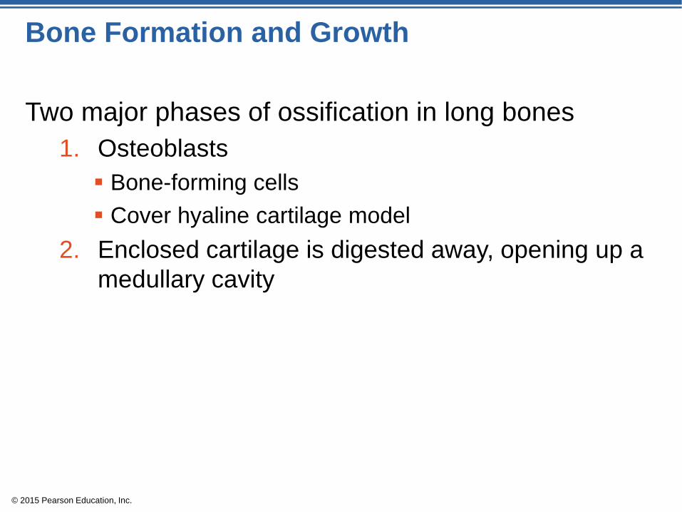
Bone Formation and Growth
Two major phases of ossification in long bones
1. Osteoblasts
Bone-forming cells
Cover hyaline cartilage model
2. Enclosed cartilage is digested away, opening up a
medullary cavity

© 2015 Pearson Education, Inc.
Bone Formation and Growth
By birth, most cartilage is converted to bone except
for two regions in a long bone:
1. Articular cartilages
2. Epiphyseal plates
New cartilage is formed continuously on external
face of these two cartilages
Old cartilage is broken down and replaced by bony
matrix

© 2015 Pearson Education, Inc.
Figure 5.6 Growth and remodeling of long bones.
Bone grows in
length because:
1
2
1
2 3
3
4
Growing shaft is
remodeled as:
Bone growth Bone remodeling
Articular cartilage
Epiphyseal plate
Bone is
resorbed here.
Bone is added
by appositional
growth here.
Bone is
resorbed here.
Cartilage
grows here.
Cartilage
is replaced
by bone here.
Cartilage
grows here.
Cartilage
is replaced by
bone here.
© 2015 Pearson Education, Inc.
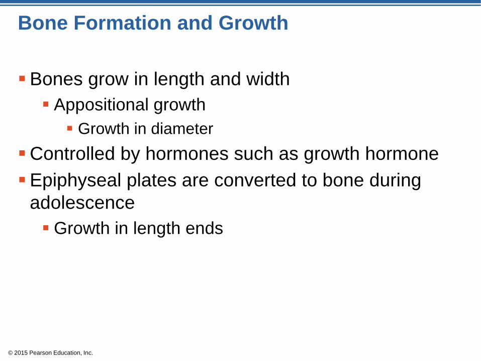
Bone Formation and Growth
Bones grow in length and width
Appositional growth
Growth in diameter
Controlled by hormones such as growth hormone
Epiphyseal plates are converted to bone during
adolescence
Growth in length ends

© 2015 Pearson Education, Inc.
Bone Remodeling
Bones are lengthened until growth stops
Bones are remodeled throughout life in response to
two factors:
1. Blood calcium levels
2. Pull of gravity and muscles on the skeleton

© 2015 Pearson Education, Inc.
Bone Remodeling
Parathyroid hormone (PTH)
Released when blood calcium levels are low
Activates osteoclasts (bone-destroying cells)
Osteoclasts break down bone and release calcium
ions into the blood
Hypercalcemia (high blood calcium levels) prompts
calcium storage to bones

© 2015 Pearson Education, Inc.
Bone Fractures
Fracture: break in a bone
Types of bone fractures
Closed (simple) fracture: break that does not
penetrate the skin
Open (compound) fracture: broken bone penetrates
through the skin

© 2015 Pearson Education, Inc.
Bone Fractures
Bone fractures are treated by reduction and
immobilization
Closed reduction: bones are manually coaxed into
position by physician’s hands
Open reduction: bones are secured with pins or
wires during surgery

© 2015 Pearson Education, Inc.
Repair of Bone Fractures
Hematoma (blood-filled swelling) is formed
Fibrocartilage callus forms
Cartilage matrix, bony matrix, collagen fibers splint
the broken bone
Bony callus replaces the fibrocartilage callus
Osteoblasts and osteoclasts migrate in
Bone remodeling occurs in response to mechanical
stresses

© 2015 Pearson Education, Inc.
Figure 5.7 Stages in the healing of a bone fracture.
Hematoma
1
External
callus
Internal
callus
(fibrous
tissue and
cartilage)
New
blood
vessels
Spongy
bone
trabecula
Hematoma
forms.
2 3 Fibrocartilage
callus forms.
Bony callus
forms.
Bone
remodeling
occurs.
Bony
callus of
spongy
bone
Healed
fracture
4

© 2015 Pearson Education, Inc.
Common Types of Fractures
Comminuted: bone breaks into many fragments
Compression: bone is crushed
Depressed: broken bone portion is pressed inward
Impacted: broken bone ends are forced into each
other
Spiral: ragged break occurs when excessive
twisting forces are applied to a bone
Greenstick: bone breaks incompletely

© 2015 Pearson Education, Inc.
Table 5.2 Common Types of Fractures.

© 2015 Pearson Education, Inc.
The Axial Skeleton
Forms the longitudinal axis of the body
Divided into three parts
1. Skull
2. Vertebral column
3. Bony thorax

© 2015 Pearson Education, Inc.
Figure 5.8a The human skeleton.
Skull
(a) Anterior view
Thoracic
cage
Vertebral
column
(ribs and
sternum)
Sacrum
Facial bones
Cranium
Clavicle
Scapula
Sternum
Rib Humerus
Vertebra Radius Ulna
Femur Patella
Tibia
Fibula
Tarsals Metatarsals Phalanges
Carpals
Phalanges
Metacarpals

© 2015 Pearson Education, Inc.
Figure 5.8b The human skeleton.
(b) Posterior view
Bones of
pectoral
girdle
Upper
limb
Bones of
pelvic
girdle
Lower
limb
Cranium
Clavicle
Scapula
Rib Humerus
Vertebra Radius Ulna
Carpals
Phalanges
Metacarpals Femur
Tibia
Fibula

© 2015 Pearson Education, Inc.
Concept Link

© 2015 Pearson Education, Inc.
The Skull
Two sets of bones
1. Cranium bones enclose the brain
2. Facial bones
Hold eyes in anterior position
Allow facial muscles to express feelings
Bones are joined by sutures
Only the mandible is attached by a freely movable
joint

© 2015 Pearson Education, Inc.
The Skull
8 cranial bones protect the brain
1. Frontal bone
2. Occipital bone
3. Ethmoid bone
4. Sphenoid bone
5–6. Parietal bones (pair)
7–8. Temporal bones (pair)

© 2015 Pearson Education, Inc.
The Skull
There are 14 facial bones. All are paired except for
the single mandible and vomer.
1–2. Maxillae
3–4. Zygomatics
5–6. Palatines
7–8. Nasals
9–10. Lacrimals
11–12. Inferior nasal conchae
13. Mandible
14. Vomer
© 2015 Pearson Education, Inc.
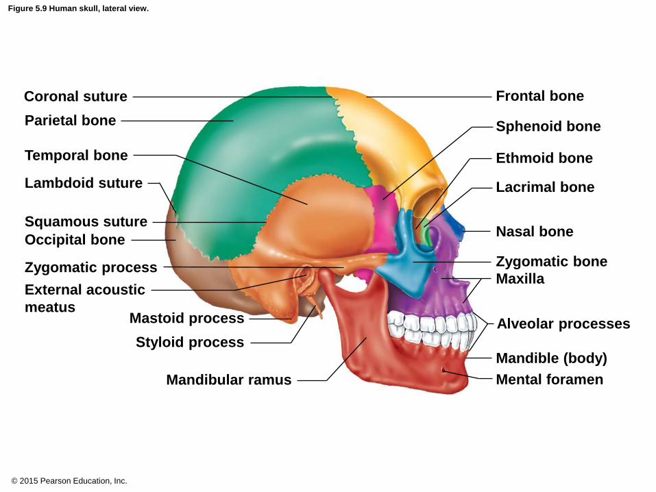
Figure 5.9 Human skull, lateral view.
Coronal suture
Parietal bone
Temporal bone
Lambdoid suture
Squamous suture
Occipital bone
Zygomatic process
External acoustic
meatus Mastoid process
Styloid process
Mandibular ramus
Frontal bone
Sphenoid bone
Ethmoid bone
Lacrimal bone
Nasal bone
Zygomatic bone
Maxilla
Alveolar processes
Mandible (body)
Mental foramen

© 2015 Pearson Education, Inc.
Figure 5.10 Human skull, superior view (top of cranium removed).
Sphenoid
bone
Temporal bone
Internal
acoustic meatus
Parietal bone
Occipital bone
Foramen magnum
Frontal bone
Ethmoid
bone
Cribriform plate
Crista galli
Optic canal
Sella turcica
Foramen ovale
Jugular foramen

© 2015 Pearson Education, Inc.
Figure 5.11 Human skull, inferior view (mandible removed).
Maxilla
Sphenoid bone
(greater wing)
Foramen ovale
Carotid canal
Jugular foramen
Occipital condyle
Foramen magnum
Maxilla
(palatine process)
Palatine bone
Hard
palate
Zygomatic bone
Temporal bone
(zygomatic process)
Vomer
Mandibular fossa
Styloid process
Mastoid process
Temporal bone
Parietal bone
Occipital bone

© 2015 Pearson Education, Inc.
Figure 5.12 Human skull, anterior view.
Mandible
Coronal suture
Parietal bone
Nasal bone
Sphenoid bone
Ethmoid bone
Lacrimal bone
Zygomatic bone
Maxilla
Frontal bone
Superior orbital
fissure
Temporal bone
Optic canal
Middle nasal concha
of ethmoid bone
Vomer
Inferior nasal concha
Alveolar processes
© 2015 Pearson Education, Inc.
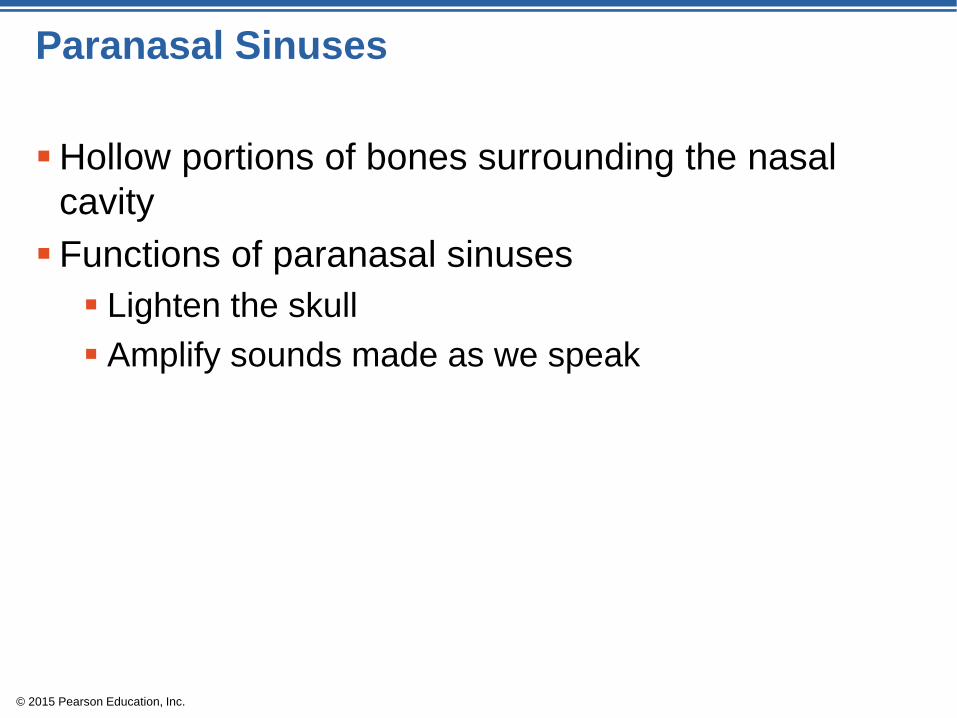
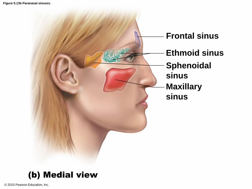
Paranasal Sinuses
Hollow portions of bones surrounding the nasal
cavity
Functions of paranasal sinuses
Lighten the skull
Amplify sounds made as we speak

© 2015 Pearson Education, Inc.
Figure 5.13a Paranasal sinuses.
Frontal sinus
Ethmoid sinus
Sphenoidal
sinus
Maxillary
sinus
(a) Anterior view
© 2015 Pearson Education, Inc.
Figure 5.13b Paranasal sinuses.
Frontal sinus
Ethmoid sinus
Sphenoidal
sinus
Maxillary
sinus
(b) Medial view

© 2015 Pearson Education, Inc.
The Hyoid Bone
Closely related to mandible and temporal bones
The only bone that does not articulate with another
bone
Serves as a movable base for the tongue
Aids in swallowing and speech

© 2015 Pearson Education, Inc.
Figure 5.14 Anatomical location and structure of the hyoid bone.
Greater horn
Lesser horn
Body

© 2015 Pearson Education, Inc.
The Fetal Skull
The fetal skull is large compared to the infant’s total
body length
Fetal skull is 1/4 body length compared to adult skull,
which is 1/8 body length
Fontanels are fibrous membranes connecting the
cranial bones
Allow skull compression during birth
Allow the brain to grow during later pregnancy and
infancy
Convert to bone within 24 months after birth

© 2015 Pearson Education, Inc.
Figure 5.15a The fetal skull.
Frontal bone
Parietal
bone
Posterior fontanel
(a)
Anterior
fontanel
Occipital
bone

© 2015 Pearson Education, Inc.
Figure 5.15b The fetal skull.
Parietal bone
Posterior
fontanel
Anterior fontanel
Sphenoidal
fontanel
Frontal
bone
Temporal bone
Occipital
bone
Mastoid
fontanel

© 2015 Pearson Education, Inc.
Vertebral Column (Spine)
Vertebral column provides axial support
Extends from skull to the pelvis
26 single vertebral bones are separated by
intervertebral discs
7 cervical vertebrae are in the neck
12 thoracic vertebrae are in the chest region
5 lumbar vertebrae are associated with the lower
back

© 2015 Pearson Education, Inc.
Vertebral Column (Spine)
9 vertebrae fuse to form two composite bones
Sacrum formed by the fusion of 5 vertebrae
Coccyx (tailbone) formed by the fusion of 3 to 5
vertebrae

© 2015 Pearson Education, Inc.
Figure 5.16 The vertebral column.
1st lumbar vertebra
Intervertebral foramen
Intervertebral disc
Spinous process
Transverse process
1st thoracic vertebra
1st cervical vertebra
(atlas)
2nd cervical vertebra
(axis)
Anterior Posterior
Cervical curvature (concave)
7 vertebrae, C1 – C7
Thoracic curvature (convex)
12 vertebrae, T1 – T12
Lumbar curvature (concave)
5 vertebrae, L1 – L5
Sacral curvature (convex)
5 fused vertebrae
Coccyx
4 fused vertebrae
© 2015 Pearson Education, Inc.
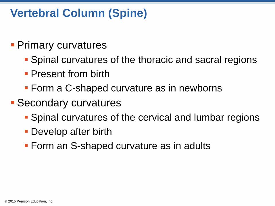
Vertebral Column (Spine)
Primary curvatures
Spinal curvatures of the thoracic and sacral regions
Present from birth
Form a C-shaped curvature as in newborns
Secondary curvatures
Spinal curvatures of the cervical and lumbar regions
Develop after birth
Form an S-shaped curvature as in adults

© 2015 Pearson Education, Inc.
Figure 5.17 The C-shaped spine typical of a newborn.

© 2015 Pearson Education, Inc.
Figure 5.18 Abnormal spinal curvatures.
(a) Scoliosis (b) Kyphosis (c) Lordosis
© 2015 Pearson Education, Inc.
Vertebral Column (Spine)
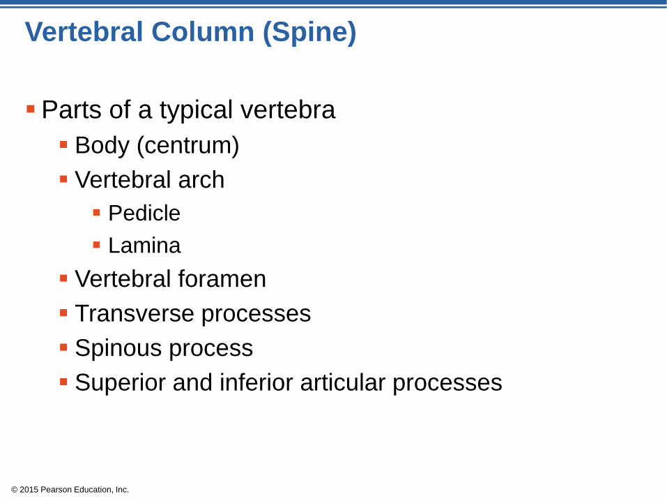
Parts of a typical vertebra
Body (centrum)
Vertebral arch
Pedicle
Lamina
Vertebral foramen
Transverse processes
Spinous process
Superior and inferior articular processes

© 2015 Pearson Education, Inc.
Figure 5.19 A typical vertebra, superior view.
Lamina Posterior
Vertebral
arch Transverse
process
Spinous
process
Vertebral
foramen
Body
Anterior
Pedicle
Superior
articular
process
and
facet

© 2015 Pearson Education, Inc.
Figure 5.20a Regional characteristics of vertebrae.
Transverse
process
(a) ATLAS AND AXIS
Posterior
arch
Superior view of axis (C2)
Anterior arch
Superior view of atlas (C1)
Transverse
process
Spinous process
Dens
Body
Facet on
superior
articular
process

© 2015 Pearson Education, Inc.
Figure 5.20b Regional characteristics of vertebrae.
(b) TYPICAL CERVICAL VERTEBRAE
Right lateral view
Spinous process Facet on
superior
articular
process
Vertebral
foramen
Transverse
process Superior view
Body
Facet on inferior
articular process
Transverse
process
Superior articular
process
Spinous
process
© 2015 Pearson Education, Inc.
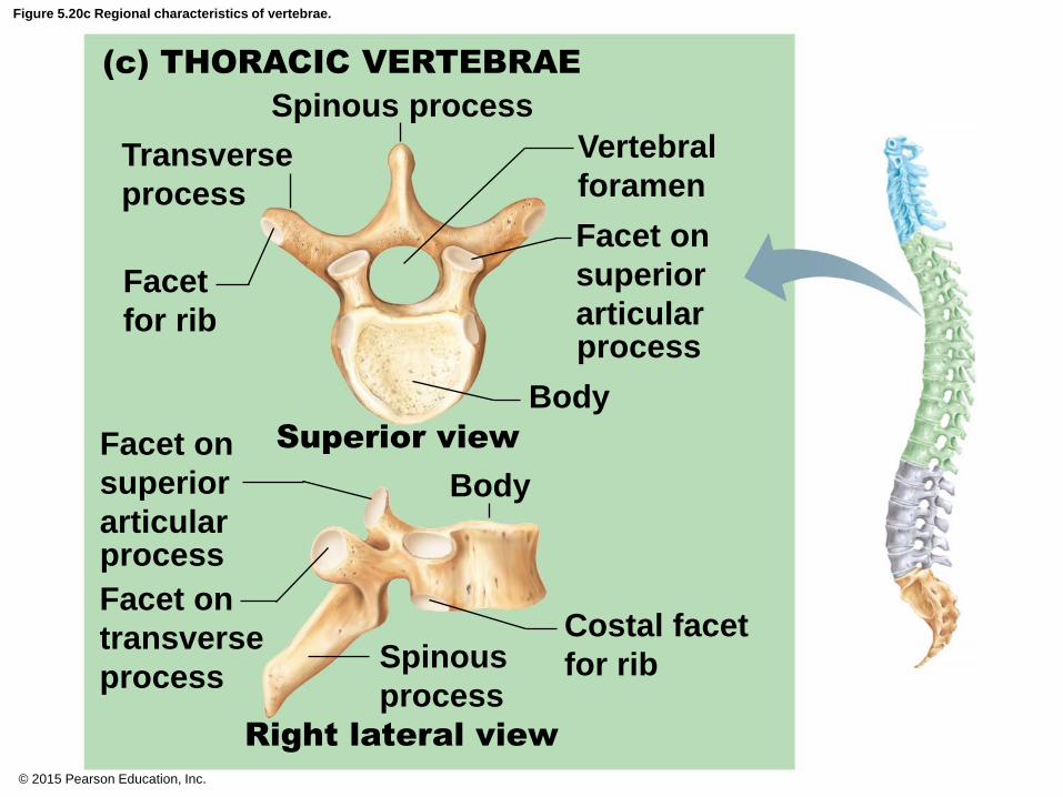
Figure 5.20c Regional characteristics of vertebrae.
(c) THORACIC VERTEBRAE
Right lateral view
Transverse
process
Body
Body
Spinous process
Facet
for rib
Vertebral
foramen
Facet on
transverse
process Spinous
process
Costal facet
for rib
Superior view
process
Facet on
superior
articular
Facet on
superior
articular
process

© 2015 Pearson Education, Inc.
Figure 5.20d Regional characteristics of vertebrae.
(d) LUMBAR VERTEBRAE
Right lateral view
Transverse
process
Vertebral
foramen
Facet on
superior
articular
process Body
Body Superior
articular
process
Spinous
process Facet on inferior
articular process
Superior view
Spinous process

© 2015 Pearson Education, Inc.
Figure 5.21 Sacrum and coccyx, posterior view.
Ala
Sacrum
Coccyx Sacral
hiatus
Posterior
sacral
foramina
Median
sacral
crest
Body
Auricular
surface
Superior
articular
process Sacral
canal

© 2015 Pearson Education, Inc.
The Bony Thorax
Forms a cage to protect major organs
Consists of three parts
1. Sternum
2. Ribs
True ribs (pairs 1–7)
False ribs (pairs 8–12)
Floating ribs (pairs 11–12)
3. Thoracic vertebrae

© 2015 Pearson Education, Inc.
Figure 5.22a The bony thorax (thoracic cage).
Clavicular notch
True
ribs
(1–7)
False
ribs
(8–12)
(a)
Floating
ribs (11, 12)
L1
vertebra Costal cartilage
Intercostal
spaces
T1 vertebra
Jugular notch
Manubrium
Sternal angle
Body
Xiphisternal
joint
Xiphoid
process
Sternum

© 2015 Pearson Education, Inc.
Figure 5.22b The bony thorax (thoracic cage).
Jugular
notch
Sternal
angle
Heart
Xiphisternal
joint
T2
T3
T4
T9
(b)

© 2015 Pearson Education, Inc.
The Appendicular Skeleton
Composed of 126 bones
Limbs (appendages)
Pectoral girdle
Pelvic girdle

© 2015 Pearson Education, Inc.
Figure 5.8a The human skeleton.
Skull
(a) Anterior view
Thoracic
cage
Vertebral
column
(ribs and
sternum)
Sacrum
Facial bones
Cranium
Clavicle
Scapula
Sternum
Rib Humerus
Vertebra Radius Ulna
Femur Patella
Tibia
Fibula
Tarsals Metatarsals Phalanges
Carpals
Phalanges
Metacarpals

© 2015 Pearson Education, Inc.
Figure 5.8b The human skeleton.
(b) Posterior view
Bones of
pectoral
girdle
Upper
limb
Bones of
pelvic
girdle
Lower
limb
Cranium
Clavicle
Scapula
Rib Humerus
Vertebra Radius Ulna
Carpals
Phalanges
Metacarpals Femur
Tibia
Fibula

© 2015 Pearson Education, Inc.
The Pectoral (Shoulder) Girdle
Composed of two bones that attach the upper limb
to the axial skeletal
1. Scapula
2. Clavicle
Pectoral girdle (2)
Light, poorly reinforced girdle
Allows the upper limb a great deal of freedom

© 2015 Pearson Education, Inc.
Figure 5.23a Bones of the shoulder girdle.
Scapula
Clavicle Acromio-
clavicular joint
(a) Articulated right shoulder (pectoral)
girdle showing the relationship to
bones of the thorax and sternum

© 2015 Pearson Education, Inc.
Figure 5.23b Bones of the shoulder girdle.
Inferior view
(b) Right clavicle, superior and
inferior views
Posterior
Acromial end
Anterior
Sternal
(medial)
end
Acromial
(lateral) end
Superior view
Anterior
Posterior
Sternal end

© 2015 Pearson Education, Inc.
Figure 5.23c Bones of the shoulder girdle.
Suprascapular notch
Superior
angle
Spine
Medial
border
Lateral border
Coracoid process
Acromion
Glenoid cavity
at lateral angle
(c) Right scapula, posterior aspect

© 2015 Pearson Education, Inc.
Figure 5.23d Bones of the shoulder girdle.
Suprascapular notch Acromion
Coracoid
process
Glenoid
cavity
Superior border
Superior
angle
Lateral
(axillary)
border
Medial
(vertebral)
border
Inferior angle
(d) Right scapula, anterior aspect

© 2015 Pearson Education, Inc.
Bones of the Upper Limbs
Humerus
Forms the arm
Single bone
Proximal end articulation
Head articulates with the glenoid cavity of the scapula
Distal end articulation
Trochlea and capitulum articulate with the bones of
the forearm

© 2015 Pearson Education, Inc.
Figure 5.24a Bones of the right arm and forearm.
Greater
tubercle
(a)
Lesser
tubercle
Head of
humerus
Anatomical
neck Intertubercular
sulcus
Deltoid
tuberosity
Medial
epicondyle
Trochlea
Radial
fossa Coronoid
fossa
Capitulum

© 2015 Pearson Education, Inc.
Figure 5.24b Bones of the right arm and forearm.
(b)
Head of
humerus
Anatomical
neck Surgical
neck
Radial
groove
Deltoid
tuberosity
Olecranon
fossa
Lateral
epicondyle
Medial
epicondyle
Trochlea

© 2015 Pearson Education, Inc.
Bones of the Upper Limbs
The forearm has two bones
1. Ulna—medial bone in anatomical position
Proximal end articulation
Coronoid process and olecranon articulate with the
humerus
2. Radius—lateral bone in anatomical position
Proximal end articulation
Head articulates with the capitulum of the humerus

© 2015 Pearson Education, Inc.
Figure 5.24c Bones of the right arm and forearm.
(c)
Head
Neck
Radial
tuberosity
Radius
Trochlear
notch
Olecranon
Coronoid
process
Proximal
radioulnar
joint
Ulna
Inter-
osseous
membrane
Ulnar
styloid
process Distal
radioulnar joint
Radial
styloid
process
© 2015 Pearson Education, Inc.
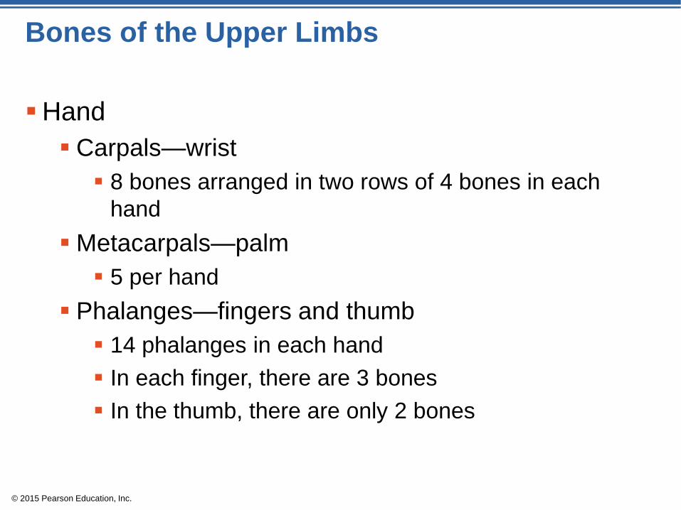
Bones of the Upper Limbs
Hand
Carpals—wrist
8 bones arranged in two rows of 4 bones in each
hand
Metacarpals—palm
5 per hand
Phalanges—fingers and thumb
14 phalanges in each hand
In each finger, there are 3 bones
In the thumb, there are only 2 bones

© 2015 Pearson Education, Inc.
Figure 5.25 Bones of the right hand, anterior view.
Distal
Middle
Proximal
Phalanges
(fingers)
Metacarpals
(palm)
Carpals
(wrist)
Hamate
Pisiform
Triquetrum
Lunate
Ulna
Trapezium
Trapezoid
Scaphoid
Capitate
Radius
1
2 3 4 5

© 2015 Pearson Education, Inc.
Bones of the Pelvic Girdle
Formed by 2 coxal (ossa coxae) bones
Composed of three pairs of fused bones
1. Ilium
2. Ischium
3. Pubis
Pelvic girdle 2 coxal bones, sacrum
Bony pelvis 2 coxal bones, sacrum, coccyx

© 2015 Pearson Education, Inc.
Bones of the Pelvic Girdle
The total weight of the upper body rests on the
pelvis
Pelvis protects several organs
Reproductive organs
Urinary bladder
Part of the large intestine

© 2015 Pearson Education, Inc.
Figure 5.26a The bony pelvis.
Ilium
Pubis
Ischium
(a)
Coxal bone
(or hip bone) Sacrum
Coccyx
Pubic arch
Iliac crest
Sacroiliac
joint
Pelvic brim
Ischial spine
Acetabulum
Pubic symphysis

© 2015 Pearson Education, Inc.
Figure 5.26b The bony pelvis.
Ilium
Iliac crest
Anterior superior
iliac spine
Anterior inferior
iliac spine
Acetabulum
Body of pubis
Pubis
Inferior pubic
ramus
Obturator
foramen
Ala
Posterior
superior
iliac spine
Posterior
inferior
iliac spine
Greater sciatic
notch
Ischial body
Ischial spine
Ischial
tuberosity
Ischium
Ischial ramus (b)

© 2015 Pearson Education, Inc.
Gender Differences of the Pelvis
The female’s pelvis:
Inlet is larger and more circular
Pelvis as a whole is shallower, and the bones are
lighter and thinner
Ilia flare more laterally
Sacrum is shorter and less curved
Ischial spines are shorter and farther apart; thus, the
outlet is larger
Pubic arch is more rounded because the angle of the
pubic arch is greater

© 2015 Pearson Education, Inc.
Figure 5.26c The bony pelvis.
False pelvis
Inlet of
true
pelvis
Pelvic brim
False pelvis
Inlet of
true
pelvis
Pelvic brim
(c)
Pubic arch
(less than 90 ° )
Pubic arch
(more than 90 ° )

© 2015 Pearson Education, Inc.
Bones of the Lower Limbs
Femur—thigh bone
The heaviest, strongest bone in the body
Proximal end articulation
Head articulates with the acetabulum of the coxal
(hip) bone
Distal end articulation
Lateral and medial condyles articulate with the tibia in
the lower leg

© 2015 Pearson Education, Inc.
Figure 5.27a Bones of the right thigh and leg.
Neck
Inter-
trochanteric
line
Lesser
trochanter
(a)
Lateral
condyle
Patellar
surface
Head

© 2015 Pearson Education, Inc.
Figure 5.27b Bones of the right thigh and leg.
Lesser
trochanter
Gluteal
tuberosity
Greater
trochanter
Inter-
trochanteric
crest
(b)
Lateral
condyle
Head
Intercondylar
fossa
Medial
condyle
© 2015 Pearson Education, Inc.
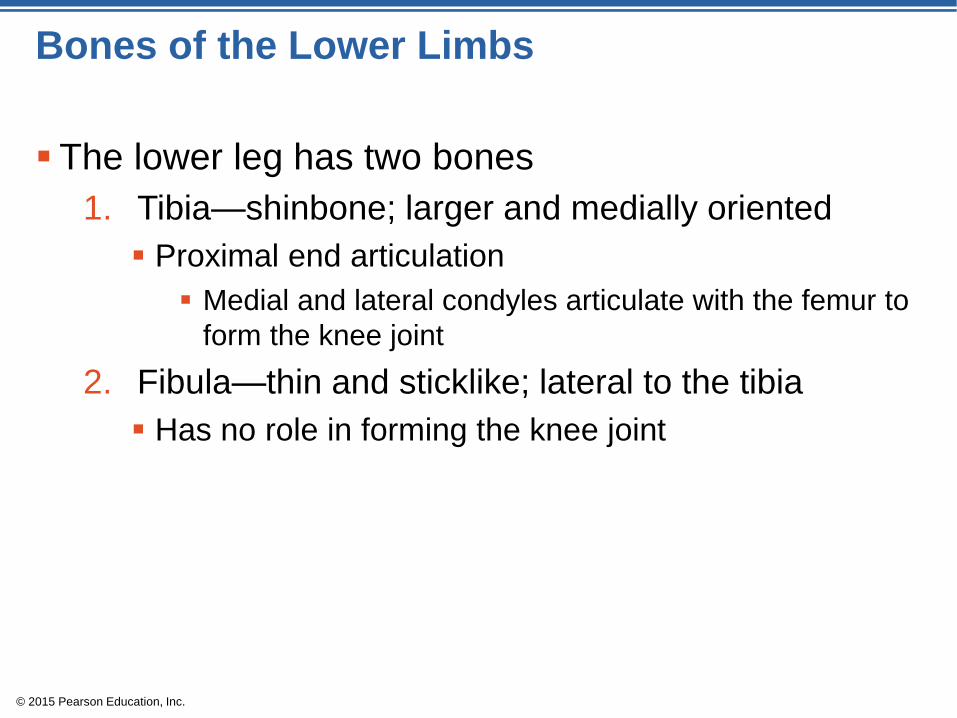
Bones of the Lower Limbs
The lower leg has two bones
1. Tibia—shinbone; larger and medially oriented
Proximal end articulation
Medial and lateral condyles articulate with the femur to
form the knee joint
2. Fibula—thin and sticklike; lateral to the tibia
Has no role in forming the knee joint

© 2015 Pearson Education, Inc.
Figure 5.27c Bones of the right thigh and leg.
Intercondylar
eminence
Lateral
condyle
Head
Proximal
tibiofibular
joint
Medial
condyle
Tibial
tuberosity
Interosseous
membrane
Anterior
border
Tibia
Medial
malleolus
Distal
tibiofibular
joint
Lateral
malleolus (c)
Fibula

© 2015 Pearson Education, Inc.
Bones of the Lower Limbs
The foot
Tarsals—7 bones
Two largest tarsals
Calcaneus (heel bone)
Talus
Metatarsals—5 bones form the sole of
the foot
Phalanges—14 bones form the toes

© 2015 Pearson Education, Inc.
Figure 5.28 Bones of the right foot, superior view.
Medial
cuneiform
Tarsals:
Phalanges:
Metatarsals
Tarsals:
Intermediate
cuneiform
Navicular
Talus
Distal
Middle
Proximal
Lateral
cuneiform
Cuboid
Calcaneus

© 2015 Pearson Education, Inc.
Arches of the Foot
Bones of the foot are arranged to form three strong
arches
Two longitudinal
One transverse

© 2015 Pearson Education, Inc.
Figure 5.29 Arches of the foot.
Medial longitudinal arch
Transverse arch
Lateral longitudinal
arch
© 2015 Pearson Education, Inc.
Joints
Joints are articulations
Two or more bones meet
Functions of joints
Hold bones together
Allow for mobility
Two ways joints are classified
Functionally
Structurally

© 2015 Pearson Education, Inc.
Functional Classification of Joints
Synarthroses
Immovable joints
Amphiarthroses
Slightly movable joints
Diarthroses
Freely movable joints

© 2015 Pearson Education, Inc.
Structural Classification of Joints
Fibrous joints
Generally immovable
Cartilaginous joints
Immovable or slightly movable
Synovial joints
Freely movable

© 2015 Pearson Education, Inc.
Concept Link

© 2015 Pearson Education, Inc.
Fibrous Joints
Bones united by fibrous tissue
Types
Sutures
Immobile
Syndesmoses
Allow more movement than sutures but still immobile
Example: Distal end of tibia and fibula
Gomphosis
Immobile

© 2015 Pearson Education, Inc.
Figure 5.30a Types of joints.
Fibrous
connective
tissue
(a) Suture
Fibrous joints

© 2015 Pearson Education, Inc.
Figure 5.30b Types of joints.
(b) Syndesmosis
Tibia
Fibula
Fibrous
connective
tissue
Fibrous joints

© 2015 Pearson Education, Inc.
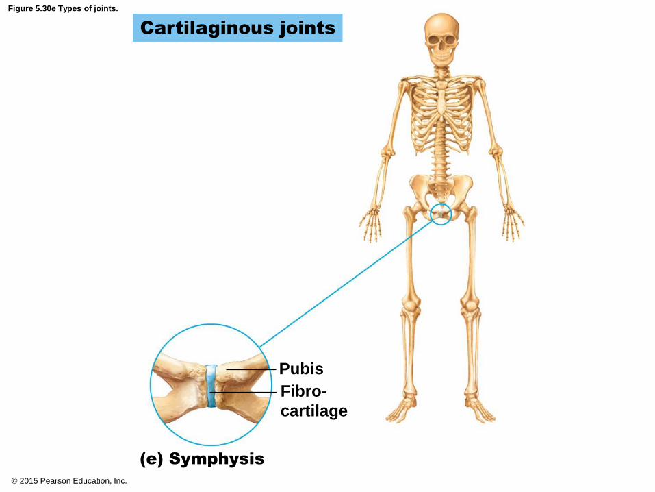
Cartilaginous Joints
Bones connected by fibrocartilage
Types
Synchrondrosis
Immobile
Symphysis
Slightly movable
Example: Pubic symphysis, intervertebral joints

© 2015 Pearson Education, Inc.
Figure 5.30c Types of joints.
First rib
Cartilaginous joints
(c) Synchondrosis
Hyaline
cartilage
Sternum

© 2015 Pearson Education, Inc.
Figure 5.30d Types of joints.
(d) Symphysis
Vertebrae
Fibro-
cartilage
Cartilaginous joints
© 2015 Pearson Education, Inc.
Figure 5.30e Types of joints.
(e) Symphysis
Pubis
Fibro-
cartilage
Cartilaginous joints

© 2015 Pearson Education, Inc.
Synovial Joints
Articulating bones are separated by a joint cavity
Synovial fluid is found in the joint cavity
Four distinguishing features of synovial joints
1. Articular cartilage
2. Articular capsule
3. Joint cavity
4. Reinforcing ligaments

© 2015 Pearson Education, Inc.
Figure 5.30f Types of joints.
(f) Multiaxial joint
Humerus
Articular (hyaline)
cartilage
(shoulder joint)
Scapula
Articular
capsule
Synovial joints

© 2015 Pearson Education, Inc.
Figure 5.30g Types of joints.
(g) Uniaxial joint
Ulna
Radius
Articular capsule
Articular (hyaline)
cartilage
Humerus
(elbow joint)
Synovial joints

© 2015 Pearson Education, Inc.
Figure 5.30h Types of joints.
Synovial joints
(h) Biaxial joint
(intercarpal joints of hand)
Carpals
Ulna
Radius
Articular
capsule

© 2015 Pearson Education, Inc.
Synovial Joints
Bursae—flattened fibrous sacs
Lined with synovial membranes
Filled with synovial fluid
Not actually part of the joint
Tendon sheath
Elongated bursa that wraps around a tendon

© 2015 Pearson Education, Inc.
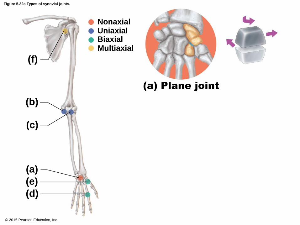
Synovial Joints
Types of synovial joints based on shape:
Plane joint
Hinge joint
Pivot joint
Condylar joint
Saddle joint
Ball-and-socket joint

© 2015 Pearson Education, Inc.
Figure 5.31 General structure of a synovial joint.
Acromion of
scapula
Ligament
Bursa
Ligament
Tendon
sheath
Tendon of
biceps muscle
Joint cavity
containing
synovial fluid
Articular
(hyaline)
cartilage
Synovial membrane
Fibrous layer of the
articular capsule
Humerus
© 2015 Pearson Education, Inc.
Figure 5.32a Types of synovial joints.
(f)
(a) Plane joint
Nonaxial Uniaxial Biaxial Multiaxial
(c)
(a)
(e)
(d)
(b)

© 2015 Pearson Education, Inc.
Figure 5.32b Types of synovial joints.
(f)
Nonaxial Uniaxial Biaxial Multiaxial
(c)
(a)
(e)
(d)
(b)
Humerus
Ulna
(b) Hinge
joint

© 2015 Pearson Education, Inc.
Figure 5.32c Types of synovial joints.
Nonaxial Uniaxial Biaxial Multiaxial
(f)
(c)
(a)
(e)
(d)
(b) (c) Pivot joint
Ulna
Radius

© 2015 Pearson Education, Inc.
Figure 5.32d Types of synovial joints.
Nonaxial Uniaxial Biaxial Multiaxial
(f)
(c)
(a)
(e)
(d)
(b) Metacarpal Phalanx
(d) Condylar joint

© 2015 Pearson Education, Inc.
Figure 5.32e Types of synovial joints.
Nonaxial Uniaxial Biaxial Multiaxial
(f)
(c)
(a)
(e)
(d)
(b) (e) Saddle joint
Carpal Metacarpal #1

© 2015 Pearson Education, Inc.
Figure 5.32f Types of synovial joints.
Nonaxial Uniaxial Biaxial Multiaxial
(f)
(c)
(a)
(e)
(d)
(b)
Head of
humerus
Scapula
(f) Ball-and-socket joint

© 2015 Pearson Education, Inc.
Inflammatory Conditions Associated with Joints
Bursitis—inflammation of a bursa, usually caused
by a blow or friction
Tendonitis—inflammation of tendon sheaths
Arthritis—inflammatory or degenerative diseases of
joints
Over 100 different types
The most widespread crippling disease in the United
States
Initial symptoms: pain, stiffness, swelling of the joint

© 2015 Pearson Education, Inc.
Clinical Forms of Arthritis
Osteoarthritis (OA)
Most common chronic arthritis
Probably related to normal aging processes
Rheumatoid arthritis (RA)
An autoimmune disease—the immune system
attacks the joints
Symptoms begin with bilateral inflammation of
certain joints
Often leads to deformities

© 2015 Pearson Education, Inc.
Figure 5.33 X-ray image of a hand deformed by rheumatoid arthritis.

© 2015 Pearson Education, Inc.
Clinical Forms of Arthritis
Gouty arthritis (gout)
Inflammation of joints is caused by a deposition of
uric acid crystals from the blood
Can usually be controlled with diet
More common in men

© 2015 Pearson Education, Inc.
Developmental Aspects of the Skeletal System
Fontanels
Allow brain growth and ease birth passage
Present in the skull at birth
Completely replaced with bone within 2 years after
birth

© 2015 Pearson Education, Inc.
Developmental Aspects of the Skeletal System
Growth of cranium after birth is related to brain
growth
Increase in size of the facial skeleton follows tooth
development and enlargement of the respiratory
passageways.

© 2015 Pearson Education, Inc.
Figure 5.34 Ossification centers in the skeleton of a 12-week-old fetus are indicated by the darker areas. Lighter regions are still fibrous or cartilaginous.
Frontal
bone
of skull
Parietal
bone
Occipita
bone
Clavicle
Scapula
Mandible
Radius
Ulna
Humerus
Femur
Tibia
Ribs
Vertebra
Hip bone

© 2015 Pearson Education, Inc.
Skeletal Changes Throughout Life
Fetus
Long bones are formed of hyaline cartilage
Flat bones begin as fibrous membranes
Flat and long bone models are converted to bone
Birth
Fontanels remain until around age 2

© 2015 Pearson Education, Inc.
Skeletal Changes Throughout Life
Adolescence
Epiphyseal plates become ossified, and long bone
growth ends
Size of cranium in relationship to body
2 years old—skull is larger in proportion to the body
compared to that of an adult
8 or 9 years old—skull is near adult size and
proportion
Between ages 6 and 11, the face grows out from the
skull

© 2015 Pearson Education, Inc.
Figure 5.35a Differences in the growth rates for some parts of the body compared to others determine body proportions.
Human newborn Human adult
(a)

© 2015 Pearson Education, Inc.
Figure 5.35b Differences in the growth rates for some parts of the body compared to others determine body proportions.
(b)
Newborn 2 yrs. 5 yrs. 15 yrs. Adult
© 2015 Pearson Education, Inc.
Skeletal Changes Throughout Life
Curvatures of the spine
Primary curvatures are present at birth and are
convex posteriorly
Secondary curvatures are associated with a child’s
later development and are convex anteriorly
Abnormal spinal curvatures (scoliosis and lordosis)
are often congenital

© 2015 Pearson Education, Inc.
Figure 5.18 Abnormal spinal curvatures.
(a) Scoliosis (b) Kyphosis (c) Lordosis

© 2015 Pearson Education, Inc.
Skeletal Changes Throughout Life
Osteoporosis
Bone-thinning disease afflicting
50 percent of women over age 65
20 percent of men over age 70
Disease makes bones fragile, and bones can easily
fracture
Vertebral collapse results in kyphosis (also known as
“dowager’s hump”)
Estrogen aids in health and normal density of a
female skeleton

© 2015 Pearson Education, Inc.
Figure 5.36 Osteoporosis.

© 2015 Pearson Education, Inc.
Figure 5.37 Vertebral collapse due to osteoporosis.
Age 40 Age 60 Age 70