nanotransporters for anticancer drug …€¦ · nanomedicine is a relatively new field of medicine...
TRANSCRIPT
In: Advances in Nanotechnology. Volume 14 ISBN: 978-1-63482-971-7 Editors: Zacharie Bartul and Jérôme Trenor © 2015 Nova Science Publishers, Inc.
Chapter 1
NANOTRANSPORTERS FOR ANTICANCER DRUG DELIVERY
Pavel Kopel1,2, Dorota Wawrzak3, Vedran Milosavljevic1, Amitava Moulick2, Marketa Vaculovicova1,2, Rene Kizek1,2
and Vojtech Adam*1,2 1Department of Chemistry and Biochemistry, Mendel University in Brno,
Brno, Czech Republic 2Central European Institute of Technology,
Brno University of Technology, Technicka, Brno, Czech Republic 3Institute of Chemistry, Envoronmental Protection and Biotechnology,
Jan Dlugosz University of Czestochowa, Czestochowa, Poland
ABSTRACT
Nanomedicine is a relatively new field of medicine aiming to overcome usual problems that appear in disease treatment. Natural or artificial nanodevices, with dimensions similar to those of biological molecules in human body, can carry drugs more efficiently and mostly with no side effects on healthy tissue than drugs alone. Nowadays, there is a huge interest in nanotechnology for detection, imaging and cancer treatment because cancer causes death of millions people every year. There are already some nanoparticle based drugs, mostly in liposomes, approved for clinical use or under clinical investigation. Many attempts are made to improve nanoparticles sizes, shapes and surface modifications that lead to prolongation of drug circulation in blood stream and targeting to cancer cells. Thus small molecules like polyethylene glycol and targeting ligands like folic acid, peptides, antibodies, aptamers and nucleic acids are bound on the surface of nanoparticles with the aim to increase specific cell uptake. Very promising are multifunctional nanoparticles that combine both diagnostic as well as delivery role together.
In this chapter, we describe recent progress on utilization of different nanotransporters including dendrimers, micelles, liposomes, protein-based carriers,
* Corresponding author, e-mail: [email protected].
The license for this PDF is unlimited except that no part of this digital document may be reproduced, stored in a retrieval system or transmitted commercially in any form or by any means. The publisher has taken reasonable care in the preparation of this digital document, but makes no expressed or implied warranty of any kind and assumes no responsibility for any errors or omissions. No liability is assumed for incidental or consequential damages in connection with or arising out of information contained herein. This digital document is sold with the clear understanding that the publisher is not engaged in rendering legal, medical or any other professional services.
Pavel Kopel, Dorota Wawrzak, Vedran Milosavljevic et al. 2
graphene, graphene oxide, carbon nanotubes, silica, gold and iron oxides nanoparticles, for transport of anticancer drugs.
INTRODUCTION Nanotechnology deals with the creation and testing of structures, the size of which at
least one dimensions is less than 100 nm. Nanomaterials beyond the specific sizes have different characteristics and properties of the same material with a lower degree of fragmentation. You cannot reduce the size of infinity, because the matter has different properties in the macro and the subatomic world, and crossing the border nano, redefine the way that appliances. Here was born the concept of nanomaterials. Nanotechnology is not only chemistry and technology of manufacture of small molecules, but also the formation and use of molecules with nano dimensions having unique properties which can be used for the preparation of new materials or construction of miniature systems. Nanomaterials have different chemical, physical and biological substances forming them than particles of larger sizes. This is due to the fact that most of the molecules forming the nanomaterial are close to the surface, and thus their electronic structure, energy levels, and the reactivity is different than if the classical form a crystal network.
Nanomaterials and nanoparticles retain characteristic physicochemical properties of these materials at the macro level, but also have a range of original features, occurring only in the nanoscale. The main reasons for the unique properties of nano-objects are their dimensions, as well as the related disclosure of quantum phenomena. The small size allows them to penetrate through most barriers, and also mean that in their case the observed effects resulting from the laws of quantum physics. Dualism of nature nanoparticles is one of their biggest advantages. Compared to the material in the macro-nano characterized inter alia: more developed specific surface area, greater hardness, greater strength and increasing plasticity occurring at the same time, sliding properties or greater biocompatibility of nano biomaterials [1]. Most of nanomaterials have a dual character: a percentage of the property does not make their use in biology, chemistry, environmental engineering and medicine becomes attractive especially in the transport and delivery of drugs and bioactive substances. The concept of using a particle measured in the nanoscale as carriers of drugs and vaccines appeared over three decades ago. Advances in nano medicine has evolved and has raised hopes for the implementation methods of striking antitumor therapy selectively in tumor mass, while reducing the risk of a wide range of side effects, which are encumbering modern pharmacology. Nanoparticles are attractive as drug delivery platforms because it is relatively easy to influence their properties and modify their features, so that they can be useful in creation of effective and precise medicine carriers. Meaningful are not only dimensions of the carrier enabling tissue penetration, but also their shape, and developed different functionalities of surface.
Current progress in the field of nanobiotechnology has led to the development of a new area of nanomedicine, associated with the application of nano biomaterials, both for diagnostic and therapeutic aims creating a new category of nano particles called theranostics. The main expectations and challenges in this regard relate to nano-magnetic properties, received bioengineering methods, with potential used in the transport of drugs, particularly anticancer drugs used in therapy determined using molecular targets. Unique physicochemical
Nanotransporters for Anticancer Drug Delivery 3
properties of magnetic nanoparticles promise hope for the development of modern cancer nanomedicine, acting, inter alia, technological breakthrough in the area of targeted drug delivery and gene therapy of cancer using magnetic hyperthermia, tissue engineering, marking the tumor cells and the molecular magnetic resonance imaging. Along with a broad interest in magnetic nanoproducts and bioengineering, in the area of special attention is the toxic potential. A considerable amount of scientific evidence to date suggests that certain properties of nanoparticles magnetic (e.g. increased surface activity, ability to penetrate cell membranes, resistance biodegradation processes) may enhance their cytotoxic potential compared to the corresponding materials lacking the size in the nanoscale. In other words, the safety evaluation conducted with respect to standard magnetic materials, may be of limited use in the risk assessment of health and environmental exposure in the case of new nano-magnetic received bioengineering methods.
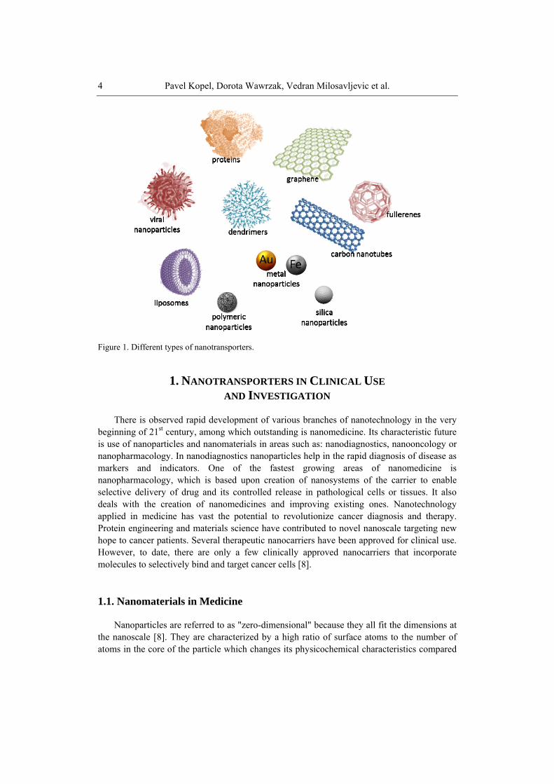
In this chapter we discuss the main directions of research conducted in experimental models in vitro and in vivo to nanoparticles (NPs), paying particular attention to the nanotransporters for anticancer drug delivery. The chapter presents also new directions in the field of research conducted in area of nanotransporters in clinical use and investigation, drug release from nanoparticles, proteins as a natural nanotransporters, liposomes and their modifications, dendrimers and polymer nanoparticles, inorganic nanoparticles as well as carbon based nanomaterials (see. Figure 1).
Nanotechnology in medicine and health care was initiated over forty years ago with deliverance of the first therapeutic and diagnostic agents in a safer and more efficient manner [2]. Convergention of diagnosis and therapy carried out through exploitation of nanoparticles resulted with increasing number of theradiagnostics went out from research stage and being commercialized or having reached clinical stage. Unique structures and physical properties which are characteristic for nanoparticles are originated because a large fraction of their volume is within “hailing distance” of the surface.
Such a structure of nanoparticles makes them useful as carriers of “heavy loads” of surface coatings, area that is structurally and compositionally different from the bulk. Coating material can rearrange all or parts of the nanoparticle structure themselves, provide a shell of different composition, or adsorb layers inorganic or organic molecules. External layers of nanoparticle are highly flexible in ability to initiate creation or building novel structures with different properties both of the core structure as adsorbed layers [3].
The fundamental complexities in structure, bonding, and interfacial interactions between a particle, its coating, and its neighboring environment, can be exploited to derive unique properties for many potential applications. The development of functional, inorganic nanoparticles (NPs) has progressed exponentially over the past two decades. Magnetic nanocrystals, luminescent particles and sophisticated systems such as up-converting NPs are some of examples from the diverse range of availabilities [4, 5]. Attainable functionalities enable the realisation of different diagnostic and therapeutic applications [6]. Although only a relatively small number of nanosized drug delivery carriers have been approved for human use so far, it is now accepted that nanotechnologies will likely constitute a growing share of the oncologist's therapeutic arsenal over the next decades to come [7-9]. There are many nanoparticle technologies under development and a great majority are still without clinical proof of concept, but advances on clinical stage show how promising and limitless is nanotechnology especially in medicine.
Pavel Kopel, Dorota Wawrzak, Vedran Milosavljevic et al. 4
Figure 1. Different types of nanotransporters.
1. NANOTRANSPORTERS IN CLINICAL USE
AND INVESTIGATION There is observed rapid development of various branches of nanotechnology in the very
beginning of 21st century, among which outstanding is nanomedicine. Its characteristic future is use of nanoparticles and nanomaterials in areas such as: nanodiagnostics, nanooncology or nanopharmacology. In nanodiagnostics nanoparticles help in the rapid diagnosis of disease as markers and indicators. One of the fastest growing areas of nanomedicine is nanopharmacology, which is based upon creation of nanosystems of the carrier to enable selective delivery of drug and its controlled release in pathological cells or tissues. It also deals with the creation of nanomedicines and improving existing ones. Nanotechnology applied in medicine has vast the potential to revolutionize cancer diagnosis and therapy. Protein engineering and materials science have contributed to novel nanoscale targeting new hope to cancer patients. Several therapeutic nanocarriers have been approved for clinical use. However, to date, there are only a few clinically approved nanocarriers that incorporate molecules to selectively bind and target cancer cells [8].
1.1. Nanomaterials in Medicine Nanoparticles are referred to as "zero-dimensional" because they all fit the dimensions at
the nanoscale [8]. They are characterized by a high ratio of surface atoms to the number of atoms in the core of the particle which changes its physicochemical characteristics compared
Nanotransporters for Anticancer Drug Delivery 5
to materials with the same chemical composition, but a normal size. This results in a change in the behavior of nanoparticles under the influence of external forces and contributes, among others, to their increased chemical reactivity and biological and other electrical and optical properties [10]. Due to the small size nanoparticles enter the body through the skin, inhalation, ingestion and can accumulate in various organs. Nanomaterials are materials which at least one dimensions less than 100 nm. They are divided into zero-dimensional nanomaterials such. Quantum dots, one-dimensional - wires and tubes, two-dimensional forming layer and forming three-dimensional materials composed of nano-crystals [10]. Due to their size they acquire new characteristic properties that differ from those obtained in the macro scale. Additional features specific for these materials include high specific surface area, the tendency to agglomerate and the ability to high specific activity [11].
1.2. Nanoparticles a Core-shell Type These nanoparticles are composed of silica core coated with a thin layer of gold, which
can be further integrated by biological ligands. Because of the ability of absorption and scattering of electromagnetic waves from the visible to near IR may be used in optical and medical imaging. Another advantage is the ability to change the scope of radiation absorbed by modifying the core thickness and number of coats. They are used mainly targeted therapy by means of photodynamic methods. In a study conducted by Hirsch et al. used in the treatment of tumor on mice in vivo and in vitro cell line SKBR3. Nanoparticles were injected interstitially to the lesion and then irradiated with low doses of near infrared radiation (820 nm; 4W/cm2). This caused high heat up of the tumor cells and eventually their destruction, while preserving all functions of the surrounding healthy tissue. The same result was obtained in studies conducted in vitro [12]. These nanoparticles, coated with antibodies were also used for the delivery of drugs in the hydrogel coating; under the effect of laser radiation dissolved and released the active drug in tumor tissue while reducing the toxicity of chemotherapy. Such nanostructures are also important in medical diagnostics for example the detection of a variety of molecules, e.g. immunoglobulin in the blood or plasma [13].
1.3. Nanoparticles of Metallic Materials Novel properties of nanoscale metallic materials, also called nanomaterials, have
attracted enormous interest compared to conventional (microcrystalline) materials. In recent years nanoscale magnetic materials showed the potential for use in many different biological and medical applications. For example, super paramagnetic iron oxide nanoparticles having an average particle diameter of about 10 nm, suspended in suitable liquid carriers are commonly referred to as ferrofluids and have excellent properties. In these materials, a wide range of both metallic and oxide magnetic nanoparticles were synthesized. Magnetic nanoparticles can be used for testing due to their high surface area and the interaction with different tissues. The main applications are the detection and analysis of bio particles directed transport of drugs, contrast during magnetic resonance imaging and hyperthermia. Effect of controlled heating of cells and tissues is one of surprising futures of ferrofluids. Each cycle of a hysteresis loop of any magnetic material involves an energy loss proportional to the area of
Pavel Kopel, Dorota Wawrzak, Vedran Milosavljevic et al. 6
the loop. Hence if magnetic nanoparticles having the required coercivity are remotely positioned at a given site in the body, perhaps the site of a malignancy, then the application of an alternating magnetic field can be used to selectively warm a given area. It has been proposed that this simple physical effect could be used both to destroy cells directly and to induce a modest increase in temperature so as to increase the efficiency either in chemotherapy or radiotherapy [14].
Rapid immunization of microorganisms to antibiotics induced intensive research of new substances with antibacterial properties. Intensive development of nanotechnology enabled the creation of nanoparticles of metals such as silver, gold and copper, which are characterized by antimicrobial properties. The largest application has silver nanoparticles due to its homogeneity, stability and functionality. From the earliest years were characterized by silver antibacterial properties, which by means of nanotechnology have been strengthened. Each silver nanoparticle contains 20 – 15 000 atoms and due to the different structures (beads, rods, cubes or wires) are generally less than 100 nm. Toxic effects of nanosilver as a germicide, antifungal and antiviral are mainly based on disruption of cell membranes, protein denaturation, the production of oxygen radicals and interference with DNA replication and inhibition of expression of proteins and enzymes constituting the respiratory chain [15]. The nano-silver antibacterial important is the construction of the bacterial cell wall. Gram-positive bacteria, for example Staphylococcus aureus, because of the thicker layer of peptidoglycan, are less sensitive to the toxic influence of nanosilver than gram-negative bacteria [16]. Combination of nanosilver with antibiotics enhances the effect of antibacterial agents such as amoxicillin, erythromycin, clindamycin, penicillin G and vancomycin as was proved by Shaverdi et al. [17]. They are also exploited as an antibacterial agent in the manufacture of bandages, dressings and surgical masks, and the coating of medical implants and ensure a long antimicrobial activity by slow release of silver ions. Silver nanoparticles are also used in medical diagnostics as biosensors, optical signal through the use of localized, surface plasmon resonance (LSPR). The method has been used in the diagnosis head and neck squamous-cell carcinoma or squamous cell cancer (SCC or SqCC), by coating with nanosilver mouse monoclonal antibody against the protein p53, which underwent overexpression in patients studied [18]. Other nanostructures used in medicine are nanoparticles of gold. Gold nanoparticles, which can be shaped and obtain different shapes, with their ease of attachment on the surface of nanoparticles additional ligands, they fulfil specific functions. For diagnostic purposes, such ligands are used as lipids or antibodies that are used for imaging tumor cells and to determine the risk of atherosclerosis - macrophages [19]. Gold nano particles are also used in the treatment of cancer using photothermal therapy (PPT). The treatment utilizes electro- magnetic radiation which is directed to nanoparticles contained in pathological tissues. They convert the radiation into heat, causing a temperature increase in pathological tissues and cell death. Nanoparticles of gold nanorods have high absorption in the near infrared and visible range, and effective generation of heat, so that the arms are promising in the treatment of cancer and other diseases [20].
Core-shell nanoparticles are of a particular interest. They comprise of a core made from one material, and a shell (or coating) made from the second one. The core/shell nanoparticles are always made from an inorganic core (i.e. oxide, nitride, carbide). The shell is made from another oxide, nitride, carbide, or an organic material (monomer, surfactant, surface active organic molecule, and organic dye). By proper selection of core and shell materials properties of nanoparticles can be combined, or the surface can be functionalized. Low toxicity of these
Nanotransporters for Anticancer Drug Delivery 7
types of nanoparticles made of gold and ease of synthesis of such nanoparticles implies that they are used as carriers for drugs and biological macromolecules such as peptides, proteins and nucleic acids. Nanoparticles such as these provide pharmaceuticals to specific sites in the body (e.g. to tumor cell), thereby increasing the effectiveness of therapy. Nanoparticles of gold can be used as a carrier of insulin according to a study by Bhumkar et al. [21]. Gold also perfectly absorbs X-rays, which can be used to assist radiation therapy [22]. Although radiotherapy is still being improved, including the use of a radiation beam with a high voltage in order to avoid damage to the skin, tomotherapy and modulating the intensity of the therapeutic beam, is still an unresolved issue is the protection of normal cells against radiation beam [23]. The solution to this problem is to use nanoparticles of gold that are accumulated in the site of the tumor and absorbing ionizing radiation allowing the use of lower therapeutic doses, which protects normal tissues. It is estimated that the strengthening of therapeutic doses using the nanoparticles of gold before the radiation reaches up to 200%. Studies Hainfeld et al. [24] showed that gold nanoparticles do not cause growth inhibition of neoplastic lesions, and irradiation causes only slow down the development of the tumor. In contrast, irradiation after the administration of nanoparticles of gold resulted in substantial reduction in tumor size or total eradication. However, in some instances, this therapy did not give positive results and tumor renew. Copper is another metal used in nanomedicine. The research results have shown that copper oxide nanoparticles (CuONPs) can be used in nosocomial infections, but their antibacterial activity is less than the nanosilver. Additionally, antiviral qualities can be used against influenza virus A and SARS virus [25]. CuONP good solubility in a low pH gives the possibility of using them in the treatment of neoplastic diseases. A study conducted by Studer et al. [26] has shown toxicity of nanoparticles on HeLa cells. Probably penetrating into the cells are located in the lysosomes and changing the osmotic pressure or to produce radicals cause the release of their contents into the lumen of the cell [26]. Another research group has demonstrated that CuONPs inhibit the proliferation of melanoma cancer cells and HeLa cells via cell cycle arrest at G0 / G1 phase, and the damage of the mitochondrial membrane to induce apoptotic pathways [27].
1.4. Fullerenes, Graphene and Carbon Nanotubes A fullerene is a molecule of carbon in the form of a hollow sphere, ellipsoid, tube, and
many other shapes. Spherical fullerenes are also called buckyballs, and they resemble the balls used in football. Cylindrical ones are called carbon nanotubes or buckytubes. Fullerenes are similar in structure to graphite, which is composed of stacked graphene sheets of linked hexagonal rings; but they may also contain pentagonal (or sometimes heptagonal) rings. Fullerenes are nanostructures with a shape similar to the sphere shell consisting of a conjugated ring consisting of five or six carbon atoms. The most popular are sixty-atomic nanostructures on the shape of a truncated icosahedron. Fullerenes are used for imaging tumors during surgery and to observe the lymph nodes closest to the tumor foci. In addition to the interior of the nanostructures can be made radioactive isotopes used in radiation therapy. The discovery of fullerenes significantly expanded the number of known carbon allotropes, which until recently were limited to graphite, diamond, and amorphous carbon such as soot and charcoal. Unique chemistry and technological applications made buckyballs and buckytubes the subject of intense research, especially in materials science, electronics, and
Pavel Kopel, Dorota Wawrzak, Vedran Milosavljevic et al. 8
nanotechnology. For the past decade, the chemical and physical properties of fullerenes have been a hot topic in the field of research, development are likely to continue to be for a long time. Fullerenes were under study for potential medicinal use: binding specific antibiotics to the structure to target resistant bacteria and even target certain cancer cells such as melanoma [28]. Fullerenes have been extensively used for several biomedical applications including the design of high-performance MRI contrast agents, X-Ray imaging contrast agents, photodynamic therapy and drug and gene delivery, summarized in several comprehensive reviews [29].
Graphene is a two-dimensional layer of carbon atoms with a thickness of single atom, of a hexagonal arrangement of atoms in a shape of honeycomb, and is often visualized as a homogeneous network of a large size. It is the basic structural element of other allotropes, including graphite, charcoal, carbon nanotubes and fullerenes. In real life, such an ideal structure does not exist, however it is possible to produce such a structure as small adjacent monolayers. Graphene has many extraordinary properties such as an extremely high mechanical strength and flexibility, good thermal and electrical conductivity, is nearly transparent. There was the bipolar transistor effect identified as a quality of graphene as well as ballistic transport of charges and large quantum oscillations in the material. Graphene is impermeable to virtually all substances, biological properties, the ability of sensory, high electron mobility and hydrophobicity (repulsion of water molecules). Of course, all these characteristics are present together only in theory. In practice, graphene is produced by various methods. The two main ones are: carbon deposition on metals and substrates of crystalline and multi-splitting (exfoliation) to a maximum of thin graphite flakes. The first process allows the fabrication of graphene monolayers mononuclear about the maximum size of the order of tens of microns. In the second one slightly thicker flakes made of several atomic layers on their surface are formed with significant amounts of oxygen. This results in formation of graphene oxide (GO), which in subsequent stages is subject to reduction (removal of oxygen) forms of reduced graphene oxide (rGO).
Medical applications of graphene are built around its bacteriostatic and bactericidal properties, which are pledged with selected other features open extremely wide field of possibilities. Biomedical applications relate to the whole family of graphene derivatives. Antibacterial properties of graphene and graphene oxide correspond to two phenomena. The first is a purely mechanical effect of destroying cell membranes by the sharp edges of graphene flakes and GO. The second is destructive to many strains of bacterial interaction of oxygen introduced into the cells on the surface of the GO [30].
Medical uses of carbon materials are intensively researched especially carbon nanotubes (CNTs). Carbon nanotubes (CNTs) take the form of a hollow cylinder with a rolled-up graphene built. It may form a structure having a diameter of several nanometers and a length of a few centimeters. Due to the number of layers that build the wall nanotubes, divided into single-wall carbon nanotubes (SWNT) and multi-walled (MWNT). CNTs are used as drug carriers enable the continuous and constant dosing of pathological cells. It may additionally comprise an antibody or specifically targeting the enzyme activity [31]. An example would be the use of MWNTs containing cisplatin, the use of which resulted in inhibition of tumor cell growth [32]. Similar results were obtained by combining doxorubicin with carbon nanotubes in breast cancer [33], or the attachment of carboplatin in the treatment of bladder cancer [34]. Nanotubes are also excellent semiconductors, with strong fluorescence and Raman scattering, can also be used as a scaffold for the immobilization of biomolecules. Such scaffolds are used
Nanotransporters for Anticancer Drug Delivery 9
in the diagnosis of biological protein microarrays in nanomatrices with high sensitivity of detection of 1 fmol / l [35, 36]. Use of the biosensor can be based on detecting changes in glucose concentration in the interstitial fluid, which is due to the increased quantity of sugar in the body which affects the fluorescence in the infrared nanotubes [36]. Carbon nanotubes are characterized by diverse morphology and unique physicochemical properties. These factors have been decided about rapid development of experimental works in the last twenty years exploring carbon nanostructures and their prospective applications. Nanomedicine is an extremely important area in which nanotubes can find a variety of uses, both in therapy and diagnosis. One of the directions is the development of biosensors, and nano-scale bioreactors, where the base is the protein or enzyme immobilization on the surface of carbon nanotubes or in the interior of the graphite cylinder [37, 38]. There are reports of electro-analytical devices based on nanotubes, which can be effectively used for the diagnosis of antigens and catalyze the enzymatic reaction [38]. By attaching specific ligand at the end of the carbon nanotube for example may be obtained useful nano diagnostic probes. Carbon nanotubes can also become a breakthrough in tissue engineering by mapping receptors on the cell surface. There are preliminary studies conducted suggesting the possibility of nanotubes acting as an electromechanical starter for artificial muscles and works on a suitable bio-functionalization of nanotubes, which are intended to provide a substrate for neuronal growth [39, 40]. Efforts are also made in attempts to combine carbon nanotubes with active particles to create modern target drug transporters, which are particularly important for the pharmaceutical industry [41, 42]. The pharmaceutical industry, and particularly the process of new drugs development, is faced with some problems, underpinned should be two important causes. First is the expiration of patents on essential drugs pharmacologically original so called blockbuster drugs. The second is inadequate bio-availability or the high toxicity of the newly discovered active substances. This forces pharmaceutical companies to take creative action to "refresh" programs of exploration and development of new drugs [42].
One strategy is to implement nanotechnology at an early stage of the process. Pharmaceutical industry offers as drug carriers liposomes, surfactants or polymeric structures [43]. Clinical studies have shown an increase in efficacy while reducing toxicity associated with doxorubicin liposomal carrier and polyethylene glycol. Systems transporting the drug substance may also affect other properties of the drug, solubility in water, allow to obtain sustained release or controlled release of the active ingredient addition can protect the drug substance against chemical degradation (hydrolysis and enzymatic), photo degradation and improve its bioavailability. The use of carbon nanotubes as a carrier is possible thanks to the progress of research on the chemical modification [44]. Carbon nanotubes can be subjected to functionalization with different active particles responsible for target recognition (targeted therapy), imaging and treatment. In this way, a multifunctional system for transporting a drug can greatly improve the pharmacological profile [44, 45]. Carbon nanotubes are also used as nanocontainers. Nanotubes filled with different chemical substances can be used in tumor therapy, diagnostic, and as contrast agents [45].
Research is carried out on "clean", efficient and reproducible synthesis of carbon nanotubes filled with iron for the treatment of cancer, using method of overheating by ferromagnetic fluid [46, 47]. The first clinical tests are run on coating with nanotubes metal or metal oxide nanoparticles, and at the same time obtaining a surface ligands (folic acid or the corresponding glycoprotein) providing transport of nanoparticles to the tumor cells. Such particles after intravenous administration to achieve the target are subjected to an external
Pavel Kopel, Dorota Wawrzak, Vedran Milosavljevic et al. 10
magnetic field, which leads to a controlled heating of the metal particles and, consequently, destruction of the transformed cells. The results indicate that this method is more accurate than chemotherapy, carries also less risk of side effects and generates lower costs.
Carbon nanotubes also fulfill a role of gene carrier. Gene therapy is a promising treatment for cancer and genetic disorders. For the transport of viral genes there are special and non-viral carriers (e. g. liposomes, polymers, micro- and nanoparticles). The first ones carry a risk of side effects such as immune response, inflammation and oncogenesis. In contrast, no viral carriers, but more secure, do not always provide the appropriate level of gene expression. Therefore, researchers are making efforts to seek new, more efficient vehicles [41]. High molecular weight and a cationic nature of functionalized carbon nanotubes (f-CNT) allow electrostatic interaction with plasmid DNA. In order to test the ability of f-CNTs to form complexes with nucleic acids and their translocation were combined in various ratios f-CNT and the plasmid DNA containing the gene of β-galactosidase marker. Obtained images demonstrated the presence of CNT-DNA complexes. F-SWCNT nanotubes were present in the form of beams, between which there plasmids in the form of annular clusters or super-coiled structures. The study of gene expression level of β-galactosidase showed penetration of these complexes to the cells. Furthermore, it was found that 5-10 times greater levels of gene expression for f-SWCNT complexes and DNA than for the same helix [48, 49].
Carbon nanotubes have also been used as carriers of antigens. Connection of the external walls of the nanotubes with synthetically produced peptides, as for example. Epitopes of antigens create a system which can induce an immune response in a living body [50].
Recently intensive research was focused on fullerenols, the water-soluble derivatives of fullerenes. Fullerenols are being intensively studied in the context of the possibility of their application in the biomedicine due to their hydrophilic properties and the ability to eliminate free radicals. Fullerenols may in the future provide a solid alternative to currently used pharmacological methods in chemotherapy, treatment of neurodegenerative diseases and radiobiology. Depending on the research protocol applied, fullerenols may also act as pro oxidants. The dualistic nature of fullerenols may contribute to finding new biomedical applications of these agents in the future, by exerting a cytotoxic or protective effect respectively against cancer cells or healthy cells [51].
1.5. Hydroxyapatite Composites Great importance for medicine has hydroxyapatite nano composite – occurring naturally
as a substrate of bone and teeth. This is flexible-HA composite hydrogel, which has a mineral-to-matrix organic ratio approximating to that of human bone. Due to the high percentage of rejection of artificial implants for hydroxyapatite is used as the coating of metal medical implants. Given its natural character it reduces the immune response and promotes healing of wounds. Additionally, it can be used as a drug carrier for bone tissue for the treatment of inflammatory or post-operation complications. Research is also progressing on the use of nanohydroxyapatite in tumor therapy by combining nanohydroxyapatite Fe3+ ions exploiting their magnetic properties [52].
Nanotransporters for Anticancer Drug Delivery 11
1.6. Magnetic Nanoparticles Magnetic nanoparticles (MNPS) are made of an inorganic core, e.g. iron oxide, cobalt or
nickel coated with substances being compatible with respect to the tissue, to which has been implemented [53]. One of the most important features is the MNPS to superparamagnetism used in clinical diagnostic techniques. Introduction of MNPS to the tested tissue bears effect of disorder of the local magnetic field in the tissue resulting in decrease of the relaxation time, the phenomenon used in magnetic resonance imaging [54, 55]. Using MNPS significantly improves possibilities of distinction between tumor and healthy tissue. Among the available contrast agents using nanoparticles there are superparamagnetic iron oxide (SPIO), used for liver imaging, or ultra small SPIO called Combidex used in the diagnosis of metastases with a diameter of 5-10 nm in the lymph nodes [56]. In addition to tumor tissue imaging MNPS are used to observe the cardiovascular system, especially in the detection of atherosclerotic plaques, and other diseases of the cardiovascular system. MNPS can be further combined with organic dyes and fluorescent like rhodamine or fluorescein isothiocyanate (FITC), allowing to define the extent of tumor resection intraoperative study.
Other application of MNPS is delivery of medicine to specific pathological tissue by utilizing the affinity of the ligands used in surface and magnetism which allows manipulating with pharmaceuticals through the external magnetic field. Biocompatibility, non-toxicity and high level of accumulation in tumors cause that magnetic nanoparticles are also used in intracellular hyperthermia. This therapy involves the use of MNPS and alternating magnetic field to produce a significant amount of heat in tumor cells. Depending on the temperature and time of generated heat it causes death of the tumor cell or at least increase their sensitivity to radiotherapy or chemotherapy [14].
1.7. Quantum Dots Quantum dots (QDs) are nanostructured semiconductors, in which the motion of
electrons is suppressed in three directions and creating by this way so-called potential barrier forming potential box. Single QD nanoparticle is composed of a core consisting of 100 -100 000 atoms of carbon mainly telluride or cadmium selenide with futures of a semiconductor. The core is surrounded by a protective coat of zinc sulfide, which can be connected to various ligands, i.e. nucleic acids, proteins and antibodies having an affinity to specific structures in the body, e.g. tumors. Additionally coat can be enriched by a variety of chemical compounds, e.g. polyethylene glycol (PEG) or dihydrolipoic acid (DHLA), which protects the QDs from the action of enzymes and hydrolysis. To reduce aggregation of the quantum dots in the suspension, additional outer layer of trioctylphosphine oxide (TOPO) is added giving nanostructure hydrophobic character. TOPO is an organophosphorus compound OP(C8H17)3, frequently referred to as TOPO, this compound is used as an extraction or stabilizing agent. It is an air-stable, white and solid at room temperature [57]. QDs are the result of a complex process that can be matched to obtain a different size and shape of the nanostructures. This is an important property of quantum dots, since the length of the emitted electromagnetic waves depends on the size of the nanoparticles [58] used in the multidimensional detection, which gives opportunity to use QDs of different colors. QDs have a wide range of radiation from the ultraviolet absorption (400 nm) to infrared (2 000
Pavel Kopel, Dorota Wawrzak, Vedran Milosavljevic et al. 12
nm), a narrow, symmetric emission spectrum and a powerful, deep and constant intensity of light [59]. It has been shown that a combination of 6 colors at 10 intensities is sufficient to encode more than one million combinations. Due to its characteristics of QDs are widely used in medicine, for detection of tumor cells with the aid of fluorescence microscope [60]. Tumor markers are detected by specific antibody attached to the quantum dots shell which, when injected into a patient prior to surgery, facilitate the work of the surgeon, thereby improving the visibility of treated surgically tumor. In vitro studies have shown a higher specificity of antibody-coated QDs to tumor cells than the melanocytes, resulting in high specificity of detection, which is very important in the diagnosis of tumors with the difficult cytological examination [60]. Using quantum dots coated with specific biomolecules can detect and track the journey of parasites, viruses and bacteria within the host. They are also used for marking the DNA and create nanosensors to determine the kinetics of biochemical reactions in the cells and the concentration of various toxic chemicals in determining the degree of poisoning of the body. Quantum dots may be used as contrast agents in computed tomography and magnetic resonance imaging.
1.8. Dendrimers Dendrimers are nanoparticles which adopt a three dimension, spherical shape with a
diameter ranging from 1 nm to several tens of nanometers. Inside comprise of a core which is surrounded by a radially radiating dendrons forming the central portion of the nanoparticle. As a result, the external layers are formed in a branch called generation. They contain free functional groups which by increasing the amount of layers surface and modifications affect the size of the nanoparticles as well as physical and chemical properties. A characteristic feature of the dendrimers is the presence of voids between their arms, which can be used to transport different substances, including antitumor compounds. As a result of the combination with cytotoxic drugs dendrimer obtains nanostructure reducing significantly skin tumors in mice, whereas the combination of ibuprofen dendrimer increased the concentrations of drug in lung cancer cells [61]. As a result of the use of polyamidoamine dendrimers (PAMAM) as carriers for sulfamethoxazole, demonstrated a significant increase in antibacterial activity and increased the solubility of the antibiotic in water [62]. PAMAM dendrimers of specific modifications adopted antiviral activity against HSV-1 and HSV-2 in vitro cultures of HFF cell line and in vivo in mice as topical antiviral agent HSV-2 [63]. Another research team demonstrated modified dendrimers called vivaGel® (SPL7013 gel) tested by ex vivo against HIV in humans adopting strong virucidal properties. The obtained results show that after administration of intravaginal gel exhibits potent antiviral activity to three hours of application. Probably such properties were gained by binding virus to the surface protein envelope gp120, which is responsible for viral entry into cells [63]. Dendrimers due to the positive charge derived from the amine groups can connect with the negatively charged phosphate groups to serve as a non-viral vector of nucleic acid. Due to the high rate of removal from a human body dendrimers may be used as contrast compounds in the magnetic resonance imaging e.g. PAMAM of the 2nd and 6th generation enriched with gadolinium salts enhancing properties exploited in blood vessels imaging [64]. In clinical use dendrimers play considerable role depending on construction and the nature of dendrimer in use. For example, nanoparticles of less than 60 kDa are suitable for imaging of kidney, while larger, of

Nanotransporters for Anticancer Drug Delivery 13
hydrophilic character, are much proper and useful to analyze the lymphatic system. Dendrimers containing antibodies can also be used as tumor markers [65].
1.9. Nanomaterials Produced by Microorganisms Bacterial cellulose (BC) is nano biomaterial consisting of β-1,4-glucan produced by
Gluconoacetobacter xylinus (formerly Acetobacter xylinum). The resulting polymer is skin look-alike with micro fibriles below 100 nm thickness, and given the appropriate spatial structure, flexible and with ability to retain water is used as bandage. In addition, BC is not mutagenic and does not cause allergic reactions. The dressing made of BC protects the wound from external factors at the same time does not adhere to the wound tissue, relieving pain and its properties can be enhanced by the addition of nano-silver or antibiotics. Tubular implants or vascular trachea can be obtained from bacterial cellulose with any length and diameter [66].
2. DRUG RELEASE FROM NANOPARTICLES Nanoparticles are able to enter various types of cells and can interact with subcellular
structures. The size, shape and chemistry of the nanoparticles affect cellular uptake, subcellular localization, and ability to catalyze oxidative products [67]. The nanoparticles are internalized by the cells using a possible mechanism of passive uptake or adhesive interaction. Van der Waals forces, steric interactions, electrostatic charges, or interfacial tension effects can initiate this uptake which does not result in the formation of vesicles [68, 69]. After this uptake, the nanoparticles are not needed to be placed within a phagosome. Porter et al. suggested that C60 molecules enter cells and can be found along the nuclear membrane, and within the nucleus [70]. Non-phagocytic uptake and free movement within the cell can cause very problematic situation by having direct access to cytoplasm proteins and organelles. Nanoparticles can be found in different locations inside cell, such as the cytoplasm, outer-cell membrane [71], mitochondria [67], lipid vesicles, along the nuclear membrane or within the nucleus [67, 71].
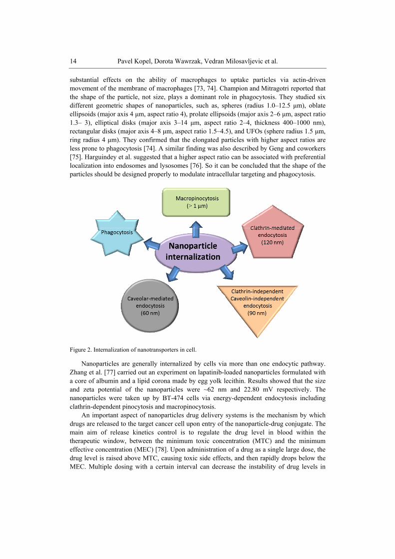
Cells generally use two main endocytic pathways to internalize nanoparticles: Phagocytosis and pinocytosis. Phagocytosis is generally found in neutrophils, dendritic cells, and macrophages [72] whereas Pinocytosis can be found in almost all types of cells. There different types of pinocytosis: clathrin-mediated endocytosis, caveolae-mediated endocytosis, clathrin/caveolae-independent endocytosis, and macropinocytosis (see Figure 2).
A number of researches are carried out to explore the internalization of nanoparticle via phagocytosis. The cellular uptake of nanoparticles through phagocytosis in macrophages employs attractive forces (i.e., ionic, electrostatic, hydrophobic/ hydrophilic, van der Waals) between the surfaces of the cells and nanoparticle. The phagocytosis can be initiated by the receptor-mediated recognition of opsonins adsorbed on the nanoparticle surfaces.
The geometry of the particle can help in controlling their cellular uptake through phagocytosis. Because of different shapes, the particles can generate different angles between the membrane and themselves at the point of cell attachment and this angle of contact shows
Pavel Kopel, Dorota Wawrzak, Vedran Milosavljevic et al. 14
substantial effects on the ability of macrophages to uptake particles via actin-driven movement of the membrane of macrophages [73, 74]. Champion and Mitragotri reported that the shape of the particle, not size, plays a dominant role in phagocytosis. They studied six different geometric shapes of nanoparticles, such as, spheres (radius 1.0–12.5 μm), oblate ellipsoids (major axis 4 μm, aspect ratio 4), prolate ellipsoids (major axis 2–6 μm, aspect ratio 1.3– 3), elliptical disks (major axis 3–14 μm, aspect ratio 2–4, thickness 400–1000 nm), rectangular disks (major axis 4–8 μm, aspect ratio 1.5–4.5), and UFOs (sphere radius 1.5 μm, ring radius 4 μm). They confirmed that the elongated particles with higher aspect ratios are less prone to phagocytosis [74]. A similar finding was also described by Geng and coworkers [75]. Harguindey et al. suggested that a higher aspect ratio can be associated with preferential localization into endosomes and lysosomes [76]. So it can be concluded that the shape of the particles should be designed properly to modulate intracellular targeting and phagocytosis.
Figure 2. Internalization of nanotransporters in cell.
Nanoparticles are generally internalized by cells via more than one endocytic pathway. Zhang et al. [77] carried out an experiment on lapatinib-loaded nanoparticles formulated with a core of albumin and a lipid corona made by egg yolk lecithin. Results showed that the size and zeta potential of the nanoparticles were ~62 nm and 22.80 mV respectively. The nanoparticles were taken up by BT-474 cells via energy-dependent endocytosis including clathrin-dependent pinocytosis and macropinocytosis.
An important aspect of nanoparticles drug delivery systems is the mechanism by which drugs are released to the target cancer cell upon entry of the nanoparticle-drug conjugate. The main aim of release kinetics control is to regulate the drug level in blood within the therapeutic window, between the minimum toxic concentration (MTC) and the minimum effective concentration (MEC) [78]. Upon administration of a drug as a single large dose, the drug level is raised above MTC, causing toxic side effects, and then rapidly drops below the MEC. Multiple dosing with a certain interval can decrease the instability of drug levels in
Nanotransporters for Anticancer Drug Delivery 15
plasma but can cause non-compliance issues in patient. For this reason, it is needed to develop specific drug carriers that provide controlled release of a drug with a low dosing frequency. Hence, a constant drug release rate (zero-order drug release profile) is frequently followed [78, 79]. On the other hand, pulsatile or stimuli-responsive drug release became interesting topic of research also to achieve timely drug release [80-82].
Several factors regulate the release of drugs from carrier such as the composition (drug, polymer, and additives), their ratio, physical and/or chemical interactions among the components, and the preparation methods. The drug release can be classified into four categories on the basis of the mechanism by which a drug escapes a carrier (solvent, diffusion, chemical reaction, and stimuli controlled release) [83, 84].
Solvent transport into a drug carrier can affect the release behaviour of the drug from carriers. There are two types of solvent-controlled release: swelling-controlled release and osmosis-controlled release [83].
If glassy hydrophilic polymeric systems are put in aqueous solutions (eg. body fluids), water diffuses into the system. The uptake of the water causes the swelling of the polymeric particles followed by drug release (swelling-controlled release). The rate of the release of the drug can be determined by the diffusion rate of water and the chain relaxation rate of polymers [85]. Different groups of researchers reported that the swelling-controlled systems can be made by polymeric materials with three dimensionally crosslinked network such as hydrogels, where the mesh size plays a central role in controlling the behaviour of drug release [85, 86]. It was found that the swelling-controlled systems can achieve a zero-order drug release, depending on the polymer composition [87] or initial drug distribution in the system [88].
Osmosis-controlled release can be found in a carrier covered with a semi-permeable polymeric membrane. Water can flow through this membrane from outside of the carrier (with a low drug concentration) to the drug-loaded core (with a high drug concentration). It has been reported that this mechanism can show a zero-order release profile as long as a constant concentration gradient is maintained across the membrane [89].
In case of diffusion-controlled drug release, a drug can be dissolved or dispersed in a core surrounded by polymeric membrane [90]. The different concentrations of the drugs across the membrane drive this diffusion. In this case, the drug initially dissolves in the core then diffuses through the membrane. The diffusion-controlled release profile can also be found in matrix-type nanospheres, where drug molecules are dispersed throughout the polymer matrix. Here, no membrane acts as a diffusion barrier. As a result, this matrix-type system generally shows high initial release, followed by a decreasing release rate with the increasing diffusion distance for drug molecules located inside of the carrier.
Different types of biodegradable polymeric drug carriers such as polyamides, polyesters, poly(amino acids), and polysaccharides release drugs by hydrolytic and/or enzymatic degradation of amide, ester, and hydrazone bonds in their backbones [91-93]. Bulk degradations are found from matrices made of polymers like poly(lactic-co-glycolic acid) (PLGA), polylactic acid (PLA), and polycaprolactone (PCL). This process can result in simultaneous degradation of entire matrices. In a small-dimension matrix like nanoparticls, the distance of water diffusion is short and the domain size of crystallization is found to be restricted. In this case, the polymer degradation is significantly enhanced, and these polymers do not necessarily follow the typical surface erosion behaviour but can show a bulk degradation (constant particle size during polymer degradation) [94].
Pavel Kopel, Dorota Wawrzak, Vedran Milosavljevic et al. 16
The behaviour of the release of the drug from stimuli-responsive nanocarriers is regulated by internal or external factors such as pH, temperature, ionic strength, sound, and electric or magnetic fields [80]. As the stimuli can be localized, these carriers were explored for target-specific drug delivery. Researchers have developed some nanocarriers with pH-sensitive linkers for tumor-specific drug delivery [95, 96]. To increase the contrast between intracellular and extracellular drug release pH-sensitive carriers have been developed [96-98]. In case of thermosensitive drug carriers, the temperature-induced phase transition of the polymer causes the release of the drugs [99, 100].
3. LIPOSOMES AND THEIR MODIFICATIONS Nanotransporters have been explored for delivering drugs for over 35 years. Liposomes
have been the most successful drug delivery carriers. Liposomes are extensively used as carriers for numerous molecules in cosmetic and pharmaceutical industries. Additionally, liposomes can entrap unstable compounds (for example, antimicrobials, antioxidants, flavors and bioactive elements) and can trap both hydrophobic and hydrophilic compounds, avoid decomposition of the entrapped compounds and release the entrapped at designated targets. Because of their biocompatibility, biodegradability, low toxicity and possibility to trap both hydrophilic and lipophilic drugs and simplify site-specific drug delivery to tumor tissues, liposomes have increased rate of both as an investigational system and commercially as a drug delivery system. The drugs inside the liposomes are protected from oxidation and degradation during circulation in bloodstream. This protective phospholipid shield or barrier remains undamaged until the contents of the liposome are delivered to the exact target gland, organ, or system where the contents will be utilized [101]. Since the introduction of Doxil (a PEGylated liposomal doxorubicin), several products have been approved worldwide [102]. Doxil was approved by the US FDA in 1995 [103]. Liposomes are mostly used for the passive targeting having blood lifetime one or two days. It is required that the size of liposomes is less than 200 nm to facilitate fenestration through the leaky blood vessels around the tumour site. In general, the release of drug from liposome has to be slow enough not to let free drug to be removed quickly from the bloodstream. Liposome involves an aqueous core entrapped ba one or more bilayers composed of natural or synthetic lipids. Drugs with different lipophilicity can be encapsulated into liposomes i.e. lipophilic drugs are located in the lipid bilayer whereas hydrophilic drugs are in the core and those intermediary between lipid and aqueous phases.
Doxorubicin was loaded into the aqueous core of the liposome at a concentration exceeding saturation. This high loading (~ 12.5% by weight of the lipids) was achieved through a special technique called active loading using an ammonium sulphate gradient [104]. Undissolved doxorubicin is in the core in the form of crystals. This method of preparation serves to prolongation of the drug release beyond the circulation halftime.
Most of the liposomes for cancer treatment were approved on the basis of reduced side effects due to their passive targeting capabilities. Other liposomal anticancer products, such as DaunoXome and Myocet were primarily used to reduce toxicity in comparison to free doxorubicin and not to sustained release of encapsulated drug [105].
Nanotransporters for Anticancer Drug Delivery 17
Except of conventional liposomes there are modified ones called stealth liposomes. These were designed to improve properties of transporters. The main reason for modification of liposomes is that although they behave like biomembranes they are still foreign objects of the body. Therefore, liposomes are known by the mononuclear phagocytic system (MPS) after contact with plasma proteins. Accordingly, liposomes can be removed from the blood stream. These stability difficulties are solved through the use of synthetic phospholipids, particle coated with amphipathic polyethylene glycol (PEG), coating liposomes with chitin derivatives, freeze drying, polymerization, micro-encapsulation of gangliosides [106]. Coating liposomes with PEG reduces the percentage of uptake by macrophages and leads to a prolonged circulation and, therefore, make available abundant time for these liposomes to leak from the circulation. Stealth liposomes are transporters with a membrane composed of phospholipid bilayer used to deliver drugs into a cell. A liposome can be made of naturally phospholipids with mixed lipid chains coated by polymers of PEG and colloidal in nature. Stealth liposomes are a new generation of compounds used for controlled drug release [107]. This stealth principle has been used to develop the successful doxorubicin-loaded liposome product that is presently marketed as Doxil (Janssen Biotech, Inc., Horsham, USA) or Caelyx (Schering-Plough Corporation, Kenilworth, USA) for the treatment of solid tumors [108].
Very often, poly(ethylene glycol) molecules (PEG) (molar mass ~ 2000 Da ) are situated on to the surface of the liposomes by mixing the PEGylated lipids with the main lipids that form liposome [103]. The combination of properties (PEGylation, active loaded crystallized drug and small size) enables some selectivity of action towards tumor tissue, thus reducing side effects of the drug. For example, the biodistribution characteristics of liposomes surface-modified with the mixture of polyethylene glycol (PEG) and polyvinyl alcohol (PVA) as a drug carrier for passive targeting of drugs was studied [109]. The liposomes were made of (egg phosphatidylcholine: cholesterol = 55:40, molar ratio) modified with both PEG and PVA (4:1 molar ratio).
Clinically have been approved these drugs in liposomes – Doxil (Caelyx in Europe), Myocet, DaunoXome and DepoCyt [101]. Doxorubicin is known for its cardiotoxicity which is minimized by closing in the first two liposomal drugs. Doxil is a PEGylated liposome and it was already mentioned that use of PEG increases circulation time, which result in delaying of capture by the reticuloendothelial system [110]. Doxil is used for treatment of metastatic breast cancer, ovarian cancer, multiple myeloma and also in treatment of AIDS-related Kaposi’s sarcoma (KS).
It was also shown that taxane-resistant breast cancer patients treated with PEGylated liposomal doxorubicin exhibited slightly increased survival results compared to those treated with vinorelbine or vinblastine in combination with mitomycin C [111]. The effectiveness of doxorubicin and its PEGylated form is nearly the same but cardiotoxicity, myelosuppression, vomiting and alopecia was decreased in the groups of patients treated with Doxil [112]. The same result was obtained for Kaposi’s sarcoma treatment [113, 114]. Doxil was also compared with topotecan used for ovarian cancer. Doxil show much lower toxicity and effectiveness of therapy is comparable [115]. Increased efficiency gives Doxil in combination with other chemotherapeutic drugs docetaxel or bortezomib but unfortunately this combination leads to higher toxicity [116, 117]. Doxil, in combination with radiotherapy, has shown increased anticancer effect [118]. Myocet is a liposomal doxorubicin used for the treatment of breast cancer in combination with cyclophosphamide. In the trials and in comparison with free doxorubicin it was found that Myocet show lower cardiotoxicity and
Pavel Kopel, Dorota Wawrzak, Vedran Milosavljevic et al. 18
neutropenia and has the same effectiveness of cancer treatment [119, 120]. Another liposomal product DaunoXome containing daunorubicin was showing better results than doxorubicin, bleomycin and vincristine, in Kaposi’s sarcoma treatment [121]. DepoCyt is commercially available non-PEGylated liposomal form for cytarabine, which belong to a group of hydrophilic chemotherapeutic drugs. It can be used for treatment of lymphomatous meningitis, leukaemia and glioblastoma.
There are plenty of anticancer liposomes under clinical trials. To the group belong PEGylated lipoplatin, S-CKD602 [122] and NL CPT-11 containing cisplatin, CKD-602 (a camptothecin analogue) and irinotecan (CPT-11), respectively. From the trials it follows that lipoplatin is less toxic and of the same activity like cisplatin applicable for various cancers [123, 124, 125].
Drug irinotecan was encapsulated in liposomes and products are under names NL CPT-11, CPX-1 and LE-SN38. The drugs are tested for glioma and colorectal cancer treatment [126]. Liposomes can be modified for specific targeting. Such example can be MBP-426 containing oxaliplatin and transferrin which is bonded to phosphatidylethanolamine of liposome [127]. MCC-465 is a PEGylated liposome encapsulating doxorubicin with surface modification with a fragment of the human monoclonal antibody, able to identify cell surface molecules of different cancer cells [128, 129]. CPX-351 is a liposome encapsulated formulation of cytarabine and daunorubicin that exploits molar ratio-dependent drug-drug synergy to enhance antileukemic efficacy. The phase II study shows on lower mortality of patients [130]. Trastuzumab combined with sequential chemotherapy with taxanes and anthracyclines as primary systemic therapy achieved high rates of pathologic complete response. Non-pegylated liposome-encapsulated doxorubicin has shown equal efficacy but minor cardiotoxicity compared to doxorubicin. This phase II study aimed to evaluate the activity and safety of trastuzumab with sequential chemotherapy for early or locally advanced HER2 positive BC.
No cardiac toxicity or discontinuation of trastuzumab was reported. This study confirms that integrating anti-HER2 therapy in primary treatment for HER2 positive breast cancer is active [131].
3.1. Modified Liposomes To improve efficacy of cancer treatment by liposomes it is necessary to modify either
liposomes or their surface by peptides, RNA or antibodies which also serve for targeted delivery to specific cancer tissue. Some examples of recent development in the field are mentioned hereinafter.
Dual-ligand liposomal delivery system for targeting the delivery of paclitaxel to lung cancer was prepared. The specific ligand peptide HAIYPRH (T7) and the cationic cell-penetrating peptide TAT were connected with phospholipid via a polyethylene glycol (PEG) spacer to prepare the dual-ligand liposomes (T7/TAT-LP-PTX) [132].
Active targeting molecules displayed better cell selectivity but were shadowed by the poor tumor penetration effect. Cell penetrating peptides could increase the uptake of the carriers but were limited by their non-specificity. Dual-ligand system may possess a synergistic effect and create a more ideal drug delivery effect. Thus, liposome system modified with RGD, TAT and cleavable PEG was designed. The RGD specifically
Nanotransporters for Anticancer Drug Delivery 19
recognized the integrins overexpressed on various malignant tumors and mediated efficient internalization in the synergistic effect of the RGD and TAT. In vitro cellular uptake and 3D tumor spheroids penetration studies demonstrated that the system could not only be selectively and efficiently taken up by cells overexpress ingintegrins but also penetrate the tumor cells to reach the depths of the avascular tumor spheroids. In vivo imaging and fluorescent images of tumor section further demonstrated that this system achieved profoundly improved distribution within tumor tissues, and the RGD and TAT ligands on C-R/T liposomes produced a strong synergistic effect that promoted the uptake of liposomes into cells after the systemic administration of L-cysteine. The results of this study demonstrated a tremendous potential of this multistage liposomes for efficient delivery to tumor tissue and selective internalization into tumor cells [133].
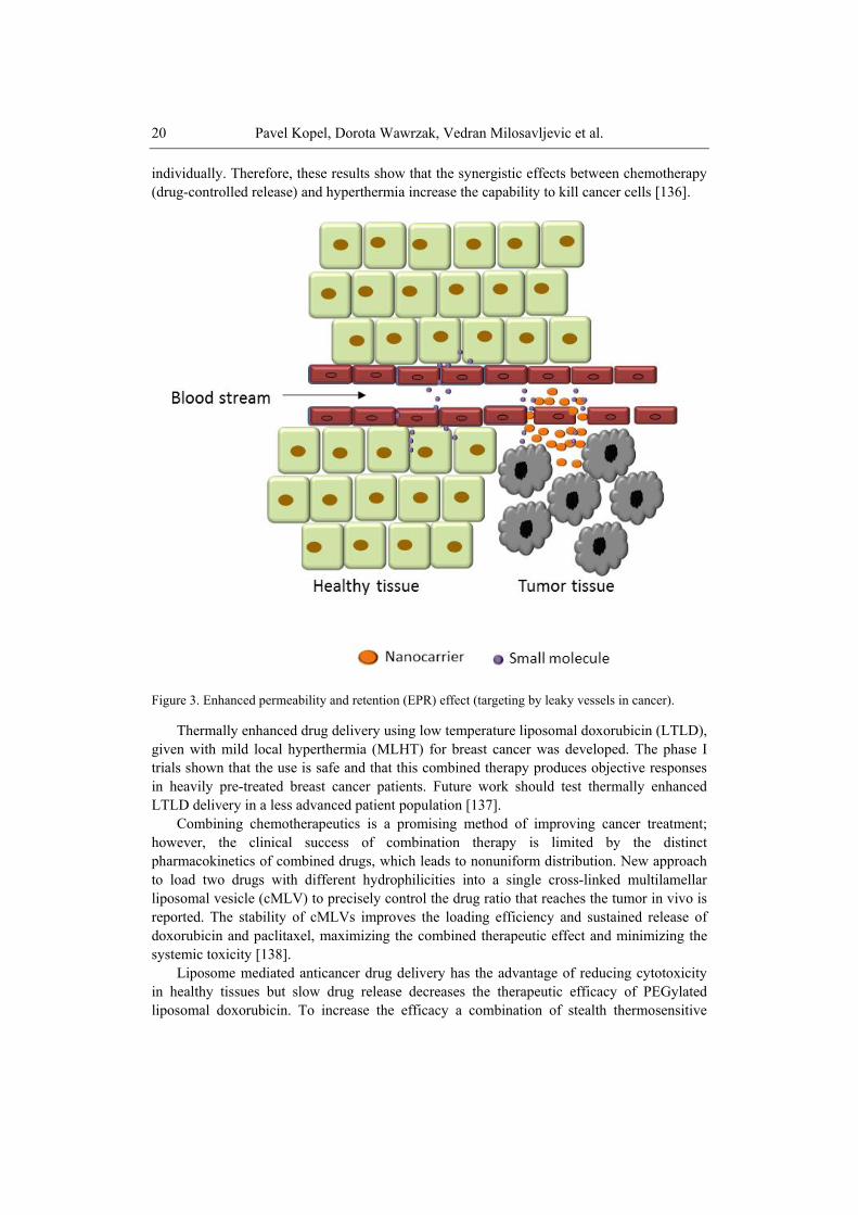
Liposomal drug delivery system conjugated with cyclic arginine-glycine-aspartic acid-tyrosine-lysine peptide (cRGDyk) was developed to improve therapeutic efficacy in a mice model of bone metastasis from prostate cancer. Compared with free cisplatin and cRGDyk-free liposomes, cRGDyk conjugated liposomes showed significantly higher cellular uptake and higher cytotoxicity of loaded cisplatin, as evidenced by in vitro cell experiments. In vivo results revealed that free cisplatin and free cRGDyk could relieve tumor-induced pain but had no contributions to tumor regression and overall survival improvement. cRGDyk-free liposomal drug system with prolonged blood circulation time could accumulated in the tumor sites in the bone through enhanced permeability and retention (EPR) effects (see Figure 3) and however, did not exhibit desirable therapeutic efficacy superior to free cisplatin and free cRGDyk. Inspired by their enhanced therapeutic efficacy and low organ toxicity, cRGDyk conjugated liposomes could serve as an effective drug system for targeted and synergistic therapy of bone metastases [134].
Novel, acid-sensitive liposomes that respond to physiopathological pH for tumour targeting applications were obtained by surface modification with 1,2-distearoyl-sn-glycero-3-phosphoethanolamine-N-[methoxy(polyethyleneglycol)] (mPEG-DSPE) and stearoyl-poly(ethyleneglycol)-poly(methacryloylsulfadimethoxine) copolymer (stearoyl-PEG-polySDM). All of the liposome formulations were stable at pH 7.4, even in the presence of foetal bovine serum, but they underwent rapid size increase at pH 6.5. At pH 6.5, these liposomes displayed higher cytotoxicity than at pH 7.4 or compared to non-responsive control liposomes at both incubation pH. Notably, treatment with free gemcitabine did not yield cytotoxic effects, indicating that the carrier can efficiently deliver the anticancer drug to the cytosolic compartment [135].
The cancer treatment through the combination of chemotherapy and thermotherapy using doxorubicin-loaded magnetic liposomes is reported. The citric acid-coated magnetic nanoparticles (CAMNP, ca. 10 nm) and doxorubicin were encapsulated into the liposome (HSPC/DSPE/cholesterol = 12.5:1:8.25) by rotary evaporation and ultrasonication process. In vitro cytotoxicity of the liposome was investigated through 3-(4,5-dimethylthiazol-2-yl)-2,5-diphenyltetrazolium bromide (MTT) assay using L-929 cells, as the mammalian cell model. In vitro cytotoxicity and hyperthermia (inductive heating) studies were evaluated against colorectal cancer (CT-26 cells) with high-frequency magnetic field (HFMF) exposure. MTT assay revealed that these drug carriers exhibited no cytotoxicity against L-929 cells, suggesting excellent biocompatibility. When the magnetic liposomes with doxorubicin was used to treat CT-26 cells in combination with HFMF exposure, approximately 56% cells were killed and found to be more effective than either hyperthermia or chemotherapy treatment
Pavel Kopel, Dorota Wawrzak, Vedran Milosavljevic et al. 20
individually. Therefore, these results show that the synergistic effects between chemotherapy (drug-controlled release) and hyperthermia increase the capability to kill cancer cells [136].
Figure 3. Enhanced permeability and retention (EPR) effect (targeting by leaky vessels in cancer).
Thermally enhanced drug delivery using low temperature liposomal doxorubicin (LTLD), given with mild local hyperthermia (MLHT) for breast cancer was developed. The phase I trials shown that the use is safe and that this combined therapy produces objective responses in heavily pre-treated breast cancer patients. Future work should test thermally enhanced LTLD delivery in a less advanced patient population [137].
Combining chemotherapeutics is a promising method of improving cancer treatment; however, the clinical success of combination therapy is limited by the distinct pharmacokinetics of combined drugs, which leads to nonuniform distribution. New approach to load two drugs with different hydrophilicities into a single cross-linked multilamellar liposomal vesicle (cMLV) to precisely control the drug ratio that reaches the tumor in vivo is reported. The stability of cMLVs improves the loading efficiency and sustained release of doxorubicin and paclitaxel, maximizing the combined therapeutic effect and minimizing the systemic toxicity [138].
Liposome mediated anticancer drug delivery has the advantage of reducing cytotoxicity in healthy tissues but slow drug release decreases the therapeutic efficacy of PEGylated liposomal doxorubicin. To increase the efficacy a combination of stealth thermosensitive
Nanotransporters for Anticancer Drug Delivery 21
liposomes and local mild hyperthermia was investigated to increase bioavailable doxorubicin levels in tumors. It was found that the combination of PEGylated thermosensitive liposome and local mild hyperthermia offers promising clinical opportunities to improve liposomal doxorubicin delivery to solid tumors [139].
A single nanoparticle platform has been developed through the modular and controlled layer-by-layer process to codeliver siRNA that knocks down a drug-resistance pathway in tumor cells and a chemotherapy drug to challenge a highly aggressive form of triple-negative breast cancer. Layer-by-layer films were formed on nanoparticles by alternately depositing siRNA and poly-L-arginine; a single bilayer on the nanoparticle surface could effectively load up to 3500 siRNA molecules, and the resulting nanoparticles exhibit an extended serum half-life of 28 h. In animal models, one dose via intravenous administration significantly reduced the target gene expression in the tumors by almost 80%. By generating the siRNA-loaded film on doxorubicin-loaded liposome, the efficacy was enhanced by 4 fold in vitro and led to up to an 8 fold decrease in tumor volume compared to the control treatments with no observed toxicity [140].
Liposomal drug delivery has expanded considerably over the past few decades, and several liposomal drugs are already providing improved clinical outcomes. Liposomes have now progressed beyond simple, inert drug carriers and can be designed to be highly responsive in vivo, with active targeting, increased stealth, and controlled drug-release properties. Ligand targeted liposomes have the potential to revolutionize the treatment of cancer. Recent challenges in ligand targeted liposomes are described [141].
4. NATURAL NANOTRANSPORTERS – PROTEINS Ferritin belongs to proteins with cage like structure, which can be used to bind molecules
in its cavity. Ferritin molecules are present in most living organisms and are used to store iron ions as their hydrated hydroxide-oxide Fe(III) to avoid their toxicity due to free radicals that can be generated with Fe(III), which is readily reduced to Fe(II). There are two kinds of ferritins namely maxiferritins and miniferritins. These have distinctly different inner and outer diameters and molecular weights. Maxiferritins are formed from 24 subunits 12 nm in diameter with 8 nm cavities with MW = 480 kDa and miniferritins formed from 12 subunits 8 nm in diameter with 5 nm diameter cavities of MW = 240 kDa [142]. Mostly maxiferritins are used, especially horse spleen ferritin for its commercial availability. Ferritin wide occurrence as well as its ability to reversibly store and release iron ions to the living cells has attracted the interest of researchers worldwide.
It was found that the cavity can be utilized for storage of other ions and molecules and can be utilized for synthesis of nanoparticles with defined size. Apart from interior cavity, the surface of apoferritin can be modified. This offers further possibility of delivering encapsulated drug to a target cell in more effective way and minimalizing thus side effects particularly toxicity to nontarget organs by drugs.
There are two possible ways for preparation of apoferritin loaded compounds. These routes can be called reassembly and nanoreactor routes. Reassembly route comprise of disassembly of the apoferritin shell into protein subunits at low pH by addition of an acid. Then the solution of complex, drug or nanoparticles is added. Afterwards, pH of the solution
Pavel Kopel, Dorota Wawrzak, Vedran Milosavljevic et al. 22
is adjusted with addition of a base to pH around 8. Protein subunits are reassembled again to form apoferritin with loaded compounds in the cavity [143]. Nanoreactor route comprise of utilization of ion or nanoparticles diffusion through channels in apoferritin structure. Mostly, ions are added to the apoferritin solution and mixture is shaken, afterwards, anions are added which form nanoparticles in the cavity. Reactants and precipitates outside of apoferritin are than removed by centrifugation, dialysis or ultrafiltration.
Selective dose-dependent antitumor activity of horse spleen apoferritin encapsulated PbS quantum dots against two human-derived colorectal carcinoma cell lines is reported (GI50 similar to 70 µg mL-1) [144]. Following in vitro exposure to PbS, CRC cells fail to recover proliferative capacity, and undergo apoptosis triggered by the generation of reactive oxygen species (ROS). In contrast, the apoferritin-PbS nanocomposites do not affect the growth and cell cycle of non-tumor human endothelial HMEC-1 cells. Neither adverse health nor behavioral indicators were observed throughout the 15 day study in mice. The photoluminescence combined with selective antitumor activity offer potential for simultaneous non-invasive imaging and treatment of malignant tissue. Apoferritin was employed to encapsulate anticancer drugs cisplatin and carboplatin [145, 146]. It is well known, that clinical application of platinum-based anticancer drugs is largely limited by severe general toxicity and drug resistance. Drug delivery systems with tumor-targeting potential are highly desired for improving the efficacy and applicability of these drugs. The delivery of platinum drugs cisplatin, carboplatin and oxaliplatin by encapsulating each of them in the cavity of apoferritin was studied. The encapsulation was achieved through reassembly route at pH 2.0 and 7.4, respectively, in saturated drug solution. UV-vis spectrometry, circular dichroism spectrometry, dynamic light scattering, and inductively coupled plasma mass spectrometry were used to characterize the apoferritin-drug complexes. The loading capacity of apoferritin varies with respective drugs and the structural integrity of the protein shell remains intact after encapsulation. In vitro assays on the rat pheochromocytoma cell line (PC12) show that cisplatin inhibits the cells in a slow but sustaining mode and the cellular uptake of platinum drug is enhanced by apoferritin. Carboplatin and oxaliplatin complexes in apoferritin exhibit a marginal cytotoxicity towards this cell line under similar concentrations [146]. A novel antibody-drug conjugate was synthesized incorporating ferritin cisplatin nanoparticles [147]. An average of three molecules of monoclonal antibody (mAb) Ep1 to the human melanoma-specific antigen CSPG4 were conjugated to a single ferritin cage encapsulating about 50 cisplatin molecules. The flow cytometry demonstrated specific binding to a CSPG4(+) melanoma cell line, but not to a CSPG4(-) breast carcinoma cell line. As compared to the cisplatin-containing ferritin nanoparticle alone, which inhibited thymidine incorporation more efficiently in breast carcinoma than melanoma cells, the mAb-derivatized nanoparticle had a 25-fold preference for the latter. Anticancer activity was also studied on a methylene blue-encapsulated apoferritin complex. The complex shows cytotoxic effects on MCF-7 human breast adenocarcinoma cells when irradiated at the appropriate wavelength [148].
Ferritin can be genetically modified to present a peptide sequence on the surface [149]. Thus Cys-Asp-Cys-Arg-Gly-Asp-Cys-Phe-Cys (RGD4C)-modified ferritin can efficiently target tumors through RGD-integrin interaction. It was shown that after being precomplexed with Cu(II), doxorubicin can be loaded onto RGD modified apoferritin with high efficiency (up to 73.49 wt %). These doxorubicin-loaded ferritin nanocages showed on U87MG subcutaneous tumor models a longer circulation half-life, higher tumor uptake, better tumor
Nanotransporters for Anticancer Drug Delivery 23
growth inhibition, and less cardiotoxicity than free doxorubicin. Such a technology might be extended to load a broad range of therapeutics and holds great potential in clinical translation [149]. RGD4C-modified ferritin can also serve as a safe and efficient photosensitizer vehicle [150]. Zinc hexadecafluorophthalocyanine (ZnF16Pc), a potent photosensitizer with a high quantum yield but poor water solubility, can be encapsulated into ferritin and tests on U87MG subcutaneous tumor models, P-RFRTs showed a high tumor accumulation rate and minimal toxicity to the skin and other major organs.
Ma-Ham et al. [151] studied daunomycin, an anthracycline antibiotic drug that is used for specific types of cancer treatment such as acute myeloid leukemia and acute lymphocytic leukemia, encapsulated within apoferritin cage. They used reassembly route to load the therapeutic compound daunomycin into the cavity of apoferritin. At experimental pH 5 conditions the interaction between the apoferritin interior cage and daunomycin was weak making it difficult to encapsulate the drug effectively within the protein cage. The incorporation of poly-L aspartic acid, a polypeptide and biodegradable material that does not increase the toxicity of the drug delivery system and is negatively charged at pH 5.0, into the drug delivery system resulted in a substantial improvement in the drug encapsulation. The binding properties of free daunomycin with DNA were compared to the synthesized apoferritin protein based drug delivery system. Encapsulation of the daunomycin within the apoferritin protein cage had little effect upon the intrinsic binding constant, K(i), or the exclusion parameter n as compared to the free daunomycin model. The study resulted in the design and optimization of a unique protein based drug delivery platform using the protein cage apoferritin for potential therapeutic administration of the anticancer agent daunomycin.
Doxorubicin belongs to the group of anthracycline antibiotics with very effective anticancer properties. On the other hand, the cardiotoxic effects limit its application. To overcome this obstacle, encapsulation of this drug into the protective nanotransporter such as apoferritin is beneficial. The apoferritin-doxorubicin complex has been formed by reassembly of the apoferritin sphere in the presence of doxorubicin [152]. The doxorubicin encapsulation was carried out using direct and step-wise change of pH of the solution from 2.5 to 7.4. Non-denaturing polyacrylamide gels showed that the protein cage of the complex successfully self-assembles into its nanosphere form. It was found that up to 28 molecules of doxorubicin can be capsulated per apoferritin protein and no significant drug leakage occurs during the first two days.
Magnetic particle mediated transport in combination with nanomaterial based drug carrier has a great potential for targeted cancer therapy. Doxorubicin encapsulated into the apoferritin was conjugated with magnetic particles and investigated by capillary electrophoresis with laser-induced fluorescence detection (CE-LIF). This combination of magnetic particles and drug encapsulated in apoferritin can be potentially used for magnetic resonance imaging, thermotherapy and chemotherapy [153].
Voltammetry was used for detection of doxorubicin (DOX) and encapsulated doxorubicin in apoferritin structure at a carbon paste electrode [154]. The samples were measured by differential pulse voltammetry in phosphate buffer (pH 5.5). The experimental conditions as time of accumulation and deposition potential were optimized. The study also shows on the release of doxorubicin at different pH which is very important for cancer treatment. Fluorescent behaviour of doxorubicin in various solvents was studied [155]. Ethanol, acetonitrile and dimethyl sulfoxide were tested and the best linearity of the calibration curve was obtained when above 50 % of the solvent was present in the binary mixture with water.
Pavel Kopel, Dorota Wawrzak, Vedran Milosavljevic et al. 24
Moreover, pH influence on the DOX fluorescence was also observed within the range of 4-10. Further, the DOX behavior in capillary electrophoresis (CE) was investigated. Electrophoretic mobilities (CE) in various pH of the background electrolyte were determined and CE was also used to monitor the encapsulation of DOX into the cavity of apoferritin as well as the pH-triggered release [155]. Apoferritin and liposome encapsulated forms of doxorubicin were prepared and their toxicity were compared with doxorubicin alone and Myocet on prostate cell lines [156]. Three different prostatic cell lines PNT1A, 22Rv1, and LNCaP were chosen. The toxicity was compared using the MTT assay, real-time cell impedance-based cell growth method (RTCA), and flow cytometry. The efficiency of doxorubicin entrapment was 56% in apoferritin cages and 42% in the liposome carrier. Apodox IC50 was determined as follows: 603.1, 1344.2, and 931.2 nM for PNT1A, 22Rv1, and LNCaP, respectively. Ferritin nanocages can carry high doses of doxorubicin for tumor-specific targeting and killing without any targeting ligand functionalization or property modulation [157]. Doxorubicin loaded apoferritin specifically bound and subsequently internalized into tumor cells via interaction with overexpressed transferrin receptor 1 and released doxorubicin in the lysosomes. In vivo in the mouse, it exhibited more than 10-fold higher intratumoral drug concentration than free doxorubicin and significantly inhibited tumor growth after a single-dose injection. It also displayed an excellent safety profile that significantly reduced healthy organ drug exposure and improved the maximum tolerated dose by fourfold compared with free doxorubicin [157].
Ferritins can be used not only for drug transport but can be applied for magnetic resonance imaging, optical imaging and cell tracking. The progress in the field can be also found in some reviews that appeared just recently [158-161].
5. DENDRIMERS AND POLYMER NANOPARTICLES
5.1. Polymeric Nanoparticles Polymeric nanoparticles represent biomaterial widely used for drug delivery application,
which arise from their remarkable biodegradability and biocompatibility with other molecules. Compared with other drug delivery materials (especially with liposome) the polymeric nanoparticles demonstrate higher stability, sharper size distribution, more tunable physicochemical properties, sustained and more controllable drug-release profiles, and higher loading capacity for poorly water soluble drugs. The applications of the polymers in drug delivery have been investigated extensively for group of natural and as well as for synthetic polymers [162, 163]. Natural polymers such as albumin, chitosan and heparin have been a material of choice for the delivery of oligonucleotides, DNA, protein and drugs. The clinical application of natural polymers as transporter for anticancer drug delivery were reported in the case of metastatic breast cancer, non–small-cell lung cancer (phase II trial) and advanced nonhematologic malignancies (phase I and pharmacokinetics trials using abraxane which represents nanometer-sized albumin-bound paclitaxel [164-166]. However, the synthetic polymers enable the delivery of various drugs due to their great modification potential allowing particles to be tailored for specific needs. N-(2-hydroxypropyl)-methacrylamide copolymer (HPMA), poly(lactico-glycolic acid) (PLGA) and polycaprolactone (PCL),
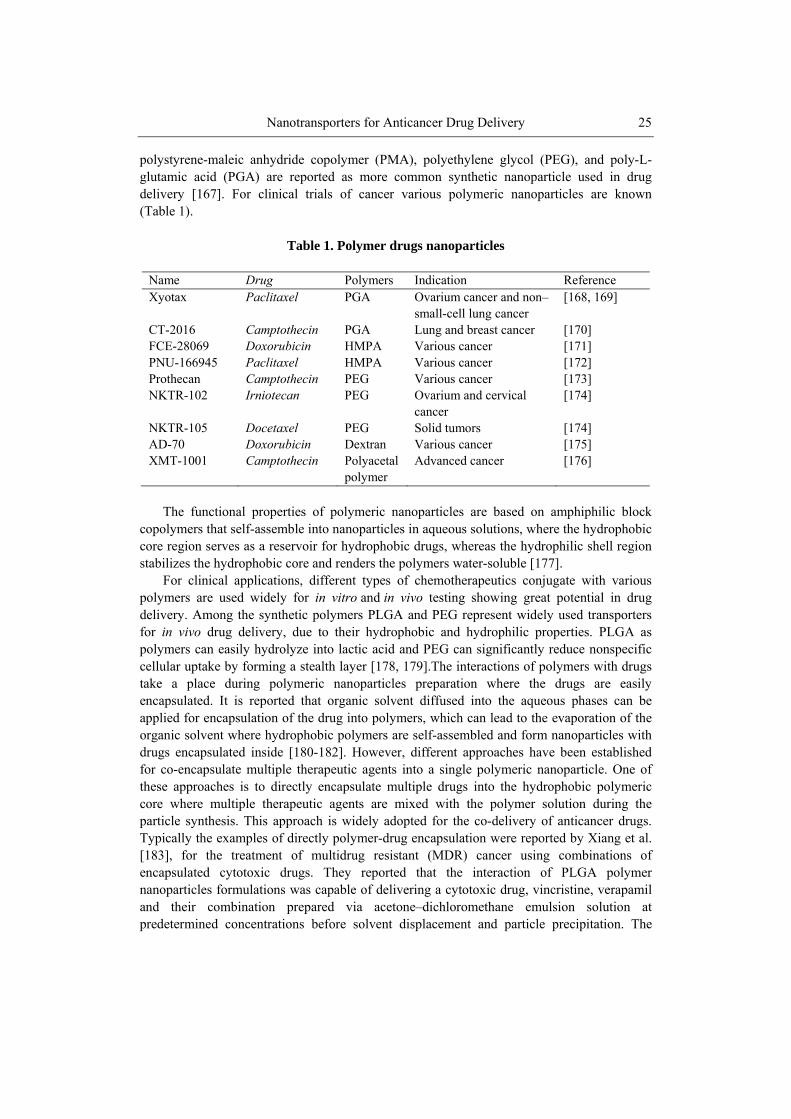
Nanotransporters for Anticancer Drug Delivery 25
polystyrene-maleic anhydride copolymer (PMA), polyethylene glycol (PEG), and poly-L-glutamic acid (PGA) are reported as more common synthetic nanoparticle used in drug delivery [167]. For clinical trials of cancer various polymeric nanoparticles are known (Table 1).
Table 1. Polymer drugs nanoparticles
Name Drug Polymers Indication Reference Xyotax Paclitaxel PGA Ovarium cancer and non–
small-cell lung cancer [168, 169]
CT-2016 Camptothecin PGA Lung and breast cancer [170] FCE-28069 Doxorubicin HMPA Various cancer [171] PNU-166945 Paclitaxel HMPA Various cancer [172] Prothecan Camptothecin PEG Various cancer [173] NKTR-102 Irniotecan PEG Ovarium and cervical
cancer [174]
NKTR-105 Docetaxel PEG Solid tumors [174] AD-70 Doxorubicin Dextran Various cancer [175] XMT-1001 Camptothecin Polyacetal
polymer Advanced cancer [176]
The functional properties of polymeric nanoparticles are based on amphiphilic block
copolymers that self-assemble into nanoparticles in aqueous solutions, where the hydrophobic core region serves as a reservoir for hydrophobic drugs, whereas the hydrophilic shell region stabilizes the hydrophobic core and renders the polymers water-soluble [177].
For clinical applications, different types of chemotherapeutics conjugate with various polymers are used widely for in vitro and in vivo testing showing great potential in drug delivery. Among the synthetic polymers PLGA and PEG represent widely used transporters for in vivo drug delivery, due to their hydrophobic and hydrophilic properties. PLGA as polymers can easily hydrolyze into lactic acid and PEG can significantly reduce nonspecific cellular uptake by forming a stealth layer [178, 179].The interactions of polymers with drugs take a place during polymeric nanoparticles preparation where the drugs are easily encapsulated. It is reported that organic solvent diffused into the aqueous phases can be applied for encapsulation of the drug into polymers, which can lead to the evaporation of the organic solvent where hydrophobic polymers are self-assembled and form nanoparticles with drugs encapsulated inside [180-182]. However, different approaches have been established for co-encapsulate multiple therapeutic agents into a single polymeric nanoparticle. One of these approaches is to directly encapsulate multiple drugs into the hydrophobic polymeric core where multiple therapeutic agents are mixed with the polymer solution during the particle synthesis. This approach is widely adopted for the co-delivery of anticancer drugs. Typically the examples of directly polymer-drug encapsulation were reported by Xiang et al. [183], for the treatment of multidrug resistant (MDR) cancer using combinations of encapsulated cytotoxic drugs. They reported that the interaction of PLGA polymer nanoparticles formulations was capable of delivering a cytotoxic drug, vincristine, verapamil and their combination prepared via acetone–dichloromethane emulsion solution at predetermined concentrations before solvent displacement and particle precipitation. The
Pavel Kopel, Dorota Wawrzak, Vedran Milosavljevic et al. 26
preparation of polyalkylcyanoacrylate (PACA) nanoparticles in combination with cyclosporin A and doxorubicin were reported by Soma et al. [184], as direct encapsulation of drug into polymer by emulsion polymerization process in which the hydrophobic cyclosporin A was added together with doxorubicin to the polymerization medium to achieve dual drug loading. Both of this studies show in vitro cytotoxicity for multidrug resistant cancer cell, enabling the use of a great potential of polymeric particles for the drug transports and cancer treatment. However, the problem is that it can limit this way of encapsulation come from little possibility to control the release of different drugs.
The drug releasing problem of direct encapsulation method can be avoided using alternative multicompartment approach. This approach is conceived using the advantages of highly functional surface of polymeric nanoparticles. Zhang et al. [162], reported a delivery of hydrophobic and hydrophilic drugs by aptamer-nanoparticles. They produced PLGA targeted polymeric nanoparticles which were conjugate with docetaxel (Dtxl) as a hydrophobic drug and doxorubicin (Dox) as a hydrophilic drug. PLGA polymer nanoparticles ware surface modified with oligonucleotides which was use as targeting ligands and as intercalation sites for a hydrophilic doxorubicin. The efficiency of PLGA nanoparticles was demonstrated against prostate cancer cell resulting in high targeting delivery of docetaxel and doxorubicine as well as effective drug releasing kinetics. The release profiles of Dtxl and Dox from the PLGA nanoparticles-aptamer bioconjugates showed a high releasing efficiency of 80 % after 6 hours for doxorubicin comparing with lower efficiency of docetaxel where after 6 hours it was released only 50 %. These pathways provide excellent opportunities for the delivery of two different class of drug for cancer treatment allowing application of combined therapy. This targeted co-delivery system may also allow for temporally distinct release of drugs, which may have implications for delivery to distinct anatomical locations [180].
The control of drug releasing was reported by Sengupta et al. [180], who synthesized a nanocell consisting of a PLGA core and a lipid envelope to demonstrate the potential of differential drug release in combination with anticancer therapy. The particles were fabricated by encapsulating doxorubicin after the conjugation with PLGA to achieve a slow release profile. Combretastatin from the other side were conjugated with PLGA-liposome allowed optimal loading of drug by partitioning into the lipid bilayer. Since doxorubicin is linked to the PLGA polymer, its release was determined by the hydrolytic degradation rate of the polymer and was much slower than the release of combretastatin resulted in a complete ablation of the tumor cells without affecting the endothelial cells. In compare with PLGA-liposomal co-encapsulation of combretastatin, liposomal shell faster degradation and releasing of drug resulted in a rapid collapse of the vascular network without affecting the tumor cells. The therapeutic efficacy of this treatment were examines on B16/F10 melanomas and Lewis lung carcinoma cell resulting in better tumor reduction, longer median survival time, as well as lower systemic toxicity. Thus, the concept can enables a significant advance in cancer therapy over current approaches.
One more approach for encapsulating and delivering drug by polymer is to capture multiple drugs into single polymeric nanoparticles by conjugation of multiple drugs to the polymer backbone. Bea et al. [185] reported the conjugation of doxorubicin and wortmannin to PEG–poly(aspartate hydrazide) which blocks copolymers through an acid-labile hydrazone bond where the hydrazone linker allows the release of the functional moieties by undergoing rapid hydrolysis in the acidic endosomal and lysosomal environments. According to the authors, the benefits of this approach lies in the fact that the ratio of the drugs can be precisely
Nanotransporters for Anticancer Drug Delivery 27
controlled by varying the drug content during the conjugate synthesis process. The temporal control on anticancer drug releasing has been reported by Lammers et al. [181], using peptide linkers that are susceptible to intracellular proteases. In this experiment, the chemotherapeutic agents gemcitabine and doxorubicin interact with HPMA polymer nanoparticle by Gly-Phe-Leu-Gly peptide linker. This peptide sequence is a known substrate to a lysosomal cysteine protease, cathepsin B, where on this substrate drug release kinetics is observed at pH 6 indicating that after 10 hours 100 % of gemcitabine was released compared with doxorubicin were after 30 hours only 30 % was released increasing its toxicity, and it more strongly inhibited angiogenesis and induced apoptosis. According to the author, the poor doxorubicin releasing is directly connected with their structure which might hinder the cathepsin activity by blocking the peptide substrate. However, these findings demonstrate that passively tumor-targeted polymeric drug carriers can be used for delivering two different chemotherapeutic agents to tumors simultaneously.
The selective control of drug concentration and distribution within the tumor microenvironment is one of the most important factors to achieve effective and safe cancer chemotherapy. Controlled drug delivery systems using macromolecular bioconjugates have been shown to offer benefits for improving the delivering of two different pharmacologically active agents to tumors simultaneously. Among all the polymeric nanoparticles, polymeric micelles from amphiphilic which block copolymers are considered as the most promising drug carriers, and characterized by excellent biocompatibility, high drug-loading content, and markedly improved biodistribution.
5.2. Dendrimers Nanoparticles Dendrimers are a novel class of nanoparticles widely used in cancer therapy composed of
multiple highly branched monomers that emerge radially from the central core. Dendrimer-based drug delivery systems have been developed in order to improve the biodistribution of a drug in the body and to allow the controlled release of the drug at its target site. Their high aqueous solubility, low toxicity, compact globular shape and controlled surface functionalities with defined structure of initiator core, layers of branched repeating units and functional end groups on the outermost layer made them ideal carriers for water soluble and insoluble anti-cancer drugs [186]. The internal cavity of the dendrimers could be used for the encapsulation of hydrophobic anti-cancer drugs, offering the advantage of subsequent controlled release of the drug to the tumors and on the other hand multivalent surface of the dendrimers allows conjugation with hydrophilic drugs. However, it is reported that dendrimers are suitable for small anticancer drugs, where solubility and toxicity of the anticancer drugs against cancer cells can increase the encapsulation into dendrimers [187]. The dendrimer-drug conjugates generally consist of an anti-neoplastic agent covalently attached to the peripheral groups of the dendrimer and various types of this conjugates are reported. Among the dendrimers PGLSA poly(glycerol succinic acid) represent widely used transporters for delivery of camptothecins drugs, who are naturally-derived hydrophobic compounds with anticancer activity. Morgan et al. [188], reported an use of two dendrimes transporters, G4-PGLSA dendrimers with hydroxyl (G4-PGLSA-OH) peripheral groups and G4-PGLSA dendrimers with carboxylate (G4-PGLSA-COONa) peripheral groups, for encapsulation of 10-hydroxycamptothecin (10-HCPT) applied on MCF-7 human breast cancer cells. However, the
Pavel Kopel, Dorota Wawrzak, Vedran Milosavljevic et al. 28
results showed that G4-PGLSA dendrimers with hydroxyl group after mixing with 10-HCPT was precipitated, which was explain as lower water solubility of dendrimers and they reported that the unloaded dendrimer didn’t show any anticancer effect. In contrary to the dendrimers with hydroxyl group, the dendrimers with carboxylate group are excellent soluble in water. 10-HCPT was successfully encapsulated into dendrimer, which showed a significant reduction of cell viability, demonstrating that 10-HCPT retains the activity upon encapsulation. Similar results ware reported by same group [189], only in this experiment they synthesized dendrimer modified with PEG (G4-PGLSA-OH)2-PEG3400, and the anti-cancer activity was examined using HT-29 human colon cancer cells. In this case the same cytotoxicity was reported for encapsulated 10-HCPT as well as for non-encapsulated 10-HCPT. This lead to the conclusion that G4-PGLSA-COONa dendrimer show better abilities for delivering a highly potent lipophilic camptothecin derivative.
Poly(amido amine)-PAMAM dendrimer transporters for delivering of anticancer drugs modified with PEG and poly(γ-caprolactone) were reported by Wang et al. [190]. Doxorubicin and etoposide were successfully encapsulated into a formation of micelle dendrimer and applied on kidney epithelial cells. The results showed that the unloaded dendrimer didn’t cause any cytotoxicity in comparison with loaded drugs, where they showed similar cytotoxicity for the cell at the same concentration. A cytotoxicity assay demonstrated that the star-PCL-PEG copolymer is nontoxic in cell culture. This type of block copolymer can be used as a drug delivery carrier. The anticancer drug delivery by PAMAM dendrimer was also reported by Malik et al., [191], where his group encapsulat cisplatin after interaction with carboxylate surface of dendrimer, enabling synthesis of highly water soluble dendrimer-platinate which slowly released cisplatinum. In vivo experiment was conducted on B16F10 melanomas resulting in an accumulation of cisplatin in tumor cells with high cytotoxicity and less toxicity in normal tissues by 3-to 15-fold compared to the free drug in the solution. This model can open the way for delivery of anticancer drugs. To avoid the solubility problem of doxorubicine and secondary effect on health tissue Kojima and his group encapsulated doxorubicin into PEGylated PAMAM dendrimers with chain end of third or fourth generation via urethane bond [192]. They reported that the encapsulation efficacy of the drug are increasing with dendrimer generation and chain length of poly(ethylene glycol) grafts, where the highest ability have dendrimer of fourth generation with 6.5 doxorubicin molecules per dendrimer molecule and 100 % of drug releasing after 2 hours. PEGylated-PAMAM dendrimers for delivery of anti-cancer drug 5-fluorouracil was studied byBhadra and his group [193]. They reported the synthesis of PAMAM dendrimers, using ethylenediamine as a core and methylmethacrylate as a propagating agent and N-hydroxysuccinimide-activated carboxymethyl MPEG-5000 for PEGylation. The drug loading capacity into the dendrimer was increased by this formulation, but it was also found that the releasing rate and hemolytic activity of the drugs reduced. The PEGylation systems were found suitable for prolonged delivery of an anti-cancer drug in in vitro and for the reduction of drug leakage and hemolytic toxicity, providing excellent system for drug loading in body.
As it is mentioned above many of the drugs have water solubility problem, especially paclitaxel is limited by its poor solubility in water. Wu et al. [194] overcome this problem encapsulating paclitaxel with poly(glycerol) dendrimer formulations which led to a drug water solubility 400-fold higher than that of the free drug. The results showed that the fifth-generation of poly(glycerol) dendrimers increased paclitaxel solubility. They suggested that paclitaxel is not incorporated within the core of these dendrimer, but instead of that methyne
Nanotransporters for Anticancer Drug Delivery 29
groups and aromatic rings of the paclitaxel are surrounded by the dendrimer structure leading to hydrotropic solubilization.
To improve its bioavailability and its poor water solubility Neerman and his group [195], used melamine-based dendrimers to solubilize and reduce toxicity of anticancer drugs methotrexate and 6-mercaptopurine. They reported that the intraperitoneal injection of the known hepatotoxic drug methotrexate and 6-mercaptopurine into mice liver significantly reduce hepatotoxicity and decrease the levels of alanine transaminase for 27% in case of methotrexate and 36% of 6-mercaptopurine compared to those obtained with the free drug.
The delivery of anticancer drugs by the encapsulation into denrimers is limited by the very small capacity of the void spaces within the dendrimers and the problem with long time releasing of encapsulated anticancer drugs. For this reason, drug-encapsulated dendrimer systems may be the best to utilized via direct intra-tumor injection or conjugate then to the surface of the dendrimers.
6. INORGANIC NANOPARTICLES FOR ANTICANCER DRUG DELIVERY Inorganic nanoparticles as drug carrier for cancer treatment can be defined as particles of
metal oxide or metallic composition possessing great opportunity due to their advantages such us easy modification of targeting molecules, drugs or other molecules on them, effective delivery to target sites, resulting in high therapeutic efficacy and controlling drug release by external/internal stimuli. Inorganic nanoparticles provide significantly chemical, physical, and biological properties, and functionality due to their nanoscale size, which is connect high surface area per unit volume and the ability to be functionalized with a large number of ligands to enhance their affinity towards target molecules, local delivery of the drug, reducing of side effect, and provide higher efficiency of the therapeutic molecule. However, synthesis of inorganic nanoparticles offers different ways of preparation depending on the applying drugs [196, 197]. One of the traditional methods for synthesis of inorganic nanoparticles is by sol-gel route, where producing of inorganic nanoparticles are directly connected with influence on pH or thermal conditions in solution. Controlling the hydrolysis and condensation reaction by mineralizers metal oxide species can be synthesis using different type of inorganic precursor such as metal salts, metal halides, and inorganic alkoxides [196]. However, it is also reported more friendly synthesis such us spray-drying process, enabling by spraying homogenization of inorganic precursors solution in special chamber at temperatures bellows then boiling point of the solvent. Using flowing gas, solution is sprayed into chamber as droplet leading to fast evaporation of solution by hot air or nitrogen enable formation of nanoparticles [198]. Using of gas-phase methods can be effective rout for synthesis of inorganic nanoparticles, which can include the use of a combustion flame, laser ablation, chemical vapor deposition, and spray-pyrolysis [199]. Microemulsions present one of thermodynamically stable method for preparation of inorganic nanoparticles without significant mechanical agitation making it a rather simple technique [200]. Among, inorganic nanoparticles, we focus on iron based nanoparticles.
Pavel Kopel, Dorota Wawrzak, Vedran Milosavljevic et al. 30
6.1. Iron Based Nanoparticles Iron based nanoparticles have been actively investigated as the next generation of
nanoparticles systematically administered but directed towards a specific target in the human body while remaining ultimately localized, by means of an applied magnetic field. Benefit of iron based NPs is, the use of localized magnetic field gradients to attract the particles to a chosen site, and the possibility to hold them there until the therapy is complete and then to remove them. It is reported different ways of iron based nanoparticles synthesis in different form such as γ-Fe2O3 (maghemite), Fe3O4 (magnetite), α-Fe2O3 (hermatite), magnetite-based, silica-coated MNP (SiMNP), superparamagnetic Fe3O4 poly ɛ-caprolactone (PCL), silica-coated iron–carbon nanoparticles, MgFe2O4 magnetic nanoparticle and other form iron oxide nanoparticles [201-204]. Superparamagnetic nanoparticle ferumoxtran using as drug delivery system for the lymphatic biodistribution were reported by Rety et al., [205]. Ferumoxtran was applied on rat subdiaphragmatic lymph nodes for investigation of uptake mechanism, were 0.3 ml of lymph was collected over 45 minutes for 24 hours. Cytology results demonstrated that immediately after injection high concentrations of nanoparticles were found in the thoracic lymph, but without any nanoparticles in lymph cells indicating that ferumoxtran was extracellular in the lymph fluid, concluding that transcapillary pathway and subsequent lymph drainage have a main roll to drug pathway into lymph node.
Conjugations of magnetic nanoparticles with anticancer drug doxorubicin were reported by Sadighian et al., [206], demonstrated that the doxorubicin-magnetic nanoparticles can be applied on tumor-bearing tissues with dual targeting effect using magnetic attraction and the pH-sensitive cleavage of the drug–nanoparticle. Also releasing of drug was longer then in case of not loaded drug directly reduces the side effect of anticancer drug. Magnetic field and pH were effective in increasing the DOX-nanoparticle bioavailability in acidic condition then in neutral pH, enabling using this conjugate for targeting and increasing therapeutic efficiency of anticancer drug. The biodistribution of doxorubicin and methotrexate conjugate with magnetite nanoparticles are reported by Samra et al., [207]. Immunosorbent assay and FTIR spectroscopy demonstrated that the doxorubicin 18 % and methotrexate 27 % were immobilization with magnetic nanoparticles respectively. Result showed that modified magnetic nanoparticles with anticancer drug applied on Hella cells binding between 41 and 45 % comparing with B cells which binding 20 to 26 %, with higher releasing of anticancer drug in acid condition (pH-5) of 45 %, than in neutral condition (pH-7.4) of 10 %. These indicate that this methodology can be effective as mechanism for anticancer drug delivery.
Superparamagnetic Fe3O4 poly ɛ-caprolactone (PCL) nanoparticles may provide therapeutic effect on xenografts of human HPAC pancreatic adenocarcinoma cells [208]. Gang and his group reported pharmacokinetic behavior of superparamagnetic poly ɛ-caprolactone nanoparticles conjugated with gemcitabine on tumor cells, resulting in drug loading content of 18.6 % with entrapment efficiency of 52.2%. They were found widely distribution of anticancer drug with 15-fold higher dose in comparison with free gemcitabine in tumor cells, indicating the efficiency of magnetic PCL nanoparticles in drug delivery. Poly- (N-isopropylacrylamide-methyl methacrylic acid, (PNIPAAm-MAA) grafted magnetic nanoparticles as anticancer drug delivery transporter are reported by Akbarzadeh et al. [209], were polymerization of N-isopropylacrylamide and methacrylic acid was conduct using silan coated magnetic nanoparticles. Block copolymer-coated magnetite nanoparticles conjugate with doxorubicin were applied on A549 lung cancer cell line for investigation of targeting and
Nanotransporters for Anticancer Drug Delivery 31
controlled drug delivery. Results show that polymer chains were effectively grafted onto the surface of the Fe3O4 nanoparticles and encapsulate doxorubicin with loading efficiency of 75%, which results in longer time of drug release at pH 5.8 comparing with neutral pH at 7.4. These nanoparticles show potential as anticancer drug delivery system.
6.2. Iron Based Nanoparticles with Silica Modification of iron carbon magnetic nanoparticles with silica are used as an anticancer
drug delivery platform, were chemoadsorptive properties of activated carbon are provide drug loading and magnetic Fe are used for targeting [210]. Silica-coated iron–carbon composite nanoparticles are loaded with doxorubicin and biodistributed in the left hepatic lobe of pigs under external magnetic field. Results show that nanoparticles loaded with doxorubicin was 24 times higher in hepatic tissue then in tissue that is not targeted, leading to penetration of magnetic nanoparticles trough capillary wall encircling the tissue interstitium and hepatic cells. These indicating that the novel silica-coated iron-carbon composite particles could be a potential application in targeted treatment for some kinds of tumor as an effective drug carrier. Applications of arsenic trioxide Mg-Fe ferrite magnetic nanoparticles as anticancer drug delivery system were reported by Yang et al., [211], where their effect was evaluated by in vivo experiment. Particles were prepared by solvent-displacement method using Poly-D,L-lactic-co-glycolic acid (PLGA) in process of synthesis. Cytotoxicity of nanoparticles was investigated on Saos-2 cells showing no significant toxicity, also they investigated concentration of As2O3–MNPs in liver resulting in four time higher concentration then non-magnetically vectored group and kidney where concentration was lower than in non-magnetically vectored group leading to conclusion that polymer-loaded magnetic nanoparticle composed of arsenic trioxide show great possibility for targeting and drug delivery.
7. CARBON BASED NANOMATERIALS Among the several types of nanomaterials invented in recent years, carbon based
materials have attracted tremendous attention due to their unique properties as one of the most promising nanomaterials for a variety of biomedical applications. Different types of carbon based nanomaterials such as graphene, carbon nanotubes, fullerene, nanodiamond and carbon nanoparticles (CNPs) have been developed especially as delivery vehicles for drugs.
In 1960s, nanodiamond was first studied by researchers and recently started to be highlighted in different medical applications [212]. The fullerene was discovered in 1985 as a new type of carbon molecules [213]. The carbon nanotube (CNT) was first discovered by Iijima in 1991 [214]. In comparison with other nanomaterials, CNTs (an allotrope of carbon) can be considered as more dynamic nanomaterials in their biological application. For example, not like other nanomaterials, CNTs have the potential to be used not only in imaging but also for drug delivery and thermal ablation [215]. Generally, it is prepared using three main techniques: laser ablation, thermal or plasma enhanced chemical vapour deposition and electric arc discharge [216]. The application of CNTs in drug delivery to their cite of action has become one of the main areas of interest for different researchers due to their
Pavel Kopel, Dorota Wawrzak, Vedran Milosavljevic et al. 32
special characteristics including their unique physical, chemical, and biological properties, hollow monolithic structure, nanoneedle shape, and their ability to acquire the desired functional groups on their surface [217]. The shape of CNTs allows them to penetrate cells using various methods including endocytosis or passive diffusion across the lipid bilayer. In case of endocytosis, the CNT attaches to the cell surface and is subsequently engulfed by the cell membrane [217, 218]. The factors which make CNTs as a promising drug carrier are the hollow monolithic structure of CNTs and their ability to bind desired functional groups on the surface [217, 219].
CNTs can be mainly classified into two categories on the basis of the number of cylindrical graphene layers: single-walled carbon nanotubes (SWCNTs) and multi-walled carbon nanotubes (MWCNTs). SWCNTs have a single cylindrical carbon layer whose diameter varies from 0.4 to 2 mm depending on the temperature at which they have been prepared [220]. It has been reported that a higher growth temperature is responsible to give a larger diameter. On the other hand, MWCNTs are usually consists of several cylindrical carbon layers with diameters in the range of 2–100 nm for the outer tubes and 1–3 nm for the inner tubes [221].
Different research groups reported that functionalized water soluble CNTs can enter cells. The exact uptake mechanism may depend on the size and surface chemistry of CNTs and the type of the cells. The majority of reports have revealed that CNTs functionalized by oxidization, wrapped by DNA, and coated by surfactants or amphiphilic polymers can be engulfed by cells using an energy-dependent endocytosis pathway [222-227]. The similar entryway can be found for other nanomaterials used in biomedicine.
Some researchers also showed that CNTs can enter cells via passive diffusion owing to the needle-like structure of nanotubes after functionalization by the 1,3-dipolar cycloaddition, or Prato reaction [228-230]. The interactions of CNTs and cell depend not only the surface chemistry but also the size of them. Kang et al. reported that the endocytosis pathway for SWNTs with length of 100 – 200 nm was mainly via clathrin-coated pits, whereas shorter SWNTs (50 – 100 nm) were internalized through clathrincoated vesicles as well as the caveolae pathway [231]. Another experiment suggested that individual MWCNT is able enter cells through direct penetration while the bundles of MWCNTs are taken up by cells through endocytosis [232].
After the process of endocytosis, CNTs generally stays inside cell lysosomes and endosomes. CNTs can also move away from these membrane-bound compartments inside cells depending on their sizes and surface modifications. SWCNTs and MWCNTs show difference in nature inside the cells. Various reports suggested that SWCNTs functionalized by DNA or PEG are mostly retained inside endosomes and lysosomes [224, 225, 233, 234]. On the other hand, it has been found that individualized MWCNTs are able to travel through various cellular barriers and even entered the nucleus [230, 232]. In a different experiment, Zhou et al. showed that by conjugating different molecules to PEG functionalized SWCNTs, nanotubes show the ability to localize in specific subcellular organelles like mitochondria [235].
Chemotherapeutic agents show some limitations after their application to body due to their toxic side effects. So it is needed to develop cell-targeting drug formulations with a wide therapeutic index. Drug delivery can be considered as one of the most widely explored applications of CNTs in biomedicine. CNTs have the ability to deliver drugs directly to cancer cells [219, 236]. Anticancer drugs can be attached on the outer surface of the CNTs
Nanotransporters for Anticancer Drug Delivery 33
through two main ways: covalent bonding and noncovalent bonding (hydrophobic, π-π stacking, and electrostatic interactions) [237-239].
In case of covalent conjugation, the drug molecules are linked to the functional groups on the CNT surface or to the polymer coating of CNTs, generally using cleavable bonds. The 1, 3-dipolar cycloaddition is used to functionalize CNTs via amide bonds to link anti-cancer drugs [240, 241].
Liu et al. studied paclitaxel (PTX), a widely used cancer chemotherapy drug, with SWCNTs. In this experiment the paclitaxel had been delivered to cancer cells via covalent attachment of paclitaxel to the outer surface of the SWCNT. The paclitaxel was initially reacted with succinic anhydride, which helped to add carboxylic acid groups on the surface of paclitaxel. Then, a sonication was carried out for 30 minutes for SWCNTs in a 0.2 mmol/L solution of DSPE-PEG 5000-4-arm-(PEG-amine) and they were centrifugation at 24,000 g for 6 hours. This process helped in formation of SWCNTs noncovalently attached to PEG-NH2. Subsequently, the carboxylic acid-coated paclitaxel was reacted with the product in presence of coupling agents: 1-ethyl-3-(3-dimethylaminopropyl) carbodiimide hydrochloride (EDC) 5 mmol/L and N- ydroxysulfosuccinimide (NHS) 5 mmol/L. A filtration process was carried out to remove unconjugated paclitaxel. Then, UV-visible near infrared spectra of SWCNTs were recorded before and after the conjugation with paclitaxel. In vitro delivery of the drug samples showed that paclitaxel attached to SWCNTs can suppress tumor growth more efficiently than paclitaxel alone. This experiment suggested that a higher concentration of paclitaxel can be delivered to breast cancer cells using SWCNT-paclitaxel conjugates compared to the delivery of paclitaxel alone [242].
Another group of researcher have showed that platinum (IV) can also be delivered to cancer cells by conjugation to SWCNTs. In this experiment, SWCNTs were initially sonicated with platinum (IV)-PEG-NH2 for 1 hour followed by a centrifugation at 2.4 × 10⁴ g for 6 hours to remove catalysts and large aggregates. Then an ultrafiltration was carried out to remove excess free platinum (IV)-PEG-NH2. Result showed that the platinum (IV) conjugated with SWCNTs, was able to kill cancer cells at higher rates than platinum alone. Platinum (IV)-SWCNT complex showed an enhanced cytotoxicity which suggests that SWCNTs can successfully deliver platinum (IV) improving the cellular uptake of the drug [243].
Wu et al. reported that 10-hydroxy camptothecin (HCPT) (an antitumor agent) can be attached to the outer surface of the MWCNTs by covalent bonding. Succinic anhydride was used to obtain carboxylic groups on the surface of HCPT. They were then reacted together in the presence of NHS and EDC as coupling agents. The linkage of the MWCNTs to HCPT was confirmed by UV-visible near infrared spectrometry [238].
Besides covalent linkage, it has been found that some aromatic molecules with a flat structure can also be attached to the surface of CNTs using non-covalent π-π stacking, hydrophobic interaction, or electrostatic adsorption. Liu et al. reported that doxorubicin can be loaded on the surface of PEGylated SWCNTs with a significantly high loading capacity of up to 4 grams of drug per 1 gram of nanotubes. The ultrahigh surface area of SWCNTs allowed the drug to be loaded at amount [244]. It has been suggested that that the pH-dependent drug binding and releasing behaviors can be useful for drug release in lysosomes and endosomes, as well as in tumor micro-environments with acidic pH. Different groups of researchers suggested that this π-π stacking based drug loading strategy can be applied to MCWNTs and nano-graphene also [245-248]. However, the main limitation of the
Pavel Kopel, Dorota Wawrzak, Vedran Milosavljevic et al. 34
noncovalent bonding is the lack of efficient attachment, which can result in release of the drug before it reaches its site of action. [249, 250].
Graphene is an sp2-bonded novel 2D carbon material with unique physical and chemical properties. It has gained tremendous attention since researchers isolated a graphene sheet from graphite crystal in 2004 [251]. From that time, graphene and graphene oxide (GO) have been used in different ways in the field of biomedicine [252, 253]. Graphene shares a similar chemical structure with CNTs and thus can be used as a carrier for drug delivery. Yang et al. studied the in vivo behaviours of nanographene sheets (NGS) with polyethylene glycol (PEG) coating by a fluorescent labelling method. Results showed that NGS has several interesting in vivo behaviours including highly efficient tumour passive targeting and relatively low retention in reticuloendothelial systems. The strong optical absorbance of NGS in the near-infrared (NIR) region was employed for in vivo photothermal therapy. They showed that the intravenous administration of NGS and low-power NIR laser irradiation can cause ultraefficient tumour ablation [254].
Yang et al. studied multi-functionalized graphene oxide as an anticancer drug-carrier. Initially they prepared a superparamagnetic GO–Fe3O4 nanohybrid. Then, folic acid, a targeting agent toward some tumour cells, was attached with the Fe3O4 nanoparticles. This chemical linkage was formed with amino groups of the 3-aminopropyl triethoxysilane (APS) modified superparamagnetic GO–Fe3O4 nanohybrid. Doxorubicin hydrochloride (Dox) was attached on the surface of this multi-functionalized GO using π–π stacking. This experiment suggested that this multi-functionalized GO can be used as a potential carrier for targeted delivery and the controlled release of anticancer drugs [255].
CONCLUSION The main objective of research in recent years is to provide a multifunctional
nanoparticles and nanomaterials whose properties can be controlled in the body through the local environment and external factors, e. g. external magnetic field. Many pharmaceutical companies have their own research programs aimed at the introduction of new products based on nanoparticles and nanomaterials and improve current pharmaceuticals. As a result of intensive and lengthy analysis the structure and properties of nano materials and nano particles were examined what let find out a lot of new qualities of nanocels. Nanosubstances appeared to be commercially and started to be used widely in the diagnosis or treatment of cancer, among others. Intensive nanotechnology research in the future will lead to extend the functions of nanoparticles in nanodiagnostics, in nanopharmacology and in many new medical applications. Now nanoparticles are mainly used as carriers of drugs and substances with antibacterial and anti-virus. Also play a major role in diagnosis, where they are used in immunohistochemistry, genetic studies and for the detection of pathogens and cancer. They increase the speed, accuracy and sensitivity bioassays with of small sample volumes. In addition to the use of nanoparticles for diagnostic imaging, the future nanotechnologies also involve the targeted treatment of cancer. Despite the many advantages and applications not only in the medical field and in the protection of the environment and in various technologies, it is important to test the nanoparticles and nanosubstances for cytotoxicity.
Nanotransporters for Anticancer Drug Delivery 35
ACKNOWLEDGMENTS The financial support of NANOSEMED KAN208130801 is highly acknowledged.
REFERENCES
[1] Zhao, JS, and Castranova, V (2011). Toxicology of nanomaterials used in nanomedicine. J. Toxicol. Env. Health-Pt b-Crit. Rev., 14, 593-632.
[2] Strebhardt, K, and Ullrich, A (2008). Paul Ehrlich's magic bullet concept: 100 years of progress. Nat. Rev. Cancer, 8, 473-480.
[3] Navrotsky, A (2003). Energetics of nanoparticle oxides: interplay between surface energy and polymorphism. Geochem. Trans., 4, 34-37.
[4] Reiss, P, Protiere, M, and Li, L (2009). Core/Shell Semiconductor Nanocrystals. Small, 5, 154-168.
[5] Wang, HQ, and Nann, T (2009). Monodisperse Upconverting Nanocrystals by Microwave-Assisted Synthesis. ACS Nano, 3, 3804-3808.
[6] Bear, J, Charron, G, Fernandez-Arguelles, MT, Massadeh, S, McNaughter, P, and Nann, T (2011). In Vivo Applications of Inorganic Nanoparticles. In Betasys: Systems Biology of Regulated Exocytosis in Pancreatic Beta-Cells, Volume 2, B. BoossBavnbek, B. Klosgen, J. Larsen, F. Pociot and E. Renstrom, eds. (New York: Springer), pp. 185-220.
[7] Scheinberg, DA, Villa, CH, Escorcia, FE, and McDevitt, MR (2010). Conscripts of the infinite armada: systemic cancer therapy using nanomaterials. Nat. Rev. Clin. Oncol., 7, 266-276.
[8] Peer, D, Karp, JM, Hong, S, FaroKhzad, OC, Margalit, R, and Langer, R (2007). Nanocarriers as an emerging platform for cancer therapy. Nat. Nanotechnol., 2, 751-760.
[9] Kim, BYS, Rutka, JT, and Chan, WCW (2010). Current Concepts: Nanomedicine. N. Engl. J. Med., 363, 2434-2443.
[10] Murthy, SK (2007). Nanoparticles in modern medicine: State of the art and future challenges. Int. J. Nanomed., 2, 129-141.
[11] Oberdorster, E (2004). Manufactured nanomaterials (Fullerenes, C-60) induce oxidative stress in the brain of juvenile largemouth bass. Environ. Health Perspect., 112, 1058-1062.
[12] Hirsch, LR, Stafford, RJ, Bankson, JA, Sershen, SR, Rivera, B, Price, RE, Hazle, JD, Halas, NJ, and West, JL (2003). Nanoshell-mediated near-infrared thermal therapy of tumors under magnetic resonance guidance. Proc. Natl. Acad. Sci. U. S. A., 100, 13549-13554.
[13] Freitas, RA (2005). Current status of nanomedicine and medical nanorobotics. J. Comput. Theor. Nanosci., 2, 1-25.
[14] Maier-Hauff, K, Rothe, R, Scholz, R, Gneveckow, U, Wust, P, Thiesen, B, Feussner, A, von Deimling, A, Waldoefner, N, Felix, R, et al. (2007). Intracranial thermotherapy using magnetic nanoparticles combined with external beam radiotherapy: Results of a feasibility study on patients with glioblastoma multiforme. J. Neuro-Oncol., 81, 53-60.
Pavel Kopel, Dorota Wawrzak, Vedran Milosavljevic et al. 36
[15] Kim, JS, Kuk, E, Yu, KN, Kim, JH, Park, SJ, Lee, HJ, Kim, SH, Park, YK, Park, YH, Hwang, CY, et al. (2007). Antimicrobial effects of silver nanoparticles. Nanomed.-Nanotechnol. Biol. Med., 3, 95-101.
[16] Shrivastava, S, Bera, T, Roy, A, Singh, G, Ramachandrarao, P, and Dash, D (2007). Characterization of enhanced antibacterial effects of novel silver nanoparticles. Nanotechnology, 18.
[17] Shahverdi, AR, Fakhimi, A, Shahverdi, HR, and Minaian, S (2007). Synthesis and effect of silver nanoparticles on the antibacterial activity of different antibiotics against Staphylococcus aureus and Escherichia coli. Nanomed.-Nanotechnol. Biol. Med., 3, 168-171.
[18] Zhou, W, Ma, YY, Yang, HA, Ding, Y, and Luo, XG (2011). A label-free biosensor based on silver nanoparticles array for clinical detection of serum p53 in head and neck squamous cell carcinoma. Int. J. Nanomed., 6, 381-386.
[19] Thaxton, CS, Daniel, WL, Giljohann, DA, Thomas, AD, and Mirkin, CA (2009). Templated Spherical High Density Lipoprotein Nanoparticles. J. Am. Chem. Soc., 131, 1384-1385.
[20] Huang, XH, Jain, PK, El-Sayed, IH, and El-Sayed, MA (2008). Plasmonic photothermal therapy (PPTT) using gold nanoparticles. Lasers Med. Sci., 23, 217-228.
[21] Bhumkar, DR, Joshi, HM, Sastry, M, and Pokharkar, VB (2007). Chitosan reduced gold nanoparticles as novel carriers for transmucosal delivery of insulin. Pharm. Res., 24, 1415-1426.
[22] Ahn, S, Jung, SY, and Lee, SJ (2013). Gold Nanoparticle Contrast Agents in Advanced X-ray Imaging Technologies. Molecules, 18, 5858-5890.
[23] Dong, P, Yu, V, Nguyen, D, Demarco, J, Woods, K, Boucher, S, Low, DA, and Sheng, K (2014). Feasibility of using intermediate x-ray energies for highly conformal extracranial radiotherapy. Med. Phys., 41, 1-12.
[24] Hainfeld, JF, Dilmanian, FA, Slatkin, DN, and Smilowitz, HM (2008). Radiotherapy enhancement with gold nanoparticles. J. Pharm. Pharmacol., 60, 977-985.
[25] Ren, GG, Hu, DW, Cheng, EWC, Vargas-Reus, MA, Reip, P, and Allaker, RP (2009). Characterisation of copper oxide nanoparticles for antimicrobial applications. Int. J. Antimicrob. Agents, 33, 587-590.
[26] Studer, AM, Limbach, LK, Van Duc, L, Krumeich, F, Athanassiou, EK, Gerber, LC, Moch, H, and Stark, WJ (2010). Nanoparticle cytotoxicity depends on intracellular solubility: Comparison of stabilized copper metal and degradable copper oxide nanoparticles. Toxicol. Lett., 197, 169-174.
[27] Wang, Y, Zi, XY, Su, J, Zhang, HX, Zhang, XR, Zhu, HY, Li, JX, Yin, M, Yang, F, and Hu, YP (2012). Cuprous oxide nanoparticles selectively induce apoptosis of tumor cells. Int. J. Nanomed., 7, 2641-2652.
[28] Tegos, GP, Demidova, TN, Arcila-Lopez, D, Lee, H, Wharton, T, Gali, H, and Hamblin, MR (2005). Cationic fullerenes are effective and selective antimicrobial photosensitizers. Chem. Biol., 12, 1127-1135.
[29] Montellano, A, Da Ros, T, Bianco, A, and Prato, M (2011). Fullerene C-60 as a multifunctional system for drug and gene delivery. Nanoscale, 3, 4035-4041.
[30] Geim, AK, and Novoselov, KS (2007). The rise of graphene. Nat. Mater., 6, 183-191. [31] Terranova, ML, Sessa, V, and Rossi, M (2006). The world of carbon nanotubes: An
overview of CVD growth methodologies. Chem. Vapor Depos., 12, 315-325.
Nanotransporters for Anticancer Drug Delivery 37
[32] Tripisciano, C, Kraemer, K, Taylor, A, and Borowiak-Palen, E (2009). Single-wall carbon nanotubes based anticancer drug delivery system. Chem. Phys. Lett., 478, 200-205.
[33] Ali-Boucetta, H, Al-Jamal, KT, and Kostarelos, K (2008). Multi-Walled Carbon Nanotube-Doxorubicin Supramolecular Complexes for Cancer Therapeutics, (Boca Raton: Crc Press-Taylor & Francis Group).
[34] Hampel, S, Kunze, D, Haase, D, Kramer, K, Rauschenbach, M, Ritscbel, M, Leonbardt, A, Thomas, J, Oswald, S, Hoffmann, V, et al. (2008). Carbon nanotubes filled with a chemotherapeutic agent: a nanocarrier mediates inhibition of tumor cell growth. Nanomedicine, 3, 175-182.
[35] Liu, Z, Tabakman, S, Welsher, K, and Dai, HJ (2009). Carbon Nanotubes in Biology and Medicine: In vitro and in vivo Detection, Imaging and Drug Delivery. Nano Res., 2, 85-120.
[36] Barone, PW, Baik, S, Heller, DA, and Strano, MS (2005). Near-infrared optical sensors based on single-walled carbon nanotubes. Nat. Mater., 4, 86-U16.
[37] Baughman, RH, Cui, CX, Zakhidov, AA, Iqbal, Z, Barisci, JN, Spinks, GM, Wallace, GG, Mazzoldi, A, De Rossi, D, Rinzler, AG, et al. (1999). Carbon nanotube actuators. Science, 284, 1340-1344.
[38] Rege, K, Raravikar, NR, Kim, DY, Schadler, LS, Ajayan, PM, and Dordick, JS (2003). Enzyme-polymer-single walled carbon nanotube composites as biocatalytic films. Nano Lett., 3, 829-832.
[39] Lin, Y, Taylor, S, Li, HP, Fernando, KAS, Qu, LW, Wang, W, Gu, LR, Zhou, B, and Sun, YP (2004). Advances toward bioapplications of carbon nanotubes. J. Mater. Chem., 14, 527-541.
[40] Mattson, MP, Haddon, RC, and Rao, AM (2000). Molecular functionalization of carbon nanotubes and use as substrates for neuronal growth. J. Mol. Neurosci., 14, 175-182.
[41] Bianco, A, Kostarelos, K, and Prato, M (2005). Applications of carbon nanotubes in drug delivery. Curr. Opin. Chem. Biol., 9, 674-679.
[42] Prato, M, Kostarelos, K, and Bianco, A (2008). Functionalized carbon nanotubes in drug design and discovery. Accounts Chem. Res., 41, 60-68.
[43] Malmsten, M (2006). Soft drug delivery systems. Soft Matter, 2, 760-769. [44] Cui, DX, Tian, FR, Ozkan, CS, Wang, M, and Gao, HJ (2005). Effect of single wall
carbon nanotubes on human HEK293 cells. Toxicol. Lett., 155, 73-85. [45] Pastorin, G, Wu, W, Wieckowski, S, Briand, JP, Kostarelos, K, Prato, M, and Bianco,
A (2006). Double functionalisation of carbon nanotubes for multimodal drug delivery. Chem. Commun., 1182-1184.
[46] Costa, S, Borowiak-Palen, E, Bachmatiuk, A, Rummeli, MH, Gemming, T, and Kalenezuk, RJ (2007). Filling of carbon nanotubes for bio-applications. Phys. Status Solidi B-Basic Solid State Phys., 244, 4315-4318.
[47] Gooding, JJ, Wibowo, R, Liu, JQ, Yang, WR, Losic, D, Orbons, S, Mearns, FJ, Shapter, JG, and Hibbert, DB (2003). Protein electrochemistry using aligned carbon nanotube arrays. J. Am. Chem. Soc., 125, 9006-9007.
[48] Pantarotto, D, Singh, R, McCarthy, D, Erhardt, M, Briand, JP, Prato, M, Kostarelos, K, and Bianco, A (2004). Functionalized carbon nanotubes for plasmid DNA gene delivery. Angew. Chem.-Int. Edit., 43, 5242-5246.
Pavel Kopel, Dorota Wawrzak, Vedran Milosavljevic et al. 38
[49] Singh, R, Pantarotto, D, McCarthy, D, Chaloin, O, Hoebeke, J, Partidos, CD, Briand, JP, Prato, M, Bianco, A, and Kostarelos, K (2005). Binding and condensation of plasmid DNA onto functionalized carbon nanotubes: Toward the construction of nanotube-based gene delivery vectors. J. Am. Chem. Soc., 127, 4388-4396.
[50] Ito, A, Shinkai, M, Honda, H, and Kobayashi, T (2005). Medical application of functionalized magnetic nanoparticles. J. Biosci. Bioeng., 100, 1-11.
[51] Grebowski, J, Kazmierska, P, and Krokosz, A (2013). Fullerenols as a New Therapeutic Approach in Nanomedicine. Biomed Res. Int., 1-10.
[52] Vedakumari, WS, Priya, VM, and Sastry, TP (2014). Deposition of superparamagnetic nanohydroxyapatite on iron-fibrin substrates: Preparation, characterization, cytocompatibility and bioactivity studies. Colloid Surf. B-Biointerfaces, 120, 208-214.
[53] Mnneil, SE (2005). Nanotechnology for the biologist. J. Leukoc. Biol., 78, 585-594. [54] Jain, TK, Richey, J, Strand, M, Leslie-Pelecky, DL, Flask, CA, and Labhasetwar, V
(2008). Magnetic nanoparticles with dual functional properties: Drug delivery and magnetic resonance imaging. Biomaterials, 29, 4012-4021.
[55] Sun, C, Lee, JSH, and Zhang, MQ (2008). Magnetic nanoparticles in MR imaging and drug delivery. Adv. Drug Deliv. Rev., 60, 1252-1265.
[56] Harisinghani, MG, Barentsz, J, Hahn, PF, Deserno, WM, Tabatabaei, S, van de Kaa, CH, de la Rosette, J, and Weissleder, R (2003). Noninvasive detection of clinically occult lymph-node metastases in prostate cancer. N. Engl. J. Med., 348, 2491-U2495.
[57] Watson, EK, and Rickelton, WA (1992). A review of the industrial and recent potential applications of trioctylphosphine oxide. Solvent Extr. Ion Exch., 10, 879-889.
[58] Gao, XH, Cui, YY, Levenson, RM, Chung, LWK, and Nie, SM (2004). In vivo cancer targeting and imaging with semiconductor quantum dots. Nat. Biotechnol., 22, 969-976.
[59] Jaiswal, JK, Mattoussi, H, Mauro, JM, and Simon, SM (2003). Long-term multiple color imaging of live cells using quantum dot bioconjugates. Nat. Biotechnol., 21, 47-51.
[60] Chan, WCW, Maxwell, DJ, Gao, XH, Bailey, RE, Han, MY, and Nie, SM (2002). Luminescent quantum dots for multiplexed biological detection and imaging. Curr. Opin. Biotechnol., 13, 40-46.
[61] Kolhe, P, Misra, E, Kannan, RM, Kannan, S, and Lieh-Lai, M (2003). Drug complexation, in vitro release and cellular entry of dendrimers and hyperbranched polymers. Int. J. Pharm., 259, 143-160.
[62] Ma, ML, Cheng, YY, Xu, ZH, Xu, P, Qu, H, Fang, YJ, Xu, TW, and Wen, LP (2007). Evaluation of polyamidoamine (PAMAM) dendrimers as drug carriers of anti-bacterial drugs using sulfamethoxazole (SMZ) as a model drug. Eur. J. Med. Chem., 42, 93-98.
[63] Price, CF, Tyssen, D, Sonza, S, Davie, A, Evans, S, Lewis, GR, Xia, S, Spelman, T, Hodsman, P, Moench, TR, et al. (2011). SPL7013 Gel (VivaGel (R)) Retains Potent HIV-1 and HSV-2 Inhibitory Activity following Vaginal Administration in Humans. PLoS One, 6, 1-12.
[64] Wiener, EC, Auteri, FP, Chen, JW, Brechbiel, MW, Gansow, OA, Schneider, DS, Belford, RL, Clarkson, RB, and Lauterbur, PC (1996). Molecular dynamics of ion-chelate complexes attached to dendrimers. J. Am. Chem. Soc., 118, 7774-7782.
[65] Svenson, S, and Tomalia, DA (2005). Commentary - Dendrimers in biomedical applications - reflections on the field. Adv. Drug Deliv. Rev., 57, 2106-2129.
Nanotransporters for Anticancer Drug Delivery 39
[66] Czaja, W, Krystynowicz, A, Bielecki, S, and Brown, RM (2006). Microbial cellulose - the natural power to heal wounds. Biomaterials, 27, 145-151.
[67] Xia, T, Kovochich, M, Brant, J, Hotze, M, Sempf, J, Oberley, T, Sioutas, C, Yeh, JI, Wiesner, MR, and Nel, AE (2006). Comparison of the abilities of ambient and manufactured nanoparticles to induce cellular toxicity according to an oxidative stress paradigm. Nano Lett, 6, 1794-1807.
[68] Peters, A, Veronesi, B, Calderon-Garciduenas, L, Gehr, P, Chen, LC, Geiser, M, Reed, W, Rothen-Rutishauser, B, Schurch, S, and Schulz, H (2006). Translocation and potential neurological effects of fine and ultrafine particles a critical update. Part Fibre Toxicol, 3, 13.
[69] Geiser, M, Rothen-Rutishauser, B, Kapp, N, Schürch, S, Kreyling, W, Schulz, H, Semmler, M, Hof, VI, Heyder, J, and Gehr, P (2005). Ultrafine Particles Cross Cellular Membranes by Nonphagocytic Mechanisms in Lungs and in Cultured Cells. Environ Health Persp, 113, 1555-1560.
[70] Porter, AE, Muller, K, Skepper, J, Midgley, P, and Welland, M (2006). Uptake of C-60 by human monocyte macrophages, its localization and implications for toxicity: Studied by high resolution electron microscopy and electron tomography. Acta Biomater, 2, 409-419.
[71] Garcia-Garcia, E, Andrieux, K, Gil, S, Kim, HR, Le Doan, T, Desmaele, D, d'Angelo, J, Taran, F, Georgin, D, and Couvreur, P (2005). A methodology to study intracellular distribution of nanoparticles in brain endothelial cells. Int J Pharmaceut, 298, 310-314.
[72] Aderem, A, and Underhill, DM (1999). Mechanisms of phagocytosis in macrophages. Annu. Rev. Immunol., 17, 593-623.
[73] Champion, J, and Mitragotri, S (2009). Shape Induced Inhibition of Phagocytosis of Polymer Particles. Pharm Res, 26, 244-249.
[74] Champion, JA, and Mitragotri, S (2006). Role of target geometry in phagocytosis. P Natl Acad Sci USA, 103, 4930-4934.
[75] Geng, Y, Dalhaimer, P, Cai, S, Tsai, R, Tewari, M, Minko, T, and Discher, DE (2007). Shape effects of filaments versus spherical particles in flow and drug delivery. Nat Nano, 2, 249-255.
[76] Harguindey, S, Arranz, JL, Wahl, ML, Orive, G, and Reshkin, SJ (2009). Proton transport inhibitors as potentially selective anticancer drugs. Anticancer Res, 29, 2127-2136.
[77] Zhang, L, Zhang, S, Ruan, S-b, Zhang, Q-y, He, Q, and Gao, H-l (2014). Lapatinib-incorporated lipoprotein-like nanoparticles: preparation and a proposed breast cancer-targeting mechanism. Acta Pharmacol Sin, 35, 846-852.
[78] Siegel, R, and Rathbone, M (2012). Overview of Controlled Release Mechanisms. In Fundamentals and Applications of Controlled Release Drug Delivery, J. Siepmann, R.A. Siegel and M.J. Rathbone, eds. (Springer US), pp. 19-43.
[79] Bajpai, AK, Shukla, SK, Bhanu, S, and Kankane, S (2008). Responsive polymers in controlled drug delivery. 33, 1088-1118.
[80] Abouelmagd, SA, Hyun, H, and Yeo, Y (2014). Extracellularly activatable nanocarriers for drug delivery to tumors. Expert Opin Drug Del, 11, 1601-1618.
[81] Ehrbar, M, Schoenmakers, R, Christen, EH, Fussenegger, M, and Weber, W (2008). Drug-sensing hydrogels for the inducible release of biopharmaceuticals. Nat Mater, 7, 800-804.
Pavel Kopel, Dorota Wawrzak, Vedran Milosavljevic et al. 40
[82] Mura, S, Nicolas, J, and Couvreur, P (2013). Stimuli-responsive nanocarriers for drug delivery. Nat Mater, 12, 991-1003.
[83] Langer, R, and Peppas, N (1983). Chemical and Physical Structure of Polymers as Carriers for Controlled Release of Bioactive Agents: A Review. J Macrom Sci C, 23, 61-126.
[84] Siegel, RA, and Rathbone, MJ (2012). Overview of Controlled Release Mechanisms. In Fundamentals and Applications of Controlled Release Drug Delivery, J. Siepmann, R.A. Siegel and M.J. Rathbone, eds., pp. 19-43.
[85] Peppas, NA, Bures, P, Leobandung, W, and Ichikawa, H (2000). Hydrogels in pharmaceutical formulations. Eur J Pharm Biopharm, 50, 27-46.
[86] Lin, C-C, and Metters, AT (2006). Hydrogels in controlled release formulations: Network design and mathematical modeling. Adv Drug Del Rev, 58, 1379-1408.
[87] Kaity, S, Isaac, J, and Ghosh, A (2013). Interpenetrating polymer network of locust bean gum-poly (vinyl alcohol) for controlled release drug delivery. Carbohyd Polym, 94, 456-467.
[88] Lee, PI (1984). Novel approach to zero-order drug delivery via immobilized nonuniform drug distribution in glassy hydrogels. J Pharm Sci, 73, 1344-1347.
[89] Herrlich, S, Spieth, S, Messner, S, and Zengerle, R (2012). Osmotic micropumps for drug delivery. Adv Drug Del Rev, 64, 1617-1627.
[90] Cauchetier, E, Deniau, M, Fessi, H, Astier, A, and Paul, M (2003). Atovaquone-loaded nanocapsules: influence of the nature of the polymer on their in vitro characteristics. Int J Pharmaceut, 250, 273-281.
[91] Lee, SH, Mok, H, Lee, Y, and Park, TG (2011). Self-assembled siRNA–PLGA conjugate micelles for gene silencing. J Control Release, 152, 152-158.
[92] Prabaharan, M, Grailer, JJ, Pilla, S, Steeber, DA, and Gong, S (2009). Amphiphilic multi-arm-block copolymer conjugated with doxorubicin via pH-sensitive hydrazone bond for tumor-targeted drug delivery. Biomaterials, 30, 5757-5766.
[93] Yoo, HS, and Park, TG (2001). Biodegradable polymeric micelles composed of doxorubicin conjugated PLGA-PEG block copolymer. J Control Release, 70, 63-70.
[94] Lee, W-C, and Chu, IM (2008). Preparation and degradation behavior of polyanhydrides nanoparticles. J Biomed Mater Res, 84B, 138-146.
[95] Min, KH, Kim, J-H, Bae, SM, Shin, H, Kim, MS, Park, S, Lee, H, Park, R-W, Kim, I-S, Kim, K, et al. (2010). Tumoral acidic pH-responsive MPEG-poly(β-amino ester) polymeric micelles for cancer targeting therapy. J Control Release, 144, 259-266.
[96] Talelli, M, Iman, M, Varkouhi, AK, Rijcken, CJF, Schiffelers, RM, Etrych, T, Ulbrich, K, van Nostrum, CF, Lammers, T, Storm, G, et al. (2010). Core-crosslinked polymeric micelles with controlled release of covalently entrapped doxorubicin. Biomaterials, 31, 7797-7804.
[97] Lai, P-S, Lou, P-J, Peng, C-L, Pai, C-L, Yen, W-N, Huang, M-Y, Young, T-H, and Shieh, M-J (2007). Doxorubicin delivery by polyamidoamine dendrimer conjugation and photochemical internalization for cancer therapy. J Control Release, 122, 39-46.
[98] Yuba, E, Harada, A, Sakanishi, Y, Watarai, S, and Kono, K (2013). A liposome-based antigen delivery system using pH-sensitive fusogenic polymers for cancer immunotherapy. Biomaterials, 34, 3042-3052.
Nanotransporters for Anticancer Drug Delivery 41
[99] Chang, C, Wei, H, Quan, C-Y, Li, Y-Y, Liu, J, Wang, Z-C, Cheng, S-X, Zhang, X-Z, and Zhuo, R-X (2008). Fabrication of thermosensitive PCL-PNIPAAm-PCL triblock copolymeric micelles for drug delivery. J Polym Sci Pol Chem, 46, 3048-3057.
[100] Li, W, Li, J, Gao, J, Li, B, Xia, Y, Meng, Y, Yu, Y, Chen, H, Dai, J, Wang, H, et al. (2011). The fine-tuning of thermosensitive and degradable polymer micelles for enhancing intracellular uptake and drug release in tumors. Biomaterials, 32, 3832-3844.
[101] Hofheinz, RD, Gnad-Vogt, SU, Beyer, U, and Hochhaus, A (2005). Liposomal encapsulated anti-cancer drugs. Anti-Cancer Drugs, 16, 691-707.
[102] Allen, TM, and Cullis, PR (2013). Liposomal drug delivery systems: From concept to clinical applications. Adv. Drug Deliv. Rev., 65, 36-48.
[103] Barenholz, Y (2012). Doxil (R) - The first FDA-approved nano-drug: Lessons learned. J. Control. Release, 160, 117-134.
[104] Haran, G, Cohen, R, Bar, LK, and Barenholz, Y (1993). Transmembrane ammonium-sulfate gradients in liposomes produce efficient and stable entrapment of amphipathic weak bases. Biochim Biophys Acta, 1151, 201-215.
[105] Huynh, NT, Passirani, C, Saulnier, P, and Benoit, JP (2009). Lipid nanocapsules: A new platform for nanomedicine. Int. J. Pharm., 379, 201-209.
[106] Sharma, A, and Sharma, US (1997). Liposomes in drug delivery: progress and limitations. Int. J. Pharm., 154, 123-140.
[107] Gabizon, AA (2001). Stealth liposomes and tumor targeting: One step further in the quest for the magic bullet. Clin. Cancer Res., 7, 223-225.
[108] Romberg, B, Hennink, WE, and Storm, G (2008). Sheddable coatings for long-circulating nanoparticles. Pharm. Res., 25, 55-71.
[109] Shehata, T, Ogawara, K, Higaki, K, and Kimura, T (2008). Prolongation of residence time of liposome by surface-modification with mixture of hydrophilic polymers. Int. J. Pharm., 359, 272-279.
[110] Gaitanis, A, and Staal, S (2010). Liposomal Doxorubicin and nab-Paclitaxel: Nanoparticle Cancer Chemotherapy in Current Clinical Use. In Cancer Nanotechnology: Methods and Protocols, Volume 624, S.R. Grobmyer and B.M. Moudgil, eds. (Totowa: Humana Press Inc), pp. 385-392.
[111] Keller, AM, Mennel, R, Georgoulias, VA, Nabholtz, JM, Erazo, A, Lluch, A, Vogel, CL, Kaufmann, M, von Minckwitz, G, Henderson, IC, et al. (2004). Randomized phase III trial of pegylated liposomal doxorubicin versus vinorelbine or mitomycin C plus vinblastine in women with taxane-refractory advanced breast cancer. J. Clin. Oncol., 22, 3893-3901.
[112] O'Brien, MER, Wigler, N, Inbar, M, Rosso, R, Grischke, E, Santoro, A, Catane, R, Kieback, DG, Tomczak, P, Ackland, SP, et al. (2004). Reduced cardiotoxicity and comparable efficacy in a phase III trial of pegylated liposomal doxorubicin HCl (CAELYX (TM)/Doxil (R)) versus conventional doxorubicin for first-line treatment of metastatic breast cancer. Ann. Oncol., 15, 440-449.
[113] Northfelt, DW, Dezube, BJ, Thommes, JA, Miller, BJ, Fischl, MA, Friedman-Kien, A, Kaplan, LD, Du Mond, C, Mamelok, RD, and Henry, DH (1998). Pegylated-liposomal doxorubicin versus doxorubicin, bleomycin, and vincristine in the treatment of AIDS-related Kaposi's sarcoma: Results of a randomized phase III clinical trial. J. Clin. Oncol., 16, 2445-2451.
Pavel Kopel, Dorota Wawrzak, Vedran Milosavljevic et al. 42
[114] Stewart, S, Jablonowski, H, Goebel, FD, Arasteh, K, Spittle, M, Rios, A, Aboulafia, D, Galleshaw, J, Dezube, BJ, and Int Pegylated Liposomal Doxorubicin Study, G (1998). Randomized comparative trial of pegylated liposomal doxorubicin versus bleomycin and vincristine in the treatment of AIDS-related Kaposi's sarcoma. J. Clin. Oncol., 16, 683-691.
[115] Gordon, AN, Fleagle, JT, Guthrie, D, Parkin, DE, Gore, ME, and Lacave, AJ (2001). Recurrent epithelial ovarian carcinoma: A randomized phase III study of pegylated liposomal doxorubicin versus topotecan. J. Clin. Oncol., 19, 3312-3322.
[116] Sparano, JA, Makhson, AN, Semiglazov, VF, Tjulandin, SA, Balashova, OI, Bondarenko, IN, Bogdanova, NV, Manikhas, GM, Oliynychenko, GP, Chatikhine, VA, et al. (2009). Pegylated Liposomal Doxorubicin Plus Docetaxel Significantly Improves Time to Progression Without Additive Cardiotoxicity Compared With Docetaxel Monotherapy in Patients With Advanced Breast Cancer Previously Treated With Neoadjuvant-Adjuvant Anthracycline Therapy: Results From a Randomized Phase III Study. J. Clin. Oncol., 27, 4522-4529.
[117] Orlowski, RZ, Nagler, A, Sonneveld, P, Blade, J, Hajek, R, Spencer, A, Miguel, JS, Robak, T, Dmoszynska, A, Horvath, N, et al. (2007). Randomized phase III study of pegylated liposomal doxorubicin plus bortezomib compared with bortezomib alone in relapsed or refractory multiple myeloma: Combination therapy improves time to progression. J. Clin. Oncol., 25, 3892-3901.
[118] Davies, CDL, Lundstrom, LM, Frengen, J, Eikenes, L, Bruland, OS, Kaahus, O, Hjelstuen, MHB, and Brekken, C (2004). Radiation improves the distribution and uptake of liposomal doxorubicin (Caelyx) in human osteosarcoma xenografts. Cancer Res., 64, 547-553.
[119] Harris, L, Batist, G, Belt, R, Rovira, D, Navari, R, Azarnia, N, Welles, K, Winer, E, and Grp, TDS (2002). Liposome-encapsulated doxorubicin compared with conventional doxorubicin in a randomized multicenter trial as first-line therapy of metastatic breast carcinoma. Cancer, 94, 25-36.
[120] Batist, G, Ramakrishnan, G, Rao, CS, Chandrasekharan, A, Gutheil, J, Guthrie, T, Shah, P, Khojasteh, A, Nair, MK, Hoelzer, K, et al. (2001). Reduced cardiotoxicity and preserved antitumor efficacy of liposome-encapsulated doxorubicin and cyclophosphamide compared with conventional doxorubicin and cyclophosphamide in a randomized, multicenter trial of metastatic breast cancer. J. Clin. Oncol., 19, 1444-1454.
[121] Gill, PS, Wernz, J, Scadden, DT, Cohen, P, Mukwaya, GM, vonRoenn, JH, Jacobs, M, Kempin, S, Silverberg, I, Gonzales, G, et al. (1996). Randomized phase III trial of liposomal daunorubicin versus doxorubicin, bleomycin, and vincristine in AIDS-related Kaposi's sarcoma. J. Clin. Oncol., 14, 2353-2364.
[122] Zamboni, WC, Ramalingam, S, Friedland, DM, Edwards, RP, Stoller, RG, Strychor, S, Maruca, L, Zamboni, BA, Belani, CP, and Ramanathan, RK (2009). Phase I and Pharmacokinetic Study of Pegylated Liposomal CKD-602 in Patients with Advanced Malignancies. Clin. Cancer Res., 15, 1466-1472.
[123] Boulikas, T (2009). Clinical overview on Lipoplatin (TM): a successful liposomal formulation of cisplatin. Expert Opin. Investig. Drugs, 18, 1197-1218.
[124] Canta, A, Chiorazzi, A, Carozzi, V, Meregalli, C, Oggioni, N, Sala, B, Crippa, L, Avezza, F, Forestieri, D, Rotella, G, et al. (2011). In vivo comparative study of the
Nanotransporters for Anticancer Drug Delivery 43
cytotoxicity of a liposomal formulation of cisplatin (lipoplatin (TM)). Cancer Chemother. Pharmacol., 68, 1001-1008.
[125] Casagrande, N, De Paoli, M, Celegato, M, Borghese, C, Mongiat, M, Colombatti, A, and Aldinucci, D (2013). Preclinical evaluation of a new liposomal formulation of cisplatin, lipoplatin, to treat cisplatin-resistant cervical cancer. Gynecol. Oncol., 131, 744-752.
[126] Batist, G, Gelmon, KA, Chi, KN, Miller, WH, Chia, SKL, Mayer, LD, Swenson, CE, Janoff, AS, and Louie, AC (2009). Safety, Pharmacokinetics, and Efficacy of CPX-1 Liposome Injection in Patients with Advanced Solid Tumors. Clin. Cancer Res., 15, 692-700.
[127] Sankhala, KK, Mita, AC, Adinin, R, Wood, L, Beeram, M, Bullock, S, Yamagata, N, Matsuno, K, Fujisawa, T, and Phan, A (2009). A phase I pharmacokinetic (PK) study of MBP-426, a novel liposome encapsulated oxaliplatin. J. Clin. Oncol., 27, 1-2.
[128] Matsumura, Y, Gotoh, M, Muro, K, Yamada, Y, Shirao, K, Shimada, Y, Okuwa, M, Matsumoto, S, Miyata, Y, Ohkura, H, et al. (2004). Phase I and pharmacokinetic study of MCC-465, a doxorubicin (DXR) encapsulated in PEG immunoliposome, in patients with metastatic stomach cancer. Ann. Oncol., 15, 517-525.
[129] Egusquiaguirre, SP, Igartua, M, Hernandez, RM, and Pedraz, JL (2012). Nanoparticle delivery systems for cancer therapy: advances in clinical and preclinical research. Clin. Transl. Oncol., 14, 83-93.
[130] Cortes, JE, Goldberg, SL, Feldman, EJ, Rizzeri, DA, Hogge, DE, Larson, M, Pigneux, A, Recher, C, Schiller, G, Warzocha, K, et al. (2015). Phase II, Multicenter, Randomized Trial of CPX-351 (cytarabine: daunorubicin) Liposome Injection Versus Intensive Salvage Therapy in Adults With First Relapse AML. Cancer, 121, 234-242.
[131] Saracchini, S, Foltran, L, Tuccia, F, Bassini, A, Sulfaro, S, Micheli, E, Del Conte, A, Bertola, M, Gion, M, Lorenzon, M, et al. (2013). Phase II study of liposome-encapsulated doxorubicin plus cyclophosphamide, followed by sequential trastuzumab plus docetaxel as primary systemic therapy for breast cancer patients with HER2 overexpression or amplification. Breast, 22, 1101-1107.
[132] Wang, RH, Cao, HM, Tian, ZJ, Jin, B, Wang, Q, Ma, H, and Wu, J (2015). Efficacy of dual-functional liposomes containing paclitaxel for treatment of lung cancer. Oncol. Rep., 33, 783-791.
[133] Mei, L, Fu, L, Shi, KR, Zhang, QY, Liu, YY, Tang, J, Gao, HL, Zhang, ZR, and He, Q (2014). Increased tumor targeted delivery using a multistage liposome system functionalized with RGD, TAT and cleavable PEG. Int. J. Pharm., 468, 26-38.
[134] Wang, FF, Chen, L, Zhang, R, Chen, ZP, and Zhu, L (2014). RGD peptide conjugated liposomal drug delivery system for enhance therapeutic efficacy in treating bone metastasis from prostate cancer. J. Control. Release, 196, 222-233.
[135] Bersani, S, Vila-Caballer, M, Brazzale, C, Barattin, M, and Salmaso, S (2014). pH-sensitive stearoyl-PEG-poly(methacryloyl sulfadimethoxine) decorated liposomes for the delivery of gemcitabine to cancer cells. Eur. J. Pharm. Biopharm., 88, 670-682.
[136] Hardiansyah, A, Huang, LY, Yang, MC, Liu, TY, Tsai, SC, Yang, CY, Kuo, CY, Chan, TY, Zou, HM, Lian, WN, et al. (2014). Magnetic liposomes for colorectal cancer cells therapy by high-frequency magnetic field treatment. Nanoscale Res. Lett., 9, 1-13.
[137] Zagar, TM, Vujaskovic, Z, Formenti, S, Rugo, H, Muggia, F, O'Connor, B, Myerson, R, Stauffer, P, Hsu, IC, Diederich, C, et al. (2014). Two phase I dose-
Pavel Kopel, Dorota Wawrzak, Vedran Milosavljevic et al. 44
escalation/pharmacokinetics studies of low temperature liposomal doxorubicin (LTLD) and mild local hyperthermia in heavily pretreated patients with local regionally recurrent breast cancer. Int. J. Hyperthermia, 30, 285-294.
[138] Liu, YR, Fang, JX, Kim, YJ, Wong, MK, and Wang, P (2014). Codelivery of Doxorubicin and Paclitaxel by Cross-Linked Multilamellar Liposome Enables Synergistic Antitumor Activity. Mol. Pharm., 11, 1651-1661.
[139] Li, L, ten Hagen, TLM, Hossann, M, Suss, R, van Rhoon, GC, Eggermont, AMM, Haemmerich, D, and Koning, GA (2013). Mild hyperthermia triggered doxorubicin release from optimized stealth thermosensitive liposomes improves intratumoral drug delivery and efficacy. J. Control. Release, 168, 142-150.
[140] Deng, ZJ, Morton, SW, Ben-Akiva, E, Dreaden, EC, Shopsowitz, KE, and Hammond, PT (2013). Layer-by-Layer Nanoparticles for Systemic Codelivery of an Anticancer Drug and siRNA for Potential Triple-Negative Breast Cancer Treatment. ACS Nano, 7, 9571-9584.
[141] Noble, GT, Stefanick, JF, Ashley, JD, Kiziltepe, T, and Bilgicer, B (2014). Ligand-targeted liposome design: challenges and fundamental considerations. Trends Biotechnol., 32, 32-45.
[142] Theil, EC, Liu, XFS, and Tosha, T (2008). Gated pores in the ferritin protein nanocage. Inorg. Chim. Acta, 361, 868-874.
[143] Hennequin, B, Turyanska, L, Ben, T, Beltran, AM, Molina, SI, Li, M, Mann, S, Patane, A, and Thomas, NR (2008). Aqueous Near-Infrared Fluorescent Composites Based on Apoferritin-Encapsulated PbS Quantum Dots. Adv. Mater., 20, 3592-3596.
[144] Bradshaw, TD, Junor, M, Patane, A, Clarke, P, Thomas, NR, Li, M, Mann, S, and Turyanska, L (2013). Apoferritin-encapsulated PbS quantum dots significantly inhibit growth of colorectal carcinoma cells. J. Mat. Chem. B, 1, 6254-6260.
[145] Yang, Z, Wang, XY, Diao, HJ, Zhang, JF, Li, HY, Sun, HZ, and Guo, ZJ (2007). Encapsulation of platinum anticancer drugs by apoferritin. Chem. Commun., 3453-3455.
[146] Xing, RM, Wang, XY, Zhang, CL, Zhang, YM, Wang, Q, Yang, Z, and Guo, ZJ (2009). Characterization and cellular uptake of platinum anticancer drugs encapsulated in apoferritin. J. Inorg. Biochem., 103, 1039-1044.
[147] Falvo, E, Tremante, E, Fraioli, R, Leonetti, C, Zamparelli, C, Boffi, A, Morea, V, Ceci, P, and Giacomini, P (2013). Antibody-drug conjugates: targeting melanoma with cisplatin encapsulated in protein-cage nanoparticles based on human ferritin. Nanoscale, 5, 12278-12285.
[148] Yan, F, Zhang, Y, Yuan, HK, Gregas, MK, and Vo-Dinh, T (2008). Apoferritin protein cages: a novel drug nanocarrier for photodynamic therapy. Chem. Commun., 4579-4581.
[149] Zhen, ZP, Tang, W, Chen, HM, Lin, X, Todd, T, Wang, G, Cowger, T, Chen, XY, and Xie, J (2013). RGD-Modified Apoferritin Nanoparticles for Efficient Drug Delivery to Tumors. ACS Nano, 7, 4830-4837.
[150] Zhen, ZP, Tang, W, Guo, CL, Chen, HM, Lin, X, Liu, G, Fei, BW, Chen, XY, Xu, BQ, and Xie, J (2013). Ferritin Nanocages To Encapsulate and Deliver Photosensitizers for Efficient Photodynamic Therapy against Cancer. ACS Nano, 7, 6988-6996.
Nanotransporters for Anticancer Drug Delivery 45
[151] Ma-Ham, AH, Wu, H, Wang, J, Kang, XH, Zhang, YY, and Lin, YH (2011). Apoferritin-based nanomedicine platform for drug delivery: equilibrium binding study of daunomycin with DNA. J. Mater. Chem., 21, 8700-8708.
[152] Kilic, MA, Ozlu, E, and Calis, S (2012). A Novel Protein-Based Anticancer Drug Encapsulating Nanosphere: Apoferritin-Doxorubicin Complex. J. Biomed. Nanotechnol., 8, 508-514.
[153] Blazkova, I, Nguyen, HV, Dostalova, S, Kopel, P, Stanisavljevic, M, Vaculovicova, M, Stiborova, M, Eckschlager, T, Kizek, R, and Adam, V (2013). Apoferritin Modified Magnetic Particles as Doxorubicin Carriers for Anticancer Drug Delivery. Int. J. Mol. Sci., 14, 13391-13402.
[154] Tmejova, K, Hynek, D, Kopel, P, Dostalova, S, Smerkova, K, Stanisavljevic, M, Nguyen, HV, Nejdl, L, Vaculovicova, M, Krizkova, S, et al. (2013). Electrochemical Behaviour of Doxorubicin Encapsulated in Apoferritin. Int. J. Electrochem. Sci., 8, 12658-12671.
[155] Konecna, R, Nguyen, HV, Stanisavljevic, M, Blazkova, I, Krizkova, S, Vaculovicova, M, Stiborova, M, Eckschlager, T, Zitka, O, Adam, V, et al. (2014). Doxorubicin Encapsulation Investigated by Capillary Electrophoresis with Laser-Induced Fluorescence Detection. Chromatographia, 77, 1469-1476.
[156] Gumulec, J, Fojtu, M, Raudenska, M, Sztalmachova, M, Skotakova, A, Vlachova, J, Skalickova, S, Nejdl, L, Kopel, P, Knopfova, L, et al. (2014). Modulation of Induced Cytotoxicity of Doxorubicin by Using Apoferritin and Liposomal Cages. Int. J. Mol. Sci., 15, 22960-22977.
[157] Liang, MM, Fan, KL, Zhou, M, Duan, DM, Zheng, JY, Yang, DL, Feng, J, and Yan, XY (2014). H-ferritin-nanocaged doxorubicin nanoparticles specifically target and kill tumors with a single-dose injection. Proc. Natl. Acad. Sci. U. S. A., 111, 14900-14905.
[158] Fan, KL, Gao, LZ, and Yan, XY (2013). Human ferritin for tumor detection and therapy. Wiley Interdiscip. Rev.-Nanomed. Nanobiotechnol., 5, 287-298.
[159] Bellini, M, Mazzucchelli, S, Galbiati, E, Sommaruga, S, Fiandra, L, Truffi, M, Rizzuto, MA, Colombo, M, Tortora, P, Corsi, F, et al. (2014). Protein nanocages for self-triggered nuclear delivery of DNA-targeted chemotherapeutics in Cancer Cells. J. Control. Release, 196, 184-196.
[160] Heger, Z, Skalickova, S, Zitka, O, Adam, V, and Kizek, R (2014). Apoferritin applications in nanomedicine. Nanomedicine, 9, 2233-2245.
[161] Zhen, ZP, Tang, W, Todd, T, and Xie, J (2014). Ferritins as nanoplatforms for imaging and drug delivery. Expert Opin. Drug Deliv., 11, 1913-1922.
[162] Zhang, L, Radovic-Moreno, AF, Alexis, F, Gu, FX, Basto, PA, Bagalkot, V, Jon, S, Langer, RS, and Farokhzad, OC (2007). Co-delivery of hydrophobic and hydrophilic drugs from nanoparticle-aptamer bioconjugates. Chemmedchem, 2, 1268-1271.
[163] Putnam, D, and Kopecek, J (1995). Polymer conjugates with anticancer activity. In Advances in Polymer Science; Biopolymers II, Volume 122, N.A. Peppas and R.S. Langer, eds., pp. 55-123.
[164] Gradishar, WJ, Tjulandin, S, Davidson, N, Shaw, H, Desai, N, Bhar, P, Hawkins, M, and O'Shaughnessy, J (2005). Phase III trial of nanoparticle albumin-bound paclitaxel compared with polyethylated castor oil-based paclitaxel in women with breast cancer. J Clin Oncol, 23, 7794-7803.
Pavel Kopel, Dorota Wawrzak, Vedran Milosavljevic et al. 46
[165] Green, MR, Manikhas, GM, Orlov, S, Afanasyev, B, Makhson, AM, Bhar, P, and Hawkins, MJ (2006). Abraxane((R)), a novel Cremophor((R))-free, albumin-bound particle form of paclitaxel for the treatment of advanced non-small-cell lung cancer. Ann Oncol, 17, 1263-1268.
[166] Nyman, DW, Campbell, KJ, Hersh, E, Long, K, Richardson, K, Trieu, V, Desai, N, Hawkins, MJ, and Von Hoff, DD (2005). Phase I and pharmacokinetics trial of ABI-007, a novel nanoparticle formulation of paclitaxel in patients with advanced nonhematologic malignancies. J Clin Oncol, 23, 7785-7793.
[167] Li, C (2002). Poly(L-glutamic acid) - anticancer drug conjugates. Adv Drug Del Rev, 54, 695-713.
[168] Auzenne, E, Ghosh, SC, Khodadadian, M, Rivera, B, Farquhar, D, Price, RE, Ravoori, M, Kundra, V, Freedman, RS, and Klostergaard, J (2007). Hyaluronic acid-paclitaxel: Antitumor efficacy against CD44(+) human ovarian carcinoma xenografts. Neoplasia, 9, 479-486.
[169] Sabbatini, P, Aghajanian, C, Dizon, D, Anderson, S, Dupont, J, Brown, JV, Peters, WA, Jacobs, A, Mehdi, A, Rivkin, S, et al. (2004). Phase II study of CT-2103 in patients with recurrent epithelial ovarian, fallopian tube, or primary peritoneal carcinoma. J Clin Oncol, 22, 4523-4531.
[170] Bhatt, RL, de Vries, P, Tulinsky, J, Bellamy, G, Baker, B, Singer, JW, and Klein, P (2003). Synthesis and in vivo antitumor activity of poly(L-glutamic acid) conjugates of 20(S)-camptothecin. J Med Chem, 46, 190-193.
[171] Seymour, MT, Maughan, TS, Ledermann, JA, Topham, C, James, R, Gwyther, SJ, Smith, DB, Shepherd, S, Maraveyas, A, Ferry, DR, et al. (2007). Different strategies of sequential and combination chemotherapy for patients with poor prognosis advanced colorectal cancer (MRC FOCUS): a randomised controlled trial. Lancet, 370, 143-152.
[172] Atkins, JH, and Gershell, LJ (2002). Selective anticancer drugs. Nat Rev Cancer, 2, 645-645.
[173] Rowinsky, EK, Schwartz, GH, Gollob, JA, Thompson, JA, Vogelzang, NJ, Figlin, R, Bukowski, R, Haas, N, Lockbaum, P, Li, YP, et al. (2004). Safety, pharmacokinetics, and activity of ABX-EGF, a fully human anti-epidermal growth factor receptor monoclonal antibody in patients with metastatic renal cell cancer. J Clin Oncol, 22, 3003-3015.
[174] Pasut, G, and Veronese, FM (2009). PEG conjugates in clinical development or use as anticancer agents: An overview. Adv Drug Del Rev, 61, 1177-1188.
[175] Watanabe, T, Komuro, Y, Kiyomatsu, T, Kanazawa, T, Kazama, Y, Tanaka, J, Tanaka, T, Yamamoto, Y, Shirane, M, Muto, T, et al. (2006). Prediction of sensitivity of rectal cancer cells in response to preoperative radiotherapy by DNA microarray analysis of gene expression profiles. Cancer Res, 66, 3370-3374.
[176] Yurkovetsky, Z, Skates, S, Lomakin, A, Nolen, B, Pulsipher, T, Modugno, F, Marks, J, Godwin, A, Gorelik, E, Jacobs, I, et al. (2010). Development of a Multimarker Assay for Early Detection of Ovarian Cancer. J Clin Oncol, 28, 2159-2166.
[177] Adams, ML, Lavasanifar, A, and Kwon, GS (2003). Amphiphilic block copolymers for drug delivery. J Pharm Sci, 92, 1343-1355.
[178] Bazile, D, Prudhomme, C, Bassoullet, MT, Marlard, M, Spenlehauer, G, and Veillard, M (1995). Stealth Me.PEG-PLA nanoparticles avoid uptake by the mononuclear phagocytes system J Pharm Sci, 84, 493-498.
Nanotransporters for Anticancer Drug Delivery 47
[179] Allemann, E, Brasseur, N, Benrezzak, O, Rousseau, J, Kudrevich, SV, Boyle, RW, Leroux, JC, Gurny, R, and Vanlier, JE (1995). PEG-coated poly(lactic acid) nanoparticles for the delivery of hexadecafluoro zinc phthalocyanine to ETM-6 mouse mammary-tumors. J Pharm Pharmacol, 47, 382-387.
[180] Sengupta, S, Eavarone, D, Capila, I, Zhao, GL, Watson, N, Kiziltepe, T, and Sasisekharan, R (2005). Temporal targeting of tumour cells and neovasculature with a nanoscale delivery system. Nature, 436, 568-572.
[181] Lammers, T, Subr, V, Ulbrich, K, Peschke, P, Huber, PE, Hennink, WE, and Storm, G (2009). Simultaneous delivery of doxorubicin and gemcitabine to tumors in vivo using prototypic polymeric drug carriers. Biomaterials, 30, 3466-3475.
[182] Abeylath, SC, Turos, E, Dickey, S, and Lim, DV (2008). Glyconanobiotics: Novel carbohydrated nanoparticle antibiotics for MRSA and Bacillus anthracis. Bioorgan Med Chem, 16, 2412-2418.
[183] Song, XR, Cai, Z, Zheng, Y, He, G, Cui, FY, Gong, DQ, Hou, SX, Xiong, SJ, Lei, XJ, and Wei, YQ (2009). Reversion of multidrug resistance by co-encapsulation of vincristine and verapamil in PLGA nanoparticles. Eur J Pharm Sci, 37, 300-305.
[184] Soma, CE, Dubernet, C, Bentolila, D, Benita, S, and Couvreur, P (2000). Reversion of multidrug resistance by co-encapsulation of doxorubicin and cyclosporin A in polyalkylcyanoacrylate nanoparticles. Biomaterials, 21, 1-7.
[185] Bae, Y, Diezi, TA, Zhao, A, and Kwon, GS (2007). Mixed polymeric micelles for combination cancer chemotherapy through the concurrent delivery of multiple chemotherapeutic agents. J Control Release, 122, 324-330.
[186] Liu, MJ, and Frechet, JMJ (1999). Designing dendrimers for drug delivery. Pharmaceut Sci Technol Today, 2, 393-401.
[187] Wolinsky, JB, and Grinstaff, MW (2008). Therapeutic and diagnostic applications of dendrimers for cancer treatment. Adv Drug Del Rev, 60, 1037-1055.
[188] Morgan, MT, Carnahan, MA, Immoos, CE, Ribeiro, AA, Finkelstein, S, Lee, SJ, and Grinstaff, MW (2003). Dendritic molecular capsules for hydrophobic compounds. J Am Chem Soc, 125, 15485-15489.
[189] Morgan, MT, Carnahan, MA, Finkelstein, S, Prata, CAH, Degoricija, L, Lee, SJ, and Grinstaff, MW (2005). Dendritic supramolecular assemblies for drug delivery. Chem Commun, 4309-4311.
[190] Wang, F, Bronich, TK, Kabanov, AV, Rauh, RD, and Roovers, J (2005). Synthesis and evaluation of a star amphiphilic block copolymer from poly(epsilon-caprolactone) and poly(ethylene glycol) as a potential drug delivery carrier. Bioconjugate Chem, 16, 397-405.
[191] Malik, N, Evagorou, EG, and Duncan, R (1999). Dendrimer-platinate: a novel approach to cancer chemotherapy. Anti-Cancer Drug, 10, 767-776.
[192] Kojima, C, Kono, K, Maruyama, K, and Takagishi, T (2000). Synthesis of polyamidoamine dendrimers having poly(ethylene glycol) grafts and their ability to encapsulate anticancer drugs. Bioconjugate Chem, 11, 910-917.
[193] Bhadra, D, Bhadra, S, Jain, S, and Jain, NK (2003). A PEGylated dendritic nanoparticulate carrier of fluorouracil. Int J Pharmaceut, 257, 111-124.
[194] Wu, G, Barth, RF, Yang, WL, Kawabata, S, Zhang, LW, and Green-Church, K (2006). Targeted delivery of methotrexate to epidermal growth factor receptor-positive brain
Pavel Kopel, Dorota Wawrzak, Vedran Milosavljevic et al. 48
tumors by means of cetuximab (IMC-C225) dendrimer bioconjugates. Mol Cancer Ther, 5, 52-59.
[195] Neerman, MF, Chen, H-T, Parrish, AR, and Simanek, EE (2004). Reduction of drug toxicity using dendrimers based on melamine. Mol Pharmaceut, 1, 390-393.
[196] Lee, EJH, Ribeiro, C, Longo, E, and Leite, ER (2006). Growth kinetics of tin oxide nanocrystals in colloidal suspensions under hydrothermal conditions. Chem Phys, 328, 229-235.
[197] Mann, S, Burkett, SL, Davis, SA, Fowler, CE, Mendelson, NH, Sims, SD, Walsh, D, and Whilton, NT (1997). Sol-gel synthesis of organized matter. Chem Mater, 9, 2300-2310.
[198] Vasiliev, PO, Faure, B, Ng, JBS, and Bergstroem, L (2008). Colloidal aspects relating to direct incorporation of TiO2 nanoparticles into mesoporous spheres by an aerosol-assisted process. J Colloid Interf Sci, 319, 144-151.
[199] Tenne, R, Remskar, M, Enyashin, A, and Seifert, G (2008). Inorganic nanotubes and fullerene-like structures (IF). In Carbon Nanotubes, Volume 111, A. Jorio, G. Dresselhaus and M.S. Dresselhaus, eds., pp. 631-671.
[200] Kishida, M, Fujita, T, Umakoshi, K, Ishiyama, J, Nagata, H, and Wakabayashi, K (1995). Novel preparation of metal-supported catalysts by colloidal microparticles in a water-in-oil microemulsion - catalytic-hydrogenation of carbon-dioxide. Chem Commun, 763-764.
[201] Mu, Q, Yang, L, Davis, JC, Vankayala, R, Hwang, KC, Zhao, J, and Yan, B (2010). Biocompatibility of polymer grafted core/shell iron/carbon nanoparticles. Biomaterials, 31, 5083-5090.
[202] Corot, C, Robert, P, Idee, J-M, and Port, M (2006). Recent advances in iron oxide nanocrystal technology for medical imaging. Adv Drug Del Rev, 58, 1471-1504.
[203] Chang, Y, Meng, X, Zhao, Y, Li, K, Zhao, B, Zhu, M, Li, Y, Chen, X, and Wang, J (2011). Novel water-soluble and pH-responsive anticancer drug nanocarriers: Doxorubicin-PAMAM dendrimer conjugates attached to superparamagnetic iron oxide nanoparticles (IONPs). J Colloid Interf Sci, 363, 403-409.
[204] Jiang, W, Zhang, X, Sun, Z, Fang, Y, Li, F, Chen, K, and Huang, C (2011). Preparation and mechanism of magnetic carbonaceous polysaccharide microspheres by low-temperature hydrothermal method. J Magn Magn Mater, 323, 2741-2747.
[205] Rety, F, Clement, O, Siauve, N, Cuenod, CA, Carnot, F, Sich, M, Buisine, A, and Frija, G (2000). MR lymphography using iron oxide nanoparticles in rats: Pharmacokinetics in the lymphatic system after intravenous injection. J Magn Reson Imaging, 12, 734-739.
[206] Sadighian, S, Rostamizadeh, K, Hosseini-Monfared, H, and Hamidi, M (2014). Doxorubicin-conjugated core-shell magnetite nanoparticles as dual-targeting carriers for anticancer drug delivery. Colloid Surface B, 117, 406-413.
[207] Samra, ZQ, Ahmad, S, Javeid, M, Dar, N, Aslam, MS, Gull, I, and Ahmad, MM (2013). Anticancer medicines (doxorubicin and methotrexate) conjugated with magnetic nanoparticles for targeting drug delivery through iron. Prep Biochem Biotech, 43, 781-797.
[208] Gang, J, Park, S-B, Hyung, W, Choi, EH, Wen, J, Kim, H-S, Shul, Y-G, Haam, S, and Song, SY (2007). Magnetic poly epsilon-caprolactone nanoparticles containing Fe3O4
Nanotransporters for Anticancer Drug Delivery 49
and gemcitabine enhance anti-tumor effect in pancreatic cancer xenograft mouse model. J Drug Target, 15, 445-453.
[209] Akbarzadeh, A, Zarghami, N, Mikaeili, H, Asgari, D, Goganian, AM, Khiabani, HK, Samiei, M, and Davaran, S (2012). Synthesis, characterization, and in vitro evaluation of novel polymer-coated magnetic nanoparticles for controlled delivery of doxorubicin. Nanotech sci appl, 5, 13-25.
[210] Cao, H, Gan, J, Wang, S, Xuan, S, Wu, Q, Li, C, Wu, C, Hu, C, and Huang, G (2008). Novel silica-coated iron-carbon composite particles and their targeting effect as a drug carrier. J Biomed Mater Res A, 86A, 671-677.
[211] Yang, G-f, Li, X-h, Zhao, Z, and Wang, W-b (2009). Preparation, characterization, in vivo and in vitro studies of arsenic trioxide Mg-Fe ferrite magnetic nanoparticles. Acta Pharmacol Sin, 30, 1688-1693.
[212] Schrand, AM, Hens, SAC, and Shenderova, OA (2009). Nanodiamond Particles: Properties and Perspectives for Bioapplications. Crit Rev Solid State 34, 18-74.
[213] Kroto, HW, Heath, JR, O'Brien, SC, Curl, RF, and Smalley, RE (1985). C60: Buckminsterfullerene. Nature, 318, 162-163.
[214] Iijima, S (1991). Helical microtubules of graphitic carbon. Nature, 354, 56-58. [215] Utreja, P, Jain, S, and Tiwary, AK (2010). Novel Drug Delivery Systems for Sustained
and Targeted Delivery of Anti-Cancer Drugs: Current Status and Future Prospects. Curr Drug Deliv, 7, 152-161.
[216] Baughman, RH, Zakhidov, AA, and de Heer, WA (2002). Carbon Nanotubes--the Route Toward Applications. Science, 297, 787-792.
[217] Sahoo, NG, Bao, H, Pan, Y, Pal, M, Kakran, M, Cheng, HKF, Li, L, and Tan, LP (2011). Functionalized carbon nanomaterials as nanocarriers for loading and delivery of a poorly water-soluble anticancer drug: a comparative study. Chem Commun, 47, 5235-5237.
[218] Lamprecht, C, Liashkovich, I, Neves, V, Danzberger, J, Heister, E, Rangl, M, Coley, HM, McFadden, J, Flahaut, E, Gruber, HJ, et al. (2009). AFM imaging of functionalized carbon nanotubes on biological membranes. Nanotechnology, 20.
[219] Beg, S, Rizwan, M, Sheikh, AM, Hasnain, MS, Anwer, K, and Kohli, K (2011). Advancement in carbon nanotubes: basics, biomedical applications and toxicity. J Pharm Pharmacol, 63, 141-163.
[220] Klumpp, C, Kostarelos, K, Prato, M, and Bianco, A (2006). Functionalized carbon nanotubes as emerging nanovectors for the delivery of therapeutics. Biochim Biophys Acta, 1758, 404-412.
[221] Bekyarova, E, Ni, Y, Malarkey, EB, Montana, V, McWilliams, JL, Haddon, RC, and Parpura, V (2005). Applications of Carbon Nanotubes in Biotechnology and Biomedicine. J Biomed Nanotechnol, 1, 3-17.
[222] Liu, Z, and Qian, L (2009). Changepoint Estimation in a Segmented Linear Regression via Empirical Likelihood. Commun Stat - Simul C, 39, 85-100.
[223] Tans, SJ, Devoret, MH, Dai, H, Thess, A, Smalley, RE, Geerligs, LJ, and Dekker, C (1997). Individual single-wall carbon nanotubes as quantum wires. Nature, 386, 474-477.
[224] Kam, NWS, Liu, Z, and Dai, H (2006). Carbon Nanotubes as Intracellular Transporters for Proteins and DNA: An Investigation of the Uptake Mechanism and Pathway. Angew Chem Int Edit, 45, 577-581.
Pavel Kopel, Dorota Wawrzak, Vedran Milosavljevic et al. 50
[225] Jin, H, Heller, DA, and Strano, MS (2008). Single-Particle Tracking of Endocytosis and Exocytosis of Single-Walled Carbon Nanotubes in NIH-3T3 Cells. Nano Lett, 8, 1577-1585.
[226] Wu, P, Chen, X, Hu, N, Tam, UC, Blixt, O, Zettl, A, and Bertozzi, CR (2008). Biocompatible Carbon Nanotubes Generated by Functionalization with Glycodendrimers. Angew Chem Int Edit, 47, 5022-5025.
[227] Chen, X, Tam, UC, Czlapinski, JL, Lee, GS, Rabuka, D, Zettl, A, and Bertozzi, CR (2006). Interfacing Carbon Nanotubes with Living Cells. J Am Chem Soc, 128, 6292-6293.
[228] Pantarotto, D, Briand, J-P, Prato, M, and Bianco, A (2004). Translocation of bioactive peptides across cell membranes by carbon nanotubes. Chem Commun, 16-17.
[229] Bianco, A, Kostarelos, K, Partidos, CD, and Prato, M (2005). Biomedical applications of functionalised carbon nanotubes. Chem Commun, 571-577.
[230] Kostarelos, K, Lacerda, L, Pastorin, G, Wu, W, WieckowskiSebastien, Luangsivilay, J, Godefroy, S, Pantarotto, D, Briand, J-P, Muller, S, et al. (2007). Cellular uptake of functionalized carbon nanotubes is independent of functional group and cell type. Nat Nano, 2, 108-113.
[231] Kang, B, Chang, S, Dai, Y, Yu, D, and Chen, D (2010). Cell Response to Carbon Nanotubes: Size-Dependent Intracellular Uptake Mechanism and Subcellular Fate. Small, 6, 2362-2366.
[232] Mu, Q, Broughton, DL, and Yan, B (2009). Endosomal Leakage and Nuclear Translocation of Multiwalled Carbon Nanotubes: Developing a Model for Cell Uptake. Nano Lett, 9, 4370-4375.
[233] Kam, NWS, and Dai, H (2005). Carbon Nanotubes as Intracellular Protein Transporters: Generality and Biological Functionality. J Am Chem Soc, 127, 6021-6026.
[234] Kam, NWS, Liu, Z, and Dai, H (2005). Functionalization of Carbon Nanotubes via Cleavable Disulfide Bonds for Efficient Intracellular Delivery of siRNA and Potent Gene Silencing. J Am Chem Soc, 127, 12492-12493.
[235] Zhou, F, Xing, D, Wu, B, Wu, S, Ou, Z, and Chen, WR (2010). New Insights of Transmembranal Mechanism and Subcellular Localization of Noncovalently Modified Single-Walled Carbon Nanotubes. Nano Lett, 10, 1677-1681.
[236] Jin, H, Heller, DA, and Strano, MS (2009). Single-particle tracking of endocytosis and exocytosis of single-walled carbon nanotubes in NIH-3T3 cells. Abstr Am Chem Soc, 237.
[237] Liu, Z, Chen, K, Davis, C, Sherlock, S, Cao, Q, Chen, X, and Dai, H (2008). Drug delivery with carbon nanotubes for in vivo cancer treatment. Cancer Res, 68, 6652-6660.
[238] Wu, W, Li, R, Bian, X, Zhu, Z, Ding, D, Li, X, Jia, Z, Jiang, X, and Hu, Y (2009). Covalently Combining Carbon Nanotubes with Anticancer Agent: Preparation and Antitumor Activity. ACS Nano, 3, 2740-2750.
[239] Li, Y, Cousins, BG, Ulijn, RV, and Kinloch, IA (2009). A Study of the Dynamic Interaction of Surfactants with Graphite and Carbon Nanotubes using Fmoc-Amino Acids as a Model System. Langmuir, 25, 11760-11767.
Nanotransporters for Anticancer Drug Delivery 51
[240] Pastorin, G, Wu, W, Wieckowski, S, Briand, J-P, Kostarelos, K, Prato, M, and Bianco, A (2006). Double functionalisation of carbon nanotubes for multimodal drug delivery. Chem Commun, 1182-1184.
[241] Wu, W, Wieckowski, S, Pastorin, G, Benincasa, M, Klumpp, C, Briand, J-P, Gennaro, R, Prato, M, and Bianco, A (2005). Targeted Delivery of Amphotericin B to Cells by Using Functionalized Carbon Nanotubes. Angew Chem Inter Edit, 44, 6358-6362.
[242] Liu, Z, Chen, K, Davis, C, Sherlock, S, Cao, QZ, Chen, XY, and Dai, HJ (2008). Drug delivery with carbon nanotubes for in vivo cancer treatment. Cancer Res., 68, 6652-6660.
[243] Dhar, S, Liu, Z, Thomale, J, Dai, H, and Lippard, SJ (2008). Targeted single-wall carbon nanotube-mediated Pt(IV) prodrug delivery using folate as a homing device. J Am Chem Soc, 130, 11467-11476.
[244] Liu, Z, Sun, X, Nakayama-Ratchford, N, and Dai, H (2007). Supramolecular Chemistry on Water-Soluble Carbon Nanotubes for Drug Loading and Delivery. ACS Nano, 1, 50-56.
[245] Liu, Z, Robinson, JT, Sun, X, and Dai, H (2008). PEGylated Nanographene Oxide for Delivery of Water-Insoluble Cancer Drugs. J Am Chem Soc, 130, 10876-10877.
[246] Sun, X, Zhang, Y, Zhang, X, Yu, J, Li, Y, Yang, X, Dai, Z, and Li, M (2008). The clinical evaluation of Iressa first-line treatment of senium advanced-stage non-small cell lung cancer. Chin. -Ger. J. Clin. Oncol., 7, 203-206.
[247] Ali-Boucetta, H, Al-Jamal, KT, McCarthy, D, Prato, M, Bianco, A, and Kostarelos, K (2008). Multiwalled carbon nanotube-doxorubicin supramolecular complexes for cancer therapeutics. Chem Commun, 459-461.
[248] Robinson, JT, Tabakman, SM, Liang, Y, Wang, H, Sanchez Casalongue, H, Vinh, D, and Dai, H (2011). Ultrasmall Reduced Graphene Oxide with High Near-Infrared Absorbance for Photothermal Therapy. J Am Chem Soc, 133, 6825-6831.
[249] Kam, NWS, Jessop, TC, Wender, PA, and Dai, HJ (2004). Nanotube molecular transporters: Internalization of carbon nanotube-protein conjugates into mammalian cells. J Am Chem Soc, 126, 6850-6851.
[250] Gottesman, MM, Fojo, T, and Bates, SE (2002). Multidrug resistance in cancer: Role of ATP-dependent transporters. Nat Rev Cancer, 2, 48-58.
[251] Geim, AK, and Novoselov, KS (2007). The rise of graphene. 6, 183-191. [252] Zhang, Y, Nayak, TR, Hong, H, and Cai, W (2012). Graphene: a versatile
nanoplatform for biomedical applications. Nanoscale, 4, 3833-3842. [253] Yang, K, Feng, L, Shi, X, and Liu, Z (2013). Nano-graphene in biomedicine:
theranostic applications. Chem Soc Rev, 42, 530-547. [254] Yang, K, Zhang, S, Zhang, G, Sun, X, Lee, S-T, and Liu, Z (2010). Graphene in Mice:
Ultrahigh In Vivo Tumor Uptake and Efficient Photothermal Therapy. Nano Lett, 10, 3318-3323.
[255] Yang, X, Wang, Y, Huang, X, Ma, Y, Huang, Y, Yang, R, Duan, H, and Chen, Y (2011). Multi-functionalized graphene oxide based anticancer drug-carrier with dual-targeting function and pH-sensitivity. J Mater Chem, 21, 3448-3454.