naming skeletal muscles - · pdf filenaming skeletal muscles action origin & insertion...
TRANSCRIPT
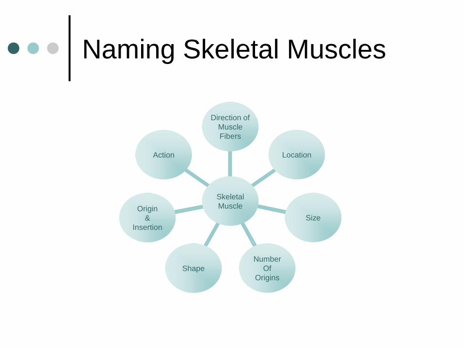
Naming Skeletal Muscles
Action
Origin
&
Insertion
Shape
Number
Of
Origins
Size
Location
Direction of
Muscle
Fibers
Skeletal
Muscle

Direction of Muscle Fibers
Relative to the Midline
RECTUS = parallel to the
midline
Rectus Abdominus
TRANSVERSE =
perpendicular to midline
Transverse Abdominus
OBLIQUE = diagonal to
midline
External Oblique
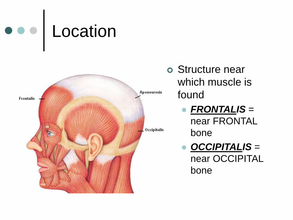
Location
Structure near
which muscle is
found
FRONTALIS =
near FRONTAL
bone
OCCIPITALIS =
near OCCIPITAL
bone

Size
Relative Size of Muscle
MAXIMUS = largest
Gluteus Maximus
MEDIUS = middle
Gluteus Medius
MINIMUS = smallest
Gluteus Minimus
LONGUS = longest
Fibularis Longus
BREVIS = short
Fibularis Brevis
TERTIUS = shortest
Fibularis Tertius
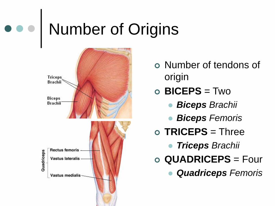
Number of Origins
Number of tendons of
origin
BICEPS = Two
Biceps Brachii
Biceps Femoris
TRICEPS = Three
Triceps Brachii
QUADRICEPS = Four
Quadriceps Femoris

Shape
Relative Shape of the Muscle
DELTOID = triangular shape Δ
TRAPEZIUS = trapezoid shape SERRATUS = saw-toothed ♒
RHOMBOIDEUS = rhomboid shape
TERES = round ○

Origin & Insertion
Origin – attachment to an immoveable bone
Insertion –attachment to a movable bone
ILIO COSTALIS= attaches to the ilium & ribs (costal = ribs)

Action
NAME ACTION EXAMPLE
FLEXOR Decrease angle at a joint Flexor Carpi Radialis
EXTENSOR Increase angle at a joint Extensor Carpi Ulnaris
ABDUCTORMove bone away from
midlineAbductor Pollicis Longus
ADDUCTOR Move bone toward midline Adductor Longus
LEVATOR Produce upward movement Levator Scapulae
DEPRESSORProduce downward
movementDepressor Labii Inferioris
SUPINATOR Turn palm upward/anterior Supinator
PRONATORTurn palm
downward/posteriorPronator Teres
Types of Muscle--Actions
Prime mover (Agonist) – muscle with the
major responsibility for a certain movement
Antagonist – muscle that opposes or
reverses a prime mover
Synergist – muscle that aids a prime mover
in a movement and helps prevent rotation
Fixator – stabilizes the origin of a prime
mover

Characteristics of Muscles
Muscle cells are elongated
(muscle cell = muscle fiber)
Contraction of muscles is due to the
movement of microfilaments
All muscles share some terminology
Prefix myo refers to muscle
Prefix mys refers to muscle
Prefix sarco refers to flesh

Cardiac muscle tissue• Makes up myocardium of heart
• Unconsciously (involuntarily) controlled
• Microscopically appears striated
• Cells are short, branching & have a single
nucleus
Types of Muscle Tissue:
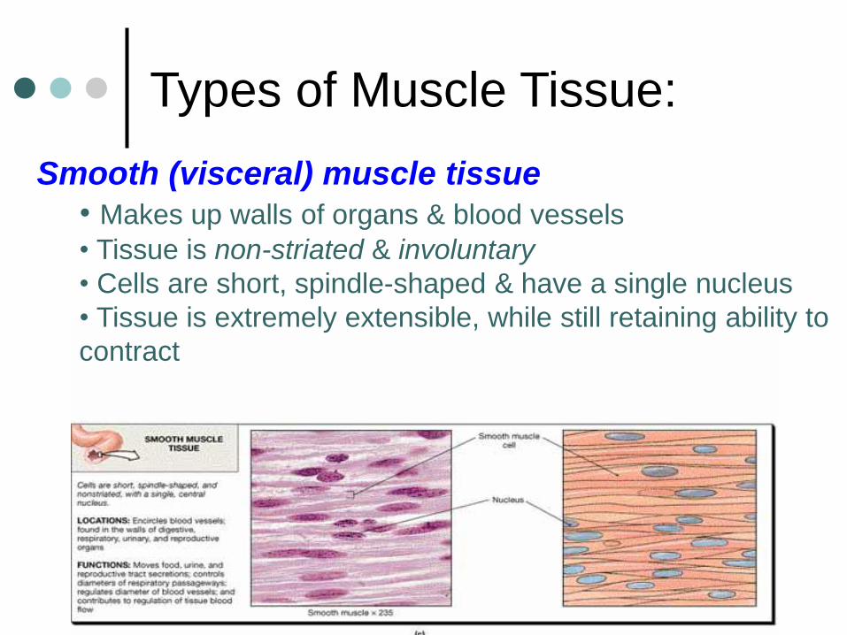
Smooth (visceral) muscle tissue
• Makes up walls of organs & blood vessels
• Tissue is non-striated & involuntary
• Cells are short, spindle-shaped & have a single nucleus
• Tissue is extremely extensible, while still retaining ability to
contract
Types of Muscle Tissue:
Types of Muscle Tissue:
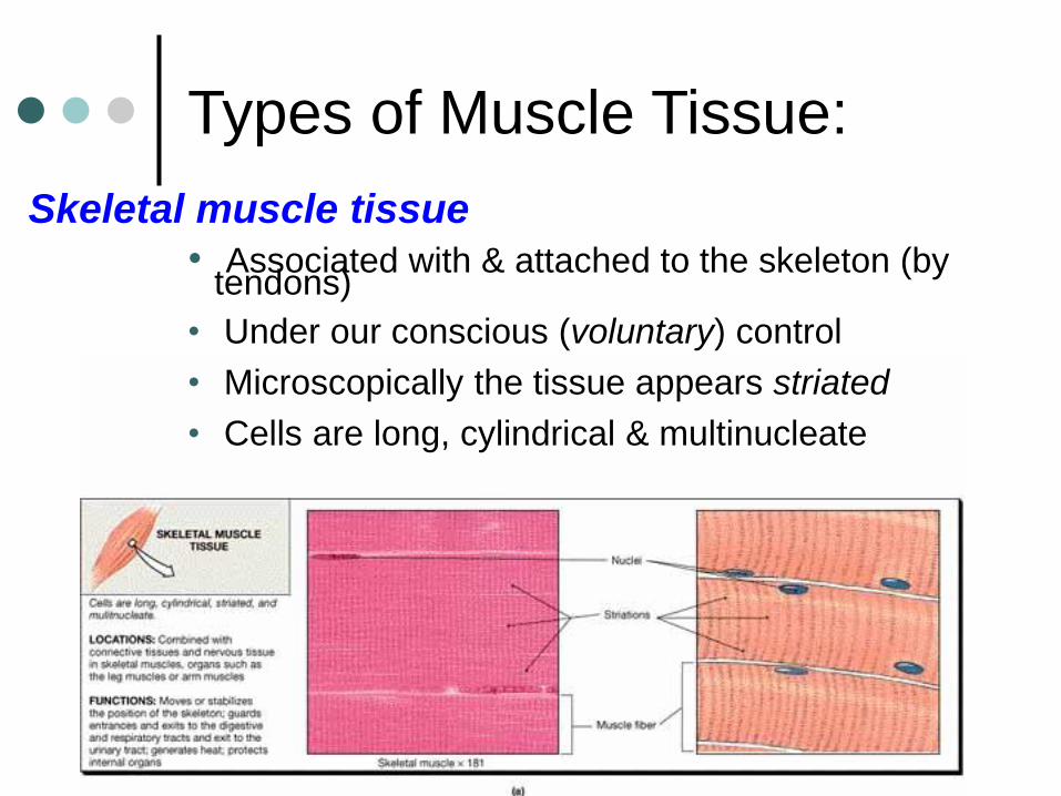
Skeletal muscle tissue
• Associated with & attached to the skeleton (by tendons)
• Under our conscious (voluntary) control
• Microscopically the tissue appears striated
• Cells are long, cylindrical & multinucleate

Skeletal Muscle
Functions of Skeletal Muscle Produce Movement
Maintain posture
Stabilize joints
Generate Heat
Sites of Muscle Attachment Bones
Cartilage
Connective tissue coverings
Muscle Fibers blend into a connective tissue attachment Tendon—cordlike structure
Aponeurosis—sheet-like structure
Properties of Muscle Irritability – ability to receive
and respond to a stimulus
Contractibility – ability to shorten when an adequate stimulus is received
Extensibility – ability to lengthen when an adequate stimulus is received
Elasticity – ability to return to normal shape

Anatomy of skeletal muscles (pg. 131)
Skeletal
muscle
fiber (cell)
Muscle
Fascicle
Surrounded by
perimysium
Surrounded by
endomysium
endomysium
perimysium
Skeletal
muscle
Surrounded by
epimysium
epimysium
tendon
Play IP Anatomy of Skeletal muscles (IP p. 4-6)

Microanatomy of a Muscle Fiber

Organization of Skeletal Muscles
This is skeletal muscle
The bundled muscle
fascicles are surrounded
by epimysium
Epimysium
Muscle fascicle
Organization of Skeletal Muscles
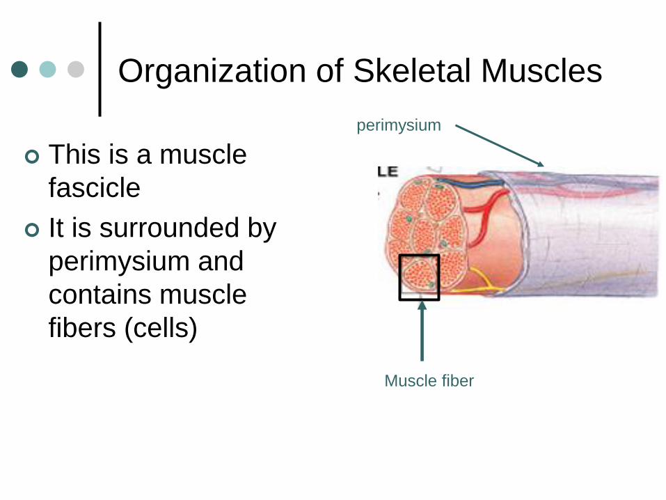
This is a muscle
fascicle
It is surrounded by
perimysium and
contains muscle
fibers (cells)
perimysium
Muscle fiber

Organization of Skeletal Muscles
This is a muscle fiber.
Each muscle fiber is a
single cell with multiple
nuclei
It is surrounded by
endomysium and
contains many myofibrils
endomysium
myofibril

Organization of Skeletal Muscle
This is a myofibril
Each myofibril is
surrounded by
sarcoplasmic reticulum
(smooth ER)
Myofibrils are made up of
many sarcomeres (from
Z line to Z line)
Sarcoplasmic reticulum
Sarcomere (unit
between the Z lines)
Organization of Skeletal Muscle
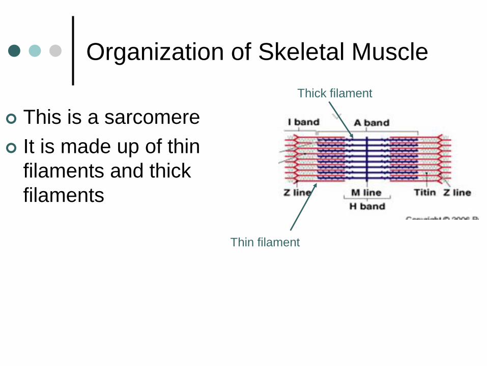
This is a sarcomere
It is made up of thin
filaments and thick
filaments
Thick filament
Thin filament
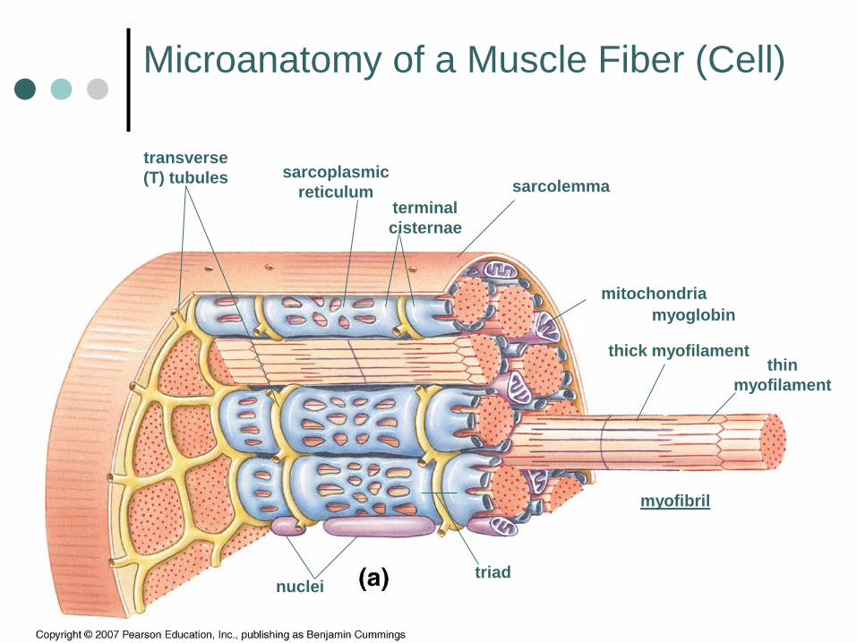
Microanatomy of a Muscle Fiber (Cell)
sarcolemma
transverse
(T) tubules sarcoplasmic
reticulumterminal
cisternae
myofibril
thin
myofilament
thick myofilament
triad
mitochondria
nuclei
myoglobin
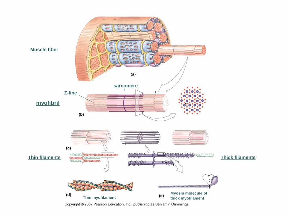
Muscle fiber
myofibril
Thin filaments Thick filaments
Thin myofilamentMyosin molecule of
thick myofilament
sarcomere
Z-line
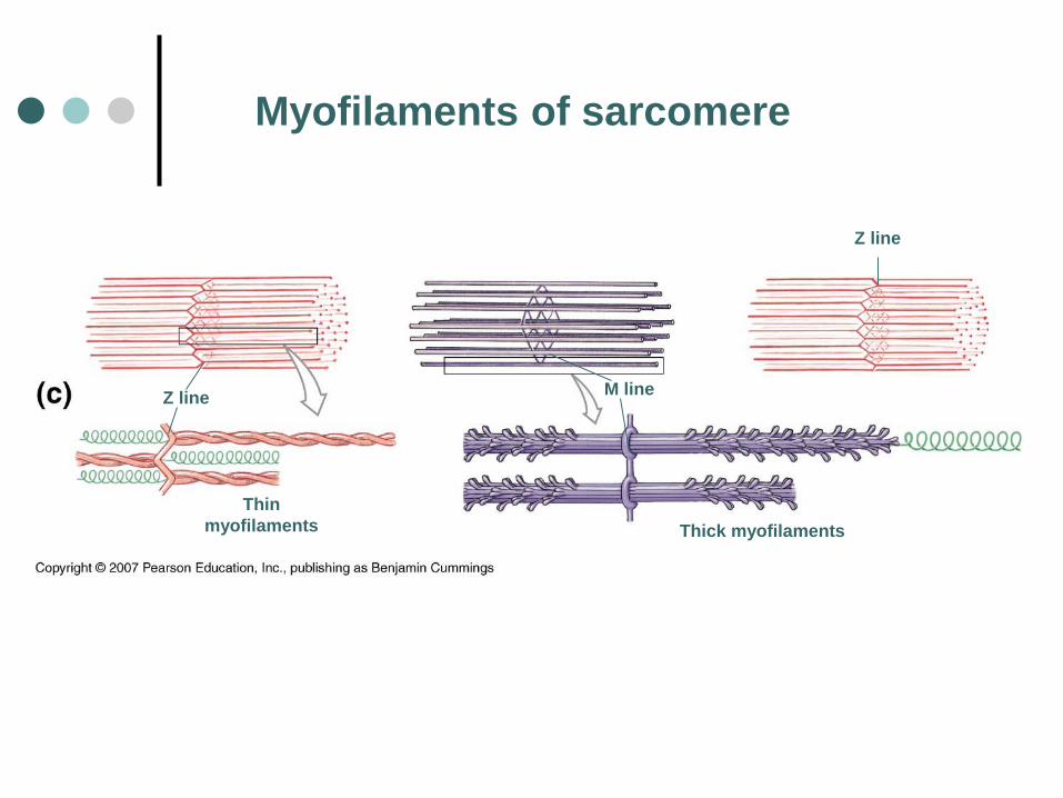
Z line
Thin
myofilaments
M line
Thick myofilaments
Z line
Myofilaments of sarcomere

Thin Myofilament
(myosin binding site)

Thick myofilament
(has ATP
& actin
binding
site)
*Play IP sliding filament theory p.5-14 for overview of thin &
thick filaments

Sarcomere
Z lineZ line
A band
H zone
I band Zone of
overlap M lineZone of
overlapThin
myofilaments Thick
myofilaments

How do skeletal muscles
contract?
1. A signal is sent from the brain or the spinal
cord to the muscle via neurons
2. An action potential is generated in the
neuron, releasing Ca++ in the neuromuscular
junction
3. The influx of calcium ions causes
acetylcholine (AcH) to be released in the
synaptic cleft
4. AcH binds to the AcH receptors present in
the sarcolemma, increasing its permeability
5. Na++enter the sarcolemma, changing its
polarity and creating an action potential
6. Ca++ are released by the sarcoplasmic
reticulum, as the action potential travels down
the T-tubules in the muscle fiber
7. Ca++ bind with troponin C, causing the
myosin to bind on actin sites

8. ATP is hydrolyzed into ADP and phosphorus,
releasing energy
9. Myosin head bends and actin slides over the
myosin surface (contraction)
10. Myosin releases the ADP molecule
11. Actin is released and returns to original
position (relaxation)


Sliding Filament Theory

Sliding Filament Theory1. Myosin heads attach to actin molecules (at binding [active]
site)
2. Myosin “pulls” on actin, causing thin myofilaments to slide
across thick myofilaments, towards the center of the sarcomere
3. Sarcomere shortens, I bands get smaller, H zone gets smaller,
& zone of overlap increases
4. As sarcomeres shorten, myofibril shortens. As myofibrils
shorten, so does muscle fiber
• Once a muscle fiber begins to contract, it will contract maximally
• This is known as the “all or none” principle


Head & Neck Muscles

Head & Neck Muscles
Frontalis: elevate eyebrows
Orbicularis Oculi: close eyelid
Zygomaticus: draw angle of lip upward
Buccinator: draws cheeks against teeth
Orbicularis Oris: closes mouth
Platysma: draws lower lip down & back
Cranial Aponeurosis: connects frontalis to occipitalis
Temporalis: elevates mandible
Occipitalis: draws scalp back
Masseter: elevates mandible
Sternocleidomastoid: Flexes head
Draws head toward shoulder
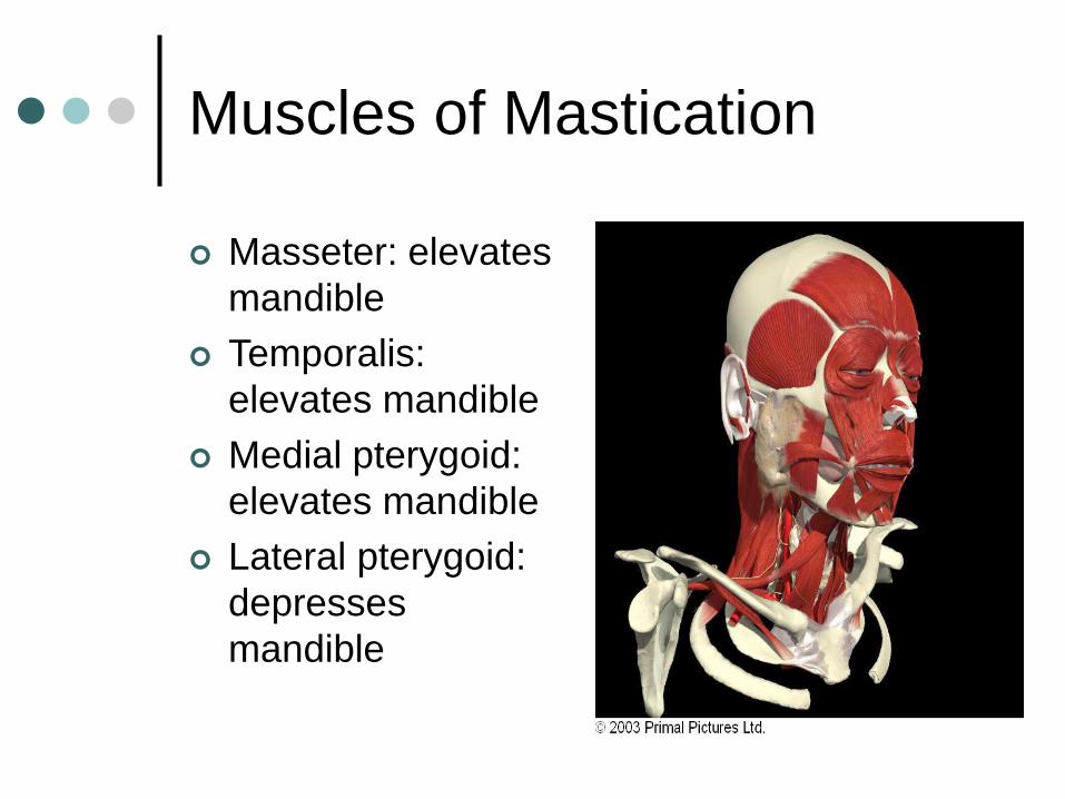
Muscles of Mastication
Masseter: elevates
mandible
Temporalis:
elevates mandible
Medial pterygoid:
elevates mandible
Lateral pterygoid:
depresses
mandible

Key Muscles of Facial Expression
Smiling Muscles
Orbicularis Oculi
Nasalis
Levator Labii
Superioris
Levator Anguli
Superioris
Zygomaticus
Risorius
Frowning Muscles
Frontalis
Orbicularis Oris
Depressor Anguli
Oris
Depressor Labii
Inferioris
Mentalis
Platysma
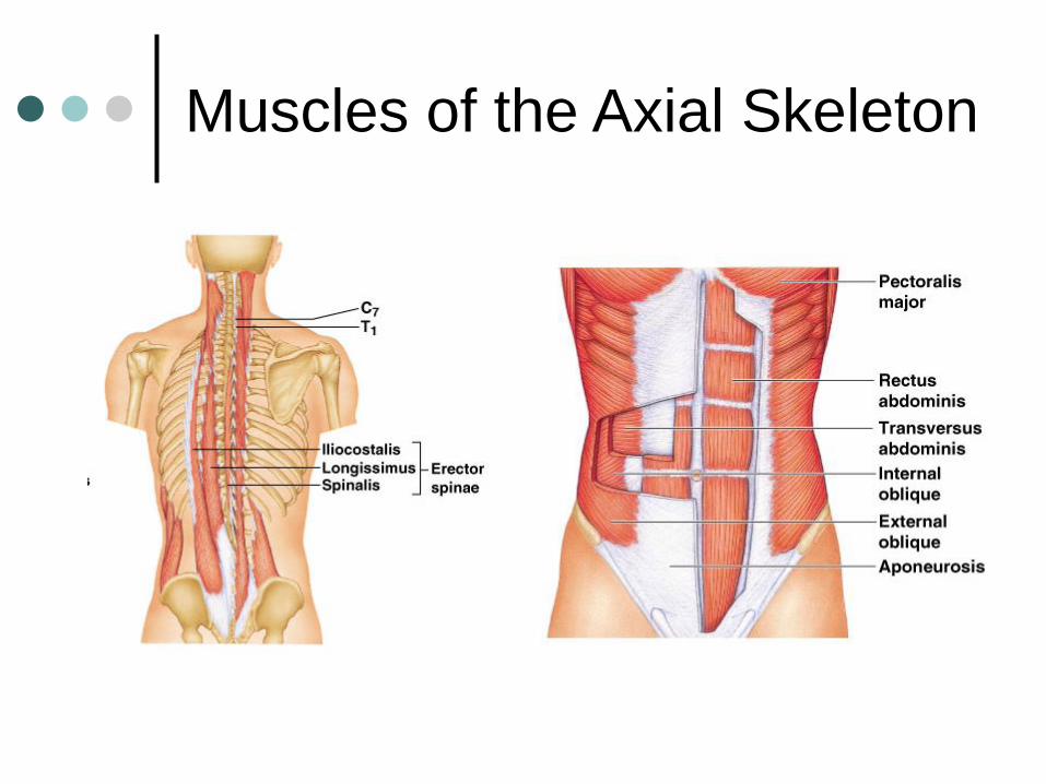
Muscles of the Axial Skeleton

Muscles of the Axial Skeleton
Intrinsic Muscles Erector Spinae:
maintain posture of back/extension
• Spinalis
• Longissimus
• Iliocostalis
Oblique Muscles: rotation of the vertebrae
• Semispinalis
• Multifidus
• Rotatores
Muscles of Quiet Respiration Diaphragm
External Intercostals
Internal Intercostals—deep breaths
Abdominal Muscles External Obliques
Internal Obliques
Transverse Abdominus
Rectus Abdominus
Quadratus Lumborum
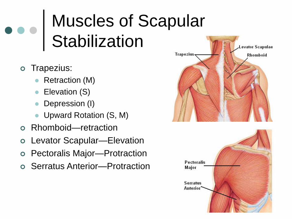
Muscles of Scapular
Stabilization
Trapezius:
Retraction (M)
Elevation (S)
Depression (I)
Upward Rotation (S, M)
Rhomboid—retraction
Levator Scapular—Elevation
Pectoralis Major—Protraction
Serratus Anterior—Protraction
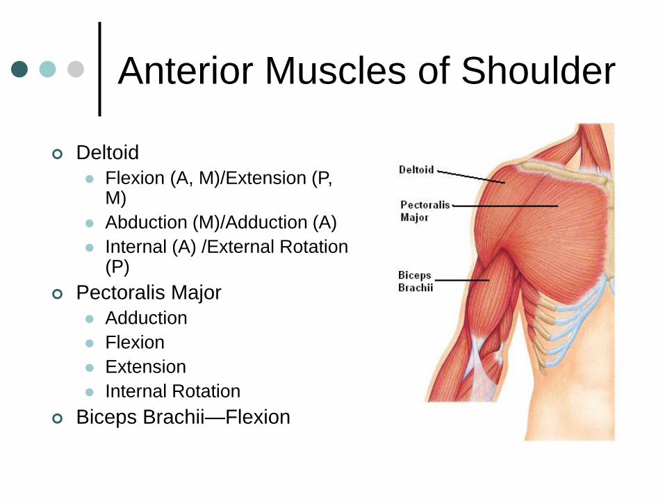
Anterior Muscles of Shoulder
Deltoid
Flexion (A, M)/Extension (P, M)
Abduction (M)/Adduction (A)
Internal (A) /External Rotation (P)
Pectoralis Major
Adduction
Flexion
Extension
Internal Rotation
Biceps Brachii—Flexion

Posterior Muscles of Shoulder
Teres Major
Adduction
Extension
Internal Rotation
Latissimus Dorsi
Adduction
Extension
Internal Rotation
Triceps Brachii
Adduction
Extension
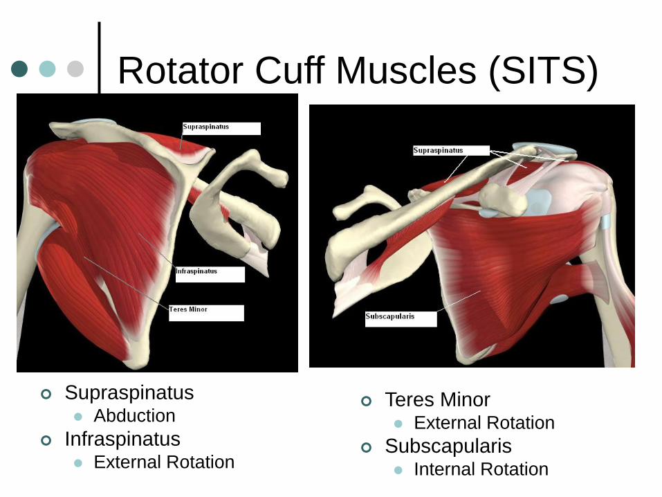
Rotator Cuff Muscles (SITS)
Supraspinatus Abduction
Infraspinatus External Rotation
Teres Minor External Rotation
Subscapularis Internal Rotation

Muscles of the
Elbow/Forearm
Triceps Brachii—Extension
Bicep Brachii— Flexion
Supination
Brachialis—Flexion
Brachioradialis— Flexion
Pronation
Pronator Teres
Pronator Quadratus
Supinator Longus

Muscles of the Wrist & Hand
Flexor Carpi Ulnaris
Flexor Carpi Radialis
Flexor Digitorum
Extensor Carpi Ulnaris
Extensor Carpi Radialis
Extensor Digitorum
Anterior (Palmar) View Posterior (Dorsal) View
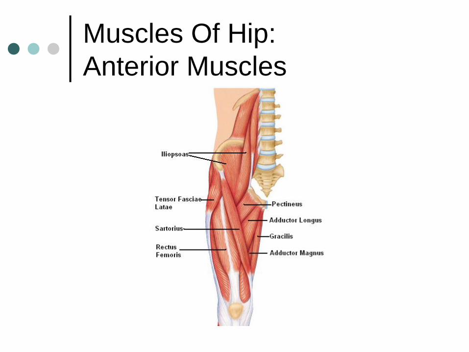
Muscles Of Hip:
Anterior Muscles

Muscles of Hip:
Anterior Muscles
Medial/Adductor
Muscles:
Adductor Magnus
Adductor Longus
Adductor Brevis
Gracilis
Anterior Muscles
Iliopsoas—Flexion
Pectineus—
• Flexion
• Adduction
Sartorius—
• Flexion
• Lateral Rotation

Muscles of Hip:
Gluteal Muscles
Gluteus Maximus—Extension
Gluteus Medius—Abduction
Gluteus Minimus—Abduction
Tensor Fasciae Latae—
Flexion
Abduction
** Gluteus Minimus is under the
Gluteus Medius

Muscles of Anterior Thigh
“Quadriceps”
Rectus Femoris—
• Hip flexion
• Knee extension
Vastus Lateralis—knee extension
Vastus Medialis—knee extension
Vastus Intermedius—knee extension
Sartorius—
• Hip & Knee Flexion
• Lateral Hip Rotation**Vastus Intermedius is
beneath Rectus Femoris

Muscles of Posterior Thigh
“Hamstrings”
Responsible for
Knee Flexion & Hip
Extension
Semimembranosus
Semitendinosus
Biceps Femoris
Gastrocnemius
Knee Flexion

Muscles of the Lower Leg
Anterior Compartment Tibialis Anterior—Dorsiflexion &
inversion
Extensor Digitorum Longus
Fibularis Tertius—dorsiflexion & eversion
Posterior Compartment Gastrocnemius—plantarflexion,
knee flexion
Soleus—plantarflexion
Lateral Compartment Fibularis Longus—plantarflexion
& eversion
Fibularis Brevis—plantarflexion & eversion

Throwing Movement

Running & Kicking