interactions of skeletal muscles skeletal muscles work _________________________ or in _ muscles...
Post on 20-Dec-2015
230 views
TRANSCRIPT
Interactions of Skeletal Muscles
• Skeletal muscles work _________________________ or in _
• Muscles only _______________(never push)
• As muscles shorten, the insertion generally moves toward the origin
Muscles: Functional Groups
• – provide the major force for producing a specific movement
• – oppose or reverse a particular movement
• – Add force to a movement– Reduce undesirable or unnecessary movement
• – synergists that immobilize a bone or muscle’s origin
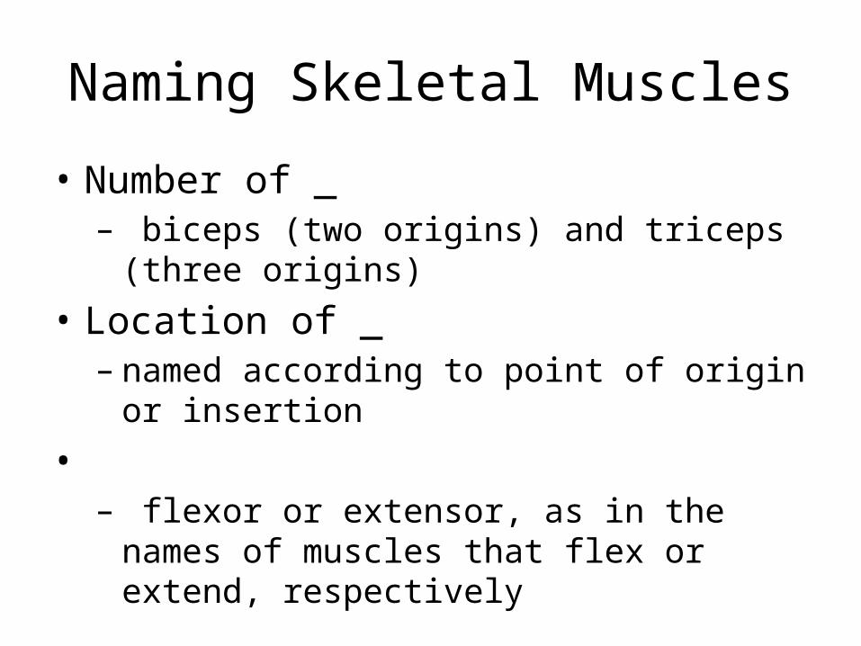
Naming Skeletal Muscles
• – bone or body region associated with the muscle
• – e.g., the deltoid muscle (deltoid = triangle)
Naming Skeletal Muscles
• – maximus (largest), minimus (smallest), longus (long)
• – rectus (fibers run straight), transversus, and oblique
(fibers run at angles to an imaginary defined axis)
Naming Skeletal Muscles
• Number of _– biceps (two origins) and triceps (three origins)
• Location of _– named according to point of origin or insertion
• – flexor or extensor, as in the names of muscles that
flex or extend, respectively
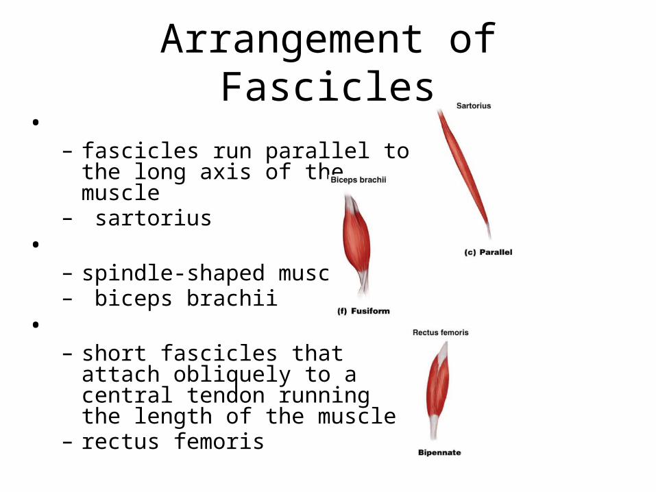
Arrangement of Fascicles• – fascicles run parallel to the long axis
of the muscle – sartorius
• – spindle-shaped muscles – biceps brachii
• – short fascicles that attach obliquely
to a central tendon running the length of the muscle
– rectus femoris
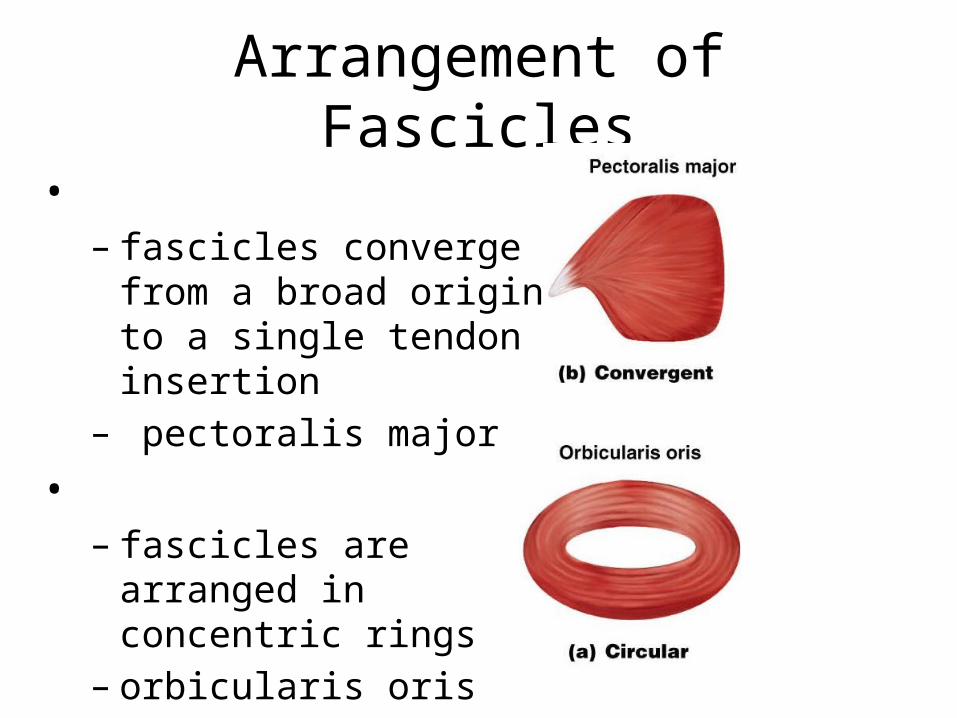
Arrangement of Fascicles
• – fascicles converge from a
broad origin to a single tendon insertion
– pectoralis major • – fascicles are arranged in
concentric rings – orbicularis oris
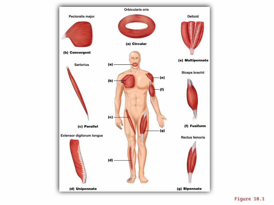
Figure 10.1
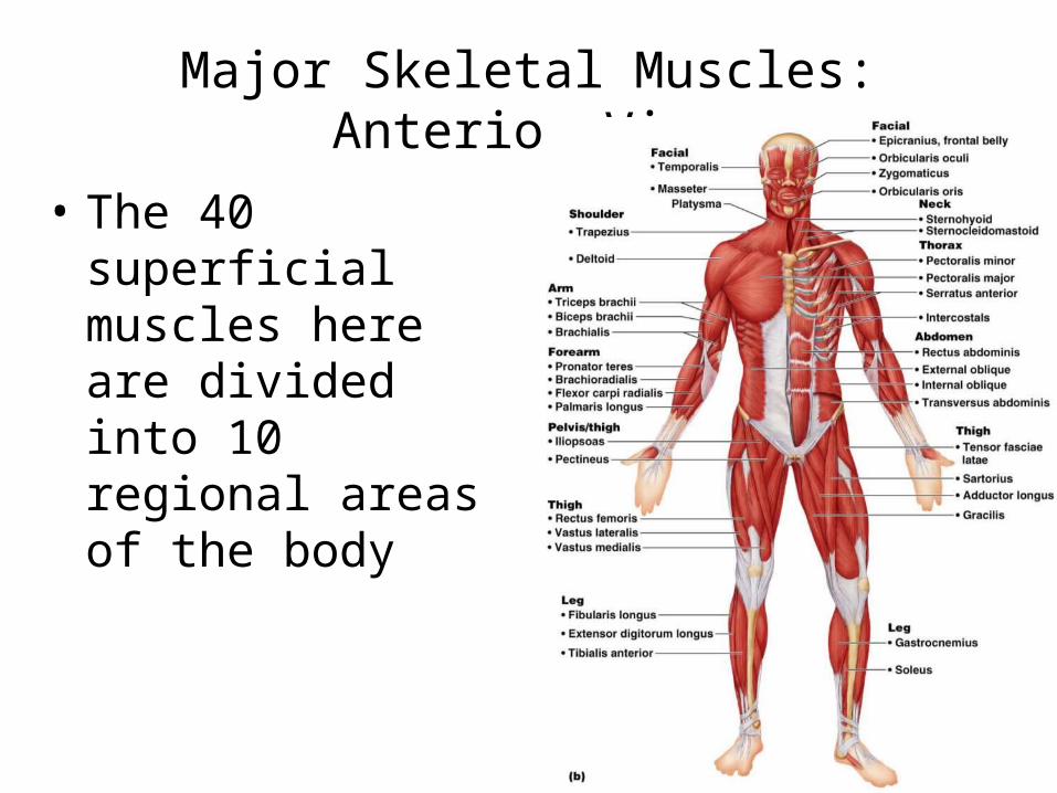
Major Skeletal Muscles: Anterior View
• The 40 superficial muscles here are divided into 10 regional areas of the body
Figure 10.4b
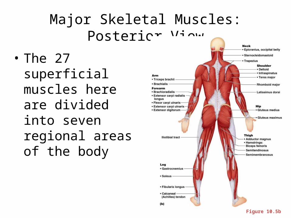
Major Skeletal Muscles: Posterior View
• The 27 superficial muscles here are divided into seven regional areas of the body
Figure 10.5b
Muscles: Name, Action, and Innervation
• Name and description of the muscle – be alert to information given in the name
• Origin and insertion–
• Action – best learned by
_________________________________________ on one’s own body
• Nerve supply– name of major nerve that innervates the muscle
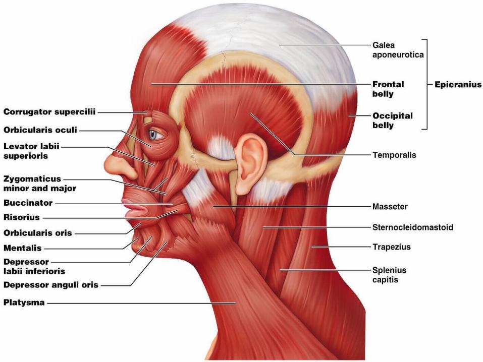
Muscles of the Scalp
• Epicranius (___________________________) – bipartite muscle consisting of the:– – – Galea aponeurotica – cranial
___________________________________ connecting above muscles
• These two muscles have alternate actions of pulling the scalp forward and backward
Muscles of the Face
• 11 muscles are involved in lifting the eyebrows, flaring the nostrils, opening and closing the eyes and mouth, and smiling
• All are innervated by __________________________________ (facial nerve)
• Usually insert ______________________ (rather than bone), and adjacent muscles often fuse
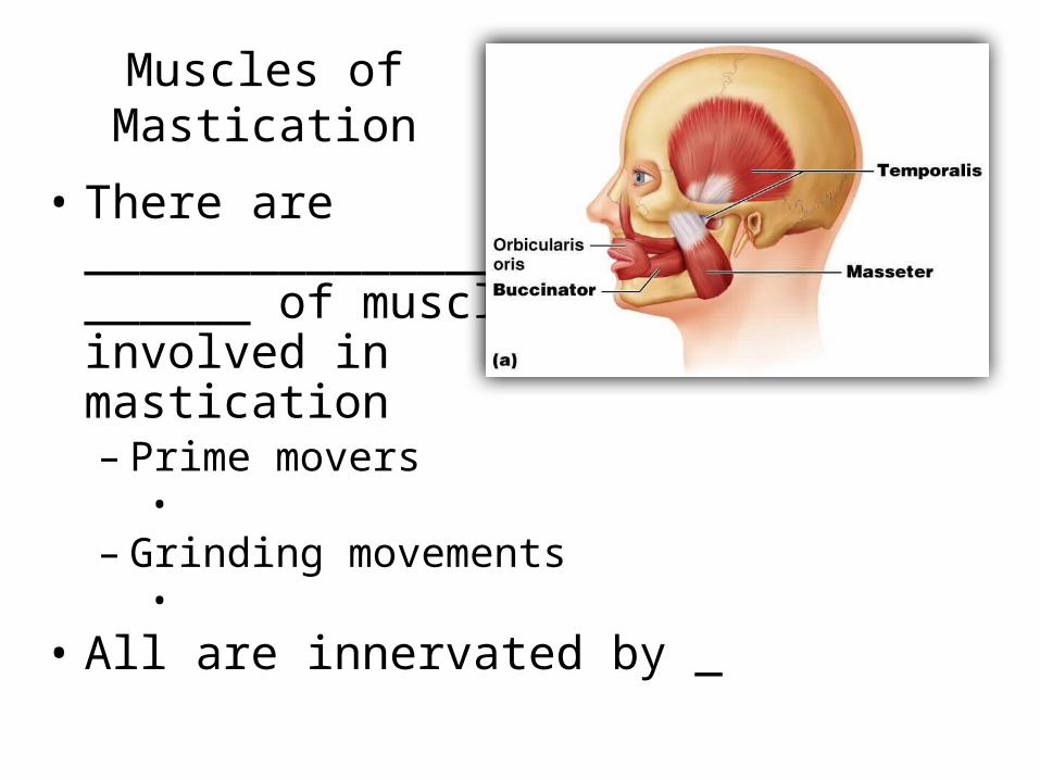
Muscles of Mastication
• There are ____________________________________ of muscles involved in mastication– Prime movers •
– Grinding movements •
• All are innervated by _
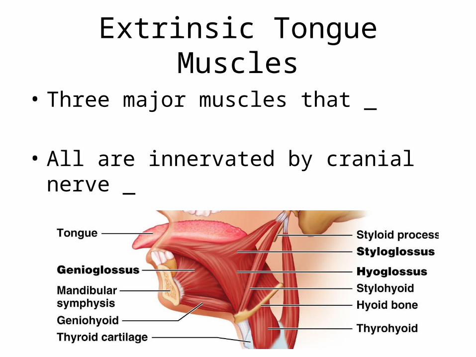
Extrinsic Tongue Muscles
• Three major muscles that _
• All are innervated by cranial nerve _
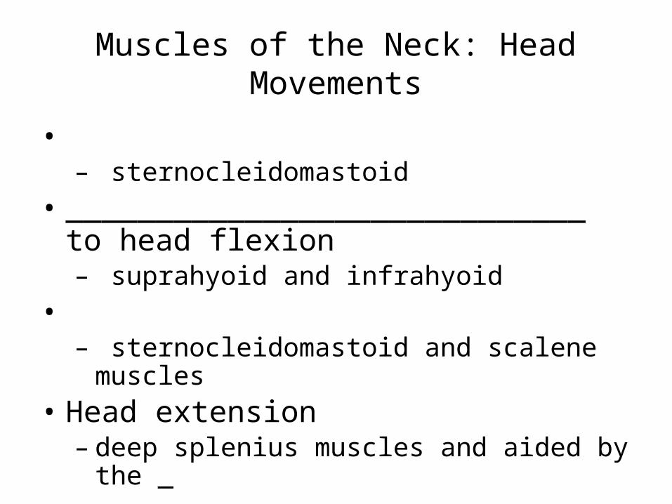
Muscles of the Neck: Head Movements
• – sternocleidomastoid
• _____________________________ to head flexion – suprahyoid and infrahyoid
• – sternocleidomastoid and scalene muscles
• Head extension – deep splenius muscles and aided by the _
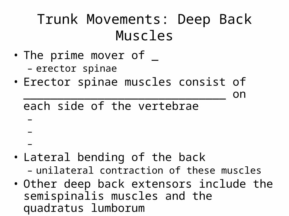
Trunk Movements: Deep Back Muscles
• The prime mover of _– erector spinae
• Erector spinae muscles consist of ______________________________ on each side of the vertebrae – – –
• Lateral bending of the back – unilateral contraction of these muscles
• Other deep back extensors include the semispinalis muscles and the quadratus lumborum
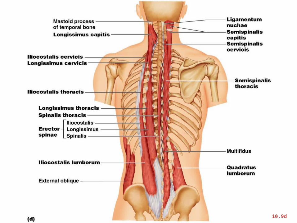
Figure 10.9d
Trunk Movements: Short Muscles
• Four short muscles extend from one vertebra to another
• These muscles are synergists in ________________________________ of the spine
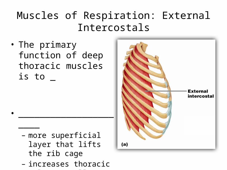
Muscles of Respiration: External Intercostals
• The primary function of deep thoracic muscles is to _
• ______________________– more superficial layer that
lifts the rib cage – increases thoracic volume
to allow inspiration
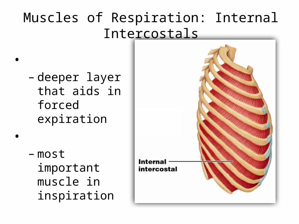
Muscles of Respiration: Internal Intercostals
• – deeper layer that
aids in forced expiration
• – most important
muscle in inspiration
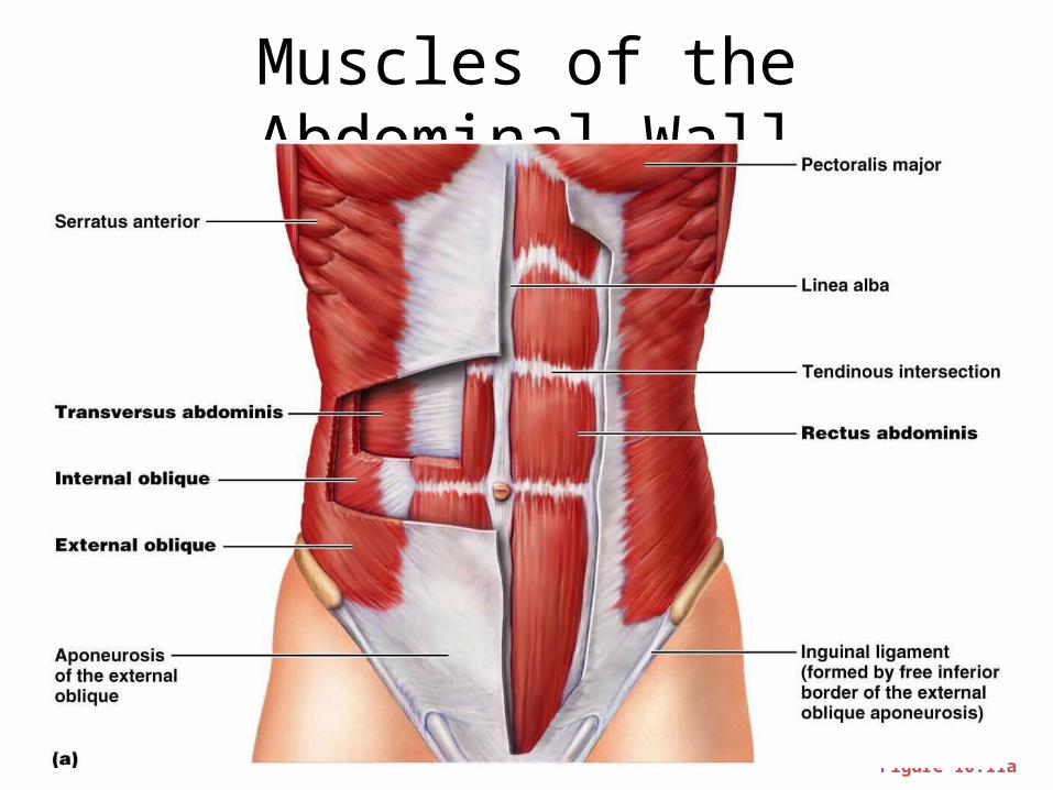
Muscles of the Abdominal Wall• The abdominal wall is composed of four paired
muscles– – external obliques, – – rectus abdominis), – their fasciae, and their _
• Fascicles of these muscles run at right and oblique angles to one another, giving the abdominal wall added strength
Muscles of the Abdominal Wall
• In addition to forming the abdominal wall, these muscles:– Are involved with ________________________
and rotation of the trunk
– Help promote __________________________, defecation, ___________________________, vomiting, coughing, and _
Muscles of the Abdominal Wall
Figure 10.11a
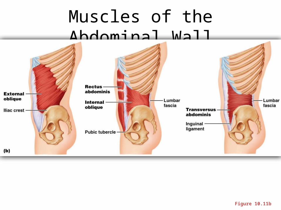
Muscles of the Abdominal Wall
Figure 10.11b
Extrinsic Shoulder Muscles• Muscles of the thorax– Anterior:
• pectoralis major, pectoralis minor, serratus anterior, and subclavius
– Posterior: • latissimus dorsi, trapezius muscles, levator scapulae, and
rhomboids
– These muscles are involved with the _________________________________________ including elevation, depression, rotation, and lateral and medial movements
• Prime movers of shoulder elevation are the _
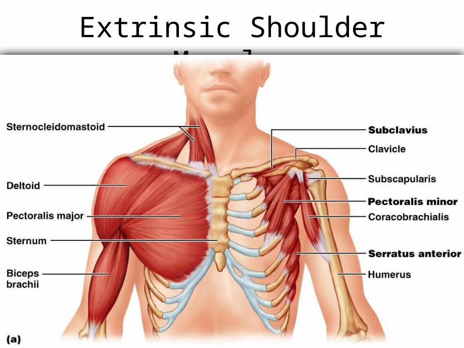
Extrinsic Shoulder Muscles
Figure 10.13a
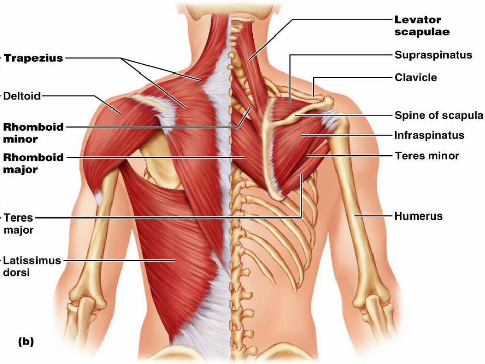
Figure 10.13b
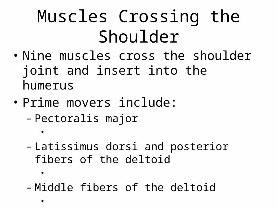
Muscles Crossing the Shoulder
• Nine muscles cross the shoulder joint and insert into the humerus
• Prime movers include:– Pectoralis major •
– Latissimus dorsi and posterior fibers of the deltoid •
– Middle fibers of the deltoid •
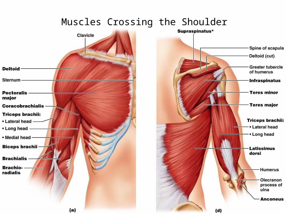
Muscles Crossing the Shoulder
Muscles Crossing the Shoulder
• Rotator cuff muscles– – – –
• Function mainly to reinforce the capsule of the shoulder– Secondarily act as synergists and fixators
Muscles Crossing the Elbow
• Forearm extension– prime mover of forearm extension•
– weak synergist • The
Muscles Crossing the Elbow
• Forearm flexion
– chief forearm flexors•
– synergist • The _• helps stabilize the _
Muscles of the Forearm
• Forearm muscle groups: – those that cause _– those that move the _
• These muscles insert via the flexor and extensor retinacula
• Most _• Most _
Muscles of the Forearm
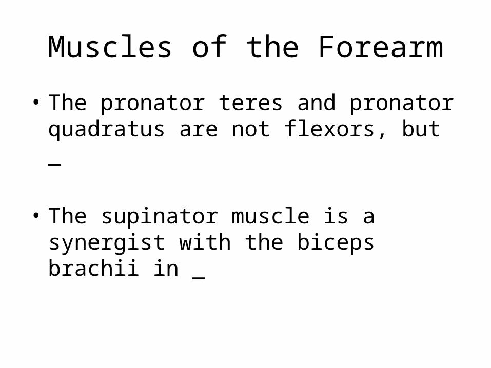
• The pronator teres and pronator quadratus are not flexors, but _
• The supinator muscle is a synergist with the biceps brachii in _
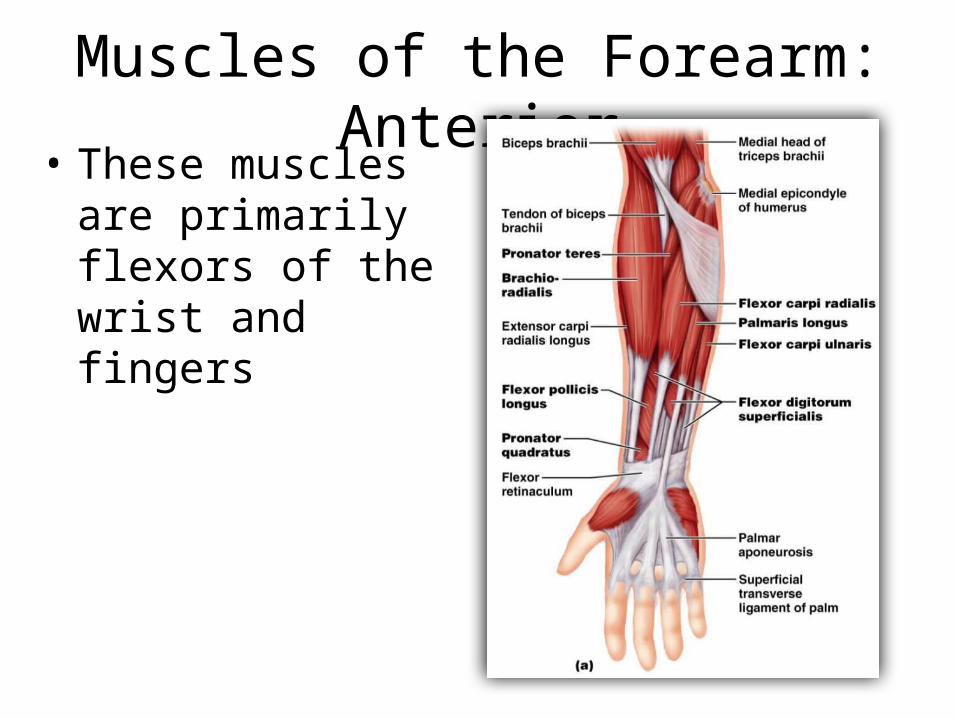
Muscles of the Forearm: Anterior• These muscles are
primarily flexors of the wrist and fingers
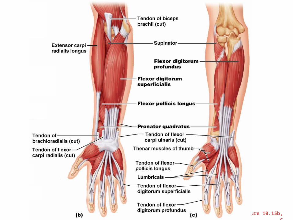
Figure 10.15b, c
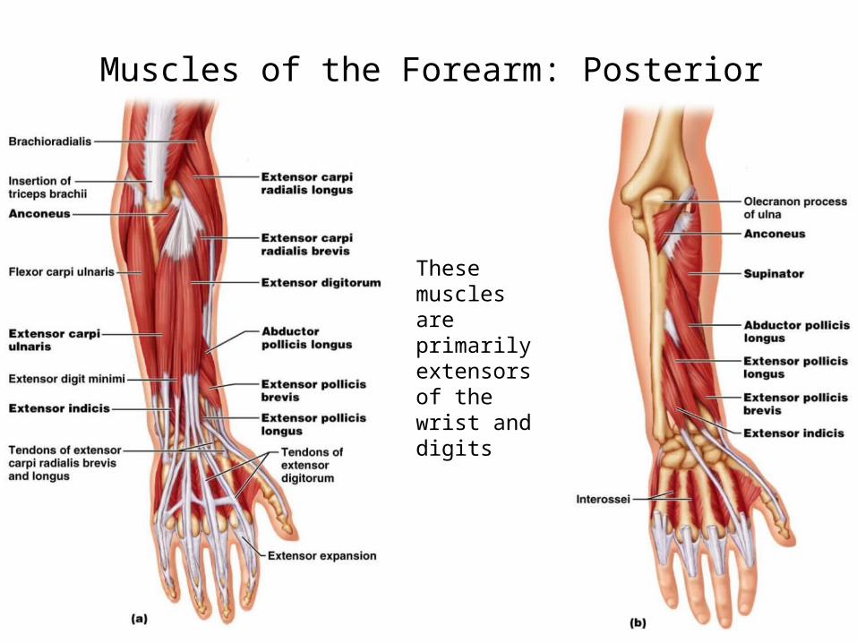
Muscles of the Forearm: Posterior
Figure 10.16a
These muscles are primarily extensors of the wrist and digits
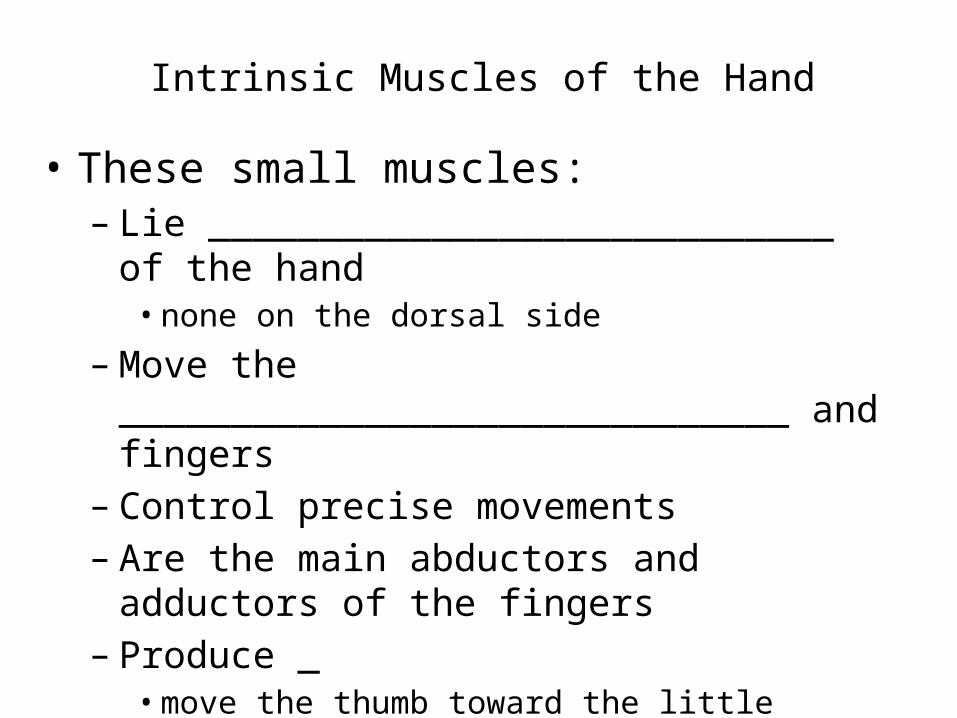
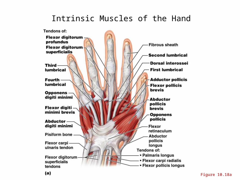
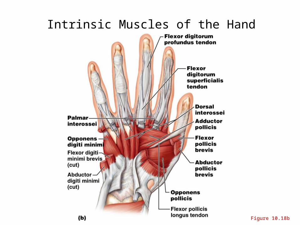
Intrinsic Muscles of the Hand
• These small muscles: – Lie ____________________________ of the hand• none on the dorsal side
– Move the ______________________________ and fingers
– Control precise movements – Are the main abductors and adductors of the
fingers– Produce _• move the thumb toward the little finger
Intrinsic Muscles of the Hand
Figure 10.18a
Intrinsic Muscles of the Hand
Figure 10.18b
Finger and Thumb Movements
• Flexion– Thumb – – Fingers –
• Extension– Thumb – – Fingers –
Intrinsic Muscles of the Hand: Groups
• There are _____________________ groups of intrinsic hand muscles
• The thenar eminence and hypothenar eminence– each have a _
• The midpalm muscles, the lumbricals and interossei,
–
• The _– abduct and adduct the fingers
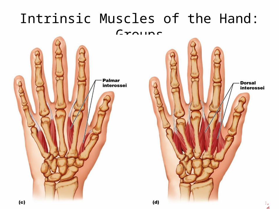
Intrinsic Muscles of the Hand: Groups
Figure 10.18c, d
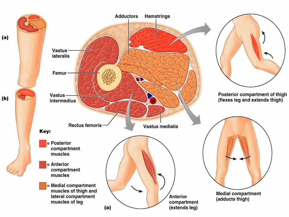
Muscles Crossing Hip and Knee Joints
• Most ___________________________________ muscles of the hip and thigh – –
• Posterior compartment muscles of the hip and thigh – extend _– flex _
• The medial compartment muscles–
• These three groups are enclosed by the fascia lata
Movements of the thigh at the Hip: Flexion and Extension
• The ball-and-socket hip joint permits – – Extension– – Adduction– – Rotation
• The most important thigh flexors – – –
• The medially located adductor muscles and sartorius assist in thigh flexion
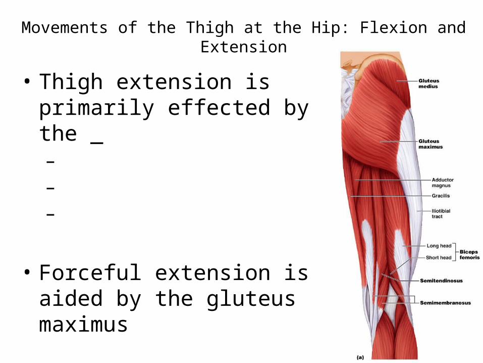
Movements of the Thigh at the Hip: Flexion and Extension
• Thigh extension is primarily effected by the _– – –
• Forceful extension is aided by the gluteus maximus
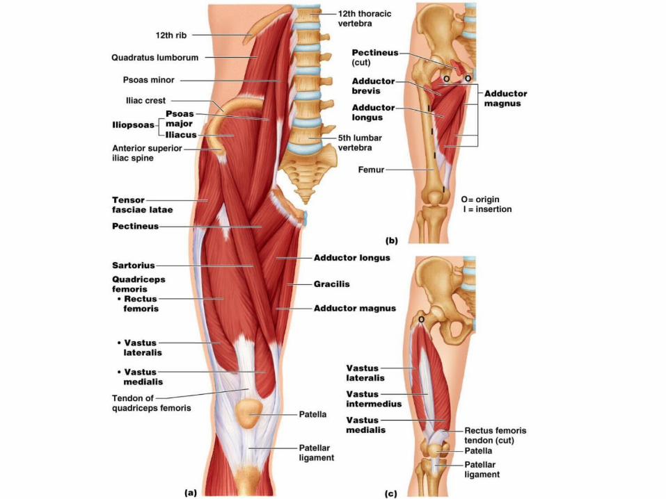
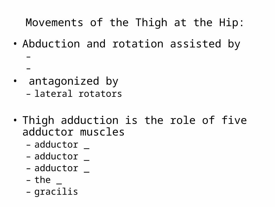
Movements of the Thigh at the Hip:
• Abduction and rotation assisted by – –
• antagonized by – lateral rotators
• Thigh adduction is the role of five adductor muscles – adductor _– adductor _– adductor _– the _– gracilis
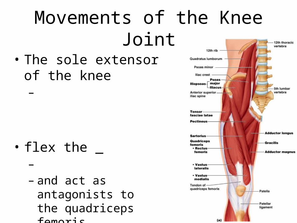
Movements of the Knee Joint
• The sole extensor of the knee –
• flex the _– – and act as antagonists to
the quadriceps femoris
Figure 10.19a
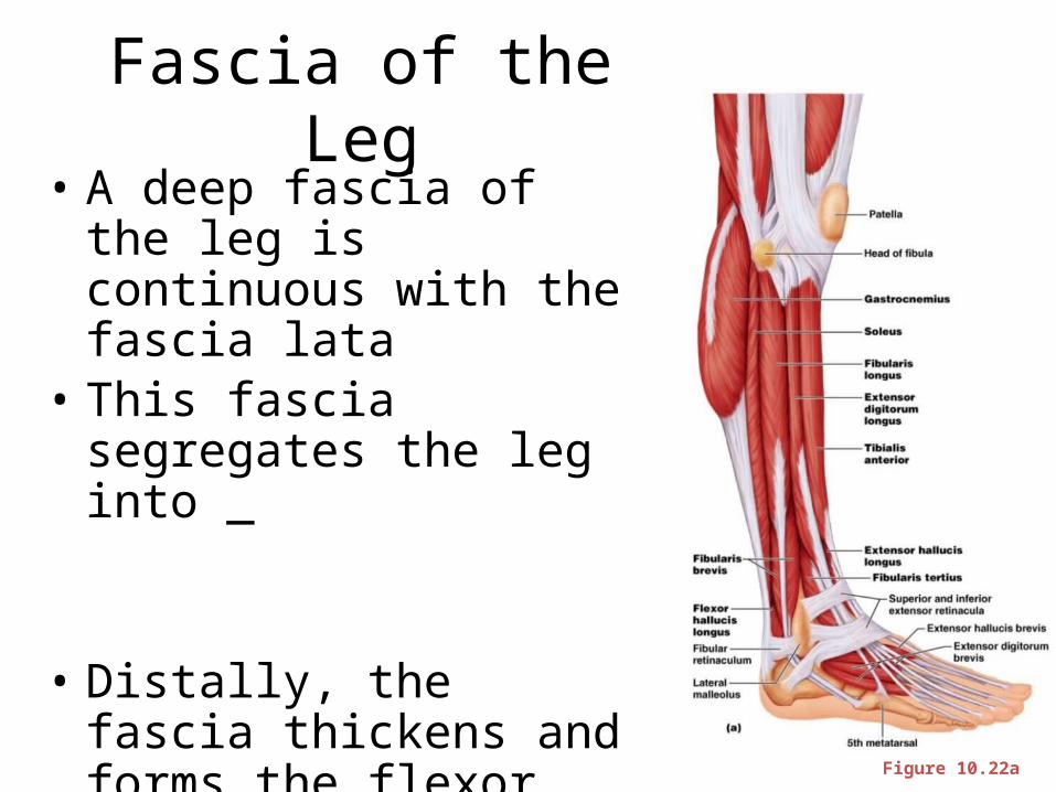
Fascia of the Leg• A deep fascia of the leg is
continuous with the fascia lata
• This fascia segregates the leg into _
• Distally, the fascia thickens and forms the flexor, extensor, and fibular retinaculae
Figure 10.22a
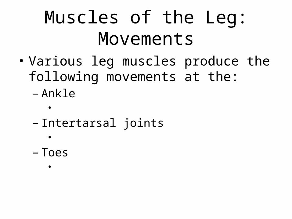
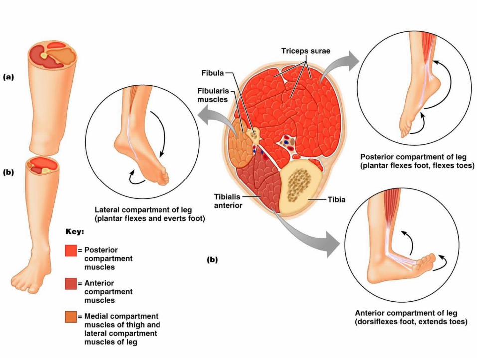
Muscles of the Leg: Movements
• Various leg muscles produce the following movements at the:– Ankle•
– Intertarsal joints•
– Toes•
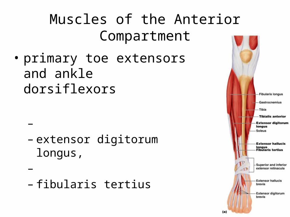
Muscles of the Anterior Compartment
• primary toe extensors and ankle dorsiflexors
– – extensor digitorum longus, – – fibularis tertius
Figure 10.21a
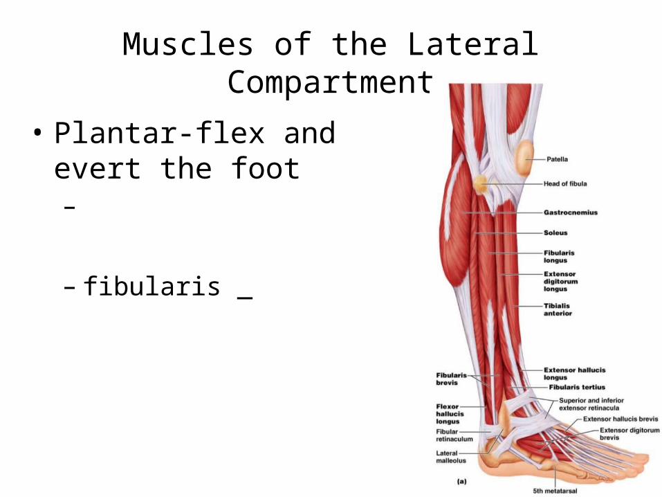
Muscles of the Lateral Compartment
• Plantar-flex and evert the foot–
– fibularis _
Figure 10.22a
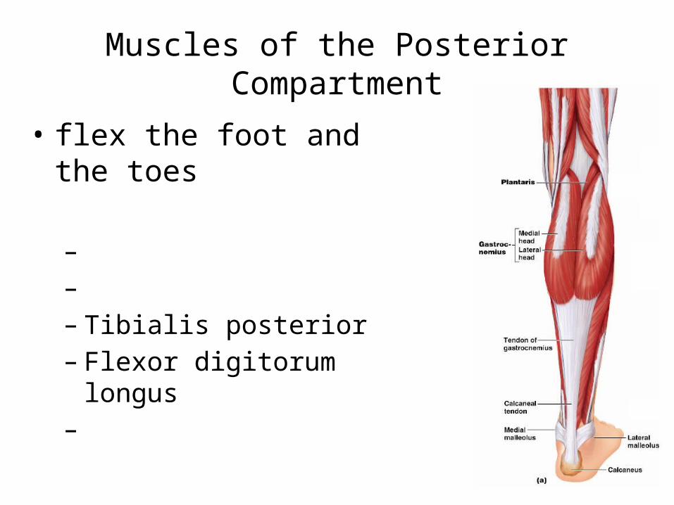
Muscles of the Posterior Compartment
• flex the foot and the toes
– – – Tibialis posterior – Flexor digitorum longus–
Figure 10.23a
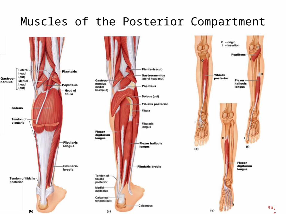
Muscles of the Posterior Compartment
Figure 10.23b, c
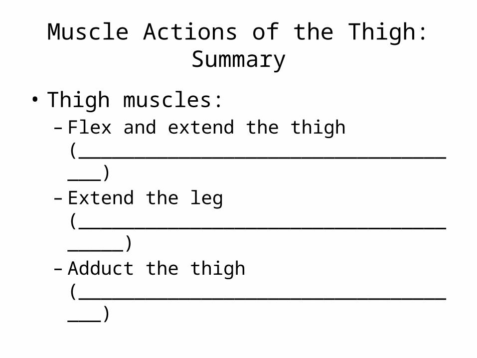
Muscle Actions of the Thigh: Summary
• Thigh muscles: – Flex and extend the thigh
(____________________________________)– Extend the leg
(______________________________________)– Adduct the thigh
(____________________________________)
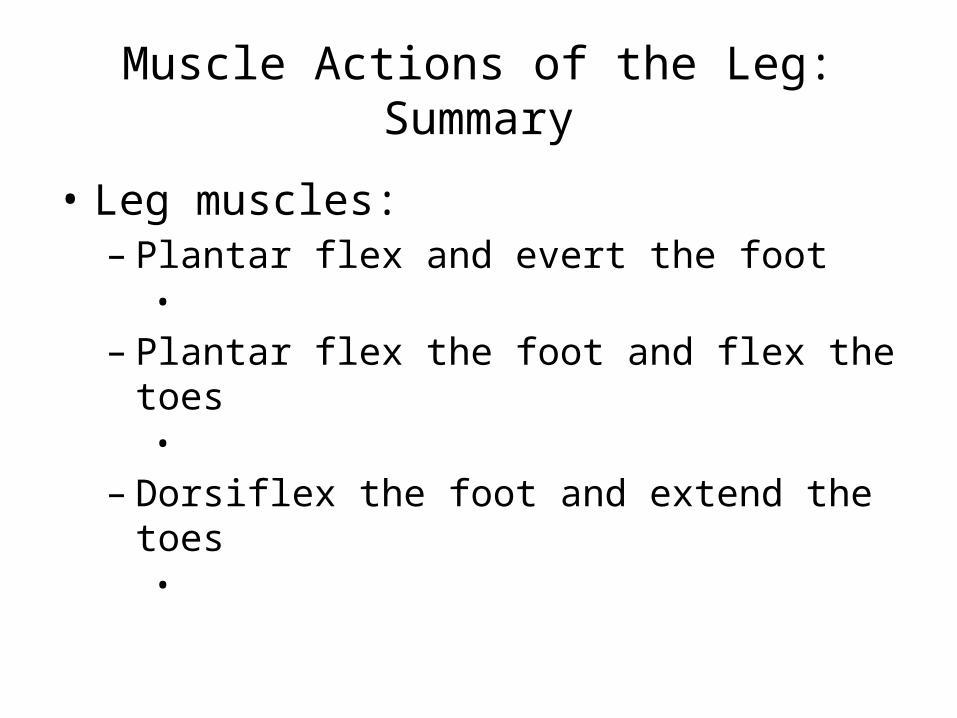
Muscle Actions of the Leg: Summary
• Leg muscles:– Plantar flex and evert the foot •
– Plantar flex the foot and flex the toes •
– Dorsiflex the foot and extend the toes •
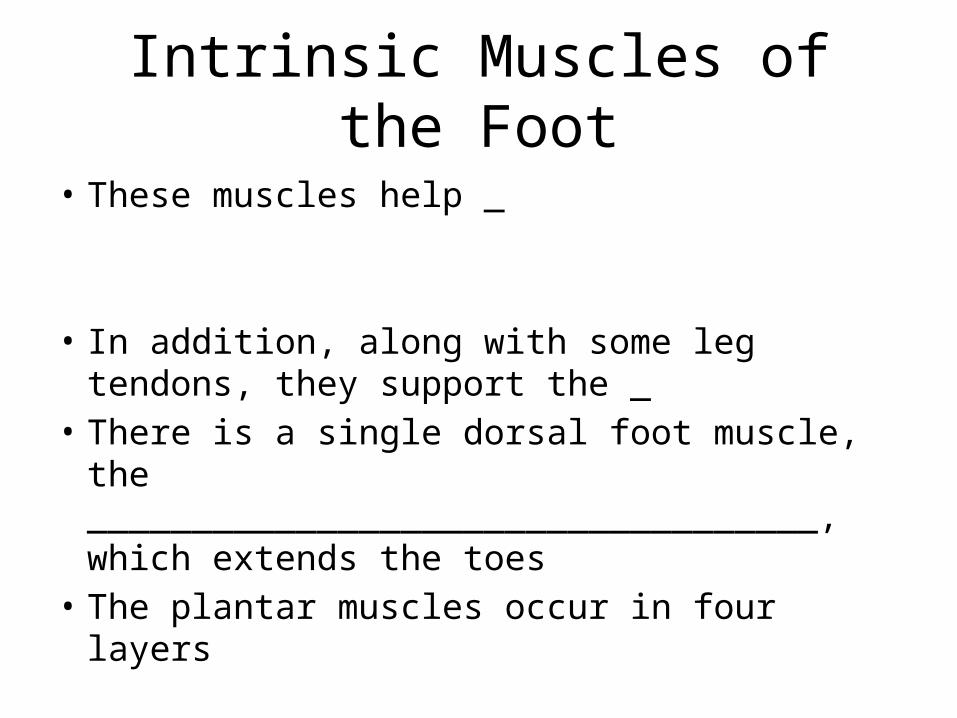
Intrinsic Muscles of the Foot
• These muscles help _
• In addition, along with some leg tendons, they support the _
• There is a single dorsal foot muscle, the ___________________________________, which extends the toes
• The plantar muscles occur in four layers
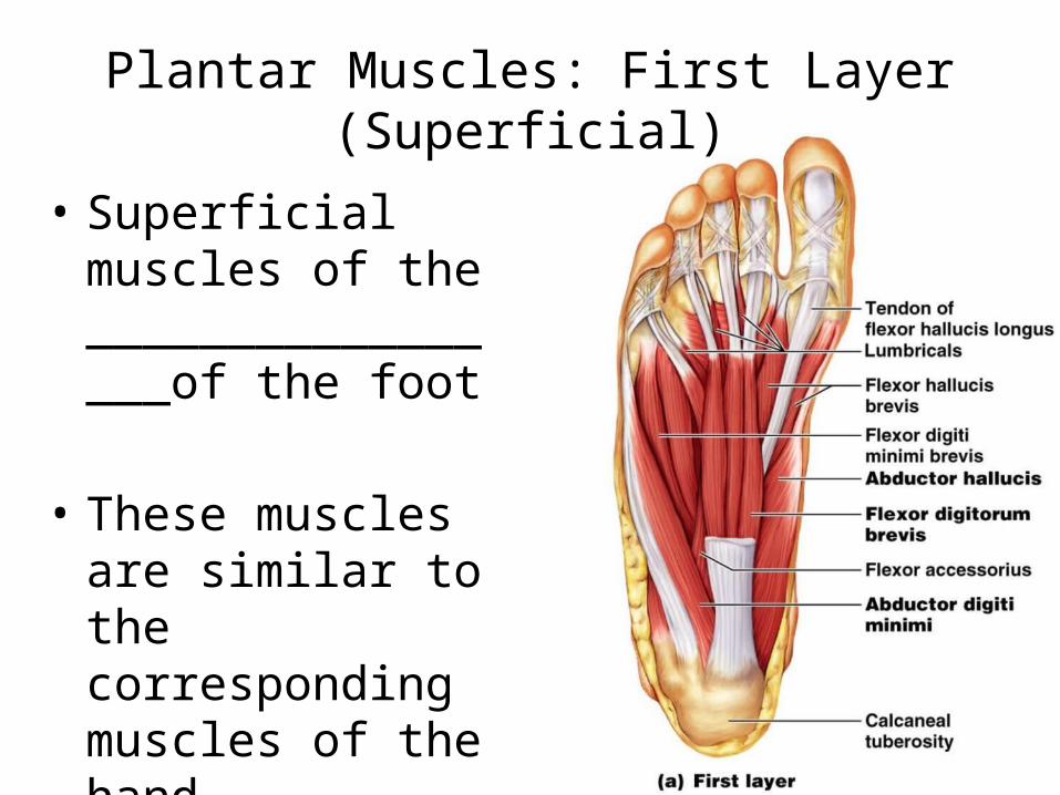
Plantar Muscles: First Layer (Superficial)
• Superficial muscles of the _________________of the foot
• These muscles are similar to the corresponding muscles of the hand
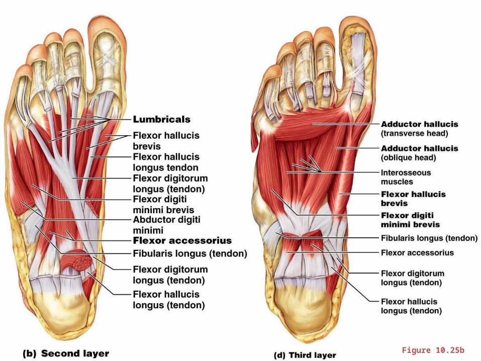
Figure 10.25b
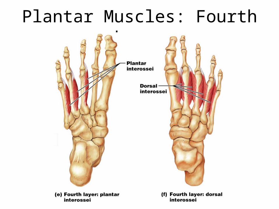
Plantar Muscles: Fourth Layer