redalyc.purificación de cinco isoformas de la hormona ...redalyc.org/pdf/423/42335205.pdf · vet....
TRANSCRIPT
Veterinaria México
ISSN: 0301-5092
Universidad Nacional Autónoma de México
México
Perera Marín, Gerardo; Falcón Alcántara, Andrés; Murcia Mejía, Clara; Hernández Cerón, Joel;
González Padilla, Everardo
Purificación de cinco isoformas de la hormona luteinizante bovina (bLH). Caracterización
fisicoquímica, biológica e inmunológica
Veterinaria México, vol. 35, núm. 2, abril-junio, 2004, pp. 129-145
Universidad Nacional Autónoma de México
Distrito Federal, México
Available in: http://www.redalyc.org/articulo.oa?id=42335205
How to cite
Complete issue
More information about this article
Journal's homepage in redalyc.org
Scientific Information System
Network of Scientific Journals from Latin America, the Caribbean, Spain and Portugal
Non-profit academic project, developed under the open access initiative
129Vet. Méx., 35 (2) 2004
Purifi cación de cinco isoformas de la hormona luteinizante bovina (bLH). Caracterización fi sicoquímica, biológica e inmunológica
Purifi cation of fi ve bovine luteinizing hormone (bLH) isoforms. Chemical, physical, biological and immunological
characterization
Gerardo Perera Marín*
Andrés Falcón Alcántara**
Clara Murcia Mejía*
Joel Hernández Cerón*
Everardo González Padilla*
Recibido el 22 de abril de 2003 y aceptado el 14 de agosto de 2003.
* Departamento de Reproducción, Facultad de Medicina Veterinaria y Zootecnia, Universidad Nacional Autónoma de México,
04510, México, D.F.
** Departamento de Neurobiología Celular y Molecular, Laboratorio de Farmacología Marina, Instituto de Neurobiología,
Universidad Nacional Autónoma de México, A.P.1-1141, Juriquilla, Querétaro, México.
Correspondencia: Gerardo Perera Marín, Departamento de Reproducción, Facultad de Medicina Veterinaria y Zootecnia,
Universidad Nacional Autónoma de México, 04510, México, D.F., Tel. (55)56225860, Correo electrónico: pererag @ servidor.
unam.mx
Abstract
The study describes the purifi cation of fi ve LH isoforms from bovine pituitary extracts. Cationic inter-change chromatography was used to obtain fractions at a pH of 6.8 and 9.5. These were then repurifi ed under identical conditions in DEAE-Sephacel to obtain LH-I (1.2 mg/kg of pituitary), and LH-II (1.8 mg/kg), LH-III (1.5 mg/kg), LH-IV (1.2 mg/kg) and LH-V (7.0 mg/kg), respectively. The relative mobil-ity (Rf) of each isoform was determined using polyacrylamide gel electrophoresis under native condi-tions. LH-I presented an Rf of 0.16 cm; LH- II, LH-III and LH-IV had an Rf of 0.044 cm, while LH-V had a 0.11 cm Rf. The molecular weight (MW) of LH-I and LH-V was 37.5 kDa, while LH-II, LH-III and LH-IV had a MW of 38.5 kDa. The immunotransference analysis identifi ed MW bands weighing 59.0, 37.5 and 22.0 kDa in the standards as well as in the different isoforms. The in vivo biological potency corresponded to 1.04 U/mg (LH-I); 5.9 U/mg (LH-II); 0.16 U/mg (LH-III); 0.86 U/mg (LH-IV) and 1.4 U/mg (LH-V) and their LH radioinmmunoassay (RIA) 50% expected dose (ED50) were 2.27 ng/tube (LH-I), 1.19 ng/tube (LH-II), 1.35 ng/tube (LH-III), 0.66 ng/tube (LH-IV) and 1.71 ng/tube (LH-V). These data show that it is possible to obtain two LH isoforms at pH 6.8 (I and V) and three at pH 9.5 (II, III and IV), with heterogeneous charges, RIA and biological activity, with suffi cient yield as to try to develop simpler and more precise analytical methods.
Key words: PITUITARY, BOVINE, LUTENIZING HORMONE (LH) ISOFORMS, IONIC INTERCHANGE.
Resumen
El estudio describe la purifi cación de cinco isoformas de LH a partir del extracto hipofi sario bovino. Las fracciones obtenidas a pH 6.8 y a pH 9.5 de la cromatografía de intercambio catiónico, se repurifi caron en condiciones idénticas en DEAE-Sephacel para obtener a la LH-I (1.2 mg/kg de hipófi sis) y las LH- II (1.8 mg/kg), LH-III (1.5 mg/kg) LH-IV (1.2 mg/kg) y LH-V (7.0 mg/kg), respectivamente. La movilidad relativa (Rf) de cada isoforma se determinó por una electroforesis en geles de poliacrilamida. La LH-I presentó un Rf de 0.16 cm; las LH-II, III y IV de 0.044 cm y la LH-V de 0.11cm. El peso molecular (PM) de las LH-I y LH-V correspondió a 37.5 kilodaltones (kDa), y de 38.5 kDa para la LH-II, LH-III y LH-IV. El análisis por inmunotransferencia identifi có bandas de PM de 59.0, 37.5 y 22.0 kDa en los estándares como en las isoformas. La potencia biológica in vivo correspondió a 1.04 U/mg (LH-I); 5.9 U/mg (LH-II), 0.16 U/mg (LH-III), 0.86 U/mg (LH-IV) y 1.4 U/mg (LH-V) y la concentración de LH por radioinmunoensayo (RIA) a la dosis ED50 fue de 2.27 ng/tubo (LH-I), 1.19 ng/tubo (LH-II), 1.35 ng/tubo (LH-III), 0.66 ng/tubo (LH-IV) y 1.71 ng/tubo (LH-V). Los datos muestran la obtención de

130
dos isoformas de elución a pH 6.8 (I y V) y tres isoformas a pH 9.5 (II, III y IV), con heterogeneidad de carga, diferente actividad biológica y concentración inmunorreactiva de LH en un sistema estándar, con rendimientos superiores para intentar el desarrollo de sistemas analíticos más precisos.
Palabras clave: HIPÓFISIS, BOVINOS, ISOFORMAS DE HORMONA LUTEINIZANTE (LH), INTERCAMBIO IÓNICO.
Introduction
The luteinizing hormone (LH) is a protein composed by two subunits ( and ) in a non-covalent bond.1 Subunit is common in
gonadotropins, while subunit confers biological and immune specifi city.2 LH presents a polymorphism that, in recent years, has been partly attributed to the diverse internal structure of its oligosaccharides,1,3,4
which have an effect on its physicochemical,5 biologi-cal and immune3 properties.
This polymorphism has been identifi ed in pitui-tary gland, serum and urine6 in various species under distinct physiological conditions,7 using methods such as ion exchange,8-14 hydrophobic interaction chroma-tography15,16 and chromatofocusing.5,17 However, the number and quantity of each of the identifi ed iso-forms differs depending on the employed method.
In ruminants, especially in bovine species, LH poly-morphism has been described in pituitary extracts and the heterogeneity of LH present in bovine18 and caprine19 serum has been recently reported. It was demonstrated that the proportion of basic (characte-rized by high in vitro biological activity and short circulating half-life) and acid (lower in vitro biologi-cal activity and longer time in circulation) isofoms varies according to the studied physiological status, which suggests the differential participation of each isofom in the reproductive process of these species. It is important to develop accurate and specifi c analytic systems for each of the LH isoforms20-23 to facilitate the study of their participation in different processes of the reproductive physiology. On the other hand, multiple evidence has presented different immunore-activity values for the same isoforms, results depend-ing on the employed radioimmunoassay system type (RIA; homologous or heterologous) and antibody type (monoclonal or polyclonal). It is therefore perti-nent to obtain and purify enough amount of each iso-form, to secure the material required for the develop-ment of a specifi c and simple quantitation system for each isoform. The aim of this study was to obtain and purify LH isoforms from glycoprotein extracts of bovine ante-rior pituitary and to determine their physicochemical characteristics, immunoreactive concentration (stan-dard RIA) and biological activity.
Introducción
La hormona luteinizante (LH) es una proteína compuesta de dos subunidades ( y ) en unión no covalente.1 La subunidad es común
en las gonadotropinas, mientras que la subunidad les confi ere especifi cidad biológica e inmunológica.2
La LH presenta un polimorfi smo que en los últimos años se ha atribuido, en parte, a la variada estructura interna de sus oligosacáridos,1,3,4 lo que repercute en sus propiedades fi sicoquímicas,5 biológicas e inmunológicas.3
Este polimorfi smo se ha identifi cado en la hipófi sis, suero y orina6 en diversas especies bajo distintas condiciones fi siológicas,7 empleando métodos como el intercambio iónico,8-14 la cromatografía de interacción hidrofóbica15,16 y el cromatoenfoque;5,17 no obstante, el número y cantidad de cada una de las isoformas identifi cadas varían con el método.
En los rumiantes, particularmente en la especie bovina, el polimorfi smo de la LH se ha descrito en extractos hipofi sarios y recientemente se ha informado la heterogeneidad de la LH presente en suero de la especie bovina18 y caprina,19 en donde se señala que la proporción de isoformas básicas (caracterizadas por alta actividad biológica in vitro y baja vida media en la circulación) y ácidas (menor actividad biológica in vitro y mayor tiempo en la circulación) varía de acuerdo con el estado fi siológico estudiado, lo que sugiere la participación diferencial de cada isoforma en el proceso reproductivo de estas especies. Resulta importante desarrollar sistemas analíticos, precisos y específi cos para cada una de las isoformas de la LH,20-23 para facilitar el estudio de su participación en diferentes procesos de la fi siología reproductiva. Por otro lado, múltiples evidencias han presentado diferentes valores de inmunorreactividad para una misma isoforma; los resultados dependen del tipo de sistema de radioinmunoanálisis (RIA; homólogo o heterólogo) y tipo de anticuerpo (monoclonal o policlonal) utilizado; por ello es relevante obtener y purifi car cantidades sufi cientes de cada una de las isoformas, con el fi n de contar con el material necesario para el desarrollo de un sistema de cuantifi cación específi co y sencillo para cada isoforma.
El objetivo de este estudio fue obtener y purifi car

131Vet. Méx., 35 (2) 2004
Material and methods
One thousand bovine glands were collected at the State Slaughterhouse of La Habana, Cuba, during six months; the glands were obtained 30 to 45 min after sacrifi ce and preserved in acetone until their process-ing and transportation to Mexico City. All extraction and purifi cation phases were performed at 4ºC with analytic grade reagents.
Glycoprotein fraction (GLP)
Glands were dissected from the surrounding tissue and the posterior lobes were separated; the anterior lobes were homogenized in acidifi ed water (pH 5.1) containing a protease inhibitor (phenylmethylsulfo-nyl fl uoride, PMSF) at 0.02%; the homogenate was mechanically stirred for 16 h. The proteins of the centrifuge-obtained supernatant (3 000 g/ 30 min /4°C) were precipitated in ethanol, thoroughly dia-lyzed with water, and lyophilized for further studies. Six volumes of 10%, pH 5.1 ammonium acetate and four volumes of 96% ethanol with PMSF (0.02%) were added for each gram of the obtained precipitate. This mixture was mechanically stirred for 48 h and cen-trifuged at 6 000 g for 30 min. The precipitate was resuspended and the whole process repeated; the last residue was preserved frozen for further studies. The proteins of each supernatant were previously adjusted to pH 5.1 and precipitated with cold ethanol, drop by drop with constant stirring. The mixtures were left to rest for 48 h, and the supernatant was subsequently extracted and the precipitate was centrifuged in the mentioned conditions. The precipitates corresponded to the glycoprotein fraction (GLP) and were resus-pended in water and dialyzed* for 24 h, with three water changes; fi nally, the precipitates were lyophi-lized.**
Cation exchange chromatography (CM-Sepharose)***
The glycoprotein fraction was purifi ed in accordance with the method described for ruminants.10,11 The gly-coprotein extract (61.4 mg of protein) was dissolved in 27 ml of 0.005 M, pH 5.1 ammonium acetate; the insoluble material was discarded by centrifugation at 6 000 g for 30 min and the supernatant was applied to the cation exchange column (1.5 cm × 27 cm), bal-anced with 0.005 M, pH 5.1 ammonium acetate. The column was eluted applying a scaled gradient of buf-fers with a pH range of 5.1 to 9.5, which corresponded to 0.005 M, pH 5.1 ammonium acetate, 0.1 M, pH 6.8 ammonium acetate and 1.0 M ammonium acetate plus 0.1 M, pH 9.5 glycine, with a 23 ml/h fl ow. Frac-
isoformas de LH a partir de extractos glicoproteínicos de adenohipófi sis de bovinos, y determinar sus características fi sicoquímicas, concentración inmuno-rreactiva (RIA estándar) y actividad biológica.
Material y métodos
Se colectaron mil glándulas bovinas en el Rastro Estatal de la Habana, Cuba, durante seis meses, después de 30 a 45 min del sacrifi cio se conservaron en acetona hasta su procesamiento. En estas condiciones las glándulas se transportaron a la ciudad de México. Todos los pasos de extracción y purifi cación se realizaron a 4°C con reactivos de grado analítico.
Fracción glicoproteínica (GLP)
Las glándulas se disecaron del tejido que las rodea y se separaron los lóbulos posteriores, los lóbulos anteriores se homogeneizaron en agua acidifi cada (pH 5.1) que contenía un inhibidor de proteasas (fenilsulfonilmetilfl uoruro, PMSF) al 0.02%; el homogeneizado se agitó mecánicamente durante 16 h. Las proteínas del sobrenadante obtenido por centrifugación (3 000 g/ 30 min/4°C) se precipitaron con etanol, se dializaron exhaustivamente con agua y se liofi lizaron para estudios posteriores. Por cada gramo de precipitado obtenido se añadieron seis volúmenes de acetato de amonio al 10%, pH 5.1 y cuatro volúmenes de etanol al 96% que contenía PMSF (0.02%). Esta mezcla se agitó mecánicamente durante 48 h y se centrifugó a 6 000 g durante 30 min. El precipitado se resuspendió nuevamente y se repitió el proceso; el último residuo se conservó en congelación para estudios posteriores. Las proteínas de cada sobrenadante fueron previamente ajustadas a pH 5.1 y precipitadas con etanol frío, agregado gota a gota y con agitación constante. Las mezclas se mantuvieron en reposo durante 48 h, después se extrajo el sobrenadante y el precipitado se centrifugó en las condiciones señaladas. Los precipitados correspondieron a la fracción glicoproteínica (GLP) y se resuspendieron en agua y se dializaron* durante 24 h, con tres cambios de agua; fi nalmente se liofi lizaron.**
Cromatografía de intercambio catiónico (CM-Sepharosa)***
La fracción glicoproteínica se purifi có de acuerdo con el método descrito para rumiantes.10,11 En breve
* Membrana de diálisis con límite de exclusión de 12 a
14 000 daltones (Spectrapor 4).
** Liofi lizadora (Labconco 5).
*** Carboximetil-Sepharosa 6B (Pharmacia, Biotech).

132
tions of 3 ml were collected and protein detection was performed by spectrometry at 280 nm.
Anion exchange chromatography (DEAE-Sephacel)*
The protein fractions obtained with the second (CM-2ab, 7.6 mg of protein) and third buffers (CM-3ab, 27.7 mg of protein) of the GLP run in CM-Sepharose were resuspended in 0.1 M, pH 9.5 glycine buffer and placed independently into the DEAE-Sep-hacel columns (1.2 cm × 34 cm; 1.5 cm × 24 cm), bal-anced with 0.1 M, pH 9.5 glycine. Column elution was performed with a 18 ml/h and 23 ml/h fl ow, respec-tively; a pH gradient within a range of 9.5 to 5.1 was added with the buffers: 0.1 M, pH 9.5 glycine, 0.1 M, pH 6.8 ammonium acetate and 1.0 M, pH 5.1 ammo-nium acetate. Fractions of 3 ml were collected and proteins were detected at 280 nm.
Physical-chemical characterization
Quantitation of total protein
This was performed in accordance with the method described by Bradford,24 modifi ed25 using bovine serum albumin as standard.
Electrophoresis under native conditions (TRIS-PAGE)
Each fraction obtained from the extraction and puri-fi cation was determined for relative mobility (Rf) or electric charge through tube electrophoresis, in native conditions, using 7.0 %, pH 8.3 polyacrylamide.26
Then, 100 µg of protein were placed in the tube and a constant voltage of 1.5 m A/tube was applied for 3 h. At the end of the run, gels were dyed with 0.5 % Amido-Black 10B (Bio-Rad) dissolved in 5 % acetic acid. Discoloration was done with 5 % acetic acid.
Sodium dodecyl sulfate-polyacrylamide gel electrophoresis (SDS-PAGE)
The molecular weight of each fraction obtained by the DEAE-Sephacel was determined through plate elec-trophoresis,27 using 12.5 %, pH 8.6 polyacrylamide gels, in the presence of -mercaptoethanol (reduc-ing conditions) or in its absence (non-reducing con-ditions). The gels were dyed with silver* and used as standard reference of low molecular weight.**
el extracto glicoproteínico (61.4 mg de proteína) se disolvió en 27 ml de acetato de amonio 0.005 M, pH 5.1; el material insoluble se descartó por centrifugación a 6 000 g durante 30 min y el sobrenadante se aplicó a la columna (1.5 × 27 cm) de intercambio catiónico, equilibrada con acetato de amonio 0.005 M, pH 5.1. La columna se eluyó aplicando un gradiente escalonado de amortiguadores con un rango de pH de 5.1 a 9.5, que correspondió a acetato de amonio 0.005 M, pH 5.1, acetato de amonio 0.1 M, pH 6.8 y acetato de amonio 1.0 M más glicina 0.1 M pH 9.5, con fl ujo de 23 ml/h. Se colectaron fracciones de 3 ml y la detección de proteína se realizó por espectrometría a 280 nm.
Cromatografía de intercambio aniónico (DEAE-Sephacel)*
Las fracciones proteínicas obtenidas con el segundo (CM-2ab, 7.6 mg de proteína) y tercer amortiguadores (CM-3ab, 27.7 mg de proteína) de la corrida del EGP en la CM-Sepharosa se resuspendieron en amortiguador de glicina 0.1 M, pH 9.5 y se aplicaron de manera independiente a columnas (1.2 × 34 cm; 1.5 × 24 cm) de DEAE-Sephacel, equilibradas con glicina 0.1 M, pH 9.5. La elución de las columnas se realizó con un fl ujo de 18 y 23 ml/h, respectivamente; se aplicó gradiente de pH con rango de pH de 9.5 a 5.1, con los amortiguadores: glicina 0.1 M, pH 9.5, acetato de amonio 0.1 M, pH 6.8 y acetato de amonio 1.0 M, pH 5.1. Se colectaron fracciones de 3 ml y la detección de proteína se realizó a 280 nm.
Caracterización fi sicoquímica
Cuantifi cación de proteínas totales
Se realizó de acuerdo con el método descrito por Bradford,24 modifi cado25 utilizando albúmina sérica bovina como estándar.
Electrofóresis en condiciones nativas (TRIS-PAGE)
A cada fracción obtenida durante la extracción y purifi cación se le determinó la movilidad relativa (Rf) o carga eléctrica por medio de electrofóresis en tubo, en condiciones nativas, utilizando poliacrilamida al 7.0%, pH 8.3.26 En breve se colocaron 100 µg de proteína/tubo y se aplicó un voltaje constante de 1.5 m A/tubo durante 3 h. Al término de la corrida, los geles se tiñeron con Amido-Black 10B (Bio-Rad) al 0.5% disuelto en ácido acético al 5%. La decoloración se realizó con ácido acético al 5%.
* Dietilaminoetil-Sephacel (Pharmacia, Biotech).
133Vet. Méx., 35 (2) 2004
LH identifi cation
Immunoblotting
An SDS-PAGE electrophoresis was developed apply-ing protein concentration, 100 ng per lane, in order to identify only the bands that corresponded to LH in the fractions obtained with the anion exchanger (DEAE-sephacel). Following the electrophoresis, pro-tein bands were transferred to a nitrocellulose mem-brane (0.45 µm trans, blot, Bio-Rad) for 90 min at 150 volts.11,28 The transferred proteins were incubated with the fi rst antibody (anti-oLH, CSU-204, diluted 1:500) for 24 h, and then incubated for 2 h with the second goat antibody (1:3000) against rabbit IgG*** coupled to alkaline phosphatase. Immunoreactive bands† were developed with 1 ml of A color agent (tetrazolium blue in dimethylformamide DMF ), 1 ml of B color agent (5-bromo-4-chloro-3-indolyl phosphate in DMF), dis-solved in 100 ml of Tris buffer (0.1 M).
Radioimmunoassay (RIA) for luteinizing hormone (LH)
LH concentration present in the fi nal fractions of the purifi cation process was determined by liquid phase RIA with 20 h of incubation at 4°C. The hor-mone NIDDK-oLH-I-2, AFP-7071B was used as tracer, marked with Na125I by the IODO-GEN method,29 while NIDDK-oLH-26 was used as reference or standard, at doses from 0.325 ng/tube to 45.0 ng/tube. The fi rst antibody (NIDDK-anti-oLH-1, AFP-192279) was used at a fi nal dilution of 1:500 000 and the antigen-anti-body complex was obtained after the addition of 1 ml of a cell suspension of Staphylococcus aureus, coupled to protein A* (1.5 µg/tube). The LH concentration for each purifi ed fraction was analyzed three times, with protein concentrations similar to that of the standard one, and LH immunoreactivity was calculated based on the 50% expected dose (ED50) of Logit-log linear regression of the dose-response curve for any studied fraction.30 Parallelism was determined through the comparison of each slope and the correlation coeffi -cient between the standard and the proteins in this study.
Radioimmunoassay for the follicle-stimulating hormone (FSH)
Contamination with FSH present in the fi nal fraction of each purifi cation was determined through liquid phase RIA for FSH, with an incubation period of 48 h at room temperature, using the second antibody as a separation system. The hormone USDA-bFSH-I-2, AFP-5318C was used as tracer and as reference for
Electrofóresis en geles de poliacrilamida-dodecil sulfato de sodio (SDS-PAGE)
El peso molecular para cada una de las fracciones obtenidas en DEAE-Sephacel se determinó mediante electrofóresis en placa,27 utilizando geles de poliacrilamida al 12.5%, pH 8.6, en presencia de
-mercaptoetanol (condiciones reductoras) o en su ausencia (condiciones no reductoras). Los geles se tiñeron con plata* y se utilizaron como referencia estándares de bajo peso molecular.**
Identifi cación de la LH
Inmunotransferencia
Con el propósito de identifi car sólo las bandas de proteínas correspondientes a LH en las fracciones obtenidas en el intercambiador aniónico (DEAE-Sephacel) se desarrolló electrofóresis de SDS-PAGE aplicando concentración de proteína, 100 ng por carril. Una vez realizada la electroforesis las bandas de proteína se transfi rieron a una membrana de nitrocelulosa (0.45 µm trans, blot, Bio-Rad) durante 90 minutos a 150 volts.11,28 Transferidas las proteínas se incubaron con el primer anticuerpo (anti-oLH, CSU-204, diluido 1:500) durante 24 h, posteriormente se incubaron durante dos horas con el segundo anticuerpo (1:3 000) de cabra dirigida contra IgG de conejo*** acoplado a fosfatasa alcalina. El revelado de las bandas inmunorreactivas† se realizó con 1 ml de agente color A (azul de tetrazolio en dimetilformamida [DMF]), 1 ml de agente de color B (5-bromo-4-cloro-3-indolilfosfato en DMF), disuelto en 100 ml de amortiguador TRIS (0.1 M).
Radioinmunoanálisis (RIA) para la hormona luteinizante (LH)
La determinación de la concentración de la LH presente en las fracciones fi nales de la purifi cación se realizó mediante RIA en fase líquida, con 20 h de incubación a 4°C. Para ello se utilizó como trazador a la hormona NIDDK-oLH-I-2, AFP-7071B que se marcó con Na125I, por el método del IODO-GEN,29 en tanto que la NIDDK-oLH-26 se utilizó como referencia o estándar, a las dosis de 0.325 a 45.0 ng/tubo. El primer anticuerpo (NIDDK-anti-oLH-1, AFP-192279) se utilizó a una dilución fi nal de 1:500 000 y el complejo antígeno-anticuerpo, se obtuvo después de
* Estuche de tinción de plata (Bio-Rad).
** Marcadores de bajo peso molecular (Bio-Rad).*** Conjugado de fosfatasa alcalina, anti IgG de conejo
(Jackson Immuno Research).† Estuche del sustrato para fosfatasa alcalina (Bio-Rad).
134
the doses ranging from 0.2 ng/tube to 12.8 ng/tube. The fi rst antibody (anti -bFSH-USDA) was used at a fi nal dilution of 1:9 600 in presence of 1:1 600 normal rabbit serum. The antigen-antibody complex was pre-cipitated with a second ovine antibody (1:40)against rabbit IgG. Fractions were analyzed three times from 0.4 ng/tube to 204 ng/tube. Contamination with FSH present in each fraction was determined, based on the 50% expected dose at (ED50) of the Logit-log regres-sion obtained from the reference standard curve.
Radioimmunoassay for thyroid-stimulating hormone (TSH)
TSH concentration in LH fractions was quantifi ed with bovine liquid phase RIA for TSH with an incu-bation period of 20 h at room temperature. The hor-mone USDA-bTSH-I-1 was used as tracer and standard for the doses from 0.1 ng/tube to 12.8 ng/tube. The fi rst antibody (anti-bTSH, AFP-284246) was used at a fi nal dilution of 1:500 000. Precipitation of the anti-gen-antibody complex and TSH concentration in the analyzed fractions were determined as for LH RIA.
Biological activity
Biological potency was determined by the in vivo bio-assay of the ascorbic acid ovarian depletion in pseu-dopregnant rats.11 Three independent bioassays were performed: the fi rst one included LH-I and LH-II; the second one included LH- III and LH-IV; and the third one included LH-V and also LH-I. Each bioassay was performed under the same conditions as described previously.11 Quantitation of ascorbic acid was done through densitometry at 245 nm. Fifty Wistar rats, 23 days old and with a mean weight of 40-50 g, were used in each bioassay. Pseudopregnancy was induced by a subcutaneous injection of 50 UI of PMSG/animal in 0.5 ml of saline solution; 60 h later, 25 UI of HCG were injected to each animal by the same procedure. The bioassay was developed eight days later; animals were randomly distributed into ten groups: control group (n = 5), standard groups (NIDDK-oLH-26) were administered doses of 0.75 µg (n = 5), 3 µg (n = 5) and 12 g of protein (n = 5), respectively, while the same number of animals (n = 15) was distributed into three groups (n = 5 each) which were used for LH-I and LH-II, receiving the hormone isomorph at the same protein doses as those used with the stan-dard. Bioassays 2 and 3 were performed in a way sim-ilar as bioassay 1, using the same standard doses in each situation. Pseudopregnant animals received 0.5 ml of intraperitoneal saline solution (control group) or protein solution (standard or problem groups) with the afore-cited concentrations. Four hours after
la aplicación de 1 ml de una suspensión celular de Staphylococus aureus, acoplada a la proteína A*
(1.5 µg/tubo). La concentración de LH para cada fracción purifi cada se analizó por triplicado, con concentraciones de proteína semejantes a las dosis del estándar y la inmunorreactividad de la LH se calculó en función de la dosis esperada del 50% (ED50) de la regresión lineal Logit-log de la curva dosis-respuesta para cualquier fracción estudiada.30 El paralelismo se determinó mediante la comparación de cada pendiente y el coefi ciente de correlación entre el estándar y las proteínas de este estudio.
Radioinmunoanálisis para la hormona estimulante del folículo (FSH)
La contaminación con FSH presente en cada fracción fi nal de purifi cación se determinó mediante RIA para FSH en fase líquida, con periodo de incubación de 48 h a temperatura ambiente, utilizando como sistema de separación al segundo anticuerpo. La hormona USDA-bFSH-I-2, AFP-5318C se utilizó como trazador, así como de referencia a las dosis de 0.2 a 12.8 ng/tubo. El primer anticuerpo (anti -bFSH-USDA) se utilizó a dilución fi nal de 1:9 600 en presencia de suero normal de conejo 1:1 600. El complejo antígeno-anticuerpo se precipitó con segundo anticuerpo (1:40) de ovino dirigido contra IgG de conejo. Las fracciones se analizaron por triplicado desde 0.4 a 204 ng de proteína/tubo. La contaminación de FSH presente en cada fracción se determinó en función de la dosis esperada al 50% (ED50) de la regresión logit-log obtenida de la curva estándar de referencia.
Radioinmunoanálisis para la hormona estimulante de la tiroides (TSH)
La concentración de TSH en las fracciones de LH se cuantifi có con RIA bovino para TSH en fase líquida con 20 h de incubación a temperatura ambiente. La hormona USDA-bTSH-I-1 se utilizó como trazador y estándar a las dosis de 0.1 a 12.8 ng/tubo. El primer anticuerpo (anti-bTSH, AFP-284246) se utilizó a dilución fi nal de 1:500 000. La precipitación del complejo antígeno-anticuerpo, así como la concentración de TSH en las fracciones analizadas se determinaron de manera idéntica a lo señalado en el RIA de LH.
Actividad biológica
La potencia biológica se determinó por medio del bioensayo in vivo de la depleción ovárica del ácido ascórbico en ratas seudopreñadas.11 Se hicieron tres bioensayos independientes; el primero incluyó a la

135Vet. Méx., 35 (2) 2004
LH-I y LH-II, el segundo a las LH- III y LH-IV y el tercero a la LH-V y de nuevo a la LH-I. Cada bioensayo se desarrolló bajo las mismas condiciones previamente descritas,11 la cuantifi cación del ácido ascórbico se hizo por densitometría a 245 nm. Para cada bioensayo se utilizaron 50 ratas de la cepa Wistar, de 23 días de edad, con peso promedio de 40-50 g. La seudopreñez en los animales se indujo con aplicación de 50 UI de PMSG/animal en 0.5 ml de solución salina por vía subcutánea, 60 h después se administró por la misma vía 25 UI de HCG a cada animal. Ocho días después el bioensayo se desarrolló, para ello los animales se distribuyeron al azar en diez grupos: grupo testigo (n = 5), el estándar (NIDDK-oLH-26) se administró a las dosis de 0.75 µg (n = 5), 3 µg (n = 5) y 12 g de proteína (n = 5), respectivamente; en tanto que para LH-I y LH-II se utilizaron el mismo número de animales (n = 15) distribuidos en tres grupos (n = 5 c/u) a las que se les administró la isoforma de hormona en las mismas dosis de proteína que las utilizadas con el estándar. Los bioensayos dos y tres se realizaron de manera semejante al bioensayo uno, utilizando las mismas dosis del estándar en cada ocasión. A los animales seudopreñados se les administró por vía intraperitoneal 0.5 ml de solución salina (grupo testigo) o solución de proteína (estándar o problema) con las concentraciones ya señaladas. Cuatro horas después de la aplicación de las hormonas, los animales se sacrifi caron con dióxido de carbono y se disecó el ovario izquierdo de cada animal que se pesó y posteriormente se homogeneizó en 10 ml de ácido metafosfórico al 2.5%. El homogeneizado se fi ltró (Whatman 40-42, 11 cm) e inmediatamente se leyó a 245 nm. Los resultados obtenidos se interpolaron en una curva de ácido ascórbico (1 a 256 µg/ml) disuelto en ácido metafosfórico a la misma concentración. Los criterios de validez del bioensayo y los cálculos de la actividad biológica se refi eren a los ensayos de dosis 3 + 3 o ensayos de seis puntos; tres del estándar y tres de cada isoforma. Para cada caso se calculó el índice de precisión ( = desviación estándar promedio por dosis pendiente del logaritmo de las dosis) y el paralelismo entre las pendientes de la dosis-respuesta del estándar y de cada isoforma en cada ensayo (g). En ambos casos se consideraron como valores aceptables los menores a 1.0.
Resultados
El patrón de elución de la fracción glicoproteínica en CM-Sepharosa se presenta en la Figura 1a, se identifi caron por espectrometría tres picos de proteína con la aplicación del gradiente escalonado de amortiguadores con 91.8% de recuperación de proteína. La fracción de proteína no retenida en
hormone application, animals were sacrifi ced with carbon dioxide and the left ovary of each animal was dissected, weighed and then homogenized in 10 ml of 2.5 % metaphosphoric acid. The homogenate was fi l-tered (Whatman 40-42, 11 cm) and immediately read at 245 nm. The obtained results were interpolated in a curve of ascorbic acid (1 - 256 µg/ml) dissolved in metaphosphoric acid at the same concentration. The bioassay validation criteria and the biological activity calculations refer to the 3 + 3 dose assays or six point assays: three corresponded to the standard and three corresponded to each isomorph. The accuracy index ( = mean standard deviation per slope dose of the doses log) and parallelism between the slopes of the dose-response of the standard and each isomorph in each assay (g) were calculated. In both cases, values smaller than 1.0 were considered as acceptable.
Results
Figure 1a shows the elution pattern of the glycopro-tein fraction in CM-sepharose; three protein peaks were identifi ed with the application of the scale gradi-ent of buffers with 91.8% of protein recovery. The pro-tein fraction not retained in the column (designated as CM-1ab) was 32.4% of the protein quantity of the EGP applied to the column, while the fraction that eluted with 0.1 M, pH 6.8 ammonium acetate (desig-nated as CM-2ab) represented 14.2% and the fraction obtained with 1 M ammonium acetate plus 0.1 M, pH 9.5 glycine (designated as CM-3ab) was 45.2%. Figure 1b depicts the chromatographic pattern of CM-2ab fraction repurifi ed by anion exchange (DEAE-Sepha-cel), where a small fraction of proteins that eluted with the second buffer (0.1 M, pH 6.8 ammonium ace-tate) was obtained, which corresponded to the protein named as LH-I, and represented 16.1% of the total protein added to the column, while a protein fraction with 8.4 % yield was obtained with 1 M, pH 5.1 ammo-nium acetate, which presented a relative mobility (Rf) of 0.48 cm with molecular weight of 47.6 kilodaltons (kDa), and FSH biological activity of 421 U/mg and FSH immune concentration of 1.218 ng/ml, reason for which it was discarded from the study. Figure 1c shows the CM-3ab fraction repurifi ed in DEAE-Sepha-cel; three protein fractions eluted with the fi rst buffer (0.1M, pH 9.5 glycine), which corresponded to LH II, III and IV, and represented 6.4%, 5.6% and 4.3% of the recovered protein, respectively. The peak corre-sponding to LH-V eluted with the second buffer (0.1 M, pH 6.8 ammonium acetate), which represented 25.3% of the recovered protein. A fi fth peak of protein eluted with physicochemical, biological and immune features similar to those of FSH with the third buffer (1 M, pH 5.1 ammonium acetate); therefore, it was not

136
considered in this study. Results are summarized in Table 1.
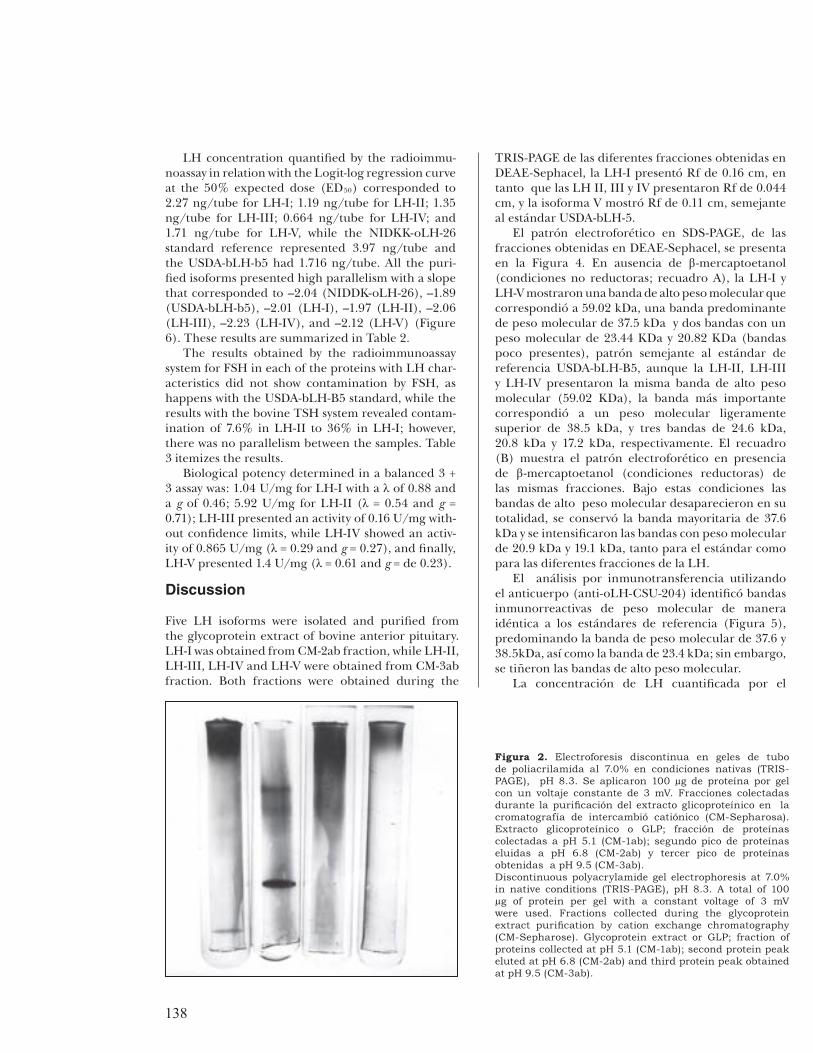
Electrophoretic patterns under native conditions (TRIS-PAGE) of each fraction obtained by the cation exchange chromatography (CM-Sepharose) are shown in Figure 2. CM-1ab fraction presented two bands with relative mobility of 0.39 cm and 0.49 cm, while fraction CM-2ab presented Rf of 0.12 cm and 0.38 cm; fi nally, CM-3ab fraction presented a band right next to the application line with Rf of 0.049 cm.
la columna (designada CM-1ab), fue 32.4% de la cantidad de proteína del EGP aplicado a la columna, en tanto que la fracción que eluyó con acetato de amonio 0.1 M, pH 6.8 (designada CM-2ab) representó 14.2% y la fracción obtenida con acetato de amonio 1 M más glicina 0.1 M, pH 9.5 (designada CM-3ab) fue 45.2%. La Figura 1b muestra el patrón cromatográfi co de la fracción CM-2ab repurifi cada en intercambio aniónico (DEAE-Sephacel), donde se obtuvo una pequeña fracción de proteínas que eluyeron con el
0
1
2
3
4
5
1
50
99
148
197
246
1ab2ab
3ab
AcO
NH
4, 0
.00
5M
, p
H 5
.1
AcO
NH
4,
0.1
M, p
H 6
. 8
AcO
NH
4, 1
.0M
/ G
ly 0
.1M
, p
H 9
.5
a)
0
0.5
1
1.5
1
50
99
148
FSH
LH-I
Gly
0.0
1M
, p
H 9
.5
AcO
NH
4,
0.1
M, p
H 6
.8
AcO
NH
4, 1
M, p
H 5
.1
b)
0
0.5
1
1.5
2
1
50
99
148
LH-II
LH-IIILH-IV
LH-V
FSH
Gly
0.0
1M
, p
H 9
.5
AcO
NH
4, 0
.1M
pH
6.8
AcO
NH
4, 1
M, p
H 5
.1
C
D.O
. 2
80
nm
Fraction Number
c)
Figura 1. a) Cromatografía de intercambio catiónico
(CM-Sepharosa) de la fracción glicoproteínica (GLP: 61.86
mg de proteína). La columna (1.5 × 27 cm) se equilibró con
acetato de amonio 0.005 M, pH 5.1, con un fl ujo de 23 ml/h,
se colectaron fracciones de 3 ml. La fracción no retenida en
la columna (CM-1ab) eluyó con este amortiguador, mientras
que las fracciones CM-2ab y CM-3ab se obtuvieron con
acetato de amonio 0.1 M, pH 6.8 y acetato de amonio
1 M más glicina 0.1 M, pH 9.5, respectivamente. b)
Cromatografía de intercambio aniónico (DEAE-Sephacel)
de la fracción CM-2ab (7.6 mg de proteína), la columna (1.5
× 25 cm) se equilibró con glicina 0.1 M pH 9.5, con fl ujo
de 18 ml/h, y se colectaron fracciones de 3 ml. La fracción
LH-I eluyó con acetato de amonio 0.1 M, pH 6.8 y la FSH,
eluyó con acetato de amonio 1 M, pH 5.1. c) Cromatografía
de intercambio aniónico (DEAE-Sephacel) de la fracción
CM-3ab (27.72 mg de proteína), la columna (1.5 × 24.5 cm)
se equilibró con glicina 0.1 M pH 9.5, con fl ujo 22.9 ml/h
y se colectaron fracciones de 3 ml. Las fracciones LH-II,
LH-III y LH-IV eluyeron con glicina 0.1M, pH 9.5, la LH-V
eluyó con acetato de amonio 0.1 M pH 6.8 y las fracciones
de FSH eluyeron con acetato de amonio 1 M, pH 5.1.
a) Cation exchange chromatography (CM-Sepharose) of
the glycoprotein fraction (GLP: 61.86 mg of protein). The
column (1.5 cm × 27 cm) was balanced with 0.005 M,
pH 5.1 ammonium acetate, with a 23 ml/h fl ow; 3 ml
fractions were collected. The fraction not retained in the
column (CM-1ab) was eluted with this buffer, while frac-
tions CM-2ab and CM-3ab were obtained with 0.1 M, pH
6.8 ammonium acetate and 1 M ammonium acetate plus
0.1 M, pH 9.5 glycine, respectively. b) Anion exchange chro-
matography (DEAE-Sephacel) of CM-2ab fraction (7.6 mg
of protein), the column (1.5 cm × 25 cm) was balanced with
0.1 M, pH 9.5 glycine, with a 18 ml/h fl ow; fractions of 3
ml were collected. Fraction LH-I eluted with 0.1 M, pH 6.8
ammonium acetate and fraction FSH eluted with 1 M, pH
5.1 ammonium acetate. c) Anion exchange chromatogra-
phy (DEAE-Sephacel) of fraction CM-3ab (27.72 mg of pro-
tein), the column (1.5 cm × 24.5 cm) was balanced with
0.1 M, pH 9.5 glycine, with 22.9 ml/h fl ow; fractions of 3
ml were collected. Fractions LH-II, LH-III and LH-IV eluted
with 0.1M, pH 9.5 glycine, fraction LH-V eluted with 0.1 M,
pH 6.8 ammonium acetate and fractions FSH eluted with 1
M, pH 5.1 ammonium acetate.

137Vet. Méx., 35 (2) 2004
segundo amortiguador (acetato de amonio 0.1 M, pH 6.8) que correspondió a la proteína que se denominó LH-I, y representó 16.1% del total de proteína aplicado a la columna, mientras que con acetato de amonio 1 M, pH 5.1 se obtuvo una fracción de proteína con rendimiento del 8.4%, que presentó una movilidad relativa (Rf) de 0.48 cm con peso molecular de 47.6 kilodaltones (kDa), y actividad biológica de FSH de 421 U/mg y una concentración inmunológica de FSH de 1.218 ng/ml, por lo que se descartó del estudio. En cuanto a la fracción CM-3ab repurifi cada en DEAE-Sephacel, se presenta en la Figura 1c; con el primer amortiguador (glicina 0.1M, pH 9.5) eluyeron tres fracciones proteínicas que correspondieron a las LH II, III y IV, que representaron 6.4%, 5.6% y 4.3% de la proteína recuperada, respectivamente. Con el segundo amortiguador (acetato de amonio 0.1 M, pH 6.8) se eluyó el pico correspondiente a la LH-V, que constituyó 25.3% de la proteína recuperada. Con el tercer amortiguador (acetato de amonio 1 M, pH 5.1) eluyó un quinto pico de proteína con características fi sicoquímicas, biológicas e inmunológicas de FSH, por lo que no se consideró en este estudio. Los rendimientos se resumen en el Cuadro 1.
Los patrones electroforéticos en condiciones nativas (TRIS-PAGE) de cada fracción obtenida durante la cromatografía de intercambio catiónico (CM-Sepharosa) se presentan en la Figura 2. La fracción CM-1ab presentó dos bandas con movilidad relativa de 0.39 cm y 0.49 cm, en tanto que la fracción CM-2ab presentó un Rf de 0.12 cm y 0.38 cm, fi nalmente la fracción CM-3ab presentó una banda pegada a la línea de aplicación con un Rf de 0.049 cm. La Figura 3 presenta los patrones en
Figure 3 shows the TRIS-PAGE patterns in the vari-ous fractions obtained in DEAE-sephacel; LH-I pre-sented Rf of 0.16 cm, while LH II, III and IV pre-sented Rf of 0.044 cm, and isoform V showed Rf of 0.11 cm, similar to the USDA-bLH-5 standard.
The electrophoretic pattern in SDS-PAGE of the fractions obtained by DEAE-Sephacel is shown in Figure 4. In absence of -mercaptoethanol (non-reducing conditions; panel A), LH-I and LH-V showed a high molecular weight band that corresponded to 59.02 kDa, a predominant band with molecular weight of 37.5 kDa and two bands with molecular weight of 23.44 KDa and 20.82 KDa (less present bands), and a pattern similar to the USDA-bLH-B5 standard ref-erence, although LH-II, LH-III and LH-IV presented the same high molecular weight band (59.02 KDa). The most important band corresponded to a molecu-lar weight slightly higher than 38.5 kDa, and three bands of 24.6 kDa, 20.8 kDa and 17.2 kDa, respec-tively. Panel (B) depicts the electrophoretic pattern in presence of -mercaptoethanol (reducing condi-tions) of the same fractions. Under these conditions, the high molecular weight bands disappeared com-pletely; the predominant band of 37.6 kDa was con-served and the molecular weight bands of 20.9 kDa and 19.1 kDa were intensifi ed, both for standard and for the different LH fractions.
The immunoblotting analysis using the antibody (anti-oLH-CSU-204) identifi ed immunoreactive bands with molecular weight similar to that of the ref-erence standards (Figure 5), with predominance of the band with molecular weight of 37.6 kDa and 38.5 kDa, as well as the band of 23.4 kDa; however, the high molecular weight bands were dyed.
Cuadro 1
FRACCIONES OBTENIDAS DURANTE LA EXTRACCIÓN Y PURIFICACIÓN DE LAS ISOFORMAS DE LH
FRACTIONS OBTAINED DURING EXTRACTION AND PURIFICATION OF LH ISOFORMS
Fraction mg of powder/kg of
pituitary µg of protein/mg of powder mg de protein/kg of pituitary
GLP1 660 93 61.4 CM-1ab2 99.5 200 19.9 CM-2ab2 33.7 258.7 8.7 CM-3ab2 138.6 200 27.2
LH-I3 7.2 170.3 1.2 LH-II3 15.2 117 1.8 LH-III3 15.9 97 1.5 LH-IV3 15 80 1.2 LH-V3 40.7 172 7
1 Glycoprotein extract. 2 Fractions obtained in cation exchange chromatography (CM-Sepharose). 3 Fractions obtained in anion exchange chromatography (DEAE-Sephacel).
138
LH concentration quantifi ed by the radioimmu-noassay in relation with the Logit-log regression curve at the 50% expected dose (ED50) corresponded to 2.27 ng/tube for LH-I; 1.19 ng/tube for LH-II; 1.35 ng/tube for LH-III; 0.664 ng/tube for LH-IV; and 1.71 ng/tube for LH-V, while the NIDKK-oLH-26 standard reference represented 3.97 ng/tube and the USDA-bLH-b5 had 1.716 ng/tube. All the puri-fi ed isoforms presented high parallelism with a slope that corresponded to –2.04 (NIDDK-oLH-26), –1.89 (USDA-bLH-b5), –2.01 (LH-I), –1.97 (LH-II), –2.06 (LH-III), –2.23 (LH-IV), and –2.12 (LH-V) (Figure 6). These results are summarized in Table 2.
The results obtained by the radioimmunoassay system for FSH in each of the proteins with LH char-acteristics did not show contamination by FSH, as happens with the USDA-bLH-B5 standard, while the results with the bovine TSH system revealed contam-ination of 7.6% in LH-II to 36% in LH-I; however, there was no parallelism between the samples. Table 3 itemizes the results.
Biological potency determined in a balanced 3 + 3 assay was: 1.04 U/mg for LH-I with a of 0.88 and a g of 0.46; 5.92 U/mg for LH-II ( = 0.54 and g = 0.71); LH-III presented an activity of 0.16 U/mg with-out confi dence limits, while LH-IV showed an activ-ity of 0.865 U/mg ( = 0.29 and g = 0.27), and fi nally, LH-V presented 1.4 U/mg ( = 0.61 and g = de 0.23).
Discussion
Five LH isoforms were isolated and purifi ed from the glycoprotein extract of bovine anterior pituitary. LH-I was obtained from CM-2ab fraction, while LH-II, LH-III, LH-IV and LH-V were obtained from CM-3ab fraction. Both fractions were obtained during the
TRIS-PAGE de las diferentes fracciones obtenidas en DEAE-Sephacel, la LH-I presentó Rf de 0.16 cm, en tanto que las LH II, III y IV presentaron Rf de 0.044 cm, y la isoforma V mostró Rf de 0.11 cm, semejante al estándar USDA-bLH-5.
El patrón electroforético en SDS-PAGE, de las fracciones obtenidas en DEAE-Sephacel, se presenta en la Figura 4. En ausencia de -mercaptoetanol (condiciones no reductoras; recuadro A), la LH-I y LH-V mostraron una banda de alto peso molecular que correspondió a 59.02 kDa, una banda predominante de peso molecular de 37.5 kDa y dos bandas con un peso molecular de 23.44 KDa y 20.82 KDa (bandas poco presentes), patrón semejante al estándar de referencia USDA-bLH-B5, aunque la LH-II, LH-III y LH-IV presentaron la misma banda de alto peso molecular (59.02 KDa), la banda más importante correspondió a un peso molecular ligeramente superior de 38.5 kDa, y tres bandas de 24.6 kDa, 20.8 kDa y 17.2 kDa, respectivamente. El recuadro (B) muestra el patrón electroforético en presencia de -mercaptoetanol (condiciones reductoras) de las mismas fracciones. Bajo estas condiciones las bandas de alto peso molecular desaparecieron en su totalidad, se conservó la banda mayoritaria de 37.6 kDa y se intensifi caron las bandas con peso molecular de 20.9 kDa y 19.1 kDa, tanto para el estándar como para las diferentes fracciones de la LH.
El análisis por inmunotransferencia utilizando el anticuerpo (anti-oLH-CSU-204) identifi có bandas inmunorreactivas de peso molecular de manera idéntica a los estándares de referencia (Figura 5), predominando la banda de peso molecular de 37.6 y 38.5kDa, así como la banda de 23.4 kDa; sin embargo, se tiñeron las bandas de alto peso molecular.
La concentración de LH cuantifi cada por el
Figura 2. Electroforesis discontinua en geles de tubo
de poliacrilamida al 7.0% en condiciones nativas (TRIS-
PAGE), pH 8.3. Se aplicaron 100 µg de proteína por gel
con un voltaje constante de 3 mV. Fracciones colectadas
durante la purifi cación del extracto glicoproteínico en la
cromatografía de intercambió catiónico (CM-Sepharosa).
Extracto glicoproteínico o GLP; fracción de proteínas
colectadas a pH 5.1 (CM-1ab); segundo pico de proteínas
eluidas a pH 6.8 (CM-2ab) y tercer pico de proteínas
obtenidas a pH 9.5 (CM-3ab).
Discontinuous polyacrylamide gel electrophoresis at 7.0%
in native conditions (TRIS-PAGE), pH 8.3. A total of 100
µg of protein per gel with a constant voltage of 3 mV
were used. Fractions collected during the glycoprotein
extract purifi cation by cation exchange chromatography
(CM-Sepharose). Glycoprotein extract or GLP; fraction of
proteins collected at pH 5.1 (CM-1ab); second protein peak
eluted at pH 6.8 (CM-2ab) and third protein peak obtained
at pH 9.5 (CM-3ab).

139Vet. Méx., 35 (2) 2004
purifi cation of the glycoprotein extract in CM-Sepha-rose and repurifi ed under the same conditions in an anion exchanger (DEAE-Sephacel). The chromato-graphic pattern of the glycoprotein extract of bovine anterior pituitary in CM-Sepharose was similar to that described for bovine10 and caprine11 glycoprotein extracts in the same conditions; although the fraction obtained from 0.005 M, pH 5.1 ammonium acetate, designated as CM-1ab, was discarded from the study due to the high FSH yield and activity.8,10 Perera et al.11 reported the obtention of a molecular morph of LH in caprines from this fraction. Probably, another LH isoform may be obtained from this fraction in bovines. Although the yield (mg of protein per kg of tissue) obtained for each of the fractions consid-ered as LH was lower than that reported for other species,8,11,30-33 none of these studies described the obtention of four proteins with physicochemical, bio-logical and immune LH features from CM-3ab frac-tion, considered as an important source of TSH.10
The electrophoretic pattern in TRIS-PAGE of the different LH isoforms confi rmed the charge het-erogeneity of this protein, since LH-I and LH-V showed a less cationic behavior, a pattern similar for equine,30,34 human,8,9 bovine,10,33 rat35 and caprine11
isoforms. LH-II, LH-III and LH-IV eluted at pH 9.5, presented a cationic-type electrophoretic pattern sim-ilar to USDA-bLH-5, AFP5500 and NIDDK-oLH-26, AFP5551B standards, as well as a variant of caprine LH,11 equine LH30 and human LH.36 It has been sug-gested that the difference among the various electro-phoretic patterns in TRIS-PAGE (charge heterogene-ity) for LH that eluted at pH 6.8 and those that eluted at pH 9.5 is due to the different composition of the carbohydrate units of the molecule and the quantity of terminal sulfate in these carbohydrates.1,4, 20,37
radioinmunoensayo con relación a la curva de regresión Logit-log a la dosis esperada al 50% (ED50)correspondió a 2.27 ng/tubo para la LH-I; 1.19 ng/tubo en la LH-II; 1.35 ng/tubo para la LH-III; 0.664 ng/tubo en la LH-IV; 1.71 ng/tubo para la LH-V, en tanto que los estándares de referencia NIDKK-oLH-26 presentaron 3.97 ng/tubo y el USDA-bLH-b5 de 1.716 ng/tubo. Todas las isoformas purifi cadas presentaron un alto paralelismo con una pendiente que correspondió a –2.04 (NIDDK-oLH-26), –1.89 (USDA-bLH-b5), –2.01 (LH-I), –1.97 (LH-II), –2.06 (LH-III), –2.23 (LH-IV), y –2.12 (LH-V) (Figura 6), cuyos resultados se resumen en el Cuadro 2.
Los resultados obtenidos con el sistema de radioinmunoensayo para FSH en cada una de las proteínas con características de LH no mostraron contaminación de FSH, como se presentó con el estándar USDA-bLH-B5; en tanto que los resultados con el sistema de TSH bovino mostraron contaminación del 7.6% en la LH-II al 36% en la LH I; sin embargo, no existió paralelismo con las muestras. El Cuadro 3 resume los resultados.
La potencia biológica determinada en un bioensayo 3 + 3 balanceado correspondió: Para la LH-I a 1.04 U/mg con una de 0.88 y una g de 0.46; para la LH-II de 5.92 U/mg ( = 0.54 y g = 0.71); la LH-III presentó una actividad de 0.16 U/mg sin límites de confi anza, en tanto que la LH-IV fue de 0.865 U/mg ( = 0.29 y una g = 0.27), y fi nalmente la LH-V fue de 1.4 U/mg ( = 0.61 y g = de 0.23).
Discusión
Se aislaron y purifi caron del extracto glicopro-teínico de adenohipófi sis bovinas, cinco isoformas de la
Figura 3. Electroforesis discontinua en geles de tubo
de poliacrilamida al 7.0% en condiciones nativas
(TRIS-PAGE), pH 8.3. Se aplicaron de 100 µg de
proteína por gel, con un voltaje constante de 3 mV.
USDA-bLH-b5 (Rf = 0.11 cm.); LH-I (Rf = 0.16 cm.);
LH-II,III, IV (Rf = 0.04) y LH-V (Rf = 0.11).
Discontinuous polyacrylamide gel electrophoresis at
7.0% in native conditions (TRIS-PAGE), pH 8.3. A total
of 100 µg of protein per gel with a constant voltage of 3
mV was used. USDA-bLH-b5 (Rf = 0.11 cm.); LH-I (Rf
= 0.16 cm.); LH-II, LH-III, LH-IV (Rf = 0.04) and LH-V
(Rf = 0.11).

140
The electrophoretic analysis of the SDS-PAGE system, in comparison with the standard USDA-bLH-B5 in non-reducing conditions was used in order to determine the molecular weight of the LH iso-forms. An important band with relative molecular weight of 37.5-38.5 kDa was present in all cases, close to that reported for ovines, bovines, porcines and caprines,11 suggesting that this band, present in the various purifi ed fractions in this study, cor-responds to the native form of this protein. On the other hand, high molecular weight proteins were identifi ed, mainly in LH-I and LH-V, which, when analyzed in reducing conditions (in presence of -mercaptoethanol), disappeared suggesting that these proteins are molecular weight aggregates of LH, since the band of 37.5 kDa was slightly conserved and the bands with relative molecular weight of 23.4 kDa and 20.8 kDa were enriched. Results similar to those reported for this hormone10-12,30 suggest that the high molecular weight forms correspond to aggre-gates of the native forms, and that this, in turn, is composed by two subunits corresponding to the bands with lower molecular weight.
Immunoblotting analysis for each of the purifi ed LH fractions identifi ed high molecular weight pro-teins, an important band with a relative molecular weight of 37.5 kDa, as well as a protein with a molecu-lar weight of 23.4 kDa. These data confi rmed that the various fractions purifi ed in this study corresponded to LH. In order to analyze the specifi city of the anti-body used in this system USDA-bTSH was simultane-
LH. La LH-I se obtuvo a partir de la fracción CM-2ab, mientras que la LH-II, LH-III, LH-IV y LH-V se obtuvieron de la fracción CM-3ab, ambas fracciones obtenidas durante la purifi cación del extracto glicoproteínico en CM-Sepharosa y repurifi cadas en las mismas condiciones en un intercambiador aniónico (DEAE-Sephacel). El patrón cromatográfi co del extracto glicoproteínico de adenohipófi sis bovina en CM-Sepharosa fue similar a lo descrito para extractos glicoproteínicos bovinos10
y caprinos,11 en las mismas condiciones. Aunque la fracción obtenida a partir de acetato de amonio 0.005 M, pH 5.1, designada como CM-1ab se descartó del estudio debido al alto rendimiento y actividad de FSH;8,10 Perera et al.11 informaron de la obtención de una forma molecular de la LH en la especie caprina a partir de esta fracción, probablemente en la especie bovina se obtenga otra isoforma de LH a partir de esta fracción. Aunque el rendimiento (mg de proteína/kg de tejido) obtenido para cada una de las fracciones consideradas como LH fueron inferiores a lo notifi cado en otras especies,8,11,30-33
ninguno de estos estudios describen la obtención de cuatro proteínas con características fi sicoquímicas, biológicas e inmunológicas de LH a partir de la fracción CM-3ab, considerada como una fuente importante de TSH.10
El patrón electroforético en TRIS-PAGE de las diferentes isoformas de la LH confi rmó la heterogeneidad de carga de esta proteína, ya que la LH-I y la LH-V mostraron un comportamiento
Cuadro 2
ACTIVIDAD BIOLÓGICA Y CONCENTRACIÓN INMUNORREACTIVA DE LH EN LAS ISOFORMAS PURIFICADAS
BIOLOGICAL ACTIVITY AND IMMUNOREACTIVE CONCENTRATION OF LH IN PURIFIED ISOFORMS
Biological Activity1 Immunoreactive concentration of
LH2 Hormone
pH of
elution
U/mg
g
ED50
(ng/tube)
Slope
NIDDK-oLH-26 ------- 2.3 ----- ----- 3.97 − 2.04 USDA-bLH-5 ------- 2.1 ---- ----- 1.72 − 1.89
LH-I 6.8 1.04 0.88 0.46 2.27 − 2.01 LH-II 9.5 5.93 0.53 0.71 1.19 − 1.97 LH-III 9.5 0.15 1.02* 3.20* 1.35 − 2.06 LH-IV 9.5 0.87 0.29 0.27 0.66 − 2.23 LH-V 6.8 1.40 0.61 0.23 1.71 − 2.12
1 In vivo bioassay consisting of ascorbic acid depletion in prepubescent pseudopregnant rats (n = 5), reference NIDDK-oLH-26 standard, with a biological activity of 2.3 U/mg. The statistical analysis of the biological activity was performed with a 3 + 3 analysis with 95% confidence limits for the bioassay. 2 Radioimmunoassay of ovine LH, NIDDK-oLH-1-2 was marked with Na125 I using the IODO-GEN technique and NIDDK-oLH-26 was used as standard. The immunoreactive LH concentration was determined based on the quantity of hormone required for the displacement to 50% of the B/BO ratio. Parallelism was determined by the slopes with a P > 0.05. * The values of and g indicate that the bioassay was not valid.
λ
λ
141Vet. Méx., 35 (2) 2004
ously included, which was recognized with very low intensity bands of 37.5 kDa and 23.4 kDa, confi rming that the standard presented contamination with LH and also suggesting that the band with low molecular weight corresponded to the subunit alpha, common among glycoproteins.
The radioimmunoassay system revealed a paralle-lism of the different LH isoforms, which was confi r-med with the slope values in comparison with the oLH and bLH standard values. This confi rmed the presence of LH. The amount of immunoreactive LH required for the ED50 of the dose-response curve was lower in the proteins that eluted at pH 9.5 (LH-II, LH-III and LH-IV) than in those that eluted at pH 6.8 (LH-I and LH-V). The difference in response can be explained based on the type of antibody used in this study, which was produced with an LH obtained at pH 9.5 (data reported by USDA and NIDDK).
The contamination degree of LH isoforms with structurally similar proteins (FSH and TSH) was not observed in the systems used for LH identifi cation, particularly with FSH, which demonstrates that these isoforms were not contaminated with FSH. In con-trast, contamination of 7.6 % for LH-III and 36.2 % for LH-I was observed with the TSH system, while percentage in LH-V was minimum or not detectable; the dose-response curves were not parallel, which was refl ected in their slope establishing that there was no identity; however, the antibody used in the TSH system can recognize antigenic sites common with the LH, since both proteins are very similar in struc-ture; therefore, the various proteins purifi ed in this
menos catiónico, patrón semejante para isoformas equinas;30,34 humana;8,9 bovina;10,33 rata35 y caprina.11
La LH-II, LH-III y LH-IV eluyeron a pH 9.5, presentaron patrón electroforético del tipo catiónico, semejante a los estándares USDA-bLH-5, AFP5500 y NIDDK-oLH-26, AFP5551B, así como una variante de LH caprina,11 LH equina30 y humana.36 La diferencia entre los diferentes patrones electroforéticos en TRIS-PAGE (heterogeneidad de carga) para las LH que eluyeron a pH 6.8 y aquellas que eluyeron a pH 9.5, se ha sugerido que se debe a la distinta composición de las unidades de carbohidratos de la molécula y a la cantidad de sulfato terminal que se observa en estos carbohidratos.1,4, 20,37
Con el fi n de determinar el peso molecular de las isoformas de LH, se recurrió al análisis electroforético en el sistema SDS-PAGE, en comparación con el estándar USDA-bLH-B5 en condiciones no reductoras; en todos los casos se presentó una banda importante con peso molecular relativo de 37.5 a 38.5 kDa, cercano a lo señalado para las especies ovina, bovina, porcina y caprina,11 sugiriendo que esta banda presente en las diferentes fracciones purifi cadas de este estudio, corresponde a la forma nativa de esta proteína. Por otro lado, se identifi caron proteínas de alto peso molecular, principalmente en la LH-I y LH-V, que cuando se analizaron en condiciones reductoras (en presencia de -mercaptoetanol) desaparecieron, ello sugiere que estas proteínas son agregados de peso molecular de la LH, ya que se conserva ligeramente la banda de 37.5 kDa y se enriquecen las bandas de peso molecular relativo de
Cuadro 3
PORCENTAJE DE CONTAMINACIÓN DE FSH Y TSH PARA CADA ISOFORMA DE LH PURIFICADA
PERCENTAGE OF FSH AND TSH CONTAMINATION FOR EACH PURIFIED LH ISOFORM
RIA-TSH1 RIA-FSH2
Hormone ED503 (ng/tube) Contamination (%) ED50
3 (ng/tube) Contamination (%)
USDA-bLH-5 62.0 1.22 ND ND LH-I 2.10 36.2 52.8 3.69 LH-II 5.6 13.6 ND ND LH-III 10.0 7.6 ND ND LH-IV 3.3 23.0 ND ND LH-V ND ND ND ND
NIDDK-oLH-26 ND ND ND ND 1 Bovine homologous liquid phase radioimmunoassay, 20 h incubation at room temperature. Standard USDA-bTSH-I-1 (0.1-12.8 ng/tube), anti-bTSH, AFP-284246, final dilution of 1:500 000. 2 Bovine homologous liquid phase radioimmunoassay, 48 h incubation at room temperature. Standard USDA-bFSH-I-2, AFP-5318C (0.2-12.8 ng/tube), antiβ-bFSH-USDA, final dilution of 1:40,000. 3 The percentage of FSH and TSH contamination was calculated based on the ED50 of the Logit-log linear regression of the dose-response curve for any studied hormone, in relation to standard USDA-bTSH-I-1 (0.76 ng/tube) and USDA-bFSH-I (1.95 ng/tube), respectively. ND = Not detectable by the system.

142
Figura 4. Electroforesis discontinua en
condiciones desnaturalizantes en SDS-
PAGE, al 12.5%, pH 8.6. El panel (A)
corresponde a las condiciones no re-
ductoras (ausencia de -mercaptoetanol)
y el panel (B) corresponde a las
condiciones reductoras (presencia de
-mercaptoetanol): Marcadores de peso
molecular; USDA-bLH-b5; LH-I; LH-II;
LH-III; LH-IV y LH-V. La concentración
de proteína en cada carril correspondió
a 1 µg. La tinción se realizó con nitrato
de plata (BIO-Rad). Las fl echas a la
izquierda indican los marcadores de peso
molecular. Las fl echas de la derecha
señalan agregados de la forma nativa
(59.02 kDa); la forma nativa de la LH (37.6
kDa). Las bandas de 23.4 kDa y 20.8 kDa
corresponden a la subunidad alfa y beta
de LH, respectivamente.
Denaturing discontinuous electrophore-
sis in SDS-PAGE, at 12.5%, pH 8.6. Panel
(A) corresponds to non-reducing condi-
tions (absence of -mercaptoethanol) and
panel (B) corresponds to reducing con-
ditions (presence of -mercaptoethanol).
Molecular weight markers; USDA-bLH-b5;
LH-I; LH-II; LH-III; LH-IV and LH-V. Pro-
tein concentration in each lane corre-
sponds to 1 µg. Dying was performed
with silver nitrate (BIO-Rad). The arrows
at the left indicate the molecular weight
markers. The arrows at the right indicate
native aggregates (59.02 kDa); the native
morph of LH (37.6 kDa). The bands of
23.4 kDa and 20.8 kDa correspond to LH
subunits alpha and beta, respectively.
Figura 5. Inmunotransferencia de las
diferentes isoformas de la LH purifi cadas
en este estudio. El anticuerpo (anti-oLH-
CSU-204) se utilizó a una dilución 1:500,
con un periodo de incubación de 24 h USDA-
bTSH; NIDDK-oLH-26; USDA-bLH-b5; LH-V;
LH-IV; LH-III; LH-II; LH-I. Marcadores de
peso molecular (92.5; 62.5, 45.0, 31.0,
21.5 y 14.4 kDa). La banda de 37.5 kDa
corresponde a la forma nativa de la LH y la
banda de 23.4 kDa, muestra la subunidad
alfa.
Immunoblotting of the different LH iso-
morphs purifi ed in this study. The anti-
body (anti-oLH-CSU-204) was used at a
dilution of 1:500, with a 24 h incubation
period USDA-bTSH; NIDDK-oLH-26; USDA-
bLH-b5; LH-V; LH-IV; LH-III; LH-II; LH-I.
Molecular weight markers (92.5 kDa, 62.5
kDa, 45.0 kDa, 31.0 kDa, 21.5 kDa and 14.4
kDa). The band of 37.5 kDa correspond to
the native morph of LH and the band of 23.4
kDa shows the alpha subunit.
(A)
(B)

143Vet. Méx., 35 (2) 2004
study are LH with null immune activity of FSH and TSH.
Recently, Ulloa-Aguirre et al.20 pointed out the sig-nifi cance of the relationship of biological and immune activity as an index (B/I) of the biopotency of indivi-dual isoforms, which varies according to protein aci-dity. For example, Castro-Fernandez et al.38 reported that the relationship decreases as acidity increases, data that confi rms the results obtained for LH-II (elu-tion at pH 9.5), when compared to isoforms that eluted at pH 6.8 (LH-I and LH-V); however, variants LH-III and LH-IV showed a very low relationship, although they eluted at pH 9.5. May be, this dif-ference in biological activity is due to the fact that the biological approach (ascorbic acid depletion) used for their determination was not suffi ciently sensitive or specifi c for them, since the antibody recognized each isoforms without distinction in all cases.
Therefore, physicochemical, biological and immu-nological results of this study confi rm that the used methodology allowed the purifi cation of fi ve LH iso-forms from the bovine glycoprotein extract with satis-factory yield for each relatively pure isoform. The iso-lated isoforms showed differences among each other in biological activity and in their interaction with a reference antibody.
Acknowledgements
We wish to thank the United States Department of Agriculture (USDA) and the National Institute of Dia-
23.4 y 20.8 kDa. Resultados semejantes a lo informado para esta hormona10-12,30 sugieren que las formas de alto peso molecular corresponden a agregados de la forma nativa y ésta, se compone de dos subunidades que correspondieron a las bandas de menor peso molecular.
El análisis por medio de la inmuno-transferencia para cada una de las fracciones purifi cadas de la LH identifi caron proteínas de alto peso molecular, una banda importante con peso molecular relativo de 37.5 kDa, así como una proteína con un peso molecular de 23.4 kDa. Estos datos confi rman que las diferentes fracciones purifi cadas en este estudio corresponden a la LH. Con el fi n de analizar la especifi cidad de anticuerpo utilizado en este sistema, simultáneamente se incluyó a la USDA-bTSH, que fue reconocida con bandas de 37.5 kDa y 23.4 kDa de muy poca intensidad, confi rmando que el estándar presenta contaminación con LH y también sugiere que la banda de peso molecular pequeño corresponde a la subunidad alfa, común entre las glicoproteínas.
El sistema de radioinmunoensayo permitió observar un paralelismo de las diferentes isoformas de la LH, lo que se pudo confi rmar en los valores de las pendientes, en comparación con los de los estándares de oLH y bLH; ello confi rma la presencia de LH. La cantidad de LH inmunorreactiva necesaria para la dosis del 50% (ED50) de la curva dosis-respuesta, fue inferior en las proteínas que eluyeron a pH 9.5 (LH-II, LH-III y LH-IV) en comparación con la de elución a pH 6.8 (LH-I y LH-V). La diferencia
0.1 1 10 100 1000
−4
−3
−2
−1
0
1
2L
ogit
(%
B/B
0)
USDA-bLH-B5
LH-I
LH-II
LH-III
LH-IV
LH-V
Figura 6. Curvas dosis-respuesta de las isoformas de la
LH bovina. Se presenta la transformación logística (Logit %
B/Bo) del porcentaje de unión máxima en relación con el
logaritmo de las dosis en cada curva.
Dose-response curves of bovine LH isoforms. The logistic
transformation (Logit % B/Bo) of the maximum bond
percentage in relation to the dose log in each curve is
presented.

144
betes & Digestive & Kidney Diseases (NIDDK) for the donation of reference standards for the immunologi-cal and biological quantitation and characterization of the proteins for this study, as well as the Mexican National Council for Science and Technology (Con-sejo Nacional de Ciencia y Tecnologia) for the fi nan-cial support of the project 25748-B, and Gerardo Arrellin Rosas, the person in charge of the Animal House of the Institute of Biomedical Research, UNAM, Mexico and Dr. Carlos Gutierrez Aguilar for his com-ments on this work.
Referencias
de respuesta se puede explicar en función del tipo de anticuerpo utilizado en este estudio, el cual se generó con una LH obtenida a pH 9.5 (datos señalados por USDA y NIDDK).
El grado de contaminación de las isoformas de la LH, con proteínas estructuralmente semejantes (FSH y TSH) no se observó en los sistemas utilizados para la identifi cación de LH, en particular con FSH, lo que demuestra que estas isoformas no tienen contaminación con FSH. En contraparte, con el sistema de TSH se observó contaminación de 7.6% para la LH-III y de 36.2% para la LH-I, mientras que en la LH-V el porcentaje fue mínimo o no detectable; el comportamiento de sus curvas dosis-respuesta no fueron paralelas, lo que se refl ejó en su pendiente y establece que no hay identidad; sin embargo, el anticuerpo utilizado en el sistema de TSH puede reconocer sitios antigénicos comunes con la LH, ya que ambas proteínas son estructuralmente muy semejantes; por tanto, las diferentes proteínas purifi cadas en este estudio son LH con nula actividad inmunológica de FSH y TSH.
Recientemente, Ulloa-Aguirre et al.20 enfatizan la importancia de la relación de la actividad biológica e inmunológica (B/I) como un índice (B/I) de la biopotencia de isoformas individuales, que varía con la acidez de la proteína; por ejemplo, Castro-Fernández et al.38 señalan que la relación disminuye con el incremento de la acidez, datos que se confi rman con los resultados obtenidos para la LH-II (elución a pH 9.5), cuando se comparó con las isoformas que eluyeron a pH 6.8 (LH-I, V); sin embargo, las variantes LH-III y LH-IV mostraron una relación muy inferior a pesar de haber eluido a pH 9.5. Quizá esta discrepancia en actividad biológica se deba a que el modelo biológico (depleción del ácido ascórbico) utilizado para su determinación no es sufi cientemente sensible o específi co para ellas, ya que en todos los casos el anticuerpo reconoció de manera indistinta a cada isoforma.
Por tanto, los resultados fi sicoquímicos, bioló-gicos e inmunológicos de este estudio confi rman que la metodología empleada permitió la purifi cación de cinco isoformas de LH a partir del extracto glicoproteínico bovino, con adecuado rendimiento para cada isoforma, relativamente puras. Las isoformas aisladas muestran entre sí diferencias en su actividad biológica y en su interacción ante un anticuerpo de referencia.
Agradecimientos
Se agradece al United States Departament of Agriculture (USDA) y al National Institute of Diabetes & Digestive & Kidney diseases (NIDDK), la donación
Combarnous Y. Structure and structure-function rela-tionships in gonadotrophins. Reprod Nutr Dev 1988; 28:211-228.Pierce JG, Parson TF. Glycoprotein hormones: structure and function. Annu Rev Biochem 1981; 50:465-495.Creus S, Chaia Z, Pellizzari EH, Cigorraga SB, Ulloa-Aguirre A, Campo S. Human FSH isoforms: carbohy-drate complexity as determinant of in-vitro bioactivity. Mol Cell Endocrinol 2001;174: 41-49.Baenzinger JU, Green ED. Pituitary glycoprotein hormone oligosaccharides: structure, synthesis and function of the asparagine-like oligosaccharides on lutropin, follitropin and thyrotropin. Biochim Bio-phys Acta 1988; 947:287-306.Kojima FN, Cupp AS, Stumpf TT, Zalesky DD, Rob-ertson MS, Werth LA. Effects of 17 -estradiol on dis-tribution of pituitary isoforms of luteinizing hormone and follicle-stimulating hormone during the follicu-lar phase of the bovine estrous cycle. Biol Reprod 1995; 52:297-304.Ulloa-Aguirre A, Midgley AR, Beitins IZ, Padma-nabhan V. Follicle-Stimulating Isohormone: Charac-terization and Physiological Relevance. Endocr Rev 1995;16:765-787.Cooke DJ, Crowe MA, Roche JF, Headon DR. Gonad-otrophin heterogeneity and its role in farm animal reproduction. Anim Reprod Sci 1996; 41:77-99.Stockell Hartree A. Separation and partial purifi ca-tion of the protein hormones from human pituitary glands. J Biol Chem 1966; 100:754-761.Bates RW, Garrison MM, Cooper JA, Condliffe PG. Further studies on the purifi cation of human thyro-tropin. Endocrinology 1968; 83:721-730.Carranza SME, Amezcua MEV, Neri BR, Salas VA. Extracción y purifi cación de la hormona luteinizante bovina. Tec Pecu Mex 1994;32: 5-17.Perera MG, Ortiz RF, Gamboa VJJ, Reynoso MW, Falcón AA, Salas VA. Obtención, purifi cación y caracterización de dos formas de hormona luteinizante de la adenohipófi sis caprina (gLH). Vet Mex 1996; 27: 1-10.Stanton PG, Burgon PG, Hearn MTW, Robertson DM. Structural and functional characterization of
1.
2.
3.
4.
5.
6.
7.
8.
9.
10.
11.
12.
145Vet. Méx., 35 (2) 2004
de los estándares de referencia para la cuantifi cación y caracterización inmunológica y biológica de las proteínas de este estudio, así como al Consejo Nacional de Ciencia y Tecnología, de México, el fi nanciamiento al proyecto 25748-B, de igual manera a Gerardo Arrellin Rosas, encargado del Bioterio del Instituto de Investigaciones Biomédicas y al Dr. Carlos Gutiérrez Aguilar sus comentarios a este trabajo.
13.
14.
15.
16.
17.
18.
19.
20.
21.
22.
23.
24.
25.
26.
hFSH and hLH isoforms. Mol Cell Endocrinol 1996; 125:133-141.Burgon PG, Stanton PG, Robertson DM. In vivo bio-activities and clearance patterns of highly purifi ed human luteinizing hormone isoforms. Endocrinol-ogy 1996; 137:4827-4836.Chlenov MA, Kandyba EI, Nagornaya LV, Orlova IL, Volgin YV. High-performance liquid chromatogra-phy of human glycoprotein hormones. J Chromatogr 1993; 631:261-267.Jack GW, Blazek R, James K, Boyd JE, Micklem LR. The automated production by immunoaffi nity chro-matography of the human pituitary glycoprotein hor-mones thyrotropin, follitropin and lutropin. J Chem Technol Biotechnol 1987; 39: 45-58.Hiyama J, Surus A, Renwick AGC. Purifi cation of human pituitary LH and thyrotrophin by hydro-phobic chromatography. J Endocrinol 1990; 125: 493-500.Grotjan HE, Schanbacher BD, Keel BD. Ovine lutein-izing hormone. Signifi cance of fl ow-through peaks observed during chromatofocusing as revealed by var-ious methods of sample preparation and application. J Chromatogr 1991; 549:141-152.Perera MG, Rojas MS, Murcia MC, Hernandez CJ, Gonzalez PE. Identifi cation of molecular forms of luteinizing hormone in the serum of cows and goats during the preovulatory peak. 27th World Veterinary Congress; 2002 September 25-29; Tunis -Tunisia, Ed. Dr. Faouzi Kechrid, 2002, Res 446.Rojas MS, Perera MG, Murcia MC, Hernández CJ, Zarco QL y González PE Formas moleculares de la hormona luteinizante (LH) en el pico preovulatorio de cabras ciclando o en el inducido con GnRH durante el anestro. XXXVII Reunión Nacional de Investigación Pecuaria; 2001 octubre 9-12; Chiapas-(México). México (D.F.): 2001,20.Ulloa-Aguirre A, Timossi C, Mendez JP. Is there any physiological role for gonadotrophin oligosaccha-ride heterogeneity in humans? Hum Reprod 2001;16: 599-604.Rose PM, Das Gaines RE, Balen HA. Defi nition and measurement of follicle stimulating hormone. Endocr Rev 2000;21:5-22.Padmanabhan V, Lee JS, Beitins IZ. Follicle-stimu-lating isohormones: regulation and biological signifi -cance. J. Reprod Fertil Suppl 1999; 54: 87-89.Ulloa-Aguirre A, Midgley R Jr., Beitins IZ, Padmanab-ham V. Follicle-stimulating isohormones: Character-ization and physiological relevance. Endocr Rev 1995; 16:765-787.Bradford MM. A rapid and sensitive method for the quantization of microgram quantities of protein uti-lizing the protein-dye binding. Anal Biochem 1976; 72:248-254.Bollag DM, Edelstein SJ. Protein Methods. New York: Wiley-Liss, 1991.Nicoll SC, Linch P. Evolutionary biology of prolactin and somatotropin. II Electrophoretic comparison of
tetrapod somatotropin. Gen Comp Endocrinol 1971; 17:490-507.Laemmli LVK. Cleavage of structural protein during the assembly of head of bacteriophage T4. Nature 1970; 224: 680-685.Towbin H, Staehelin T, Gordon J. Electrophoretic transfer of protein from polyacrylamide gels to nitro-cellulose sheets: procedure and applications. Proc Natl Acad Sci USA 1979; 76:4350-4354.Perera MG, Falcón AA, Salas VA. Estandarización de la técnica de radiomarcaje con Iodo-Gen. Memorias del XXXIX Congreso Nacional de Ciencias Fisiológicas; 1996 septiembre 24-26; Puebla (Puebla) México. México (DF). Sociedad Mexicana de Ciencias Fisiológicas, AC, 1996: 40.Matteri RL, Papkoff DA, Swedlow JR, Chang Y-S. Iso-lation and characterization of three forms of lutein-izing hormone from the pituitary gland of the horse. Biol Reprod 1986; 34: 571-578.Alatorre FS. Programa manual para bioensayos. Unidad de Investigaciones Biomédicas. México (DF): Centro Médico Nacional, Instituto Mexicano del Seguro Social 1976 Finney DJ. Statistical methods in biological assay. 2ª Ed London Griffi n, 1978.Muralidhar K, Rajendrakumar T, Sharma HP. Het-erogeneity in buffalo lutropin. Indian J Biochem Bio-phys 1992; 29:168-172.Braselton Jr. WE, Mcshan WH. Purifi cation and prop-erties of follicle stimulating and luteinizing hormones from horse pituitary glands. Arch Biochem Biophys 1970; 139:45-58.Ward DN, Reichert Jr. JE, Fitak BA, Nahm HS, Swee-ney CM, Neill JD. Isolation and properties of subunits of rat pituitary luteinizing hormone. Biochemistry 1971; 10:1796-180.Stockell-Hartree A, Renwick GC. Molecular struc-tures of glicoprotein hormones and functions of their carbohydrate components. Biochem J 1992; 287:665-679.Dias JA. Is there any physiological role for gonadotro-phin oligosaccharide heterogeneity in humans? Hum Reprod 2001; 16: 825-830.Castro-Fernandez C,Olivares A, Soderlung D, Lopez-Alvarenga JC, Zambrano E, Veldhuis JD, et al. Apreponderance of circulating basic isoforms is associ-ated with decreased plasma half-life and biological to immunological ratio of gonadotropin-releasing hor-mone-releasable luteinizing hormone in obese men. J Clin Endocrinol Metab 2000; 85: 4603-4610.
27.
28.
29.
30.
31.
32.
33.
34.
35.
36.
37.
38.