myocardial postconditioning: anesthetic considerations · mia-reperfusión si se aplican breves...
TRANSCRIPT

Arch Cardiol Mex 2011;81(1):33-46
1405-9940/$ - see front matter © 2011 Instituto Nacional de Cardiología Ignacio Chávez. Publicado por Elsevier México. Todos los derechos reservados.
Corresponding author: Martín Martínez, Department of Physiology. Instituto Nacional de Cardiología Ignacio Chávez, México, DF. Juan Badiano No. 1. Colonia Sección 16, México, DF. CP 14080. E-mail: [email protected]
REVIEW ARTICLE
Myocardial postconditioning: anesthetic considerations
Pastor Luna-Ortiz,1 Juan Carlos Torres,1 Gustavo Pastelin,1 Martín Martínez-Rosas2
1Department of Pharmacology2Department of PhysiologyInstituto Nacional de Cardiología Ignacio Chávez, México, D.F.
Recibido el 12 de marzo de 2010; aceptado el 12 de enero de 2011.
KEYWORDSPostconditioning; cardioprotection and isquemia-reperfusion injury; Mexico.
AbstractRecently, it has been shown that the heart can be protected against the ischemia-reperfusion injury if brief coronary occlusions are performed just at the beginning of the reperfusion. This procedure has been called postconditioning (PostC). It can also be elicited by pharmacological interventions, which are named pharmacological PostC. In general, PostC reduces the reperfu-sion-induced injury, blunts oxidant-mediated damages and attenuates the local inflammatory response to reperfusion, decreases infarct size, diminishes apoptosis, neutrophil activation, and endothelial dysfunction. The mechanisms that participate in PostC are still not completely understood. In this regard, adenosine, glycine, bradykinin, ciclosporin A are involved in PostC triggering. Similar to ischemic preconditioning, PostC triggers several signaling pathways and molecular components, including nitric oxide (NO), protein kinase C, adenosine triphosphate-sensitive potassium channels, the Reperfusion Injury Salvage Kinases (RISK) pathway, which comprises phosphatidylinositol-3-OH kinase (PI3K) and extracellular signal-regulated kinase (ERK 1/2), and, finally, the Survivor Activating Factor Enhancement (SAFE) pathway. In this review, we describe the mechanisms of reperfusion-induced injury as well as the proposed protective pathways activated by PostC, which seem to converge in inhibition of mitochondrial permeability transition pores opening. On the other hand, experimental evidence indicates that volatile anesthetics and opioids are capable of exerting cardioprotective effects under certain conditions, constituting a very useful pharmacological PostC. Thus, the first minutes of reperfusion represent a window of opportunity for triggering the aforementioned mediators, which acting in concert lead to protection of the myocardium against reperfusion injury. Phar-macological, especially anesthetic, PostC may have a promising future in the clinical scenarios in the operating room.

Pastor Luna-Ortiz et al34
IntroductionDespite advances in prevention and treatment, cardiovascu-lar disease remains as the number one cause of death in men and women in the United States.1 Acute myocardial infarction (AMI) is still a frequent and disabling disease being the infarct size a major determinant of myocardial functional recovery and mortality after an AMI.2 Thus,
Posacondicionamiento miocárdico; consideraciones anestésicas
ResumenRecientemente, se ha demostrado que el corazón puede protegerse contra el daño por isque-mia-reperfusión si se aplican breves oclusiones coronarias justo al inicio de la reperfusión. Este procedimiento ha sido llamado posacondicionamiento y puede ser producido mediante inter-venciones farmacológicas, las cuales constituyen el posacondicionamiento farmacológico. En general, el posacondicionamiento reduce el daño inducido por la reperfusión, disminuyendo el daño oxidativo y atenuando la respuesta inflamatoria local durante la reperfusión, así también disminuye el tamaño del infarto, disminuyendo el proceso de apoptosis, la activación neutrofí-lica y la disfunción endotelial. Los mecanismos que participan en el posacondicionamiento aún no son bien entendidos, aunque se sabe que moléculas como la adenosina, la glicina, la bra-dicinina y la ciclosporina A están involucradas en la activación del posacondicionamiento. De manera similar al preacondicionamiento isquémico, el posacondicionamiento activa rutas de señalización en las cuales participan diversos componentes moleculares como el óxido nítrico, la proteína cinasa C, los canales sensibles a ATP, la ruta de aumento del factor de activación de sobrevivencia, así como la ruta de las cinasas de salvamento de la lesión por reperfusión las cuales comprenden la cinasa de fosfatidilinositol-3-0H y la cinasa regulada por señales extrace-lulares. En esta revisión describimos los mecanismos de daño inducido por la reperfusión así como las vías protectoras propuestas activadas por el posacondicionamiento, las cuales pare-cen converger en una inhibición de la apertura de los poros de transición de la permeabilidad mitocondrial. Por otro lado, la evidencia experimental indica que los anestésicos volátiles y los opiáceos son capaces de ejercer efectos cardioprotectores bajo ciertas condiciones, constitu-yendo un posacondicionamiento farmacológico muy útil. De esta manera, los primeros minutos de la reperfusión representan una ventana de oportunidad para activar los mediadores antes mencionados, los cuales actúan en concierto para llevar a la protección del miocardio contra el daño por reperfusión. El posacondicionamiento farmacológico especialmente el anestésico puede tener un futuro promisorio en los escenarios clínicos de las salas de operaciones.
PALABRAS CLAVEPosacondicionamiento; cardioprotección ydaño por isquemia-reperfusión; México.
List of abbreviations
PostC = postconditioningIPC = ischemic preconditioningNO = nitric oxidePKC = Protein Kinase CIKATP = adenosine triphosphate-sensitive potassium channelsMitoKATP = mitochondrial adenosine triphosphate-sensitive potassium channelsPI3K = phosphatidylinositol-3-OH kinaseERK1/2 = Extracellular signal-regulated kinaseSAFE= Survivor Activating Factor Enhancement pathwaymPTP= mitochondrial permeability transition poreAMI= acute myocardial infarctionPCI= percutaneous coronary interventionATP= adenosine triphosphateROS = reactive oxygen speciesRISK = Reperfusion Injury Salvage KinasesAkt = protein kinase BCypD = cyclophilin DPPIases = peptidyl-prolyl cis-trans insomerasesVDAC = voltage-dependent anion channelANT = adenine nucleotide translocator
MEK kinases = MAPK/ERK kinaseMAPK = mitogen-activated protein kinaseGSK3b = Glycogen synthase kinase-bBAD = Bcl-2-associated death promoterGIK = glucose-insulin-potassium solutionTGF b = Tumor growth factor bGLP-1 = glucagon-like peptideAPC = anesthetic postconditioningGly = glycineGlyR = glycine receptorMPG = N-(2-mercaptopropionyl)-glycineLDH = lactate deshidrogenaseICU = intensive care unitTNFα = tumor necrosis factor-alpha STAT-3 = Signal Transducer and Activator of TranscriptioneNOS = endothelial Nitric Oxide SynthaseJAKs = Janus kinasep70s6K = serine/threonine kinase that acts downstream in the PI3 kinase pathway. Its target substrate is the S6 ribosomal protein. The phosphorylation of S6 induces protein synthesis at the ribosome.
limitation of infarct size represents an appropriate strate-gy to prevent postinfarction heart failure and to improve survival. In the last 15 years, there has been a significant improvement in the outcome of AMI patients. This impro-vement is mainly based on the introduction of efficient reperfusion strategies, such as primary percutaneous coronary intervention (PCI) and thrombolysis.3 However,

Myocardial postconditioning: anaesthetic considerations 35
coronary heart disease still presents a mortality rate of around 10% at one year for all AMI patients.1 This remai-ning high mortality rate is partly a consequence of the phenomenon called “myocardial reperfusion injury”,4 ori-ginally described by Jennings and Sommers5 in the sixties in an experimental model of myocardial infarction. In the myocardial reperfusion injury, the reperfusion causes additional functional and structural damage to the myo-cardium after a period of ischemia when the myocardium is acutely reperfused.5 Studies of AMI in animal models suggest that lethal reperfusion injury, which starts imme-diately after the opening of the culprit coronary artery, accounts for up to 50% of the final size of a myocardial infarct.4 In human clinical studies, this phenomenon is as-sociated with detrimental myocardial remodeling, ventri-cular arrhythmias, and no-reflow, and predicts a negative prognosis in myocardial infarction patients.6 Thus, lethal reperfusion injury has become a major therapeutic target in the search for improvement in AMI patient’s recovery and numerous strategies have been tested in experimen-tal settings to reduce reperfusion injury. Recently, it has been shown that the heart can be effectively protected from reperfusion injury not only with ischemic precon-ditioning (IPC), which is a classic strategy of cardiopro-tection, but also with brief episodes of ischemia during
the early reperfusion period after a prolonged period of ischemia. This process is called: ischemic PostC.
In this review, we describe the mechanisms of reper-fusion injury, the possible protective mechanisms of the PostC and pharmacological PostC, especially using anes-thetics to induce the heart protection.
Mechanisms of reperfusion injuryThe heart is a pump that converts chemical energy into mechanical work. The power produced by the cardiac muscle is generated almost entirely by oxidation of car-bon fuels with oxygen. The fuels are provided to a great extent by coronary blood flow. Oxidative metabolism is the function of mitochondria within the cell, and thus the bulk of cardiac energy is supplied by oxidative phos-phorylation within cardiac mitochondria. Like many cells, when deprived of oxygen (anoxia), cardiac cells can main-tain ATP levels by anaerobic glycolysis, and can then re-vert smoothly to oxidative metabolism on reperfusion.7 However, if blood flow is restricted, as in myocardial in-farct, the cells accumulate glycolytic by-products (lacta-te, H+) in addition to suffering from oxygen deprivation.8 This is a condition known as ischemia and can damage cardiac cells irreversibly. Paradoxically, however, the major damage to ischemic cells comes on the re-intro-duction of oxygen (reperfusion). During reperfusion, the cells typically undergo further contraction (hypercontrac-ture) and membrane damage, followed by cell death.9,10
The reperfusion injury of the myocardium is a complex phenomenon that encompasses a list of events including: reperfusion arrhythmias, microvascular damage, reversi-ble myocardial mechanical dysfunction (“stunned” myo-cardium), and cell death (due to apoptosis or necrosis). These events may occur together or separately. Most of the detrimental effects of reperfusion are triggered within the first minutes following the re-opening of the occluded coronary artery.10 However, most of the cellular disturban-ces that occur at the time of reperfusion are determined by the abnormalities induced during the ischemic period (Figure 1). During ischemia, the increase in anaerobic glycolysis results in the progressive accumulation of pro-tons and lactic acid, with increased acidosis eventua-lly inhibiting glycolytic flux and synthesis of ATP. As the cardiomyocyte attempts to correct acidosis through the Na+/H+ exchanger (this transporter moves H+ out of the cell and Na+ into the cell), the Na+ is accumulated and this event activates the Na+/K+ ATPase to pump Na+ out of the cell. However, taking in account the diminished ATP production, the Na+/K+ ATPase progressively fails wi-thout sufficient ATP. Subsequent activation of the Na+/Ca2+ exchanger, in its reverse mode, helps to move Na+ out of the cell, but favors cytosolic accumulation of Ca2+. Prolonged ischemia induces a progressive failure of io-nic homeostasis, which ultimately causes accumulation of intracellular Na+ and Ca2+ and a decline in ATP levels. If ischemia continues for a long time, a hypercontracture is developed and cell death occurs.
During early reperfusion (in the first minutes), occurs a rapid correction of acidosis (low intracellular pH) through the Na+/H+ exchanger, and through the entry of the ion HCO3
- by the Na/HCO3 symporter (Figure 2). Additionally,
Figure 1. Schematic representation of the main events contri-buting to rapid lethal cardiomyocyte injury during ischemia and hypercontracture-cell death.

Pastor Luna-Ortiz et al36
the lactate, previously accumulated, is washed out with the restored blood flow diminishing the acidosis. However, all these events cause accumulation of Na+. This additional augment in intracellular sodium induces a secondary ac-tivation of the Na+/Ca2+ exchanger in the reverse mode to move out Na+ ions; however, this aggravates cytosolic Ca2+
accumulation. On the other hand, the abrupt re-exposure of the ischemia-inhibited mitochondrial respiratory chain to oxygen generates a membrane potential to drive ATP synthesis, which leads to a rapid overload of Ca2+ in the matrix and massive production of reactive oxygen spe-cies (ROS), which by themselves are capable of damaging cellular membranes and to induce oxidative stress. The-se two factors (cytosolic Ca accumulation and oxidative stress) trigger the mPTP to open, and this pore is a key element in cell death.11 The mPTP is a nonspecific channel located on the inner mitochondrial membrane. The mPTP was originally proposed to consist of three major compo-nents: a voltage-dependent anion channel, the adenine nucleotide translocase, and cyclophilin D, contained wi-thin the mitochondrial matrix, however, its structure is not completely defined. Under normal physiological condi-tions, the mitochondrial inner membrane is impermeable
to almost all metabolites and ions, and the mPTP is in a closed conformation. Under some stress conditions, the mPTP may open and trigger several deleterious events. Opening of the mPTP abolishes the mitochondrial mem-brane potential (∆ym) inhibiting oxidative phosphoryla-tion. The mPTP opening allows the equilibration of mo-lecules that are smaller than approximately 1500 daltons. The osmotic force generated by matrix proteins results in matrix swelling (mitochondrial swelling), leading to fur-ther rupture of the outer membrane and to the release of proapoptotic factors, such as cytochrome c, into the cytosol. These actions rapidly produce cell death. In addi-tion, disruption of the mitochondrial membrane potential also causes the ATP synthase to behave as an ATPase and accelerates energy depletion secondary to the ischemic insult.12
Experimental evidence supports that mPTP opening is a postischemic event. In the isolated rat heart model, it has been demonstrated that the cytosolic release of NAD+ (this molecule was used as a marker of mPTP ope-ning) occurs at the time of reperfusion following a pro-longed ischemic insult.13 Griffiths and Halestrap used the [3H]2-deoxyglucose entrapment technique to investigate
Figure 2. Increasing ATP dissipation during ischemia leads to rises in resting cytosolic free [Ca2+]. Reperfusion leads to excessive mito-chondrial Ca2+ uptake. Mitochondrial Ca2+ overload together with oxidative stress and the prevailing low ATP induce mPTP opening. These events initiate a “vicious cycle”, i.e., inner-membrane depolarization, ATP hydrolysis by the mitochondrial ATP synthase, further increases in cytosolic Ca2+, and so on, leading, finally, to cell death.

Myocardial postconditioning: anaesthetic considerations 37
the kinetics of in situ mPTP opening, and demonstrated that mPTP opening does not happen during ischemia, but occurs within the first 5 minutes of reflow following a 30-minute period of ischemia in the isolated rat heart.14 Importantly, the time course of mPTP opening appeared to match the rapid correction of pH that occurs at re-perfusion. Recent in vivo studies support this concept by showing that PostC may mediate its cardioprotective effects through prolonged transient acidosis during the early reperfusion phase.15
Inflammatory changes and endothelial function during reperfusionDuring reperfusion there are several altered responses in other tissues or systems besides the cardiac muscle for-ming a set of events that make up the reperfusion dama-ge. Alterations of endothelial function are pivotal in the development of reperfusion damage and the no-reflow phenomenon. The inflammatory process, characterizing early and late periods of reperfusion, is an important as-pect of changes leading to tissue damage by its effects on endothelial cells. Neutrophils feature prominently in the inflammatory component of postischemic injury. This
occurs because ischemia-reperfusion prompts a release of oxygen free radicals, cytokines, and other pro-inflam-matory mediators that activate both the neutrophils and the coronary vascular endothelium.16 Activation of the-se cells promotes the expression of adhesion molecules on both neutrophils and the endothelium, which recruit neutrophils on the endothelial surface and initiate a spe-cific cascade of cell-cell interaction. This leads first to the adhesion of neutrophils to the vascular endothelium and subsequently to their transendothelial migration and their direct interactions with the interstitial matrix and myocytes.17,18 This specific series of events is a prerequi-site for the full expression of reperfusion injury, including endothelial dysfunction, microvascular collapse, and im-pairment of blood flow (the “no reflow” phenomenon), myocardial infarction and apoptosis.
Postconditioning to protect the heartIn the procedure called myocardial PostC, the heart can be protected against the ischemia-reperfusion injury with brief coronary occlusions performed just at the beginning of the reperfusion. PostC was first described by Zhao and colleagues in dogs,19 in which it reduced the myocardial injury to an extent comparable to IPC. Beneficial outco-mes observed with PostC include reduction in infarct size,20,21 in endothelial dysfunction, in neutrophil adheren-ce,19,21 and in apoptosis.22 Recently, it has been shown that a range of pharmacological agents given at the moment of reperfusion after an ischemic insult can significantly protect the myocardium, and this effect has been shown to involve protection against necrosis and apoptosis. This maneuver has been called: pharmacological PostC.
Proposed mechanisms of PostCStudies have documented that several pathways and mo-lecular components are involved in the cardioprotective effects of PostC. These include NO,23 phosphatidylinosi-tol 3-kinase (PI3K),24 extracellular signal-regulated kinase (ERK),25 PKC,26 and mitochondrial adenosine triphosphate-sensitive potassium channels (mitoKATP),27 Reperfusion Injury Salvage Kinases (RISK), and Survivor Activating Fac-tor Enhancement (SAFE) pathways. However, there is not a scheme that could help to understand the role of each described component and the final or direct effectors of PostC.
mPTP as potential end-effector of PostCAs described, mitochondria and mPTP opening have been proposed to play an essential role in reperfusion injury28 and, in this way, inhibition of mPTP opening has been repor-ted to be an important mechanism underlying PostC pro-tection.29 A number of studies have discovered a common finding with regard to the timing of PostC. The protection induced can only be taken advantage if PostC is initiated at the onset of reperfusion and is lost if it is delayed by few minutes in rat30 and rabbit models.31 Therefore, this would suggest that the end-effector of protection must exert its actions during the initial stages of reperfusion. In this regard, pharmacological inhibition of the mPTP during
Figure 3. Scheme showing the proposed mechanism of protection induced by ischemic postconditioning (that could also be triggered by ischemia reperfusion). The gradual reperfusion may have an effect via activation of phosphatidylinositol 3-kinase (PI3K)-Akt or ERK 1/2, phophorylates downstream targets such as glycogen syn-thase kinase-3b (GSK-b), BAD/Bax, and endothelial (NO) synthase (eNOS), producing NO, which inhibits mitochondrial permeability transition pore (mPTP) opening. Phosphorylation of p70s6K confers protection by inactivating Bcl-2-associated death promoter (BAD) or through protein translation.

Pastor Luna-Ortiz et al38
the early minutes of reperfusion32 has been shown to be cardioprotective, however delaying this inhibition by 15 minutes abolished this protection. Similarly, delaying the administration of insulin, which is known to activate the RISK pathway, until after the first 15 minutes of reperfu-sion abrogates its infarct-limiting effect.33 Therefore, one could speculate that the mPTP, which is known to regulate cell death during the first few minutes of reperfusion,34 is potentially the main candidate as the end-effector. Other studies have assessed the interaction between the mPTP opening, and the postconditioning interventions. In a rabbit model of ischemia-reperfusion, Argaud and co-lleagues found that both, mechanical (initiating with a smooth flow at the beginning of reperfusion) and phar-macological PostC with cyclosporin A (CsA) or NIM811, a specific inhibitor of the mPTP, that was given 1 minute before reperfusion, limit infarct size by at least 45% com-pared with control animals.35 Thus, they demonstrated that the suppression of mPTP opening at reperfusion pro-vided powerful antinecrotic and anti-apoptotic protec-tion to the ischemic myocardium. They also showed that both mechanical and pharmacological PostC increase the Ca2+ load that is required to open the mPTP.36 Additional evidence for a major role of the mPTP in lethal reper-fusion injury recently came from the use of transgenic
mice lacking cyclophilin D (CypD).37,38 CypD, which is re-cognized as a key molecular component of the mPTP, is a mitochondrial member of the family of peptidyl-prolyl cis-trans isomerases (PPIases). Although still debated, it was reported that, in the presence of a high matrix Ca2+
concentration, CypD modifies the conformation of the inner membrane proteins to form a mega-channel. The molecular structure of the mPTP remains poorly known and, besides, CypD might involve several other proteins including voltage-dependent anion channel (VDAC) or adenine nucleotide translocator (ANT), as mentioned above. Unfortunately, their precise role is still elusive and no pharmacological agent targeting these proteins is currently available for clinical trials. In vivo, CypD-deficient mice develop smaller infarcts after a prolonged coronary artery occlusion followed by reperfusion.37, 38 Recently, Lim et al. reported that CypD-deficient mice cannot be postconditioned, further suggesting that le-thal reperfusion injury is mediated by mPTP opening.39 These results strongly support the proposal that mPTP opening, triggered by mitochondrial Ca2+ overload and overproduction of ROS, plays a central role in lethal re-perfusion injury, and specific inhibition of mPTP opening at the time of reperfusion is central to the cardioprotec-tive effect induced by PostC.
Figure 4. Protective effects of potent inhalation of anesthetics. APD = action potential duration.
Myocardial postconditioning: anaesthetic considerations 39
Additional mechanisms of PostC
RISK pathwayDuring the early stages of reperfusion, there is an up-re-gulation of pro-survival kinases termed: the Reperfusion Injury Salvage Kinase (RISK) pathway, which, recently, has promoted a renewed interest in cardioprotective reperfu-sion strategies40 (Figure 3). While the mechanisms invol-ved in reperfusion injury are known to involve apoptosis and necrosis, among others, it is also realized that cells have an inherent program for survival after ischemia-re-perfusion insults, via the recruitment of innate prosurvival kinase cascades. This pathway comprises to PI3-kinases and p42/p44 extra-cellular signal-regulated kinases (ERK 1/2), which could be activated by ischemia-reperfusion injury or by PostC or pharmacological PostC. The PI3-ki-nases (PI3Ks) are a family of enzymes involved in several functions such as cell growth, proliferation, differentia-tion, motility, survival and intracellular trafficking. These enzymes are capable of phosphorylating the 3-position hydroxyl group of the inositol ring of phosphatidylinosi-tol. Particularly, the PI3K-Akt (Akt = protein kinase B) and MEK kinases (MEK kinase = MAPK/ERK kinase; mitogen-ac-tivated protein kinase/extracellular signal-regulated ki-nase) have shown to be important components of the cell survival pathway and have antiapoptotic effects. There are several potential mechanisms, none of them mutually exclusive, through which this pathway mediates inhibition of mPTP opening. These are: 1) phosphorylating and in-hibiting GSK3b;41 2) phosphorylating eNOS and producing nitric oxide, which has been demonstrated to inhibit mPTP opening42; and 3) phosphorylating BAD, either directly or indirectly via p70S6 kinase,33,43,44 thereby negating its pro-apoptotic effect. Recently, Davidsson and colleagues.45 found that insulin-mediated phosphorylation of PI3K-Akt protects the myocyte against oxidative stress by inhibiting mPTP. This suggests that the pharmacological activation of the RISK pathway by insulin at the onset of reperfusion protects against ischemia-reperfusion injury by reducing the probability that the mPTP will open. This mechanism could participate in the cardioprotective effect of “GIK” solution, which is composed by insulin in combination with glucose and potassium. However, clinical studies have su-ggested that the GIK solution may be cardioprotective fo-llowing myocardial ischemia, via a direct, non-metabolic cardioprotective effect.46, 47 Albeit, whether the RISK pa-thway kinases and the mPTP are involved in the clinical setting remains to be examined. At this moment, it is necessary to ascertain the potential mechanism whereby the RISK pathway delays mPTP opening and protects the cells from injury, and this issue is part of the setting of preconditioning or protection against reperfusion-induced injury in experimental models.
Postconditioning the human heartThe ultimate validation and utility of a cardioprotective therapy is its application to humans presenting with coro-nary artery disease and concomitant risk factors. Howe-ver, at this date there are very few studies conducted in humans; this situation is understandable because of the high risks posed by this approach. Two studies have
recently reported that conventional PostC is an effective treatment in a select patient population with coronary artery disease. In a study by Laskey,48 17 patients under-going PCI were enrolled to receive standard angioplasty involving 90s of uninterrupted balloon inflation without further treatment (n = 7) or repeated balloon inflation (“conditioning”, n = 10) of 90 s duration applied 3 to 5 min after the angioplasty inflation. “Conditioning” after an-gioplasty reduced the magnitude of ST-segment elevation compared to controls, and accelerated the rate at which ST elevation normalized after reperfusion. Furthermore, blood flow velocity reserve was significantly improved in “conditioned” hearts. Staat and colleagues 49 reported a multi-center randomized clinical trial in 37 patients with total coronary artery occlusion undergoing angioplasty/stenting. Patients that achieved a TIMI flow grade of 2 to 3 at completion of the angioplasty/stent procedure were randomized to receive either standard treatment thereaf-ter or PostC with 4 cycles of 1-min re-inflation, followed by 1-min deflation of the angioplasty balloon. Infarct size was significantly less, and the coronary blood flow achie-ved was greater in the postconditioned patients. There were no adverse events in the postconditioned patients. Together, these two studies suggest that PostC represents a safe and efficient cardioprotective intervention for the treatment of reperfusion injury in patients with ischemic heart disease. These data must be reproduced by other clinical trials, and in broader patient populations, particu-larly those presenting with severe coronary artery disease involving high risk factors (hypertension, hypercholeste-rolemia, obesity, diabetes).
Pharmacological PostCDespite the evidence mentioned above, in the clinical si-tuations, ischemic PostC may be conceptually difficult to introduce, i.e., reintroduction of ischemia at the time of reperfusion may lead to potential complications. These complications are the cause of the initial refusal to use this approach in clinical settings. For example, during pri-mary angioplasty, repetitive inflations and deflations of the balloon may result in coronary plaque rupture with consequences for restenosis or embolic events. During coro-nary bypass surgery, interruptions to reperfusion via the newly grafted conduit may only lead to regional myocar-dial protection in the area supplied by that bypass. An alternative strategy during bypass surgery would be repe-titive clamping and unclamping of the ascending aorta to achieve ischemic PostC, a concept, however, that many cardiac surgeons would be unwilling to perform due to the high risk of disrupting atheromatous plaque debris and subsequent risk of stroke. The use of ischemic PostC would not be possible in an acute myocardial infarction patient referred for thrombolysis, in case of unstable an-gina, in patients presenting with non-ST segment eleva-tion myocardial infarction or in the event of a cardiac arrest. Such a protocol might be possible to apply in a patient with myocardial infarction referred for PCI, elec-tive PCI, or at the time of cardiac surgery. However, the concept of “pharmacological PostC” by the administration of agents that activate the RISK pathway and mediate the protective effects of PostC is a more practical solution. Agents such as insulin,44 atorvastatin,50 bradikinin,51
Pastor Luna-Ortiz et al40
tumor growth factor (TGF)-B,52 and glucagon–like peptide 1 (GLP-1),53 anesthetics,54-59 opiods60-63 could individually be used as adjuvant to current reperfusion strategies, such as thrombolytics and primary PCI, to limit lethal re-perfusion injury and could form the basis of much needed and important reperfusion strategies.
Anesthetic postconditioning (APostC)Experimental evidence indicates that volatile anesthe-tics are capable of exerting cardioprotective effects under these conditions. For example, halothane preven-ted reoxygenation-induced hypercontracture of cardiac myocytes in vitro, a potential cause of myocyte necrosis during early reperfusion.64 Halothane also reduced reper-fusion injury after regional myocardial ischemia in rabbit hearts.65 Desflurane and sevoflurane reduced infarct size when administered during the first 15 minutes of reperfu-sion in rabbits.66 Isoflurane enhanced the functional reco-very of isolated rat hearts when administered solely during reperfusion.67 The administration of sevoflurane after is-chemia also improved contractile and metabolic function concomitant with reduced myoplastic calcium loading in isolated guinea pig hearts.68 These and other findings in-dicate that volatile anesthetics may prevent intracellular
calcium overloading during early reperfusion, presumably by virtue of their actions as voltage-dependent calcium channel antagonist. Isoflurane and sevoflurane also redu-ced postischemic adhesion of neutrophils,69 an important source of oxygen-derived free radicals during reperfusion that are known to be critical mediators of reperfusion in-jury.70 Several protective effects of anesthetics inhalation have been proposed (Figure 4).
Opioid and postCActivation of G protein-coupled receptors such as ade-nosine and opioid receptors has been demonstrated to initiate preconditioning. In the case of PostC, the G pro-tein-coupled receptor activation may serve as essential mechanism that also triggers protection. Indeed, recent reports have addressed that adenosine A2a and A2b recep-tors are responsible for the protection granted by PostC.71 Recently, Gross and colleagues, demonstrated that opioids can reduce infarct size when administered just before reper-fusion, an effect that was similar to that observed when opioids were given before ischemia.72 Furthermore, Wei-hrauch and colleagues73 reported that morphine enhanced the isoflurane-induced PostC in rabbit hearts. Therefore, it is possible that PostC protects the heart by activating
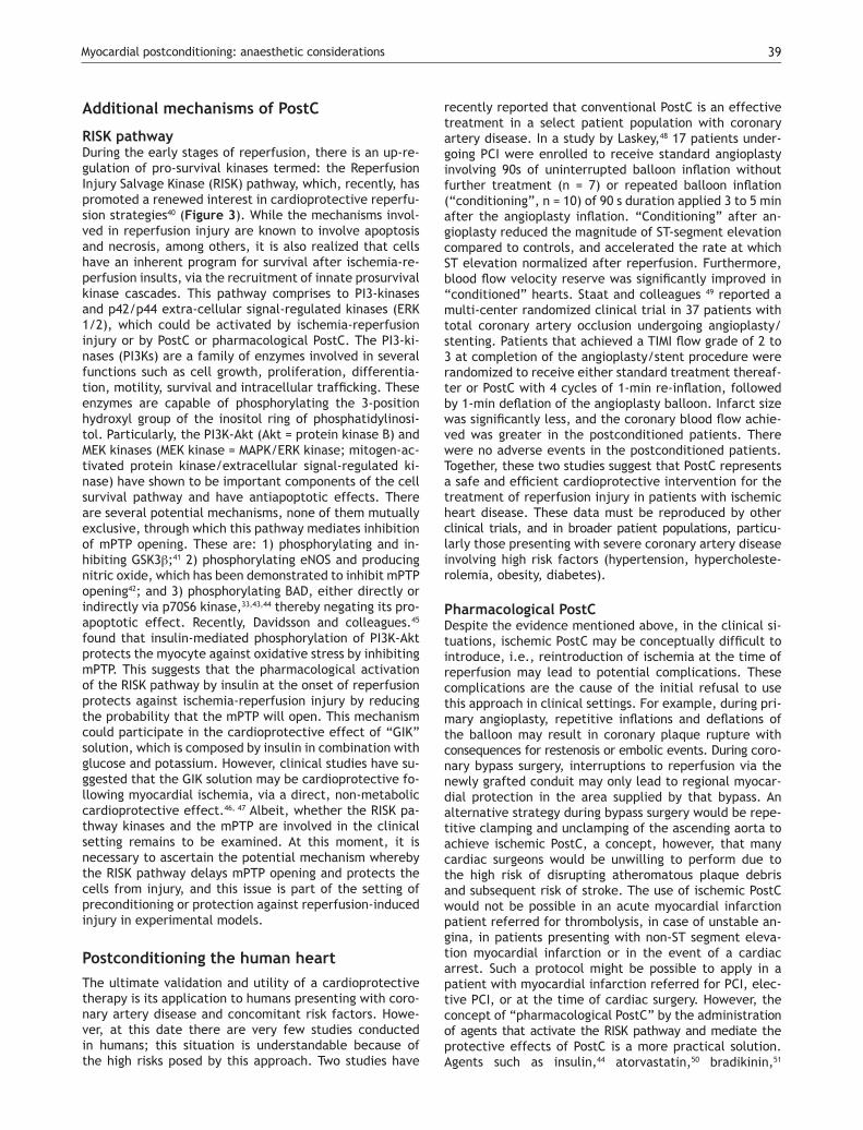
Figure 5. Scheme of the activation on the RISK pathway induced by d- and k-opioid receptor. ROS=reactive oxygen species; PKC = protein kinase C.

Myocardial postconditioning: anaesthetic considerations 41
opioid receptors during reperfusion. On the other hand, activation of delta opioid receptors plays an important role in PostC and may protect the heart at reperfusion by modulating the mPTP opening via a signaling cascade involving delta and kappa opioid receptors, NO, PKC, and other pathways (Figure 5). Gross and colleagues72 and Fryer and colleagues,74 showed that opioid-induced PostC was mediated by activation of G protein-coupled delta 1 opioid receptors, PI3K-mediated signaling, and mitoKA-TP channels. Two selective delta 1 opioid receptor ago-nists (TAN-67 and BW373U86) and morphine were initially shown to augment isoflurane preconditioning in rats,75 and the nonselective opioid antagonist, naloxone, inhibi-ted these beneficial actions. Interestingly, naloxone pre-treatment also abolished reductions in infarct size produ-ced by administration of isoflurane alone before ischemia and reperfusion, showing for the first time that opioid receptors mediate anesthetic preconditioning. Our group, at the pharmacology department at Instituto Nacional de Cardiología Ignacio Chávez, showed that remifentanil, an agonist of opioid receptors, elicited antiarrhythmic and cardioprotective effects in experimental ventricular arr-hythmias induced by digoxin and, in a model of two atrial arrhythmias induced by aconitine and by electrical stimu-lation.76
Glycine cardioprotectionThe amino acid, glycine (Gly), has been shown to exert a protective effect against cell death after diverse insults
including ischemia-reperfusion injury in different cell ty-pes other than cardiomyocytes77 (Figure 6). Gly is an in-hibitory neurotransmitter in the central nervous system that has been demonstrated by a wide range of studies in the past few years. Gly prevents or mitigates a variety of pathological processes in experimental models, such as inflammation, shock, endotoxemic shock, and those as-sociated with transplantation. The classic inhibitory gly receptor (GlyR) in the central nervous system is a ligand-gated membrane-spanning ion channel.78,79 GlyR consists of three types of subunits: (alpha), a ligand binding subu-nit; (beta), a structural subunit; and gephyrin, a cytoplas-mic anchoring protein.80 Gly in the central nervous system exerts its inhibitory actions by binding the GlyR that is located mainly on the postsynaptic neuronal membranes. Activation of GlyR leads to increase in chloride conduc-tance.81 An influx of chloride hyperpolarizes postsynaptic membranes, thus counteracting the depolarizing action of excitatory neurotransmitters. It is proposed that hy-perpolarization decreases the opening of calcium chan-nels, thereby blocking the movement of calcium across the plasma membrane and the subsequent events in the inflammatory process. Recent studies have provided phar-macological and molecular evidence for the existence of GlyR in endothelial cells,82 renal proximal tubular cells,83 and most leukocytes.84 Several investigators reported the benefits of N-(2-mercaptopropionyl)-glycine (MPG), a hydroxyl radical scavenger, for ischemic-reperfused hearts; treatment of anesthetized animals with this agent
Figure 6. Proposed role of glycine (Gly) during reperfusion injury.

Pastor Luna-Ortiz et al42
reduced ischemia-reperfusion-induced formation of the infarct size,85 and improved ischemia-reperfusion induced myocardial stunning.86 Furthermore, treatment of perfu-sed hearts with MPG enhanced recovery of the contractile function and preserved the ultrastructural integrity of the myocardium.87 Ruiz-Meana’s and colleagues showed that the amino acid Gly has a powerful and previously unrecog-nized inhibitory effect on mPTP, similar of that of low pH, in rat heart mitochondria. They also demonstrated for the first time that addition of Gly during reoxygenation pre-vents acute necrotic cell death associated with pH nor-malization in cultured cardiac myocytes and LDH release in isolated rat hearts. Moreover, exposure to Gly during reoxygenation not only reverses cellular Gly depletion ob-served in cells subjected to ischemia-reoxygenation, but markedly raises intracellular Gly content up to the range proven to inhibit mPTP in isolated mitochondria.88
Postconditioning the human heart with ade-nosineAdenosine is a primary mediator of postconditioning effects. Studies performed in animal models indica-te that adenosine receptor activation is involved in the cardioprotection conferred by PostC. The underlying
mechanism may involve delaying the washout of endo-genously released adenosine during the early minutes of reperfusion.89 The protective effects could be blocked by the nonselective adenosine receptor antagonist, 8SPT,90 and by A2a and A3 selective antagonists given before PostC. Adenosine has also been implicated in the cardio-protection exerted by remote postC.91 In a study of 60 patients with rheumatic heart valve disease undergoing heart valve replacement operations, patients were ran-domized to an adenosine (1.5 mg/kg) or saline (control) bolus injection through an arterial catheter immediately after the aorta cross-clamp had been removed. The re-sults of this study were the inotrope scores in the intensi-ve care unit (ICU) were much lower, and the ICU time was significantly shorter in the adenosine group. More impor-tant, cardiac troponin I release was lower, especially at 12 and 24 hours after reperfusion. That study demonstrated that pharmacologic PostC with adenosine produces pro-tective effects against myocardial reperfusion injury in cardiac operations.92
Advances in PostCActivation of the protective survivor activating factor enhancement (SAFE) pathway against reperfusion injury.
Figure 7. The JAK/STAT signaling pathway. JAK-2 is activated in response to a ligand (like TNFα). Activated JAK-2 recruits STAT-3 (signal transducer and activator of transcription) from the cytoplasm and phosphorylates STAT3 on a tyrosine residue, which allows STAT-3 to form homodimers and translocate to the nucleus where it up regulates transcription. STAT-3 is further phosphorylated on a serine residue to improve DNA interaction.

Myocardial postconditioning: anaesthetic considerations 43
Novel strategies to protect the heart during myocardial re-perfusion have emerged as very promising experimental therapies against lethal reperfusion injury. One of them, is the activation of the protective survivor activating fac-tor enhancement (SAFE) pathway.93 This path includes the participation of TNFα and STAT-3. TNFα levels (blood and tissue) are increased in acute myocardial infarction and, ge-nerally, TNFα is implicated as a mediator of adverse remode-ling in heart failure.94 TNFα exerts its action after binding onto its cell surface receptors, TNFα receptor 1 (TNFR1 or p55) and TNFα receptor 2 (TNFR2 o p75). Both receptor subtypes are found in human and rat cardiac myocytes.95 There are controversial findings that have been descri-bed with beneficial and detrimental effects for TNFα.96,97 These differences about its effects can be explained de-pending on which TNFα receptor is activated. Thus, TNFα 1 may be cardiotoxic whereas TNFα receptor 2 could be cardioprotective. Lecour and colleagues,98 have demons-trated that exogenous TNFα, in a dose and time depen-dent manner, could mimic ischemic preconditioning in the rat or mouse model.
The mechanisms proposed for the protective effect of TNFα is the following: After activation of the membrane receptors by TNFα (other receptors could be participa-ting, like interleukin 6 or growth factors receptors), two
adjacent juxtaposed JAKs (Janus kinase) are transphos-phorylated and subsequently activate the STAT pathway. JAKs are a family of tyrosine kinases that are associated with membranes receptors and play a major role in the transduction of signals from the cytosol to the nucleus, and they are activated during ischemia and ischemia re-perfusion in the heart. The STAT family of transcription factor proteins consists of seven identified members: STAT-1, STAT-2, STAT-3, STAT-4, STAT-5A, STAT-5B, and STAT6. After activation with JAKs, STATs form homo- or hetero-dimers that are translocated to the nucleus, resulting in gene transcription (Figure 7). The activation of the JAK/STAT pathway plays a crucial role in the expression of stress-responsive genes that could be participating in the protective effects on the heart.99
Acting together SAFE and RISK pathwaysThese distinct pathways are activated at the time of re-perfusion following an ischemic preconditioning insult, but whether these two pathways may interact together or be totally independent remains unclear. Lecour’s group has found that TNFα-mediated preconditioning acts inde-pendently of the activation of the RISK pathway, therefo-re suggesting that activation of both pathways at the time of reperfusion is not always mandatory to protect the heart against reperfusion injury.98 However, the inhibition of either the RISK or the SAFE pathway in ischemic pre-conditioning totally abolished the protection in ischemic preconditioning, suggesting the possibility of a cross-talk between the two pathways at the level of PI3K/Akt and JAK/STAT-3.98-100 (Figure 8).
The precise molecular mechanisms and pathways of signaling protective cascades are far from being fully elu-cidated. However, there is now strong evidence for the existence of at least two pathways with TNFα that play a key role in the immune system. Therefore, they represent a novel and alternative protective SAFE path during both ischemic pre- and post-conditioning.
It is clear now that it is necessary to gather more ex-perimental evidence, besides those mentioned above, in order to encourage the development of minimal risk pro-cedures that could be used in the clinical setting.
ConclusionAlthough many of the pathways involved in PostC have also been identified in IPC, recent evidences shows that both processes could be different in their pathways, being the end-effector the mPTP opening. Thus, we could get benefits from the different timing of these approaches and apply the PostC at the onset of reperfusion in a clini-cal setting (angioplasty, cardiac surgery, transplantation, etc). The PostC represents a new therapeutic tool that could be considered part of existing therapeutic resour-ces. The experimental evidence reviewed in this work supports its possible clinical application in an immediate future, albeit, more randomized, double-blind, and large-scale studies are needed. We have no doubts that in co-ming years this therapeutic objective will become widely accepted.
Figure 8. Activation of the RISK and SAFE pathways following is-chemic pre- or post-conditioning. Extracellular signal-regulated protein kinase (ERK).
Pastor Luna-Ortiz et al44
References
1. Lloyd-Jones D, Adams RJ, Brown TM, et al. Heart disease and stroke statistics-2010 update: a report from the American Heart Association Circulation 2010;121:e46-e215.
2. Gibbons RJ, Valeti US, Araoz PA, et al. The quantification of the infarct size. J Am Coll Cardiol. 2004;44:1533-1542.
3. McGovern PG, Pankow JS, Shahar E, et al. Recent trends in acu-te coronary heart disease-mortality, morbidity, medical care and risk factors. The Minnesota Heart Survey Investigators. N Engl J Med 1996;334:884-890.
4. Yellon DM, Hausenloy DJ. Myocardial reperfusion injury. N Engl J Med 2007;357:1121-1135.
5. Jennings RB, Sommers HM, Smyth GA, et al. Myocardial necrosis induced by temporary occlusion of a coronary artery in the dog. Arch Pathol 1960;70:68-78.
6. Wu KC, Zerhouni EA, Judd RM, et al. Prognostic significan-ce of microvascular obstruction by magnetic resonance ima-ging in patients with acute myocardial infarction. Circulation 1998;97:765-772.
7. Das AM, Harris DA. Regulation of the mitochondrial ATP synthase in intact rat cardiomyocytes. Biochem J 1990;266:355–361.
8. Dennis S C, Gevers W, Opie LH. Protons in ischemia: whe-re do they come from; where do they go? J Mol Cell Cardiol 1991;23:1077–1086.
9. Schlüter KD, Jakob G, Ruiz-Meana M, et al. Protection of reoxy-genated cardiomyocytes against osmotic fragility by NO donors. Am J Physiol 1996;271:H428–H434.
10. Piper HM, Abdallah Y, Schafer C. The first minutes of reperfu-sion: a window of opportunity for cardioprotection. Cardiovasc Res 2004; 61:365-371.
11. Crompton M. The mitochondrial permeability transition pore and its role in cell death. Biochem J 1999;341:233-249.
12. Halestrap AP, Clarke SJ, Javadov SA. Mitochondrial permeabi-lity transition pore opening during myocardial reperfusion –a target for cardioprotection. Cardiovasc Res 2004;61:372-385.
13. Di Lisa F, Menabo R, Canton M, et al. Opening of the mitochon-drial permeability transition pore causes depletion of mitochon-drial and cytosolic NAD+ and is a causative event in the death of myocytes in postischemic reperfusion of the heart. J Biol Chem 2001;276:2571-2575.
14. Griffiths EJ, Halestrap AP. Mitochondrial non-specific pores re-main closed during cardiac ischaemia, but open upon reperfu-sion. Biochem J 1995;307:93-98.
15. Cohen MV, Yang XM, Downey JM. Acidosis, oxygen, and in-terference with mitochondrial permeability transition pore formation in the early minutes of reperfusion are critical to postconditioning´s success. Basic Res Cardiol 2008;103;464-471.
16. Jordan JE, Zhao ZQ, Vinten-Johansen J. The role of neutrophils in myocardial ischemia-reperfusion injury. Cardiovasc Res 1999; 43: 860-878.
17. Engler RL, Schmid-Schönbein GW, Pavelec RS. Leukocyte ca-pillary plugging in myocardial ischemia and reperfusion in the dog. Am J Pathol 1983;111:98-111.
18. Moore KL, Patel KD, Bruel RE. P-selectin glycoprotein lig-and-1 mediates rolling of human neutrophils on P-selectin. J Cell Biol 1995;128:661-671.
19. Zhao ZQ, Corvera JS, Halkos ME. Inhibition of myocardial injury by ischemic postconditioning during reperfusion: comparison with ischemic preconditioning. Am J Physiol Heart Circ Physiol 2003;285:H579-H588.
20. Kin H, Zatta AJ, Lofye MT, et al. Postconditioning reduces in-farct size via adenosine receptor activation by endogenous ade-nosine. Cardiovasc Res 2005;67:124-133.
21. Halkos ME, Kerendi F, Corvera JS, et al. Myocardial protection with postconditioning is not enhanced by ischemic preconditio-ning. Ann Thorac Surg 2004;78:961-969.
22. Sun HY, Wang NP, Halkos M. Postconditioning attenuates car-diomyocyte apoptosis via inhibition of JNK and p38 mito-gen activated protein kinase signaling pathways. Apoptosis 2006;11:1583-1593.
23. Yang X-M, Proctor JB, Cui L, et al. Multiple, brief coronary occlu-sions during early reperfusion protect rabbit hearts by targeting cell signaling pathways. J Am Coll Cardiol 2004;44:1103-1110.
24. Yang X-M, Philipp S, Downey JM, et al. Postconditioning’s pro-tection is not dependent on circulating blood factors or cells but involves adenosine receptors and requires PI3Kinase and guanylyl cyclase activation. Basic Res Cardiol 2005;100:57-63.
25. Tsang A, Hausentoy DJ, Mocanu MM, et al. Postconditioning a form of “modified reperfusion” protects the myocardium by ac-tivating the phosphatidylinositol 3-kinase-akt pathway. Circ Res 2004;95:230-232.
26. Zatta AJ, Kin H, Lee G, et al. Infarct-sparing effect of myocar-dial postconditioning is dependent on protein kinase C signa-ling. Cardiovasc Res 2006;70:315-324.
27. Jang Y, Xi J, Wang H, et al. Postconditioning prevents reper-fusion injury by activating δ-opioid receptors. Anaesthesiology 2006;108:243-250.
28. Griffiths EJ, Halestrap AP. Protection by cyclosporin A of ische-mia/reperfusion-induced damage in isolated rat hearts. J Mol Cell Cardiol 1993;25:1461-1469.
29. Argaud I, Gateau-Roesch O, Risky O, et al. Postconditioning inhibits mitocondrial permeability transition. Circulation 2005;111:194-197.
30. Philipp S, Downey JM, Cohen MV. Postconditioning must be ini-tiated in less than 1 minute following reperfusion and is de-pendent on adenosine receptors and PI3-kinase. Circulation 2004;110(Suppl 111): 804-806.
31. Hausenloy DJ, Duchen MR, Yellon DM. Inhibiting mitochondrial permeability transition pore opening at reperfusion protects against ischaemia-reperfusion injury. Cardiovasc Res 2003; 60: 617-625.
32. Hausenloy DJ, Maddock HL, Baxter GF, et al. Inhibiting mito-chondrial permeability transition pore opening: a new paradigm for myocardial preconditioning? Cardiovasc Res 2002;55:534-543.
33. Jonassen AK, Sack MN, Mjos OD, et al. Myocardial protection by insulin at reperfusion requires early administration and is mediated via Akt and p70s6 kinase cell-survival signaling. Circ Res 2001;89:1191-1198.
34. Sun K, Liu ZS, Sun Q. Role of mitochondria in cell apoptosis du-ring hepatic ischemia-reperfusion injury and protective effect of ischemic postconditioning. World J Gastroenterol 2004; 10:1934-1938.
35. Argaud L, Gateau-Roesch O, Raisky O, Loufouat J, Robert D and Ovize M. Postconditioning inhibits mitochondrial permeability transition. Circulation 2005; 111: 194-197.
36. Argaud L, Gomez L, Gateau-Roesch O, et al. Trimetazidine in-hibits mitochondrial permeability transition pore opening and prevents lethal ischemia-reperfusion injury. J. Mol Cell Cardiol 2005;39:893-899.
37. Baines CP, Kaise RA, Purcell NH, Blair NS, Osinska H, Hambleton MA, Brunskill EW, Sayen MR, Gottlieb RA, Dorn GW, et al. Loss of cyclophilin D reveals a critical role for mitochondrial permeabi-lity transition in cell death. Nature 2005;434;658-662.
38. Nakagawa T, Shimizu S, Watanabe T, et al. Cyclophilin D-dependent mitochondrial permeability transition regu-lates some necrotic but no apoptotic cell death. Nature 2005;434:652-658.
39. Lim SY, Davidson SM, Hausenly DJ et al. Preconditioning and postconditioning: the essential role of the mitochondrial per-meability transition pore. Cardiovasc Res 2007;75:530-535.
40. Hausenby DJ, Yellon DM. New directions for protecting the heart against ischaemia-reperfusion injury: targeting the re-perfusion injury salvage kinase (RISK)-pathway. Cardiovasc Res 2004; 61: 448-460.
41. Juhaszova M, Zorov DB, Kim SH, et al. Glycogen synthase kina-se-3 beta mediates convergence of protection signaling to inhi-bit the mitochondrial permeability transition pore. J Clin Invest 2004;113:1535-1549.
Myocardial postconditioning: anaesthetic considerations 45
42. Kim JS, Ohshima S, Pediaditakis P, et al. Nitric oxide protects rat hepatocytes against reperfusion injury mediated by the mi-tochondrial permeability transition. Hepatology 2004;39:1533-1543.
43. Jonassen AK, Mjos OD, Sack MN. p70s6 kinase is a functional target of insulin activated Akt cell-survival signaling. Biochem Biophys Res Commun 2004; 315:160-165.
44. Jonassen AK, Brar BK, Mjøs OD, et al. Insulin administered at reoxygenation exerts a cardioprotective effect in myocytes by a possible anti-apoptotic mechanism. J Mol Cell Cardiol 2000;32:757-764.
45. Davidson SM, Hausenloy D, Duchen MR, et al. Signalling via the reperfusion injury signalling kinase (RISK) pathway links closure of the mitochondrial permeability transition pore to cardiopro-tection. The International Journal of Biochemistry & Cell Biolo-gy 2006;38:414-419.
46. Doenst T, Bothe W, Beyersdorf F. Therapy with insulin in cardiac surgery: Controversies and possible solutions. Ann Thorac Surg 2003;75:S721-728.
47. Sack MN Yellon DM. Insulin therapy as an adjunct to reperfusion after acute coronary ischemia: A proposed direct myocardial cell survival effect independent of metabolic modulation. J Am Coll Cardiol 2003;41:1404-1407.
48. Laskey WK. Brief repetitive balloon occlusions enhance reper-fusion during percutaneous coronary intervention for acute myocardial infarction: a pilot study. Catheter Cardiovasc Interv 2005;65:361-367.
49. Staat P, Rioufol G, Piot C, et al. Postconditioning the human heart. Circulation 2005;112:2143-2148.
50. Bell RM, Yellon DM. Atorvastatin, administered at the onset of reperfusion and independent of lipid lowering, protects the myocardium by up-regulating a pro-survival pathway. J Am Coll Cardiol 2003;41:508-515.
51. Bell RM, Yellon DM. Bradykinin limits infarction when adminis-tered as an adjunct to reperfusion in mouse heart: the role of PI3k, Akt and eNOS. J Mol Cell Cardiol 2003;35:185-193.
52. Baxter GF, Mocanu MM, Brar BK, et al. Cardioprotective effects of transforming growth factor-beta 1 during early reoxygena-tion of reperfusion are mediated by p42/p44 MAPK. J Cardio-vasc Pharmacol 2001;38:930-939.
53. Nikolaidis LA, Mankad S, Sokos GG, et al. Effects of glucagon-like peptide-1 in patients with acute myocardial infarction and left ventricular dysfunction after successful reperfusion. Circu-lation 2004;109:962-965.
54. Shlack W, Preckel B, Barthel H. Halothane reduces reperfusion injury after regional ischaemia in the rabbit heart in vivo. Br J Anaesth 1997;79:88-96.
55. Preckel B, Schlack W, Comfere T. Effects of enflurane, isoflu-rane, sevoflurane and desflurane on reperfusion injury after regional myocardial ischaemia in the rabbit heart in vivo. Br J Anaesth 1998; 81: 905-912.
56. Schlack W, Preckel B, Stunneck D. Effects of halothane, enflura-ne, isoflurane, sevoflurane and desflurane on myocardial reper-fusion injury in the isolated rat heart. Br J Anaesth 1998;81:913-919.
57. Varadarajan SG, An J, Novalija E. Sevoflurane before or after ischemia improves contractile and metabolic function while re-ducing myoplasmic Ca2 loading in intact hearts. Anesthesiology 2002;96:125-133.
58. HeindI B, Reichle FM, Zahler S. Sevoflurane and isoflurane pro-tect the reperfused guinea pig heart by reducing postischemic adhesion of polymorphonuclear neutrophils. Anesthesiology 1999;91:521-530.
59. Vinten-Johansen J. Involvement of neutrophils in the patho-genesis of lethal myocardial reperfusion injury. Cardiovasc Res 2004;61:481-497.
60. Jang Y, Xi J, Wang H, et al. Postconditioning prevents reper-fusion injury by activating δ-opioid receptors. Anaesthesiology 2006;108:243-250.
61. Gross ER, Hsu AK, Gross GJ. Opioid-induced cardioprotection occurs via glycogen synthase kinase β inhibition during reperfu-sion in intact rat hearts. Circ Res 2004;94:960-966.
62. Weihrauch D, Riotikowski JG, Bienengracher M, et al. Morphine enhances isoflurane-induced postconditioning against myocar-dial infarction: The role of phosphatidylinositol-3-kinase and opioid receptors in rabbits. Anesth Analg 2005;101:942-949.
63. Gross ER, Hsu AK, Gross GJ. GSK3β inhibition and KATP channel opening mediate acute opioid-induced cardioprotection at re-perfusion. Basic Res Cardiol 2007;102: 341-349.
64. Siegmund B, Schlack W, Ladilov YV. Halothane protects car-diomyocytes against reoxygenation-induced hypercontracture. Circulation 1997;96: 4372-4379.
65. Shlack W, Preckel B, Barthel H. Halothane reduces reperfusion injury after regional ischaemia in the rabbit heart in vivo. Br J Anaesth 1997;79: 88-96.
66. Preckel B, Schlack W, Comfere T. Effects of enflurane, isoflu-rane, sevoflurane and desflurane on reperfusion injury after regional myocardial ischaemia in the rabbit heart in vivo. Br J Anaesth 1998;81:905-912.
67. Schlack W, Preckel B, Stunneck D. Effects of halothane, enflura-ne, isoflurane, sevoflurane and desflurane on myocardial reper-fusion injury in the isolated rat heart. Br J Anaesth 1998;81:913-919.
68. Varadarajan SG, An J, Novalija E. Sevoflurane before or after ischemia improves contractile and metabolic function while re-ducing myoplasmic Ca2+ loading in intact hearts. Anesthesiology 2002;96:125-133.
69. HeindI B, Reichle FM, Zahler S. Sevoflurane and isoflurane pro-tect the reperfused guinea pig heart by reducing postischemic adhesion of polymorphonuclear neutrophils. Anesthesiology 1999;91:521-530.
70. Vinten-Johansen J. Involvement of neutrophils in the patho-genesis of lethal myocardial reperfusion injury. Cardiovasc Res 2004;61:481-497.
71. Kin H, Zatta AJ, Lofye MT, et al. Postconditioning reduces in-farct size via adenosine receptor activation by endogenous ade-nosine. Cardiovasc Res 2005;67:124-133.
72. Gross ER, Hsu AK, Gross GJ. Opioid-induced cardioprotection occurs via glycogen synthase kinase β inhibition during reperfu-sion in intact rat hearts. Circ Res 2004;94:960-966.
73. Weihrauch D, Riotikowski JG, Bienengracher M, et al. Morphine enhances isoflurane-induced postconditioning against myocardial infarction: The role of phosphatidylino-sitol-3-kinase and opioid receptors in rabbits. Anesth Analg 2005;101:942-949.
74. Fryer RM, Wang Y, Hsu AK. Essential activation of PKC-delta in opioid-initiated cardioprotection. Am J Physiol Heart Circ Phy-siol 2001;280: H1346-H1353.
75. Ludwig LM, Patel HH, Gross GJ. Morphine enhances pharma-cological preconditioning by isoflurane: Role of mitochon-drial K (ATP) channels and opioid receptors. Anesthesiology 2003;98:705-711.
76. Luna-Ortiz P, Zarco-Olvera G, Ramírez-Ortega M, et al. Antiarr-hythmic and cardioprotective effects of remifentanil in anes-thetized dogs. Arch Cardiol Mex 2009;79:182-188.
77. Nishimura Y, Lemasters JJ. Glycine blocks opening of a death channel in cultured hepatic sinusoidal endothelial cells during chemical hypoxia. Cell Death Differ 2001; 8: 850-858.
78. Rajendra S, Lynch JW, Schofield PR. The glycine receptor. Phar-macol Ther 1997;73:121-146.
79. Laube B, Maksay G, Schemm R, et al. Modulation of glycine receptor function: a novel therapeutic approach for thera-peutic intervention at inhibitory synapses? Trends Pharmacol Sci 2002;23:519-527.
80. Pfeiffer F, Betz H. Solubilization of the glycine receptor from rat spinal cord. Brain Res 1981;226:273-279.
81. Werman R, Davidoff RA, Aprison MH. Is glycine a neurotransmit-ter? Nature 1967;214:681-683.

Pastor Luna-Ortiz et al46
82. Yamashima S, Konno A, Wheeler MD, et al. Endothelial cells con-tain a glycine-gated chloride channel. Nutr Cancer 2001;40:197-204.
83. Reeves WB. Effects of chloride channel blockers on hypoxic in-jury in rat proximal tubules. Kidney Int 1997;51:1529-1534.
84. Ikejima K, Limuro Y, Forman DT, et al. A diet containing glycine improves survival in endotoxic shock in the rat. Am J Physiol 1996;271:G-97-103.
85. Koerner JE, Anderson BA, Dage RC. Protection against postis-chemic myocardial dysfunction in anesthetized rabbits with scavengers of oxygen-derived free radicals. J Cardiovasc Phar-macol 1991;17:185-191.
86. Myers ML, Bolli R, Lekich RF, Hartley et al. N-2mercaptopropion-ylglycine improves recovery of myocardial function after rever-sible regional ischemia. J Am Coll Cardiol 1986;8:1161-1168.
87. Tanaka M, Fujiwara H, Yamasaki K, et al. Superoxide dismutase and N-2 mercaptopropionylglycine attenuate infarct size limita-tion effect of ischemic preconditioning in the rabbit. Cardiovasc Res 1994;28:980-986.
88. Ruiz-Meana M, Pina P, Garcia-Dorado D, et al. Glycine protects cardiomyocytes against lethal reoxygenation injury by inhibiting mitochondrial permeability transition. J Physiol 2004;558:873-882.
89. Kin H, Zatta AJ, Lofye MT, et al. Postconditioning reduces in-farct size via adenosine receptor activation by endogenous ade-nosine. Cardiovasc Res 2005;67:124-133.
90. Yang XM, Philipp S, Downey JM, et al. Postconditioning protec-tion is not dependent on circulating blood factors. Basic Res Cardiol 2005;100:57-63.
91. Kerendi F, Kin H, Halkos ME, et al. Remote postconditioning: brief renal ischemia and reperfusion applied before coronary artery reperfusion reduces myocardial infarct size via endo-genous activation of adenosine receptors. Bas Res Cardiol 2005;100:404-412.
92. Jin ZX, Zhou JJ, Xing M. Postconditioning the human heart with adenosine in heart valve replacement surgery. Ann thorac Surg 2007;83:2066-2073.
93. Lecour S. Multiple protective pathways against reperfusion in-jury: a SAFE path without Aktion? J Mol Cell Cardiol 2009;46:607-609.
94. Mann DL. Stress-activated cytokines and the heart: from adaptation to maladaptation. Annu Rev Physiol 2003;65:81-101.
95. Torre-Amione G, Kapadia S, Lee J, et al. Expression and functio-nal significance of tumor necrosis factor receptors in human myocardium. Circulation 1995;92:1487-1493.
96. Kurreimeyer KM, Michael LH, Baumgarten G, et al. Endoge-nous tumor necrosis factor protects the adult cardiac myocyte against ischemic-induced apoptosis in a murine model of acute myocardial infarction. Proc Natl Acad Sci USA 2000;97:5456-5461.
97. Flaherty MP, Guo Y, Tiwari S, et al. The role of TNF-alpha re-ceptors p55 and p75 in acute myocardial ischemia/reperfusion injury and late preconditioning. J Mol Cell Cardio 2008;45:735-741.
98. Lecour S, Suleman N, Deuchar GA, et al. Pharmacological preconditioning with tumor necrosis factor-alpha activa-tes signal transducer and activator of transcription-3 at reperfusion without involving classic prosurvival kinases (Akt and extracellular signal-regulated kinase). Circulation 2005;112:3911-3918.
99. Boengler K, Hilfiker-Kleiner D, Drexler H, Heusch G, Schulz R. The myocardial JAK/STAT pathway: from protection to failure. Pharmacol Ther 2008;120:172-185.
100. Gross ER, Hsu AK, Gross GJ. The JAK/STAT pathway is essen-tial for opioid-induced cardioprotection: JAK2 as a mediator of STAT3, Akt, and GSK-3 beta. Am J Physiol Heart Circ Physiol 2006;291:H827-834.