muscles, muscle fibres and myofibrils · properties of muscle fiber types fast fibers slow fibers....
TRANSCRIPT

Muscles, muscle fibres and myofibrils

Properties of Muscle Fiber Types
Fast fibers Slow fibers
Characteristic IIb IIa Type I
Resistance to fatigue Low High/moderate High
Predominant energy system Anaerobic Combination Aerobic
Vmax
(speed of shortening) Highest Intermediate Low
Myoglobin Low Medium High
Capillary density Low Medium High
IIx

Fibre-specific genes and their expression pattern in ADULT striated muscle
Myosin heavy chain MyHCI/slow/β MyHC2A MyHC2X MyHC2B MyHCI/slow/β MyHCα
Slow Fibres Fast FibresI IIA IIX/D IIB
Gene familyHeart
Myosin light chain 1 MLC1SA MYL3 MYL1 MYL4 MYL3Myosin light chain 2 MYL2 MYL5 MYL7 MYL2Troponin C TNNC1 TNNC2 TNNC1Troponin T TNNT1 TNNT3 TNNT2Troponin I TNNI1 TNNI2 TNNI3
Gene expressed

Motor control of muscle fibresMotor unit – the α-motor neuron and all the fibres under
its control
Motor units may control <5 muscle fibres in the eye or small hand muscles or >2000 fibres in the gastrocnemius

Importance of muscle fibre types
Athletic performance – marathon runners versus sprinters
Ageing – preferential reduction of fast fibres in sarcopenia
Disease – preferential loss of fast fibres in Duchennemuscular dystrophy; complete absence of fast fibres in some nemaline myopathy patients.
Atrophy responses – reduction of slow fibres in response to bed-rest, space flight and spinal cord injury.
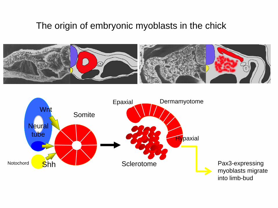
The origin of embryonic myoblasts in the chick
Shh
WntDermamyotomeEpaxial
Pax3-expressing myoblasts migrate into limb-bud
Notochord
Hypaxial
Sclerotome
SomiteNeural tube

Formation of the myotomeMuscle progenitors delaminate from the edges of the dermamyotome to form the myotome. Some cells migrate into the limb buds. At E10.5 the dermamyotome disintegrates centrally and the main myotome is formed
Expression of the myogenicregulatory factor (MRF) gene MyoD
Myogenesis in the mouse

Epaxial and hypaxial components of the myotome E11.5 mouse embryos.
Eloy-Trinquet S , Nicolas J Development 2002;129:111-122

Myogenesis
Myoblasts Myotube
Maturation hypertrophy to increase size and expression of adult myofilament genes = mature muscle fiber
Myogenicprogenitors
determination differentiationmaturation
growth hypertrophy
Proliferative phase
specification

Myoblast Myocyte MyotubeDifferentiation Fusion
Myoblast differentiation in culture
Phase contrast
Immunofluorescent detection of a ‘muscle marker’

Differentiation of primary myotubes in the mouse hind-limb (12-14 dpc) and the beginning of fibre
type patterning
MyHC expression1. Embryonic2. Neonatal3. Slow
Tendon formation from sclerotome-derived cells – marked by expression of Scleraxis (Scx). Induced by the myotome.
Fusion of myoblasts is ordered and synchronous. Nerve is not required for fusion or Myosin Heavy Chain Slow expression
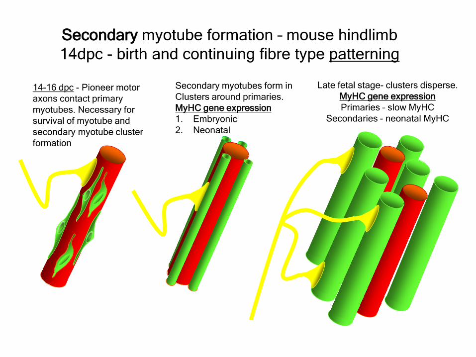
14-16 dpc - Pioneer motor axons contact primary myotubes. Necessary for survival of myotube and secondary myotube cluster formation
Secondary myotubes form inClusters around primaries.MyHC gene expression1. Embryonic 2. Neonatal
Secondary myotube formation – mouse hindlimb14dpc - birth and continuing fibre type patterning
Late fetal stage- clusters disperse.MyHC gene expressionPrimaries – slow MyHC
Secondaries - neonatal MyHC

EM sections of developing iliofibularis muscle in chick embryos
Primary myogenesis
Secondary myogenesis

Studying muscles in the mouse as a model of human muscle development – the lower hind limb
18 Tibialis anterior15 Extensor digitorum longus EDL12 Peroneus brevis and longus17 Tibialis posterior8 Soleus9 Gastrocnemius medial head5 Gastrocnemius lateral head
A
P
Approx plane of section

In situ hybridisation analysis of Troponin I isoforms in mouse crural sections
G = GastrocnemiusS = SoleusE = EDLT = Anterior tibialis
Tnni1 is the gene that encodes the inhibitory subunit of the Troponincomplex that is found in slow-twitch fibres.

Postnatal fibre CONVERSION: slow fiber number declines and neonatal MyHC is
replaced by the adult fast fibre MyHCs
TibiaTibialis anterior muscleEDL muscleFibulaSoleus muscleMedial GastrocnemiusmuscleLateral Gastrocnemiusmuscle
A
Transverse sections of hind-limbs from postnatal mice 2days and 6 weeks after birth – stained for Myosin heavy chain slow and Myosin heavy chain 2A

Plasticity and Regeneration of Adult Muscle
Muscle Adaptation to Exercise TrainingAdaptations to exercise training, particularly elevation in oxidative capacity of exercisedmuscle but also some myosin isoform changes mainly in fast subtypes.
Cross-ReinnervationBuller et al. (1960) – Motor nerves supplying the (slow) soleus and (fast) FDL muscles swapped around. Contraction speed of soleus got faster, FDL slower.
Chronic Low-Frequency Stimulation (CLFS)Artificial electrical stimulation of a nerve supplying a fast muscle with a tonic patternmimics the impulse pattern of a slow nerve and induces fast to slow transformation Pette et al. (1973).
Pure Fibers, Hybrid fibers and the “Next-Neighbour Rule”Analysis of myofilament isoforms in single fibers reveal the presence of “pure” and “hybrid” fiberscontaining, for example, MHC 2B and 2X. The percentage of hybrid fibers increases dramaticallyin transforming muscles. Transition occurs in a stepwise direction 2B->2X->2A->I. Hybrids fibres always contain a pair of “next-neighbour” isoforms.
RegenerationInjured muscle can regenerate itself using a population of stem cells that are laid down during embryogenesis – called satellite cells. Satellite cells lie between the sarcolemma and the basal lamina of each muscle fibre and activated by injury.