muscles: functions 1.movement 2.heat production 3.posture maintenance types: 1.skeletal 2.cardiac...
TRANSCRIPT
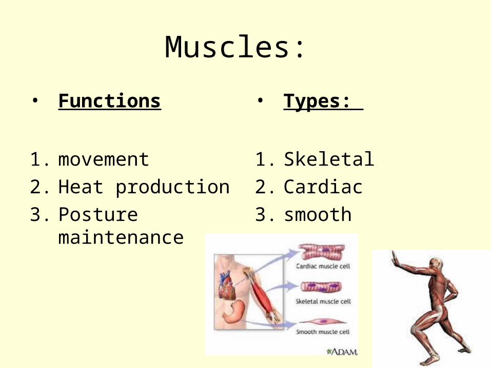
Muscles:
• Functions
1. movement
2. Heat production
3. Posture maintenance
• Types:
1. Skeletal
2. Cardiac
3. smooth
Functional characteristics
1. Excitability: ability to receive and respond to stimulus ( neural or hormonal)
2. Contractility: ability to forcibly shorten in response to stimulus
3. Extensibility: ability to stretch or extend when pulled
4. Elasticity: ability to return to original length after being stretched ( contracting)
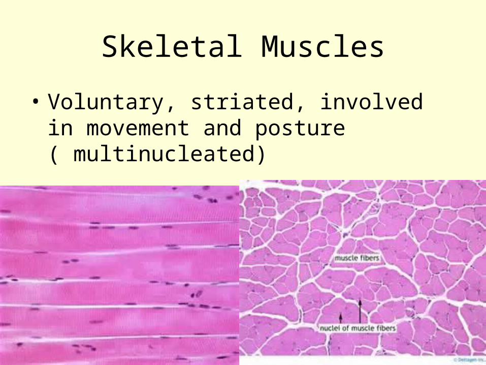
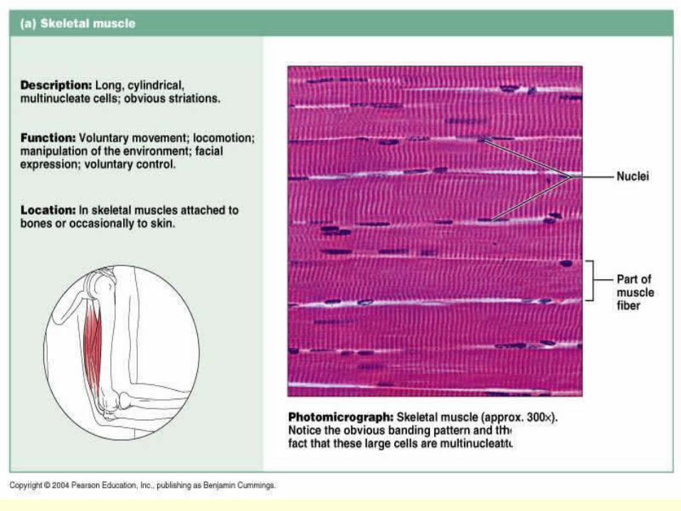
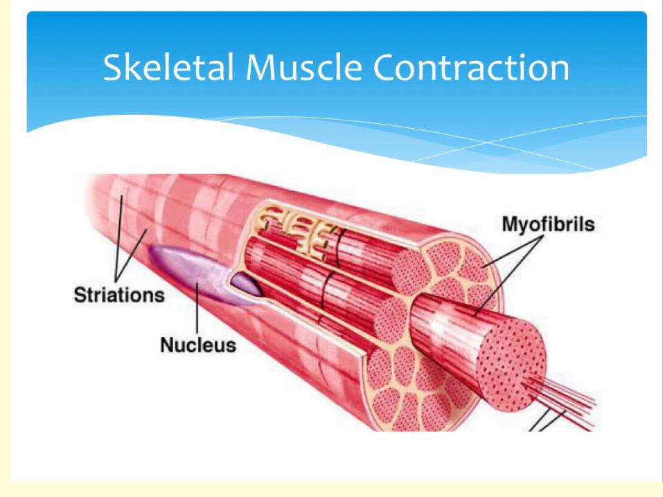
Skeletal Muscles
• Voluntary, striated, involved in movement and posture ( multinucleated)
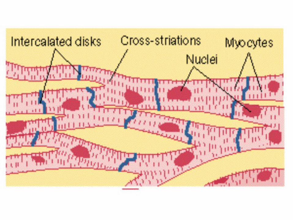
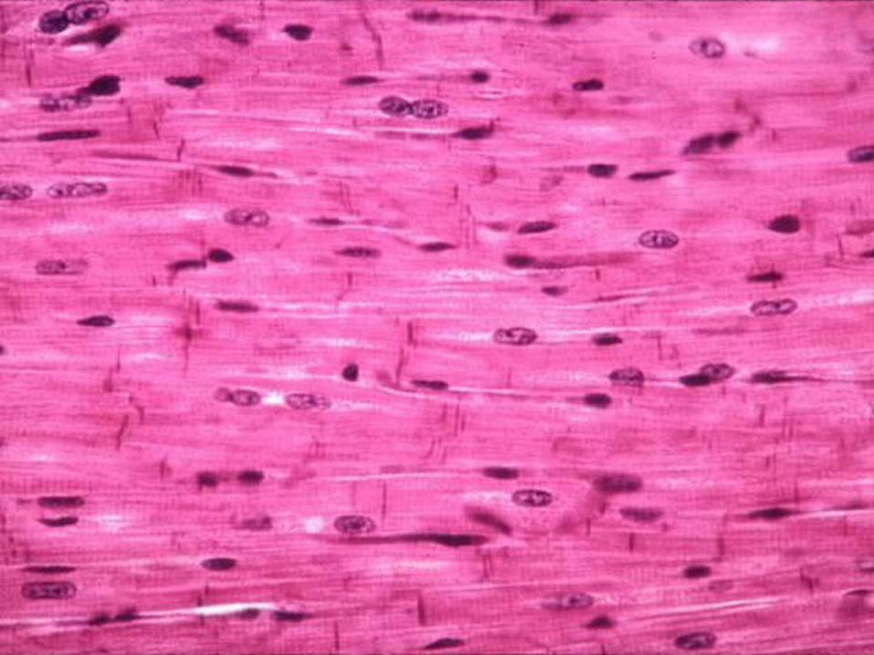
Cardiac Muscle
• Striated, involuntary, single central nucleus
• Fibers are branched, weave together forming a network
• Individual fibers connected with each other at the intercalated discs ( these discs not only tie the fibers together for strength, but also conduct impulses from one fiber to another
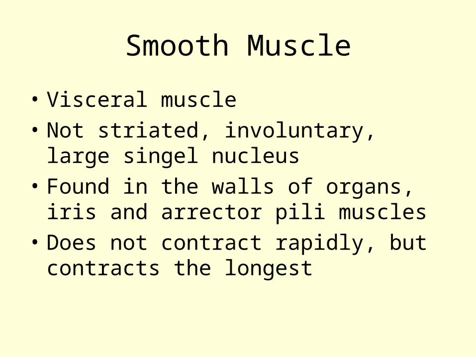
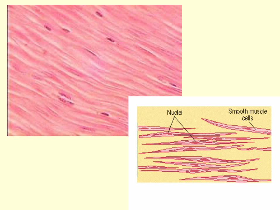
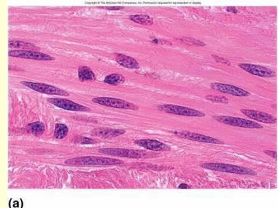
Smooth Muscle
• Visceral muscle
• Not striated, involuntary, large singel nucleus
• Found in the walls of organs, iris and arrector pili muscles
• Does not contract rapidly, but contracts the longest
Skeletal muscles
• Make up 40% of body weight and consists of over 600 muscles.
• The difference in strength, endurance, and coordination is a function of both heredity and conditioning.
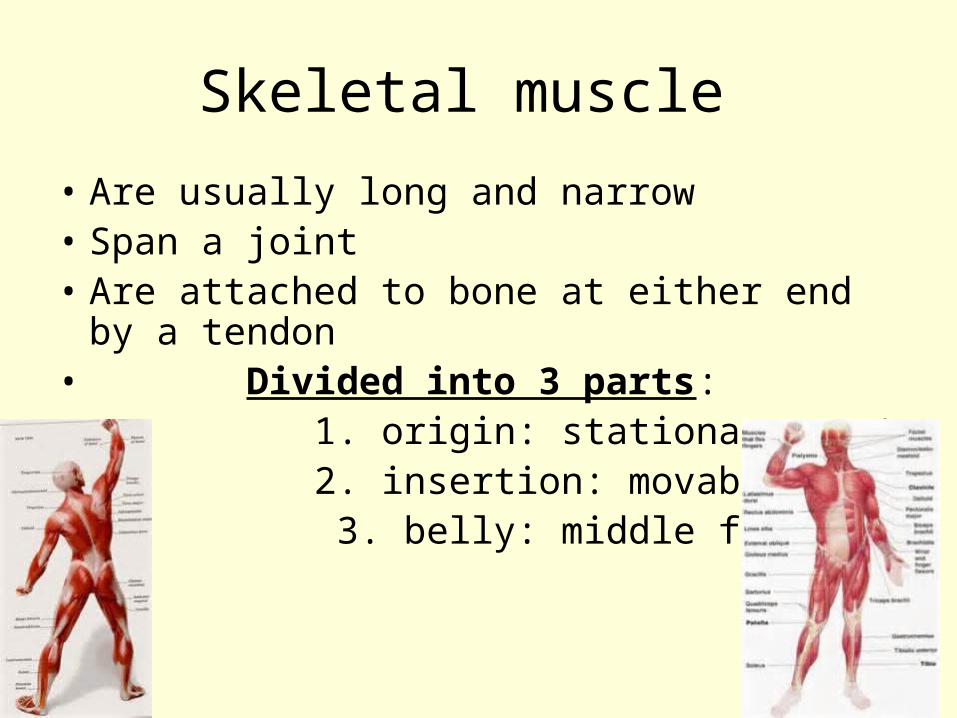
Skeletal muscle
• Are usually long and narrow• Span a joint• Are attached to bone at either end by a
tendon• Divided into 3 parts:• 1. origin: stationary part• 2. insertion: movable end• 3. belly: middle fat part
Gross Anatomy of skeletal muscles
• Fascia: layers of fibrous C.T. that covers and separates muscles
• Superficial fascia: joins skin t muscle and contains much fat ( aereolar c.t.)
• Deep fascia: joins or binds adjacent muscles and individual muscle fibers
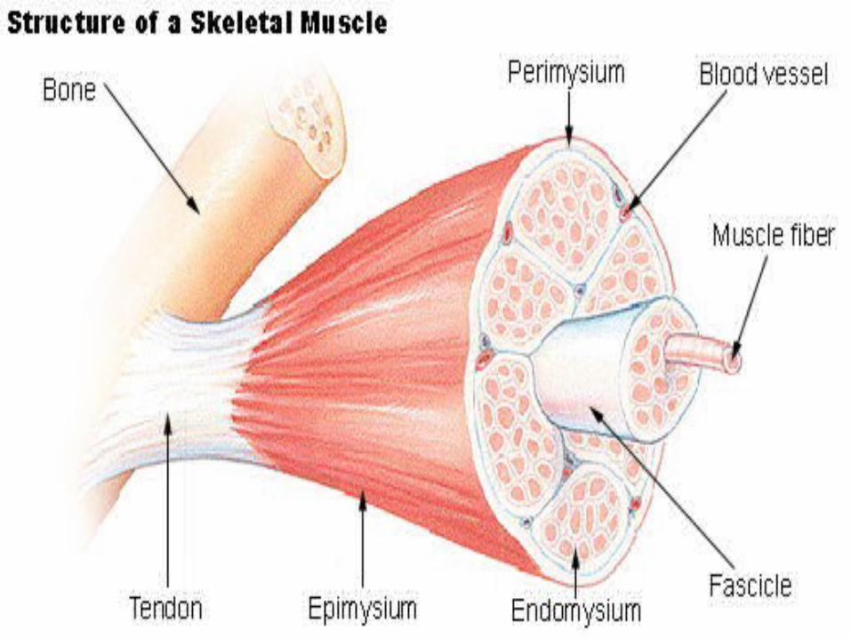
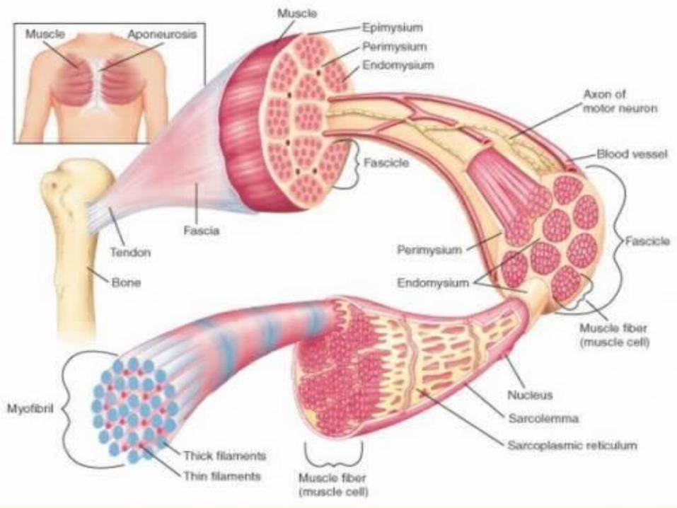
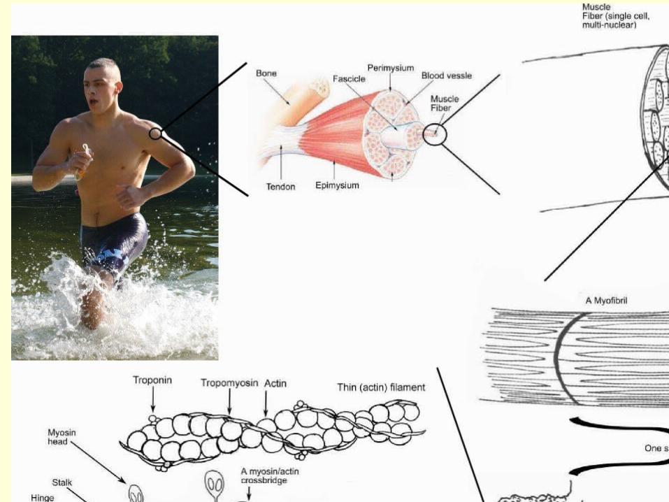
Connective Tissue Wrappings of skeletal Muscles:
• Endomysium: c.t. that holds muscle fibers together
• Muscle fiber: same a s a muscle cell• Tendon: attaches muscle to bone (ligament:
bone to bone)• Perimysium: c.t that surrounds the groups of
muscle fibers• Fascicle: group of muscle fibers• Epimysium: c.t. that holds groups of muslce
fibers together
Microscopic Anatomy
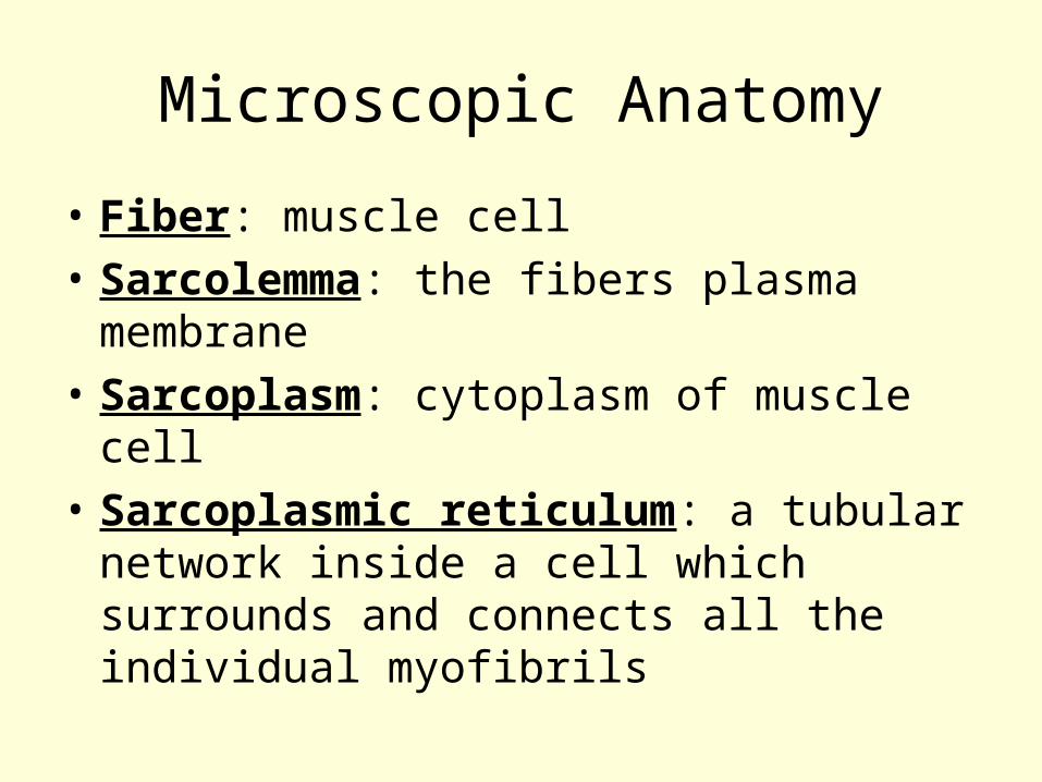
• Fiber: muscle cell
• Sarcolemma: the fibers plasma membrane
• Sarcoplasm: cytoplasm of muscle cell
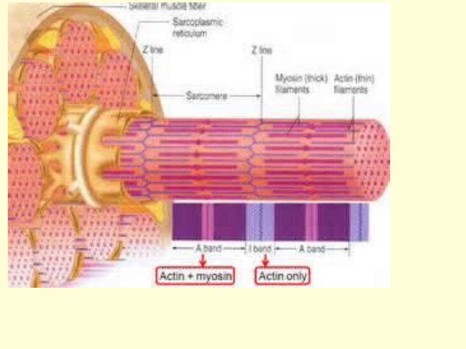
• Sarcoplasmic reticulum: a tubular network inside a cell which surrounds and connects all the individual myofibrils
Microscopic Anatomy
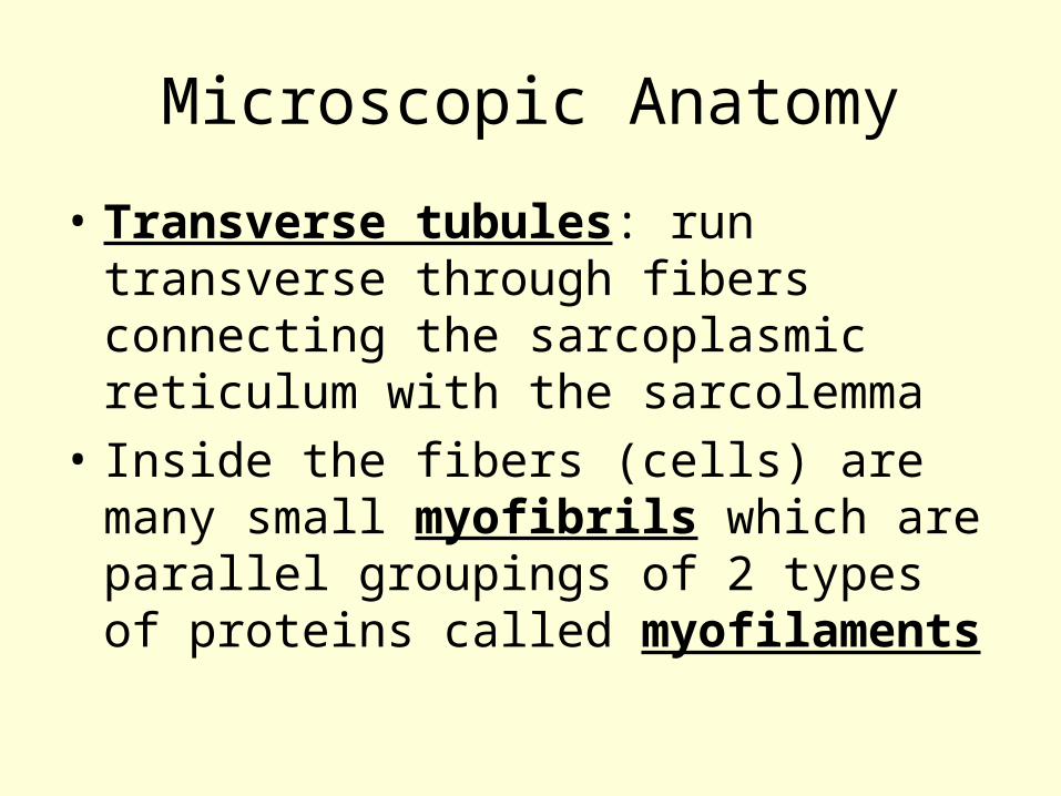
• Transverse tubules: run transverse through fibers connecting the sarcoplasmic reticulum with the sarcolemma
• Inside the fibers (cells) are many small myofibrils which are parallel groupings of 2 types of proteins called myofilaments
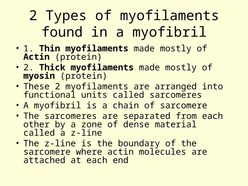
2 Types of myofilaments found in a myofibril
• 1. Thin myofilaments made mostly of Actin (protein)
• 2. Thick myofilaments made mostly of myosin (protein)
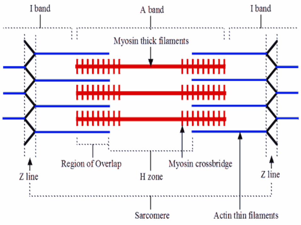
• These 2 myofilaments are arranged into functional units called sarcomeres
• A myofibril is a chain of sarcomere• The sarcomeres are separated from each other
by a zone of dense material called a z-line• The z-line is the boundary of the sarcomere
where actin molecules are attached at each end
Muscle contraction
• During a contraction, the actin myofilaments are pulled toward the center of the sarcomere, this pulls the z lines closer to each other and thus sortens the sarcomere
• The amount of overlap between the actin and myosin changes and is seen as striations on the muscle
Mechanics of a Muscle contractionskeletal muscles contract by the following steps
1. A motor nerve signals the muscle. Some of the nuerons in the nerve develop electrical impulses which signal some fibers in the muscle. Each axon secretes a neurotransmitter ( chemical signal) called acetylchoine at the synapse (motor end plate) between the neuron and some muscle fibers. This signal excites each muscle cell.
Mechanics of a Muscle contractionskeletal muscles contract by the following steps
2. An electrical signal spreads out along the sarcolemma ( cell membrane) of each muscle cell that is signaled.
3. This signal continues transversely int the sarcoplasm of each muscle cell along the membranes of the T tubule
4. The T tubule joins the sarcoplasmic reticulum in the sarcoplasm. The signal spreads from T tubules to tubular S. R. releasing calcium ion
Mechanics of a Muscle contractionskeletal muscles contract by the following steps
5. The release of calcium from the S. R. blocks the action of troponin ( a protein in myofibriles). Troponin normally inhibits the interaction of actin and myosin
6. With troponin inhibited, actin and myosin can interact. Cross bridges on the myosin slide the actin molecule toward the center of the sarcomere. As the actin molecules attached to the z line…. This shortens the sarcomere
Mechanics of a Muscle contractionskeletal muscles contract by the following steps
7. Hydrolysis of ATP in the cells, into ADP and phosphate releases energy to drive the sliding filaments. ATP is rebuilt from an energy-storage compound creating phosphate
8. If enough sarcomeres, shorten the myofibriles shortn. If enough mofibrils shorten, then the fibers shorten. If enough fibers shorten, then the muscle contracts
9. Each muscle responds by an all or non law ( with the force of contraction dependent on the % of cells that are active)
Patterns of muscle contraction
• Tonus : muscle tone• Tension: force produced by a whole muscle when it
contracts• Load: resistance. Force exerted on a muscle by a
weight• Isometic contraction: response in which a muscle does
not contract enough to produce motion ex muscle in shoulder act isometrically if arm is pushed against an immovable wall
• Isotonic exercise: better aerobically and produces endurance
• Isometric exercise builds muscle size and strength • You should do both
Naming of skeletal muscles
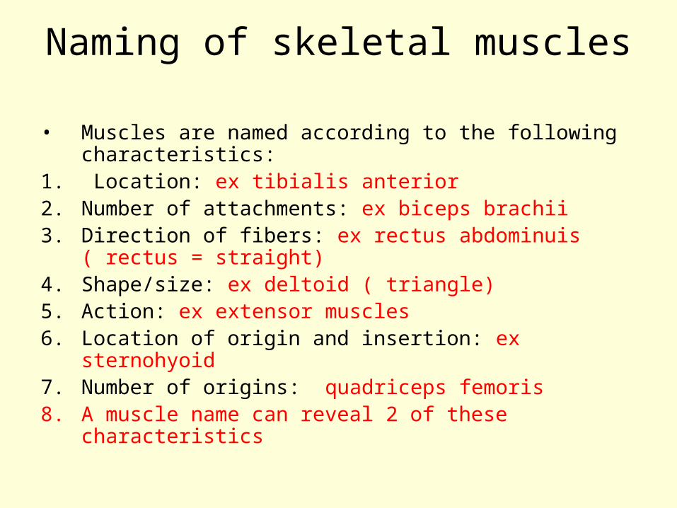
• Muscles are named according to the following characteristics:
1. Location: ex tibialis anterior2. Number of attachments: ex biceps brachii3. Direction of fibers: ex rectus abdominuis ( rectus =
straight)4. Shape/size: ex deltoid ( triangle)5. Action: ex extensor muscles6. Location of origin and insertion: ex sternohyoid7. Number of origins: quadriceps femoris8. A muscle name can reveal 2 of these characteristics
Muscle disorders:
• Can be due to infection/damage to the muscle or to its motor neuron
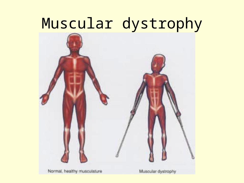
Muscular dystrophy
• Genetic causation
• Barely understood
• Involves progressive degeneration of individual skeletal muscle fibers
• Braces and exercise help post-pone inevitable
• Death occurs due to respiratory failure
Muscular dystrophy
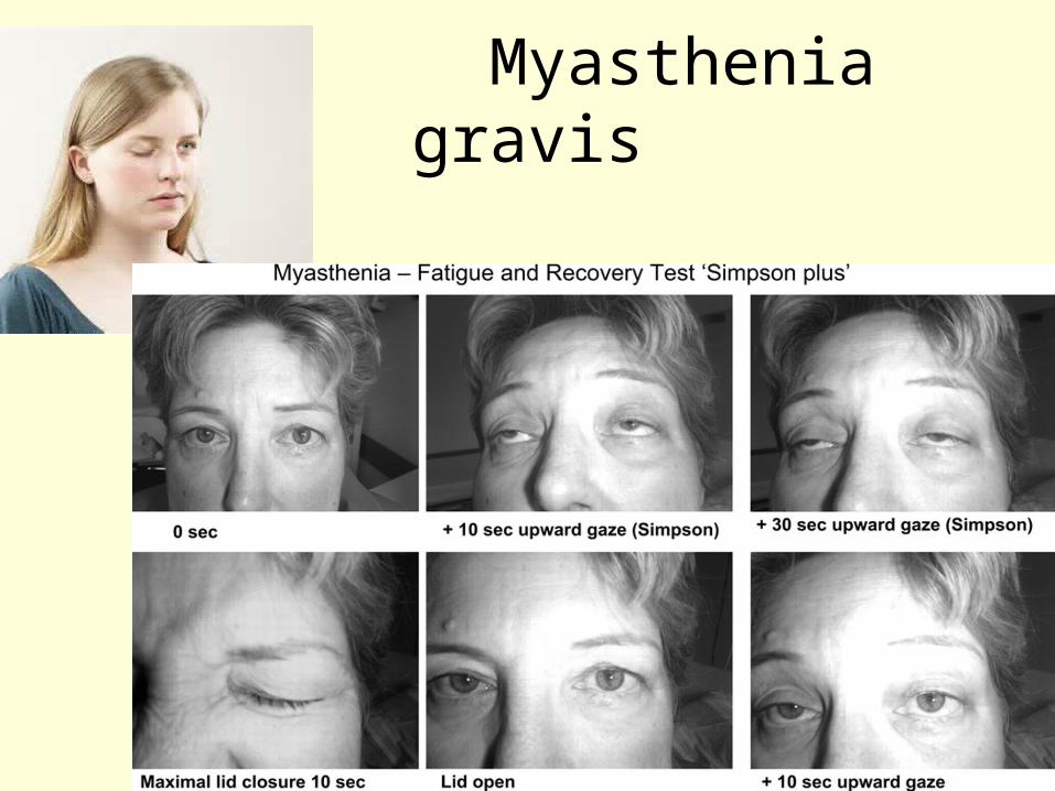
Myasthenia gravis
• Skeletal muscle becomes weak and easily fatigued due to motor endplate abnormality
• The receptor for neural impulses are blocked or destroyed
• Is an autoimmune disease that sometimes progresses to paralysis or death
Myasthenia gravis
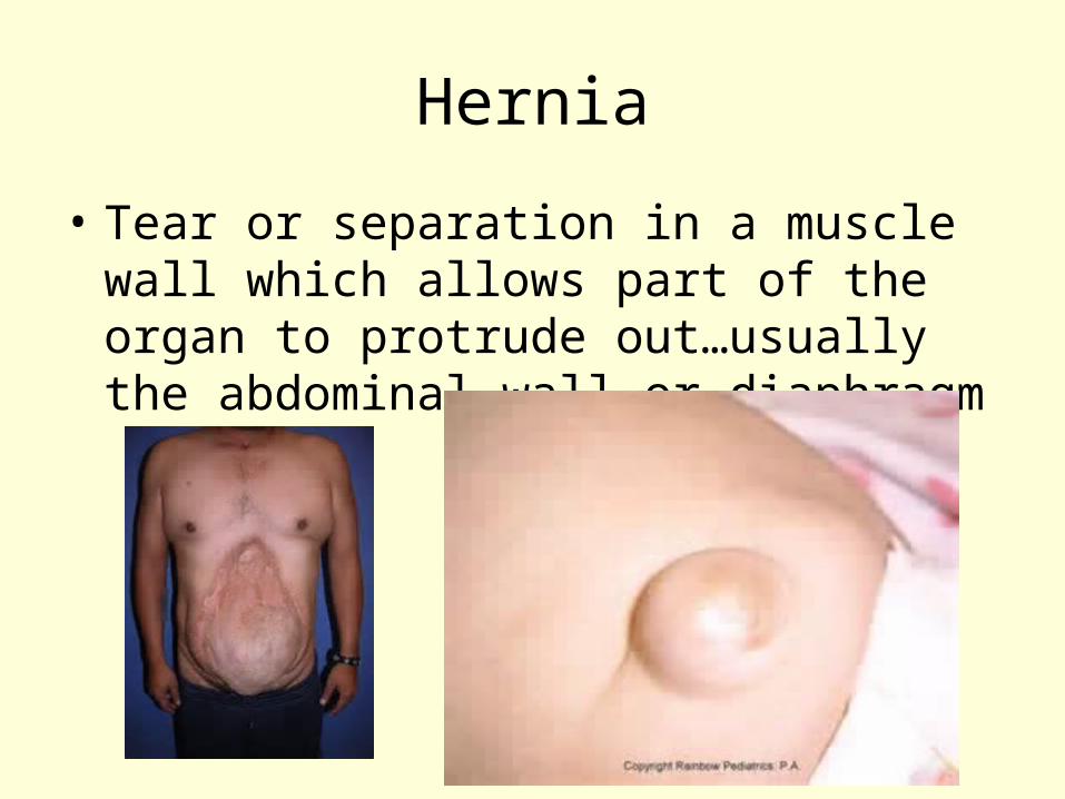
Hernia
• Tear or separation in a muscle wall which allows part of the organ to protrude out…usually the abdominal wall or diaphragm
• Spasm: contractions of a muscle, if very small are called tics, if painful they are called cramps
• Usually due to inflammation or water ion imbalances
Reasons for muscle fatigue
• Strenuous activity with and Cell respiration going anaerobic, lactic acid build up
• Poor nutrition
• Poor delivery system: respiratory or cardiovascular
• Unexplained neural disorder (rare)