mucosal immunization with shigella flexneri outer membrane...
TRANSCRIPT

1
Mucosal Immunization with Shigella flexneri Outer Membrane Vesicles induced
protection in mice
A.I. Camachoa, J. de Souzaa, S. Sánchez-Gómeza, M. Pardo-Rosa ,J. M. Iracheb, C.
Gamazoa*
aDepartment of Microbiology, University of Navarra, 31008 Pamplona, Spain
bDepartment of Pharmacy and Pharmaceutical Technology, University of Navarra,
31008 Pamplona, Spain
* Author to whom all correspondence should be addressed:
Carlos Gamazo
Departamento de Microbiología
Universidad de Navarra
31008 Pamplona (Spain)
Phone no. +34 9 48 42 56 88
Fax no. + 34 9 48 42 56 49
Email: [email protected]
Keywords: Shigella, outer membrane vesicles, vaccine, nanoparticles, adjuvant,
Running Title:
1. INTRODUCTION
According to World Health Organization (WHO), approximately 2.5 billion cases of
diarrhea occurred worldwide which results in 1.5 million deaths among children under

2
the age of five. It is a common cause of death in developing countries and the second
most common cause of infant deaths. Among the main causes,Shigellosis is responsible
of more than 165 million cases annually, leading to 1.2 million deaths [1]. Furthermore,
many cases progress into serious damages in their intestinal epithelium that will limit
the correct nutrient absorption with the subsequent sequel for life. Shigella spread
massively within the community and from person to person, and hence, prevention
relies on basic sanitary measures, which unfortunately may be not possible applied for
many countries. In addition, the increasing problem of antibiotic resistance alerts on the
urgent need of protective vaccines. In fact, the World Health Organization has made the
development of a safe and effective vaccine against Shigella a high priority [1].
The efforts have been mainly focussed on live oral vaccines with several vaccine
candidates on clinical trials [2]. However, development of such safe Shigella vaccine is
being problematical, and no vaccine is still available[3].
Currently, most vaccines in development are acellular vaccines which [2;4] in
comparison to live-attenuated or whole inactivated organism, are safer. However, these
prototypes require adjuvants to achieve a more effective immune response. The
challenge is the designing of formulations able to enhance the immunogenicity of
associated antigens, through the right activation of the immune system, and susceptible
to be administered by mucosal routes. Previous studies of our group have evaluated the
adjuvant capability of nanoparticles made from the copolymer of methyl vinyl ether and
maleic anhydride (Gantrez AN®). These nanoparticles demonstrated their ability to
initiate a strong and balanced mucosal immune response and then, to efficiently induce
Th-1 subset [5]. In addition, these nanoparticles loaded with different antigens have
showed to be effective against experimental challenges with Salmonella or Brucella [6-
9].

3
In this work we propose the use of outer membrane vesicles (OMVs) from Shigella as
the source of relevant antigens to be included in the acellular vaccine. OMVs are
secreted from the outer membrane of a large variety of gram negative bacteria, during in
vitro culture and during infection [10]. Currently, there have been described many
functions for this blebbing process. Functions proposed vary from facilitating the
intracellular bacterial growth within phagocytes [11], to the delivery of effectors
molecules critical for pathogen dissemination such as pathogen-associated molecular
patterns (PAMPs) and other virulence factors to host cells [12-14].
We therefore describe here the preparation, characterization and evaluation of Shigella
flexneri outer membrane vesicles in order to be used in vaccination. We obtained the
OMVs from S. flexneri 2a, being this the most common cause of shigellosis. In fact, it’s
responsible for 25 to 50 percent of all cases in the developing world [2]. The protective
efficacy of OMVs either in their free form or adjuvanted in NP were tested in the
murine pneumonia model [15] after immunization with one single dose by intradermal
or mucosal routes.
The OMVs formulations obtained and characterized here were found to induce
protection in mice after one single dose against a lethal dose of S. flexneri 2a.
2. MATERIAL AND METHODS
Preparation and characterization of outer membrane vesicles
OMVs were obtained from S. flexneri 2a (clinical isolate from Hospital de Navarra,
Pamplona, Spain). Vesicles were purified from a method adapted from Horstman and

4
Kuehn [16]. Bacteria were grown in LB broth overnight to early stationary phase. Then,
bacteria were inactivated with a solution of binary ethylenimine and formaldehyde (6
mM BEI- 0, 06% FA, 6 h, 37 ºC). BEI was prepared as a 0.1 M solution by cyclization
of 0.1 M 2-bromoethylamine hydrobromide (Sigma) in 0.175 M NaOH solution for one
hour following the method of Bahnemann [17]. Cells were removed by pelleting
(10,000 × g, 10 min). Supernatant was filtered through a 0.45 μm Durapore PVDF filter
(Millipore) and purified by ultradiafiltration via a 300-kDa tangential filtration
concentration unit (Millipore). The retentate was freezed in order to induce larger blebs
formed through reassociation of the smaller ones into multimicelles, as had been
proposed previously [18]. Final product was recovered by centrifugation at 40,000 × g,
2 h. Total protein content was determined by the method of Lowry, with bovine serum
albumin as standard. Lypopolysaccharide (LPS) content was determined by Purpald
assay[19;20]. Briefly, to 50 μL of LPS samples or standards [21] in each of the duplicate
wells in a 96-well tissue culture plate, 50 μL of 32 mM NaIO4 was added, and the plate
was incubated for 25 min followed by addition of 50 μL of 136 mM purpald reagent in
2 N NaOH. After further incubation for 20 min, 50 μL of 64mM NaIO4 was added, and
the plate was incubated for another 20 min. The foam in each well can be eliminated by
addition of 20 μL 2-propanol. The absorbance of each well was measured by a plate
reader at 550 nm. Finally, extract was resuspended in sample buffer 1× and analyzed by
SDS-PAGE and immunoblotting, using polyclonal pool sera from patient infected with
S. flexneri (Clínica Universidad de Navarra) or anti IpaC mAb (kindly provided by A.
Phalipon, Institut Pasteur). The morphology of the vesicles was examined by Field
Emission Scanning Electron Microscope.
Outer membrane proteins (OMPs) from S. flexneri were prepared by sequential
detergent extraction of cell envelopes [18]. Briefly, after the disruption of cells by
5
sonication (4 pulses × 5 min, power 2, Branson Sonifier 450), whole bacteria were
removed by centrifugation at 6000 × g, 15 min. Cell envelopes were recovered from
supernatant by centrifugation (40 000 × g, 1h). Pellet was resuspended in 1% Sarkosyl
(N-Lauryl sarcosine, Sigma Chemical Co., St. Louis, USA), incubated for 30 min and
further centrifuged at 40 000g, 1h, twice. The enriched sediment in outer membrane
proteins was suspended in 0.5 M Tris-HCl (pH 6.8) with 10% SDS (Lauryl sulfate,
Sigma) and boiled for 15 min and finally, centrifuged (20 000 × g; 30 min). The OMPs
of S. flexneri were present in the final supernatant.
Ipa (invasion plasmid antigens) proteins secretion assay. Secretion of Ipa proteins
through the TTSS (Type three secretion system) was induced using a Congo Red
secretion assay [22]. Exponential-phase bacteria were harvested, resuspended in 10 μM
Congo Red/PBS, and incubated at 37 ºC for 30 min. Following incubation, bacteria
were pelleted by centrifugation, and supernatants were collected and passed through a
0.22 μm-pore filter. Proteins in the supernatants, which represent proteins secreted
through the TTSS, were then concentrated by tricholoroacetic acid precipitation.
Finally, extract was resuspended in sample buffer 1× and analyzed by SDS-PAGE and
immunoblotting using anti-IpaB or -IpaC mAb (kindly provided by A. Phalipon, Institut
Pasteur).
Preparation and characterization of nanoparticles
Poly (anhydride) nanoparticles were prepared by a modification of the solvent
displacement method [6;23]. Briefly, 100 mg of the copolymer of methyl vinyl ether
and maleic anhydride (PVM/MA) (Gantrez®AN 119; M.W. 200 KDa) was dissolved in
5 ml acetone under magnetic stirring at room temperature. On the other hand, 5 mg
OMVs were dispersed by ultrasonication with a probe Microson TM (Misonix Inc.,

6
New York, USA) in 10 ml water for 1 min. After dispersion, nanoparticles were formed
by addition of this water phase containing OMVs. The agitation was maintained during
15 min in order to allow the stabilization of the system. Organic solvents were removed
under reduced pressure (Büchi R-144, Switzerland). The obtained nanoparticles were
collected by centrifugation (27.000 × g, 20 min, 4 ºC) and washed with water twice.
Finally, particles were freeze-dried using sucrose 5% as crioprotector.
The preparation of empty nanoparticles was performed in the same way in the absence
of OMVs.
Characterization of nanoparticles. The particle size and the zeta potential of
nanoparticles were determined by photon correlation spectroscopy (PCS) and
electrophoretic laser Doppler anemometry, respectively, using a Zetamaster analyzer
system (Malvern Instruments Ltd., Worcestershire, UK). The diameter of the
nanoparticles was determined after dispersion in ultrapure water (1/10) and measured at
25ºC by dynamic light scattering angle of 90ºC. The zeta potential was determined as
follows: 200 μL of the samples was diluted in 2 mL of a 0.1 mM KCl solution adjusted
to pH 7.4. The morphology of the vesicles was examined by Field Emission Scanning
Electron Microscope (Carl Zeiss, model Ultra Plus). For this purpose freeze-dried
formulations were resuspended in ultrapure water and centrifuged at 27,000 × g for
20 min at 4 °C. Then, supernatants were rejected and the obtained pellets were mounted
on TEM grids. The yield of the nanoparticles preparation process was determined by
gravimetry as described previously [23]. Briefly, poly (anhydride) nanoparticles, freshly
prepared, were freeze-dried. Then, the yield was calculated as the difference between
the initial amount of the polymer used to prepare nanoparticles and the weight of the
freeze-dried carriers.

7
Loading capacity of nanoparticles. The yield of nanoparticles was calculated from the
difference between the initial amount of the polymer used to prepare the particles and
the weight of the freeze-dried samples. The ability of PVM/MA nanoparticles to entrap
the complex antigen was directly determined after degradation of loaded nanoparticles
with NaOH. Briefly, OMVs-loaded Gantrez nanoparticles (15 mg) were dispersed in
water vortexing 1 min. After centrifugation (27.000 × g, 15 min) pellet was resupended
in NaOH 0,1 M sonicated (MicrosonTM Ultrasonic cell disruptor) and incubated for 1 h
to assess the total delivery of the associated antigen. After this time, the amount of
antigen released from the nanoparticles was determined using microbicin choninic acid
(microBCA) protein assay (Pierce, Rockford, CA, USA). In order to avoid interferences
of the process, calibration curves were made with degraded blank nanoparticles, and all
measurements were performed in triplicate.
Determination of the structural integrity and antigenity of OMVs. Western-blot
analysis was used as a qualitative tool to examine the structure of the antigens,
complementing the quantification performed by microBCA. To accomplish this
analysis, the protocol for nanoparticle degradation was modified in order to avoid any
interference of the enzyme. In this case, after nanoparticle isolation, 15 mg of loaded
nanoparticles were dispersed in water vortexing 1 min. After centrifugation (27 000 × g,
15 min) pellet was resupended in 2 ml of a mixed of dimethilformamide: acetone (1:3)
(-80 ºC, 1h). After centrifugation, pellet was resuspended in acetone (-80 ºC, 30 min).
Finally, extract was resuspended in sample buffer 1× and analyzed by SDS-PAGE and
immunoblotting using polyclonal sera from hyperimmunized rabbit with S. flexneri
[24].
SDS-PAGE and Immunoblotting

8
SDS-PAGE was performed in 12% acrylamide slabs (Criterion XT, Bio Rad
Laboratories, CA) with the discontinuous buffer system of Laemmli and gels stained
with Coomassie blue or silver staining. After electrophoresis, gels were electroblotted to
a PVDF (polyvinylidene fluoride) membrane at 0.8 mA/cm2 for 30 min. Then,
membranes were soaked overnight in a blocking solution containing 3% (w/v) of non-
fat milk and then incubated in the presence of different sera, described above. After the
incubation, the membranes were washed five times; the anti-rabbit or human Ig-alkaline
phosphatase conjugate was added, followed by incubation for an additional hour. The
membranes were exhaustively washed and the antibody–antigen complexes were
visualized after addition of the substrate/chromogen solution (H2O2/cloronaftol).
Active immunization and challenge.
All mice were treated in accordance with institutional guidelines for treatment of
animals (Protocol 087/06 of animal treatment, approved in 1 October 2007 by the
Ethical Comity for the Animal Experimentation, CEEA, of the University of Navarra).
Nine-week-old BALB/c mice (20±1 g) were separated in randomized groups of 6
animals and immunized with OMVs either free or encapsulated in PVM/MA NPs by
intradermal, nasal, ocular (20 μg of extract) or oral route (100 μg of extract). The
scheme of administration and doses are summarized in Table 1.
Challenge infection was performed on day 35 intranasally with a lethal dose of 1×10⁷
UFC/Mouse of Shigella flexneri 2a (clinical isolate) grown to logarithmic phase and
suspended in 20 µl of prewarmed PBS. The number of dead mice after challenge was
recorded daily.
Measurement of immune response in the mouse.

9
Blood samples were collected from the reto-orbital plexures of anesthetized mice
ELISA. The antibody response was measured by an enzyme-linked immunosorbent
assay (ELISA). In brief, 96-well microtiter plates (MaxiSorb; Nunc, Wiesbaden,
Germany) were coated with 100 uL of 10 μg/ml OMVs in coating buffer (60 mM
carbonate buffer, pH 9.6). Afterwards, unspecific binding sites were saturated with 3%
bovine serum albumin (BSA) in PBS for 1 h at RT. Sera from mice were serially diluted
in PBS with 1% BSA and incubated overnight at RT. After intense washing with PBS
Tween 20 (PBS-T) buffer, the alkaline phosphatase (AP)-conjugated detection antibody,
class-specific goat anti-mouse IgG/IgA (Sigma) for sera, was added for 1h at 37ºC. The
detection reaction was performed by incubating the sample with ABTS substrate for 20
min at room temperature. Absorbance was measured with an ELISA reader (Sunrise
remote; Tecan-Austria, Groeding, Austria) at a wavelength of 405 nm.
Quantification of cytokines from sera. Cytokines (IL-2, IL-4, IL-5, IL-6, IL-10, IL-
12(p40), IL-12(p70), IL-13, IL17, IFN-, and tumor necrosis factor) were quantified
from serum by luminex-based multiplex assay (Milliplex; Millipore, Billerica, MA)
using a Bioplex analyzer (Bio-Rad, Hercules, CA).
Statistics
Statistical analyses were performed using GraphPad Prism 5 for Mac OS X. All
experiments were performed with n=6. Statistical comparisons between antibody serum
levels were performed using Kruskal-Wallis test, followed by Dunn´s post-hoc test. The
statistical significance was set at P < 0.05. For cytokine levels, it was performed using
single-factor analysis of variance, followed by Turkey´s post hoc test. The statistical
significance was set at P<0.001. The Kaplan-Meyer curves were used for analysis of the
protection experiment.
10
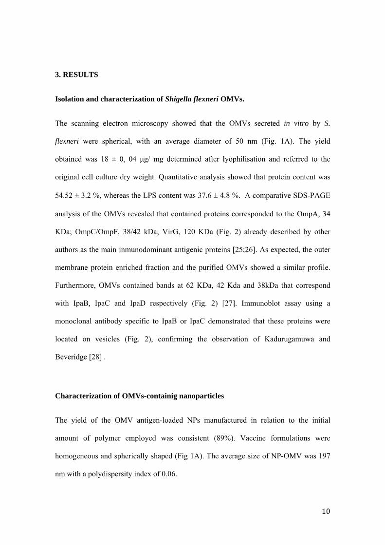
3. RESULTS
Isolation and characterization of Shigella flexneri OMVs.
The scanning electron microscopy showed that the OMVs secreted in vitro by S.
flexneri were spherical, with an average diameter of 50 nm (Fig. 1A). The yield
obtained was 18 ± 0, 04 μg/ mg determined after lyophilisation and referred to the
original cell culture dry weight. Quantitative analysis showed that protein content was
54.52 ± 3.2 %, whereas the LPS content was 37.6 4.8 %. A comparative SDS-PAGE
analysis of the OMVs revealed that contained proteins corresponded to the OmpA, 34
KDa; OmpC/OmpF, 38/42 kDa; VirG, 120 KDa (Fig. 2) already described by other
authors as the main inmunodominant antigenic proteins [25;26]. As expected, the outer
membrane protein enriched fraction and the purified OMVs showed a similar profile.
Furthermore, OMVs contained bands at 62 KDa, 42 Kda and 38kDa that correspond
with IpaB, IpaC and IpaD respectively (Fig. 2) [27]. Immunoblot assay using a
monoclonal antibody specific to IpaB or IpaC demonstrated that these proteins were
located on vesicles (Fig. 2), confirming the observation of Kadurugamuwa and
Beveridge [28] .
Characterization of OMVs-containig nanoparticles
The yield of the OMV antigen-loaded NPs manufactured in relation to the initial
amount of polymer employed was consistent (89%). Vaccine formulations were
homogeneous and spherically shaped (Fig 1A). The average size of NP-OMV was 197
nm with a polydispersity index of 0.06.
11
The Z potential of NP was tested before and after OMV encapsulation. Results suggest
that OMV is at least partially bound on the NP surface, indicated by the change in Z of
NP. Zeta potential of free OMVs was -14.1 +/- 3 mV. The encapsulation of the extract
in nanoparticles resulted in a change of Z potential from -44 +/- 4 mV to -27 +/- 4 mV
when OMVs were loaded into PVM/MA nanoparticles.
To further confirm OMV encapsulation into NPs, BCA protein determination and SDS-
PAGE/immunoblotting were also performed. The procedure involved the use of a
purification step in order to discard unbound OMV. S. flexneri OMVs were efficiently
associated with PVM/MA nanoparticles, as they showed a loading encapsulation of 20
g OMVs/ mg of polymer. Besides, an immunoblotting was carried out using sera from
rabbit hyper-immunized with S. flexneri. Results indicate that entrapment in
nanoparticles did not alter its antigenic properties (Fig. 1B).
Evaluation of the immunogenicity and protection conferred by OMVs vaccine.
Groups of 6 mice were immunized once by intradermal or mucosal routes with OMVs
(20 µg/mouse), either free or encapsulated in NPs. A control group of non-immunized
mice was also included. All animals immunized by nasal or ocular routes remained in
good health, exhibiting no respiratory difficulties, changes in body temperature, or
abnormal behaviour. Oral immunized mice showed a transient abdominal swelling a
few hours after immunization. By contrast, mice immunized intradermally
experimented sweating and lethargy during 2 days post-immunization, which
disappeared thereafter.
Specific IgG2a and IgG1 against OMVs antigens were determined by indirect-ELISA at
days 0, 15 and 35 post-immunization (Fig. 3). Results expressed that the OMV
12
immunization by either route elicited significant levels of serum IgG1 and IgG2a with
respect control mice (Fig. 3). Higher levels of IgG were found in groups immunized
intradermally. Overall the levels of IgG2a (Th1 response) were higher than that those of
IgG1 (Th2). An adjuvant effect after encapsulation was observed on the
immunogenicity (global specific antibody response) especially after oral immunization.
There were not found significant differences in the mucosal levels of the IgA elicited
after intradermal or mucosal deliveries.
Levels of serum cytokines were determined at day 15 post-immunization (Fig. 4). The
encapsulation of OMVs in NPs induced an increase in the level of IL-12 (p40) and a
decrease of IL-10 �with respect to the free form, by intradermal or oral delivery. In
contrast, after ocular or nasal immunization, the inverse switching phenomenon was
observed.
At day 35 after immunization, mice were challenged with S. flexneri via intranasal route
and monitored for survival over 30 days (n = 6 mice/group) (Fig. 5). Nasal or ocular
immunizations with free OMVs provided complete protection. Non-significant
differences were found between OMV free or nano-encapsulated in groups immunized
by nasal, ocular or oral route. In contrast, the intradermally delivery of free OMVs was
not protective, while the encapsulated extract conferred full protection.
4. DISCUSSION
Currently, live vaccines provide better protection as compared to the inactivated
vaccines, including the acellular ones[2;29]. However, it is always difficult to properly
calibrate attenuation to achieve the minimum of toxicity with the optimal
immunogenicity. Besides, the use of live Shigella vaccines is questionable since this

13
pathogen is able to strongly interfere with the immune response, by inducing an
immunosuppressive condition that favors infective process. In our present experimental
study, we support the use of mucosal immunization with acellular vaccines. Results
demonstrated a significant efficacy and no reactogenicity in the mice pulmonary model.
The best prophylactic measure probably would be to prevent bacterial invasion by
neutralizing key surface virulence factors. The outer membrane (OM) of Shigella
contains several main virulence factors, including outer membrane proteins (OMP),
protein adhesins, the highly conserved virulence-plasmid-encoded Ipa proteins [28] as
well as LPS. These are essential components in the invasion process, and can alter the
course of infection and the host responses, and therefore their neutralization for the host
will succeed in protective immunity. [25;30-33].
Outer membrane vesicles (OMVs) consist of OM and soluble periplasmic components
shed from gram-negative bacteria. This blebbing process is considered as a peculiar
bacterial extracellular secretion system than enable bacterial colonization and impairs
host immune response [34]. Therefore, it is plausible to think on Shigella OMVs as
ideal candidates for an acellular vaccine. The capacity of OMV-based vaccines to
stimulate a protective immune response has already been exploited against several
bacterial pathogens, such as Brucella ovis [18], S. typhimurium [35], Flavobacterium
[36] Porfiromonas [37] or Neisseria meningitides B, with over 55 million doses
administered to date of the former [38].
As many gram-negative bacteria, Shigella bleb off membrane vesicles during normal
growth. Kadurugamuwa, et al. already obtained and characterized membrane vesicles
from S. flexneri [39]. In order to obtain this material massively, we developed an
extraction protocol that also maximize OMV purity. Vesicles were isolated from

14
concentrated, cell-free culture supernatant leading to an appropriate antigenic profile as
well as high purity grade. Besides, final product was ultradiafiltered in order to avoid
interferences in the encapsulation process.
OMV used here contain key alarm signals such as LPS, OMPs and Ipa recognized by
the innate immune system, including epthelial cells, MALT and antigen presenting
cells[40;41], and therefore have the capacity to either enhance bacterial clearance or
cause host tissue damage by activating an inflammatory response. It is interesting to
note that these components provide a prolonged stimulation of the inflammatory
response that, at first instance, facilitates bacterial survival in the tissues. However, this
fact will lead to the bacteria elimination by the host immune system[42].
In fact, our results indicate that a single dose of non-adjuvated OMVs delivered by
mucosal routes is able to protect against a lethal challenge with S. flexneri. Vaccines
that stimulate protective mucosal immune responses often need an adjuvant for proper
delivery and presentation to the mucosal immune tissues. The mechanisms underlying
the effectiveness of free OMV without external adjuvant may be explained by the nature
of some individual components contained within this “proteoliposome” or/and by the
biophysical properties of these vesicles [43]. Besides, Ipa containing OMVs may
contribute to its adjuvanticity by their ability to interact with host cell receptors which
facilitate OMVs transcytosis across mucosal epithelial barriers [27]. On the other hand,
the amphipatic properties of OMVs may facilitate its own movement through mucosal
tissues, enhancing antigen presentation to drive a protective response.
In this study, we measured the levels of cytokines in OMVs vaccinated mice two weeks
after the immunization. Then, we analyzed their association with the challenge outcome.
A strong association between the ratio of IL-12p40/ IL-10 and protection was found.

15
Moreover, low levels of IFN-γ correlated with protection. However, conclusions from
these particular data must be taken with caution since cytokine levels were measured
directly from serum. At this point, further studies are being carried out to really
establish a correlation of these parameters and protection.
After oral administration, under steady-state conditions, some factors released by
enterocytes, such as retinoic acid, thymic stromal lymphopoietin and TGF-β, will
“condition” non-activated resident DCs to elicit a Th2 or regulatory responses [44].
However, following an inflammatory stimulus, a recruitment of DC expressing
CX3CR1 to the mucosal tissues is observed, increasing the number of DC extending
dendrites into intestinal lumen. Under this state of high activation, DC-expressing
massively co-stimulatory molecules, present the antigenic determinant to the specific T
naïve cells in the T area MALT. The substantial distinctive release of IL-12 from those
DCs will also contribute to the further differentiation of naïve cells to Th1/Th2/Th17,
linked to an inflammatory response. Actually, our results would support it since OMVs
adjuvanted into NPs induced increasing levels of IL-12 (p40) and decreasing IL-10 with
respect to the free form, either by intradermal or oral delivery. NPs can enhance the
delivery of the loaded antigen to the gut lymphoid cells due to their ability to be
captured and internalized by cells of the gut-associated lymphoid tissue (GALT), and to
induced maturation of DCs with a significant upregulation of CD40, CD80, and CD86
and a Th1 response in animal models. The mechanisms responsible for DC maturation
may be related to TLR-NP specific interaction [5].
On the other hand, the encapsulation of OMVs in NPs induced an increase in the level
of IL-10 and a decrease of IL-12 (p40) �with respect to the free form, by ocular or
nasal routes, which is characteristic of mucosal adjuvants that usually stimulate a Th2

16
T-cell response [45-47], characterized by increased secretory IgA, high proportions of
antigen-specific serum IgG1, and the stimulation and synthesis of IL-4, IL-5, and IL-10.
The specific immune mechanisms that mediate resistance to Shigella infection have not
been clearly defined and are currently being debated. Thus, in humans, up regulation of
both proinflammatory and anti-inflammatory are observed during the first stages of
infection. Later, in relation with the convalescent stage of shigellosis, an increase in
IFN-γ is observed. Summing up, although Th1 is effective to control infection, a Th2
response may be also as effective but shorter-lasting.
Concerning the antibody response elicited after OMV immunization, we can not
establish a relation between antibody levels and protection. Serum and mucosal
antibodies to LPS and the Ipa proteins have been demonstrated during human
shigellosis [48;49]. However, it has not been established the role of these antibodies to
limit the spread or severity of the infection. The apparent inconsistency between IgG
subclass response and cytokine profile may be due to immune cells other than T helper
cells.
The ultimate goal for vaccination is to stimulate long-lasting protective immunological
memory. Toll-like receptors [50] generally promote adaptive immune responses
indirectly by activating innate immune cells. It has been recently shown that the use of
multiple TLR-agonists carried by nanoparticles influence in the induction of long-term
memory cells [51].
Recent studies report that in a murine model of acute bacterial infection with S. flexneri
the T cell response is dominated by the induction of long-term memory Shigella-
specific Th17 cells that contribute to mediate protective immunity against reinfection
[52]

17
Now, new research shows an unexpected direct role for TLR2 signalling in T cells
themselves, promoting the differentiation and proliferation of T helper 17 (TH17) cells
[50]. Taking into account these data and together with previous results from our own
group about the high ability of PVM/MA to stimulate TLR2 [5] suggest that these
nanoparticles are good adjuvant candidate for further investigation. OMVs are safe and
protective in mice, therefore, the use of OMVs adjuvanted into NP to trigger mucosal
immunity and effectively neutralize Shigella infection open the door to safely deals with
vaccination, especially critical when young children are the primary target.
Acknowledgments
This research was financially supported by Health Department of “Gobierno de
Navarra” (28/2007), “Instituto de Salud Carlos III” (PS09/01083 and PI070326), from
Spain. Ana Camacho was also financially supported by “Instituto de Salud Carlos III”
(FI08/00432).
Reference List
[1] Kotloff KL, Winickoff JP, Ivanoff B, et al. Global burden of Shigella infections: implications for vaccine development and implementation of control strategies. Bull World Health Organ 1999;77(8):651-66.
[2] Levine MM, Kotloff KL, Barry EM, Pasetti MF, Sztein MB. Clinical trials of Shigella vaccines: two steps forward and one step back on a long, hard road. Nat Rev Microbiol 2007 Jul;5(7):540-53.
[3] Kweon MN. Shigellosis: the current status of vaccine development. Curr Opin Infect Dis 2008 Jun;21(3):313-8.
[4] Kaminski RW, Oaks EV. Inactivated and subunit vaccines to prevent shigellosis. Expert Rev Vaccines 2009 Dec;8(12):1693-704.
[5] Tamayo I, Irache JM, Mansilla C, Ochoa-Reparaz J, Lasarte JJ, Gamazo C. Poly(anhydride) nanoparticles act as active Th1 adjuvants through Toll-like receptor exploitation. Clin Vaccine Immunol 2010 Sep;17(9):1356-62.

18
[6] Ochoa J, Irache JM, Tamayo I, Walz A, DelVecchio VG, Gamazo C. Protective immunity of biodegradable nanoparticle-based vaccine against an experimental challenge with Salmonella Enteritidis in mice. Vaccine 2007 May 30;25(22):4410-9.
[7] Gomez S, Gamazo C, San RB, Vauthier C, Ferrer M, Irachel JM. Development of a novel vaccine delivery system based on Gantrez nanoparticles. J Nanosci Nanotechnol 2006 Sep;6(9-10):3283-9.
[8] Salman HH, Gamazo C, Campanero MA, Irache JM. Salmonella-like bioadhesive nanoparticles. J Control Release 2005 Aug 18;106(1-2):1-13.
[9] Gomez S, Gamazo C, Roman BS, Ferrer M, Sanz ML, Irache JM. Gantrez AN nanoparticles as an adjuvant for oral immunotherapy with allergens. Vaccine 2007 Jul 20;25(29):5263-71.
[10] Kulp A, Kuehn MJ. Biological functions and biogenesis of secreted bacterial outer membrane vesicles. Annu Rev Microbiol 2010 Oct 13;64:163-84.
[11] Fernandez-Moreira E, Helbig JH, Swanson MS. Membrane vesicles shed by Legionella pneumophila inhibit fusion of phagosomes with lysosomes. Infect Immun 2006 Jun;74(6):3285-95.
[12] Kesty NC, Mason KM, Reedy M, Miller SE, Kuehn MJ. Enterotoxigenic Escherichia coli vesicles target toxin delivery into mammalian cells. EMBO J 2004 Nov 24;23(23):4538-49.
[13] Kadurugamuwa JL, Beveridge TJ. Virulence factors are released from Pseudomonas aeruginosa in association with membrane vesicles during normal growth and exposure to gentamicin: a novel mechanism of enzyme secretion. J Bacteriol 1995 Jul;177(14):3998-4008.
[14] Wai SN, Lindmark B, Soderblom T, et al. Vesicle-mediated export and assembly of pore-forming oligomers of the enterobacterial ClyA cytotoxin. Cell 2003 Oct 3;115(1):25-35.
[15] Mallett CP, Hale TL, Kaminski RW, et al. Intransal or intragastric immunization with proteosome-Shigella lipopolysaccharide vaccines protects against lethal pneumonia in a murine model of Shigella infection. Infect Immun 1995 Jun;63(6):2382-6.
[16] Horstman AL, Kuehn MJ. Enterotoxigenic Escherichia coli secretes active heat-labile enterotoxin via outer membrane vesicles. J Biol Chem 2000 Apr 28;275(17):12489-96.
[17] Bahnemann HG. Inactivation of viral antigens for vaccine preparation with particular reference to the application of binary ethylenimine. Vaccine 1990 Aug;8(4):299-303.
[18] Gamazo C, Winter AJ, Moriyon I, Riezu-Boj JI, Blasco JM, Diaz R. Comparative analyses of proteins extracted by hot saline or released

19
spontaneously into outer membrane blebs from field strains of Brucella ovis and Brucella melitensis. Infect Immun 1989 May;57(5):1419-26.
[19] Marolda CL, Lahiry P, Vines E, Saldias S, Valvano MA. Micromethods for the characterization of lipid A-core and O-antigen lipopolysaccharide. Methods Mol Biol 2006;347:237-52.
[20] Lee CH, Tsai CM. Quantification of bacterial lipopolysaccharides by the purpald assay: measuring formaldehyde generated from 2-keto-3-deoxyoctonate and heptose at the inner core by periodate oxidation. Anal Biochem 1999 Feb 1;267(1):161-8.
[21] Horstman AL, Bauman SJ, Kuehn MJ. Lipopolysaccharide 3-deoxy-D-manno-octulosonic acid (Kdo) core determines bacterial association of secreted toxins. J Biol Chem 2004 Feb 27;279(9):8070-5.
[22] Bahrani FK, Sansonetti PJ, Parsot C. Secretion of Ipa proteins by Shigella flexneri: inducer molecules and kinetics of activation. Infect Immun 1997 Oct;65(10):4005-10.
[23] Arbos P, Wirth M, Arangoa MA, Gabor F, Irache JM. Gantrez AN as a new polymer for the preparation of ligand-nanoparticle conjugates. J Control Release 2002 Oct 30;83(3):321-30.
[24] Diaz R, Bosseray N. [Study of the antigenic relations between Yersina enterocolitica serotype 9 and other gram negative bacterial strains]. Microbiol Esp 1974 Jan;27(1):1-14.
[25] Mukhopadhaya A, Mahalanabis D, Chakrabarti MK. Role of Shigella flexneri 2a 34 kDa outer membrane protein in induction of protective immune response. Vaccine 2006 Aug 14;24(33-34):6028-36.
[26] Al-Hasani K, Navarro-Garcia F, Huerta J, Sakellaris H, Adler B. The immunogenic SigA enterotoxin of Shigella flexneri 2a binds to HEp-2 cells and induces fodrin redistribution in intoxicated epithelial cells. PLoS One 2009;4(12):e8223.
[27] Parsot C, Menard R, Gounon P, Sansonetti PJ. Enhanced secretion through the Shigella flexneri Mxi-Spa translocon leads to assembly of extracellular proteins into macromolecular structures. Mol Microbiol 1995 Apr;16(2):291-300.
[28] Kadurugamuwa JL, Beveridge TJ. Delivery of the non-membrane-permeative antibiotic gentamicin into mammalian cells by using Shigella flexneri membrane vesicles. Antimicrob Agents Chemother 1998 Jun;42(6):1476-83.
[29] Levine MM. Immunogenicity and efficacy of oral vaccines in developing countries: lessons from a live cholera vaccine. BMC Biol 2010;8:129.
[30] Canh DG, Lin FY, Thiem VD, et al. Effect of dosage on immunogenicity of a Vi conjugate vaccine injected twice into 2- to 5-year-old Vietnamese children. Infect Immun 2004 Nov;72(11):6586-8.

20
[31] Jennison AV, Verma NK. Shigella flexneri infection: pathogenesis and vaccine development. FEMS Microbiol Rev 2004 Feb;28(1):43-58.
[32] Sansonetti PJ, Tran Van NG, Egile C. Rupture of the intestinal epithelial barrier and mucosal invasion by Shigella flexneri. Clin Infect Dis 1999 Mar;28(3):466-75.
[33] Li A, Rong ZC, Ekwall E, Forsum U, Lindberg AA. Serum antibody responses against shigella lipopolysaccharides and invasion plasmid-coded antigens in shigella infected Swedish patients. Scand J Infect Dis 1993;25(5):569-77.
[34] Ellis TN, Kuehn MJ. Virulence and immunomodulatory roles of bacterial outer membrane vesicles. Microbiol Mol Biol Rev 2010 Mar;74(1):81-94.
[35] Alaniz RC, Deatherage BL, Lara JC, Cookson BT. Membrane vesicles are immunogenic facsimiles of Salmonella typhimurium that potently activate dendritic cells, prime B and T cell responses, and stimulate protective immunity in vivo. J Immunol 2007 Dec 1;179(11):7692-701.
[36] Aoki M, Kondo M, Nakatsuka Y, Kawai K, Oshima S. Stationary phase culture supernatant containing membrane vesicles induced immunity to rainbow trout Oncorhynchus mykiss fry syndrome. Vaccine 2007 Jan 5;25(3):561-9.
[37] Zhang T, Hashizume T, Kurita-Ochiai T, Yamamoto M. Sublingual vaccination with outer membrane protein of Porphyromonas gingivalis and Flt3 ligand elicits protective immunity in the oral cavity. Biochem Biophys Res Commun 2009 Dec 18;390(3):937-41.
[38] Holst J, Martin D, Arnold R, et al. Properties and clinical performance of vaccines containing outer membrane vesicles from Neisseria meningitidis. Vaccine 2009 Jun 24;27 Suppl 2:B3-12.
[39] Kadurugamuwa JL, Beveridge TJ. Delivery of the non-membrane-permeative antibiotic gentamicin into mammalian cells by using Shigella flexneri membrane vesicles. Antimicrob Agents Chemother 1998 Jun;42(6):1476-83.
[40] Schroeder GN, Hilbi H. Molecular pathogenesis of Shigella spp.: controlling host cell signaling, invasion, and death by type III secretion. Clin Microbiol Rev 2008 Jan;21(1):134-56.
[41] Biswas A, Banerjee P, Mukherjee G, Biswas T. Porin of Shigella dysenteriae activates mouse peritoneal macrophage through Toll-like receptors 2 and 6 to induce polarized type I response. Mol Immunol 2007 Feb;44(5):812-20.
[42] Phalipon A, Sansonetti PJ. Shigella's ways of manipulating the host intestinal innate and adaptive immune system: a tool box for survival? Immunol Cell Biol 2007 Feb;85(2):119-29.
[43] Sanders H, Feavers IM. Adjuvant properties of meningococcal outer membrane vesicles and the use of adjuvants in Neisseria meningitidis protein vaccines. Expert Rev Vaccines 2011 Mar;10(3):323-34.

21
[44] Rimoldi M, Chieppa M, Salucci V, et al. Intestinal immune homeostasis is regulated by the crosstalk between epithelial cells and dendritic cells. Nat Immunol 2005 May;6(5):507-14.
[45] Petrovsky N, Aguilar JC. Vaccine adjuvants: current state and future trends. Immunol Cell Biol 2004 Oct;82(5):488-96.
[46] Lindblad EB. Aluminium compounds for use in vaccines. Immunol Cell Biol 2004 Oct;82(5):497-505.
[47] Bungener L, Geeraedts F, Ter VW, Medema J, Wilschut J, Huckriede A. Alum boosts TH2-type antibody responses to whole-inactivated virus influenza vaccine in mice but does not confer superior protection. Vaccine 2008 May 2;26(19):2350-9.
[48] Coster TS, Hoge CW, VanDeVerg LL, et al. Vaccination against shigellosis with attenuated Shigella flexneri 2a strain SC602. Infect Immun 1999 Jul;67(7):3437-43.
[49] Oberhelman RA, Kopecko DJ, Salazar-Lindo E, et al. Prospective study of systemic and mucosal immune responses in dysenteric patients to specific Shigella invasion plasmid antigens and lipopolysaccharides. Infect Immun 1991 Jul;59(7):2341-50.
[50] Bird L. T cells: TLRs deliver a direct hit to TH17 cells. Nat Rev Immunol 2010 Jun;10(6):384.
[51] Kasturi SP, Skountzou I, Albrecht RA, et al. Programming the magnitude and persistence of antibody responses with innate immunity. Nature 2011 Feb 24;470(7335):543-7.
[52] Sellge G, Magalhaes JG, Konradt C, et al. Th17 cells are the dominant T cell subtype primed by Shigella flexneri mediating protective immunity. J Immunol 2010 Feb 15;184(4):2076-85.

22
Figure legends Figure 1. (A). Scanning electron micrograph images of outer membrane vesicles
(OMVs) from Shigella flexneri 2a (up), or loaded in nanoparticles (NP-OMVs) (down).
Scale bar indicates 200 nm. (B) Integrity and antigenicity of the outer membrane
vesicles components antigenic components after encapsulation into nanoparticles.
Panel shows the immunoblotting developed with a pool of sera from rabbit
hyperimmunized with whole cells from Shigella flexneri: lanes correspond with the
following samples: (1) free OMVs, (2) OMVs released from OMV-loaded NPs.
Figure 2. Comparative analysis of Shigella flexneri outer membrane vesicles. SDS-
PAGE with silver staining for proteins (A) or for LPS (B), and immunoblotting (C) of:
(1) outer membrane vesicles (OMVs), (2) extract enrich in outer membrane proteins
(OMPs), and (3) extract enrich in Ipa proteins. Immunoblots were developed with
polyclonal antibodies from a patient infected with S. flexneri (lane a), anti-IpaC mAb
(lanes b) and anti-IpaB mAb (lane c). Molecular weight markers and identity of some
bands are indicated.
Figure 3. Antibody immune response induced after vaccination of BALB/c mice.
Serum IgG1, IgG2a and IgA titers in vaccinated mice (n=6/group) with either free
extract (OMVs) or loaded in nanoparticles (NP-OMVs) at weeks 0, 2 and 5 after
immunization. Broken line indicates first dilution tested. Data are mean value (*, P < 0,
05 for immunized mice vs. control).

23
Figure 4. Immune response induced after vaccination of BALB/c mice. Cytokines
serum level (IL-10, IL-12 (p40), IL-12 (p70), IL-5, and IFN-) detected at day 15 after
immunization with either free outer membrane vesicles (OMVs) (gray bars) or loaded in
nanoparticles (NP-OMVs) (black bars). Broken line indicates serum level before
immunization. Data are mean value (*, P < 0,001).
Figure 5. Protection study against Shigella flexneri. BALB/c mice (20 ±1 g) were
immunized with 20 μg of outer membrane vesicles either free (OMVs) or loaded into
nanoparticles of PVM/MA (NP-OMVs) by intradermal (■), nasal (▲), ocular ( ) or
oral (♦), routes. An extra group was included as non-immunized control (×). At day 35
after immunization, all groups received an intranasal lethal challenge of 107 UFC/mouse
of Shigella flexneri 2a (clinical isolate). Graphs indicate the percentage of mice that
survived the infective challenge at the indicated days after immunization (*, P<0, 01,
Logrank test)

24
200 nm200 nm200 nm200 nm200 nm200 nm
A B
Figure1
Figure2

25
Figure3

26
Figure4Figure5