molecular genetics of intracranial meningiomas with emphasis on
TRANSCRIPT
cancers
Review
Molecular Genetics of Intracranial Meningiomas withEmphasis on Canonical Wnt Signalling
Nives Pecina-Šlaus 1,2,*, Anja Kafka 1,2 and Mirna Lechpammer 3
1 Laboratory of Neuro-Oncology, Croatian Institute for Brain Research, School of Medicine,University of Zagreb, Salata 12, Zagreb 10000, Croatia; [email protected]
2 Department of Biology, School of Medicine, University of Zagreb, Salata 3, Zagreb 10000, Croatia3 Department of Pathology & Laboratory Medicine, University of California, Davis,
Medical Center 4400 V Street, Sacramento, CA 95817, USA; [email protected]* Correspondence: [email protected]; Tel.: +385-1-4621-140; Fax: +385-1-4590-199 or +385-1-4920-050
Academic Editors: Renée van Amerongen and Walter BirchmeierReceived: 13 April 2016; Accepted: 7 July 2016; Published: 15 July 2016
Abstract: Research over the last decade recognized the importance of novel molecular pathwaysin pathogenesis of intracranial meningiomas. In this review, we focus on human brain tumoursmeningiomas and the involvement of Wnt signalling pathway genes and proteins in this commonbrain tumour, describing their known functional effects. Meningiomas originate from the meningeallayers of the brain and the spinal cord. Most meningiomas have benign clinical behaviour andare classified as grade I by World Health Organization (WHO). However, up to 20% histologicallyclassified as atypical (grade II) or anaplastic (grade III) are associated with higher recurrent rate andhave overall less favourable clinical outcome. Recently, there is emerging evidence that multiplesignalling pathways including Wnt pathway contribute to the formation and growth of meningiomas.In the review we present the synopsis on meningioma histopathology and genetics and discuss ourresearch regarding Wnt in meningioma. Epithelial-to-mesenchymal transition, a process in whichWnt signalling plays an important role, is shortly discussed.
Keywords: meningioma; Wnt signalling; meningioma genetics; E-cadherin; APC; beta-catenin;AXIN1; p53
1. Introduction
In the recent years, genome- and exome-wide sequencing approaches revealed a number of genemutations and pathways associated with meningioma initiation and progression. Despite recentadvances in understanding molecular, genomic and epigenetic profile of meningiomas, one of themost common intracranial tumours, further understanding of the factors that drive meningiomaformation and progression is needed for successful treatment and subsequent clinical outcome.Most meningiomas are benign, indolent, slowly growing tumours, effectively treated with grosstotal surgical resection. However, up to 20% of the cases will have aggressive behaviour with highpropensity for recurrence, which leads to an increased morbidity and mortality [1]. Even meningiomasthat lack histological features of malignancy can recur. The origin of meningioma lies in progenitorcells that give rise to arachnoidal cap cells positioned outside of the thin arachnoid layer that coversthe brain and spinal cord (Latin arachnoidea encephali; arachnoidea spinalis). The layer was termedarachnoid since its thin trabeculae form delicate web resembling a spider web (Latin aranea) [2].Arachnoidal cap cells are a high metabolically active subgroup of arachnoid cells involved in thereabsorption of cerebrospinal fluid. In order to fulfil their functions, arachnoid cells form a variety ofcell junctions [3]. To maintain strong adhesion these cells contain numerous desmosomes. Nevertheless,gap, tight, intermediate junctions and hemidesmosomes are present on these cells and play a role
Cancers 2016, 8, 67; doi:10.3390/cancers8070067 www.mdpi.com/journal/cancers
Cancers 2016, 8, 67 2 of 22
in adhesion. On the other hand, they also need flexibility which is provided by adherens junctionmolecule E-cadherin. Since arachnoid lacks vascularization intercellular circulation is very important.Metabolites and ions communicate through gap-junctions. The reason why meningioma cells arethought to be derived from arachnoid cells is that both cells share many ultrastructural and functionalfeatures. For instance desmosomes, tight junctions, pinocytic vesicles and cistern-like extracellularspaces are morphological features found in both cells [2].
The biological territories that need to be investigated in meningiomas are diverse. Currently,there is a lack of understanding of adhesion, migration, cell-to-cell communication, proliferation,differentiation, cell survival, apoptosis and tissue homeostasis in pathogenesis of meningioma.New research shows that multiple signalling pathways including Wnt pathway are involved information and growth of meningiomas. Today, a lot of expectation is placed on high throughputtechniques and analyses which can provide us with knowledge about specific disease on a large scalebasis. In this article we focus on recent molecular aspects of meningioma genetics and pathology anddiscuss several signalling pathways involved, including Wnt pathway. We will also give an overviewof our research findings on the role of Wnt signalling in meningioma and comment on its potentialrole in epithelial-to-mesenchymal transition (EMT).
2. Epidemiology and Histopathological Classification
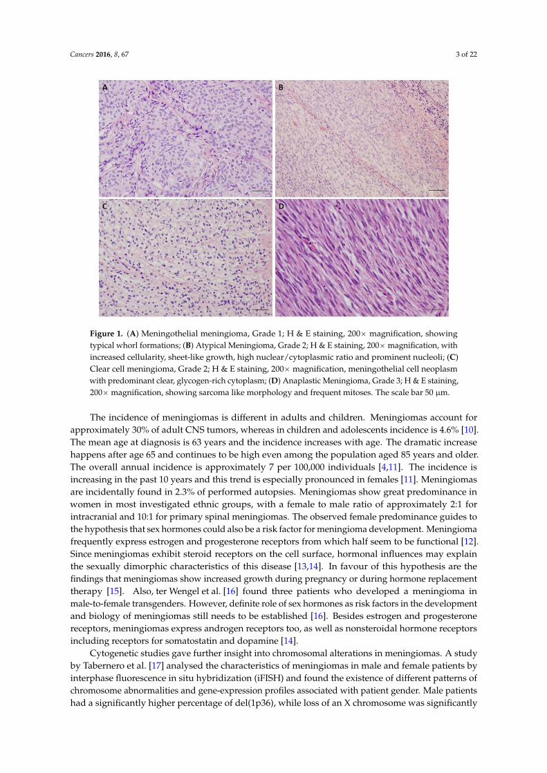
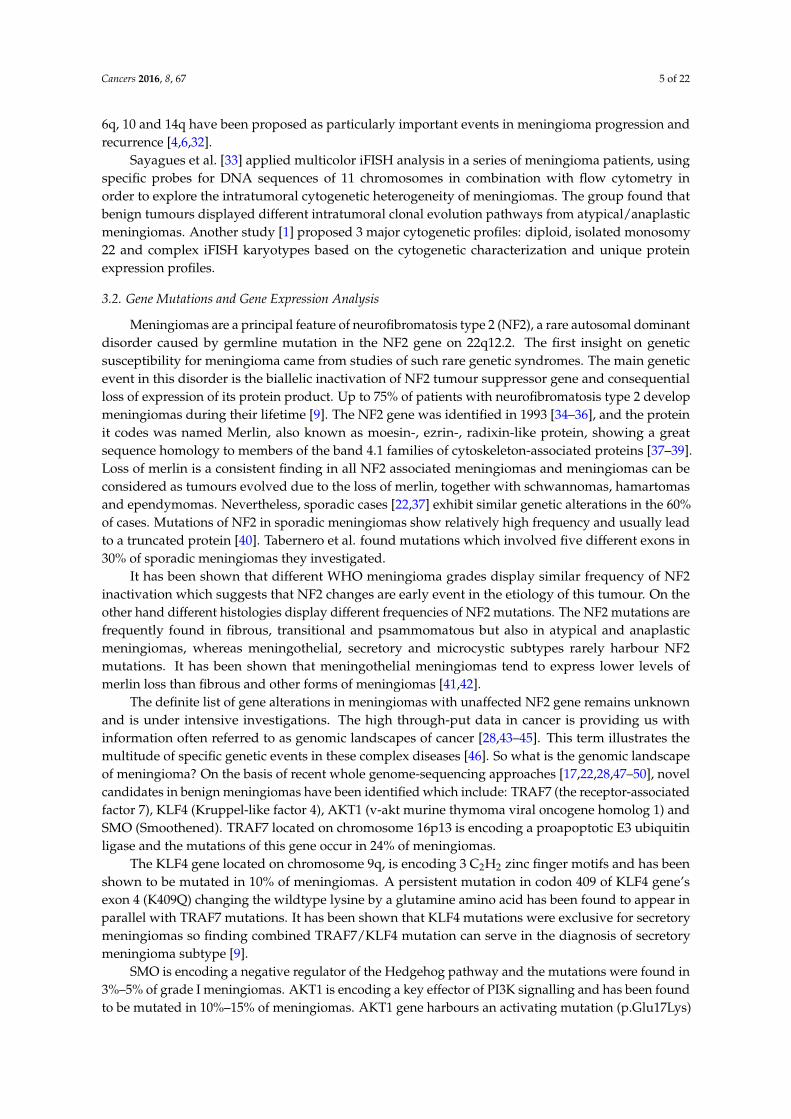
Sixteen different variants or subtypes of meningiomas are classified into three grades accordingto 2007 WHO classification [4–6]. The majority of meningiomas (80%) correspond to grade I and thusare considered to be benign, slowly growing tumours [4,5]. Grade I meningiomas exhibit a widerange of histological subtypes, including meningothelial, fibrous (fibroblastic), transitional (mixed),psammomatous, angiomatous, microcystic, secretory, lyphoplasmacyte-rich and metaplastic subtypes(Figure 1). Yet, these classifications are not always specific in respect to prediction of patient outcome,recurrence or response to treatment. Although most meningiomas histologically classified as “benign”have indolent biological behavior when adequately resected, there is substantial morbidity associatedwith recurrence. Meningiomas associated with less favourable clinical outcome, significantly higherrates of recurrence, morbidity and mortality correspond to grade II (atypical) and grade III (anaplastic).The incidence of atypical meningioma is 10%–15% and the incidence of anaplastic is fortunately lowwith 2%–5% of cases.
Atypical meningiomas are defined by histopathological criteria and two histological variants(clear cell and chordoid). Criteria include presence of at least 4 mitosis per 10 high power fields (HPF)or presence of at least three of the following histological features: sheet-like growth, spontaneousnecrosis, high nuclear-to-cytoplasmic ratio, prominent nucleoli and increased cellularity. Anaplastic(grade III) meningiomas have excessive mitotic index defined as 20 or more mitoses per 10 high powerfields (ě20/10 HPF) and the presence of frank anaplasia, including malignant cytology resemblingcarcinoma, melanoma or sarcoma. The brain invasion is a criterion for atypical meningioma [7].Papillary and rhabdoid variant have been classified as variants of grade III. Aggressive meningiomasare usually highly vascularized and express high levels of vascular endothelial growth factor (VEGF).Current treatment options for recurring higher-grade tumours are inadequate [4,6,8,9].
Cancers 2016, 8, 67 3 of 22
Cancers 2016, 8, 67 3 of 21
functional [12]. Since meningiomas exhibit steroid receptors on the cell surface, hormonal influences
may explain the sexually dimorphic characteristics of this disease [13,14]. In favour of this hypothesis
are the findings that meningiomas show increased growth during pregnancy or during hormone
replacement therapy [15]. Also, ter Wengel et al. [16] found three patients who developed a
meningioma in male‐to‐female transgenders. However, definite role of sex hormones as risk factors
in the development and biology of meningiomas still needs to be established [16]. Besides estrogen
and progesterone receptors, meningiomas express androgen receptors too, as well as nonsteroidal
hormone receptors including receptors for somatostatin and dopamine [14].
Figure 1. (A) Meningothelial meningioma, Grade 1; H & E staining, 200× magnification, showing
typical whorl formations; (B) Atypical Meningioma, Grade 2; H & E staining, 200× magnification, with
increased cellularity, sheet‐like growth, high nuclear/cytoplasmic ratio and prominent nucleoli; (C)
Clear cell meningioma, Grade 2; H & E staining, 200× magnification, meningothelial cell neoplasm
with predominant clear, glycogen‐rich cytoplasm; (D) Anaplastic Meningioma, Grade 3; H & E
staining, 200× magnification, showing sarcoma like morphology and frequent mitoses. The scale bar
50 μm.
Cytogenetic studies gave further insight into chromosomal alterations in meningiomas. A study
by Tabernero et al. [17] analysed the characteristics of meningiomas in male and female patients by
interphase fluorescence in situ hybridization (iFISH) and found the existence of different patterns of
chromosome abnormalities and gene‐expression profiles associated with patient gender. Male
patients had a significantly higher percentage of del(1p36), while loss of an X chromosome was
significantly associated to meningiomas from female patients. The group also showed a higher
frequency of chromosome losses, other than monosomy 22, in meningiomas arising in male patients,
while female patients displayed a higher frequency of chromosome gains or monosomy 22 alone.
Eight genes displayed a significantly different expression pattern in male versus female patients and
they were all localized to the sex chromosomes: two in chromosome X (DDX3X, XIST); and six in
chromosome Y (RPS4Y1, DDX3Y, JARID1D, EIF1AY, USP9Y, and CYorf15B) [17].
The firmly established risk factor associated with development of meningiomas is primarily
ionizing radiation. This environmental risk factor is usually associated to radiotherapy for primary
intracranial tumours in childhood [18–20]. It has been shown that radiation‐induced meningiomas
Figure 1. (A) Meningothelial meningioma, Grade 1; H & E staining, 200ˆ magnification, showingtypical whorl formations; (B) Atypical Meningioma, Grade 2; H & E staining, 200ˆmagnification, withincreased cellularity, sheet-like growth, high nuclear/cytoplasmic ratio and prominent nucleoli; (C)Clear cell meningioma, Grade 2; H & E staining, 200ˆmagnification, meningothelial cell neoplasmwith predominant clear, glycogen-rich cytoplasm; (D) Anaplastic Meningioma, Grade 3; H & E staining,200ˆmagnification, showing sarcoma like morphology and frequent mitoses. The scale bar 50 µm.
The incidence of meningiomas is different in adults and children. Meningiomas account forapproximately 30% of adult CNS tumors, whereas in children and adolescents incidence is 4.6% [10].The mean age at diagnosis is 63 years and the incidence increases with age. The dramatic increasehappens after age 65 and continues to be high even among the population aged 85 years and older.The overall annual incidence is approximately 7 per 100,000 individuals [4,11]. The incidence isincreasing in the past 10 years and this trend is especially pronounced in females [11]. Meningiomasare incidentally found in 2.3% of performed autopsies. Meningiomas show great predominance inwomen in most investigated ethnic groups, with a female to male ratio of approximately 2:1 forintracranial and 10:1 for primary spinal meningiomas. The observed female predominance guides tothe hypothesis that sex hormones could also be a risk factor for meningioma development. Meningiomafrequently express estrogen and progesterone receptors from which half seem to be functional [12].Since meningiomas exhibit steroid receptors on the cell surface, hormonal influences may explainthe sexually dimorphic characteristics of this disease [13,14]. In favour of this hypothesis are thefindings that meningiomas show increased growth during pregnancy or during hormone replacementtherapy [15]. Also, ter Wengel et al. [16] found three patients who developed a meningioma inmale-to-female transgenders. However, definite role of sex hormones as risk factors in the developmentand biology of meningiomas still needs to be established [16]. Besides estrogen and progesteronereceptors, meningiomas express androgen receptors too, as well as nonsteroidal hormone receptorsincluding receptors for somatostatin and dopamine [14].
Cytogenetic studies gave further insight into chromosomal alterations in meningiomas. A studyby Tabernero et al. [17] analysed the characteristics of meningiomas in male and female patients byinterphase fluorescence in situ hybridization (iFISH) and found the existence of different patterns ofchromosome abnormalities and gene-expression profiles associated with patient gender. Male patientshad a significantly higher percentage of del(1p36), while loss of an X chromosome was significantly
Cancers 2016, 8, 67 4 of 22
associated to meningiomas from female patients. The group also showed a higher frequency ofchromosome losses, other than monosomy 22, in meningiomas arising in male patients, while femalepatients displayed a higher frequency of chromosome gains or monosomy 22 alone. Eight genesdisplayed a significantly different expression pattern in male versus female patients and they were alllocalized to the sex chromosomes: two in chromosome X (DDX3X, XIST); and six in chromosome Y(RPS4Y1, DDX3Y, JARID1D, EIF1AY, USP9Y, and CYorf15B) [17].
The firmly established risk factor associated with development of meningiomas is primarilyionizing radiation. This environmental risk factor is usually associated to radiotherapy for primaryintracranial tumours in childhood [18–20]. It has been shown that radiation-induced meningiomas arehighly proliferative and more commonly high grade. Surprisingly, radiation-induced meningiomasrarely display NF2 alterations, both allelic losses and mutations in comparison to sporadic caseswhich may indicate that NF2 plays a less important role in the pathogenesis of radiation-inducedmeningiomas [18]. Besides, ionizing radiation, head trauma, hormone-replacement therapy andadvanced age are also the established risk factors [21]. The use of mobile phones does not seem tobe associated with the increased risk of meningioma development although there are controversialreports [9,22].
Survival rates of meningioma patients differ according to assigned grade [10,20], the 5-year overallsurvival is 92% for grade I, 78% for grade II and 47% (37.7% according to Champeauy et al. [23]) forgrade III meningioma. Benign meningiomas have recurrence rates of ~7%–25%; atypical 29%–52%and anaplastic 50%–94% [5]. In the study by Perry et al. [24], tumour recurrence rates were 7%–20%of benign (Grade I), 29%–40% of atypical (Grade II), 50%–78% of anaplastic (Grade III) meningiomas.Age at diagnosis has a significant effect on relative survival: 10 years survival for ages 24–44 is 85.2%and for patients older than 75 it is 29.1% [11]. The risk of meningioma recurrence depends on multipleclinical and biological factors, histological grade, extent of surgical resection, tumour size, location,patient age and gender, increased mitotic activity, as well as genetic characteristics of the tumour [25].Espinosa et al. [26] analysed the cytogenetic relationship between primary and subsequent recurrentmeningiomas developed in the same individual and found that in most cases similar tumour cellclones identified in the initial lesion were also detected in the subsequent recurrent tumour samples.The authors concluded that the development of recurrent meningiomas after gross total tumourresection is usually due to regrowth of the primary tumour and rarely to the emergence of an unrelatedmeningioma. A prognostic signature for meningioma prognosis was reported by Chen et al. [27] whoanalysed genome wide expression profiles of 119 meningioma samples from two previously publishedDNA microarray studies [17,28] using the Cox proportional hazards regression models and found37 genes to be specifically related to meningioma overall survival.
The current treatment and management options for meningioma patients include observationand surgical resection. It is important to individualize treatments for each patient, but the majorityof patients are treated with surgery. Radiotherapy is reserved for some special circumstances oras adjuvant therapy. Chemotherapy is rarely utilized, since effective chemotherapeutic agents formeningioma are still in the investigation phase [2].
3. Genetics and Signalling Pathways
3.1. Chromosome Aberrations
Besides being histologically heterogeneous, meningiomas are also showing great cytogeneticalheterogeneity. Chromosome gains and losses have been found to occur frequently. The most commonalteration observed in meningiomas is monosomy of chromosome 22, observed in 40%–70% gradeI meningiomas (WHO, 2007). Other most common cytogenetic alterations in meningioma, besidesabnormalities in the 22q locus, are the deletion of the short arm of chromosome 1 (specific regions1p33-34 and 1p36), loss of chromosomes 6, 10, 14, 18 and 19 [6,29,30] and gains of chromosomes 1q, 9q,12q, 15q, 17q and 20q of which many are associated with tumour grade [21,31]. Besides 22q, losses of
Cancers 2016, 8, 67 5 of 22
6q, 10 and 14q have been proposed as particularly important events in meningioma progression andrecurrence [4,6,32].
Sayagues et al. [33] applied multicolor iFISH analysis in a series of meningioma patients, usingspecific probes for DNA sequences of 11 chromosomes in combination with flow cytometry inorder to explore the intratumoral cytogenetic heterogeneity of meningiomas. The group found thatbenign tumours displayed different intratumoral clonal evolution pathways from atypical/anaplasticmeningiomas. Another study [1] proposed 3 major cytogenetic profiles: diploid, isolated monosomy22 and complex iFISH karyotypes based on the cytogenetic characterization and unique proteinexpression profiles.
3.2. Gene Mutations and Gene Expression Analysis
Meningiomas are a principal feature of neurofibromatosis type 2 (NF2), a rare autosomal dominantdisorder caused by germline mutation in the NF2 gene on 22q12.2. The first insight on geneticsusceptibility for meningioma came from studies of such rare genetic syndromes. The main geneticevent in this disorder is the biallelic inactivation of NF2 tumour suppressor gene and consequentialloss of expression of its protein product. Up to 75% of patients with neurofibromatosis type 2 developmeningiomas during their lifetime [9]. The NF2 gene was identified in 1993 [34–36], and the proteinit codes was named Merlin, also known as moesin-, ezrin-, radixin-like protein, showing a greatsequence homology to members of the band 4.1 families of cytoskeleton-associated proteins [37–39].Loss of merlin is a consistent finding in all NF2 associated meningiomas and meningiomas can beconsidered as tumours evolved due to the loss of merlin, together with schwannomas, hamartomasand ependymomas. Nevertheless, sporadic cases [22,37] exhibit similar genetic alterations in the 60%of cases. Mutations of NF2 in sporadic meningiomas show relatively high frequency and usually leadto a truncated protein [40]. Tabernero et al. found mutations which involved five different exons in30% of sporadic meningiomas they investigated.
It has been shown that different WHO meningioma grades display similar frequency of NF2inactivation which suggests that NF2 changes are early event in the etiology of this tumour. On theother hand different histologies display different frequencies of NF2 mutations. The NF2 mutations arefrequently found in fibrous, transitional and psammomatous but also in atypical and anaplasticmeningiomas, whereas meningothelial, secretory and microcystic subtypes rarely harbour NF2mutations. It has been shown that meningothelial meningiomas tend to express lower levels ofmerlin loss than fibrous and other forms of meningiomas [41,42].
The definite list of gene alterations in meningiomas with unaffected NF2 gene remains unknownand is under intensive investigations. The high through-put data in cancer is providing us withinformation often referred to as genomic landscapes of cancer [28,43–45]. This term illustrates themultitude of specific genetic events in these complex diseases [46]. So what is the genomic landscapeof meningioma? On the basis of recent whole genome-sequencing approaches [17,22,28,47–50], novelcandidates in benign meningiomas have been identified which include: TRAF7 (the receptor-associatedfactor 7), KLF4 (Kruppel-like factor 4), AKT1 (v-akt murine thymoma viral oncogene homolog 1) andSMO (Smoothened). TRAF7 located on chromosome 16p13 is encoding a proapoptotic E3 ubiquitinligase and the mutations of this gene occur in 24% of meningiomas.
The KLF4 gene located on chromosome 9q, is encoding 3 C2H2 zinc finger motifs and has beenshown to be mutated in 10% of meningiomas. A persistent mutation in codon 409 of KLF4 gene’sexon 4 (K409Q) changing the wildtype lysine by a glutamine amino acid has been found to appear inparallel with TRAF7 mutations. It has been shown that KLF4 mutations were exclusive for secretorymeningiomas so finding combined TRAF7/KLF4 mutation can serve in the diagnosis of secretorymeningioma subtype [9].
SMO is encoding a negative regulator of the Hedgehog pathway and the mutations were found in3%–5% of grade I meningiomas. AKT1 is encoding a key effector of PI3K signalling and has been foundto be mutated in 10%–15% of meningiomas. AKT1 gene harbours an activating mutation (p.Glu17Lys)
Cancers 2016, 8, 67 6 of 22
named AKT1E17K. A fraction of meningiomas (~13%), most frequently targeted with this mutation,were WHO grade I meningothelial and transitional meningiomas. Tumours grade II rarely harbouredAKT1E17K mutation while it was absent in grades III [9].
A strong up-regulation of secreted frizzled-related protein 1 (SFRP1) expression was suggestedin all meningiomas with AKT1E17K mutation. Therefore the use of SFRP1 immunohistochemistrymay be a reliable marker for the detection of AKT1E17K mutations [51]. Abedalthagafi et al. [52]found oncogenic PI3K mutations in meningioma and demonstrated that they are as common as AKT1and SMO mutations. Of note is that the mutations found in the above-mentioned genes are mutuallyexclusive of NF2 mutations.
Another gene thought to be involved early in meningioma pathogenesis and also being a memberof the 4.1 family, is DAL1 with its gene product protein 4.1B [4]. Martinez-Glez et al. [53] performed amutational study of DAL1 and found mutations in its exons 13 and 19, intron 18 and a polymorphismin exon 14. In approximately 60% of investigated meningiomas its reduced protein expression wasfound regardless of histological grade [9]. Lack of DAL1 protein was only slightly, and not significantly,more frequent in anaplastic meningiomas than in benign and atypical meningiomas, suggesting thatit represents an early event in meningioma tumorigenesis. Combined loss of DAL1 and merlin wasdetected in 58% of investigated cases, suggesting that they belong to different signalling pathways [18].
In addition other candidate genes include SMARCB1 (INI1) involved in chromatin remodelling.The found germline missense mutation in SMARCB1’s exon 2 predisposes individuals to thedevelopment of multiple meningiomas and schwannomas [20,22]. Since loss of chromosome22 region is a common event in meningioma, the region represents interesting genetic territoryfor search of additional candidate genes. Putative genes on chromosome 22q include LARGE(Like-Glycosyltransferase), BAM22 (AP1B1, Adaptor-Related Protein Complex 1, Beta 1 Subunit,also known as ADTB1 or beta-adaptin) and MN1 (Meningioma (Disrupted In Balanced Translocation)1). LARGE encodes a member of the N-acetylglucosaminyltransferase gene family involved in thesynthesis of glycoprotein and glycosphingolipid sugar chains. Also localized in the meningioma criticalchromosomal region is BAM22. The gene codes for the subunit of clathrin-associated adaptor proteincomplex 1 [54], a member of the adaptin protein family. The molecule plays a role in protein sorting inthe trans-Golgi network. BAM22 is part of the complexes that mediate both the recruitment of clathrinto membranes and the recognition of sorting signals within the cytosolic tails of transmembranereceptors. MN1 gene located on 22q12.1 [55,56] was found to be disrupted in its first exon by balancedtranslocation (4; 22) in a meningioma patient. Resent research [44,56] indicates that MN1 (a putativemeningioma tumour suppressor) was found to be differently expressed in malignant and benignmeningiomas. Chang et al. [44] assessed gene expression levels and copy number variants usingmicroarray platform and showed that MN1 was significantly repressed in all the malignant samplesanalysed in their study. Zhang et al. [56] from exome sequencing data, identified two novel potentialdriver mutations in MN1 which nominated MN1 as a candidate gene for malignant transformationof meningiomas.
Other genes alterations associated with meningioma include the well-known TP53 gene. Althoughmutations of the TP53 gene have been reported to be rare in meningiomas, low frequency of pointmutations is constantly found and reported [57–59]. In addition, relatively high incidences of somaticmutations and enhanced expression in meningiomas have been reported for sis, myc, ras, fos, mos,TP73, BCL-2 and STAT3 oncogenes [56].
Several genes have been associated with malignant progression in meningioma.Tumour suppressor genes CDKN2A (encoding p16INK4a), ARF (encoding p14ARF), and CDKN2B(encoding p15INK4b) residing on chromosome 9p21 are all associated with the anaplastic grade [30,38].Homozygous deletions or mutations of the above mentioned genes are found in most anaplasticmeningiomas [4,9].
Another candidate engaged in meningioma progression is the TIMP3 (the tissue inhibitor ofmetalloproteinase 3) gene on 22q12, because it has been shown that anaplastic meningiomas showed
Cancers 2016, 8, 67 7 of 22
much higher hypermethylation of its promoter than atypical and benign cases [6]. The maternallyexpressed gene 3 (MEG3) located in the 14q32 region has been implicated in meningioma progression,too. Allelic losses, promoter hypermethylation and reduced expression of MEG3 gene have beenassociated to aggressive meningioma phenotype. It has been reported that MEG3 has anti-proliferativeand anti-tumour activity in meningiomas. The gene encodes a non-coding RNA, whose expressionwas reduced in aggressive meningiomas [6].
Lusis et al. [60] using DNA microarray techniques identified a new meningioma associatedcandidate gene NDRG2 (N-myc downstream regulated gene 2) on chromosome 14q11.2. This tumoursuppressor is a Myc-repressed gene and is supposed to participate in cell growth, differentiationand p53-mediated apoptosis [25]. NDRG2 was found to be commonly inactivated in meningiomaprogression. It is down-regulated in anaplastic meningiomas and atypical meningiomas withaggressive clinical behaviour. The reduced expression of NDRG2 was associated with promoterhypermethylation [6]. Skiriute et al. [25] observed statistically significant differences in NDRG2gene expression level between primary and recurrent meningioma groups and between benign(WHO grade I) and atypical (WHO grade II) meningiomas measured at the mRNA level. Interestingly,NDRG2 contributes to the regulation of the Wnt signalling pathway. It down-regulatesCTNNB1-mediated transcriptional activation of target genes, such as CCND1 and may thereby act as atumour suppressor [61,62].
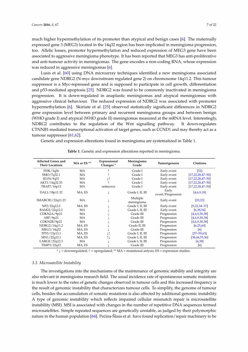
Genetic and expression alterations found in meningioma are systematized in Table 1.
Table 1. Genetic and expression alterations reported in meningioma.
Affected Genes andTheir Locations MA or ES ** Expressional
Changes *Meningioma
Grade Tumorigenesis Citations
PI3K/3q26 MA Ò Grade I Early event [52]SMO/7q32.1 MA Ò Grade I Early event [17,22,28,47–50]KLF4/9q31 MA ÓÒ Grade I Early event [17,22,28,47–50]
AKT1/14q32.33 MA Ò Grade I Early event [17,22,28,47–50]TRAF7/16p13 MA unknown Grade I Early event [17,22,28,47–50]
DAL1/18p11.32 MA; ES Ó Grade I, II, III Earlyevent/Progression [4,6,9,18]
SMARCB1/22q11.23 MA ÓMultiple
meningioma Early event [20,22]
NF2/22q12.2 MA; ES Ó Grade I, II, III Early event [9,22,34–37]BAM22/22q12.2 MA Ó Grade I, II, III Early event [6,38,54]CDKN2A/9p21 MA Ó Grade III Progression [4,6,9,30,38]
ARF/9p21 MA Ó Grade III Progression [4,6,9,30,38]CDKN2B/9p21 MA Ó Grade III Progression [4,6,9,30,38]
NDRG2/14q11.2 MA; ES Ó Grade II, III Progression [6,25,60]MEG3/14q32 MA; ES Ó Grade III Progression [6]TP53/17p13.1 MA; ES ÓÒ Grade I, II, III Progression [57–59,63]MN1/22q12.1 MA; ES ÒÓ Grade I, II, III Progression [38,44,55,56]
LARGE/22q12.3 MA Ó Grade I, II, III Progression [4,38]TIMP3/22q12 MA; ES Ó Grade III Progression [6]
* Ó = downregulated; Ò = upregulated; ** MA = mutational anlysis; ES = expression studies.
3.3. Microsatellite Instability
The investigations into the mechanisms of the maintenance of genomic stability and integrity arealso relevant in meningioma research field. The usual incidence rate of spontaneous somatic mutationsis much lower to the rates of genetic changes observed in tumour cells and this increased frequency isthe result of genomic instability that characterizes tumour cells. To simplify, the genome of tumourcells, besides the accumulation of somatic mutations is also affected by additional genomic instability.A type of genomic instability which reflects impaired cellular mismatch repair is microsatelliteinstability (MSI). MSI is associated with changes in the number of repetitive DNA sequences termedmicrosatellites. Simple repeated sequences are genetically unstable, as judged by their polymorphicnature in the human population [64]. Pecina-Šlaus et al. have found replication/repair machinery to be
Cancers 2016, 8, 67 8 of 22
constantly targeted in the meningiomas they investigated in two different studies [65]. Microsatellitemarkers specific for two different Wnt genes that were used (CDH1, AXIN1), revealed a fractionof meningiomas with MSI. This is indicative of malfunctioning of replication/repair genes (hMLH1or hMSH2, hPMS1, hPMS2), opening a potential area of interest in meningioma studies. D16S752microsatellite tetranucleotide marker for E-cadherin gene (CDH1) revealed 11% of samples with MSI.All MSI samples were reamplified and repeatedly analysed on both Spreadex and polyacrylamidegels. The samples demonstrating MSI were confirmed by direct sequencing. One meningothelial,one transitional and one anaplastic case harboured MSI [65].
There are other studies that show that certain proportions of meningiomas demonstrate featuresof genomic instability. Pykett et al. [66] have reported that 25% of meningiomas exhibit MSI,Sobrido et al. [67] have reported on 6.3% of meningiomas with MSI at 2 or 3 loci, which is similar tothe findings of Zhu et al. [68] of 2.4%. Bethke et al. [69] analysed single nucleotide polymorphismsof the DNA repair genes in association to meningioma predisposition and found that some DNArepair gene variants are connected to higher risk of meningioma development. The similar results onDNA repair variants affecting the risk of meningioma are reported by Rajaraman et al. [70]. A studyby Chen et al. [71] investigated the roles of the methylation of hMLH1 and MSI in meningiomas andfound 4.66% of cases to exhibit MSI. Hypermethylation of a promoter of hMLH1 was found in 18% ofinvestigated meningiomas and associated to meningioma progression. In their paper Yang et al. [72]showed that a tumour suppressor gene—CHEK2, involved in DNA repair and genome stability,contributes to the genomic instability in meningiomas. Alternative splicing and frequent codeletion ofCHEK2 with NF2 in meningiomas harbouring chromosome 22q deletions impaired DNA repair intheir study and increased chromosomal instability, thus promoting meningioma progression.
3.4. Epigenetic Studies in Meningioma
New evidence suggests that altered epigenetic regulation which include: altered DNAmethylation, microRNA expression, histone and chromatin modifications, plays an important role inthe pathogenesis of meningiomas [21,73]. Of note, aberrant promoter hypermethylation of a variety ofgenes has been identified as a frequent event in atypical and anaplastic meningiomas, suggesting thatepigenetic changes are substantially involved in meningioma progression [6]. The detailed descriptionof these findings is beyond the scope of this article.
3.5. Signalling Pathways
Understanding the genetic basis and molecular etiology of meningioma is essential for clinicalphenotype determination as well as patient outcome. The involvement of multiple pathways alsosuggests that therapy could be targeted against specific signalling level [74].
Molecular pathways driving meningioma progression still need elucidation. Our knowledge onspecific genetic drivers of malignant transformation is also incomplete [75]. Novel findings [41,74,76,77]suggest that activation of multiple growth factor receptors and their signalling pathways areresponsible for the growth of meningiomas.
One of the first gene expression profiling studies of meningiomas was performed byWatson and co-workers [78] who identified gene transcripts differentially expressed betweennormal leptomeningeal cells and meningiomas of different grades. Gene expression study byTabernero et al. [17] showed a relationship of expression profiles to the cytogenetic subgroups ofmeningiomas and patient outcome. Domingues et al. [1] investigated the different protein expressionprofiles by immunophenotyping of individual meningioma cells and also found association withtumour cytogenetics.
Comparative tissue proteomic profiling of meningioma shed light on molecular basis of thetumorigenesis using another approach. A study by Sharma et al. [77] investigated alterations intissue proteome in different grades of human meningiomas. Combining the results obtained fromtwo mass spectrometric platforms, the authors identified 2367 proteins that exhibited differential
Cancers 2016, 8, 67 9 of 22
expression in meningiomas. Functional analysis of the identified differentially expressed proteinsconfirmed the modulation of signal transduction pathways, including integrin signalling, Wntsignalling, Ras signalling, FGF signalling, EGF growth signalling, apoptosis signalling and ubiquitinproteasome signalling.
Merlin acts as a tumour suppressor and is capable of modulating a wide range of signallingpathways [79–82]. It interacts with cell-surface proteins, proteins involved in cytoskeletal dynamicsand proteins involved in regulating ion transport. Merlin-lacking cells are also known to containdefective adherens junctions [39]. It has been shown that merlin inhibits signalling and the activationof downstream pathways, including the Ras/Raf/MEK, PI3K/AKT/mTOR, Rac/PAK/JNK andWnt/β-catenin pathways [82].
Already from the listing of genes involved in meningioma in the previous paragraphs, we canassume which signalling pathways to suspect for meningioma development. Primarily involved is thenotorious Ras/Raf/MEK signalling pathway. It has been shown that receptor tyrosine kinases such asepidermal growth factor receptor (EGFR) and platelet-derived growth factor receptor (PDGFR) arewidely expressed in meningioma tumour cells. Equally important is the Pi3K/Akt/mTOR signallingpathway [9,47,74]. Both pathways lie downstream of receptor tyrosine kinases. Hilton et al. [74] havedemonstrated the expression of phosphorylated Jnk and Mek, in addition to Erk, pS6RP and Akt, in themajority of meningiomas of all grades. Besides EGFR and PDGFR, the kinases known to be involved areErb-B2 Receptor Tyrosine Kinase 2 (ERBB2), insulin-like growth factor 1 receptor (IGF1R) and vascularendothelial growth factor receptors (VEGFRs) [82]. Alterations in the RB retinoblastoma protein andp53 pathways are presumed because of the dysregulation of p16INK4a, p15INK4b, and p14ARF.The involvement of TGFbeta/SMAD as well as Hedgehog pathways is also noted as important [38].Integrin mediated signalling via Rac/PAK/JNK [83,84] has proved to be particularly interesting too.Studies linking the NF2 tumour suppressor as a modulator of growth factor and extracellular matrixsignals that trigger Rac1-dependent cytoskeleton-associated processes indicate its important role in theprocesses of cell adhesion and migration.
Insulin-like growth factor (IGF) signalling cascade [85] has been shown to be involved too,since both IGF-II and IGFBP2 are expressed in meningiomas, with increased concentrations of IGFIIassociated with invasiveness and malignant progression [4]. Gene expression transcript profilingstudy [86] revealed the deregulation of Notch pathway [73].
4. Wnt Signalling
Signalling pathways build complex molecular network within the cell and their accuratefunctioning maintains cellular homeostasis. Signal transduction pathways which regulate cell survival,proliferation and migration are also fundamental in tumorigenesis. Alongside other well-knownsignalling pathways is Wnt signalling pathway, primarily studied in development which regulateskey intercellular signalling events during embryogenesis [87,88] and plays an important role in thedevelopment of central nervous system. Components of Wnt signalling regulate multiple aspectsof brain development in vertebrate embryos. Wnt ligands have been identified as key regulatorsof regional identity in the early developing of the forebrain [89]. They modulate axon pathfinding,dendritic development and synaptic assembly [90]. Yu and Malenka [91] identified beta-catenin,the Wnt pathway’s main effector signalling molecule, as a critical mediator of dendritic morphogenesis.Specifically, overexpression of a stabilized beta-catenin in transgenic neural precursors causes massiveexpansion of the cerebral cortex, while loss-of-function mutations in individual Wnts cause deletionsor malformations of distinct brain regions [92]. A study by Lang et al. [93] showed that adenomatouspolyposis coli (APC), yet another key component of Wnt signalling, enhances proliferation ofoligodendroglial progenitor cells (OPCs). It is known that lymphoid-enhancer factor 1 (LEF1) andTcf4 (Transcription Factor 4; T cell factor 4), pathway’s transcription factors, are required for proneuraland neuronal gene expression, for neuronal differentiation in the posterior hypothalamus [94] and
Cancers 2016, 8, 67 10 of 22
for oligodendrocyte differentiation [95]. Huang et al. [96] reported that Wnt pathway co-receptors,Lrp 5 and 6, are required for the development of the cerebellum.
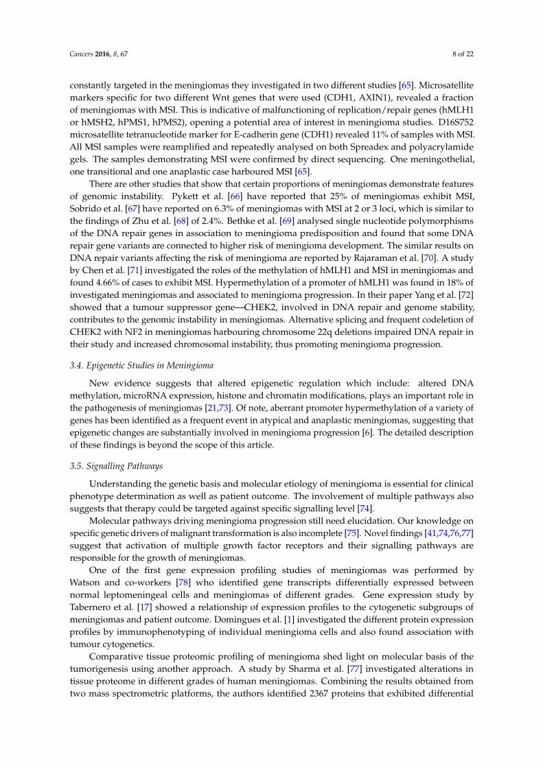
The pathway has two distinct branches, the canonical (or beta-catenin) and non-canonical(or planar cell polarity (PCP) and Wnt-Ca2+ pathways). Among two Wnt signalling cascades thecanonical is the longest known and the best studied one [97].
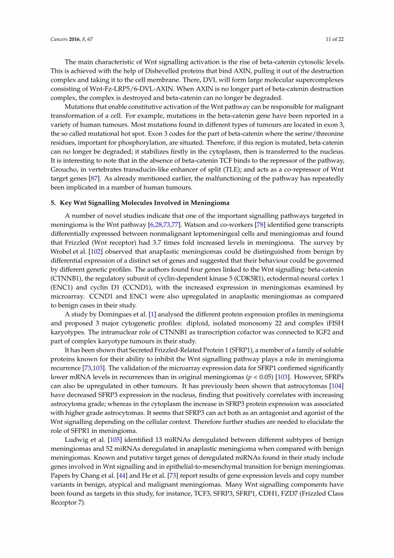
The pathway has two modes, active and inactive (Figure 2). When inactive, levels of beta-cateninare downregulated and kept low. This is achieved by beta-catenin destruction complex wherebeta-catenin is being phosphorylated by glycogen synthase kinase 3 beta (GSK3β) and casein kinase1 (CK1). The phosphorylated beta-catenin is targeted for quick ubiquitinilation and degradation inthe proteosome. Beta-catenin cellular levels are regulated by capturing it in the destruction complexwhere AXIN serves as a backbone. Besides AXIN, the complex is also composed of APC, GSK3βand CK1 [88,98,99]. Once bound to this protein complex, beta-catenin is sequentially phosphorylatedon four relevant amino acids: serines 45, 37 and 33; and threonine at 41, ultimately resulting in thetargeting of beta-catenin for degradation [100,101].Cancers 2016, 8, 67 10 of 21
Figure 2. A schematic illustration of the canonical Wnt signal transduction cascade. Panel (A), in the
absence of Wnt ligand, a destruction complex consisting of AXIN, APC, GSK3‐β and CK1 resides in
the cytosol. β‐catenin is phosphorylated by CK1 and GSK3‐β and targeted for degradation by the
proteosomal machinery; Panel (B), with Wnt stimulation, some components of protein complex
dislocate from the cytosol to the plasma membrane. The destruction complex falls apart and β‐catenin
is stabilized. Dvl is also recruited to the membrane and binds to Fz and Axin which is bound to
phosphorylated LRP5/6. Stablized β‐catenin is translocated to the nucleus where it associates to
LEF/TCF transcription factors, displacing co‐repressor TLE and recruiting additional co‐activators to
Wnt target genes. The activated Wnt pathway is associated to meningioma.
The main characteristic of Wnt signalling activation is the rise of beta‐catenin cytosolic levels.
This is achieved with the help of Dishevelled proteins that bind AXIN, pulling it out of the destruction
complex and taking it to the cell membrane. There, DVL will form large molecular supercomplexes
consisting of Wnt‐Fz‐LRP5/6‐DVL‐AXIN. When AXIN is no longer part of beta‐catenin destruction
complex, the complex is destroyed and beta‐catenin can no longer be degraded.
Mutations that enable constitutive activation of the Wnt pathway can be responsible for
malignant transformation of a cell. For example, mutations in the beta‐catenin gene have been
reported in a variety of human tumours. Most mutations found in different types of tumours are
located in exon 3, the so called mutational hot spot. Exon 3 codes for the part of beta‐catenin where
the serine/threonine residues, important for phosphorylation, are situated. Therefore, if this region is
mutated, beta‐catenin can no longer be degraded; it stabilizes firstly in the cytoplasm, then is
transferred to the nucleus. It is interesting to note that in the absence of beta‐catenin TCF binds to the
repressor of the pathway, Groucho, in vertebrates transducin‐like enhancer of split (TLE); and acts as
a co‐repressor of Wnt target genes [87]. As already mentioned earlier, the malfunctioning of the
pathway has repeatedly been implicated in a number of human tumours.
5. Key Wnt Signalling Molecules Involved in Meningioma
A number of novel studies indicate that one of the important signalling pathways targeted in
meningioma is the Wnt pathway [6,28,73,77]. Watson and co‐workers [78] identified gene transcripts
differentially expressed between nonmalignant leptomeningeal cells and meningiomas and found
that Frizzled (Wnt receptor) had 3.7 times fold increased levels in meningioma. The survey by Wrobel
et al. [102] observed that anaplastic meningiomas could be distinguished from benign by differential
expression of a distinct set of genes and suggested that their behaviour could be governed by different
genetic profiles. The authors found four genes linked to the Wnt signalling: beta‐catenin (CTNNB1),
Figure 2. A schematic illustration of the canonical Wnt signal transduction cascade. Panel (A), in theabsence of Wnt ligand, a destruction complex consisting of AXIN, APC, GSK3-β and CK1 residesin the cytosol. β-catenin is phosphorylated by CK1 and GSK3-β and targeted for degradation bythe proteosomal machinery; Panel (B), with Wnt stimulation, some components of protein complexdislocate from the cytosol to the plasma membrane. The destruction complex falls apart and β-cateninis stabilized. Dvl is also recruited to the membrane and binds to Fz and Axin which is bound tophosphorylated LRP5/6. Stablized β-catenin is translocated to the nucleus where it associates toLEF/TCF transcription factors, displacing co-repressor TLE and recruiting additional co-activators toWnt target genes. The activated Wnt pathway is associated to meningioma.
The pathway is active in response to Wnt ligands, highly conserved secreted signalling molecules.In this mode beta-catenin cannot be degraded and it accumulates in the cytoplasm. Upon cytoplasmicstabilization it enters the cell nucleus and since it is unable to bind to the DNA lacking necessarydomains it finds a partner among members of the DNA binding protein family LEF/TCF. Bound tosuch transcription cofactors it impacts the expression of target genes including cyclin D1, c-myc, fra-1and c-jun.
Cancers 2016, 8, 67 11 of 22
The main characteristic of Wnt signalling activation is the rise of beta-catenin cytosolic levels.This is achieved with the help of Dishevelled proteins that bind AXIN, pulling it out of the destructioncomplex and taking it to the cell membrane. There, DVL will form large molecular supercomplexesconsisting of Wnt-Fz-LRP5/6-DVL-AXIN. When AXIN is no longer part of beta-catenin destructioncomplex, the complex is destroyed and beta-catenin can no longer be degraded.
Mutations that enable constitutive activation of the Wnt pathway can be responsible for malignanttransformation of a cell. For example, mutations in the beta-catenin gene have been reported in avariety of human tumours. Most mutations found in different types of tumours are located in exon 3,the so called mutational hot spot. Exon 3 codes for the part of beta-catenin where the serine/threonineresidues, important for phosphorylation, are situated. Therefore, if this region is mutated, beta-catenincan no longer be degraded; it stabilizes firstly in the cytoplasm, then is transferred to the nucleus.It is interesting to note that in the absence of beta-catenin TCF binds to the repressor of the pathway,Groucho, in vertebrates transducin-like enhancer of split (TLE); and acts as a co-repressor of Wnttarget genes [87]. As already mentioned earlier, the malfunctioning of the pathway has repeatedlybeen implicated in a number of human tumours.
5. Key Wnt Signalling Molecules Involved in Meningioma
A number of novel studies indicate that one of the important signalling pathways targeted inmeningioma is the Wnt pathway [6,28,73,77]. Watson and co-workers [78] identified gene transcriptsdifferentially expressed between nonmalignant leptomeningeal cells and meningiomas and foundthat Frizzled (Wnt receptor) had 3.7 times fold increased levels in meningioma. The survey byWrobel et al. [102] observed that anaplastic meningiomas could be distinguished from benign bydifferential expression of a distinct set of genes and suggested that their behaviour could be governedby different genetic profiles. The authors found four genes linked to the Wnt signalling: beta-catenin(CTNNB1), the regulatory subunit of cyclin-dependent kinase 5 (CDK5R1), ectodermal-neural cortex 1(ENC1) and cyclin D1 (CCND1), with the increased expression in meningiomas examined bymicroarray. CCND1 and ENC1 were also upregulated in anaplastic meningiomas as comparedto benign cases in their study.
A study by Domingues et al. [1] analysed the different protein expression profiles in meningiomaand proposed 3 major cytogenetic profiles: diploid, isolated monosomy 22 and complex iFISHkaryotypes. The intranuclear role of CTNNB1 as transcription cofactor was connected to IGF2 andpart of complex karyotype tumours in their study.
It has been shown that Secreted Frizzled-Related Protein 1 (SFRP1), a member of a family of solubleproteins known for their ability to inhibit the Wnt signalling pathway plays a role in meningiomarecurrence [73,103]. The validation of the microarray expression data for SFRP1 confirmed significantlylower mRNA levels in recurrences than in original meningiomas (p < 0.05) [103]. However, SFRPscan also be upregulated in other tumours. It has previously been shown that astrocytomas [104]have decreased SFRP3 expression in the nucleus, finding that positively correlates with increasingastrocytoma grade; whereas in the cytoplasm the increase in SFRP3 protein expression was associatedwith higher grade astrocytomas. It seems that SFRP3 can act both as an antagonist and agonist of theWnt signalling depending on the cellular context. Therefore further studies are needed to elucidate therole of SFPR1 in meningioma.
Ludwig et al. [105] identified 13 miRNAs deregulated between different subtypes of benignmeningiomas and 52 miRNAs deregulated in anaplastic meningioma when compared with benignmeningiomas. Known and putative target genes of deregulated miRNAs found in their study includegenes involved in Wnt signalling and in epithelial-to-mesenchymal transition for benign meningiomas.Papers by Chang et al. [44] and He et al. [73] report results of gene expression levels and copy numbervariants in benign, atypical and malignant meningiomas. Many Wnt signalling components havebeen found as targets in this study, for instance, TCF3, SFRP3, SFRP1, CDH1, FZD7 (Frizzled ClassReceptor 7).
Cancers 2016, 8, 67 12 of 22
Sharma et al. [77] in their tissue proteome study of meningioma found Wnt signalling cascade asone of the significantly modulated pathways. Frizzleds, Casein Kinase 1 Alpha 1 (CSNK1A1), alsoknown as CK1 and SFRPs were all upregulated, while serine/threonine protein phosphatase B (PP2A)was downregulated.
Pecina-Slaus et al. investigated the involvement of Wnt signalling pathway in meningioma byanalysing its key signalling molecules, APC, beta-catenin, E-cadherin and AXIN1. They showed [106]significant association between APC genetic changes and lack of wild type protein expression,or presence of mutant APC proteins in meningiomas indicating involvement of this tumour suppressorgene. Thirty-three meningiomas were analysed regarding genetic changes of this tumour suppressorgene. Two genetic markers, Rsa I in APC’s exon 11 and Msp I in its exon 15 were used to testgenetic changes using the polymerase chain reaction/loss of heterozygosity (LOH) and RFLP method.Gross deletions of the APC gene were found in 47% of investigated meningiomas. The observed geneticchanges of the APC gene were dispersed among different types of benign meningioma, indicatingthat APC is not likely to be the first event in the advancement of this tumour. Meningiomas that wereharboring LOHs were also accompanied with the absence of APC protein expression or presence ofmutant APC proteins (Chi square = 13.81, df = 2, p < 0.001).
APC changes also influenced beta-catenin expression and nuclear localization. Beta-catenin wasupregulated and transferred to the nucleus in 71.2% of meningiomas and its nuclear localizationcorrelated to gross deletions of APC gene (Chi square = 21,96, df = 2, p < 0.0001). This high frequencyof nuclear transfer is indicative of beta-catenin’s importance in the biology of meningioma.
Together with APC in the β-catenin destruction complex is a scaffold protein AXIN1, functioningas a tumour suppressor in cancer. AXIN1, 16p13.3, 96 kDa, is an inhibitor of Wnt signalling.It down-regulates beta-catenin by facilitating its phosphorylation by GSK3-beta. It binds directlyto APC, beta-catenin, GSK3-beta and Dishevelled [107,108]. There is emerging evidence suggestingthat AXIN plays critical roles in the regulation of synaptic functions, formation of synaptic proteincomplexes and anchoring postsynaptic proteins in the central nervous system. LOH of AXIN1 genewas found in 21.1% of meningiomas. The majority of investigated samples showed moderate or strong(78.2%) levels of expression for AXIN1. Nevertheless, seven out of 32 samples (21.9%) demonstratednegative or very weak AXIN1 expression levels with exclusive cytoplasmic localization when comparedto the levels of AXIN1 in healthy brain tissues. Strong statistical correlations were observed betweencytoplasmic localization of AXIN1 and its weak expression; and also between the simultaneouscytoplasmic and nuclear localizations; and moderate and strong expression levels (p < 0.000) [109].
E-cadherin (gene CDH1 at 16q22.1, encodes a 120-kDa glycoprotein) is considered an indirectmodulator of Wnt signalling. Bound to beta-catenin, it is localized on the surfaces of cells in regionsof cell-cell contacts known as adherens junctions, while its intracellular domain interacts with theactin cytoskeleton. The downregulation, or loss of E-cadherin expression is considered responsiblefor dysfunction in cell-cell adhesion. We assume that the disruption of bound beta-catenin can risecytoplasmic levels of this molecule and thus indirectly modulate the activation of Wnt signalling.
The results of analysis on E-cadherin in meningiomas showed downregulation or loss of its proteinexpression in 73% of the total meningioma samples investigated [65]. Downregulation observed inmeningioma subtypes was in 50% of meningothelial, 80% of fibrous, 80% of transitional, 90% ofangiomatous, 80% of atypical and in 80% of anaplastic. Gross deletions of the CDH1 gene were alsodetected in 32% of investigated meningiomas. Altogether nine samples with LOH of the CDH1 geneout of 28 heterozygous patients were observed with the gross deletions distributed as follows: 2 in11 informative meningothelial meningiomas; 4 in 6 informative fibrous; 3 in 4 informative angiomatous.Next, significant association between the genetic changes of CDH1 and the nuclear localization ofbeta-catenin protein was found (Chi square = 5.25, df = 1. p < 0.022).
The results on E-cadherin in meningioma by other authors show similar patterns of expression.Schwechheimer et al. [110] found that E-cadherin’s expression was absent from the majority ofmalignant meningiomas they examined and Utsuki et al. [111] also reported on negative E-cadherin
Cancers 2016, 8, 67 13 of 22
immunostaining of their meningioma sample. Brunner et al. [112] believe that it is unlikely that lossof NF2 expression is associated with loss of the proper localization of beta-catenin and E-cadherinin meningiomas. Saydam et al. [113] evidenced that miR-200a has a role in meningioma growth viaE-cadherin and Wnt/beta-catenin signalling pathway. Downregulated miR-200a in meningiomaspromoted tumour growth by reducing E-cadherin and activating the Wnt pathway. A direct correlationbetween the downregulation of MiR-200a and the upregulation of beta-catenin was demonstrated inthis study.
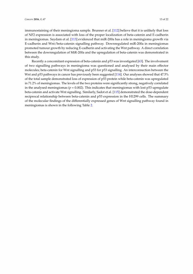
Recently a concomitant expression of beta-catenin and p53 was investigated [63]. The involvementof two signalling pathways in meningioma was questioned and analysed by their main effectormolecules; beta-catenin for Wnt signalling and p53 for p53 signalling. An interconnection between theWnt and p53 pathways in cancer has previously been suggested [114]. Our analyses showed that 47.5%of the total sample demonstrated loss of expression of p53 protein while beta-catenin was upregulatedin 71.2% of meningiomas. The levels of the two proteins were significantly strong, negatively correlatedin the analysed meningiomas (p = 0.002). This indicates that meningiomas with lost p53 upregulatebeta-catenin and activate Wnt signalling. Similarly, Sadot et al. [115] demonstrated the dose-dependentreciprocal relationship between beta-catenin and p53 expression in the H1299 cells. The summaryof the molecular findings of the differentially expressed genes of Wnt signalling pathway found inmeningiomas is shown in the following Table 2.
Cancers 2016, 8, 67 14 of 22
Table 2. Differentially expressed genes of Wnt signalling pathway found in meningiomas.
Gene Locus Product Function Deregulation Meningioma Effect Citation
FZD2 17q21.1 Frizzled class receptor 2 receptor for Wnt signaling proteins upregulation tumorigenesis [78]
FZD7 2q33 Frizzled class receptor 7 receptor for Wnt signaling proteins upregulation [44,73]
CSNK1A1 5q32 Casein kinase 1, alpha 1 transferring phosphorus-containinggroups protein tyrosine kinase activity upregulation tumorigenesis [77]
APC 5q22.2 Adenomatous polyposis coli negative regulator of Wnt signalingtumor suppressor loss of heterozygosity tumorigenesis [106]
AXIN1 16q13.3 Axin1 negative regulator of Wnt signalingtumor suppressor
gross deletions,downregulation, MSI cell growth and tumor progression [107,108]
CTNNB1 3p22.1 β-cateninkey downstream component of thecanonical Wnt signalingtranscription cofactor
upregulationcell growth and tumor progressionassociated to complexkaryotype meningiomas
[1,63,73,102,114,115]
PPP2CA 4q24 Serine/threonine proteinphosphatase 2B
negative control of cell growthand division downregulation tumorigenesis [77]
TCF3 19p13.3 Transcription factor 3 (T cell factor 3) transcription factor upregulation tumorigenesis [44,73]
CCND1 11q13.3 Cyclin D1 regulator (progression through) ofcell cycle upregulation cell growth and tumor progression [28,102]
ENC1 5q13.3 Ectodermal-neural cortex 1 role in the oxidative stress response arole in malignant transformation upregulation cell growth and tumor progression [102]
FRZB (SFRP3) 2q32.1 Secreted frizzled-related protein 3 modulator of Wnt signaling upregulation tumorigenesis [44]
SFRP1 8p11.21 Secreted frizzled-related protein 1 tumor suppressor downregulation, recurrence[28,41,44,73,102,103]
upregulation when AKT1E17K mutation is present
CDH1 16q22.1 E-cadherin regulator of cell-cell adhesions Downregulation loss offunction, gross deletion, MSI cell growth and tumor progression [65,73,78,110–113]
NDRG2 14q11.2 N-myc downstream regulator 2 transcription factor tumor suppressor Downregulation, promoterhyperventilation cell growth and tumor progression [6,25,61,62]
CDK5R1 17q11.2 Cyclin-dependent kinase 5,regulatory subunit 1
G1/S transition of mitotic cell cycle,development of the centralnervous system
upregulation cell growth and tumor progression [73,102]
Cancers 2016, 8, 67 15 of 22
6. Epithelial-to-Mesenchymal Transduction
Wnt signalling is strongly involved in epithelial-to-mesenchymal transition. This processis essential for embryogenesis where EMT enables controlled and precise movement of cells ingastrulation and neural crest formation. Cells that are till then closely held together acquirefibroblast resemblance and start to move individually. When cells gain such migratory mesenchymalphenotype they become plastic and able to perform conversions between epithelial-to-mesenchymaland mesenchymal-to-epithelial transitions (MET). In embryonic development cells undergo manyrounds of EMT and MET.
Similar to embryonic development EMT takes place during tumour progression and metastasis.Here loss of tissue integrity leads to local invasion where previously noninvasive tumour cells acquiremotility, leave the tissue parenchyma, enter the systemic circulations and ultimately disseminate todistant organs. The reverse process of MET enables the migratory cells to acquire epithelial phenotypeonce they reach their destination in order to form metastasis.
Although extremely uncommon it has been reported that meningiomas can metastasize.Metastatic dissemination of malignant meningiomas can be intra-cranial or to distant organs. Similarto other malignant neoplasms, malignant meningiomas can also infiltrate neighbouring tissues andform metastatic deposits [4]. It has been reported that extracranial meningiomas (0.1% of cases) canmetastasize to lungs, liver, pleura, bone and kidney locations. The lung is the most frequently involvedsite. Brain invasion and intra-cranial dissemination can also occur and could be explained by molecularmechanisms of EMT. Although metastatic spread of meningioma is more likely to occur in WHOgrades II and III, grade I lesions can also metastasize [4].
The molecular mechanisms driving meningioma invasion are still not well understood. It ishypothesised that the events of EMT may play role in it. Meningiomas display both mesenchymaland epithelial characteristics [116]. The meninges at the skull base are derived from the mesoderm,and the telencephalic meninges are derived from the neural crest [9]. One of the known functions ofarachnoidal cap cells is the production of collagen and fibroblast-like ECM proteins. Kida et al. [117]have shown the presence of tight junctions in arachnoid cells lining the arachnoid granulations.The expression of E-cadherin in the arachnoid membrane, arachnoid granulations and meningiomahas been confirmed by IHC [3]. The most prominent feature of EMT is the loss of expression ofthe cell-cell adhesion molecule E-cadherin and it has been observed in many carcinomas [118]. It isknown that E-cadherin can be inactivated through many different mechanisms including deletions andmutation, transcriptional repression as well as its promoter hypermethylation. The mechanism thatkeeps adherens junctons strength is the amount of cadherin molecules present at the cell membrane.
The phenomenon that happens in EMT during normal development, the so called cadherinswitch, has been known to happen in meningiomas too. In this phenomenon, E-cadherin is replacedby N-cadherin and in tumours it is regarded as a sign of invasive behaviour. Therefore E-cadherin isconsidered as an invasion suppressor gene. Another important molecular event involved in EMT isbeta-catenin’s translocation to the nucleus [119–121]. The stabilization and nuclear accumulation ofbeta-catenin can induce EMT because it can enhance the expression of two transcriptional repressorsSNAIL1 and SNAIL2 (also known as SLUG). It has been demonstrated that SNAIL1 and SNAIL2 canbind to E-cadherin promoter region and repress its expression, thus weakening adherens junctionsand inducing EMT [122–124]. As discussed in the previous paragraphs beta-catenin was progressivelyupregulated and transferred to the nucleus in our study on meningiomas.
The migratory mesenchymal cells when having reached their target destinations have thegenetic potential to revert to their original epithelial phenotype through a process known as MET.The incitement of the mechanisms of MET in invasive or metastasing cells is a tempting idea for thetherapeutic approaches in cancer, including aggressive meningioma.
Cancers 2016, 8, 67 16 of 22
7. Conclusions and Future Perspectives
The identification of molecular markers of meningioma recurrence is essential for clinicalphenotype determination as well as patient outcome.
The synthesis of knowledge on genetics, cellular signalling pathways, histopathologicalphenotypes and clinical parameters will lead to the classification of individual patient’s tumouraccording to signature alterations, with a final goal of subsequent development of novel and successfultreatment options in the dawn of personalized and precision medicine. The observed pathwaysinvolved in meningioma etiology will provide opportunities to improve prognostic markers formeningioma and predict clinical behaviour, recurrence and response to therapy.
New research indicates that alongside other well-known signalling pathways also stands the Wntpathway with important roles in meningioma formation and progression.
Acknowledgments: This work was supported by grant No. 6625 from Croatian Science Foundation.
Author Contributions: Nives Pecina-Šlaus produced the idea, designed the paper, wrote the manuscript,contributed to data interpretation, revised it for important intellectual content, and approved the final version ofthe manuscript. Anja Kafka contributed to the idea, wrote the manuscript, revised it for important intellectualcontent, and approved the final version of the manuscript. Mirna Lechpammer contributed to the paper designand data interpretation, manuscript editing, revised the manuscript for important intellectual content, andapproved the final version.
Conflicts of Interest: The authors declare no conflict of interests.
References
1. Domingues, P.H.; Teodósio, C.; Otero, Á.; Sousa, P.; Gonçalves, J.M.; Nieto, A.B.; Lopes, M.C.; de Oliveira, C.;Orfao, A.; Tabernero, M.D. The protein expression profile of meningioma cells is associated with distinctcytogenetic tumour subgroups. Neuropathol. Appl. Neurobiol. 2015, 41, 319–332. [CrossRef] [PubMed]
2. Lee, Y.H. Meningiomas, Diagnosis Treatment and Outcome, 1st ed.; Springer: London, UK, 2009.3. Mehta, B.C.; Holman, D.W.; Grzybowski, D.M.; Chalmers, J.J. Characterization of Arachnoidal Cells Cultured
on Three-Dimensional Nonwoven PET Matrix. Tissue Eng. 2007, 13, 1269–1279. [CrossRef] [PubMed]4. Riemenschneider, M.J.; Perry, A.; Reifenberger, G. Histological classification and molecular genetics of
meningiomas. Lancet Neurol. 2006, 5, 1045–1054. [CrossRef]5. Louis, D.N.; Ohgaki, H.; Wiestler, O.D.; Cavenee, W.K.; Burger, P.C.; Jouvet, A.; Scheihauer, B.W.; Kleihues, P.
The 2007 WHO Classification of Tumors of the Central Nervous System. Acta Neuropathol. 2007, 114, 97–109.[CrossRef] [PubMed]
6. Mawrin, C.; Perry, A. Pathological classification and molecular genetics of meningiomas. J. Neurooncol. 2010,99, 379–391. [CrossRef] [PubMed]
7. Louis, D.N.; Perry, A.; Reifenberger, G.; von Deimling, A.; Figarella-Branger, D.; Cavenee, W.K.; Ohgaki, H.;Wiestler, O.D.; Kleihues, P.; Ellison, D.W. The 2016 World Health Organization Classification of Tumors ofthe Central Nervous System: A summary. Acta Neuropathol. 2016, 131, 803–820. [CrossRef] [PubMed]
8. Claus, E.B.; Bondy, M.L.; Wiemels, J.L.; Wrensch, M.; Black, P.M. Epidemiology of intracranial meningioma.Neurosurgery 2005, 57, 1088–1094. [CrossRef] [PubMed]
9. Mawrin, C.; Chung, C.; Preusser, M. Biology and clinical management challenges in meningioma. Am. Soc.Clin. Oncol. Educ. Book 2015, e106–e115. [CrossRef] [PubMed]
10. Fathi, A.R.; Roelcke, U. Meningioma. Curr. Neurol. Neurosci. Rep. 2013. [CrossRef] [PubMed]11. Ostrom, Q.T.; Gittleman, H.; Farah, P.; Ondracek, A.; Chen, Y.; Wolinsky, Y.; Stroup, N.E.; Kruchko, C.;
Barnholtz-Sloan, J.S. CBTRUS Statistical Report: Primary Brain and Central Nervous System TumorsDiagnosed in the United States in 2006–2010. Neuro-Oncology 2013, 15, ii1–ii56. [CrossRef] [PubMed]
12. Speirs, V.; Boyle-Walsh, E.; Fraser, W.D. Constitutive co-expression of estrogen and progesterone receptormRNA in human meningiomas by RT-PCR and response of in vitro cell cultures to steroid hormones.Int. J. Cancer 1997, 72, 714–719. [CrossRef]
13. Chang, Z.N.; Guo, C.-L.; Ahronowitz, I.; Stemmer-Rachamimov, A.O.; MacCollin, M.; Nunes, F.P. A role forthe p53 pathway in the pathology of meningiomas with NF2 loss. J. Neurooncol. 2009, 91, 265–270. [CrossRef][PubMed]
Cancers 2016, 8, 67 17 of 22
14. Trott, G.; Pereira-Lima, J.F.S.; Leaes, C.G.S.; Ferreira, N.P.; Barbosa-Coutinho, L.M.; Oliveira, M.C. Abundantimmunohistochemical expression of dopamine D2 receptor and p53 protein in meningiomas: Follow-up,relation to gender, age, tumor grade, and recurrence. Braz. J. Med. Biol. Res. 2015, 48, 415–419. [CrossRef][PubMed]
15. Benson, V.S.; Kirichek, O.; Beral, V.; Green, J. Menopausal hormone therapy and central nervous systemtumor risk: Large UK prospective study and meta-analysis. Int. J. Cancer 2015, 136, 2369–2377. [CrossRef][PubMed]
16. Ter Wengel, P.V.; Martin, E.; Gooren, L.; Den Heijer, M.; Peerdeman, S.M. Meningiomas in threemale-to-female transgender subjects using oestrogens/progestogens and review of the literature. Andrologia2016. [CrossRef] [PubMed]
17. Tabernero, M.D.; Maillo, A.; Gil-Bellosta, C.J.; Castrillo, A.; Sousa, P.; Merino, M.; Orfao, A. Gene expressionprofiles of meningiomas are associated with tumor cytogenetics and patient outcome. Brain Pathol. 2009, 19,409–420. [CrossRef] [PubMed]
18. Lamszus, K. Meningioma pathology, genetics, and biology. J. Neuropathol. Exp. Neurol. 2004, 63, 275–286.[CrossRef] [PubMed]
19. Marosi, C.; Hassler, M.; Roessler, K.; Reni, M.; Sant, M.; Mazzab, E.; Vecht, C. Meningioma. Crit. Rev.Oncol. Hematol. 2008, 67, 153–171. [CrossRef] [PubMed]
20. Vranic, A.; Peyre, M.; Kalamarides, M. New insights into meningioma: From genetics to trials.Curr. Opin. Oncol. 2012, 24, 660–665. [CrossRef] [PubMed]
21. Murnyák, B.; Bognár, L.; Klekner, Á.; Hortobágyi, T. Epigenetics of meningiomas. BioMed Res. Int. 2015,2015, 6. [CrossRef] [PubMed]
22. Shibuya, M. Pathology and molecular genetics of meningioma: recent advances. Neurol. Med. Chir. 2015.[CrossRef] [PubMed]
23. Champeaux, C.; Wilson, E.; Brandner, S.; Shieff, C.; Thorne, L. World Health Organization grade IIImeningiomas. A retrospective study for outcome and prognostic factors assessment. Br. J. Neurosurg.2015, 29, 693–698. [CrossRef] [PubMed]
24. Perry, A.; Scheithauer, B.W.; Stafford, S.L.; Lohse, C.M.; Wollean, P.C. “Malignancy” in meningiomas:A clinicopathologic study of 116 patients, with grading implications. Cancer 1999, 85, 2046–2056. [PubMed]
25. Skiriute, D.; Tamasauskas, S.; Asmoniene, V.; Saferis, V.; Skauminas, K.; Deltuva, V.; Tamasauskas, A. Tumorgrade-related NDRG2 gene expression in primary and recurrent intracranial meningiomas. J. Neurooncol.2011, 102, 89–94. [CrossRef] [PubMed]
26. Espinosa, A.B.; Tabernero, M.D.; Maíllo, A.; Sayagués, J.M.; Ciudad, J.; Merino, M.; Alguero, M.C.;Lubombo, A.M.; Sousa, P.; Santos-Briz, A.; et al. The cytogenetic relationship between primary and recurrentmeningiomas points to the need for new treatment strategies in cases at high risk of relapse. Clin. Cancer Res.2006, 12, 772–780. [CrossRef] [PubMed]
27. Chen, F.; Xiang, C.X.; Zhou, Y.; Ao, X.S.; Zhou, D.Q.; Peng, P.; Zhang, H.Q.; Liu, H.D.; Huang, X. Geneexpression profile for predicting survival of patients with meningioma. Int. J. Oncol. 2015, 46, 791–797.[CrossRef] [PubMed]
28. Lee, Y.; Liu, J.; Patel, S.; Cloughesy, T.; Lai, A.; Farooqi, H.; Seligson, D.; Dong, J.; Liau, L.; Becker, D.;Mischel, P.; Shams, S.; Nelson, S. Genomic landscape of meningiomas. Brain Pathol. 2010, 20, 751–762.[CrossRef] [PubMed]
29. Lee, J.Y.; Finkelstein, S.; Hamilton, R.L.; Rekha, R.; King, J.T., Jr.; Omalu, B. Loss of heterozygosity analysis ofbenign, atypical, and anaplastic meningiomas. Neurosurgery 2004, 55, 1163–1173. [CrossRef] [PubMed]
30. Simon, M.; Boström, J.P.; Hartmann, C. Molecular genetics of meningiomas: From basic research to potentialclinical applications. Neurosurgery 2007, 60, 787–798. [CrossRef] [PubMed]
31. Barnholtz-Sloan, J.S.; Kruchko, C. Meningiomas: Causes and risk factors. Neurosurg. Focus 2007. [CrossRef][PubMed]
32. Weber, R.G.; Bostrom, J.; Wolter, M.; Baudis, M.; Collins, V.P.; Reifenberger, G.; Lichter, P. Analysis of genomicalterations in benign, atypical, and anaplastic meningiomas: Toward a genetic model of meningiomaprogression. Proc. Natl. Acad. Sci. USA 1997, 94, 14719–14724. [CrossRef] [PubMed]
Cancers 2016, 8, 67 18 of 22
33. Sayagués, J.M.; Tabernero, M.D.; Maíllo, A.; Espinosa, A.; Rasillo, A.; Díaz, P.; Ciudad, J.; López, A.;Merino, M.; Gonçalves, J.M.; et al. Intratumoral patterns of clonal evolution in meningiomas as definedby multicolor interphase fluorescence in situ hybridization (FISH): Is there a relationship betweenhistopathologically benign and atypical/anaplastic lesions? J. Mol. Diagn. 2004, 6, 316–325. [CrossRef]
34. Rouleau, G.A.; Merel, P.; Lutchman, M.; Sanson, M.; Zucman, J.; Marineau, C.; Hoang-Xuan, K.; Demczuk, S.;Desmaze, C.; Plougastel, B.; et al. Alteration in a new gene encoding a putative membrane-organizingprotein causes neurofibromatosis type 2. Nature 1993, 363, 515–521. [CrossRef] [PubMed]
35. Trofatter, J.A.; MacCollin, M.M.; Rutter, J.L.; Murrell, J.R.; Duyao, M.P.; Parry, D.M.; Eldridge, R.; Kley, N.;Menon, A.G.; Pulaski, K.; et al. A novel moesin-, ezrin-, radixin-like gene is a candidate for theneurofibromatosis 2 tumor suppressor. Cell 1993, 72, 791–800. [CrossRef]
36. Gusella, J.F.; Ramesh, V.; MacCollin, M.; Jacoby, L.B. Merlin: The neurofibromatosis 2 tumor suppressor.Biochim. Biophys. Acta 1999, 1423, M29–M36. [CrossRef]
37. Fuller, C.E.; Perry, A. Molecular diagnostics in central nervous system tumors. Adv. Anat. Pathol. 2005, 12,180–194. [CrossRef] [PubMed]
38. Ragel, B.T.; Jensen, R.L. Molecular genetics of meningiomas. Neurosurg. Focus 2005, 119, 1–8. [CrossRef]39. Pecina-Šlaus, N. Merlin the NF2 gene product. Pathol. Oncol. Res. 2013, 19, 365–373. [CrossRef] [PubMed]40. Tabernero, M.; Jara-Acevedo, M.; Nieto, A.B.; Caballero, A.R.; Otero, A.; Sousa, P.; Gonçalves, J.;
Domingues, P.H.; Orfao, A. Association between mutation of the NF2 gene and monosomy 22 in menopausalwomen with sporadic meningiomas. BMC Med. Genet. 2013, 14, 114. [CrossRef] [PubMed]
41. Miller, R., Jr.; DeCandio, M.L.; Dixon-Mah, Y.; Giglio, P.; Vandergrift, W.A., 3rd; Banik, N.L.; Patel, S.J.;Varma, A.K.; Das, A. Molecular Targets and Treatment of Meningioma. J. Neurol. Neurosurg. 2014, 1,PMC4255716.
42. Pavelin, S.; Becic, K.; Forempoher, G.; Tomic, S.; Capkun, V.; Drmic-Hofman, I.; Mrklic, I.; Lušic, I.;Pogorelic, Z. The Significance of Immunohistochemical Expression of Merlin, Ki-67, and p53 in Meningiomas.Appl. Immunohistochem. Mol. Morphol. 2014, 22, 46–49. [CrossRef] [PubMed]
43. Bell, D.W. Our changing view of the genomic landscape of cancer. J. Pathol. 2010, 220, 231–243. [CrossRef][PubMed]
44. Chang, X.; Shi, L.; Gao, F.; Russin, J.; Zeng, L.; He, S.; Chen, T.C.; Giannotta, S.L.; Weisenberger, D.J.;Zada, G.; et al. Genomic and transcriptome analysis revealing an oncogenic functional module inmeningiomas. Neurosurg. Focus 2013. [CrossRef] [PubMed]
45. Vogelstein, B.; Papadopoulos, N.; Velculescu, V.E.; Zhou, S.; Diaz, L.A., Jr.; Kinzler, K.W. Cancer genomelandscapes. Science 2013, 339, 1546–1558. [CrossRef] [PubMed]
46. Liu, M.; Zhang, K.; Zhao, Y.; Guo, Q.; Guo, D.; Zhang, J. Evidence for involvement of steroid receptors andcoactivators in neuroepithelial and meningothelial tumors. Tumour Biol. 2015, 36, 3251–3261. [CrossRef][PubMed]
47. Aarhus, M.; Lund-Johansen, M.; Knappskog, P.M. Gene expression profiling of meningiomas: Current statusafter a decade of microarray-based transcriptomic studies. Acta Neurochir. 2011, 153, 447–456. [CrossRef][PubMed]
48. Brastianos, P.K.; Horowitz, P.M.; Santagata, S.; Jones, R.T.; McKenna, A.; Getz, G.; Ligon, K.L.;Palescandolo, E.; van Hummelen, P.; Ducar, M.D.; et al. Genomic sequencing of meningiomas identifiesoncogenic SMO and AKT1 mutations. Nat. Genet. 2013, 45, 285–289. [CrossRef] [PubMed]
49. Clark, V.E.; Erson-Omay, E.Z.; Serin, A.; Yin, J.; Cotney, J.; Ozduman, K.; Avsar, T.; Li, J.; Murray, P.B.;Henegariu, O.; et al. Genomic analysis of non-NF2 meningiomas reveals mutations in TRAF7, KLF4, AKT1,and SMO. Science 2013, 339, 1077–1080. [CrossRef] [PubMed]
50. Bi, W.L.; Abedalthagafi, M.; Horowitz, P.; Agarwalla, P.K.; Mei, Y.; Aizer, A.A.; Brewster, R.; Dunn, G.P.;Al-Mefty, O.; Alexander, B.M.; et al. Genomic landscape of intracranial meningiomas. J. Neurosurg. 2016,1–11. [CrossRef] [PubMed]
51. Sahm, F.; Bissel, J.; Koelsche, C.; Schweizer, L.; Capper, D.; Reuss, D.; Böhmer, K.; Lass, U.; Göck, T.;Kalis, K.; et al. AKT1E17K mutations cluster with meningothelial and transitional meningiomas and can bedetected by SFRP1 immunohistochemistry. Acta Neuropathol. 2013, 126, 757–762. [CrossRef] [PubMed]
52. Abedalthagafi, M.; Bi, W.L.; Aizer, A.A.; Merrill, P.H.; Brewster, R.; Agarwalla, P.K.; Listewnik, M.L.;Dias-Santagata, D.; Thorner, A.R.; Van Hummelen, P.; et al. Oncogenic PI3K mutations are as common asAKT1 and SMO mutations in meningioma. Neuro-Oncology 2016, 18, 649–655. [CrossRef] [PubMed]
Cancers 2016, 8, 67 19 of 22
53. Martinez-Glez, V.; Bello, M.J.; Franco-Hernandez, C.; de Campos, J.M.; Isla, A.; Vaquero, J.; Rey, J.A.Mutational analysis of the DAL-1/4.1B tumour-suppressor gene locus in meningiomas. Int. J. Mol. Med.2005, 16, 771–774. [CrossRef] [PubMed]
54. Peyrard, M.; Fransson, I.; Xie, Y.G.; Han, F.Y.; Ruttledge, M.H.; Swahn, S.; Collins, J.E.; Dunham, I.;Collins, V.P.; Dumanski, J.P. Characterization of a new member of the human beta-adaptin gene familyfrom chromosome 22q12, a candidate meningioma gene. Hum. Mol. Genet. 1994, 3, 1393–1399. [CrossRef][PubMed]
55. Lekanne Deprez, R.H.; Riegman, P.H.; Groen, N.A.; Warringa, U.L.; van Biezen, N.A.; Molijn, A.C.;Bootsma, D.; de Jong, P.J.; Menon, A.G.; Kley, N.A.; et al. Cloning and characterization of MN1, a gene fromchromosome 22q11, which is disrupted by a balanced translocation in a meningioma. Oncogene 1995, 10,1521–1528. [PubMed]
56. Zhang, X.; Jia, H.; Lu, Y.; Dong, C.; Hou, J.; Wang, Z.; Wang, F.; Zhong, H.; Wang, L.; Wang, K. Exomesequencing on malignant meningiomas identified mutations in neurofibromatosis type 2 (NF2) andmeningioma 1 (MN1) genes. Discov. Med. 2014, 18, 301–311. [PubMed]
57. Mashiyama, S.; Murakami, Y.; Yoshimoto, T.; Sekiya, T.; Hayashi, K. Detection of p53 gene mutations inhuman brain tumors by single-strand conformation polymorphism analysis of polymerase chain reactionproducts. Oncogene 1991, 6, 1313–1318. [PubMed]
58. Wang, J.-L.; Zhang, Z.-J.; Hartman, M.; Smits, A.; Westermark, B.; Muhr, C.; Nistér, M. Detection ofTP53 gene mutation in human meningiomas: A study using immunohistochemistry, polymerase chainreaction/single-strand conformation polymorphism and DNA sequencing techniques on paraffin-embeddedsamples. Int. J. Cancer 1995, 64, 223–228. [CrossRef] [PubMed]
59. Joachim, T.; Ram, Z.; Rappaport, Z.H.; Simon, M.; Schramm, J.; Wiestler, O.D.; von Deimling, A. Comparativeanalysis of the NF2, TP53, PTEN, KRAS, NRAS and HRAS genes in sporadic and radiation-induced humanmeningiomas. Int. J. Cancer 2001, 94, 218–221. [CrossRef] [PubMed]
60. Lusis, E.A.; Watson, M.A.; Chicoine, M.R.; Lyman, M.; Roerig, P.; Reifenberger, G.; Gutmann, D.H.; Perry, A.Integrative genomic analysis identifies NDRG2 as a candidate tumor suppressor gene frequently inactivatedin clinically aggressive meningioma. Cancer Res. 2005, 65, 7121–7126. [CrossRef] [PubMed]
61. Kim, J.T.; Kim, J.W.; Kang, Y.H.; Kim, K.D.; Lee, S.J.; Choi, S.C.; Kim, K.S.; Chae, S.K.; Kim, J.W.; Lim, J.S.; et al.NDRG2 and PRA1 interact and synergistically inhibit T-cell factor/β-catenin signaling. FEBS Lett. 2012, 586,3962–3968. [CrossRef] [PubMed]
62. Kim, M.J.; Lim, J.; Yang, Y.; Lee, M.S.; Lim, J.S. N-myc downstream-regulated gene 2 (NDRG2) suppressesthe epithelial-mesenchymal transition (EMT) in breast cancer cells via STAT3/Snail signaling. Cancer Lett.2014, 354, 33–42. [CrossRef] [PubMed]
63. Pecina-Šlaus, N.; Kafka, A.; Vladušic, T.; Tomas, D.; Logara, M.; Skoko, J.; Hrašcan, R. Loss of p53 expressionis accompanied with upregulation of beta-catenin in meningiomas: A concomitant reciprocal expression.Int. J. Exp. Pathol. 2016, 97, 159–169. [CrossRef] [PubMed]
64. Ionov, Y.; Peinado, M.A.; Malkhosyan, S.; Shibata, D.; Perucho, M. Ubiquitous somatic mutations in simplerepeated sequences reveal a new mechanism for colonic carcinogenesis. Nature 1993, 363, 558–561. [CrossRef][PubMed]
65. Pecina-Šlaus, N.; Nikuševa Martic, T.; Deak, A.J.; Zeljko, M.; Hrašcan, R.; Tomas, D.; Musani, V. Genetic andprotein changes of E-cadherin in meningiomas. J. Cancer Res. Clin. Oncol. 2010, 136, 695–702. [CrossRef][PubMed]
66. Pykett, M.J.; Murphy, M.; Harnish, P.R.; George, D.L. Identification of a microsatellite instability phenotypein meningiomas. Cancer Res. 1994, 54, 6340–6343. [PubMed]
67. Sobrido, M.J.; Pereira, C.R.; Barros, F.; Forteza, J.; Carracedo, A.; Lema, M. Low frequency of replicationerrors in primary nervous system tumours. J. Neurol. Neurosurg. Psychiatry 2000, 69, 369–375. [CrossRef][PubMed]
68. Zhu, J.; Guo, S.Z.; Beggs, A.H.; Maruyama, T.; Santarius, T.; Dashner, K.; Olsen, N.; Wu, J.K.; Black, P.Microsatellite instability analysis of primary human brain tumors. Oncogene 1996, 12, 1417–1423. [PubMed]
69. Bethke, L.; Murray, A.; Webb, E.; Schoemaker, M.; Muir, K.; McKinney, P.; Hepworth, S.; Dimitropoulou, P.;Lophatananon, A.; Feychting, M.; et al. Comprehensive analysis of DNA repair gene variants and risk ofmeningioma. J. Natl. Cancer Inst. 2008, 100, 270–276. [CrossRef] [PubMed]

Cancers 2016, 8, 67 20 of 22
70. Rajaraman, P.; Hutchinson, A.; Wichner, S.; Black, P.M.; Fine, H.A.; Loeffler, J.S.; Selker, R.G.; Shapiro, W.R.;Rothman, N.; Linet, M.S.; et al. DNA repair gene polymorphisms and risk of adult meningioma, glioma, andacoustic neuroma. Neuro-Oncology 2010, 12, 37–48. [CrossRef] [PubMed]
71. Chen, M.N.; Wang, P.; Zhang, J.; Zhou, B.Y.; Mao, Q.; Liu, Y.H. Analysis of the role of hMLH1hypermethylation and microsatellite instability in meningioma progression. Genet. Mol. Res. 2012, 11,3933–3941. [CrossRef] [PubMed]
72. Yang, H.W.; Kim, T.M.; Song, S.S.; Shrinath, N.; Park, R.; Kalamarides, M.; Park, P.J.; Black, P.M.; Carroll, R.S.;Johnson, M.D. Alternative splicing of CHEK2 and codeletion with NF2 promote chromosomal instability inmeningioma. Neoplasia 2012, 14, 20–28. [CrossRef] [PubMed]
73. He, S.; Pham, M.H.; Pease, M.; Zada, G.; Giannotta, S.L.; Wang, K.; Mack, W.J. A review of epigenetic andgene expression alterations associated with intracranial meningiomas. Neurosurg. Focus 2013. [CrossRef][PubMed]
74. Hilton, D.A.; Shivane, A.; Kirk, L.; Bassiri, K.; Enki, D.G.; Hanemann, C.O. Activation of multiple growthfactor signalling pathways is frequent in meningiomas. Neuropathology 2015, 36, 250–261. [CrossRef][PubMed]
75. Cimino, P.J. Malignant progression to anaplastic meningioma: Neuropathology, molecular pathology, andexperimental models. Exp. Mol. Pathol. 2015, 99, 354–359. [CrossRef] [PubMed]
76. Zali, H.; Rezaei Tavirani, M. Meningioma protein-protein interaction network. Arch. Iran. Med. 2014, 17,262–272. [PubMed]
77. Sharma, S.; Ray, S.; Mukherjee, S.; Moiyadi, A.; Sridhar, E.; Srivastava, S. Multipronged quantitativeproteomic analyses indicate modulation of various signal transduction pathways in human meningiomas.Proteomics 2015, 15, 394–407. [CrossRef] [PubMed]
78. Watson, M.A.; Gutmann, D.H.; Peterson, K.; Chicoine, M.R.; Kleinschmidt-DeMasters, B.K.; Brown, H.G.;Perry, A. Molecular characterization of human meningiomas by gene expression profiling using high-densityoligonucleotide microarrays. Am. J. Pathol. 2002, 161, 665–672. [CrossRef]
79. Stamenkovic, I.; Yu, Q. Merlin, a “magic” linker between the extracellular cues and intracellular signalingpathways that regulate cell motility, proliferation, and survival. Curr. Protein Pept. Sci. 2010, 11, 471–484.[CrossRef] [PubMed]
80. Sughrue, M.E.; Yeung, A.H.; Rutkowski, M.J.; Cheung, S.W.; Parsa, A.T. Molecular biology of familial andsporadic vestibular schwannomas: Implications for novel therapeutics. J. Neurosurg. 2011, 114, 359–366.[CrossRef] [PubMed]
81. Ammoun, S.; Hanemann, C.O. Emerging therapeutic targets in schwannomas and other merlin-deficienttumors. Nat. Rev. Neurol. 2011, 7, 392–399. [CrossRef] [PubMed]
82. Zhou, L.; Hanemann, C.O. Merlin, a multi-suppressor from cell membrane to the nucleus. FEBS Lett. 2012,586, 1403–1408. [CrossRef] [PubMed]
83. Sherman, L.S.; Gutmann, D.H. Merlin: Hanging tumor suppression on the Rac. Trends Cell Biol. 2001, 11,442–444. [CrossRef]
84. Huveneers, S.; Danen, E.H. Adhesion signalling—Crosstalk between integrins, Src and Rho. J. Cell Sci. 2009,122, 1059–1069. [CrossRef] [PubMed]
85. Nordqvist, A.C.; Mathiesen, T. Expression of IGF-II, IGFBP-2, -5, and -6 in meningiomas with different braininvasiveness. J. Neurooncol. 2002, 57, 19–26. [CrossRef] [PubMed]
86. Cuevas, I.C.; Slocum, A.L.; Jun, P.; Costello, J.F.; Bollen, A.W.; Riggins, G.J.; McDermott, M.W.; Lal, A.Meningioma transcript profiles reveal deregulated Notch signaling pathway. Cancer Res. 2005, 65, 5070–5075.[CrossRef] [PubMed]
87. Logan, C.Y.; Nusse, R. The Wnt signaling pathway in development and disease. Annu. Rev. Cell Dev. Biol.2004, 20, 781–810. [CrossRef] [PubMed]
88. Lien, W.H.; Fuchs, E. Wnt some lose some: Transcriptional governance of stem cells by Wnt/β-cateninsignaling. Genes Dev. 2014, 28, 1517–1532. [CrossRef] [PubMed]
89. Harrison-Uy, S.J.; Pleasure, S.J. Wnt Signaling and Forebrain Development. Cold Spring Harb. Perspect. Biol.2012. [CrossRef] [PubMed]
90. Salinas, P.C. Wnt signaling in the vertebrate central nervous system: From axon guidance to synapticfunction. Cold Spring Harb. Perspect. Biol. 2012. [CrossRef] [PubMed]
Cancers 2016, 8, 67 21 of 22
91. Yu, X.; Malenka, R.C. Beta-catenin is critical for dendritic morphogenesis. Nat. Neurosci. 2003, 6, 1169–1177.[CrossRef] [PubMed]
92. Chenn, A. Wnt/β-catenin signaling in cerebral cortical development. Organogenesis 2008, 4, 76–80. [CrossRef][PubMed]
93. Lang, J.; Maeda, Y.; Bannerman, P.; Xu, J.; Horiuchi, M.; Pleasure, D.; Guo, F. Adenomatous Polyposis Coliregulates oligodendroglial development. J. Neurosci. 2013, 33, 3113–3130. [CrossRef] [PubMed]
94. Lee, J.E.; Wu, S.F.; Goering, L.M.; Dorsky, R.I. Canonical Wnt signaling through Lef1 is required forhypothalamic neurogenesis. Development 2006, 133, 4451–4461. [CrossRef] [PubMed]
95. Zhao, C.; Deng, Y.; Liu, L.; Yu, K.; Zhang, L.; Wang, H.; He, X.; Wang, J.; Lu, C.; Wu, L.N.; et al. Dualregulatory switch through interactions of Tcf7l2/Tcf4 with stage-specific partners propels oligodendroglialmaturation. Nat. Commun. 2016. [CrossRef] [PubMed]
96. Huang, Y.; Zhang, Q.; Song, N.N.; Zhang, L.; Sun, Y.L.; Hu, L.; Chen, J.Y.; Zhu, W.; Li, J.; Ding, Y.Q. Lrp5/6are required for cerebellar development and for suppressing TH expression in Purkinje cells via β-catenin.Mol. Brain 2016. [CrossRef] [PubMed]
97. Kafka, A.; Bašic-Kinda, S.; Pecina-Šlaus, N. The cellular story of dishevelleds. Croat. Med. J. 2014, 55, 459–467.[CrossRef] [PubMed]
98. Hart, M.J.; de los Santos, R.; Albert, I.N.; Rubinfeld, B.; Polakis, P. Downregulation of β-catenin by humanAxin and its association with the APC tumor suppressor, β-catenin and GSK-3β. Curr. Biol. 1998, 8, 573–581.[CrossRef]
99. Cliffe, A.; Hamada, F.; Bienz, M. A role of Dishevelled in relocating Axin to the plasma membrane duringwingless signaling. Curr. Biol. 2003, 13, 960–966. [CrossRef]
100. Polakis, P. Wnt signaling in cancer. Cold Spring Harb. Perspect. Biol. 2012. [CrossRef] [PubMed]101. Fagotto, F. Looking beyond the Wnt pathway for the deep nature of β-catenin. EMBO Rep. 2013, 14, 422–433.
[CrossRef] [PubMed]102. Wrobel, G.; Roerig, P.; Kokocinski, F.; Neben, K.; Hahn, M.; Reifenberger, G.; Lichter, P. Microarray-based
gene expression profiling of benign, atypical and anaplastic meningiomas identified novel genes associatedwith meningioma progression. Int. J. Cancer 2005, 114, 249–256. [CrossRef] [PubMed]
103. Pérez-Magán, E.; Rodríguez de Lope, A.; Ribalta, T.; Ruano, Y.; Campos-Martín, Y.; Pérez-Bautista, G.;García, J.F.; García-Claver, A.; Fiaño, C.; Hernández-Moneo, J.-L.; et al. Differential expression profilinganalyses identifies downregulation of 1p, 6q, and 14q genes and overexpression of 6p histone cluster 1 genesas markers of recurrence in meningiomas. Neuro-Oncology 2010, 12, 1278–1290. [CrossRef] [PubMed]
104. Pecina-Šlaus, N.; Kafka, A.; Varošanec, A.M.; Markovic, L.; Krsnik, Ž.; Njiric, N.; Mrak, G. Expressionpatterns of Wnt signaling component sFRP3 in astrocytoma and glioblastoma. Mol. Med. Rep. 2016, 13,4245–4251. [CrossRef] [PubMed]
105. Ludwig, N.; Kim, Y.J.; Mueller, S.C.; Backes, C.; Werner, T.V.; Galata, V.; Sartorius, E.; Bohle, R.M.; Keller, A.;Meese, E. Posttranscriptional deregulation of signaling pathways in meningioma subtypes by differentialexpression of miRNAs. Neuro-Oncology 2015, 17, 1250–1260. [CrossRef] [PubMed]
106. Pecina-Šlaus, N.; Nikuševa Martic, T.; Tomas, D.; Beroš, V.; Zeljko, M.; Cupic, H. Meningiomas exhibit loss ofheterozygosity of the APC gene. J. Neurooncol. 2008, 87, 63–70. [CrossRef] [PubMed]
107. Cong, F.; Varmus, H. Nuclear-cytoplasmic shuttling of Axin regulates subcellular localization of beta-catenin.Proc. Natl. Acad. Sci. USA 2004, 101, 2882–2887. [CrossRef] [PubMed]
108. Chen, Y.; Fu, A.K.Y.; Ip, N.Y. Axin: An emerging key scaffold at the synapse. IUBMB Life 2013, 65, 685–691.[CrossRef] [PubMed]
109. Pecina-Šlaus, N.; Kafka, A.; Vladušic, T.; Pecina, H.I.; Hrašcan, R. AXIN1’s expression and localization inmeningiomas and association to changes of APC and E-cadherin. 2016. in press.
110. Schwechheimer, K.; Zhou, L.; Birchmeier, W. E-Cadherin in human brain tumours: Loss of immunoreactivityin malignant meningiomas. Virchows Arch. 1998, 432, 163–167. [CrossRef] [PubMed]
111. Utsuki, S.; Oka, H.; Sato, Y.; Kawano, N.; Tsuchiya, B.; Kobayashi, I.; Fujii, K. Invasive meningioma isassociated with a low expression of E-cadherin and beta-catenin. Clin. Neuropathol. 2005, 24, 8–12. [PubMed]
112. Brunner, E.C.; Romeike, B.F.; Jung, M.; Comtesse, N.; Meese, E. Altered expression of beta-catenin/E-cadherinin meningiomas. Histopathology 2006, 49, 178–187. [CrossRef] [PubMed]
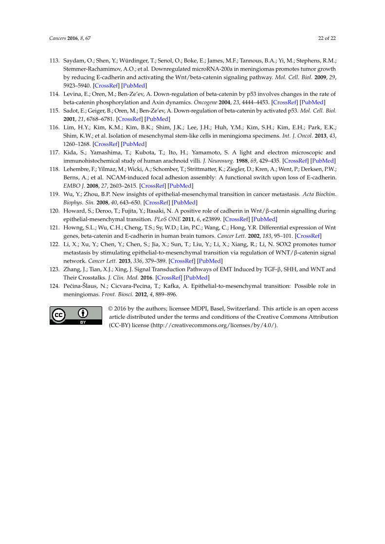
Cancers 2016, 8, 67 22 of 22
113. Saydam, O.; Shen, Y.; Würdinger, T.; Senol, O.; Boke, E.; James, M.F.; Tannous, B.A.; Yi, M.; Stephens, R.M.;Stemmer-Rachamimov, A.O.; et al. Downregulated microRNA-200a in meningiomas promotes tumor growthby reducing E-cadherin and activating the Wnt/beta-catenin signaling pathway. Mol. Cell. Biol. 2009, 29,5923–5940. [CrossRef] [PubMed]
114. Levina, E.; Oren, M.; Ben-Ze’ev, A. Down-regulation of beta-catenin by p53 involves changes in the rate ofbeta-catenin phosphorylation and Axin dynamics. Oncogene 2004, 23, 4444–4453. [CrossRef] [PubMed]
115. Sadot, E.; Geiger, B.; Oren, M.; Ben-Ze’ev, A. Down-regulation of beta-catenin by activated p53. Mol. Cell. Biol.2001, 21, 6768–6781. [CrossRef] [PubMed]
116. Lim, H.Y.; Kim, K.M.; Kim, B.K.; Shim, J.K.; Lee, J.H.; Huh, Y.M.; Kim, S.H.; Kim, E.H.; Park, E.K.;Shim, K.W.; et al. Isolation of mesenchymal stem-like cells in meningioma specimens. Int. J. Oncol. 2013, 43,1260–1268. [CrossRef] [PubMed]
117. Kida, S.; Yamashima, T.; Kubota, T.; Ito, H.; Yamamoto, S. A light and electron microscopic andimmunohistochemical study of human arachnoid villi. J. Neurosurg. 1988, 69, 429–435. [CrossRef] [PubMed]
118. Lehembre, F.; Yilmaz, M.; Wicki, A.; Schomber, T.; Strittmatter, K.; Ziegler, D.; Kren, A.; Went, P.; Derksen, P.W.;Berns, A.; et al. NCAM-induced focal adhesion assembly: A functional switch upon loss of E-cadherin.EMBO J. 2008, 27, 2603–2615. [CrossRef] [PubMed]
119. Wu, Y.; Zhou, B.P. New insights of epithelial-mesenchymal transition in cancer metastasis. Acta Biochim.Biophys. Sin. 2008, 40, 643–650. [CrossRef] [PubMed]
120. Howard, S.; Deroo, T.; Fujita, Y.; Itasaki, N. A positive role of cadherin in Wnt/β-catenin signalling duringepithelial-mesenchymal transition. PLoS ONE 2011, 6, e23899. [CrossRef] [PubMed]
121. Howng, S.L.; Wu, C.H.; Cheng, T.S.; Sy, W.D.; Lin, P.C.; Wang, C.; Hong, Y.R. Differential expression of Wntgenes, beta-catenin and E-cadherin in human brain tumors. Cancer Lett. 2002, 183, 95–101. [CrossRef]
122. Li, X.; Xu, Y.; Chen, Y.; Chen, S.; Jia, X.; Sun, T.; Liu, Y.; Li, X.; Xiang, R.; Li, N. SOX2 promotes tumormetastasis by stimulating epithelial-to-mesenchymal transition via regulation of WNT/β-catenin signalnetwork. Cancer Lett. 2013, 336, 379–389. [CrossRef] [PubMed]
123. Zhang, J.; Tian, X.J.; Xing, J. Signal Transduction Pathways of EMT Induced by TGF-β, SHH, and WNT andTheir Crosstalks. J. Clin. Med. 2016. [CrossRef] [PubMed]
124. Pecina-Šlaus, N.; Cicvara-Pecina, T.; Kafka, A. Epithelial-to-mesenchymal transition: Possible role inmeningiomas. Front. Biosci. 2012, 4, 889–896.
© 2016 by the authors; licensee MDPI, Basel, Switzerland. This article is an open accessarticle distributed under the terms and conditions of the Creative Commons Attribution(CC-BY) license (http://creativecommons.org/licenses/by/4.0/).