molecular genetics andpathogenesis ofclostridium perfringens · 622 roodandcole introduction the...
TRANSCRIPT
MICROBIOLOGICAL REVIEWS, Dec. 1991, p. 621-648 Vol. 55, No. 40146-0749/91/040621-28$02.00/0Copyright C 1991, American Society for Microbiology
Molecular Genetics and Pathogenesis of Clostridium perfringensJULIAN I. ROOD1* AND STEWART T. COLE2
Department of Microbiology, Monash University, Clayton, Victoria 3168, Australia,1 and Laboratoirede Genetique Moleculaire Bacterienne, Institut Pasteur, 75724 Paris Cedex 15, France2
INTRODUCTION ...................................................... 622BACTERIOPHAGES AND BACTERIOCINS ...................................................... 622
Bacteriophages of C. perfringens.................................................... 622Bacteriocinogenic Strains of C. perfringens ...................................................... 623
PLASMIDS...................................................... 623Bacteriocin Plasmids: an Overview ...................................................... 623Bacteriocin Plasmid pIP404 ...................................................... 623
Replication of pIP404 ...................................................... 624Control of replication and plasmid maintenance ...................................................... 624Bacteriocin production and immunity ...................................................... 625Transcription signals required for UV-inducible expression ......................................................626
Conjugative Plasmids ...................................................... 626Other Plasmids ...................................................... 627
ANTIBIOTIC RESISTANCE DETERMINANTS ...................................................... 627The Tetracycline Resistance Determinant, Tet P ...................................................... 627The Chloramphenicol Resistance Determinants, CAT P and CAT Q .............................................628
Identification of the CAT P and CAT Q determinants .................................................... 628Relationship between clostridial CAT enzymes .................................................... 628Characterization of the transposons Tn4451 and Tn4452 .................................................... 628
The Erythromycin Resistance Determinants, Erm BP and Erm Q ................................................629GENETIC MANIPULATION OF C. PERFRINGENS................................................... 630
Transformation of C. perfringens ................................................... 630Protoplast transformation .................................................... 630Electroporation .................................................... 630
C. perfringens-E. coli Shuttle Plasmids.....................................................631Transposon Mutagenesis of C. perfringens ................................................... 632
GENETICS AND CHROMOSOME MAPPING .................................................... 633Classical Genetics and the Advent of Cloning .................................................... 633Chromosome Mapping .................................................... 633Housekeeping Genes .................................................... 633Transcriptional Signals.................................................... 634Translational Signals.................................................... 635
TOXINS AND VIRULENCE FACTORS.................................................... 635a-Toxin or Phospholipase C .................................................... 637
Cloning, mapping, and expression of plc .................................................... 637Structure-function relationships.................................................... 637
fl-Toxin.................................................... 638e-Toxin .................................................... 638L-Toxin .................................................... 6380-Toxin .................................................... 639Neuraminidase.................................................... 639FL-Toxin .................................................... 639Other Toxins.................................................... 639
ENTEROTOXIN.................................................... 640Biochemical Properties of Enterotoxin .................................................... 640Molecular Genetics of Enterotoxin .................................................... 640
Cloning of the enterotoxin structural gene .................................................... 640Structure-function relationships.................................................... 640Gene location and molecular epidemiology.................................................... 641
Enterotoxin and Sporulation: Facts and Myths .................................................... 641FUTURE PERSPECTIVES .................................................... 642ACKNOWLEDGMENTS .................................................... 642REFERENCES .................................................... 642
* Corresponding author.
621
on June 9, 2019 by guesthttp://m
mbr.asm
.org/D
ownloaded from
622 ROOD AND COLE
INTRODUCTION
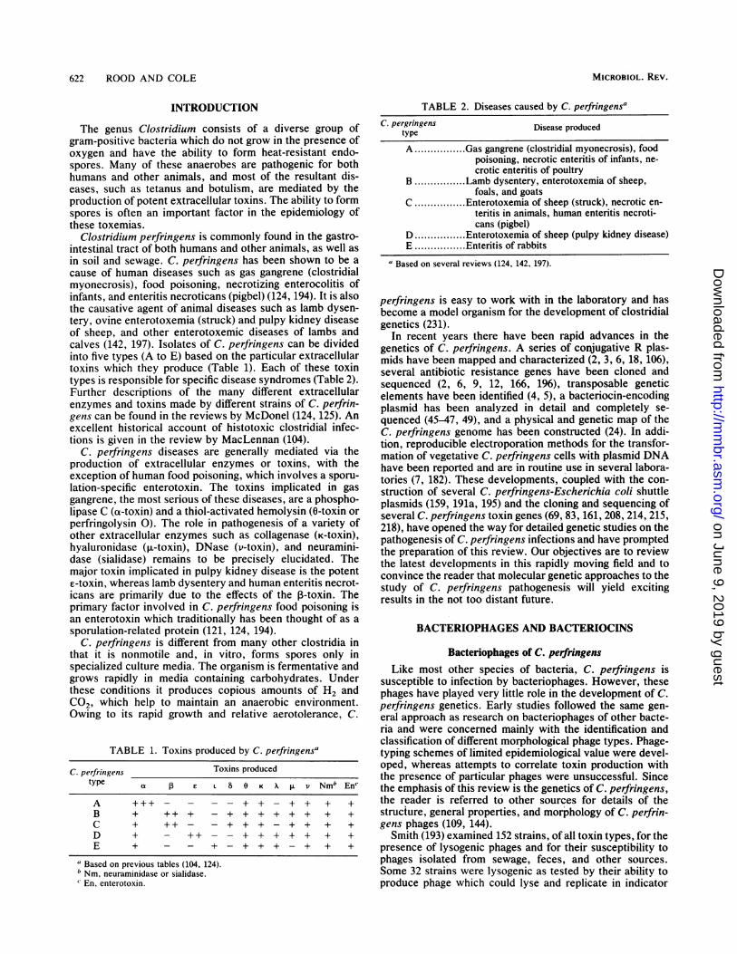
The genus Clostridium consists of a diverse group ofgram-positive bacteria which do not grow in the presence ofoxygen and have the ability to form heat-resistant endo-spores. Many of these anaerobes are pathogenic for bothhumans and other animals, and most of the resultant dis-eases, such as tetanus and botulism, are mediated by theproduction of potent extracellular toxins. The ability to formspores is often an important factor in the epidemiology ofthese toxemias.
Clostridium perfringens is commonly found in the gastro-intestinal tract of both humans and other animals, as well as
in soil and sewage. C. perfringens has been shown to be acause of human diseases such as gas gangrene (clostridialmyonecrosis), food poisoning, necrotizing enterocolitis ofinfants, and enteritis necroticans (pigbel) (124, 194). It is alsothe causative agent of animal diseases such as lamb dysen-tery, ovine enterotoxemia (struck) and pulpy kidney diseaseof sheep, and other enterotoxemic diseases of lambs andcalves (142, 197). Isolates of C. perfringens can be dividedinto five types (A to E) based on the particular extracellulartoxins which they produce (Table 1). Each of these toxintypes is responsible for specific disease syndromes (Table 2).Further descriptions of the many different extracellularenzymes and toxins made by different strains of C. perfrin-gens can be found in the reviews by McDonel (124, 125). Anexcellent historical account of histotoxic clostridial infec-tions is given in the review by MacLennan (104).
C. perfringens diseases are generally mediated via theproduction of extracellular enzymes or toxins, with theexception of human food poisoning, which involves a sporu-lation-specific enterotoxin. The toxins implicated in gasgangrene, the most serious of these diseases, are a phospho-lipase C (a-toxin) and a thiol-activated hemolysin (0-toxin orperfringolysin 0). The role in pathogenesis of a variety ofother extracellular enzymes such as collagenase (K-toxin),hyaluronidase (>i-toxin), DNase (v-toxin), and neuramini-dase (sialidase) remains to be precisely elucidated. Themajor toxin implicated in pulpy kidney disease is the potente-toxin, whereas lamb dysentery and human enteritis necrot-icans are primarily due to the effects of the ,-toxin. Theprimary factor involved in C. perfringens food poisoning isan enterotoxin which traditionally has been thought of as asporulation-related protein (121, 124, 194).
C. perfringens is different from many other clostridia inthat it is nonmotile and, in vitro, forms spores only inspecialized culture media. The organism is fermentative andgrows rapidly in media containing carbohydrates. Underthese conditions it produces copious amounts of H2 and
C02, which help to maintain an anaerobic environment.Owing to its rapid growth and relative aerotolerance, C.
TABLE 1. Toxins produced by C. perfringensa
C. perfringens Toxins producedtype aL E L 8 0 K A A v Nmb Enc
A +++- - --+ + - + + + +B + ++ + -+ + + + + + + +C + ++ - -+ + + - + + + +D + - + + + + + + + +E + - - +- + + + - + + +
a Based on previous tables (104, 124).b Nm, neuraminidase or sialidase.c En, enterotoxin.
TABLE 2. Diseases caused by C. perfringensaC. pergringens Disease produced
typeA. Gas gangrene (clostridial myonecrosis), food
poisoning, necrotic enteritis of infants, ne-crotic enteritis of poultry
B.Lamb dysentery, enterotoxemia of sheep,foals, and goats
C. Enterotoxemia of sheep (struck), necrotic en-teritis in animals, human enteritis necroti-cans (pigbel)
D. Enterotoxemia of sheep (pulpy kidney disease)E.Enteritis of rabbits
"Based on several reviews (124, 142, 197).
perfringens is easy to work with in the laboratory and hasbecome a model organism for the development of clostridialgenetics (231).
In recent years there have been rapid advances in thegenetics of C. perfringens. A series of conjugative R plas-mids have been mapped and characterized (2, 3, 6, 18, 106),several antibiotic resistance genes have been cloned andsequenced (2, 6, 9, 12, 166, 196), transposable geneticelements have been identified (4, 5), a bacteriocin-encodingplasmid has been analyzed in detail and completely se-quenced (45-47, 49), and a physical and genetic map of theC. perfringens genome has been constructed (24). In addi-tion, reproducible electroporation methods for the transfor-mation of vegetative C. perfringens cells with plasmid DNAhave been reported and are in routine use in several labora-tories (7, 182). These developments, coupled with the con-struction of several C. perfringens-Escherichia coli shuttleplasmids (159, 191a, 195) and the cloning and sequencing ofseveral C. perfringens toxin genes (69, 83, 161, 208, 214, 215,219), have opened the way for detailed genetic studies on thepathogenesis of C. perfringens infections and have promptedthe preparation of this review. Our objectives are to reviewthe latest developments in this rapidly moving field and toconvince the reader that molecular genetic approaches to thestudy of C. perfringens pathogenesis will yield excitingresults in the not too distant future.
BACTERIOPHAGES AND BACTERIOCINS
Bacteriophages of C. perfringensLike most other species of bacteria, C. perfringens is
susceptible to infection by bacteriophages. However, thesephages have played very little role in the development of C.perfringens genetics. Early studies followed the same gen-eral approach as research on bacteriophages of other bacte-ria and were concerned mainly with the identification andclassification of different morphological phage types. Phage-typing schemes of limited epidemiological value were devel-oped, whereas attempts to correlate toxin production withthe presence of particular phages were unsuccessful. Sincethe emphasis of this review is the genetics of C. perfringens,the reader is referred to other sources for details of thestructure, general properties, and morphology of C. perfrin-gens phages (109, 144).
Smith (193) examined 152 strains, of all toxin types, for thepresence of lysogenic phages and for their susceptibility tophages isolated from sewage, feces, and other sources.Some 32 strains were lysogenic as tested by their ability toproduce phage which could lyse and replicate in indicator
MICROBIOL. REV.
on June 9, 2019 by guesthttp://m
mbr.asm
.org/D
ownloaded from

GENETICS AND PATHOGENESIS OF C. PERFRINGENS 623
strains. In addition, a variety of phages were isolated fromthe environment. Several viruses appeared to be specific forindicator strains of particular toxin types. Since many of theC. perfringens strains were not susceptible to any of thesephages, it is clear from this research and other studies, thatC. perfringens bacteriophages have little epidemiologicalvalue (109). Four temperate phages, representing two dis-tinct classes, were isolated from strains of C. perfringenstype C, and unsuccessful attempts were made to obtaintransduction of erythromycin resistance (54). Other workersanalyzed a temperate C. perfringens phage, designated s9,and used the type A strain, Le chien, as an indicator (115).This phage recipient, more commonly known as strain 13, isnow the most readily transformable C. perfringens strain,probably because it appears to lack a restriction system(182). Although this strain is capable of being lysogenized(115), virtually no attempts have been made to use transduc-tion of strain 13 derivatives as a means of strain constructionin C. perfringens.
Further experiments were carried out on the UV-induciblebacteriophage s9 (115) and on a second phage isolated in thesame laboratory (114). A strain 13 derivative lysogenizedwith s9, as well as lysogenized and cured derivatives of theoriginal lysogenic parent strain, was tested for the ability tosporulate (202). The results indicated that the lysogensproduced heat-resistant spores more rapidly than did phage-free derivatives (3 to 4 h compared with 8 to 10 h). Thepercentage of refractile spores that were heat resistantappeared to increase significantly on lysogeny. It was sug-gested that this was an example of phage conversion, withthe phage s9 presumably carrying a gene(s) involved insporulation (202). Confirmation of this conclusion awaitsmodern molecular studies. Nonetheless, it appears to be theonly report of phage-mediated phenotypic changes in C.perfringens.The results of experiments involving the bacteriophage r
infection of a series of strain NCTC 8798 sporulation mu-tants were also used to postulate the existence of a C.perfringens restriction and modification system (33). Theresults of this study imply that the wild-type strain carries agene coding for a defective restriction endonuclease whichcan be mutated to an active form. It would be interesting toreexamine this system by using modern molecular methodsof analysis. However, it is clear from both conjugation (163)and transformation (182) studies, and the susceptibility ofDNA to cleavage by certain restriction endonucleases (44),that restriction and modification systems do exist in C.perfringens. Preliminary studies have led to the identifica-tion of a restriction endonuclease which is an isoschizomerof MboI (140a).
Early studies showed that the bacteriocinogenic C. per-fringens BP6K-N5 (now known as CPN50) carried a lyso-genic phage, 4)29 (82). The most recent work on C. perfrin-gens phages involved the mapping of the chromosomalattachment sites of 4)29 and another lysogenic phage, 4)59.Pulsed-field gel electrophoresis was used to compare wild-type and lysogenic derivatives of strain CPN50. The resultsshowed that the 4)59 attachment site was located near theMluI site at 2.9 Mb on the CPN50 map and that the 4)29 sitewas near the MluI site at 1.0 Mb (25). The 4)29 site waslocated within a 180-kb FspI fragment, which also containsthe nanH gene, which encodes neuraminidase (sialidase)production (161). The 4)59 site was located near the atpDgene, which codes for ATP synthase (24).Although phages of C. perfringens have been known for
many years, there have been no reports of the transduction
of either chromosomal or plasmid-determined genes. Simi-larly, unlike the situation for many other clostridial species,there is no solid evidence that any C. perfringens toxingenes, or other virulence factors, are phage determined.Although phage resistance has proven to be a useful markerfor the genetic analysis of transconjugants (18), studies ofbacteriophage genetics in C. perfringens have been unre-warding.
Bacteriocinogenic Strains of C. perfringensMany C. perfringens isolates have been shown to produce
bacteriocins capable of lysing indicator strains of C. perfrin-gens (see the review by Tagg et al. [207] for a list of earlypapers). C. perfringens bacteriocins have proven to be moreuseful than bacteriophages for the development of typingschemes, and the analysis of bacteriocin-encoding plasmidshas played a very important role in the development of C.perfringens genetics.
Several studies which have examined the ability of C.perfringens strains to produce bacteriocins have been car-ried out (107, 111). On the basis of the results, a C.perfringens typing scheme which relied on testing the sus-ceptibility of strains to 10 distinct bacteriocins was devel-oped (107). Analysis of the susceptibility of 274 strains of C.perfringens to these bacteriocins enabled them to be placedinto seven bacteriocin types and 50 subtypes. Only threestrains were untypable (107). Although subsequent studieshave used this scheme to type clinical isolates of C. perfrin-gens (119), it is not generally in widespread or routine usein clinical laboratories. An alternative bacteriocin-typingscheme has also been reported (177), and further develop-ments of the original scheme have also been made (179).Other studies have focused on the mechanism of action, theproduction, or the characterization of C. perfringens bacte-riocins (28, 81, 100, 108, 110, 112, 116).
PLASMIDS
Bacteriocin Plasmids: an Overview
C. perfringens CPN50 not only carries the bacteriophage4)29 but also produces a bacteriocin, known as N5 or BCN5,upon UV induction. Cured derivatives of CPN50 which nolonger produce bacteriocin were isolated after acriflavinetreatment and shown to be lacking plasmid DNA (80).Electron-microscopic analysis showed that the loss of bothbacteriocin production and immunity was associated withthe loss of a small (5.7-MDa) plasmid (79). Studies of strains28 (99) and 55 (129) also showed a relationship between thesephenotypic properties and small (5.6-MDa) plasmids. Sincebacteriocin production in all three strains was associatedwith plasmids of the same size, it is tempting to assume thatthese plasmids are very similar (129). However, no compar-ative restriction studies have been done. The plasmid carriedby strain 55, pCW4, was nonconjugative, whereas transfer ofpIP404, the bacteriocin plasmid from CPN50, has beenreported (18). Mobilization by a large conjugative plasmidpresent in strain CPN50 (79) is the most likely explanationfor these results.
Bacteriocin Plasmid pIP404Plasmid pIP404 can be considered a paradigm plasmid
analogous to ColEl in E. coli. It has been completelymapped and sequenced and is the best-studied C. perfrin-
VOL. 55, 1991
on June 9, 2019 by guesthttp://m
mbr.asm
.org/D
ownloaded from
624 ROOD AND COLE
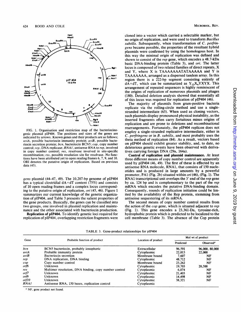
FIG. 1. Organisation and restriction map of the bacteriocino-genic plasmid pIP404. The positions and sizes of the genes areindicated by arrows. Known genes and their products are as follows;uviA, possible bacteriocin immunity protein; uviB, possible bacte-riocin secretion protein; bcn, bacteriocin BCN5; cop, copy numbercontrol; rep, DNA replicase; RNAJ, antisense RNA to rep, involvedin copy number control; res, resolvase involved in site-specificrecombination; rss, possible resolution site for resolvase. No func-tions have been attributed yet to open reading frames 6, 7, 9, and 10.
ORI denotes the putative origin of replication. Based on previousdata (45).
gens plasmid (44-47, 49). The 10,207-bp genome of pIP404has a typical clostridial dA+dT content (75%) and consistsof 10 open reading frames and a complex locus correspond-ing to the putative origin of replication, ori (45, 46). Figure 1summarizes our current knowledge of the genetic organiza-tion of pIP404, and Table 3 presents the salient properties ofthe gene products. Basically, the genes can be classified intotwo groups, one involved in plasmid replication and mainte-nance and the other associated with bacteriocin production.
Replication of pIP404. To identify genetic loci required forreplication of pIP404, overlapping restriction fragments were
cloned into a vector which carried a selectable marker, butno origin of replication, and were used to transform Bacillussubtilis. Subsequently, when transformation of C. perfrin-gens became possible, the properties of the resultant hybridplasmids were confirmed by using the homologous host. Inthis way the minimal origin of replication was defined andshown to consist of the rep gene, which encodes a 48.7-kDabasic DNA-binding protein (Table 3), and ori. The latterlocus is composed of two related families of direct repeats, Xand Y, where X is TAAAAAAATATAAAAA and Y isTAAAAAAA, arranged as a dispersed tandem array. In thisregion there is a 222-bp segment consisting entirely ofdA+dT, which can be summarized as Y12X4YXYX. Thisarrangement of repeated sequences is highly reminiscent ofthe origins of replication of numerous plasmids and phages(180). Detailed deletion analysis showed that essentially allof this locus was required for replication of pIP404 (46).The majority of plasmids from gram-positive bacteria
replicate via the rolling-circle method and use a single-stranded intermediate (65). When used as cloning vectors,such plasmids display pronounced physical instability, as theinserted fragments often carry fortuitous minus origins ofreplication and are prone to deletions and recombinationalrearrangements. Fortunately, the pIP404 replicon does notemploy a single-stranded replicative intermediate, either inC. perfringens or in B. subtilis, and most probably uses thetheta method of replication (84). As a result, vectors basedon pIP404 should exhibit greater stability, and, to date, nodeleterious genetic events have been observed with deriva-tives carrying foreign DNA (29a, 164a).
Control of replication and plasmid maintenance. At leastthree different means of copy number control are apparentlyused by pIP404 (46, 49). The first of these is effected by anantisense RNA molecule, RNA1, that consists of 150 nucle-otides and is produced in large amounts by a powerfulpromoter, PAl (Fig. 2b) situated within ori (46), (Fig. 1). TheRNA1 transcriptional unit overlaps the 3' end of the rep geneby ca. 124 bp and is complementary to the part of the repmRNA which encodes the putative DNA-binding domain.Consequently, rounds of replication initiation could be lim-ited by the availability of the Rep protein, stemming fromantisense sequestering of its mRNA.The second means of copy number control results from
the action of the cop gene, which is situated adjacent to rep(Fig. 1). This gene encodes a 23,361-Da, tyrosine-rich,hydrophobic protein which is predicted to be localized to thecell membrane (Table 3). The absence of the Cop protein
TABLE 3. Gene-product relationships for pIP404Mol wt of product
Gene Probable function of product Location of productPredicted Observeda
bcn BCN5 bacteriocin, probably ionophoric Extracellular 96,591 96,000, 80,000uviA Probable immunity protein Cytoplasmic 22,013 22,000uviB Bacteriocin secretion Membrane bound 7,607 NFrep DNA replication, DNA binding Cytoplasmic 48,712 NFcop Copy number control Membrane bound 23,262 NForf6 Unknown Cytoplasmic 19,703 20,500res Multimer resolution, DNA binding, copy number control Cytoplasmic 6,074 NForfJ7 Unknown Cytoplasmic 21,403 NForJ9 Unknown Cytoplasmic 14,498 NForflO Unknown Cytoplasmic 38,321 NFRNA] Antisense RNA, 150 bases, replication control Cytoplasmic
a NF, gene product not found.
MICROBIOL. REV.
on June 9, 2019 by guesthttp://m
mbr.asm
.org/D
ownloaded from

GENETICS AND PATHOGENESIS OF C. PERFRINGENS 625
a
Coni
"-35" < - - - - - - - 17 - - - - - - - > "-10"
P1 T T A T A a A T T T A G T T T A C A A A A T T G A A G T C A A A T T A CT T T T T A T A TT A TG
P3 T T A A T T T T T A G G T TT A C A T T T T T A A A A C T A A A C T - C T T T T T A T T T A T T A
P5 A A A A A A T A G A T A T T T A C A A A A T A G A C T A A A A A A AG C T T T T T A T A T A G T A T A A G C T T T T
P2 A A A A T A A A A A A A T T A T A A A T T T A G T T T A C A A A A T - T C A A ¢T C A A A T T A C T
*s-Cp - - A - - - A A A - T - T T G A - A A A - T T - - - A A A A T - T G - T A T A AT A A - - A T
b
PAIP4resrrnA P1rrnA P2rrnB P1rrnB P2rrng P1rrnE P2rrnHcatPermPPicorf2
GATC
"_-45". "-35" "s-10"
T T T A T T T A A A G T T T G a A A A A A A T T T T T T T A T A T T A T A T A A T C T T T G A A G A A A A G AT G T T T G G G T T T A T T C A C T T A T T T A T G A A A A A G T T G T A A A A T T A A T A C G A A C A T A TC A G T A T C A A A A A T C C A C A T T T T T G A T A C A T T A T T T T T T T G T A C A G A A A A A A G C C AG C A T A T A A A C T A A A T G T T T A T G T A A C C T T A T G T G A T A A G A T A T T A C T T G T C G C T GT G A A G A A A A A T G T T G A C A A A G T T C G A A A A T G G T G T T A A A C T A A A G A A G T C G C T T GA A A G T A A A A T T T A A A T T A A A T T A A A G A A A T G G T G T T A A G A T A A T A C C T G T C A C T AT T A A A A A A A G T G T T G A C A A A G T T C G A A G G T G A T G A T A A A A T A A A G A A G T C G C T T GA G A G A G A A G T A T T A A A T T A A A T A T G G T T A A A A T G G T A A G A T A A A T T T C G T C A G C AT G A A A A A A G T T G T T G A C A A A G T T T C T T A A A G A T G T T A A A A T A A G A A A G T C G C T T GG A A A T G A T T A T A T T A A A A G G A A G T T A T A A T T G T G A T A T C A T A A A T A T C G T C A T C AG A A G T G G G C A A G T T G A A A A A T T C A C A A A A A T G T G G T A T A A T A T C T T T G T T C A T T AG A G T G T G T T T G A T A G T G C A G T A T C T T A A A A T T T T G T A T A A T A G G A A T T G A A G T T AT T C A A A A G T T T A G T G A G G T T A T G T T A A T T A T A T G G T A T A A T T T C A A T G C G A G T G TA A A T T A T T A A A C T T A T T t T T A A T A T G A A A A T T T A C A A T T A T T A G T G T T A T A A T A A
4 4 2 3 2 4 3 3 2 1 2 4 1 - 8 1 2 1 1 2 3 1 2 1 3 4 - 1 1 - 4 6 -9 5 - - 3 1 - - 1 3 3 2 - 6 6 1 3 4 1 2 43 6 9 6 6 6 8 8 7 6 4 6 2 4 4 103 8 8 9 5 4 2 5 3 5 9 8 109 2 6 -1 4 1 137 8 12-108 6 5 8 5 2 3 4 5 7 1 2 86 3 2 5 6 4 2 3 4 6 8 311l9 1 3 4 4 5 3 6 9 9 5 6 4 4 4 3 5 8 2144 4131 7 2 -143 4 3 6 2 7 4 4 6 - - 9 7 21 1 1 - --1 - 1 1 - 1 - 1 1 - 5 1 - - - - 1 3 2 1 1 1 - - - - - - 1 - - 1 1 - 1 1 2 - 2 2 2 1 3 6 3 3 3 -
ACons-Cp --A - - - A A A - T - T T G A - A A A - T T ---A A A A T - T G-T A T A A T A A - - A T
Cons-Gram+ T- - A A A A A - - - - T T G A - A - - - - - - A - - - - - T - T G -T A T A A T A A - A -
FIG. 2. Nucleotide sequence of C. perfringens promoters. (a) Structure of four UV-inducible promoters (P1, P2, P3, and P5) from pIP404and comparison with the consensus sequence for a C. perfringens promoter. The -35 and -10 regions, as well as the transcriptional startpoints, are shown in boldface type (47). (b) Structure of 14 well-characterized promoters recognized by the major form of RNA polymeraseand the deduction of a consensus sequence. The sources of the promoters were PAl and P4 (47); res (49); rrnA, rrnB, rrnE, and rrnH (43);catP (196); ermP (120); and plc and or12 (176). The frequency of occurrence of individual nucleotides was tabulated, and when a givennucleotide appeared with a frequency of at least 50%, this was used to deduce the consensus C. perfringens (Cons-Cp) promoter sequence,which is compared with the extended promoter sequence for a gram-positive bacterium (Cons-Gram+ [63]).
leads to a fivefold increase in the copy number of pIP404-based vectors in B. subtilis (46). The effect of cop in C.perfringens is less pronounced, and cop' derivatives appearto be present in 10 to 15 copies per cell (i.e., like pIP404),whereas cop clones have an apparent copy number of 20 to25 (47a).An additional means of maintaining copy number control
and ensuring efficient distribution of plasmid molecules todaughter cells during cell division is provided by the res geneof pIP404, which encodes a resolvase highly homologous tothe site-specific recombinases of various transposons andphages (49). As for the gamma delta resolvase (64), thepromoter of the res gene is overlapped by three potentialresolution sites which could be used as resolvase substratesfor the resolution of multimers of pIP404, which result fromhomologous recombination, into the monomeric form. Al-though the effect of res on the stability of pIP404-basedvectors has not yet been evaluated, it is quite likely to bebeneficial. Additional support for this hypothesis is providedby the recent discovery of a gene analogous to res on thebroad-host-range plasmid pAMP1 from Enterococcusfaeca-lis (130a).
Bacteriocin production and immunity. Ionesco et al. (82)
showed that C. perfringens CPN50 secreted the bacteriocinBCN5 into the medium following UV irradiation, whichsuggests control of DNA damage by an SOS-like system(221). To localize the ben gene on pIP404, differential dotblot hybridization was performed with RNA prepared fromcultures before and after UV irradiation (44, 47). Thisapproach revealed that a 4-kb segment consisting of the bengene and the uviAB operon (Fig. 1) was heavily transcribedin response to DNA damage. Following activation of the bengene, copious amounts of a 96-kDa protein accumulate in thecytoplasm, to be released into the medium some 2 h afterinduction (31). The primary structure of BCN5 deduced fromthe nucleotide sequence reveals a protein of 890 amino acidresidues (molecular mass, 96 kDa), with a high glycinecontent (11.5%) which is characteristic of bacteriocins and isbelieved to facilitate their transfer across cell membranes(153, 154). Although the exact biological activity of BCN5 isunknown, the presence of an extended lipophilic region nearthe COOH terminus suggests that it might function as anionophore (44). At least part of the 96-kDa polypeptideappears to be converted to an 80-kDa form by proteolysis(44). At present we do not know whether both species havebacteriocin activity.
VOL. 55, 1991
on June 9, 2019 by guesthttp://m
mbr.asm
.org/D
ownloaded from

626 ROOD AND COLE
TABLE 4. Antibiotic resistance determinants from C. perfringens
Resistance Resistancedeterminant Origin Location mechanism
Tet P pCW3 Plasmid/chromosomal Efflux of tetracyclineCAT P pIP401 Plasmid CATCAT Q CW92 Chromosomal CATErm BP CP592 Plasmid/chromosomal rRNA methylaseErm Q CW459 Chromosomal rRNA methylase
In addition to encoding BCN5, pIP404 codes for bacterio-cin immunity (18), and a series of experiments suggests thatthe uviA gene is responsible for this activity. This gene is thepromoter-proximal cistron of the UV-inducible uviAB op-eron, situated downstream of bcn (Fig. 1), and its product isa soluble protein of 22 kDa. In contrast, the uviB geneencodes a small protein of 64 residues, with a hydrophobicNH2-terminal domain and a highly polar COOH terminus,that might play a role in BCN5 secretion by perturbing thecell membrane.
Transcription signals required for UV-inducible expression.To locate the promoters of the bcn and uviAB genes, adetailed transcriptional analysis was performed, as thesegenes could represent the prototypes of a putative clostridialSOS response and thus provide valuable regulatory insight.The mRNAs for both the bcn and uviAB transcriptional unitswere found to initiate at multiple, inducible promoters and toterminate after large stem-loop structures resembling factor-independent terminators (31). The bcn gene is transcribedfrom three promoters, P1, P2, and P3, whereas the uviABoperon is controlled by two promoters, P4 and P5. Of thesefive promoters only one, P4, bears any resemblance to theconsensus clostridial promoter sequence (Fig. 2b) and canproductively interact with purified vegetative RNA polymer-ase from C. perfringens (47, 48). We suspect that the role ofP4 is to provide immunity functions through low-level tran-scription of uviAB during normal growth conditions. Incontrast to promoter P2, which has a very unusual organi-zation, promoters P1, P3, and P5 are highly similar and haveidentical -35 and -10 regions, where the sequences TTTACA and CTTTTTAT occur, respectively (47) (Fig. 2a).
Conjugative Plasmids
C. perfringens is the only member of the genus Clostrid-ium in which conjugative antibiotic resistance plasmids (Rplasmids) have been found. All of these plasmids are closelyrelated and encode the same tetracycline resistance deter-minant (3, 6), designated Tet P in accordance with acceptedterminology (97). Some of these plasmids also carry achloramphenicol resistance determinant, designated CAT P(Table 4) (6, 18).A large number of conjugative R plasmids have been
found in various strains of C. perfringens (3, 6, 18, 70, 168).Two of these plasmids, pIP401 and pCW3, have beenextensively studied (2, 4, 6, 106) and shown to be closelyrelated (6). Genetic analysis has shown that both pIP401 andpCW3 carry genes encoding their own transfer, which oc-curs by a conjugationlike process (18, 168, 183, 184). The54-kb plasmid, pIP401, carries both the Tet P and CAT Pdeterminants, whereas pCW3 (47 kb) confers only tetracy-cline resistance. Loss of chloramphenicol resistance frompIP401 is often observed after conjugative transfer and isassociated with the loss of a 6.2-kb segment of DNA (6, 18).Subsequent studies showed that this segment comprises the
6.2 Tn 4451 0
FIG. 3. Relationship between the conjugative R plasmids pCW3and pIP401. The restriction map ofpCW3 is drawn with the arbitraryzero point at the single Sphl site as before (2). The plasmid pIP401consists of a pCW3 replicon that contains the 6.2-kb transposonTn4451 at the site indicated (4, 6). The location and direction oftranscriptional of the tet(P) (1, 3) and catP (4, 6) genes are alsoindicated.
transposable element, Tn445J (4). A closely related plasmid,pJIR27, carries a very similar chloramphenicol resistancetransposon, Tn4452 (4, 6).The entire pCW3 replicon has been cloned in E. coli by
separately cloning each of the five ClaI fragments that arepresent in pCW3. Restriction maps were elucidated for eachof these plasmids and, together with restriction data ob-tained from digests of pCW3, used to construct a detailedphysical map of the entire plasmid (2). A restriction map ofpIP401 has also been elucidated (106). Comparison of thesemaps reveals that these plasmids are very closely related(Fig. 3) and that two pIP401-derived, chloramphenicol-sensitive deletion plasmids (6, 106) are indistinguishablefrom pCW3 (6). The pIP401 replicon can therefore beconsidered to be a pCW3 plasmid which contains the chlor-amphenicol resistance transposon Tn4451 (4, 6). However, itis not known whether pCW3 is a transposon deletion deriv-ative of pIP401 or whether pIP401 is a transposon insertionderivative of pCW3.
In other studies, conjugative tetracycline-resistant strainsof C. perfringens have been isolated from diverse sources,including human and animal feces and the environment (18,70, 131, 132, 163, 167). Molecular analysis has shown thatthe conjugative plasmids harbored by all of these strainscarry the Tet P resistance determinant. In addition, three ofthese plasmids carry the CAT P determinant (3, 6). No otherphenotypic markers have been identified on any conjugativeC. perfringens plasmids (163).
Molecular analysis of these conjugative plasmids and, inparticular, comparison of their restriction profiles revealed
MICROBIOL. REV.
on June 9, 2019 by guesthttp://m
mbr.asm
.org/D
ownloaded from

GENETICS AND PATHOGENESIS OF C. PERFRINGENS 627
that they are all either identical to, or closely related to, theprototype plasmid, pCW3. Plasmids which could not bedistinguished from pCW3 by restriction analysis were de-tected in transconjugants derived from porcine, bovine,human, and environmental strains from the United States,France, Belgium, Japan, and Australia (3, 6). All of theremaining conjugative plasmids that have been reported inthe literature have at least 17 kb of restriction identity withpCW3 (3, 6, 70). The Tet P determinant is located within thisregion (2, 6).
Unlike the situation in most bacterial species, it is clearthat in C. perfringens there is only one major type ofconjugative R plasmid. All of the plasmids so far identifiedand analyzed carry the same tetracycline resistance deter-minant and were presumably derived either from pCW3 orfrom a common progenitor plasmid. Note that the sources ofthe strains from which these plasmids were isolated wereextremely diverse, from both a geographical and an environ-mental perspective. It is concluded that there must be aworldwide gene pool of antibiotic-resistant C. perfringensstrains, with ready distribution of plasmids within this genepool. The migration patterns of both humans and otheranimals would be key factors contributing to the dispersal ofR plasmids within and between these different environmen-tal niches (6).
Other Plasmids
There are many reports in the literature of cryptic C.perfringens plasmids (3, 13, 14, 18, 70, 92, 113, 118, 149, 158,162, 167, 168, 192). Plasmid profiling also has been used forstrain differentiation in C. perfringens (118, 149).Apart from the plasmids already mentioned, there are very
few C. perfringens plasmids which confer defined pheno-types. Only one nonconjugative R plasmid has been identi-fied, a large plasmid, pIP402, which confers resistance to themacrolide-lincosamide-streptogramin B (MLS) antibiotics(18). A small (3.1-kb) plasmid, pHB101, which conferscaseinase, or X-toxin, activity was identified several yearsago in a type B strain of C. perfringens (14). Despite its smallsize, this plasmid has not been analyzed in detail and thetoxin gene has not been cloned or sequenced. It is interestingthat again, unlike the situation for other bacteria, no viru-lence plasmids have been clearly identified in C. perfringens,with the possible exception of pHB101. It has been sug-gested that the C. perfringens a-toxin gene may be locatedon a large plasmid, but this report remains unconfirmed (35).
ANTIBIOTIC RESISTANCE DETERMINANTS
Penicillin has traditionally been regarded as the drug ofchoice for the treatment of C. perfringens gas gangrene, andno P-lactamase-mediated penicillin-resistant strains of C.perfringens have been reported (224). However, doubts havebeen cast upon the efficacy of penicillins and cephalosporinsin the clinical situation. Comparison of in vivo and in vitroeffectiveness of several antimicrobial agents, including thepenicillins, indicate some discrepancies between these tests.The treatment of C. perfringens infections therefore may notbe as straightforward as is commonly believed, and otherantibiotics, or combinations thereof, may be more suitablethan the penicillins (198, 199, 211, 212). It follows that it isimportant that resistance levels to various antimicrobialagents be monitored routinely and that the mechanisms bywhich C. perfringens isolates mediate and disseminate theirantimicrobial resistance be well understood.
The Tetracycline Resistance Determinant, Tet P
Tetracycline resistance is probably the most commonantibiotic resistance phenotype found in C. perfringens. Themajority of tetracycline-resistant strains do not have theability to transfer their resistance. However, conjugativetetracycline resistance plasmids can be readily detected (18,131, 132, 163, 165, 168). Tetracycline resistance in noncon-jugative isolates is generally constitutively expressed,whereas it is inducible in conjugative strains (163). Themolecular basis for this difference is not known, although ithas been postulated that plasmids like pCW3 carry a genewhich encodes a repressor of tetracycline resistance (2).The tetracycline resistance determinant from pCW3, Tet
P, has been cloned and shown to be located on two juxta-posed 1.9- and 2.1-kb EcoRI fragments on the pCW3 map(Fig. 3) (2, 195). Subcloning and transposon mutagenesisexperiments have shown that the SphI site at 0 kb and theEcoRI site at 0.8 kb (pCW3 coordinates) are both locatedwithin the Tet P determinant (1, 2). Preliminary DNAsequence data indicate that both sites are located within generegions encoding hydrophobic domains typical of membraneproteins (191a). These results are in agreement with obser-vations which indicate that the Tet P determinant mediatesan active efflux of tetracycline (126a).To determine the distribution of the Tet P determinant in
various bacteria, the internal 0.8-kb SphI-EcoRI fragmentwas purified and used as a gene-specific tet(P) hybridizationprobe in a series of Southern blot experiments. The resultsshowed that the tet(P) gene, or a gene very closely related toit, was present in eight nonconjugative isolates of C. perfrin-gens. It is clear that the same tetracycline resistance deter-minant is present in the constitutive nonconjugative isolates,where the resistance gene appears to be chromosomallydetermined, and in the inducible isolates which carry thetet(P) gene on conjugative pCW3-like plasmids (1). Addi-tional studies showed that the tet(P) probe hybridized with atetracycline-resistant strain of Clostridium paraputrificumbut did not hybridize with a resistant Clostridium sporo-genes isolate or six tetracycline-resistant Clostridium dif-ficile strains. Thus, although the Tet P determinant is notrestricted to C. perfringens, it is not widespread throughoutthe clostridia. Transfer of pIP401 from C. perfringens to C.difficile has been demonstrated, but the resultant tetracy-cline-resistant transconjugants were unstable (78). The fairlylimited dissemination of the Tet P determinant may thereforebe due to the narrow host range of pCW3-like plasmids.
In C. difficile, tetracycline resistance is mediated via a TetM-like determinant, which is located on conjugative, chro-mosomally determined transposons similar to those found inthe enterococci and streptococci (67, 137, 228). The entero-coccal tet(M)-encoding transposon Tn916 does transfer to C.perfringens, and the tet(M) gene is expressed (6a, 181a,196a). However, no naturally occurring C. perfringens iso-lates which carry a Tet M-like determinant have beenreported. Further screening of clinical isolates is required tosee whether Tet P is the only naturally occurring class oftetracycline resistance determinant found in C. perfringens.
Several different classes of tetracycline resistance deter-minants have been identified in diverse bacterial species(97). Although many of these determinants mediate theirresistance by an active efflux mechanism (96, 127), hybrid-ization experiments failed to reveal any homology betweenthese genes and tet(P) (1). The determination of the preciserelationship between these genes awaits the elucidation ofthe complete nucleotide sequence of tet(P).
VOL. 55, 1991
on June 9, 2019 by guesthttp://m
mbr.asm
.org/D
ownloaded from

628 ROOD AND COLE
The Chloramphenicol Resistance Determinants,CAT P and CAT Q
Identification of the CAT P and CAT Q determinants.Chloramphenicol resistance is less common than eithertetracycline or erythromycin resistance in C. perfringens(165, 167). Resistance is mediated by the production ofchloramphenicol acetyltransferases (CAT) (Table 4) (9, 168,234). Three conjugative tetracycline resistance plasmids(pIP401, pJIR25, and pJIR27) have been shown to carrychloramphenicol resistance determinants (6, 18), and twoother strains have also been shown to harbor chloramphen-icol resistance plasmids (166). Only one other chlorampheni-col-resistant strain, CW92 or VPI11268, has been analyzedin detail. This isolate, which is the original strain from whichpCW3 was obtained, carries the chromosomally determinedcatQ gene (166, 168).The chloramphenicol resistance gene from pIP401, catP,
has been cloned, localized, and shown to be expressed in E.coli (6). Hybridization analysis has shown that all five of theplasmid-determined C. perfringens cat genes share sequencesimilarity with catP (6, 166). However, the remaining chlor-amphenicol-resistant C. perfringens isolate that is availablefor analysis, CW92, did not hybridize with a catP-specificprobe (166). The chloramphenicol resistance gene, catQ,from a derivative of this strain (168), has also been clonedand shown to be expressed in E. coli (166). Comparativerestriction analysis and Southern blots confirmed that catPand catQ were different genes, even though both were of C.perfringens origin. Further hybridization studies showedthat neither of these genes hybridized with cat genes fromother genera (166). However, hybridization and sequenceanalysis has revealed that the catD gene from C. difficile isvery closely related to the catP gene (196, 226, 227). Thefinding that essentially identical chloramphenicol resistancedeterminants are present in C. perfringens and C. difficile isin agreement with the results obtained for the respectiveerythromycin resistance determinants (11). In contrast, quitedifferent tetracycline resistance genes are found in thesespecies (1).
Conjugative transfer of pIP401 from C. perfringens to C.difficile has been reported, but the resultant unstable tetra-cycline-resistant transconjugants were not screened forchloramphenicol resistance (78). In addition, although thecatP gene is carried within the pIP401-derived transposonTn4451 (4), the catD gene does not appear to be located oneither a plasmid or a transposon (166, 226). The differencebetween the species distribution of the catP and tet(P) genescan be explained by postulating that conjugative transfer ofpIP401-like plasmids from C. perfringens may be responsiblefor the introduction of both genes to C. difficile. However,the tetracycline resistance determinant may have been lostbecause the plasmid was unstable in C. difficile whereas thechloramphenicol resistance determinant was capable oftransposing to the C. difficile chromosome, although thecomplete transposon presumably was lost at some laterstage. If this hypothesis is correct, it may be possible to findeither complete or partial copies of Tn4451 in some chloram-phenicol-resistant C. difficile strains.
Relationship between clostridial CAT enzymes. The nucle-otide sequences of the catP (196), catD (227), and catQ (9)genes have been determined, and the amino acid sequencesof the encoded CAT enzymes have been elucidated. Thesesequences have been compared with the amino acid se-quences of 10 other CAT enzymes (9). All three clostridialCAT monomers shared considerable amino acid sequence
E. col III
E. coli I
P. mirabilis
S. acrimycini
.pC223-CAT-86
pUB112
pC221
pC194CATQ
C. coli
CATP
CATDFIG. 4. Phylogenetic relationships between CAT monomers. A
phylogenetic tree of CAT monomer amino acid sequences wasprepared by using the parsimony method in conjunction withpairwise alignment. Designations refer to either the genetic locus,plasmid, or bacterial species of origin. Reproduced with permissionfrom reference 9.
identity with the other enzymes, including the regionsknown to be involved in enzyme activity (98, 186, 187). ThecatP- and catD-encoded enzymes have 98% amino acidsequence identity and are also the only CAT monomerswhich contain a 4-amino-acid deletion between residues 38and 41 (196, 227). This deletion is not present in the CAT Qenzyme (9).To obtain an insight into the evolutionary relationships
between the CAT enzymes, their amino acid sequences wereused to construct a phylogenetic tree (9). The resultantdendrogram (Fig. 4) revealed that the CAT Q monomer wasas closely related to CAT enzymes from Staphylococcusaureus as it was to CAT P or CAT Q. Surprisingly, theCampylobacter coli CAT monomer (223) is more similar toCAT P and CAT D than the CAT Q enzyme is (9). It is clearfrom these results that the catP and catD genes are derivedfrom a fairly recent, in evolutionary terms, common ances-tor, whereas the catQ gene has evolved independently,albeit from a common primordial gene. The relationship ofthese genes to the catA and catB genes from Clostridiumbutyricum (34) remains to be determined, although hybrid-ization analysis has shown that they are not closely related(8a).
Characterization of the transposons Tn4451 and Tn4452.Analysis of the conjugative chloramphenicol resistance plas-mids indicated that the chloramphenicol resistance determi-nants were often lost on conjugation and that an identical ca.6-kb fragment was always deleted (6, 18, 184). In addition,the catP recombinant plasmids derived from pIP401 andpJIR27 were unstable in recA strains of E. coli, with identical
MICROBIOL. REV.
on June 9, 2019 by guesthttp://m
mbr.asm
.org/D
ownloaded from

GENETICS AND PATHOGENESIS OF C. PERFRINGENS 629
LI_
DR1
0- !-. Q sz,Q 1-6.- szf.
112
ermP
=_ I--
I 11 -f5.24
DR2FIG. 5. Genetic organization of the C. perfringens Erm BP determinant. The restriction map of the ermBP gene region of the recombinant
plasmid pJIR122 is shown. The ermBP gene is indicated by the arrow, and the direct repeats (DR1 and DR2) are shown by the dark boxes.Data were derived by D. Berryman and J. Rood and reproduced with permission from reference 164.
6.2-kb fragments being lost spontaneously (4). Detailedrestriction and sequence analysis showed that the end prod-ucts arising from events occurring in C. perfringens and E.coli were identical and that in each organism the deletionevent was precise (4, 5). Confirmation that the 6.2-kb regionsrepresented chloramphenicol resistance transposons wasobtained for E. coli when recA-independent transposition ofchloramphenicol resistance from temperature-sensitive plas-mids to the chromosome was demonstrated (4). Heterodu-plex analysis was done to compare the two transposonsTn4451 and Tn4452, from pIP401 and pJIR27, respectively.The results revealed that they were very closely related,differing only in a 0.4-kb region at the right end of eachtransposon (4).The mechanism(s) by which conjugative excision of
Tn4451 (and Tn4452) occurs in C. perfringens and sponta-neous excision occurs in E. coli is not known. However, it istempting to speculate that these events arise from part of thenormal transposition process, perhaps involving a circularintermediate as in the enterococcal transposon Tn916 (181).Sequence analysis of the ends of Tn4451 showed that the
termini of the transposon contain small imperfect invertedrepeats that have some similarity with the ends of the Tn3family. A CTAA sequence, which is often observed at theinternal terminus of the 38-bp Tn3 inverted repeat (188), andthe terminal GGGGTC sequence are both present in Tn4451(5). However, Tn4451 is not really a member of the Tn3family since preliminary evidence suggests that it may du-plicate a 2-bp sequence upon insertion (5), whereas Tn3transposons have been shown to duplicate a 5-bp targetsequence (188). Tn4451-R also contains an outward-firing-35 promoter consensus sequence close to the end of thetransposon (5), which is typical of many transposable ele-ments (42).
The Erythromycin Resistance Determinants,Erm BP and Erm Q
Erythromycin-resistant strains of C. perfringens are iso-lated more frequently than chloramphenicol-resistant deriv-atives. Resistance to erythromycin is always associated withresistance to clindamycin and lincomycin and is thereforereferred to as MLS resistance (18, 32, 37, 38, 165, 167). Theerythromycin resistance determinants are not located onconjugative R plasmids or on conjugative transposons, sincein mixed-plate mating experiments resistance is not transfer-able (18, 63, 165). However, one nonconjugative erythromy-cin resistance plasmid, pIP402, has been identified (18).The erythromycin resistance gene, designated ermP, from
a strain carrying pIP402 has been cloned, localized, andshown to be expressed in E. coli (11). An intragenic probewas then used in a series of hybridization experiments toexamine the distribution of the Erm P determinant. Theresults were very different from those obtained with the tetand cat probes. Only 5 of the 40 erythromycin-resistant C.
perfringens strains tested hybridized with the ermP probe(11). The Erm P determinant is therefore not widely distrib-uted in C. perfringens. We have recently cloned a seconderythromycin resistance gene, ermQ, from a different strainof C. perfringens (Table 4) and have shown that it is distinctfrom ermP (102a).Comparative hybridization analysis has shown that the
ermP gene is not restricted to C. perfringens since severalMLS-resistant isolates of both C. difficile and C. paraputri-ficum share sequence similarity with an ermP probe. Inaddition, the ermP gene hybridizes to, and shares restrictionmap identity with, the ermBlermAM gene from the promis-cuous E. faecalis plasmid pAM,1 (11). In C. difficile at leastone ermP-like erythromycin resistance determinant is lo-cated on a conjugative transposon which is chromosomallylocated and is capable of encoding the transfer of erythro-mycin resistance to other bacterial genera. The C. difficileerythromycin resistance gene, which was designated asermZ, also hybridizes with the ermBlermAM genes (66).Conjugative transfer of an MLS determinant from Clostrid-ium innocuum to C. perfringens, in the absence of plasmidtransfer has also been demonstrated (105).DNA sequence analysis has shown that the sequence of
the ermP gene is identical to the previously determinedsequence (19, 120) of the erm gene from pAMP1 and verysimilar to the other Erm B class MLS resistance determi-nants (12). On the basis of these data, and the designation ofthe E. coli erythromycin resistance determinant, which alsobelongs to the Erm B class, as the ermBC gene (20), it isproposed that the C. perfringens ermP gene be referred to asthe ermBP gene (12) and that the C. difficile ermZ gene beknown as the ermBZ gene.Sequence analysis has shown that the constitutive ermBP
gene is located between two 1.3-kb directly repeated se-quences (Fig. 5). (10a, 164). The recombinant ermBP plas-mid pJIR122, which contains both of the direct repeats, isunstable in recA strains of E. coli, with erythromycin-sensitive derivatives which lack one copy of the direct repeatbeing isolated at high frequency. As expected, plasmidscontaining one direct repeat are stable (11). The behavior ofthis determinant in E. coli is very similar to that of the C.perfringens transposons Tn4451 and Tn4452 (4). However,there is no evidence that the ErmBP determinant can trans-pose in either C. perfringens or E. coli.The enterococcal plasmid pAM,B1 is a broad-host-range
conjugative plasmid which has the ability to code for its owntransfer to many different species of bacteria (74). Conjuga-tive transfer to the clostridia has also been demonstrated(147, 155, 233). From these data it is reasonable to postulatethat the ermBP gene arose from the transfer of a pAM,1-likeplasmid into C. perfringens followed by the subsequenttransposition, or recombination, of the erm gene into thechromosome or a nonconjugative plasmid. Comparativesequence analysis of the ermBP gene region and the other
IL- -..
VOL. 55, 1991
%. Z.].I.s
on June 9, 2019 by guesthttp://m
mbr.asm
.org/D
ownloaded from

630 ROOD AND COLE
Erm B class determinants revealed that the identity betweenthese resistance determinants extended beyond the respec-tive erm genes. The identity between the pAM,1 erm geneand ermBP extends into both of the C. perfringens directrepeats. However, pAM,1 does not contain complete copiesof both direct repeats. It therefore seems likely that the C.perfringens Erm BP determinant is more closely related tothe primordial Erm B class determinant. It is suggested thatthe C. perfringens gene arose from a conjugative enterococ-cal or streptococcal plasmid which had complete copies ofthe direct repairs (lOa).
GENETIC MANIPULATION OF C. PERFRINGENS
Transformation of C. perfringens
Genetic analysis in C. perfringens was limited for manyyears because there were no known mechanisms for geneticexchange. Early studies were therefore limited to the isola-tion and characterization of relevant mutants (36, 169, 171,185, 191). Although conjugation mechanisms were discov-ered in the 1970s (18, 168, 184) and have been important inthe analysis of C. perfringens R plasmids, they have notplayed a major role in the genetic analysis of chromosomalgenes. The major advances in C. perfringens genetics haveawaited the development of transformation methods.
Protoplast transformation. The first report of transforma-tion in C. perfringens involved the polyethylene glycol-mediated transformation of protoplasts (70). L-phase vari-ants of C. perfringens 11268 CDR (a derivative of the same
strain which originally harbored pCW3) were generated bypenicillin treatment in the presence of 0.4 M sucrose. TheL-phase cells were transformed to tetracycline resistance byusing the conjugative R plasmid pJU124. The transformationprocedure involved the addition of the plasmid DNA to theprotoplasts in the presence of high concentrations of poly-ethylene glycol. Transformants were obtained at low fre-quencies (ca. 1.4 x 10⁻⁶ transformant per ,ug of DNA per
viable cell), but all of the transformed cells were still in theform of L-phase variants. Reversion to vegetative cells was
not obtained. However, it was observed that autoplasts(protoplasts derived from autolysis) were able to be regen-
erated to produce rods with cell walls and could be trans-
formed with C. perfringens plasmid DNA (70).These methods were subsequently used to introduce shut-
tle plasmids into C. perfringens (195). L-phase variantscould be transformed to tetracycline resistance with E.coli-derived plasmids but at frequencies some 2 orders of
magnitude lower than those obtained with the same plasmidisolated from C. perfringens. Unfortunately, C. perfringensautoplasts could not be transformed with DNA which orig-inated in E. coli, presumably because an outgrowth step inliquid broth was required (195). To obtain rod-shaped walled
C. perfringens cells containing a plasmid originating in E.
coli, a cumbersome two-step transformation procedure was
required. This method involved the transformation ofL-phase variants with plasmid DNA from E. coli, the extrac-
tion of plasmid DNA from the resultant transformants, thetransformation of autoplasts with that DNA, and the regen-
eration of the autoplasts to rods (195). Although thesestudies made a very valuable contribution to the develop-ment of C. perfringens genetics and enabled the first gener-
ation of shuttle vectors to be constructed (195), the proce-
dure was clearly too complex for routine use. Other
laboratories had difficulty in obtaining the correct conditions
for transformation, and the methods were not able to be used
for the isolation of transformed rods. However, a mi-cromethod based on this technique was developed for thetransformation of other C. perfringens L forms (117) andsuccessfully used for the construction of an additional shut-tle plasmid (159).
Electroporation. The major advances in transformationmethods resulted from the application of the technique ofelectroporation to C. perfringens (7, 182). Electroporationinvolves the application of a high-voltage electric field tovegetative bacterial cells for a very short period. The electricpulse creates pores in the bacterial cell membrane and allowsthe passive influx of DNA molecules (23). Initial studiesinvolved the transformation of C. perfringens 3624A with theenterococcal plasmid pAM,B1 and the shuttle plasmidpHR106. Very small numbers of transformants (1.5 x 10² to1.2 x 103 transformants per,ug of DNA) were obtained (7).This brief report was followed by another study, from thesame laboratory, in which extensive modifications of theseconditions were reported (91). When the shuttle plasmidpAK201 was used, somewhat higher transformation frequen-cies (ca. 104 transformants per,ug of DNA) were obtained.The authors commented that they routinely used late-sta-tionary-phase cells and postulated that these cells may bemore suitable for electroporation because they were partiallyautolyzed. No transformants were obtained when mid-log-phase cells were used (91). In another study, four type Astrains and one type C strain were found to be transform-able, but no transformants were obtained with the three typeB strains tested (8). In that study some minor modificationsof the original method (7) used by the workers in theBlaschek laboratory were reported (8), but, surprisingly, nomention was made of the alternative electroporation condi-tions reported from either their own (91) or other (182)laboratories, even though plasmids and strains from thosestudies were used. There is one other study in whichelectroporation was used to obtain C. perfringens transfor-mants (148). In these experiments, a modification of theglycerol method previously reported (91) was found to yieldoptimal numbers of transformants (4.4 x 10³ transformantsper ,ug of DNA).
Previous workers, in their studies on polyethylene glycol-mediated transformation of the L-form strain L-13, com-mented that there was little difference in the transformationefficiencies of plasmid DNA derived from E. coli or C.perfringens (117, 159). These data could be readily explainedif it was assumed that L-13 lacked a restriction and modifi-cation system. Derivatives of the vegetative strain 13, theparent of L-13, would therefore be excellent candidates forC. perfringens transformation recipients. A systematic studyof the parameters affecting the electroporation-mediatedtransformation of strain 13 has been carried out, and theresults have shown that it is an excellent transformationrecipient (182). The effect of growth phase, DNA concentra-tion, electric field strength, time constant, and cell densityon the efficiency of transformation of strain 13 with pHR106were all studied. The results showed that optimal numbers oftransformants (ca. 3 x 10⁵ transformants per ,ug of DNA)were obtained when the cells were harvested in early loga-rithmic growth phase and were treated at maximal fieldstrength. The optimal cell density for efficient transformationwas between 1 x 10⁸ and 5 x 10⁸ viable cells per ml. Theefficiency of transformation (ca. 2.5 x 10⁻⁴ transformant per,ug of DNA per viable cell) was high enough to allow thedirect cloning of the tet(P) gene in C. perfringens (182).Unlike the results of other studies (7), no effects on cellviability were observed after electroporation treatment. Sig-
MICROBIOL. REV.
on June 9, 2019 by guesthttp://m
mbr.asm
.org/D
ownloaded from

GENETICS AND PATHOGENESIS OF C. PERFRINGENS 631
TABLE 5. C. perfringens-E. coli shuttle plasmids
Shuttle Size C. perfringens E. coliplasmid(kb) a ~~~~~~~~~~~~~~~~~~~~~~~~~~Referenceplasmid (kb) Replicon Selection" Replicon Selectiona
pJU12 11.6 pJU121 Tc pBR322 Ap Tc 195pJU13 12.2 pJU122 Tc pBR322 Ap Tc 195pJU16 12.2 pJU122 Tc pBR322 Ap Tc 195pHR106 7.9 pJU122 Cm pSL100 Ap Cm 159pAK201 8.0 pHB101 Cm pBR322 Cm 91pSB92A2 7.9 pCP1 Cm pHG165 Ap Cm 148pTG67 6.6 pIP404 Cm pUC18 Ap Cm 46pJIR418 7.4 pIP404 Em Cm pUC18 Em Cm XG 191a
a Tc, Ap, Cm, and Em represent resistance to tetracycline, ampicillin, chloramphenicol, and erythromycin, respectively. XG represents screening for,B-galactosidase production on X-Gal medium.
nificant strain dependence was found, as two other C.perfringens strains were refractory to transformation (182).Strain variation in transformation efficiency was later re-ported by other workers (8).The initial electroporation transformation experiments
with strain 13 were unsuccessful (182). Therefore, on thebasis of results obtained with the lactic streptococci (151),the cells were pretreated with lysozyme prior to electropo-ration. No transformants were obtained. However, pretreat-ment with lysostaphin, a peptidase which cleaves the penta-glycine bridge in the S. aureus cell wall (21), yielded largenumbers of transformants. Subsequent experiments showedthat the optimal lysostaphin concentration was 2 to 20 ,ug/ml(182). Although the C. perfringens cell wall has been shownto contain glycine (178), it is not known whether the activefactor involved is lysostaphin or an enzyme which is acontaminant of commercial lysostaphin preparations. It wasassumed that lysostaphin partially digested the C. perfrin-gens cell wall so that the plasmid DNA now had access tothe pores subsequently created in the cell membrane byelectroporation. This conclusion is in agreement with thesuggestions made by previous workers regarding the poten-tial role of autolysins (91).
This lysostaphin dependence is somewhat variable, andother workers have reported that they could obtain transfor-mants of strain 13 without pretreatment with lysostaphin(159a). Subsequently, we have also, at times, obtainedtransformants without pretreatment. The reason for thisvariability is not known, but it is not due to changes in thequality of the DNA or in strain variation (181a). The mostlikely explanation is that use of different commercial batchesof culture medium results in alterations in either the thick-ness or composition of the strain 13 cell wall. Lysostaphinpretreatment is still routinely carried out in our laboratories,and some C. perfringens strain, such as derivatives ofCPN50, are transformed efficiently only under these condi-tions (47a).
In summary, it is clear that strain 13 offers the greatestpotential for use as a C. perfringens transformation recipi-ent. Strain 13 is currently the only C. perfringens strain thatyields enough transformants to enable the direct cloning ofgenes in C. perfringens. Although other strains are trans-formable, the frequencies obtained are lower and their usewill require optimization of the transformation conditions ineach case. Some strains may not be transformable. Mobili-zation, by either pCW3 or pIP401, of plasmids introducedinto strain 13 by electroporation should prove to be a usefulmeans of introducing recombinant plasmids into these or-ganisms.
C. perfringens-E. coli Shuttle Plasmids
The development of C. perfringens gene-cloning systemsrequires the construction of shuttle vectors capable of inde-pendent replication and selection in both C. perfringens andE. coli. The antibiotic resistance genes tet(P), catP, andmore recently ermBP have proven invaluable for the con-struction of these vectors because they are all expressed inboth C. perfringens and E. coli. A variety of C. perfringensreplicons have been used, but we suggest that the mostvaluable shuttle plasmids will be those based on the verywell characterized bacteriocin plasmid pIP404.The first shuttle plasmids that were constructed used two
C. perfringens cryptic plasmids (pJU121 and pJU122) andthe E. coli plasmid pBR322 as replicons and the tet(P) genesfrom pCW3 and pJU124 (195). E. coli transformants wereselected by resistance to ampicillin. The resultant plasmids(Table 5) were used to transform C. perfringens protoplasts,autoplasts, and L forms to tetracycline resistance (117, 159,195).The most commonly used shuttle plasmid has been
pHR106 (159) (Table 5), which was the first vector to use theC. perfringens catP determinant from the recombinant plas-mid pJIR62 (6). The plasmid pHR106 also uses the C.perfringens pJU122 replicon and has been used to transformboth L forms (117, 159) and vegetative cells (7, 91, 182). ThecatP gene also has been used to construct pAK201, a shuttlevector based on the caseinase-encoding plasmid pHB101(91), and pSB92A2, which incorporates the cryptic plasmidpCP1 (148).
All of the shuttle vectors reported in the literature (Table5) can be used to transform C. perfringens to either tetracy-cline or chloramphenicol resistance. However, none of themare really suitable for use in pathogenesis studies that requirethe cloning and analysis of C. perfringens toxin genes. All ofthese vectors, with the exception of pAK201, contain anintact E. coli ampicillin resistance gene, bla. Since P-lacta-mase-mediated ampicillin resistance has not been found inC. perfringens (224), plasmids containing bla genes shouldbe avoided in experiments involving the introduction ofcloned C. perfringens toxin genes onto multicopy plasmid.sin C. perfringens. The plasmid pAK201 does not carry anintact bla gene but does have a complete copy of -thecaseinase, or A-toxin, gene. This gene is not functional inpAK201, and the reasons why it is not expressed are notclear, because the gene has not been sqquenced or otherwisecharacterized. Therefore, this vector plasmid should also beavoided in pathogenesis studies. Finally, all of the C. per-fringens replicons used in the construction of the existing
VOL. 55, 1991
on June 9, 2019 by guesthttp://m
mbr.asm
.org/D
ownloaded from

632 ROOD AND COLE
Sma IcatP 3
EcoRlV 4
FIG. 6. Organization and genetic map of the C. perfringens-E.coli shuttle plasmid pJIR418. The location and direction of transcrip-tion of the ermBP gene from pIP402 (11), the catP gene from pIP401(6), the rep gene from pIP404 (45), and the lacZ' gene from pUC18(230) are indicated. The plasmid pJIR418 (191a) contains the multi-ple cloning region from pUC18 (at position 0) and both E. coli (oriECfrom pUC18) and C. perfringens (oriCP from pIP404) origins ofplasmid replication.
vectors are uncharacterized plasmids which have not beensequenced and whose mechanisms of replication are un-known. It seems obvious that the next generation of C.perfringens-E. coli shuttle vectors should use the completelysequenced and well-characterized plasmid pIP404. Since thisplasmid does not replicate by a rolling-circle mechanisminvolving a single-stranded intermediate (45, 46), derivativesof pIP404 should be stable in C. perfringens (see othersections of this review).A new shuttle plasmid, pJIR418, has been constructed in
one of our laboratories (Fig. 6) (191a). This plasmid containsthe C. perfringens catP and ermBP genes but does not havean intact bla gene. It contains the replication region, lacZ'gene, and multiple cloning site from pUC18 and the replica-tion region from pIP404. Selection in both C. perfringens andE. coli relies on either chloramphenicol or erythromycinresistance. The normal 6-base recognition sites in the multi-ple cloning region can be used for cloning in E. coli, withscreening for recombinants on 5-bromo-4-chloro-3-indolyl-P-D-galactopyranoside (X-Gal) medium. This property isabsent from all other C. perfringens-E. coli shuttle plasmids.In addition, blunt-end cloning into the EcoRV site located inthe catP gene can be used to insertionally inactivate thechloramphenicol resistance determinant, which allows fordirect screening of recombinants in C. perfringens afterselection for erythromycin resistance. The vector uses thepIP404 replicon, which, as outlined elsewhere in this review,has replication properties that are ideal for use as a cloningvector. The final important feature of pJIR418 is that theentire sequence of this plasmid is known because the se-
quence of each of its component parts was already available.
The detailed molecular analysis of recombinant clones con-structed in pJIR418 will therefore be greatly facilitated.To test the usefulness of pJIR418, we have cloned the C.
perfringens phospholipase C gene, plc, from the E. colirecombinant plasmid pTox6 (176) into pJIR418 and success-fully reintroduced the resultant recombinant plasmid into C.perfringens strain 13 (191a). The success of this relativelysimple manipulation augurs well for the use of pJIR418 instudies on the pathogenesis of C. perfringens infections. Inparticular, it should be possible to use pJIR418, in combina-tion with the electroporation-mediated transformation ofstrain 13, for the direct cloning of C. perfringens virulencegenes whose gene products are too large to be successfullyexpressed in E. coli.
Transposon Mutagenesis of C. perfiringensMolecular approaches to the study of bacterial pathogen-
esis generally involve the isolation of mutants defective inthe production of specific virulence factors, the reintroduc-tion of functional virulence genes into these mutants, and thetesting of these strains in animal or tissue culture models.Transposon mutagenesis is site specific and is therefore oneof the preferred methods for the isolation of such mutantsbecause it is relatively easy to ensure that the mutation is inthe correct gene and that only the gene being studied hasbeen altered. Although two transposons, Tn4451 andTn4452, have been characterized from C. perfringens (4), theconditions for transposition in C. perfringens have not beenelucidated; hence, these transposons are currently of limitedvalue as genetic tools. The best candidates for transposonmutagenesis are therefore the E. faecalis transposons Tn916(50, 181) and Tn917 (232), both of which have been used fortransposon mutagenesis in other gram-positive bacteria (10).To the best of our knowledge, there have been no reports ofthe use of Tn917 in C. perfringens, although the ermB genecarried on this transposon should be expressed in C. perfrin-gens.
In contrast, the conjugative transposon Tn916, whichcontains a tet(M) gene, has been successfully introduced intoC. perfringens (6a, 181a, 196a). Transfer of Tn916 from E.faecalis to an erythromycin-resistant derivative of C. per-fringens 13 has been achieved by filter mating and selectionfor tetracycline and erythromycin resistance. Transconju-gants were detected at a frequency of approximately 10"7to10-8 per viable recipient. Hybridization analysis confirmedthat the C. perfringens transconjugants carried a chromo-somal copy of Tn916. Only three of the 20 transconjugantsexamined carried more than one copy of Tn916 (196a).Transconjugants obtained in these matings have been usedsuccessfully as donors in further C. perfringens-C. perfrin-gens matings to demonstrate the feasibility of carrying outconjugative Tn916 mutagenesis directly in C. perfringens(181a, 196a).Other workers have used an alternative, equally effective
approach to Tn916 mutagenesis in C. perfringens (6a). Tn916was introduced by transformation in pAM120, an E. colirecombinant plasmid which carries Tn916 but does notreplicate in C. perfringens. Hybridization analysis showedthat Tn916 had been randomly incorporated into the C.perfringens chromosome. It was noted that multiple Tn916insertion events occurred at high frequency. All three of theresearch groups to have studied Tn916 in C. perfringenshave observed multiple insertion events (6a, 181a, 196a). Itwill be essential to show, by screening large numbers ofinsertion derivatives, that the incidence of multiple inser-
MICROBIOL. REV.
on June 9, 2019 by guesthttp://m
mbr.asm
.org/D
ownloaded from

GENETICS AND PATHOGENESIS OF C. PERFRINGENS 633
tions is within workable limits. If so, Tn916 mutagenesisshould prove to be invaluable for the analysis of genescoding for toxins and other C. perfringens virulence factors.Otherwise it may be very worthwhile investigating thepotential of mutagenesis with Tn917.
GENETICS AND CHROMOSOME MAPPING
Classical Genetics and the Advent of CloningC. perfringens is a natural auxotroph and requires exoge-
nous supplies of 11 amino acids and several vitamins forgrowth (17, 185). Although this is consistent with its sapro-phytic lifestyle and its role in the putrefaction process, it hascomplicated the classic genetic approach, namely the char-acterization of mutants with defined biosynthetic lesions.Early genetic studies, involving chemical mutagenesis, led tothe isolation of mutants which required uracil and lysine aswell as to the definition of a minimal medium suitable for theisolation of further mutants with defects in additional meta-bolic pathways (185). A similar approach was used toidentify mutations which affected the formation of spores(36). Attempts to isolate transducing phages or plasmidscapable of chromosome mobilization were unsuccessful (18),and progress in C. perfringens genetics was slow until theintroduction of recombinant DNA technology.Among the early DNA fragments cloned were various
components of resistance and bacteriocin plasmids, as de-scribed in the preceding sections. The first successful clon-ing of a gene from the chromosome of C. perfringens wasreported by Fairweather et al. (40), who isolated a hemolytictransducing phage from a lambda library and mistakenlyidentified it as a clone producing the 0-toxin. Subsequentstudies clearly attributed the hemolysis to the activity of themultifunctional ox-toxin, described below. Since that time,more than 30 known genes have been cloned or identified bycross-hybridization with heterologous probes, and genebanks have been established in phage, cosmid, and plasmidvectors.
Chromosome MappingPulsed-field gradient gel electrophoresis of macro-restric-
tion fragments has had a major impact on C. perfringensresearch and has led to the establishment of detailed physicaland genetic maps of seven strains. Construction of restric-tion maps was facilitated considerably by the high dA+dTcontent (75%) of the genome. Canard and Cole (24) reasonedthat the use of six restriction enzymes with dG+dC-richrecognition sequences would generate a detailed physicalmap with arbitrary genetic intervals (50 to 100 kb on average)suitable for the localization of cloned genes, assuming thatthe sites were distributed randomly on a typical bacterialchromosome (i.e., about 4 Mb in size). After many endonu-cleases had been tested, the six enzymes retained for usewere ApaI, FspI, MluI, NruI, SacII, and SmaI; of these,MluI proved to be particularly valuable as it cleaved onlyfive times in the genome of the type A strain CPN50. Thismeant that it was possible to isolate MluI end probes, andlinking clones which spanned the MluI site, for use inhybridization mapping analysis. The end probes were usedto produce maps of individual MIuI fragments that had beenpartially digested with the other five enzymes, and thesefragments could then be attached to the following Miulfragment by means of the linking clones. In this way, it wasestablished that CPN50 possesses a single circular chromo-
some of ca. 3.6 Mb on which more than 100 restriction siteshave been situated. On inspection of the map, it was clearthat the distribution of the sites was nonrandom and thatcertain areas were prone to multiple cleavage; these areaswere later shown to correspond to the rRNA operons (43).Nonetheless, the initial objective was achieved, and 50physical intervals, varying from less than 5 to 250 kb, weredefined on the chromosome. By means of DNA hybridiza-tion, the positions of all the cloned C. perfringens genes, aswell as those of housekeeping genes of conserved sequence,were identified (24). The current gene map is shown in Fig.7, superimposed on the most recent restriction map of thechromosome of C. perfringens CPN50.
Subsequently, this study was extended to include threemore type A strains (two from culture collections and theother isolated from a recent case of gangrene) in addition toa well-characterized member of the other serotypes, B, C,D, and E (25b, 30). The objective was to see whetherpolymorphic genomic regions could be correlated with dif-ferences in pathogenesis and host specificity. Detailed phys-ical maps were generated by the combined indirect endlabeling and linking approach outlined above for the MIuIfragments of all strains, except for the type C isolate, whichproduced copious amounts of a highly stable DNase. Com-parison of the maps revealed a remarkably constant chro-mosome organization with highly conserved restriction sites,fragment sizes, and genetic loci. Comparative genome map-ping showed the C. perfringens isolates to comprise ahomogeneous group in which the type A, B, D, and E strainsare closely related. The positions of the rare variant regionsare indicated in Fig. 7; in most cases these are due to smallinsertions and deletions and not to gross chromosome rear-rangements such as inversions. The largest insertions de-tected, which were ca. 50 kb in size, probably correspond tointegrated prophages; experimental confirmation of this hasbeen obtained in several cases (25). In contrast, although thesmallest insertions, 1 to 2 kb, may be indicative of thepresence of insertion sequences or transposons, no evidenceis available to support this suggestion. Clearly, it is temptingto attribute novel virulence functions to some of theseinsertions, but, owing to the lack of appropriate probes, nodirect proof is available. It should be possible, however, toclone the corresponding regions by using a subtractivehybridization approach and to determine whether they en-code potential factors of virulence. Alternatively, it is quiteconceivable that the genes coding for toxins not found intype A strains are borne by plasmids or episomes; this willbe discussed further below.
Housekeeping Genes
All of the strains of C. perfringens examined appear tocontain 10 rRNA operons, and this undoubtedly contributesto the fast growth of the organism (24, 43). One rRNAoperon, rrnA, is linked to the DNA gyrase genes, gyrA andgyrB, and in the related organisms B. subtilis and S. aureusthis arrangement is associated with the chromosomal originof replication, oriC (71, 73). By analogy, it is quite probablethat rrnA defines the origin of replication of C. perfringens,and, in this case, seven of the rrn operons are located on theclockwise side of the origin and the remaining three on thecounterclockwise side (Fig. 7).
Interspersed with the rRNA operons are many tRNAgenes, and these two gene families are confined to an area,spanning the putative origin of replication, which representsa mere 30% of the chromosome (24). This compact arrange-
VOL. 55, 1991
on June 9, 2019 by guesthttp://m
mbr.asm
.org/D
ownloaded from
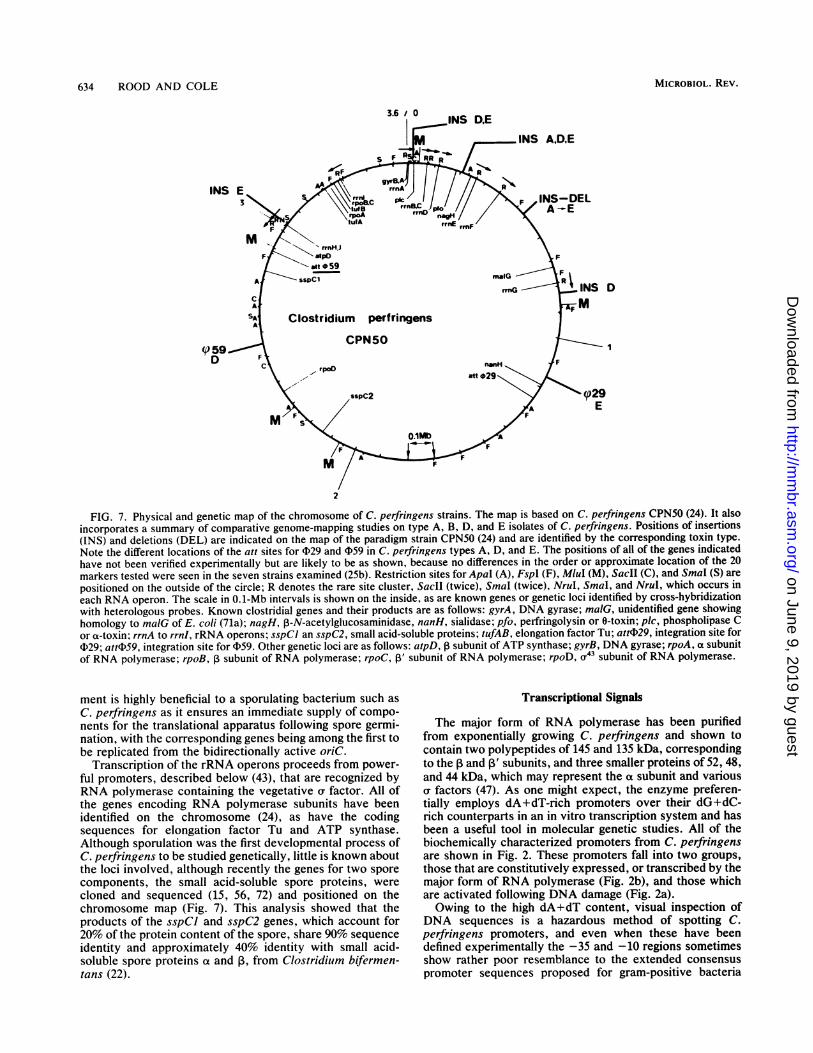
634 ROOD AND COLE
INS
()59-D
2
FIG. 7. Physical and genetic map of the chromosome of C. perfringens strains. The map is based on C. perfringens CPN50 (24). It alsoincorporates a summary of comparative genome-mapping studies on type A, B, D, and E isolates of C. perfringens. Positions of insertions(INS) and deletions (DEL) are indicated on the map of the paradigm strain CPN50 (24) and are identified by the corresponding toxin type.Note the different locations of the att sites for 4)29 and 059 in C. perfringens types A, D, and E. The positions of all of the genes indicatedhave not been verified experimentally but are likely to be as shown, because no differences in the order or approximate location of the 20markers tested were seen in the seven strains examined (25b). Restriction sites for Apal (A), FspI (F), MluI (M), SacII (C), and SmaI (S) arepositioned on the outside of the circle; R denotes the rare site cluster, SacII (twice), SmaI (twice), NruI, SmaI, and NruI, which occurs ineach RNA operon. The scale in 0.1-Mb intervals is shown on the inside, as are known genes or genetic loci identified by cross-hybridizationwith heterologous probes. Known clostridial genes and their products are as follows: gyrA, DNA gyrase; ma1G, unidentified gene showinghomology to maiG of E. coli (71a); nagH, P-N-acetylglucosaminidase, nanH, sialidase; pfo, perfringolysin or 0-toxin; plc, phospholipase Cor a-toxin; rrnA to rrnl, rRNA operons; sspCI an sspC2, small acid-soluble proteins; tufAB, elongation factor Tu; attj?29, integration site for(129; attw)59, integration site for 059. Other genetic loci are as follows: atpD, ,1 subunit of ATP synthase; gyrB, DNA gyrase; rpoA, a subunitof RNA polymerase; rpoB, 3 subunit of RNA polymerase; rpoC, p3' subunit of RNA polymerase; rpoD, (r" subunit of RNA polymerase.
D
I
ment is highly beneficial to a sporulating bacterium such asC. perfringens as it ensures an immediate supply of compo-nents for the translational apparatus following spore germi-nation, with the corresponding genes being among the first tobe replicated from the bidirectionally active oriC.
Transcription of the rRNA operons proceeds from power-ful promoters, described below (43), that are recognized byRNA polymerase containing the vegetative a factor. All ofthe genes encoding RNA polymerase subunits have beenidentified on the chromosome (24), as have the codingsequences for elongation factor Tu and ATP synthase.Although sporulation was the first developmental process ofC. perfringens to be studied genetically, little is known aboutthe loci involved, although recently the genes for two sporecomponents, the small acid-soluble spore proteins, werecloned and sequenced (15, 56, 72) and positioned on thechromosome map (Fig. 7). This analysis showed that theproducts of the sspCI and sspC2 genes, which account for20% of the protein content of the spore, share 90% sequenceidentity and approximately 40% identity with small acid-soluble spore proteins ot and P, from Clostridium bifermen-tans (22).
Transcriptional Signals
The major form of RNA polymerase has been purifiedfrom exponentially growing C. perfringens and shown tocontain two polypeptides of 145 and 135 kDa, correspondingto the a and I' subunits, and three smaller proteins of 52, 48,and 44 kDa, which may represent the a subunit and variousv factors (47). As one might expect, the enzyme preferen-tially employs dA+dT-rich promoters over their dG+dC-rich counterparts in an in vitro transcription system and hasbeen a useful tool in molecular genetic studies. All of thebiochemically characterized promoters from C. perfringensare shown in Fig. 2. These promoters fall into two groups,those that are constitutively expressed, or transcribed by themajor form of RNA polymerase (Fig. 2b), and those whichare activated following DNA damage (Fig. 2a).Owing to the high dA+dT content, visual inspection of
DNA sequences is a hazardous method of spotting C.perfringens promoters, and even when these have beendefined experimentally the -35 and -10 regions sometimesshow rather poor resemblance to the extended consensuspromoter sequences proposed for gram-positive bacteria
MICROBIOL. REV.
on June 9, 2019 by guesthttp://m
mbr.asm
.org/D
ownloaded from

GENETICS AND PATHOGENESIS OF C. PERFRINGENS 635
(63). It may be significant, in an organism with a bias againstdC and dG, that the frequency of guanosine residuesthroughout the promoter region, but especially in the 10 bppreceding the -35 hexamer, is strikingly high. Likewise, thetop strand of promoter regions is virtually free from cytosineresidues (Fig. 2b). Together with the classic sequence ele-ments, these features possibly contribute to promoter rec-ognition by RNA polymerase. On the basis of the sample ofpromoter sequences available, a consensus C. perfringenspromoter sequence has been deduced (Fig. 2). This agreeswell with the extended promoter consensus sequence pro-posed by Graves and Rabinowitz (63), although the -45region is less well conserved and the T-TG motif precedingthe -10 region is most prominent.The regulatory regions for four of the rRNA operons have
been studied in detail, and in three cases transcription occursfrom tandem promoters, as has been found in other bacteria(43). The proximal promoters contain canonical TTGACAsequences in the -35 region, whereas the distal promoterspossess no clearly defined -35 sequences. In all cases the-10 hexamers are preceded by one nucleotide by TG motifswhich are known to compensate for atypical -35 sequences(89, 150). Transcription of rrnH is from a single promoterwhich bears closer resemblance to the distal promoters ofthe other operons. All of the rrn control regions containsequence motifs which are very similar to those known tomediate the stringent response in B. subtilis (43), and it istherefore likely that a similar global control system exists inC. perfringens.Among the first promoters to be characterized were those
controlling two UV-inducible transcriptional units carried bythe bacteriocinogenic plasmid pIP404 (47, 48). Although oneof these five promoters (P4) was weakly constitutive (Fig.2b), the others were activated only after DNA damage.Three of the promoters are strikingly similar and have thesame -35 and -10 regions in TTTACA and CTTTTTAT,respectively. As none of these promoters is recognized bythe major form of RNA polymerase, this was taken asevidence that the SOS system is positively controlled in C.perfringens, unlike the situation in members of the familyEnterobacteriaceae, in which the LexA repressor shuts offexpression under normal physiological conditions (221).As C. perfringens is a sporulating bacillus, it is most
probable that the genes required for the different stages ofspore formation will be the targets of a complex regulatorynetwork involving a host of different a factors and activatormolecules, as found in B. subtilis (205). To date, the onlysporulation genes which have been cloned are the ssp genes,and indirect evidence suggests that these should be tran-scribed from forespore-specific promoters by RNA polymer-ase containing a a factor analogous to orG (72, 141). It isanticipated that as more genes are cloned and characterized,a wealth of detail about their various control systems willemerge.
Translational Signals
The translation initiation sites preceding C. perfringenscoding sequences all contain a ribosome-binding motif show-ing reasonable complementarity to the 3' end of the 16SrRNA (AAGGAGGA) (43). This site is invariably situated 6to 10 bases upstream of the initiation codon, which, in all butone of the cases thus far characterized, is AUG. Theexception is a GUG initiation codon used by a poorlyexpressed gene, rep, of pIP404 (45). By far the most strikingfeature of the mRNAs is their extraordinarily biased codon
usage, which is a reflection of the extremely high dA+dTcontent of the C. perfringens chromosome. The averagecodon usage for 21 genes of chromosomal and plasmid originis presented in Table 6 and compared with the correspondingdata from E. coli and B. subtilis. Codons composed of two ormore G - C base pairs have been selectively excluded fromC. perfringens genes by unknown evolutionary pressures,and 7 of the 61 sense codons are virtually never used. Forexample, the major codons for Arg in E. coli, CGU andCGC, are rarely found in C. perfringens mRNAs in whichAGA is predominant (143). This codon is rigorously avoidedby E. coli, and the availability of the cognate tRNA has beenidentified as a rate-limiting factor for expression in thisorganism. Other examples of codons for which the fre-quency of utilization varies between 1 and 2 orders ofmagnitude between E. coli and C. perfringens are UUA(Leu), AGU (Ser), ACA (Thr), GGA (Gly), and AUA (Ile). Itis striking that the codon usage in antibiotic resistance genesisolated from C. perfringens (catP, catQ, and ermBP) issignificantly less biased than that of chromosomal genes(data not shown), suggesting that horizontal transfer mayhave occurred.To date, no E. coli genes have been shown to be expressed
in C. perfringens, although many C. perfringens codingsequences have been cloned in the gram-negative host. Thediametrically opposed codon usage is likely to represent abarrier to expression, and to assess this possibility we haveanalyzed a number of C. perfringens genes for their rarecodon content, their context, and the degree of expressionobserved. In most cases the T7 expression system (410promoter and endogenous T7 RNA polymerase) was used todrive transcription in E. coli (206) and the amount of proteinsynthesis was determined in vitro (by labeling with [35S]me-thionine) or in vivo (Coomassie blue-stained proteins). Ascan be seen in Table 7, the number of rare codons did notlimit expression until the genes became very large. At thispoint no full-length protein was produced in vitro, althoughpeptides corresponding to translational intermediates couldbe detected (31a). Similar results have been obtained forexpression of other large clostridial genes such as thoseencoding the tetanus toxin (39) or the C. difficile cytotoxin A(87, 220). These findings are difficult to reconcile withexisting models for the limitation of gene expression in E.coli by rare codons, such as those proposed by Chen andInouye (26), who found a correlation between expressionlevels and the frequency of rare codons in the first 30codons, or Varenne and Lazdunski (219), who showed thatclusters of three or more rare codons slowed or stoppedtranslation. In the genes studied there is no correlationbetween rare-codon content, position, and context. Thegene length is the decisive factor (Table 7). The longer themRNA, the greater will be the probability that a ribosomewill stall at a rare codon. Presumably the A+T-rich mRNAthen succumbs to an exonucleolytic attack by RNases beforetranslation has the chance to recommence. These observa-tions should be borne in mind if one is considering expres-sion cloning of large C. perfringens genes in E. coli, as thechances of success are clearly much greater if the constructsare shuttled back into C. perfringens prior to screening.
TOXINS AND VIRULENCE FACTORS
As one might expect of an organism responsible for such avariety of pathological conditions, ranging from relativelymild food poisoning to life-endangering myonecrosis, C.perfringens produces at least 16 potential virulence factors
VOL. 55, 1991
on June 9, 2019 by guesthttp://m
mbr.asm
.org/D
ownloaded from

636 ROOD AND COLE
TABLE 6. Codon usage in C. perfringens genes and comparisonwith E. coli and B. subtilisa
Amino Usage (%) in:acid C. perfringens E. coli B. subtilis
Arg CGUCGCCGACGGAGAAGG
Leu CUU
CUC
CUACUGUUAUUG
Ser UCU
UCC
UCAUCGAGUAGC
Thr ACUACCACAACG
Pro CCUCCC
CCACCG
Ala GCUGCCGCAGCG
Gly GGUGGCGGAGGG
Val GUUGUCGUAGUG
Ile AUUAUCAUA
Lys AAAAAG
Asn AAUAAC
Gln CAACAG
His CAUCAC
Glu GAAGAG
9.31.52.11.5
72.213.4
14.40.19.23.0
66.56.8
21.53.4
33.00.6
33.38.2
41.86.6
47.04.6
38.11.1
57.43.4
39.98.1
48.33.7
27.45.2
59.67.8
48.23.0
44.93.9
39.95.2
54.9
73.826.2
79.820.2
85.414.6
75.624.4
78.921.1
58.135.02.33.21.20.3
8.66.61.8
69.15.88.2
26.525.68.3
11.46.5
21.6
23.850.65.9
19.7
9.06.0
19.965.1
27.918.822.930.5
47.840.84.66.8
37.512.922.926.8
37.362.20.5
76.723.3
24.275.8
26.673.4
38.961.1
73.426.6
25.217.59.1
11.127.79.4
26.19.86.3
21.822.413.6
24.412.018.710.010.724.1
TABLE 6-Continued
Amino Usage (%) in:acd Codon
acid C. perfringens E. coli B. subtilis
Asp GAU 87.4 51.0 63.8GAC 12.6 49.0 36.2
Tyr UAU 87.3 40.6 61.8UAC 12.7 59.4 38.2
Cys UGU 69.8 42.0 45.7UGC 30.2 58.0 54.3
Phe UUU 76.7 43.5 64.0UUC 23.3 56.5 36.0
Met AUG 100 100 100
Trp UGG 100 100 100
" Data for C. perfringens are compiled from the sequences of 21 knowngenes, of chromosomal or plasmid origin, comprising 6,239 codons. Data forE. coli and B. subtilis are from Ogasawara (143). Sources of the C. perfringenscodons were references 9, 25a, 45, 72, 120, 176, 196, 215, and 218.
14.8 (124, 125, 194), including 12 toxins (a to v), enterotoxin,14.1 non-a-8-0 hemolysin, neuraminidase (sialidase), and the43-3 vigorous metabolic activity of the organism. Many of these27.9 virulence factors simple hydrolytic secreted by
33.6 the organism as part of its saprophytic life style in the soil,9.8 where they are probably involved in the putrefaction pro-
19.1 cess. Their role in pathogenesis is therefore, at best, purely37.5 fortuitous. Only a selection of these factors are produced by
a given isolate of C. perfringens, and the identity of some27.5 may well be rather dubious. It is most probable that more20.0 factors of pathogenicity will be discovered as advances are27.1 made in the detailed genetic analysis of C. perfringens25.4 pathogenesis.
In the last few years there has been considerable progress25.4 in our understanding of the genetics, molecular biology, and
31.5 activity of some of the major pathogenic determinants, and13.5 these factors will now be addressed individually. Gene
cloning and expression in E. coli has been of great value to31.4 studies of the mode of action, as it has allowed toxin24.7 preparations to be obtained free from contaminating activi-24.5 ties and has enabled structure-function relationships to be19.4 elucidated.
5039.410.6
75.424.6
53.146.9
54.245.8
68.631.4
69.530.5
Continued
TABLE 7. Correlation between rare codons, gene size, andexpression in E. colia
% Rare Total no. ExpressionGene codons of codons In vitro In vivo
catP 9.9 212 + +orfF 23.7 279 + NTbnanH 18.3 382 + +plc 14.3 398 + +pfo 16.2 499 + +bcn 22.1 890 - -nagH 17.5 943tet 17.8 1,315
" Data taken from the literature and our unpublished observations: catP(196), orfF and nagH (25a), nanH (161), plc (175a, 176), pfo (214), bcn (44),tet, tetanus toxin (39).
b NT, not tested.
MICROBIOL. REV.
on June 9, 2019 by guesthttp://m
mbr.asm
.org/D
ownloaded from

GENETICS AND PATHOGENESIS OF C. PERFRINGENS 637
a-Toxin or Phospholipase C
Numerous observations suggest that the most importantvirulence factor produced by clostridial myonecrosis (gasgangrene)-causing isolates of C. perfringens is the a-toxin,the first toxin for which an enzymatic activity, phospholi-pase C, was demonstrated (103). The a-toxin is a trulymultifunctional metalloenzyme and possesses both phospho-lipase C and sphingomyelinase activities, which are respon-sible for the cytotoxicity, necrosis, and hemolysis observed(133). The 50% lethal dose for mice is only 0.75 hemolyticunits, and among the resulting symptoms are induction ofinflammatory responses, smooth muscle contraction, plate-let aggregation, shock, bradycardia, and greatly reducedmyocardial function culminating in death (201).
Cloning, mapping, and expression of plc. In 1989 thesuccessful cloning of the a-toxin gene, plc, was reported byno less than five different research groups. Four of thesegroups cloned the gene from the a-toxin-overproducingstrain NCTC 8237 (95, 145, 208, 213), whereas the fifth useda derivative of the heat-resistant strain NCTC 8798 (176).The various DNA sequences were virtually identical, andgenes homologous to plc have been detected in several otherclostridial species (210). The gene from C. bifermentans hasalso been cloned and characterized (213).
In all five toxigenic types (A to E) of C. perfringens, theplc gene is situated at the same locus, near the putativeorigin of replication and between two rRNA operons (25b).This location may well be of importance as this region of thechromosome is not only the first to be replicated but is alsoprobably the best-conserved segment of the genome, as itcontains many housekeeping genes.
plc is expressed from a constitutive promoter (Fig. 2b),which is also recognized by E. coli RNA polymerase (176).There are reports that growth medium composition affectsthe production of a-toxin (138, 139), suggesting the existenceof a possible control network which responds to the avail-ability of a given nutrient. Many bacterial virulence factorgenes are controlled as part of global regulons (130), andsome phospholipase C genes are induced in response tophosphate limitation (152). Phosphate does not appear tocontrol plc expression (31b, 139), and further work is re-quired to identify possible environmental regulatory signals.It is conceivable that the variable levels of a-toxin observedin different media are attributed to differences in the concen-tration of Zn2+ and Ca2", which are essential cofactors. Incontrast, expression of four toxin genes (0, K, X, andneuraminidase) is believed to be controlled by a singleregulatory locus (77), which may correspond to the pfoRgene (189).
Structure-function relationships. Reasonable levels ofa-toxin production have been obtained from the cloned plcgene in E. coli, where the enzyme is correctly processed andsecreted to the periplasm (95, 145, 208). Purified a-toxinfrom this host has biochemical properties virtually indistin-guishable from those obtained with toxin from C. perfrin-gens, except for a difference in its pl (5.6 in E. coli; 5.48 inC. perfringens). One major advantage of using recombinanta-toxin in biological studies is the complete absence of thecontaminating clostridial proteins which have plagued C.perfringens toxin research (134, 135). In this way it wasdemonstrated unambiguously (176) that the plc gene productpossessed both the phospholipase C and sphingomyelinaseactivities which had previously been thought to be associ-ated with a-toxin (136).The availability of the primary structure of a-toxin, de-
duced from the nucleotide sequence of the plc gene, led to amajor breakthrough in our understanding of its functionalorganization. Comparison of the amino acid sequences of the370-residue mature form of a-toxin and the 245-residuephospholipase C from Bacillus cereus revealed extensivehomologies and the presence of 65 identical equivalancedamino acid residues. In Fig. 8 the amino acid sequences oftwo a-toxins from different strains of C. perfringens arecompared with the sequences of two phospholipase C en-zymes from different isolates of B. cereus (52, 85). Thecrystal structure of one of the B. cereus enzymes, PC-PLC,has been deduced at a resolution of 0.16 nm (75), and this hasbeen of great value in interpreting the organization of theclostridial a-toxin protein.The a-toxin is clearly composed of a complete PC-PLC
module fused to a COOH-terminal domain of 120 amino acidresidues. It is apparent that all of the amino acids involved incoordinating the three zinc atoms found at the active site ofPC-PLC (75) have obvious counterparts in a-toxin (Fig. 8).These include five histidine residues, and experimental evi-dence showing the importance of histidinyl groups fora-tQxin activity has been obtained (209).The crystallographic structure of PC-PLC (75) shows the
enzyme to comprise 10 a-helical segments linked by 1-turnsof variable length. Structural predictions (176) suggest a verysimilar organization for a-toxin, with the additional COOH-terminal domain predicted to contain a further four a-heli-ces. A number of observations indicated that this supple-mentary domain could contribute to the sphingomyelinaseactivity of a-toxin, but, before discussing them, it is impor-tant to summarize the situation in B. cereus. In addition toPC-PLC, B. cereus produces a sphingomyelinase (85, 229),and the two enzymes act synergistically as a hemolysin (52).The genes for both enzymes, cerA and cerB, have beencloned and sequenced and appear to make up an operon inwhich the PC-PLC gene, cerA, is the promoter-proximalcistron. In C. perfringens both enzyme activities are unitedin a single hemolytic toxin, suggesting that the present-dayplc gene is the product of a distant gene fusion event. If so,the sequence which originated from cerB has divergedconsiderably more than the cerA module, as the 120-residueCOOH-terminal domain of a-toxin shares no significantamino acid sequence homology with the 304-residue sphin-gomyelinase from B. cereus. However, predictive studiessuggest that both polypeptides have a predominantly a-heli-cal secondary structure and similar amphiphilic profiles(175a).To test the hypothesis for a two-domain organization of
a-toxin, two studies were performed independently. Titballand coworkers (208a) constructed a truncated form of plc,which encoded the domain homologous to PC-PLC, andcharacterized the biological activities of its product. Asexpected, the 249-residue protein retained phospholipase Cactivity but was unable to hydrolyze sphingomyelin orprovoke hemolysis. Furthermore, the truncated protein ex-hibited greatly reduced toxicity. Some of these results wereconfirmed by workers in one of our laboratories (175b), whoconstructed a series of gene fusions between plc and thegenes encoding E. coli alkaline phosphatase or 1-lactamase.Hybrid proteins consisting of the 354 NH2-terminal residuesof a-toxin fused to the reporter enzymes were inactive inhemolysis or sphingomyelin hydrolysis and displayed greatlyreduced phospholipase activity. All the available evidenceindicates that a-toxin does indeed have a two-domain orga-nization. However, it is not yet clear whether the proteinpossesses two distinct active sites, one in each domain, or
VOL. 55, 1991
on June 9, 2019 by guesthttp://m
mbr.asm
.org/D
ownloaded from
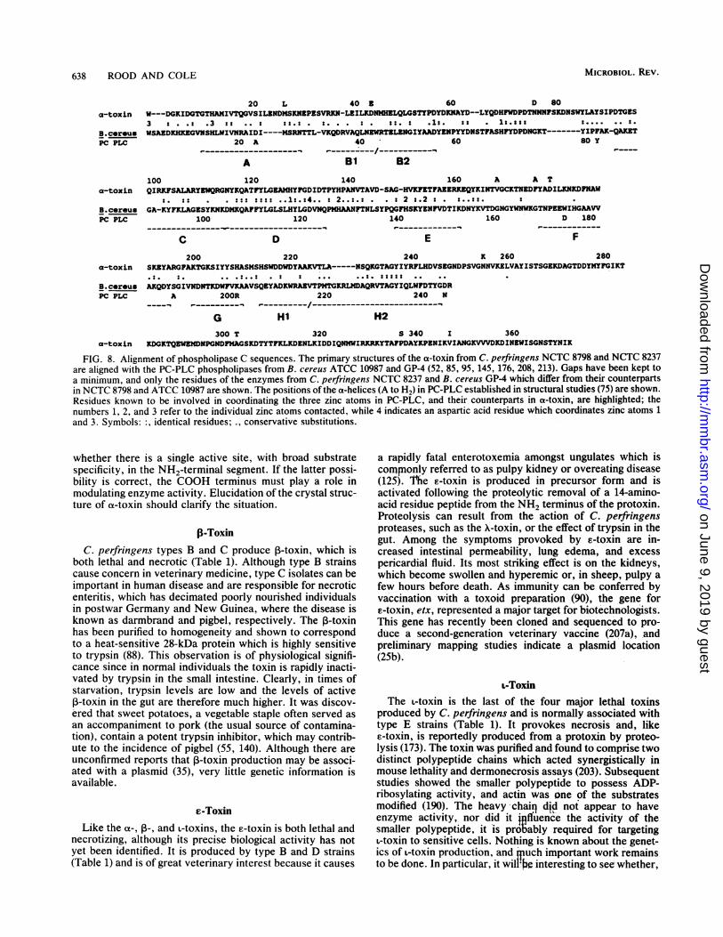
638 ROOD AND COLE
20 La-toxln
B.cereusPC PLC
40 Z 60 D 80
3 : . .: .3 :a .. : :a.: . :. . . : . :2 s .1:. . 1l.222
20 A
A
40
81 B2
60
:. . . . * . .-----w-̂ - ---
8o Y
100 120 140 160 A A TQIRKFSALARYEWQRGNYKQATFYLGEAMHYFGDIDTPYHPANVTAVD-SAG-HVKFTFAEZRKEQYKINTVGCKTNEDFYADILKNKDFNAW
s.:: . . ::: :::: ..1:.:4.. : 2..:.: . . s 2 s.2 : . :..::. :GA-KYFKLAGZSYKNKDMKQAFFYLGLSLHYLGDVNQPMHAANFTNLSYPQGFHSKYZNFVDTIKDNYKVTDGNGYWNWKGTNPEEWIHGAAW
100 120 140 160 D 180_ _ _ _ _ _-_ _ e _ _ _ _ _ _ _ _ _ _-_ _ _ _ _ _-_ _ _ ___----_ ___-_ _ _- - -- - ---%0 - - - -- -r - - - -
C200
D
220 240
E
K 260
F
280SKEYARGFAKTGKSIYYSHASMSHSWDDWDYAAKVTLA-----NSQKGTAGYIYRFLHDVSKGNDPSVGNNVKELVAYISTSGEKDAGTDDYMYFGIKT
. . .:..: . ..
AKODYSGIVNDNTXDWFVKAAVSQEYADKWRAZVTPMTGKRLMDAQRVTAGYIQLWFDTYGDRA 200R 220
._____________-.-----_.-.-I------
240 N
G300 T
HI320
H2
S 340 x 360a-toxin KDGKTQEWEMDNPGNDFMAGSKDTYTFKLKDENLXIDDIQNMWIRKRKYTAFPDAYKPENIKVIANGKVVVDKDINEWISGNSTYNIK
FIG. 8. Alignment of phospholipase C sequences. The primary structures of the a-toxin from C. perfringens NCTC 8798 and NCTC 8237are aligned with the PC-PLC phospholipases from B. cereus ATCC 10987 and GP-4 (52, 85, 95, 145, 176, 208, 213). Gaps have been kept toa minimum, and only the residues of the enzymes from C. perfringens NCTC 8237 and B. cereus GP-4 which differ from their counterpartsin NCTC 8798 and ATCC 10987 are shown. The positions of the a-helices (A to H2) in PC-PLC established in structural studies (75) are shown.Residues known to be involved in coordinating the three zinc atoms in PC-PLC, and their counterparts in a-toxin, are highlighted; thenumbers 1, 2, and 3 refer to the individual zinc atoms contacted, while 4 indicates an aspartic acid residue which coordinates zinc atoms 1and 3. Symbols: :, identical residues; ., conservative substitutions.
whether there is a single active site, with broad substratespecificity, in the NH2-terminal segment. If the latter possi-bility is correct, the COOH terminus must play a role inmodulating enzyme activity. Elucidation of the crystal struc-ture of a-toxin should clarify the situation.
j(-ToxinC. perfringens types B and C produce 1-toxin, which is
both lethal and necrotic (Table 1). Although type B strainscause concern in veterinary medicine, type C isolates can beimportant in human disease and are responsible for necroticenteritis, which has decimated poorly nourished individualsin postwar Germany and New Guinea, where the disease isknown as darmbrand and pigbel, respectively. The ,B-toxinhas been purified to homogeneity and shown to correspondto a heat-sensitive 28-kDa protein which is highly sensitiveto trypsin (88). This observation is of physiological signifi-cance since in normal individuals the toxin is rapidly inacti-vated by trypsin in the small intestine. Clearly, in times ofstarvation, trypsin levels are low and the levels of active1-toxin in the gut are therefore much higher. It was discov-ered that sweet potatoes, a vegetable staple often served as
an accompaniment to pork (the usual source of contamina-tion), contain a potent trypsin inhibitor, which may contrib-ute to the incidence of pigbel (55, 140). Although there are
unconfirmed reports that ,3-toxin production may be associ-ated with a plasmid (35), very little genetic information isavailable.
e-ToxinLike the a-, 13-, and L-toxins, the E-toxin is both lethal and
necrotizing, although its precise biological activity has notyet been identified. It is produced by type B and D strains(Table 1) and is of great veterinary interest because it causes
a rapidly fatal enterotoxemia amongst ungulates which iscomnnonly referred to as pulpy kidney or overeating disease(125). 'Me c-toxin is produced in precursor form and isactivated following the proteolytic removal of a 14-amino-acid residue peptide from the NH2 terminus of the protoxin.Proteolysis can result from the action of C. perfringensproteases, such as the X-toxin, or the effect of trypsin in thegut. Among the symptoms provoked by c-toxin are in-creased intestinal permeability, lung edema, and excesspericardial fluid. Its most striking effect is on the kidneys,which become swollen and hyperemic or, in sheep, pulpy afew hours before death. As immunity can be conferred byvaccination with a toxoid preparation (90), the gene forc-toxin, etx, represented a major target for biotechnologists.This gene has recently been cloned and sequenced to pro-duce a second-generation veterinary vaccine (207a), andpreliminary mapping studies indicate a plasmid location(25b).
L-ToxinThe L-toxin is the last of the four major lethal toxins
produced by C. perfringens and is normally'associated withtype E strains (Table 1). It provokes necrosis and, likee-toxin, is reportedly produced from a protoxin by proteo-lysis (173). The toxin was purified and found to'comprise twodistinct polypeptide chains which acted synergistically inmouse lethality and dermonecrosis assays (203). Subsequentstudies showed the smaller polypeptide to possess ADP-ribosylating activity, and actin was one of the substratesmodified (190). The heavy chain did not appear to haveenzyme activity, nor did it fluence the activity of thesmaller polypeptide, it 'is probgably required for targetingl-toxin to sensitive cells. Nothing is known about the genet-ics of l-toxin production, and qpuch important work remains
to be done. In particular, it will'f. interesting to see whether,
a-toxin
B.cereusPC PLC
a-toxin
B.cereusPC PLC
W---DGKIDGTGTHAMIVTQGVSILZNDMSKNZPZSVRKN-LEILKDNMHZLQLGSTYPDYDKNAYD--LYQDHFWDPDTNNNFSKDNSWYLAYSIPDTGES
WSAZDKHKZGVNSHLWIVNRRIDI----MSRNTTL-VKQDRVAQLNZWRTZLZNGIYARDYZNPYYDNSTFASHFYDPDNGKT-------YIPFAK-QAKZT
MICROBIOL. REV.
on June 9, 2019 by guesthttp://m
mbr.asm
.org/D
ownloaded from

GENETICS AND PATHOGENESIS OF C. PERFRINGENS 639
like diphtheria toxin, both components are encoded by thesame gene.
0-Toxin
All five types of C. perfringens produce a lethal hemoly-sin, 0-toxin, which is also known as 0-hemolysin, perfringo-lysin 0, or the thiol-activated cytolysin. The structural gene,pfoA, has recently been cloned, sequenced, and mapped (24,189, 214, 215), and related genes have been detected in otherclostridial species (213a). When introduced into E. coli, a Xphage bearing the pfoA gene exhibited marked instabilityand, although an intact coding sequence was recovered, thepromoter and regulatory region were missing (214, 215).Consequently, little could be deduced about possible controlmechanisms governing expression. However, recently thepfoA gene, and the entire region upstream of this gene, werecloned onto a low-copy-number plasmid (189). Subcloningand sequence studies showed that approximately 1.6 kbupstream ofpfoA there was a second gene, designated pfoR,which acted to enhance the expression ofpfoA. The deducedamino acid sequence of the pfoR gene product had many ofthe motifs typical of DNA-binding proteins. It appears thatthe PfoR protein acts as a cis-dominant positive activator ofpfoA gene expression (189). To our knowledge, this is firsttime that researchers have characterized a gene that isdirectly involved in the regulation of a structural generesponsible for the production of an extracellular toxin in C.perfringens.The primary sequence of 0-toxin, deduced from the pfoA
gene sequence, revealed a 494-residue preprotein fromwhich a 28-residue signal peptide is removed on secretion tothe medium. Mature 0-toxin has a predicted molecularweight of 52,469 and, when purified from C. perfringens orE. coli, appears as a 54,000-Da species (214, 215).
0-toxin is a member of a family of thiol-activated cyto-lysins produced by a diverse group of gram-positive bacteriaincluding the Bacillus, Streptococcus, Clostridium, and Lis-teria genera. These cytolysins use cholesterol as a receptorand form large pores in target cell membranes. They arereversibly inactivated by oxidation. To date, the primarystructures of five of these cytolysins (alveolysin, listerioly-sin, streptolysin, pneumolysin, and perfringolysin) havebeen deduced from a cloning and sequencing approach.Despite variations in size, there are extensive regions ofsequence identity, some of which correspond to sharedepitopes (51). An interesting stretch of conserved sequence,an undecapeptide, ECTGLAWEWWR, encompassing theunique cysteine residue, is found near the COOH terminus.For listeriolysin, replacement of the cysteine residue bysite-directed mutagenesis led to reduced activity, whereassubstitution of the tryptophan residues resulted in completeinactivation and concomitant attenuation of Listeria mono-cytogenes. This study clearly demonstrated the essentialrole of this domain in cytolysis (128).The 0-toxin plays a role in the tissue necrosis associated
with C. perfringens gas gangrene and is responsible for thedepletion of polymorphonuclear leukocytes in the affectedzone. Low concentrations of 0-toxin cause altered polymor-phonuclear leukocyte morphology, metabolism, and migra-tion (200).
Neuraminidase
All C. perfringens types produce a neuraminidase, orsialidase, which cleaves acylneuraminic acid residues from
glycoproteins, glycolipids, and oligosaccharides and mayplay a role in disease by destroying receptor molecules oncell surfaces or by disrupting connective tissue (125). Inearly studies, three distinct forms of the enzyme (64, 105,and 310 kDa) were found by gel filtration (170). The largertwo of these enzyme forms appeared to correspond todifferent multimers and appeared to be associated bothphysically and genetically with hemagglutinins (169, 171). Agene, nanH, encoding neuraminidase activity has beencloned (161), but on sequencing it was shown to correspondto a previously undetected minor form (160). The nanH geneproduct is a cytoplasmic protein of 42,770 kDa which sharessome sequence homology with other neuraminidases ofbacterial and viral origin but differs from the major form ofthe enzyme with respect to its size and activity profile (160).Interestingly, when the highly homologous nanH gene fromClostridium sordellii was cloned, DNA sequence analysisshowed that the enzyme was synthesized as a preproteinwhich is probably processed by a lipoprotein-specific signalpeptidase (174). The putative processing site includes thecysteine residue which is predicted to be covalently modifiedby attachment of lipids during maturation. Presumably, thelipid-modified enzyme is anchored in the cytoplasmic mem-brane. Although the second amino acid residue of the C.perfringens NanH protein is a cysteine, to which the lipidshould be attached, no signal peptide is predicted by theDNA sequence, suggesting that the gene may have under-gone a rearrangement. It may be significant in this respectthat a lysogenic bacteriophage has integrated into the chro-mosome near nanH (24, 25). Clearly, further work is re-quired to clarify the nature and number of neuraminidasespresent in C. perfringens and to evaluate their role inpathogenesis.
,u-Toxin
The ,u-toxin is a hyaluronidase which degrades hyaluronicacid, an important constituent of connective tissue, to re-lease glucosamine (125). It is a common contaminant incommercial preparations of neuraminidase (27). Recently,the nagH gene was cloned and sequenced (25a) and shown toencode a P-N-acetylglucosaminidase which could corre-spond to ,u-toxin. The gene is present in all C. perfringensisolates examined, and its product is a 97,000-Da secretedprotein. Truncated derivatives of nagH, containing as littleas 421 of the 943 codons, encode enzymatically activeproducts; this suggests that the enzyme may consist ofseveral domains. Although definitive identification is stillrequired, it is conceivable that the primary role of the NagHprotein/,l-toxin is in cell wall biosynthesis or autolysis andthat its action as a virulence factor is secondary.
Other Toxins
The remaining toxins produced by different C. perfringensstrains are 5-toxin, a hemolysin; K-toxin, a collagenase;X-toxin, a protease; v-toxin, a nuclease;,and the eta andgamma toxins, whose existence is dubious. Apart from theobservation that the X-toxin, or caseinase, gene is carried ona small plasmid in one type B strain of C. perfringens (14),nothing is known about their genetics. An extracellularendonuclease from one strain of C. perfringens has beencharacterized recently, but it is unclear whether this enzymeis the v-toxin (140a). There has been little biochemicalprogress on the other toxins since the last comprehensivereview (125).
VOL. 55, 1991
on June 9, 2019 by guesthttp://m
mbr.asm
.org/D
ownloaded from

640 ROOD AND COLE
TABLE 8. Principal features of the C. perfringens enterotoxinaCharacteristics of enterotoxinProperty
Primary gene product ................. 320 amino acids, mol wt 35,391Activated enterotoxin ................. 24 amino acids removed from NH2 terminus by trypsin (in vitro); new mol wt 31,472pl ................. 4.3Stokes radius ................. 2.6 nmHeat stability ................. Unstable inactivated -53°CSDS binding ................. Anomalous, 0.39 g/g of proteinSolubility ................. Low (3.94 ± 0.22 mg/ml at pH 7.0 and 25°C)Free thiol groups ................. OneDomain structure binding................. Two domains, N terminal (cytotoxic) and C terminal (receptor)Biological activity ................. Pore formation; altered permeability, ion flux, inhibition of macromolecular synthesis; Ca2+-
mediated cytoskeletal disintegration; cell lysisa According to Granum, McClane, and coworkers (55, 68, 76, 121-123, 126).
ENTEROTOXIN
Of the major virulence factors produced by C. perfrin-gens, the enterotoxin is one of the best studied. There is anenormous literature dealing with the enterotoxin, and it isbeyond the scope of this article to summarize all the avail-able knowledge. Instead, the molecular genetic aspects willbe given priority, and the reader should refer to severalrecent comprehensive reviews for additional details (55, 59,121, 125).
C. perfringens is the etiological agent of two very differenttypes of food poisoning affecting humans and is the thirdmost important source of food-borne disease after S. aureus
and Salmonella species (29, 86). Some type A strains areassociated with a mild form of food poisoning, resulting fromenterotoxin production, whereas type C strains are respon-sible for necrotic enteritis, a rare but often deadly intestinaldisease provoked by production of the ,-toxin.
C. perfringens food poisoning occurs following ingestionof heavily contaminated food, usually meat products con-taining more than 106 CFU/g. C. perfringens cells sporulatein the small intestine and concomitantly generate largequantities of enterotoxin. This can result, 12 to 24 h later, inintense nausea, diarrhea, and, more rarely, vomiting, withthe occasional fatality amongst the elderly and debilitated.Infected individuals generally recover rapidly and are un-aware of the origin of their malaise. In addition, it has alsobeen suggested that C. perfringens enterotoxin may beinvolved in antibiotic-associated diarrhea (16, 94).
Biochemical Properties of Enterotoxin
The purification of large quantities of the enterotoxin (61,126) had important consequences for the elucidation of itsmode of action and functional organization, the determina-tion of its amino acid sequence, and ultimately the cloning ofthe structural gene. The principal properties of the entero-toxin are summarised in Table 8. It is clear that the entero-toxin accumulates in large quantities intracellularly, where,owing to its limited solubility, it can form inclusion bodies(102). Following lysis of the bacteria in the gut, the toxin isliberated and, like the E-toxin, processed to a more activeform by trypsin '55, 157).The current model for enterotoxin-mediated cytotoxicity
involves two distinct phases which differ in their dependenceon Ca2+ ions (121-123). In the initial, Ca2+-dependent step,enterotoxin inserts into the brush border membrane ofepithelial cells, where it forms a complex with a two-subunitprotein receptor consisting of 50- and 70-kDa polypeptides(225). Membrane insertion is probably accompanied by a
radical conformational change (58), whereas complex forma-tion is strongly correlated with cytotoxicity (122). Intestinalcells thus affected display altered permeability and loselow-molecular-weight metabolites and ions. These earlylethal events are Ca2+ independent and have pleiotropicinhibitory effects on the synthesis of macromolecules (76).However, the presence of Ca2+ provokes the second phaseof enterotoxin cytotoxicity, and this involves morphologicdamage and more extensive permeability alterations, result-ing in the, loss of larger solutes. One possible consequence ofthe increased influx of Ca2+ is cytoskeletal collapse, whichwill culminate in cell lysis (57, 123). C. perfringens entero-toxin is quite distinct from other intestinally active toxinsand may be considered as the prototype for a new class ofmembrane-seeking cytotoxins (121).The availability of large amounts of highly purified entero-
toxin allowed the amino acid composition and sequence tobe determined (156), and this not only provided importantbiochemical information but also facilitated the subsequentcloning of the gene. Curiously, other workers (101) reportedthe purification of a second "enterotoxin" from the coatlessspore mutant, 8-6. As this strain is known to producecopious amounts of bona fide enterotoxin, the findings of thisstudy should be interpreted with caution.
Molecular Genetics of EnterotoxinCloning of the enterotoxin structural gene. Two research
groups have independently designed oligonucleotide probes,based on the protein sequence of the enterotoxin, and usedthem to screen gene banks (83, 218). Both groups identifiedclones which contained fragments of the enterotoxin gene,cpe, and in one case these overlapped sufficiently to permitthe elucidation of the complete nucleotide sequence (218).Other workers used antibodies raised against enterotoxin toscreen a C. perfringens expression library prepared in Xgtlland isolated a clone producing an immunologically cross-reacting hybrid protein (69). Despite being isolated by threedifferent research groups, an intact cpe gene has not yet beencloned, and, as a consequence, no data are available con-cerning expression in any other hosts. Furthermore, nothingis known about the transcriptional control of cpe, and thepromoter has yet to be identified. This is important as thereare several reports which suggest low-level expression in theabsence of sporulation (53, 60), although it is clear that thelevels of enterotoxin production are increased considerablyfollowing the onset of sporulation.
Structure-function relationships. Comparison of the de-duced amino acid sequence with that determined experimen-tally (156, 218) revealed reasonably good agreement, with
MICROBIOL. REV.
on June 9, 2019 by guesthttp://m
mbr.asm
.org/D
ownloaded from

GENETICS AND PATHOGENESIS OF C. PERFRINGENS 641
the exception of a small peptide which had proved refractoryto protein sequence analysis. The native enterotoxin thuscontains all 20 amino acids and comprises 320 residues togive a molecular weight of 35,391 (218) (Table 8).The current structural model for enterotoxin suggests a
two-domain organization with the N-terminal region re-quired for cytotoxicity while the C terminus interacts withcell membrane receptors, one of which is a 50-kDa proteinspecies (225). Results of a UV differential spectroscopystudy of purified enterotoxin support this model (62), andrecent independent confirmation was obtained in studieswith a recombinant C-terminal fragment (121). Hanna andMcClane (68) convincingly demonstrated that residues 171to 320 of the enterotoxin were sufficient for receptor recog-nition and binding. Although this interaction was irrevers-ible, it was not accompanied by membrane insertion or anyof the biological activities associated with enterotoxin. Thissuggests that the NH2-terminal domain is responsible forcytotoxicity. It is clear that our understanding of enterotoxinfunction and molecular organization will benefit from furtherstructural studies.Gene location and molecular epidemiology. The cloned cpe
gene has proved to be a useful tool in molecular epidemio-logical studies of numerous C. perfringens isolates fromvarious sources. In an initial survey in which specific oligo-nucleotide probes were used to screen a collection of 98randomly chosen strains isolated from the feces of farmanimals, only 6 isolates that hybridized with the probe weredetected (218). This approach was later extended to include245 strains, isolated from food and feces, associated with 186food-poisoning outbreaks of known C. perfringens etiology(217). In this study 59% of the isolates were cpe+ byhybridization, and the authors concluded that the remainderwere probably part of the normal intestinal flora and hadbeen mistakenly identified as the cause of the food poison-ing. In addition, comparative studies suggested that hybrid-ization with a cpe probe was a more reliable method thanenzyme-linked immunosorbent assay (146) for detectingpotentially enterotoxigenic C. perfringens strains (217). Thevalue of this approach was also demonstrated in studies onan outbreak of porcine diarrhea due to a type A C. perfrin-gens strain. In this example the use of a probe clearly ruledout enterotoxin production as the cause of the disease (216).By using a similar approach, the relationship between
enterotoxigenicity and plasmid profile was examined, and itwas found that the cpe gene was not associated with a widelydistributed series of small plasmids (149). The results sug-gested a chromosomal location for this gene. By means ofthe genome mapping strategy outlined above, as well asintragenic probes, the cpe gene has been located to a singlelocus between the rrnF and maiG genes on the chromosomeof the enterotoxigenic type A strain 8-6, a derivative ofNCTC 8798 (175a). It has long been known that enterotoxinproduction is not confined to type A strains but also occursin some type C and D isolates (55), and these combinedobservations suggest that the cpe gene may be located on amobile genetic element. Clearly, this requires further inves-tigation and will yield some interesting results. Finally, it isworth repeating that despite the relatively limited distribu-tion of the cpe gene, C. perfringens is a major cause offood-borne disease (29, 86).
Enterotoxin and Sporulation: Facts and Myths
Now that it is clear that there is a single cpe gene, with arelatively narrow distribution, it is easier to reevaluate the
relationship between enterotoxin production and sporulationand to reconcile some contradictory data. It is often mistak-enly believed that enterotoxin is a component of the sporecoat of C. perfringens (41) or even that it corresponds toexcess, unincorporated spore coat material (93); these ob-servations are the cause of much confusion. As virtually allEnt- strains are capable of sporulation, it is evident that theenterotoxin is not an essential spore component.
In a thorough genetic study, Duncan et al. (36) isolated agroup of sporulation mutants of strain NCTC 8798, whichcontains the cpe+ gene, and evaluated enterotoxin produc-tion. They found that all spo mutants were defective inexperimental enterotoxemia, whereas some of the oligospo-rogenous mutants, and mutants blocked in stages III, IV,and V of sporulation, produced reduced amounts of entero-toxin. This was rightly interpreted as indicating a clear-cutrelationship between sporulation and enterotoxin produc-tion. However, the low levels detected in known Ent'strains growing in batch (60) or chemostat (53) culturessuggest that the cpe gene may be expressed constitutivelyfrom a weak promoter during the exponential phase ofvegetative growth. Presumably the levels of enterotoxin thusproduced are below the levels of detection of the techniquesthat were used previously (36) with the Sp- mutants ofNCTC 8798.
Following the onset of sporulation at stage II, enterotoxinsynthesis is induced and, according to Granum et al. (60)precedes the appearance of heat-resistant spores by severalhours. Given that mutants blocked in the later stages ofsporulation (III to V) produce the toxin, it seems likely thatits gene is transcribed from a developmentally regulatedpromoter which requires the product of an unknown (stage Ior II) spo gene(s) for activation. Thus the cpe gene may playno role in sporulation per se but simply be subject tocoordinate control in response to an environmental signal.Such regulation is common place in the related organism B.subtilis, in which, for instance, the spoOA gene controlscompetence development as well as sporulation initiation(204). The availability of the cloned gene will allow thishypothesis to be tested and should shed further light on thecomplement of regulatory factors available in C. perfrin-gens.Although some workers have convincingly showed the
enterotoxin to be localized in the cytoplasm, by immuno-staining of thin sections of sporulating cells (172, 222), othersclaim that it is actually a component of the spore coat (41).Solubilization and manipulation of spore proteins are noto-riously difficult and, together with the tendency of theenterotoxin to aggregate, render localization studies hazard-ous. Recently, a Western immunoblotting analysis of sporespurified from Ent' and Ent- C. perfringens strains clearlyshowed that enterotoxin was not a major structural compo-nent of the spore coat but may have been trapped betweenthe core and coat layers (175). However, a 48-kDa proteinwhich strongly cross-reacted with antiserum raised againstenterotoxin was shown to be a minor component of thespores irrespective of the enterotoxicity of the parentstrains. As antibodies to this spore coat constituent alsorecognize enterotoxin, it would appear that the two polypep-tides have common epitopes. Although the functional signif-icance of this is obscure, it does provide a logical explana-tion for the results of localization studies publishedpreviously (41).
In conclusion, high levels of enterotoxin are produced inthe cytoplasm after activation of the cpe gene by transcrip-tional factors which also control sporulation genes. Accu-
VOL. 55, 1991
on June 9, 2019 by guesthttp://m
mbr.asm
.org/D
ownloaded from

642 ROOD AND COLE
mulation of large quantities of enterotoxin can result inaggregation, inclusion body formation, and trapping of smallamounts during spore biogenesis.
FUTURE PERSPECTIVESThe principal aim of this review was to summarize recent
advances in our knowledge and understanding of the molec-ular genetics and pathogenicity of C. perfringens. It shouldbe clear to the reader by now that the development of genetictools and systems during the last 5 years has prepared theway for a detailed analysis of the diseases associated withthis obligate anaerobe. Now that we have at our disposal thegenes for several virulence factors, as well as the means ofreintroducing them into C. perfringens, it should be rela-tively simple to construct a series of isogenic mutants bygene replacement. Alterations in the pathogenic spectrumcould then be evaluated by using a suitable animal model.
It is conceivable that after inactivation of the genes for theestablished virulence determinants, additional factors whichcontribute to pathogenesis will be discovered. In this respectit would be wise to follow closely the developments withrelated pathogens such as B. cereus, Streptococcus pneumo-
niae, and Listeria monocytogenes, as these could providefresh insight and new leads. It is interesting that in L.monocytogenes a phosphatidylinositol phospholipase hasrecently been discovered which allows the organism toescape from one cell and infect another (31c). A similarenzyme is produced by Clostridium novyi, and it is quitepossible that some C. perfringens strains also possess thisactivity. Another probable virulence determinant which hasnot yet been examined is capsule production. Many C.perfringens strains isolated from individuals with infectionsare encapsulated, and this clearly enables the organism toresist or escape phagocytosis. Although this property isoften lost after repeated passage in the laboratory, it shouldbe possible, by transposon mutagenesis, to identify thegenes involved in capsule biosynthesis.One aspect which has not been treated here is the biotech-
nological potential of C. perfringens and the development ofattenuated, or avirulent, mutants that could render theorganism more attractive to industry. C. perfringens hasmany properties of industrial interest, among the more
obvious being its short doubling time (ca. 20 min), highgrowth yield, potential to secrete proteins of interest, abilityto grow anaerobically (thus reducing fermentation costs),and relative aerotolerance (which enables genetic manipula-tion to be performed without recourse to anaerobic incuba-tion chambers). One area in which C. perfringens couldmake a valuable biotechnological contribution is as a host forhigh-level expression of large genes rich in dA+dT. Amongthe obvious candidates are other clostridial genes such as
those encoding the tetanus toxin, the botulism toxins, or theC. difficile cytotoxins, as well as those of unrelated organ-
isms such as Plasmodium spp. or trypanosomes. Anotheraspect which could be developed in the 1990s is the con-
struction, by reverse genetics, of strains suitable for use as
live veterinary vaccines.
ACKNOWLEDGMENTSWe thank the members of the C. perfringens community for
providing preprints and unpublished information. Special thanks tothe Parisien Gangrene Gang (Bruno Canard, Thierry Garnier, Anne-Marie Fargues, Hafida Fsihi, and Brigitte Saint-Joanis) and theMonash Clostridial Group (Dave Berryman, Trudi Bannam, PaulScott, and Joan Sloan) for their cheerful and enthusiastic contribu-tions.
Financial support was provided by Institut Pasteur, the InstitutNational de la Sante et de la Recherche Medicale (CRE 883003), theAustralian Research Council, the National Health and MedicalResearch Council, and the Commonwealth Serum Laboratories.
REFERENCES1. Abraham, L. J., D. I. Berryman, and J. I. Rood. 1988.
Hybridization analysis of the class P tetracycline resistancedeterminant from the Clostridium perfringens R-plasmid,pCW3. Plasmid 19:113-120.
2. Abraham, L. J., and J. I. Rood. 1985. Cloning and analysis ofthe Clostridium perfringens tetracycline resistance plasmid,pCW3. Plasmid 13:155-162.
3. Abraham, L. J., and J. I. Rood. 1985. Molecular analysis oftransferable tetracycline resistance plasmids from Clostridiumperfringens. J. Bacteriol. 161:636-640.
4. Abraham, L. J., and J. I. Rood. 1987. Identification of Tn4451and Tn4452, chloramphenicol resistance transposons fromClostridium perfringens. J. Bacteriol. 169:1579-1584.
5. Abraham, L. J., and J. I. Rood. 1988. The Clostridium perfrin-gens chloramphenicol resistance transposon Tn4451 excisesprecisely in Escherichia coli. Plasmid 19:164-168.
6. Abraham, L. J., A. J. Wales, and J. I. Rood. 1985. Worldwidedistribution of the conjugative Clostridium perfringens tetra-cycline resistance plasmid, pCW3. Plasmid 14:37-46.
6a.Allen, S., and H. Blaschek. Personal communication.7. Allen, S. P., and H. P. Blaschek. 1988. Electroporation-induced
transformation of intact cells of Clostridium perfringens. Appl.Environ. Microbiol. 54:2322-2324.
8. Allen, S. P., and H. P. Blaschek. 1990. Factors involved in theelectroporation-induced transformation of Clostridium perfrin-gens. FEMS Microbiol. Lett. 70:217-220.
8a.Bannam, T., and J. Rood. Unpublished results.9. Bannam, T. L., and J. I. Rood. 1991. The relationship between
the Clostridium perfringens catQ gene product and chloram-phenicol acetyltransferases from other bacteria. Antimicrob.Agents Chemother. 35:471-476.
10. Berg, C. M., D. E. Berg, and E. A. Groisman. 1989. Transpos-able elements and the genetic engineering of bacteria, p.879-926. In D. E. Berg and M. M. Howe (ed.), Mobile DNA.American Society for Microbiology, Washington, D.C.
10a.Berryman, D., and J. Rood. Unpublished results.11. Berryman, D. I., and J. I. Rood. 1989. Cloning and hybridiza-
tion analysis of ermP, a macrolide-lincosamide-streptograminB resistance determinant from Clostridium perfringens. Anti-microb. Agents Chemother. 33:1346-1353.
12. Berryman, D. I., and J. I. Rood. Sequence of the Clostridiumperfringens MLS resistance gene ermBP. Nucleic Acids Res.,in press.
13. Blaschek, H. P., and M. A. Klacik. 1984. Role of DNase inrecovery of plasmid DNA from Clostridium perfringens. Appl.Environ. Microbiol. 48:178-181.
14. Blaschek, H. P., and M. Solberg. 1981. Isolation of a plasmidresponsible for caseinase activity in Clostridium perfringensATCC 3626B. J. Bacteriol. 147:262-266.
15. Blom, H., M. Richardson, and P. E. Granum. 1988. Acidsoluble spore proteins from Clostridium perfringens, p. 163-168. In A. T. Ganesan and J. A. Hoch (ed.), Genetics andbiotechnology of bacilli, vol. 2. Academic Press, Inc., NewYork.
16. Borriello, S. P., H. E. Larson, A. R. Welch, F. Barclay, M. F.Stringer, and B. A. Bartholomew. 1984. Enterotoxigenic Clos-tridium perfringens: a possible cause of antibiotic-associateddiarrhoea. Lancet i:305-307.
17. Boyd, M. J., M. A. Logan, and A. A. Tytell. 1948. The growthrequirements of Clostridium perfringens BP6K. J. Biol. Chem.174:1013-1025.
18. Brefort, G., M. Magot, H. lonesco, and M. Sebald. 1977.Characterization and transferability of Clostridium perfringensplasmids. Plasmid 1:52-66.
19. Brehm, J., G. Salmond, and N. Minton. 1987. Sequence of theadenine methylase gene of the Streptococcus faecalis plasmidpAMP1. Nucleic Acids Res. 15:3177.
MICROBIOL. REV.
on June 9, 2019 by guesthttp://m
mbr.asm
.org/D
ownloaded from

GENETICS AND PATHOGENESIS OF C. PERFRINGENS 643
20. Brisson-Nel, A., M. Arthur, and P. Courvalin. 1988. Evidencefor natural gene transfer from gram-positive cocci to Esche-richia coli. J. Bacteriol. 170:1739-1745.
21. Browder, H. P., W. A. Zygmunt, J. R. Young, and P. A.Tavormina. 1965. Lysostaphin: enzymatic mode of action.Biochem. Biophys. Res. Commun. 19:383-389.
22. Cabrera-Martinez, R. M., J. M. Mason, B. Setlow, W. M.Waites, and P. Setlow. 1989. Purification and amino acidsequence of two small, acid-soluble proteins from Clostridiumbifermentans spores. FEMS Microbiol. Lett. 139:139-144.
23. Calvin, N. M., and P. C. Hanawalt. 1988. High-efficiencytransformation of bacterial cells by electroporation. J. Bacte-riol. 170:2796-2801.
24. Canard, B., and S. T. Cole. 1989. Genome organization of theanaerobic pathogen Clostridium perfringens. Proc. Natl. Acad.Sci. USA 86:6676-6680.
25. Canard, B., and S. T. Cole. 1990. Lysogenic phages of Clos-tridium perfringens: mapping of the chromosomal attachmentsites. FEMS Microbiol. Lett. 66:323-326.
25a.Canard, B., T. Garnier, B. Saint-Joanis, and S. T. Cole.Unpublished data.
25b.Canard, B., B. Saint-Joanis, and S. T. Cole. Unpublished data.26. Chen, G.-F. T., and M. Inouye. 1990. Suppression of the
negative effect of minor arginine codons on gene expression:preferential usage of minor codons within the first 25 codons ofthe Escherichia coli genes. Nucleic Acids Res. 18:1465-1473.
27. Chien, S. F., S. J. Yevich, S. C. Li, and Y. T. Li. 1975. Presenceof endo-3-N-actylglucosaminidase and protease activities inthe commercial neuraminidase preparations isolated fromClostridium perfringens. Biochem. Biophys. Res. Commun.65:683-691.
28. Clarke, D. J., R. M Robson, and J. G. Morris. 1975. Purifica-tion of two Clostridium bacteriocins by procedures appropriateto hydrophobic proteins. Antimicrob. Agents Chemother.7:256-264.
29. Cliver, D. 0. 1987. Foodborne disease in United States 1946-1986. Int. J. Food Microbiol. 4:269-277.
29a.Cole, S. T. Unpublished results.30. Cole, S. T., and B. Canard. 1991. Genome mapping of Clos-
tridium perfringens type A strains, p. 000-000. In M. Sebald(ed)., Genetics and molecular biology of anaerobes, Springer-Verlag, N.Y.; in press.
31. Cole, S. T., and T. Garnier. Molecular genetic studies ofUV-inducible Bacteriocin production in Clostridium perfrin-gens. In M. Sebald (ed.), Genetics and molecular biology ofanaerobes, in press. Springer-Verlag, New York.
31a.Cole, S. T., and T. Garnier. Unpublished data.31b.Cole, S. T. Unpublished data.32. Dornbusch, K., C. E. Nord, and A. Dahlback. 1975. Antibiotic
susceptibility of Clostridium species isolated from humaninfections. Scand. J. Infect. Dis. 7:127-134.
33. Dreyfus-Fourcade, M., M. Sebald, and M. Zavadova. 1972.Restriction et modification du phage r par Clostridium perfrin-gens NCTC 8798 et ses mutants. Ann. Inst. Pasteur (Paris)122:1117-1127.
34. Dubbert, W., H. Luczak, and W. L. Staudenbauer. 1988.Cloning of two chloramphenicol acetyltransferase genes fromClostridium butyricum and their expression in Escherichia coliand Bacillus subtilis. Mol. Gen. Genet. 214:328-332.
35. Duncan, C. L., E. A. Rokos, C. M. Christenson, and J. I. Rood.1978. Multiple plasmids in different toxigenic types of Clostrid-ium perfringens: possible control of beta toxin production, p.246-248. In D. Schlessinger (ed.), Microbiology-1978, Amer-ican Society for Microbiology, Washington, D.C.
36. Duncan, C. L., D. H. Strong, and M. Sebald. 1972. Sporulationand enterotoxin production by mutants of Clostridium perfrin-gens. J. Bacteriol. 110:378-391.
37. Dutta, G. N., and L. A. Devriese. 1980. Susceptibility ofClostridium perfringens of animal origin to fifteen antimicro-bial agents. J. Vet. Pharmacol. Ther. 3:227-236.
38. Dutta, G. N., and L. A. Devriese. 1981. Macrolide-lincosamide-streptogramin resistance patterns in Clostridium perfringensfrom animals. Antimicrob. Agents Chemother. 19:274-278.
39. Eisel, U., W. Jarausch, K. Goretzki, A. Henschen, J. Engels, U.Weller, M. Hudel, E. Habermann, and H. Niemann. 1986.Tetanus toxin: primary structure, expression in E. coli. EMBOJ. 5:2495-2502.
40. Fairweather, N. F., D. J. Pickard, P. M. Morrissey, V. A.Lyness, and G. Dougan. 1987. Genetic analysis of toxin deter-minants of Clostridium perfringens. Recent Adv. Anaerob.Bacteriol. 12:138-147.
41. Frieben, W. R., and C. L. Duncan. 1973. Homology betweenenterotoxin protein and spore structural protein in Clostridiumperfringens type A. Eur. J. Biochem. 39:393-401.
42. Galas, D. J., and M. Chandler. 1989. Bacterial insertionsequences, p. 109-162. In D. E. Berg and M. M. Howe (ed.),Mobile DNA. American Society for Microbiology, Washing-ton, D.C.
43. Garnier, T., B. Canard, and S. T. Cole. 1991. Cloning, map-ping, and molecular characterization of the rRNA operons ofClostridium perfringens. J. Bacteriol. 173:5431-5438.
44. Garnier, T., and S. T. Cole. 1986. Characterization of abacteriocinogenic plasmid from Clostridium perfringens andmolecular genetic analysis of the bacteriocin-encoding gene. J.Bacteriol. 168:1189-1196.
45. Garnier, T., and S. T. Cole. 1988. Complete nucleotide se-quence and genetic organization of the bacteriocinogenic plas-mid, pIP404, from Clostridium perfringens. Plasmid 19:134-150.
46. Garnier, T., and S. T. Cole. 1988. Identification and moleculargenetic analysis of replication functions of the bacteriocino-genic plasmid pIP404 from Clostridium perfringens. Plasmid19:151-160.
47. Garnier, T., and S. T. Cole. 1988. Studies of UV-induciblepromoters from Clostridium perfringens in vivo and in vitro.Mol. Microbiol. 2:607-614.
47a.Garnier, T., and S. T. Cole. Unpublished results.48. Garnier, T., S. F. J. Le Grice, and S. T. Cole. 1988. Charac-
terization of the promoters for two UV-inducible transcrip-tional units carried by plasmid pIP404 from Clostridium per-fringens, p. 211-214. In A. T. Ganesan and J. A. Hoch (ed.),Genetics and biotechnology of bacilli, vol. 2. Academic Press,Inc., San Diego, Calif.
49. Garnier, T., W. Saurin, and S. T. Cole. 1987. Molecularcharacterization of the resolvase gene. res, carried by a mul-ticopy plasmid from Clostridium perfringens: common evolu-tionary origin for procaryotic site-specific recombinases. Mol.Microbiol. 1:371-376.
50. Gawron-Burke, C., and D. B. Clewell. 1984. Regeneration ofinsertionally inactivated streptococcal DNA fragments afterexcision of transposon Tn9l6 in Escherichia coli: strategy fortargeting and cloning genes from gram-positive bacteria. J.Bacteriol. 159:214-221.
51. Geoffroy, C., J. Mengaud, J. E. Alouf, and P. Cossart. 1990.Alveolysin, the thiol-activated toxin of Bacillus alvei, is ho-mologous to listeriolysin 0, perfringolysin 0, pneumolysin,and streptolysin 0 and contains a single cysteine. J. Bacteriol.172:7301-7305.
52. Gilmore, M. S., A. L. Cruz-Rodz, M. Leimeister-Wachter, J.Kreft, and W. Goebel. 1989. A Bacillus cereus cytolytic deter-minant, cereolysin AB, which comprises the phospholipase Cand sphingomyelinase genes: nucleotide sequence and geneticlinkage. J. Bacteriol. 171:744-753.
53. Goldner, S. B., M. Solberg, S. Jones, and L. S. Post. 1986.Enterotoxin synthesis by nonsporulating cultures of Clostrid-ium perfringens. Appl. Environ. Microbiol. 52:407-412.
54. Grant, R. B., and H. P. Riemann. 1976. Temperate phages ofClostridium perfringens type C. Can. J. Microbiol. 22:603-610.
55. Granum, P. 1990. Clostridium perfringens toxins involved infood poisoning. Int. J. Food Microbiol. 10:101-112.
56. Granum, P., M. Richardson, and H. Blom. 1987. Isolation andamino acid sequence of an acid soluble protein from Clostrid-ium perfringens spores. FEMS Microbiol. Lett. 42:225-230.
57. Granum, P. E. 1985. The effect of Ca++ and Mg++ on theaction of Clostridium perfringens enterotoxin on Vero cells.Acta Pathol. Microbiol. Immunol. Scand. Sect. B 93:41-48.
VOL. 55, 1991
on June 9, 2019 by guesthttp://m
mbr.asm
.org/D
ownloaded from
644 ROOD AND COLE
58. Granum, P. E., and 0. Harbitz. 1985. A circular dichroismstudy of the enterotoxin from Clostridium perfringens type A.J. Food Biochem. 9:137-146.
59. Granum, P. E., and G. Stewart. Molecular biology of Clostrid-ium perfringens enterotoxin. In M. Sebald (ed.), Genetics andmolecular biology of anaerobes, in press. Springer-Verlag,New York.
60. Granum, P. E., W. Telle, and 0. Stavn. 1984. Enterotoxinformation by Clostridium perfringens during sporulation andvegetative growth. Int. J. Food Microbiol. 1:43-49.
61. Granum, P. E., and J. R. Whitaker. 1980. An improved methodfor purification of enterotoxin from Clostridium perfringenstype A. Appl. Environ. Microbiol. 39:1120-1122.
62. Granum, P. E., and J. R. Whitaker. 1980. Perturbation of thestructure of Clostridium perfringens enterotoxin by sodiumdodecyl sulfate, guanidine hydrochloride, pH and temperature.J. Food Biochem. 4:219-234.
63. Graves, M. C., and J. C. Rabinowitz. 1986. In vivo and in vitrotranscription of the Clostridium pasteurianum ferredoxin gene.J. Biol. Chem. 261:11409-11415.
64. Grindley, N. D. F., and R. R. Reed. 1985. Transpositionalrecombination in prokaryotes. Annu. Rev. Biochem. 54:863-896.
65. Gruss, A., and S. D. Ehrlich. 1989. The family of highlyinterrelated single-stranded deoxyribonucleic acid plasmids.Microbiol. Rev. 53:231-241.
66. Hachler, H., B. Berger-Bfichi, and F. H. Kayser. 1987. Geneticcharacterization of a Clostridium difficile erythromycin-clin-damycin resistance determinant that is transferable to Staph-ylococcus aureus. Antimicrob. Agents Chemother. 31:1039-1045.
67. Hachler, H., F. H. Kayser, and B. Berger-Bachi. 1987. Homol-ogy of a transferable tetracycline resistance determinant ofClostridium difficile with Streptococcus (Enterococcus) faeca-lis) transposon Tn916. Antimicrob. Agents Chemother. 31:1033-1038.
68. Hanna, P. C., and B. A. McClane. 1991. A recombinantC-terminal toxin fragment provides evidence that membraneinsertion is important for Clostridium perfringens enterotoxincytotoxicity. Mol. Microbiol. 5:225-230.
69. Hanna, P. C., A. P. Wnek, and B. A. McClane. 1989. Molecularcloning of the 3' half of the Clostridium perfringens enterotoxingene and demonstration that this region encodes receptor-binding activity. J. Bacteriol. 171:6815-6820.
70. Heefner, D. L., C. H. Squires, R. J. Evans, B. J. Kopp, andM. J. Yarus. 1984. Transformation of Clostridium perfringens.J. Bacteriol. 159:460-464.
71. Henckes, G., F. Vannier, M. Seiki, N. Ogasawara, S. Yoshi-kawa, and S. J. Seror-Laurent. 1982. Ribosomal RNA genes inthe replication origin region of Bacillus subtilis chromosome.Nature (London) 299:268-271.
71a.Holck, A. Personal communication.72. Holck, A., H. Blom, and P. E. Granum. 1990. Cloning and
sequencing of the genes encoding the acid soluble sporeproteins from Clostridium perfringens. Gene 91:107-111.
73. Hopewell, R., M. Oram, R. Briesewitz, and M. Fisher. 1990.DNA cloning and organization of the Staphylococcus aureusgyrA and gyrB genes: close homology among gyrase proteinsand implications for 4-quinolone resistance. J. Bacteriol. 172:3481-3484.
74. Horaud, T., C. Le Bouguenec, and K. Pepper. 1985. Moleculargenetics of resistance to macrolides, lincosamides and strepto-gramin B (MLS) in streptococci. J. Antimicrob Chemother.16(Suppl. A):111-135.
75. Hough, E., L. K. Hansen, B. Birknes, K. Jynge, S. Hansen, A.Hordvik, C. Little, E. Dodson, and Z. Derewenda. 1989. High-resolution (1.5 A) crystal structure of phospholipase C fromBacillus cereus. Nature (London) 338:357-360.
76. Hulkower, K. I., A. P. Wnek, and B. A. McClane. 1989.Evidence that alterations in small molecule permeability areinvolved in the Clostridium perfringens type A enterotoxin-induced inhibition of macromolecular synthesis in vero cells. J.Cell. Physiol. 140:498-504.
77. Imagawa, T., T. Tatsuki, Y. Higashi, and T. Amano. 1981.Complementation characteristics of newly isolated mutantsfrom two groups of strains of Clostridium perfringens. Biken J.24:13-21.
78. lonesco, H. 1980. Transfert de la resistance a la tetracyclinechez Clostridium difficile. Ann. Microbiol. (Paris) 131A:171-179.
79. Ionesco, H., G. Bieth, C. Dauguet, and D. Bouanchaud. 1976.Isolement et identification de deux plasmides d'une souchebactdriocinogene de Clostridium perfringens. Ann. Inst. Pas-teur (Paris) 127B:283-294.
80. lonesco, H., and D. H. Bouanchaud. 1973. Production debactdriocine lide A la prescence d'un plasmide chez Clostrid-ium perfringens type A. C.R. Acad. Sci. (Paris) Ser. D276:2855-2857.
81. lonesco, H., and A. Wolff. 1975. Sur de mode d'action de labactdriocine N5 purifide de Clostridium perfringens. C.R.Acad. Sci. (Paris) Ser. D 281:2033-2036.
82. Ionesco, H., A. Wolff, and M. Sebald. 1974. Production induitede bacteriocine et d'un bactdriophage par la souche BP6K-N5de Clostridium perfringens. Ann. Inst. Pasteur (Paris) 125B:335-346.
83. Iwanejko, L. A., M. N. Routledge, and G. S. A. B. Stewart.1989. Cloning in Escherichia coli of the enterotoxin gene fromClostridium perfringens type A. J. Gen. Mlcrobiol. 135:903-909.
84. Janniere, L., C. Braund, and S. D. Ehrlich. 1989. Structurallystabile Bacillus subtilis cloning vectors. Gene 87:53-61.
85. Johansen, T., T. Holm, P. H. Guddal, K. Sletten, F. B. Haugli,and C. Little. 1988. Cloning and sequencing of the geneencoding the phosphatidylcholine-preferring phospholipase Cof Bacillus cereus. Gene 65:293-304.
86. Johnson, C. C. 1989. Clostridium perfringens food poisoning,p. 629-638. In S. M. Finegold, and W. L. George (ed.),Anaerobic infections in humans. Academic Press, Inc. (Lon-don), Ltd., London.
87. Johnson, J. L., C. H. Dove, S. B. Price, T. W. Sickles, C. J.Phelps, and T. D. Wilkins. 1988. The toxin A gene of Clostrid-ium difficile, p. 115-123. In J. M. Hardie and S. P. Borriello(ed.), Anaerobes today. John Wiley & Sons, Ltd., Chichester,England.
88. Jolivet-Reynaud, C., M. R. Popoff, M. A. Vinit, P. Ravisse, H.Moreau, and J. E. Alouf. 1986. Enteropathogenicity of Clos-tridium perfringens ,B-toxin and other clostridial toxins. Zen-tralbl. Bacteriol. Mikrobiol. Hyg. Abt. 1 Suppl. 15:145-151.
89. Keilty, S., and M. Rosenberg. 1987. Constitutive function of apositively regulated promoter reveals new sequences essentialfor activity. J. Biol. Chem. 262:6389-6395.
90. Kennedy, K. K., S. J. Norris, W. H. Beckenhauer, and R. G.White. 1977. Clostridium perfringens type C and type Dtoxoid. Am. J. Vet. Res. 38:1515-1518.
91. Kim, A. Y., and H. P. Blaschek. 1989. Construction of anEscherichia coli-Clostridium perfringens shuttle vector andplasmid transformation of Clostridium perfringens. Appi. En-viron. Microbiol. 55:360-365.
92. Kordel, M., and G. Schallehn. 1984. Plasmid detection in amacrolide-lincosamide resistant strain of Clostridium perfrin-gens. FEMS Microbiol. Lett. 24:153-157.
93. Labbe, R. G. 1980. Relationship between sporulation andenterotoxin production in Clostridium perfringens type A.Food Technol. 34:88-90.
94. Larson, H. E., and S. P. Borriello. 1988. Infectious diarrhoeadue to Clostridium perfringens. J. Infect. Dis. 157:390-391.
95. Leslie, D., N. Fairweather, D. Pickard, G. Dougan, and M.Kehoe. 1989. Phospholipase C and haemolytic activities ofClostridium perfringens alpha-toxin cloned in Escherichia coli:sequence and homology with Bacillus cereus phospholipase C.Mol. Microbiol. 3:383-392.
96. Levy, S. B. 1984. Resistance to the tetracyclines, p. 191-204. InL. T. Bryan (ed.), Antimicrobial drug resistance. AcademicPress, Inc., New York.
97. Levy, S. B., L. M. McMurray, V. Burdett, P. Courvalin, W.Hillen, M. C. Roberts, and D. E. Taylor. 1989. Nomenclature
MICROBIOL. REV.
on June 9, 2019 by guesthttp://m
mbr.asm
.org/D
ownloaded from

GENETICS AND PATHOGENESIS OF C. PERFRINGENS 645
for tetracycline resistance determinants. Antimicrob. Agents.Chemother. 33:1373-1374.
98. Lewendon, A., I. A. Murray, C. Kleanthous, P. M. Cullis, andW. V. Shaw. 1988. Substitutions in the active site of chloram-phenicol acetyltransferase: role of a conserved aspartate. Bio-chemistry 27:7385-7390.
99. Li, A. W., P. J. Kreli, and D. E. Mahoney. 1980. Plasmiddetection in a bacteriocinogenic strain of Clostridium perfrin-gens. Can. J. Microbiol. 26:1018-1022.
100. Li, A. W., J. A. Verpoorte, R. G. Lewis, and D. E. Mahony.1982. Characterization of bacteriocin 28 produced by Clostrid-ium perfringens. Can. J. Microbiol. 28:860-873.
101. Lindsay, J. A., R. W. Sleigh, C. Ghitgas, and J. B. Davenport.1985. Purification and properties of an enterotoxin from acoatless spore mutant of Clostridium perfringens type A. Eur.J. Biochem. 149:287-293.
102. Loffler, A., and R. G. Labbe. 1985. Isolation of an inclusionbody from sporulating, enterotoxin-positive Clostridium per-fringens. FEMS Microbiol. Lett. 27:143-147.
102a.Lyristis, M., and J. Rood. Unpublished results.103. MacFarlane, M. G., and B. C. J. G. Knight. 1941. The
biochemistry of bacterial toxins. I. The lecithinase activity ofCl. welchii toxins. Biochem. J. 35:884-902.
104. MacLennan, J. D. 1962. The histotoxic clostridial infections ofman. Bacteriol. Rev. 26:177-276.
105. Magot, M. 1983. Transfer of antibiotic resistances from Clos-tridium innocuum to Clostridium perfringens in the absence ofdetectable plasmid DNA. FEMS Microbiol. Lett. 18:149-151.
106. Magot, M. 1984. Physical characterization of the Clostridiumperfringens tetracycline-chloramphenicol resistance plasmidpIP401. Ann. Inst. Pasteur Microbiol. 135B:269-282.
107. Mahony, D. E. 1974. Bacteriocin susceptibility of Clostridiumperfringens: a provisional typing schema. Appl. Microbiol.28:172-176.
108. Mahony, D. E. 1977. Induction of bacteriocins from Clostrid-ium perfringens by treatment with mitomycin C. Antimicrob.Agents Chemother. 11:1067-1068.
109. Mahony, D. E. 1979. Bacteriocin, bacteriophage and otherepidemiological typing methods for the genus Clostridium, p.1-30. In T. Bergan and J. R. Norris (ed.), Methods in micro-biology, vol. 13. Academic Press, Inc., New York.
110. Mahony, D. E. 1982. Response of Clostridium perfringens andits L form to bacteriocins of C. perfringens. Can. J. Microbiol.28:1032-1036.
111. Mahony, D. E., and M. E. Butler. 1971. Bacteriocins ofClostridium perfringens. 1. Isolation and preliminary studies.Can. J. Microbiol. 17:1-6.
112. Mahony, D. E., M. E. Butler, and R. G. Lewis. 1971. Bacteri-ocins of Clostridium perfringens. 2. Studies on mode of action.Can. J. Microbiol. 17:1435-1442.
113. Mahony, D. E., G. A. Clark, M. F. Stringer, M. C. MacDonald,D. R. Duchesne, and J. A. Mader. 1986. Rapid extraction ofplasmids from Clostridium perfringens. Appl. Environ. Micro-biol. 51:521-523.
114. Mahony, D. E., and K. B. Easterbrook. 1970. Intracellulardevelopment of a bacteriophage of Clostridium perfringens.Can. J. Microbiol. 16:983-988.
115. Mahony, D. E., and G. G. Kalz. 1968. A temperate bacterio-phage of Clostridium perfringens. Can. J. Microbiol. 14:1085-1093.
116. Mahony, D. E., and A. Li 1978. Comparative study of tenbacteriocins of Clostridium perfringens. Antimicrob. AgentsChemother. 14:886-892.
117. Mahony, D. E., J. A. Mader, and J. R. Dubel. 1988. Transfor-mation of Clostridium perfringens L forms with shuttle plasmidDNA. Appl. Environ. Microbiol. 54:264-267.
118. Mahony, D. E., M. F. Stringer, S. P. Borriello, and J. A.Mader. 1987. Plasmid analysis as a means of strain differenti-ation in Clostridium perfringens. J. Clin. Microbiol. 25:1333-1335.
119. Mahony, D. E., and C. A. Swantee. 1978. Bacteriocin typing ofClostridium perfringens in human feces. J. Clin. Microbiol.7:307-309.
120. Martin, B., G. Alloing, V. Mejean, and J.-P. Claverys. 1987.Constitutive expression of erythromycin resistance mediatedby the ermAM determinant of plasmid pAM,1 results fromdeletion of 5' leader peptide sequences. Plasmid 18:250-253.
121. McClane, B. A., P. C. Hanna, and A. P. Wnek. 1988. Clostrid-ium perfringens enterotoxin. Microb. Pathog. 4:317-323.
122. McClane, B. A., and A. P. Wnek. 1990. Studies of Clostridiumperfringens enterotoxin action at different temperatures dem-onstrate a correlation between complex formation and cyto-toxicity. Infect. Immun. 58:3109-3115.
123. McClane, B. A., A. P. Wnek, K. I. Hulkower, and P. C. Hanna.1988. Divalent cation involvement in the action of Clostridiumperfringens type A enterotoxin. J. Biol. Chem. 263:2423-2435.
124. McDonel, J. L. 1980. Clostridium perfringens toxins (type A,B, C, D, E). Pharmacol. Ther. 10:617-635.
125. McDonel, J. L. 1986. Toxins of Clostridium perfringens typesA, B, C, D and E, p. 477-517. In F. Dorner and J. Drews (ed.),Pharmacology of bacterial toxins. Permagon Press, Oxford.
126. McDonel, J. L., and B. A. McClane. 1988. Production, purifi-cation and assay of Clostridium perfringens enterotoxin. Meth-ods Enzymol. 165:94-103.
126a.McMurray, L., and S. Levy. Personal communication.127. McMurray, L., R. E. Petrucci, Jr., and S. B. Levy. 1980. Active
efflux of tetracycline encoded by four genetically differenttetracycline resistance determinants in Escherichia coli. Proc.Natl. Acad. Sci. USA 77:3974-3977.
127a.Mengaud, J., C. Braun-Breton, and P. Cossart. 1991. Identifi-cation of phosphatidylinositol-specific phospholipase C activ-ity in Listeria monocytogenes: a novel type of virulencefactor? Mol. Microbiol. 5:367-372.
128. Michel, E., K. A. Reich, R. Favier, P. Berche, and P. Cossart.1990. Attenuated mutants of the intracellular bacterium Liste-ria monocytogenes obtained by a single amino acid substitu-tion in listeriolysin 0. Mol. Microbiol. 4:2167-2178.
129. Mihelc, V. A., C. L. Duncan, and G. H. Chambliss. 1978.Characterization of a bacteriocinogenic plasmid in Clostridiumperfringens CWSS. Antimicrob. Agents Chemother. 14:771-779.
130. Miller, J. F., J. J. Mekalanos, and S. Falkow. 1989. Co-ordinateregulation and sensory transduction in the control of bacterialvirulence. Science 243:916-922.
130a.Minton, N. Personal communication.131. Miyoshi, Y. 1984. Transferability of tetracycline resistance to
Clostridium perfringens isolated from human feces. Chemo-therapy 30:170-174.
132. Miyoshi, Y., and A. Higa. 1984. Interrelationship between drugresistance and bacteriocinogeny of C. perfringens. Microbiol.Immunol. 28:281-289.
133. Mollby, R. 1978. Bacterial phospholipases, p. 367-424. In J.Jeljaszewicz and T. Wadstrom (ed.), Bacterial toxins and cellmembranes. Academic Press, Inc. (London), Ltd., London.
134. Mollby, R., C. E. Nord, and T. Wadstrom. 1973. Biologicalactivities containing preparations of phospholipase C (alpha-toxin) from Clostridium perfringens. Toxicon 11:139-147.
135. Mollby, R., M. Thelestam, and T. Wadstrom. 1974. Effect ofClostridium perfringens phospholipase C (alpha-toxin) on thehuman diploid fibroblast membrane. J. Membr. Biol. 16:313-330.
136. Moreau, H., C. Jolivet-Reynaud, and J. E. Alouf. 1988. Enzy-matic and cytolytic mechanism of Clostridium perfringensalpha-toxin, p. 65-66. In P. Falmagne, J. E. Alouf, F. J.Fehrenbach, J. Jeljaszewicz, and M. Thelestam (ed.), Bacterialprotein toxins: bacterial protein toxins. Gustav Fischer Verlag,Stuttgart, Germany.
137. Mullany, P., M. Wilks, I. Lamb, C. Clayton, B. Wren, and S.Tabaqchali. 1990. Genetic analysis of a tetracycline resistanceelement from Clostridium difficile and its conjugal transfer toand from Bacillus subtilis. J. Gen. Microbiol. 136:1343-1349.
138. Murata, R., T. Yamada, S. Kameyama, and E. Wada. 1958.Production of alpha toxin of Clostridium perfringens. II. Fail-ure to produce a potent alpha toxin in the simplified media.Jpn. J. Med. Sci. Biol. 11:415-425.
139. Murata, R., A. Yamamoto, S. Soda, and A. Ito. 1965. Nutri-
VOL. 55, 1991
on June 9, 2019 by guesthttp://m
mbr.asm
.org/D
ownloaded from

646 ROOD AND COLE
tional requirements of Clostridium perfringens PB6K for al-pha-toxin production. Jpn. J. Med. Sci. Biol. 18:189-202.
140. Murrell, T. G. C. 1989. Enteritis necroticans, p. 639-659. InS. M. Finegold and W. L. George (ed.), Anaerobic infectionsin humans. Academic Press, Inc., New York.
140a.Nedoluha, P., and H. Blaschek. Personal communication.141. Nicholson, W. L., D. Sun, B. Setlow, and P. Setlow. 1989.
Promoter specificity of (uG-containing RNA polymerase fromsporulating cells of Bacillus subtilis: identification of a group offorespore-specific promoters. J. Bacteriol. 171:2708-2718.
142. Niilo, L. 1980. Clostridium perfringens in animal disease: areview of current knowledge. Can. Vet. J. 21:141-148.
143. Ogasawara, N. 1985. Markedly unbiased codon usage in Bacil-lus subtilis. Gene 40:145-150.
144. Ogata, S., and M. Hongo. 1979. Bacteriophages of the genusClostridium. Adv. Appl. Microbiol. 25:241-273.
145. Okabe, A., T. Shimizu, and H. Hayashi. 1989. Cloning andsequencing of a phospholipase C gene of Clostridium perfrin-gens. Biochem. Biophys. Res. Commun. 160:33-39.
146. Olsvik, 0., P. E. Granum, and B. P. Berdal. 1982. Detection ofClostridium perfringens type A enterotoxin by ELISA. ActaPathol. Microbiol. Scand. Sect. B 90:445-447.
147. Oultram, J. D., and M. Young. 1985. Conjugal transfer ofplasmid pAMP1 from Streptococcus lactis and Bacillus subtilisto Clostridium acetobutylicum. FEMS Microbiol. Lett. 27:129-134.
148. Phillips-Jones, M. K. 1990. Plasmid transformation of Clostrid-ium perfringens by electroporation methods. FEMS Microbiol.Lett. 66:221-226.
149. Phillips-Jones, M. K., L. A. Iwanejko, and M. S. Longden.1989. Analysis of plasmid profiling as a method for rapiddifferentiation of food-associated Clostridium perfringens. J.Appl. Bacteriol. 67:243-254.
150. Ponnambalam, S., B. Chan, and S. Busby. 1988. Functionalanalysis of different sequence elements in the Escherichia coligalactose operon P2 promoter. Mol. Microbiol. 2:165-172.
151. Powell, I. B., M. G. Achen, A. J. Hillier, and B. E. Davidson.1988. A simple and rapid method for genetic transformation oflactic streptococci by electroporation. Appl. Environ. Micro-biol. 54:655-660.
152. Pritchard, A. E., and M. L. Vasil. 1986. Nucleotide sequenceand expression of a phosphate-regulated gene encoding asecreted hemolysin of Pseudomonas aeruginosa. J. Bacteriol.167:291-298.
153. Pugsley, A. P. 1984. The ins and outs of colicins. I. Productionand translocation across membranes. Microbiol. Sci. 1:168-175.
154. Pugsley, A. P. 1984. The ins and outs of colicins. 11. Lethalaction, immunity and ecological implications. Microbiol. Sci.1:203-205.
155. Reysset, G., and M. Sebald. 1985. Conjugal transfer of plasmid-mediated antibiotic resistance from streptococci to Clostridiumacetobutylicumn. Ann. Inst. Pasteur (Paris) 136B:275-282.
156. Richardson, M., and P. Granum. 1985. The amino acid se-quence of the enterotoxin from Clostridium perfringens typeA. FEBS Lett. 182:479-484.
157. Richardson, M., and P. E. Granum. 1983. Sequence of theamino-terminal part of the enterotoxin from Clostridium per-fringens type A. Identification of points of activation. Infect.Immun. 40:943-949.
158. Roberts, I., W. M. Holmes, and P. B. Hylemon. 1986. Modifiedplasmid isolation method for Clostridium perfringens and Clos-tridium absonum. Appl. Environ. Microbiol. 52:197-199.
159. Roberts, I., W. M. Holmes, and P. B. Hylemon. 1988. Devel-opment of a,new shuttle plasmid system for Escherichia coliand Clostridium perfringens Appl. Environ. Microbiol. 54:268-270.
159a.Roberts, I., and E. Stellwag. Personal communication.160. Roggentin, P., B. Rothe, J. B. Kaper, J. Galen, L. Lawrisuk,
E. R. Vimr, and R. Schauer. 1989. Conserved sequences inbacterial and viral sialidases. Glycoconjugate 6:349-353.
161. Roggentin, P., B. Rothe, F. Lottspeich, and R. Schauer. 1988.Cloning and sequencing of a Clostridium perfringens sialidase
gene. FEBS Lett. 238:31-34.162. Rokos, E. A., J. I. Rood, and C. L. Duncan. 1978. Multiple
plasmids in different toxigenic types of Clostridium perfrin-gens. FEMS Microbiol. Lett. 4:323-326.
163. Rood, J. I. 1983. Transferable tetracycline resistance in Clos-tridium perfringens strains of porcine origin. Can. J. Microbiol.29:1241-1246.
164. Rood, J. I. Antibiotic resistance determinants of Clostridiumperfringens. In M. Sebald (ed.), Genetics of anaerobic bacte-ria, in press. Springer-Verlag KG, Berlin.
164a.Rood, J. I. Unpublished results.165. Rood, J. I., J. R. Buddle, A. J. Wales, and R. Sidhu. 1985. The
occurrence of antibiotic resistance in Clostridium perfringensfrom pigs. Aust. Vet. J. 62:276-279.
166. Rood, J. I., S. Jefferson, T. L. Bannam, J. M. Wilkie, P.Mullany, and B. W. Wren. 1989. Hybridization analysis ofthree chloramphenicol resistance determinants from Clostrid-ium perfringens and Clostridium difficile. Antimicrob. AgentsChemother. 33:1569-1574.
167. Rood, J. I., E. A. Maher, E. B. Somers, E. Campos, and C. LDuncan. 1978. Isolation and characterization of multiply anti-biotic-resistant Clostridium perfringens strains from porcinefeces. Antimicrob. Agents Chemother. 13:871-880.
168. Rood, J. I., V. N. Scott, and C. L. Duncan. 1978. Identificationof a transferable resistance plasmid (pCW3) from Clostridiumperfringens. Plasmid 1:563-570.
169. Rood, J. I., and R. G. Wilkinson. 1975. Isolation and charac-terization of Clostridium perfringens mutants altered in bothhemagglutinin and sialidase production. J. Bacteriol. 123:419-427.
170. Rood, J. I., and R. G. Wilkinson. 1976. Relationship betweenhemagglutinin and sialidase from Clostridium perfringensCN3870: chromatographic characterization of the biologicallyactive proteins. J. Bacteriol. 126:831-844.
171. Rood, J. I., and R. G. Wilkinson. 1976. Relationship betweenhemagglutinin and sialidase from Clostridium perfringensCN3870: gel filtration of mutant and revertant activities. J.Bacteriol. 126:845-851.
172. Roper, G., J. A. Short, and P. D. Walker. 1976. The ultrastruc-ture of Clostridium perfringens spores, p. 279-296. In A. M.Baker, J. Wold, D. J. Ellar, G. H. Dring, and G. W. Gould(ed.), Spore research. Academic Press, Inc. (London), Ltd.,London.
173. Ross, H. E., M. E. Warren, and J. M. Barnes. 1949. Clostrid-ium welchii iota toxin: its activation by trypsin. J. Gen.Microbiol. 3:148-152.
174. Rothe, B., P. Roggentin, R. Frank, H. Blocker, and R. Schauer.1989. Cloning, sequencing and expression of a sialidase genefrom Clostridium sordellii G12. J. Gen. Microbiol. 135:3087-3096.
175. Ryu, S., and R. Labbe. 1989. Coat and enterotoxin-relatedproteins in Clostridium perfringens spores. J. Gen. Microbiol.135:3109-3118.
175a.Saint-Joanis, B., B. Canard, and S. T. Cole. Unpublished data.175b.Saint-Joanis, B., and S. T. Cole. Unpublished data.176. Saint-Joanis, B., T. Garnier, and S. T. Cole. 1989. Gene cloning
shows the alpha toxin of Clostridium perfringens to containboth sphingomyelinase and lecithinase activities. Mol. Gen.Genet. 219:453-460.
177. Satia, K. C., and K. G. Narayan. 1980. Passive bacteriocintyping of strains of Clostridium perfringens type A causingfood poisoning for epidemiologic studies. J. Infect. Dis. 142:899-902.
178. Schliefer, K. H., and 0. Kandler. 1972. Peptidoglycan types ofbacterial walls and their taxonomic implications. Bacteriol.Rev. 36:407-477.
179. Scott, H. G., and D. E. Mahony. 1982. Further development ofa bacteriocin typing system for Clostridium perfringens. J.Appl. Bacteriol. 53:363-369.
180. Scott, J. R. 1984. Regulation of plasmid replication. Microbiol.Rev. 48:1-23.
181. Scott, J. R., P. A. Kirchman, and M. G. Caparon. 1988. Anintermediate in transposition of the conjugative transposon
MICROBIOL. REV.
on June 9, 2019 by guesthttp://m
mbr.asm
.org/D
ownloaded from

GENETICS AND PATHOGENESIS OF C. PERFRINGENS 647
Tn916. Proc. Natl. Acad. Sci. USA 85:4809-4813.181a.Scott, P., and J. Rood. Unpublished results.182. Scott, P. T., and J. I. Rood. 1989. Electroporation-mediated
transformation of lysostaphin-treated Clostridium perfringens.Gene 82:327-333.
183. Sebald, M., D. Bouanchaud, and G. Bieth. 1975. Nature plas-midique de la resistance A plusiers antibiotiques chez C.perfringens type A, souche 659. C.R. Acad. Sci. (Paris) Ser. D280:2401-2404.
184. Sebald, M., and G. Brefort. 1975. Transfert du plasmidetetracycline-chloramphenicol chez Clostridium perfringens.C.R. Acad. Sci. (Paris) Ser. D 281:317-319.
185. Sebald, M., and R. N Costilow. 1975. Minimal growth require-ments for Clostridium perfringens and isolation of auxotrophicmutants. Appl. Microbiol. 29:1-6.
186. Shaw, W. V., and A. G. W. Leslie. 1989. Chloramphenicolacetyltransferases, p. 313-324. In L. E. Bryan (ed.), Microbialresistance to drugs. Springer-Verlag KG, Berlin.
187. Shaw, W. V. 1983. Chloramphenicol acetyltransferase: enzy-mology and molecular biology. Crit. Rev. Biochem. 14:1-46.
188. Sherrat, D. 1989. Tn3 and related transposable elements:site-specific recombination and transposition, p. 163-184. InD. E. Berg and M. M. Howe (ed.), Mobile DNA. AmericanSociety for Microbiology, Washington, D.C.
189. Shimizu, T., A. Okabe, J. Minami, and H. Hayashi. 1991. Anupstream regulatory sequence stimulates expression of theperfringolysin 0 gene of Clostridium perfringens. Infect. Im-mun. 59:137-142.
190. Simpson, L., B. Stiles, H. Zepeda, and T. Wilkins. 1987.Molecular basis for the pathological action of Clostridiumperfringens iota toxin. Infect. Immun. 55:118-122.
191. Sindar, P., M. L. Britz, and R. G. Wilkinson. 1982. Isolationand properties of metronidazole-resistant mutants of Clostrid-ium perfringens. J. Med. Microbiol. 15:503-509.
191a.Sloan, J., and J. I. Rood. Unpublished results.192. Smart, J. L., T. A. Roberts, M. F. Stringer, and N. Shah. 1979.
The incidence and serotypes of Clostridium perfringens onbeef, pork and lamb carcasses. J. Appl. Microbiol. 46:377-383.
193. Smith, H. W. 1959. The bacteriophages of Clostridium perfrin-gens. J. Gen. Microbiol. 21:622-630.
194. Smith, L. D. S. 1979. Virulence factors of Clostridium perfrin-gens. Rev. Infect. Dis. 1:254-260.
195. Squires, C. H., D. L. Heefner, R. J. Evans, B. J. Kopp, andM. J. Yarus. 1984. Shuttle plasmids for Escherichia coli andClostridium perfringens. J. Bacteriol. 159:465-471.
196. Steffen, C., and H. Matzura. 1989. Nucleotide sequence anal-ysis of a chloramphenicol-acetyltransferase coding gene fromClostridium perfringens. Gene 75:349-354.
196a.Stellwag, E. Personal communication.197. Sterne, M. 1981. Clostridial infections. Br. Vet. J. 137:443-454.198. Stevens, D. L., B. M. Laine, and J. E. Mitten. 1987. Compar-
ison of single and combination antimicrobial agents for preven-tion of experimental gas gangrene caused by Clostridiumperfringens. Antimicrob. Agents Chemother. 31:312-316.
199. Stevens, D. L., K. A. Maier, B. M. Laine, and J. E. Mitten.1987. Comparison of clindamycin, rifampin, tetracycline, met-ronidazole, and penicillin for efficacy in prevention of experi-mental gas gangrene due to Clostridium perfringens. J. Infect.Dis. 155:220-228.
200. Stevens, D. L., J. Mitten, and C. Henry. 1987. Effects of a and0 toxins from Clostridium perfringens on human polymorpho-nuclear leukocytes. J. Infect. Dis. 156:324-333.
201. Stevens, D. L., B. E. Troyer, D. T. Merrick, J. E. Mitten, andR. D. Olson. 1988. Lethal effects and cardiovascular effects ofpurified a and 0 toxins. J. Infect. Dis. 157:272-279.
202. Stewart, A. W., and M. G. Johnson. 1977. Increased numbersof heat-resistant spores produced by two strains of Clostridiumperfringens bearing temperate phage s9. J. Gen. Microbiol.103:45-50.
203. Stiles, B. G., and T. D. Wilkins. 1986. Clostridium perfringensiota toxin: synergism between two proteins. Toxicon 24:767-773.
204. Stock, J. B., A. J. Ninfa, and A. M Stock. 1989. Protein
phosphorylation and regulation of adaptive responses in bac-teria. Microbiol. Rev. 50:450-490.
205. Stragier, P., and R. Losick. 1990. Cascades of sigma factorsrevisited. Mol. Microbiol. 4:1801-1806.
206. Tabor, S., and C. C. Richardson. 1985. A bacteriophage T7RNA polymerase/promoter system for controlled exclusiveexpression of specific genes. Proc. Natl. Acad. Sci. USA82:1074-1078.
207. Tagg, J. R., A. S. Dajani, and L. W. Wannamaker. 1976.Bacteriocins of gram-positive bacteria. Bacteriol. Rev. 40:722-756.
207a.Titball, R. Personal communication.208. Titball, R. W., S. E. C. Hunter, K. L. Martin, B. C. Morris,
A. D. Shuttleworth, T. Rubidge, D. W. Anderson, and D. C.Kelly. 1989. Molecular cloning and nucleotide sequence of thealpha-toxin (phospholipase C) of Clostridium perfringens. In-fect. Immun. 57:367-376.
208a.Titball, R. W., D. L. Leslie, S. Harvey, and D. Kelly. 1991.Hemolytic and sphingomyelinase activities of Clostridium per-fringens alpha-toxin are dependent on a domain homologous tothat of an enzyme from a human arachidonic acid pathway.Infect. Immun. 59:1872-1874.
209. Titball, R. W., and T. Rubidge. 1990. The role of histidineresidues in the a-toxin of Clostridium perfringens. FEMSMicrobiol. Lett. 68:261-266.
210. Titball, R. W., H. Yeoman, and S. E. C. Hunter. Gene cloningand organisation of the alpha-toxin of Clostridium perfringens.In M. Sebald (ed.), Genetics and molecular biology of anaer-obes, in press. Springer-Verlag, New York.
211. Traub, W. H. 1986. Clostridium perfringens type A: compari-son of in vitro and in vivo activity of twelve antimicrobialdrugs. Chemotherapy 32:59-67.
212. Traub, W. H. 1988. Chemotherapy of experimental (murine)Clostridium perfringens type A gas gangrene. Chemotherapy34:472-477.
213. Tso, J. Y., and C. Siebel. 1989. Cloning and expression of thephospholipase C gene from Clostridium perfringens and Clos-tridium bifermentans. Infect. Immun. 57:468-476.
213a.Tweten, R. Personal communication.214. Tweten, R. K. 1988. Cloning and expression in Escherichia coli
of the perfringolysin 0 (theta toxin) gene from Clostridiumperfringens and characterization of the gene product. Infect.Immun. 56:3228-3234.
215. Tweten, R. K. 1988. Nucleotide sequence of the gene forperfringolysin 0 (theta toxin) from Clostridium perfringens:significant homology with the genes for streptolysin 0 andpneumolysin. Infect. Immun. 56:3235-3240.
216. Van Damme-Jongsten, M., J. Haagsma, and S. Notermans.1990. Testing strains of Clostridium perfringens type A isolatedfrom diarrhoeic piglets for the presence of the enterotoxingene. Vet. Rec. 126:191-192.
217. Van Damme-Jongsten, M., J. Rodhouse, R. Gilbert, and S.Notermans. 1990. Synthetic DNA probes for detection ofenterotoxigenic Clostridium perfringens strains isolated fromoutbreaks of food poisoning. J. Clin. Microbiol. 28:131-133.
218. Van Damme-Jongsten, M., K. Wernars, and S. Noterman. 1989.Cloning and sequencing of the Clostridium perfringens entero-toxin gene. Antonie Leeuwenhoek 56:181-190.
219. Varenne, S., and C. Lazdunski. 1986. Effect of distribution ofunfavourable codons on the maximum rate of gene expressionby a heterologous orgnaism. J. Theor. Biol. 120:99-110.
220. Von Eichel-Streiber, C., D. Suckau, M. Wachter, and U.Hadding. 1989. Cloning and characterisation of overlappingDNA fragments of the toxin A gene of Clostridium difficile. J.Gen. Microbiol. 135:55-64.
221. Walker, G. C. 1987. The SOS response of Escherichia coli, p.1346-1357. In F. C. Neidhardt, J. L. Ingraham, K. B. Low, B.Magasanik, M. Schaechter, and H. E. Umbarger (ed.), Esch-erichia coli and Salmonella typhimurium: Cellular and Molec-ular Biology. American Society for Microbiology, Washington,D.C.
222. Walker, P. D., J. A. Short, and G. Roper. 1975. Location ofantigens on ultrathin sections of sporeforming bacteria, p.
VOL. 55, 1991
on June 9, 2019 by guesthttp://m
mbr.asm
.org/D
ownloaded from

MICROBIOL. REV.
572-579. In P. Gerhardt, R. N. Costilow, and H. L. Sadoff(ed.), Spores VI. American Society for Microbiology, Wash-ington, D.C.,
223. Wang, Y., and D. E. Taylor. 1990. Chloramphenicol resistancein Campylobacter coli: nucleotide sequence, expression, andcloning vector construction. Gene 94:23-28.
224. Williamson, R. 1983. Resistance of Clostridium perfringens toP-lactam antibiotics mediated by a decreased affinity of a singleessential penicillin-binding protein. J. Gen. Microbiol. 129:2339-2343.
225. Wnek, A. P., and B. A. McClane. 1989. Preliminary evidencethat Clostridium perfringens type A enterotoxin is present in a160,000-Mr complex in mammalian membranes. Infect. Im-mun. 57:574-581.
226. Wren, B. W., P. Mullany, C. Clayton, and S. Tabaqchali. 1988.Molecular cloning and genetic analysis of a chloramphenicolacetyltransferase determinant from Clostridium difficile. Anti-microb. Agents Chemother. 32:1213-1217.
227. Wren, B. W., P. Mullany, C. Clayton, and S. Tabaqchali. 1989.Nucleotide sequence of a chloramphenicol acetyltransferasegene from Clostridium difficile. Nucleic Acids Res. 17:4877.
228. Wust, J., and U. Hardegger. 1988. Studies on the resistance ofClostridium difficile to antimicrobial agents. Zentralbl. Bacte-
riol. Mikrobiol. Hyg. Ser. A 267:383-394.229. Yamada, A., N. Tsukagoshi, S. Udaka, T. Sasaki, S. Nakino, S.
Nakamura, C. Little, M. Tomita, and H. Ikezawa. 1988. Nu-cleotide sequence and expression in Escherichia coli of thegene coding for sphingomyelinase of Bacillus cereus. Eur. J.Biochem. 175:213-220.
230. Yanisch-Perron, C., J. Vieira, and J. Messing. 1985. ImprovedM13 cloning vectors and host strains: nucleotide sequences ofthe M13mpl8 and pUC19 vectors. Gene 33:103-119.
231. Young, M., N. P. Minton, and W. L. Staudenbauer. 1989.Recent advances in the genetics of the clostridia. FEMSMicrobiol. Rev. 63:301-326.
232. Youngman, P. J., J. B. Perkins, and R. Losick. 1983. Genetictransposition and insertional mutagenesis in Bacillus subtiliswith Streptococcus faecalis transposon Tn917. Proc. Natl.Acad. Sci. USA 80:2305-2309.
233. Yu, P.-L., and L. E. Pearce. 1986. Conjugal transfer of strep-tococcal antibiotic resistance plasmids into Clostridium aceto-butylicum. Biotechnol. Lett. 8:469-474.
234. Zaidenzaig, Y., J. E. Fitton, L. C. Packman, and W. V. Shaw.1979. Characterization and comparison of chloramphenicolacetyltransferase variants. Eur. J. Biochem. 100:609-618.
648 ROOD AND COLE
on June 9, 2019 by guesthttp://m
mbr.asm
.org/D
ownloaded from