molecular cancer - oncommunity · in the reporter plasmid 6xose2-luc, luciferase expression was...
TRANSCRIPT

This Provisional PDF corresponds to the article as it appeared upon acceptance. Fully formattedPDF and full text (HTML) versions will be made available soon.
Runx2 transcriptome of prostate cancer cells: insights into invasiveness andbone metastasis
Molecular Cancer 2010, 9:258 doi:10.1186/1476-4598-9-258
Sanjeev K Baniwal ([email protected])Omar Khalid ([email protected])Yankel Gabet ([email protected])
Ruchir R Shah ([email protected])Daniel J Purcell ([email protected])
Deepak Mav ([email protected])Alice E Kohn-Gabet ([email protected])
Yunfan Shi ([email protected])Gerhard A Coetzee ([email protected])
Baruch Frenkel ([email protected])
ISSN 1476-4598
Article type Research
Submission date 8 April 2010
Acceptance date 23 September 2010
Publication date 23 September 2010
Article URL http://www.molecular-cancer.com/content/9/1/258
This peer-reviewed article was published immediately upon acceptance. It can be downloaded,printed and distributed freely for any purposes (see copyright notice below).
Articles in Molecular Cancer are listed in PubMed and archived at PubMed Central.
For information about publishing your research in Molecular Cancer or any BioMed Central journal,go to
http://www.molecular-cancer.com/info/instructions/
For information about other BioMed Central publications go to
Molecular Cancer
© 2010 Baniwal et al. , licensee BioMed Central Ltd.This is an open access article distributed under the terms of the Creative Commons Attribution License (http://creativecommons.org/licenses/by/2.0),
which permits unrestricted use, distribution, and reproduction in any medium, provided the original work is properly cited.

http://www.biomedcentral.com/
Molecular Cancer
© 2010 Baniwal et al. , licensee BioMed Central Ltd.This is an open access article distributed under the terms of the Creative Commons Attribution License (http://creativecommons.org/licenses/by/2.0),
which permits unrestricted use, distribution, and reproduction in any medium, provided the original work is properly cited.

Runx2 transcriptome of prostate cancer cells: insights into
invasiveness and bone metastasis
Sanjeev K Baniwal1,4,6
, Omar Khalid1,4,6
, Yankel Gabet1,4,6
, Ruchir R Shah7,8
, Daniel J Purcell1,6
,
Deepak Mav7, Alice E Kohn-Gabet
4,6, Yunfan Shi
4,6, Gerhard A Coetzee
3,5,6, Baruch
Frenkel1,2,4,6
1Department of Biochemistry & Molecular Biology, University of Southern California, Los
Angeles, USA
2 Department of Orthopaedic Surgery, University of Southern California, Los Angeles, USA
3 Department of Urology, University of Southern California, Los Angeles, USA
4Institute for Genetic Medicine, Keck School of Medicine at the University of Southern
California, Los Angeles, USA
5Norris Cancer Center, University of Southern California, Los Angeles, USA
6Keck School of Medicine at the University of Southern California, Los Angeles, USA
7SRA International, 2605 Meridian Parkway, Durham, USA
8Sciome LLC, 2 Davis Drive, Research Triangle Park, USA
Email addresses: “Sanjeev K Baniwal” < [email protected]>, "Omar Khalid" <[email protected]>,
"Yankel Gabet" <[email protected]>, “Ruchir R Shah” < [email protected] >, "Daniel Purcell"
<[email protected]>, “Deepak Mav” < [email protected]>, "Alice E Kohn-Gabet"
<[email protected]>, "Samantha Shi" <[email protected]>, "Gerhard Coetzee"
<[email protected]>, "Baruch Frenkel" <[email protected]>
Corresponding Authors: Baruch Frenkel and Sanjeev K. Baniwal
University of Southern California Keck School of Medicine
Institute for Genetic Medicine
2250 Alcazar Street, CSC 240
Los Angeles, CA 90033
Telephone: (323) 442-1322
FAX: (323) 442-2764
Abstract
Background: Prostate cancer (PCa) cells preferentially metastasize to bone at least in part by
acquiring osteomimetic properties. Runx2, an osteoblast master transcription factor, is aberrantly
expressed in PCa cells, and promotes their metastatic phenotype. The transcriptional programs
regulated by Runx2 have been extensively studied during osteoblastogenesis, where it activates
or represses target genes in a context-dependent manner. However, little is known about the gene
regulatory networks influenced by Runx2 in PCa cells. We therefore investigated genome wide
mRNA expression changes in PCa cells in response to Runx2.
Results: We engineered a C4-2B PCa sub-line called C4-2B/Rx2dox
, in which Doxycycline
(Dox) treatment stimulates Runx2 expression from very low to levels observed in other PCa
cells. Transcriptome profiling using whole genome expression array followed by in silico
analysis indicated that Runx2 upregulated a multitude of genes with prominent cancer associated
functions. They included secreted factors (CSF2, SDF-1), proteolytic enzymes (MMP9, CST7),
cytoskeleton modulators (SDC2, Twinfilin, SH3PXD2A), intracellular signaling molecules
(DUSP1, SPHK1, RASD1) and transcription factors (Sox9, SNAI2, SMAD3) functioning in
epithelium to mesenchyme transition (EMT), tissue invasion, as well as homing and attachment
to bone. Consistent with the gene expression data, induction of Runx2 in C4-2B cells enhanced
their invasiveness. It also promoted cellular quiescence by blocking the G1/S phase transition
during cell cycle progression. Furthermore, the cell cycle block was reversed as Runx2 levels
declined after Dox withdrawal.
Conclusions: The effects of Runx2 in C4-2B/Rx2dox
cells, as well as similar observations made
by employing LNCaP, 22RV1 and PC3 cells, highlight multiple mechanisms by which Runx2

promotes the metastatic phenotype of PCa cells, including tissue invasion, homing to bone and
induction of high bone turnover. Runx2 is therefore an attractive target for the development of
novel diagnostic, prognostic and therapeutic approaches to PCa management. Targeting Runx2
may prove more effective than focusing on its individual downstream genes and pathways.

Introduction
Runx2 together with Runx1 and Runx3 comprise the Runx class of transcription factors, defined
by their highly homologous Runt-related DNA-binding domain. As heterodimers with Cbfß,
Runx proteins bind to cognate DNA elements with the consensus nucleotide sequence 5’-
ACCACA in the promoters/enhancers of their target genes [1]. The three Runx proteins
coordinate proliferation and differentiation of various cell types [2]. Runx1 is important for
hematopoiesis [3, 4]; Runx2 is pivotal in osteogenesis [1, 5, 6]; and Runx3 is critical for
neurogenesis [7], thymopoiesis [4], and maintenance of the gastric epithelium [4, 8]. While
promoting specific cellular phenotypes, Runx proteins have evolved to inhibit cell proliferation.
Runx3 is a bona fide tumor suppressor [9] as down-regulation of its promoter by
hypermethylation contributes to the development of gastric cancer [10, 11]. Ablation of Runx1
activity leads to leukemia [12] and disruption of Runx2 results in deregulated cell proliferation
and immortalization [13-17]. Paradoxically, Runx2 is also implicated in carcinogenesis. In a
mouse screen for c-Myc-collaborating oncogenes, MLV-induced leukemia occurred most
frequently when the provirus integrated into the Runx2 locus resulting in its ectopic expression
[18]. It was suggested that Runx2 initially provides the cells with a survival advantage, and its
anti-mitogenic activity is counteracted by the CD2-Myc transgene present in the mouse model
used for this screen [2, 19]. Therefore, Runx2-mediated tumorigenesis likely requires additional
loss of check-point genes such as Trp53 or improper regulation of an oncogene such as c-Myc
[19].
Runx2 has been extensively studied in the context of osteoblastogenesis from mesenchymal
progenitors, where as a master regulator it stimulates the expression of various bone matrix
components such as osteocalcin and bone sialoprotein (BSP) [20]. Runx2-/-
mice die soon after
birth due to the lack of differentiated osteoblasts and thus a mineralized skeleton [1, 5, 6]. Runx2
haploinsufficiency in humans causes the rare skeletal disorder Cleidocranial Dysplasia [21]. In
search for hints to explain the high predilection of prostate and breast cancer to metastasize to
bone, investigators have noticed ectopic expression of Runx2 and some of its target genes in
biopsies from advanced tumors and their derivative cell lines [22-26]. In a mouse model of PCa,
conditional deletion of Pten in prostate epithelial cells resulted in the development of tumors
with progressive increase in Runx2 expression [27]. Among the osteomimetic properties of
prostate and breast cancer cells are expression of the Runx2 target genes MMP9 [28], BSP [29]
and VEGFA [30], as well as induction of mineralization [25].
In addition to promoting osteoblast differentiation, Runx2 drives the expression of
osteoclastogenic signals, both in osteoblasts [31, 32] and in the PC3 bone metastasis-derived PCa
cell line [22]. PC3 cells robustly express Runx2 [33], and its silencing decreased their
osteoclastogenic property in vitro and their growth within the bone microenvironment in vivo
[22]. Runx2 also promotes metastatic aspects not necessarily related to bone. Invasion of PC3
cells through Matrigel™, a basement membrane-like preparation, decreased after Runx2
silencing [22], and its ectopic expression in mammary epithelial cells increased their
proliferation and disrupted their normal acinar organization [34]. An oncogenic role for Runx2
has also been suggested in tumors that do not exhibit high predilection to bone, including
pancreatic ductal adenocarcinoma [35] and thyroid papillary carcinoma [36].
Whereas Runx2 is being increasingly recognized as a pro-metastatic factor, little is known about
the underlying transcriptional programs. To establish gene regulatory networks downstream of
Runx2 in aggressive PCa, we analyzed gene expression in response to Runx2 in the C4-2B PCa
cell line. These cells are castration-resistant derivatives of the androgen-dependent LNCaP cells,

and serve as a model for the aggressive stage of bone metastatic PCa [37, 38]. Although C4-2B
cells express Runx2 at levels higher than LNCaP cells [25], these levels are far lower than those
observed in PC3 cells or osteoblasts [22]. We therefore engineered a C4-2B sub-line that
allowed us to profile gene expression after induction of Runx2 with Doxycycline to levels seen
in PC3 cells. Remarkably, the most significant changes were the up-regulation of genes
implicated in cancer progression and cellular movement, and the down-regulation of genes
involved in cell cycle progression. Consistent with these changes in gene expression, Runx2
enhanced PCa cell invasiveness and inhibited their proliferation.

Results and Discussion
Establishment of C4-2B PCa cells with conditional Runx2 expression
To establish a C4-2B cell line that conditionally expresses Runx2, we employed the recently
described lentivirus-based pSLIK vector system, which allows tight Doxycycline (Dox)-
inducible, RNA PolII-mediated transcription of a gene of interest [39]. C4-2B cells were
transduced with Flag-tagged Runx2-encoding lentiviruses (Figure 1A), resulting in the C4-
2B/Rx2dox
subline. As control, we established the C4-2B/Rx2-Mdox
subline, where Dox treatment
induced expression of the transcriptionally inactive Flag-Runx2-M (Figure 1A) [40]. Western
blot analysis with anti-Flag antibodies confirmed roughly equal expression levels of the wild
type and mutant Runx2 proteins, which were strictly and dose-dependently regulated by Dox
(Figure 1B). RT-qPCR analysis revealed that the Dox treatment increased Runx2 mRNA by ~20-
fold compared to its endogenous levels, and that the induced level was comparable to that
observed in the PC3high
sub-line (Figure 1C). Western analysis using anti-Runx2 antibodies
indicated that the level of endogenous Runx2 protein was negligible in untreated C4-2B cells,
and that Dox induced expression of the exogenous Runx2 to the levels normally found in
osteoblasts (Figure 1D) [41].
The transcriptional activity of Dox-induced Runx2 was initially assessed using luciferase
reporter assay (Figure 1E). In the reporter plasmid 6XOSE2-Luc, luciferase expression was
controlled by six copies of the osteoblast-specific element 2 (OSE2) from the Runx2-regulated
osteocalcin (OC) gene promoter [42]. In the absence of Dox, 6XOSE2-luc activity was
indistinguishable from the background luciferase activity observed without any cell extract,
suggesting lack of endogenous Runx2 activity (Figure 1E). The luciferase reporter was strongly
stimulated by WT but not by the mutant form of Runx2 (Figure 1E). As shown in Figure 1F,

Runx2 also stimulated transcription of its endogenous target genes Bone Sialoprotein (BSP) [29]
and Matrix Metalloprotease-9 (MMP9) [28]. These genes were not stimulated in the Dox-treated
C4-2B/Rx2-Mdox
cells (data not shown). Interestingly, Runx2 did not significantly enhance the
expression of OC (Figure 1F) and Alkaline Phophatase (ALP; data not shown), although these
genes are strongly stimulated by Runx2 in osteoblasts [1, 43]. This observation reflects cell type-
dependent Runx2-mediated transcriptional control, and is consistent with the results of Yeung et
al. [33], who demonstrated that in PC3 cells the OC promoter is responsive to the transcription
factors AP-1 and SP1, but not Runx2. To identify Runx2-regulated genes and pathways in
advanced PCa cells in an unbiased manner, we subjected C4-2B/Rx2dox
cells to global gene
expression profiling.
Runx2-regulated global gene expression and in silico assessment of associated pathways
C4-2B/Rx2dox
cells were subjected to microarray gene expression analysis after one- and two-
days of treatment with either Dox or vehicle in biological quadruplicates (a total of 16 samples).
Of 24,526 probes represented in the microarray, 532 genes showed ≥2-fold increased expression
and 378 genes showed ≥2-fold decreased expression with high statistical significance (p<0.008)
on either day of treatment (see additional file 1). RT-qPCR analysis of 50 representative genes
conformed to the microarray data (Table 1 and additional file 2).
An unsupervised hierarchical analysis of these 910 up- and down-regulated genes resulted in a
clear separation between the Dox-treated and control samples (Figure 2). The variation among
the biological quadruplicates was small, indicating the overall robustness of the methodology
utilized. Gene clusters showing changes in expression pattern with respect to time and Dox
treatment were clearly discernable. In general, changes observed on day 1 of treatment were
maintained or intensified by day 2.

We next employed the Ingenuity Pathway Analysis (IPA™) platform to indentify disease
pathways, as well as molecular and cellular functions associated with Runx2-regulated genes.
The analysis suggested ‘cancer’ as the disease most significantly associated with both the up-
and the down-regulated gene groups (Table 2). A total of 248 genes, half from each group, had
highly significant cancer related function (see additional file 3). Additionally, the up-regulated
genes were strongly associated with genetic disorders, inflammatory responses, and
gastrointestinal diseases (Table 2A). Among the most significant molecular and cellular
functions, cellular movement, cell death, cellular growth, and proliferation were associated with
the up-regulated genes, whereas cell cycle, cell death, cellular assembly and DNA replication
functions were associated with the down-regulated genes (Table 2B).
Runx2-modulated genes are involved in tumor metastasis.
Promotion of tissue invasion, metastasis and cytoskeleton dynamics: The major functions
reported for the up-regulated genes belonged to cancer progression (Table 2). Importantly, these
genes encode transcriptional regulators, cytoskeletal components, signaling molecules and
peptidases, which have been implicated in tumor metastasis (Table 1). The transcription factors
Sox9 and SNAI2, and the extracellular-matrix (ECM) protein LCN2, all major regulators of
epithelial-to-mesenchymal transition (EMT) [44, 45], were up-regulated by ~4-fold after one day
and by >6-fold after two days of Runx2 induction, and their upregulation was confirmed by RT-
qPCR (Table 1). However, the functional significance of these EMT markers requires further
investigation in light of the unexpected increase in E-cadherin mRNA (see additional file 4).
Runx2 also enhanced the expression levels of multiple transcripts encoding matrix modifying
peptidases (Table 1). These included MMP9, a known Runx2 target in BCa cells [28] and
aspartyl proteases with fibronectin degrading activities such as Prolactin Induced Protein (PIP)

and Pepsinogen (PGC) [46, 47]. The latter two showed a rapid ~10-fold increase within 24 hours
(Table 1) and PIP exhibited the highest change (79-fold) in response to Runx2 on day 2 (Table 1
and additional file 2). PIP protein in the C4-2B/Rx2dox
culture supernatant was below detectable
levels under control conditions, but was readily detected after induction of Runx2 (Figure 3). We
also found increased transcript levels for Cystatin 7 (CST7), S100A4 and SMAD3, with a mild
~3-fold increase on day 1, but a robust >20-fold increase on day 2 (Table 1). These genes
function as metastasis promoters [48, 49]. Interestingly, S100A4 and SMAD3 physically interact
to potentiate cancer cell invasiveness [50].
Runx2 also up-regulated genes involved in cellular movement and cytoskeleton remodeling
(Table 1). SH3PXD2A, which was up-regulated by 7-fold on day 2, is a scaffold protein
involved in the formation of invadopedia [51, 52], which are matrix digesting, actin rich, short
lived protrusions observed in osteoclasts and cancer cells [53]. Runx2 up-regulated by >9-fold
the transcripts for Nav2, a scaffold protein crucial for actin cytoskeleton remodeling [53]. Other
genes that were up-regulated by >3-fold, with known roles in actin cytoskeleton dynamics
included ESPN, which interacts with the Src-homology 3 (SH3) adaptor proteins to regulate
cytoskeletal actin functions [54]; MAP1B, known to maintain cytoskeletal integrity [55];
LIMA1, which cross-links actin monomers [56]; and PTK9L (a.k.a. twinfiln), which sequesters
ADP-actin monomers in the cytoplasm and delivers them to sites of rapid actin-filament
assembly [57].
Metastasis to bone and modification of the bone microenvironment: The expression of SDF-1
and its receptor CXCR7 was enhanced by >5-fold based on the microarray analysis and by >20-
fold based on the RT-qPCR results (Table 1). Runx2-induced SDF-1 protein was also detectable
in the culture supernatant (Figure 3). SDF-1 signaling is critical for homing of hematopoietic

cells to the bone marrow space and their survival in this environment [58-65]. Within one day,
Runx2 also increased by 10-fold the mRNA for BSP (Figure 1F), whose abundant expression by
bone metastatic tumor cells facilitates their attachment to the bone matrix [66-69]. Once settled
in the bone microenvironment, the metastatic cells secrete regulatory molecules that stimulate
bone turnover [70]. Remarkably, Runx2 enhanced the expression of the osteoclastogenic
cytokine CSF2 by >50-fold within 48 hours (Table 1). This presumably occurred by direct
binding of Runx2 to the CSF2 promoter [71]. Runx2-mediated induction of CSF2 in PCa cells
likely contributes to the increased bone turnover in bone metastatic sites, similar to the role of
this cytokine in breast cancer bone metastasis [72]. CSF2 production by tumor cells may also
contribute to accumulation of macrophages, inflammatory T cells, and cytokines [73, 74] that
exacerbate morbidity and mortality [75]. Two additional Runx2-up-regulated genes associated
with osteoclast function are SPHK1, a kinase responsible for the production of sphingosine 1
phosphate (S1P), and S1P receptor 3 (S1P3) a.k.a. EDG3 (Table 1). Production of S1P in the
bone microenvironment promotes bone resorption by chemotactically attracting osteoclast
precursors [76]. The SPHK1/S1P/S1P3 axis plays additional roles in cancer progression,
including cell growth, migration, angiogenesis, and resistance to chemotherapy [77-79]. Notably,
Runx2 was the only gene differentially up-regulated in chemotherapy-resistant versus -sensitive
osteosarcoma tumors [80]. Adding to this, Runx2 also repressed the expression of GDF-15, an
osteoclastogenesis inhibitor [81] (Table 1). This repression was mild on day 1 (2-fold), but by
day 2 GDF-15 was the most repressed gene in response to Runx2 (8-fold; Table 1 and additional
file 2). Thus, Runx2-mediated alterations in gene expression may contribute to both the
predilection of PCa to bone and the subsequent pathological increase in bone turnover, which
further fuels growth of the metastatic tumors.

Angiogenesis: Runx2 has been implicated in promoting angiogenesis by stimulating VEGFA
expression during bone development [30] as well as during tumorigenesis [82]. In C4-2B/Rx2dox
cells, Runx2 increased VEGFA mRNA by 4-fold (Table 1) and the presence of VEGF in the cell
culture supernatant was detectable only after Dox treatment (Figure 3). Additionally, our study
revealed a 32-fold upregulation of the VEGFA co-receptor Syndecan-2 (SDC2; Table 1). SDC2,
which is also a Runx2 target in osteoprogenitor cells [83], is a member of the heparan sulfate
proteoglycans family, and is also implicated in cell adhesion and communication [84-86].
Interestingly, VEGFA can functionally synergize with SDF-1 to promote neoangiogenesis in
vivo [62]. Our microarray analysis also revealed Runx2-mediated induction of the angiogenic
EDN-2 gene (Table 1). Endothelins and VEGFA are secreted by PCa cells to stimulate
angiogenesis as well as differentiation of neighboring osteoblasts in the bone microenvironment
[72, 87-91].
Runx2 increases the invasion potential of C4-2B cells in vitro
Because Runx2 enhanced the expression of multiple extracellular enzymes involved in ECM
degradation (Table 1), we initially tested by in-gel zymography the presence of proteases in the
supernatant of Dox-treated C4-2B/Rx2dox
cultures. The results demonstrated that Runx2 induced
several gelatin-degrading proteins, in particular one with a molecular weight of ~140 kDa, the
identity of which remains to be determined (Figure 4A). We further investigated whether Runx2
stimulates invasion of C4-2B/Rx2dox
cells through Matrigel™, a tissue basement membrane-like
preparation containing laminin, type IV collagen, heparan sulfate proteoglycans and entactin. For
convenient and accurate assessment of cells that successfully invade through the Matrigel™
membrane, we transduced the C4-2B/Rx2dox
cells with a lentivirus constitutively expressing
luciferase. Parallel trans-wells that do not contain Matrigel™ were employed as migration
controls. Cells were incubated in the respective chambers in the presence or absence of Dox, and
the relative migration or invasion capacity was assessed. Runx2 expression led to a 2.3-fold
decrease in cell migration, but a 4.3-fold increase in invasion through Matrigel™, i.e., a ~10-fold
increase in invasiveness after adjustment for the reduced cell migration (Figure 4B). The
increased invasiveness was further confirmed in an independent experiment by histological
staining (Figure 4C). In parallel experiments, expression of Runx2-M in C4-2B cells showed no
significant effects on either migration or invasion (data not shown). Thus, expression of
transcriptionally active Runx2 is sufficient to enhance the tissue invasion potential of C4-2B
cells.
Runx2 induces cellular quiescence by reversibly inhibiting G1/S cell cycle transition
The IPA™ analysis of Runx2 down-regulated genes indicated their strong association with cell
cycle and proliferation-related functions (Table 2B). Many of these down-regulated genes, as
well as several up-regulated genes, shed light on the well-established anti-proliferative activity of
Runx2 [2, 15-17, 19]. Most striking was the >19- and >8-fold up-regulation of RASD1 and
DUSP1, respectively (Table 1). RASD1 belongs to the Ras superfamily of G-proteins, and its
expression in breast cancer suppressed cell growth [92]. DUSP1, a.k.a. MAP kinase phosphatase
1 (MKP1), is a dual specificity (Thr/Tyr) protein phophatase with anti-proliferative properties
[93]. Among the most important cell cycle regulatory genes inhibited by Runx2 was c-Myc, with
a ~3-fold decrease at the mRNA level (Table 1) and a corresponding significant decrease at the
protein level (Figure 3). In line with the down-regulation of c-Myc, the mRNA encoding its cell
cycle promoting targets E2F2 and CDK2 were also down-regulated (Table 1). CDK2 protein was
decreased below detectability (Figure 3). To further characterize effects of Runx2 on PCa cell
proliferation, we first validated by RT-qPCR the changes in the transcript levels of RASD1,
DUSP1, c-Myc and E2F2 in the day-2 samples (Figure 5A). Next, we tested the effect of Runx2
on C4-2B cell proliferation by performing MTT assays every 48 hours after Dox-mediated
Runx2 induction. Runx2 significantly restrained cell proliferation (Figure 5B). By contrast, the
transcriptionally-inactive Runx2-M did not affect proliferation (Figure 5C). Thus, Runx2
restrains PCa cell proliferation via its transcriptional activation property.
To delineate the anti-proliferative effect of Runx2, we tested its influence on apoptosis and cell
cycle progression. Apoptosis was measured using succinyl-AMC, a fluorogenic Caspase-3
substrate. Expression of Runx2, but not Runx2-M, resulted in a transient 40% increase in
apoptosis on day 4 (Figure 5D), but this could not account for the dramatic inhibition of cell
proliferation (Figure 5B). Instead, fluorescence activated cell-sorting (FACS) analysis revealed a
2.2-fold decrease in the fraction of cells in the S/G2/M phases of the cell cycle within 24 hours
of Dox treatment, and this effect persisted on day 2 (Figure 5E). Notably, the cell cycle
inhibition preceded any change in apoptosis, indicating that Runx2 restrained C4-2B cell
proliferation by inhibiting the G1/S phase transition of the cell cycle.
The prometastatic but anti-mitogenic properties of Runx2 in PCa cells suggest that it may
initially facilitate metastasis, after which it must be degraded or antagonized (e.g. by co-
repressors) to allow cell proliferation and tumor growth. Therefore, we examined if the anti-
mitogenic effect of Runx2 was reversible by withdrawing Dox from cultures after 48 hours of
treatment (Figure 5F). Dox withdrawal led to substantial clearance of Runx2 within two days,
and undetectable Runx2 levels after four days (Figure 5G). This resulted in resumption of cell
cycle progression (Figure 5H) and cell proliferation (Figure 5I). Thus, the Runx2-regulated gene
networks induce reversible cellular quiescence by blocking the G1/S phase transition of the cell
cycle.

Generality and Network modeling of Runx2-regulated genes with cancer-related functions
Our study is the first to provide genome wide analysis of Runx2-regulated genes in PCa cells.
Although we focused on the bone metastasis-derived C4-2B cells, similar responses to Runx2
were observed in the parental lymph node-derived LNCaP cells [38] (see additional file 5) as
well as in the unrelated bone metastatic 22RV1 PCa cells (see additional file 6) [94].
Furthermore, in PC3high
and PC3low
cells with high and low levels of Runx2, respectively, the
expression of six randomly selected Runx2 up-regulated genes from the present study correlated
with Runx2 expression (see additional file 7). Together, these results suggest that our
observations are relevant to various stages of PCa progression. Among the 910 genes that Runx2
up- or down-regulated by ≥2 fold, IPA™ identified 248 genes related to cancer with high
statistical significance (p<0.003; Table 2 and additional file 3). The IPA™ analysis, as well as
survey of literature on gene expression profiling in osteoblasts and fibroblasts, further suggested
that the Runx2-regulated gene network in PCa (Figure 6) bears little resemblance to its targets in
mesenchymal cells [43, 83, 95-97]. In fact, only five of the cancer-related genes in our study
have been previously reported as Runx2 targets (MMP13, CEBPA, VEGF, SMAD3, and
SMAD6) and only six others are Runx1 and/or Runx3 targets (SKP2, CSF2, IL3, ACHE,
IGFBP3, and HIPK2; Figure 6). The remaining 234 genes are therefore novel Runx2-regulated
genes related to cancer in general and to metastasis in particular. The extensive Runx2-regulated
cancer-related gene network highlights Runx2 as a viable target for the diagnosis, prognosis and
treatment of PCa.
Conclusions
Runx2, traditionally known for its master regulatory roles in the chondro-osteoblast lineage, is
emerging as a prometastatic transcription factor [22, 29, 34, 98-101]. The Runx2 transcriptome

in C4-2B cells documents gene networks that control multiple aspects of metastasis. Potentially
contributing to local invasion and dissemination are the genes known to function in EMT,
motility and ECM degradation. Additionally, the prometastatic function of Runx2 likely involves
its target genes SDF-1, CXCR7 and BSP, which promote homing and attachment to bone. We
also discovered Runx2 targets such as CSF2 and SPHK1, osteoclast activators that likely
contribute to the most important alteration that occurs in the bone microenvironment in response
to PCa metastasis, namely enhanced bone turnover. During this process, bone matrix
components such as TGFß, BMPs and calcium ions are released and further fuel tumor growth
and bone microenvironment modifications [102-105]. The regulation of SPHK1 by Runx2
probably potentiates additional aspects of the cancer phenotype, including angiogenesis (a
function assisted by the Runx2 targets VEGFA and EDN2) and drug resistance [77, 79].
The anti-mitogenic activity of Runx2 is consistent with the slow growth of PCa tumors, and may
contribute to drug resistance. We imagine that future anti-Runx2 drugs will be administered
along with traditional chemotherapy to eliminate cells that regain proliferative capacity.
Interestingly, Runx2 physically and functionally interacts with the receptors for androgens and
estrogens [99, 101]. Since these receptor proteins are targeted by many drugs for prostate and
breast cancer, it is important to investigate their effects on Runx2-regulated transcription.
Moreover, development of selective estrogen and androgen receptor modulators may benefit
from consideration of their effects on Runx2 and expression of its target genes reported in the
present study.

Methods
Cell culture reagents and antibodies: C4-2B cells were obtained from ViroMed Laboratories
(Minneapolis, MN). LNCaP and 22RV1 cells were from ATCC (Rockville, MD, USA). PC3
cells were also obtained from ATCC, but propagated for several years in either our laboratory
(PC3low
) or that of USC’s Dr. Pradip Roy-Burman (PC3high
). The cells were maintained in
RPMI-1640 medium supplemented with 10% Tet System Approved FBS from Clontech, CA,
USA. Hygromycin B was purchased from Invitrogen, Carlsbad, CA, USA and added to the
growth medium at 50 µg/ml. Doxycycline from Calibiochem, La Jolla, CA, USA was used at
0.25 ug/ml unless otherwise stated, and an equal volume of distilled water was used as vehicle
control. Mouse ANTI-FLAG® M2 monoclonal antibody for was purchased from Sigma, St
Louis, MO, USA. Mouse anti-Runx2 was from Invitrogen, Carlsbad, CA, USA. The anti-PIP
antibody (ab 62363) was purchased from abcam Inc., Cambridge, MA; and anti-GAPDH (V-18),
anti-TGFß-RIII (Sc 28975), and anti-SDF-1 (Sc-28876) antibodies were purchased from Santa
Cruz Biotechnology Inc, Santa Cruz, CA, USA. The mouse monoclonal anti-Myc and anti-
VEGFA antibodies were developed and kindly provided by USC’s Dr. Prakash S. Gill and Dr.
Young Hong, respectively. The mouse monoclonal anti-Tubulin antibody, developed by Dr.
Charles Walsh, was obtained from the Developmental Studies Hybridoma Bank under the
auspices of the NICHD and The University of Iowa, Department of Biological Sciences, Iowa
City, USA.
Plasmid construction: The cDNA encoding mouse Runx2 (MASN isoform, type-2), which is
97% identical to human type-2 Runx2 [1], was amplified using pcDNA3.0-Runx2 as template
and the Flag epitope was inserted during the PCR amplification (see primers in additional file 8).

The Flag-Runx2 cDNA was initially cloned into the SpeI/MfeI-digested lentiviral entry vector
pEN_TmiRc3 (ATCC® catalog: MBA-248), and the resulting plasmid was recombined using
Gateway® LR Clonase® II enzyme mix (Invitrogen, Carlsbad, CA, USA) with the pSLIK
(single lentivector for inducible knockdown) destination vector carrying a hygromycin resistance
gene (ATCC® catalog: MBA-237). The entry and destination vectors were kindly provided by
USC’s Dr. Elizabeth Lowler (Childrens Hospital Los Angeles). The DNA-binding mutant
Runx2-M was constructed by site directed mutagenesis of two arginine residues at positions 265
and 268 (GRSGRGK) known to contact DNA in the crystal structure [40]. Lentiviral plasmid for
constitutive Luciferase expression pCCL-c-MNDU3c-Luc-PGK-eGFP was kindly provided by
USC’s Dr. Michael Kahn at the Zilkha Neurogenetic Institute.
Lentivirus production and infection: For packaging, the lentiviral expression plasmids were
cotransfected by the calcium chloride method into HEK293T cells along with helper plasmids
pMD.G1 and pCMV R8.91[106, 107]. Culture media containing viral particles were harvested
after 48–72 hours and used for transduction of C4-2B cells in the presence of 8 µg/ml Polybrene
(Millipore Corp., MA, USA). After infection with the lentiviruses, the transduced cells were
selected with 50 µg/ml of Hygromycin.
Transient transfection and Luciferase assays: Transient transfection and Luciferase assays were
performed essentially as described by Khalid et al. [99]. Briefly, 25,000 cells were plated in a 24-
well plate 48 hours prior to transfection using Invitrogen’s Lipofectamine™ LTX reagent
according to manufacturer’s instructions. Cells were harvested and subjected to luciferase assay
using Luciferase Assay system from Promega, Madison, WI, USA.
High throughput gene expression measurement and analysis: Gene expression profiling was
performed using the BeadChipTM
platform (Illumina) and chip reference 8, version 3 for humans,

which contains 24,526 gene probes and 664 negative control probes. Details of the raw data
processing and analysis are provided in the additional file 9. Briefly, after background correction,
the normalized expression intensities for all probes were subjected to a two-way analysis of
variance (ANOVA) and the resulting p-values were adjusted for multiple testing using the
Benjimini-Hochberg method [108]. The differentially expressed probes were further investigated
using the Ingenuity Pathways Analysis package (IPA™; www.ingenuity.com) to identify
biological functions and disease categories that are significantly enriched among the
differentially expressed genes. Right-tailed Fisher’s exact test as implemented in the IPA
software was used to calculate a p-value for the probability of each network to be enriched for
Runx2-regulated genes due to chance alone. The microarray data has been deposited to the GEO
database with the accession code GSE24261.
Preparation of conditioned medium and gelatin zymography: C4-2B/Rx2dox
cells were cultured
in 10 cm culture dishes to 80% confluence, washed 3 times with RPMI-1640 and treated with
Dox in 10 mL of RPMI-1640 without FBS for 24 hrs. For zymography the 25 µl of the
conditioned media from Dox treated or control cells were analyzed by 8% acrylamide gels
containing 0.1% w/v gelatin [109] After electrophoresis, the gel was washed with 2.5% Triton
X-100 and incubated in the developing buffer (500mM Tris-HCl, pH 7.8, 2M NaCl, 50 mM
CaCl2) overnight to induce gelatin lysis. Gel was stained by Coomasie blue-250. For western blot
analysis of conditioned media, 250 µl of the supernatant was TCA-precipitated and subjected to
reducing SDS-PAGE analysis using standard procedures.
In vitro invasion and migration assays: Invasion through Matrigel™ was assessed by incubating
20,000 luciferase-expressing cells at the top of 24 well BD BioCoat™ Growth Factor Reduced
Matrigel™ chamber (BD Biosciences, MA, USA). Migration was assessed using BD BioCoat™
Control Inserts. Cells that migrated or invaded to the bottom compartment were visualized by
Diff-Quick™ staining kit (Baxter, MN, USA) or subjected to luciferase assays for quantification.
Invasion index was defined as the percentage of cells that invaded through Matrigel™ over those
that migrated under the same conditions but without the Matrigel™.
Proliferation, cell cycle and apoptosis: Cultures on different days were subjected to MTT assay
(Sigma, St Louis, MO) to measure viable cells in culture. For cell cycle analysis, 2×10⁵
to 1×10⁶
cells were harvested, washed twice with PBS (1 mL) at room temperature and stored in absolute
ethanol (4 mL) for at least 24 hours. Pelleted cells were rehydrated in 5 mL PBS for 15 minutes,
followed by staining with 1 mL of a propidium iodide (PI, Calibiochem, La jolla, CA, USA)
solution containing 3 µM PI in incubation buffer (100 mM Tris, pH 7.4, 150 mM NaCl, 1mM
CaCl2, 0.5 mM MgCl2, 0.1% Nonidet® P-40). The cell suspension (1 mL) was subjected to
fluorescence-activated cell sorting (FACScaliber, Becton Dickinson, MA, USA) and each cell
was assigned to the G1, S, G2 or M phase of the cell cycle based on the PI intensity and using
the Multicycle v3.0 software (Phoenix Flow Systems, San Diego, CA, USA). To assess
apoptosis, cells were lysed in caspase assay buffer containing 50 mM HEPES (pH 7.5), 100 mM
NaCl, 2 mM EDTA, 0.1% CHAPS, 10% sucrose, and 5 mM DTT. Aliquots of crude cell lysate
(50 µg protein) were incubated with the caspase-3 substrate Ac-DEVD-AMC
(EMD/Calbiochem, La Jolla, CA, USA) at 37°C for 30 min and the caspase-3 activity was
quantified by flow fluorimetry with excitation at 380 nm and emission
at 440 nm using
Victor3V™ from PerkinElmer, Shelton, CT, USA.
Western blot analysis: Between 1×10⁵ and 2×10
5 cells were washed once with PBS and lysed
with 200 µL of incubation buffer [100 mM Tris (pH 7.4), 500 mM NaCl, 1 mM CaCl2, 0.5 mM
MgCl2, 0.1% Nonidet®
P-40] supplemented with Complete™ protease inhibitor mix (Roche

Diagnostics, Indianapolis, IN, USA). Aliquots of 40 µg cell lysate were mixed with an equal
volume of Laemmli buffer and proteins were resolved by 10% SDS-PAGE, transferred to
Amersham Hybond™-P PVDF (GE Healthcare, Piscataway, NJ, USA) membranes, and
visualized using respective antibodies and the Western Lightning™ Plus-ECL kit (PerkinElmer
Inc, Waltham, MA, USA) followed by exposure to X-ray film (ISCBioExpress®, Kaysville, UT,
USA).
RT-qPCR: Total RNA was isolated using Aurum Total RNA kit (Bio-Rad Laboratories Inc.,
Hercules, CA, USA) following the manufacturer’s recommendations and 1 µg was reverse
transcribed using the Superscript III kit (Invitrogen, CA, USA). The cDNA was subjected to real-
time PCR amplification using iQ SYBR Green Supermix and a Opticon™2 real time PCR
machine from Bio-Rad, Hercules, CA. The sequences of primers for amplification of the cDNA
of interest and the control GAPDH are listed in the additional file 8.
GEO accession code for the microarray data: GSE24261

Competing interests
The authors declare no competing interests
Authors' contributions
Conceived and designed the experiments: SKB and BF. Performed the experiments: SKB, OK,
YG, AEG, SS and DJP. Bioinformatics analysis: RRS and DM. Analyzed data and wrote the
paper: SKB, GAC and BF. All authors have read and approved the final manuscript.
Acknowledgements
We thank Dr. Joseph Hacia and Dr. Nyam-Osor Chimge at the University of Southern California
for their critical comments during the preparation of the manuscript. We also thank Dr. Gerard
Karsenty (Columbia University, New York, NY) for the 6XOSE2-luciferase reporter and
pCDNA3.1-Runx2 (mouse) plasmids. The microarray analysis was performed at the Southern
California Genotyping Consortium at UCLA (http://scgc.genetics.ucla.edu/) under the direction
of Mr. Joseph DeYoung. Further analysis of the gene expression profiles was performed with
help from Dr. Yibu Chen at the USC Norris Medical Library. We thank Dr. Shinwu Jeong and
Dr. Gabriel M Gordon for their help with the zymography assay. We also thank Dr. Pradip Roy-
Burman and Helty Adisetiyo for kindly providing the PC3high
cell line. The anti-TGFß-RIII
antibody was kindly provided by Dr. Yang Chai at Center for Craniofacial Molecular Biology;
and anti-SDF-1 antibody was kindly provided by Dr. Henry Sucov at the University of Southern
California. This work was funded by NIH grants R01 CA 109147 and R01 CA 136924. SKB was
supported by a postdoctoral Innovative Chapter Research Award; and YG by a Meyer Young

Investigator Fellowship, both from the Arthritis Foundation Southern California Chapter. BF
holds the J. Harold and Edna L. LaBriola Chair in Genetic Orthopedic Research at USC.

Reference:
1. Ducy P, Zhang R, Geoffroy V, Ridall AL, Karsenty G: Osf2/Cbfa1: a transcriptional
activator of osteoblast differentiation. Cell 1997, 89:747-754.
2. Cameron ER, Neil JC: The Runx genes: lineage-specific oncogenes and tumor
suppressors. Oncogene 2004, 23:4308-4314.
3. Lo Coco F, Pisegna S, Diverio D: The AML1 gene: a transcription factor involved in
the pathogenesis of myeloid and lymphoid leukemias. Haematologica 1997, 82:364-
370.
4. Woolf E, Xiao C, Fainaru O, Lotem J, Rosen D, Negreanu V, Bernstein Y, Goldenberg
D, Brenner O, Berke G, et al: Runx3 and Runx1 are required for CD8 T cell
development during thymopoiesis. Proc Natl Acad Sci U S A 2003, 100:7731-7736.
5. Komori T, Yagi H, Nomura S, Yamaguchi A, Sasaki K, Deguchi K, Shimizu Y, Bronson
RT, Gao YH, Inada M, et al: Targeted disruption of Cbfa1 results in a complete lack
of bone formation owing to maturational arrest of osteoblasts. Cell 1997, 89:755-764.
6. Otto F, Thornell AP, Crompton T, Denzel A, Gilmour KC, Rosewell IR, Stamp GW,
Beddington RS, Mundlos S, Olsen BR, et al: Cbfa1, a candidate gene for cleidocranial
dysplasia syndrome, is essential for osteoblast differentiation and bone development. Cell 1997, 89:765-771.
7. Levanon D, Bettoun D, Harris-Cerruti C, Woolf E, Negreanu V, Eilam R, Bernstein Y,
Goldenberg D, Xiao C, Fliegauf M, et al: The Runx3 transcription factor regulates
development and survival of TrkC dorsal root ganglia neurons. Embo J 2002,
21:3454-3463.
8. Ito Y: Oncogenic potential of the RUNX gene family: 'overview'. Oncogene 2004,
23:4198-4208.
9. Bae SC, Choi JK: Tumor suppressor activity of RUNX3. Oncogene 2004, 23:4336-
4340.
10. Levanon D, Groner Y: Structure and regulated expression of mammalian RUNX
genes. Oncogene 2004, 23:4211-4219.
11. Oshimo Y, Oue N, Mitani Y, Nakayama H, Kitadai Y, Yoshida K, Ito Y, Chayama K,
Yasui W: Frequent loss of RUNX3 expression by promoter hypermethylation in
gastric carcinoma. Pathobiology 2004, 71:137-143.
12. Rossetti S, Van Unen L, Touw IP, Hoogeveen AT, Sacchi N: Myeloid maturation block
by AML1-MTG16 is associated with Csf1r epigenetic downregulation. Oncogene
2005, 24:5325-5332.
13. Kilbey A, Blyth K, Wotton S, Terry A, Jenkins A, Bell M, Hanlon L, Cameron ER, Neil
JC: Runx2 disruption promotes immortalization and confers resistance to oncogene-
induced senescence in primary murine fibroblasts. Cancer Res 2007, 67:11263-
11271.
14. Zaidi SK, Pande S, Pratap J, Gaur T, Grigoriu S, Ali SA, Stein JL, Lian JB, van Wijnen
AJ, Stein GS: Runx2 deficiency and defective subnuclear targeting bypass
senescence to promote immortalization and tumorigenic potential. Proc Natl Acad
Sci U S A 2007, 104:19861-19866.
15. Galindo M, Pratap J, Young DW, Hovhannisyan H, Im HJ, Choi JY, Lian JB, Stein JL,
Stein GS, van Wijnen AJ: The bone-specific expression of Runx2 oscillates during the
cell cycle to support a G1-related antiproliferative function in osteoblasts. J Biol
Chem 2005, 280:20274-20285.
16. Pratap J, Galindo M, Zaidi SK, Vradii D, Bhat BM, Robinson JA, Choi JY, Komori T,
Stein JL, Lian JB, et al: Cell growth regulatory role of Runx2 during proliferative
expansion of preosteoblasts. Cancer Res 2003, 63:5357-5362.
17. Thomas DM, Johnson SA, Sims NA, Trivett MK, Slavin JL, Rubin BP, Waring P,
McArthur GA, Walkley CR, Holloway AJ, et al: Terminal osteoblast differentiation,
mediated by runx2 and p27KIP1, is disrupted in osteosarcoma. J Cell Biol 2004,
167:925-934.
18. Blyth K, Terry A, Mackay N, Vaillant F, Bell M, Cameron ER, Neil JC, Stewart M:
Runx2: a novel oncogenic effector revealed by in vivo complementation and
retroviral tagging. Oncogene 2001, 20:295-302.
19. Blyth K, Cameron ER, Neil JC: The RUNX genes: gain or loss of function in cancer.
Nat Rev Cancer 2005, 5:376-387.
20. Komori T: Regulation of osteoblast differentiation by transcription factors. J Cell
Biochem 2006, 99:1233-1239.
21. Otto F, Kanegane H, Mundlos S: Mutations in the RUNX2 gene in patients with
cleidocranial dysplasia. Hum Mutat 2002, 19:209-216.
22. Akech J, Wixted JJ, Bedard K, van der Deen M, Hussain S, Guise TA, van Wijnen AJ,
Stein JL, Languino LR, Altieri DC, et al: Runx2 association with progression of
prostate cancer in patients: mechanisms mediating bone osteolysis and osteoblastic
metastatic lesions. Oncogene 2009:811-821.
23. Koeneman KS, Yeung F, Chung LW: Osteomimetic properties of prostate cancer
cells: a hypothesis supporting the predilection of prostate cancer metastasis and
growth in the bone environment. Prostate 1999, 39:246-261.
24. Chua CW, Chiu YT, Yuen HF, Chan KW, Man K, Wang X, Ling MT, Wong YC:
Suppression of androgen-independent prostate cancer cell aggressiveness by
FTY720: validating Runx2 as a potential antimetastatic drug screening platform. Clin Cancer Res 2009, 15:4322-4335.
25. Lin DL, Tarnowski CP, Zhang J, Dai J, Rohn E, Patel AH, Morris MD, Keller ET: Bone
metastatic LNCaP-derivative C4-2B prostate cancer cell line mineralizes in vitro. Prostate 2001, 47:212-221.
26. Selvamurugan N, Kwok S, Partridge NC: Smad3 interacts with JunB and
Cbfa1/Runx2 for transforming growth factor-beta1-stimulated collagenase-3
expression in human breast cancer cells. J Biol Chem 2004, 279:27764-27773.
27. Lim M, Zhong C, Yang S, Bell AM, Cohen MB, Roy-Burman P: Runx2 regulates
survivin expression in prostate cancer cells. Lab Invest, 90:222-233.
28. Pratap J, Javed A, Languino LR, van Wijnen AJ, Stein JL, Stein GS, Lian JB: The
Runx2 osteogenic transcription factor regulates matrix metalloproteinase 9 in bone
metastatic cancer cells and controls cell invasion. Mol Cell Biol 2005, 25:8581-8591.
29. Barnes GL, Javed A, Waller SM, Kamal MH, Hebert KE, Hassan MQ, Bellahcene A,
Van Wijnen AJ, Young MF, Lian JB, et al: Osteoblast-related transcription factors
Runx2 (Cbfa1/AML3) and MSX2 mediate the expression of bone sialoprotein in
human metastatic breast cancer cells. Cancer Res 2003, 63:2631-2637.
30. Zelzer E, Glotzer DJ, Hartmann C, Thomas D, Fukai N, Soker S, Olsen BR: Tissue
specific regulation of VEGF expression during bone development requires
Cbfa1/Runx2. Mech Dev 2001, 106:97-106.
31. Geoffroy V, Kneissel M, Fournier B, Boyde A, Matthias P: High bone resorption in
adult aging transgenic mice overexpressing cbfa1/runx2 in cells of the osteoblastic
lineage. Mol Cell Biol 2002, 22:6222-6233.
32. Maruyama Z, Yoshida CA, Furuichi T, Amizuka N, Ito M, Fukuyama R, Miyazaki T,
Kitaura H, Nakamura K, Fujita T, et al: Runx2 determines bone maturity and
turnover rate in postnatal bone development and is involved in bone loss in estrogen
deficiency. Dev Dyn 2007, 236:1876-1890.
33. Yeung F, Law WK, Yeh CH, Westendorf JJ, Zhang Y, Wang R, Kao C, Chung LW:
Regulation of human osteocalcin promoter in hormone-independent human
prostate cancer cells. J Biol Chem 2002, 277:2468-2476.
34. Pratap J, Imbalzano KM, Underwood JM, Cohet N, Gokul K, Akech J, van Wijnen AJ,
Stein JL, Imbalzano AN, Nickerson JA, et al: Ectopic runx2 expression in mammary
epithelial cells disrupts formation of normal acini structure: implications for breast
cancer progression. Cancer Res 2009, 69:6807-6814.
35. Kayed H, Jiang X, Keleg S, Jesnowski R, Giese T, Berger MR, Esposito I, Lohr M,
Friess H, Kleeff J: Regulation and functional role of the Runt-related transcription
factor-2 in pancreatic cancer. Br J Cancer 2007, 97:1106-1115.
36. Endo T, Ohta K, Kobayashi T: Expression and function of Cbfa-1/Runx2 in thyroid
papillary carcinoma cells. J Clin Endocrinol Metab 2008, 93:2409-2412.
37. Horoszewicz JS, Leong SS, Kawinski E, Karr JP, Rosenthal H, Chu TM, Mirand EA,
Murphy GP: LNCaP model of human prostatic carcinoma. Cancer Res 1983,
43:1809-1818.
38. Wu HC, Hsieh JT, Gleave ME, Brown NM, Pathak S, Chung LW: Derivation of
androgen-independent human LNCaP prostatic cancer cell sublines: role of bone
stromal cells. Int J Cancer 1994, 57:406-412.
39. Shin KJ, Wall EA, Zavzavadjian JR, Santat LA, Liu J, Hwang JI, Rebres R, Roach T,
Seaman W, Simon MI, Fraser ID: A single lentiviral vector platform for microRNA-
based conditional RNA interference and coordinated transgene expression. Proc
Natl Acad Sci U S A 2006, 103:13759-13764.
40. Tahirov TH, Inoue-Bungo T, Morii H, Fujikawa A, Sasaki M, Kimura K, Shiina M, Sato
K, Kumasaka T, Yamamoto M, et al: Structural analyses of DNA recognition by the
AML1/Runx-1 Runt domain and its allosteric control by CBFbeta. Cell 2001,
104:755-767.
41. Sudhakar S, Li Y, Katz MS, Elango N: Translational regulation is a control point in
RUNX2/Cbfa1 gene expression. Biochem Biophys Res Commun 2001, 289:616-622.
42. Ducy P, Karsenty G: Two distinct osteoblast-specific cis-acting elements control
expression of a mouse osteocalcin gene. Mol Cell Biol 1995, 15:1858-1869.
43. Vaes BL, Ducy P, Sijbers AM, Hendriks JM, van Someren EP, de Jong NG, van den
Heuvel ER, Olijve W, van Zoelen EJ, Dechering KJ: Microarray analysis on Runx2-
deficient mouse embryos reveals novel Runx2 functions and target genes during
intramembranous and endochondral bone formation. Bone 2006, 39:724-738.

44. Cheung M, Chaboissier MC, Mynett A, Hirst E, Schedl A, Briscoe J: The
transcriptional control of trunk neural crest induction, survival, and delamination. Dev Cell 2005, 8:179-192.
45. Yang J, Bielenberg DR, Rodig SJ, Doiron R, Clifton MC, Kung AL, Strong RK,
Zurakowski D, Moses MA: Lipocalin 2 promotes breast cancer progression. Proc Natl
Acad Sci U S A 2009, 106:3913-3918.
46. Caputo E, Manco G, Mandrich L, Guardiola J: A novel aspartyl proteinase from
apocrine epithelia and breast tumors. J Biol Chem 2000, 275:7935-7941.
47. Chiang L, Contreras L, Chiang J, Ward PH: Human prostatic gastricsinogen: the
precursor of seminal fluid acid proteinase. Arch Biochem Biophys 1981, 210:14-20.
48. Morita M, Yoshiuchi N, Arakawa H, Nishimura S: CMAP: a novel cystatin-like gene
involved in liver metastasis. Cancer Res 1999, 59:151-158.
49. Sherbet GV: Metastasis promoter S100A4 is a potentially valuable molecular target
for cancer therapy. Cancer Lett 2009, 280:15-30.
50. Matsuura I, Lai CY, Chiang KN: Functional interaction between Smad3 and S100A4
(metastatin-1) for TGF-beta-mediated cancer cell invasiveness. Biochem J 2010,
426:327-335.
51. Artym VV, Zhang Y, Seillier-Moiseiwitsch F, Yamada KM, Mueller SC: Dynamic
interactions of cortactin and membrane type 1 matrix metalloproteinase at
invadopodia: defining the stages of invadopodia formation and function. Cancer Res
2006, 66:3034-3043.
52. Larsen M, Artym VV, Green JA, Yamada KM: The matrix reorganized: extracellular
matrix remodeling and integrin signaling. Curr Opin Cell Biol 2006, 18:463-471.
53. Schmidt KL, Marcus-Gueret N, Adeleye A, Webber J, Baillie D, Stringham EG: The cell
migration molecule UNC-53/NAV2 is linked to the ARP2/3 complex by ABI-1. Development 2009, 136:563-574.
54. Sekerkova G, Loomis PA, Changyaleket B, Zheng L, Eytan R, Chen B, Mugnaini E,
Bartles JR: Novel espin actin-bundling proteins are localized to Purkinje cell
dendritic spines and bind the Src homology 3 adapter protein insulin receptor
substrate p53. J Neurosci 2003, 23:1310-1319.
55. Riederer BM: Microtubule-associated protein 1B, a growth-associated and
phosphorylated scaffold protein. Brain Res Bull 2007, 71:541-558.
56. Maul RS, Song Y, Amann KJ, Gerbin SC, Pollard TD, Chang DD: EPLIN regulates
actin dynamics by cross-linking and stabilizing filaments. J Cell Biol 2003, 160:399-
407.
57. Paavilainen VO, Bertling E, Falck S, Lappalainen P: Regulation of cytoskeletal
dynamics by actin-monomer-binding proteins. Trends Cell Biol 2004, 14:386-394.
58. Levoye A, Balabanian K, Baleux F, Bachelerie F, Lagane B: CXCR7 heterodimerizes
with CXCR4 and regulates CXCL12-mediated G protein signaling. Blood 2009,
113:6085-6093.
59. Mazzinghi B, Ronconi E, Lazzeri E, Sagrinati C, Ballerini L, Angelotti ML, Parente E,
Mancina R, Netti GS, Becherucci F, et al: Essential but differential role for CXCR4
and CXCR7 in the therapeutic homing of human renal progenitor cells. J Exp Med
2008, 205:479-490.
60. Kryczek I, Wei S, Keller E, Liu R, Zou W: Stroma-derived factor (SDF-1/CXCL12)
and human tumor pathogenesis. Am J Physiol Cell Physiol 2007, 292:C987-995.
61. Singh S, Singh UP, Grizzle WE, Lillard JW, Jr.: CXCL12-CXCR4 interactions
modulate prostate cancer cell migration, metalloproteinase expression and invasion. Lab Invest 2004, 84:1666-1676.
62. Kryczek I, Lange A, Mottram P, Alvarez X, Cheng P, Hogan M, Moons L, Wei S, Zou L,
Machelon V, et al: CXCL12 and vascular endothelial growth factor synergistically
induce neoangiogenesis in human ovarian cancers. Cancer Res 2005, 65:465-472.
63. Taichman RS, Cooper C, Keller ET, Pienta KJ, Taichman NS, McCauley LK: Use of the
stromal cell-derived factor-1/CXCR4 pathway in prostate cancer metastasis to bone. Cancer Res 2002, 62:1832-1837.
64. Ara T, Tokoyoda K, Sugiyama T, Egawa T, Kawabata K, Nagasawa T: Long-term
hematopoietic stem cells require stromal cell-derived factor-1 for colonizing bone
marrow during ontogeny. Immunity 2003, 19:257-267.
65. Peled A, Petit I, Kollet O, Magid M, Ponomaryov T, Byk T, Nagler A, Ben-Hur H, Many
A, Shultz L, et al: Dependence of human stem cell engraftment and repopulation of
NOD/SCID mice on CXCR4. Science 1999, 283:845-848.
66. Waltregny D, Bellahcene A, Van Riet I, Fisher LW, Young M, Fernandez P, Dewe W, de
Leval J, Castronovo V: Prognostic value of bone sialoprotein expression in clinically
localized human prostate cancer. J Natl Cancer Inst 1998, 90:1000-1008.
67. Waltregny D, Bellahcene A, de Leval X, Florkin B, Weidle U, Castronovo V: Increased
expression of bone sialoprotein in bone metastases compared with visceral
metastases in human breast and prostate cancers. J Bone Miner Res 2000, 15:834-
843.
68. Zhang JH, Tang J, Wang J, Ma W, Zheng W, Yoneda T, Chen J: Over-expression of
bone sialoprotein enhances bone metastasis of human breast cancer cells in a mouse
model. Int J Oncol 2003, 23:1043-1048.
69. Jung K, Lein M, Stephan C, Von Hosslin K, Semjonow A, Sinha P, Loening SA, Schnorr
D: Comparison of 10 serum bone turnover markers in prostate carcinoma patients
with bone metastatic spread: diagnostic and prognostic implications. Int J Cancer
2004, 111:783-791.
70. Keller ET, Zhang J, Cooper CR, Smith PC, McCauley LK, Pienta KJ, Taichman RS:
Prostate carcinoma skeletal metastases: cross-talk between tumor and bone. Cancer
Metastasis Rev 2001, 20:333-349.
71. Liu H, Holm M, Xie XQ, Wolf-Watz M, Grundstrom T: AML1/Runx1 recruits
calcineurin to regulate granulocyte macrophage colony-stimulating factor by Ets1
activation. J Biol Chem 2004, 279:29398-29408.
72. Park BK, Zhang H, Zeng Q, Dai J, Keller ET, Giordano T, Gu K, Shah V, Pei L, Zarbo
RJ, et al: NF-kappaB in breast cancer cells promotes osteolytic bone metastasis by
inducing osteoclastogenesis via GM-CSF. Nat Med 2007, 13:62-69.
73. Grivennikov SI, Greten FR, Karin M: Immunity, inflammation, and cancer. Cell 2010,
140:883-899.
74. Dranoff G: Cytokines in cancer pathogenesis and cancer therapy. Nat Rev Cancer
2004, 4:11-22.
75. Greten FR, Eckmann L, Greten TF, Park JM, Li ZW, Egan LJ, Kagnoff MF, Karin M:
IKKbeta links inflammation and tumorigenesis in a mouse model of colitis-
associated cancer. Cell 2004, 118:285-296.

76. Ishii M, Egen JG, Klauschen F, Meier-Schellersheim M, Saeki Y, Vacher J, Proia RL,
Germain RN: Sphingosine-1-phosphate mobilizes osteoclast precursors and regulates
bone homeostasis. Nature 2009, 458:524-528.
77. Shida D, Takabe K, Kapitonov D, Milstien S, Spiegel S: Targeting SphK1 as a new
strategy against cancer. Curr Drug Targets 2008, 9:662-673.
78. Visentin B, Vekich JA, Sibbald BJ, Cavalli AL, Moreno KM, Matteo RG, Garland WA,
Lu Y, Yu S, Hall HS, et al: Validation of an anti-sphingosine-1-phosphate antibody as
a potential therapeutic in reducing growth, invasion, and angiogenesis in multiple
tumor lineages. Cancer Cell 2006, 9:225-238.
79. Pchejetski D, Golzio M, Bonhoure E, Calvet C, Doumerc N, Garcia V, Mazerolles C,
Rischmann P, Teissie J, Malavaud B, Cuvillier O: Sphingosine kinase-1 as a
chemotherapy sensor in prostate adenocarcinoma cell and mouse models. Cancer
Res 2005, 65:11667-11675.
80. Sadikovic B, Thorner P, Chilton-Macneill S, Martin JW, Cervigne NK, Squire J,
Zielenska M: Expression analysis of genes associated with human osteosarcoma
tumors shows correlation of RUNX2 overexpression with poor response to
chemotherapy. BMC Cancer, 10:202.
81. Vanhara P, Lincova E, Kozubik A, Jurdic P, Soucek K, Smarda J:
Growth/differentiation factor-15 inhibits differentiation into osteoclasts--a novel
factor involved in control of osteoclast differentiation. Differentiation 2009, 78:213-
222.
82. Pratap J, Lian JB, Javed A, Barnes GL, van Wijnen AJ, Stein JL, Stein GS: Regulatory
roles of Runx2 in metastatic tumor and cancer cell interactions with bone. Cancer
Metastasis Rev 2006, 25:589-600.
83. Teplyuk NM, Haupt LM, Ling L, Dombrowski C, Mun FK, Nathan SS, Lian JB, Stein
JL, Stein GS, Cool SM, van Wijnen AJ: The osteogenic transcription factor Runx2
regulates components of the fibroblast growth factor/proteoglycan signaling axis in
osteoblasts. J Cell Biochem 2009, 107:144-154.
84. Essner JJ, Chen E, Ekker SC: Syndecan-2. Int J Biochem Cell Biol 2006, 38:152-156.
85. Park H, Kim Y, Lim Y, Han I, Oh ES: Syndecan-2 mediates adhesion and
proliferation of colon carcinoma cells. J Biol Chem 2002, 277:29730-29736.
86. Granes F, Urena JM, Rocamora N, Vilaro S: Ezrin links syndecan-2 to the
cytoskeleton. J Cell Sci 2000, 113 ( Pt 7):1267-1276.
87. Nelson JB, Hedican SP, George DJ, Reddi AH, Piantadosi S, Eisenberger MA, Simons
JW: Identification of endothelin-1 in the pathophysiology of metastatic
adenocarcinoma of the prostate. Nat Med 1995, 1:944-949.
88. Dai J, Kitagawa Y, Zhang J, Yao Z, Mizokami A, Cheng S, Nor J, McCauley LK,
Taichman RS, Keller ET: Vascular endothelial growth factor contributes to the
prostate cancer-induced osteoblast differentiation mediated by bone morphogenetic
protein. Cancer Res 2004, 64:994-999.
89. Street J, Bao M, deGuzman L, Bunting S, Peale FV, Jr., Ferrara N, Steinmetz H, Hoeffel
J, Cleland JL, Daugherty A, et al: Vascular endothelial growth factor stimulates bone
repair by promoting angiogenesis and bone turnover. Proc Natl Acad Sci U S A 2002,
99:9656-9661.
90. Cross MJ, Claesson-Welsh L: FGF and VEGF function in angiogenesis: signalling
pathways, biological responses and therapeutic inhibition. Trends Pharmacol Sci
2001, 22:201-207.
91. Ferrara N, Gerber HP, LeCouter J: The biology of VEGF and its receptors. Nat Med
2003, 9:669-676.
92. Vaidyanathan G, Cismowski MJ, Wang G, Vincent TS, Brown KD, Lanier SM: The
Ras-related protein AGS1/RASD1 suppresses cell growth. Oncogene 2004, 23:5858-
5863.
93. Horsch K, de Wet H, Schuurmans MM, Allie-Reid F, Cato AC, Cunningham J, Burrin
JM, Hough FS, Hulley PA: Mitogen-activated protein kinase phosphatase 1/dual
specificity phosphatase 1 mediates glucocorticoid inhibition of osteoblast
proliferation. Mol Endocrinol 2007, 21:2929-2940.
94. Sramkoski RM, Pretlow TG, 2nd, Giaconia JM, Pretlow TP, Schwartz S, Sy MS,
Marengo SR, Rhim JS, Zhang D, Jacobberger JW: A new human prostate carcinoma
cell line, 22Rv1. In Vitro Cell Dev Biol Anim 1999, 35:403-409.
95. Hecht J, Seitz V, Urban M, Wagner F, Robinson PN, Stiege A, Dieterich C, Kornak U,
Wilkening U, Brieske N, et al: Detection of novel skeletogenesis target genes by
comprehensive analysis of a Runx2(-/-) mouse model. Gene Expr Patterns 2007,
7:102-112.
96. Young DW, Hassan MQ, Yang XQ, Galindo M, Javed A, Zaidi SK, Furcinitti P,
Lapointe D, Montecino M, Lian JB, et al: Mitotic retention of gene expression patterns
by the cell fate-determining transcription factor Runx2. Proc Natl Acad Sci U S A
2007, 104:3189-3194.
97. Wotton S, Terry A, Kilbey A, Jenkins A, Herzyk P, Cameron E, Neil JC: Gene array
analysis reveals a common Runx transcriptional programme controlling cell
adhesion and survival. Oncogene 2008, 27:5856-5866.
98. Shore P: A role for Runx2 in normal mammary gland and breast cancer bone
metastasis. J Cell Biochem 2005, 96:484-489.
99. Khalid O, Baniwal SK, Purcell DJ, Leclerc N, Gabet Y, Stallcup MR, Coetzee GA,
Frenkel B: Modulation of Runx2 activity by estrogen receptor-alpha: implications
for osteoporosis and breast cancer. Endocrinology 2008, 149:5984-5995.
100. Javed A, Barnes GL, Pratap J, Antkowiak T, Gerstenfeld LC, van Wijnen AJ, Stein JL,
Lian JB, Stein GS: Impaired intranuclear trafficking of Runx2 (AML3/CBFA1)
transcription factors in breast cancer cells inhibits osteolysis in vivo. Proc Natl Acad
Sci U S A 2005, 102:1454-1459.
101. Baniwal SK, Khalid O, Sir D, Buchanan G, Coetzee GA, Frenkel B: Repression of
Runx2 by androgen receptor (AR) in osteoblasts and prostate cancer cells: AR binds
Runx2 and abrogates its recruitment to DNA. Mol Endocrinol 2009, 23:1203-1214.
102. Morrissey C, Vessella RL: The role of tumor microenvironment in prostate cancer
bone metastasis. J Cell Biochem 2007, 101:873-886.
103. Roodman GD: Mechanisms of bone metastasis. N Engl J Med 2004, 350:1655-1664.
104. Mundy GR: Metastasis to bone: causes, consequences and therapeutic opportunities.
Nat Rev Cancer 2002, 2:584-593.
105. Logothetis CJ, Lin SH: Osteoblasts in prostate cancer metastasis to bone. Nat Rev
Cancer 2005, 5:21-28.
106. Phillips JE, Garcia AJ: Retroviral-mediated gene therapy for the differentiation of
primary cells into a mineralizing osteoblastic phenotype. Methods Mol Biol 2008,
433:333-354.
107. Kim JH, Yang CK, Heo K, Roeder RG, An W, Stallcup MR: CCAR1, a key regulator
of mediator complex recruitment to nuclear receptor transcription complexes. Mol
Cell 2008, 31:510-519.
108. Benjamini Y, Hochberg Y: Controlling the false discovery rate: a practical and
powerful approach to multiple testing. Journal of the Royal Statistical Society Series B
1995, 57:289-300.
109. Toth M, Fridman R: Assessment of Gelatinases (MMP-2 and MMP-9 by Gelatin
Zymography Metastasis Research Protocols 2008, 57:163-174.
110. Thiery JP: Epithelial-mesenchymal transitions in tumour progression. Nat Rev
Cancer 2002, 2:442-454.
111. Dang CV, O'Donnell KA, Zeller KI, Nguyen T, Osthus RC, Li F: The c-Myc target
gene network. Semin Cancer Biol 2006, 16:253-264.
112. Guccione E, Martinato F, Finocchiaro G, Luzi L, Tizzoni L, Dall' Olio V, Zardo G, Nervi
C, Bernard L, Amati B: Myc-binding-site recognition in the human genome is
determined by chromatin context. Nat Cell Biol 2006, 8:764-770.
113. Burns JM, Summers BC, Wang Y, Melikian A, Berahovich R, Miao Z, Penfold ME,
Sunshine MJ, Littman DR, Kuo CJ, et al: A novel chemokine receptor for SDF-1 and
I-TAC involved in cell survival, cell adhesion, and tumor development. J Exp Med
2006, 203:2201-2213.
114. Bellou S, Hink MA, Bagli E, Panopoulou E, Bastiaens PI, Murphy C, Fotsis T: VEGF
autoregulates its proliferative and migratory ERK1/2 and p38 cascades by
enhancing the expression of DUSP1 and DUSP5 phosphatases in endothelial cells. Am J Physiol Cell Physiol 2009, 297:C1477-1489.
115. Liu F, Verin AD, Wang P, Day R, Wersto RP, Chrest FJ, English DK, Garcia JG:
Differential regulation of sphingosine-1-phosphate- and VEGF-induced endothelial
cell chemotaxis. Involvement of G(ialpha2)-linked Rho kinase activity. Am J Respir
Cell Mol Biol 2001, 24:711-719.
116. Liu Y, Wada R, Yamashita T, Mi Y, Deng CX, Hobson JP, Rosenfeldt HM, Nava VE,
Chae SS, Lee MJ, et al: Edg-1, the G protein-coupled receptor for sphingosine-1-
phosphate, is essential for vascular maturation. J Clin Invest 2000, 106:951-961.
117. Donaudy F, Zheng L, Ficarella R, Ballana E, Carella M, Melchionda S, Estivill X,
Bartles JR, Gasparini P: Espin gene (ESPN) mutations associated with autosomal
dominant hearing loss cause defects in microvillar elongation or organisation. J Med
Genet 2006, 43:157-161.
118. Bartles JR, Wierda A, Zheng L: Identification and characterization of espin, an actin-
binding protein localized to the F-actin-rich junctional plaques of Sertoli cell
ectoplasmic specializations. J Cell Sci 1996, 109 ( Pt 6):1229-1239.
119. Gu C, Rodriguez ER, Reimert DV, Shu T, Fritzsch B, Richards LJ, Kolodkin AL, Ginty
DD: Neuropilin-1 conveys semaphorin and VEGF signaling during neural and
cardiovascular development. Dev Cell 2003, 5:45-57.
120. Singh R, Bandyopadhyay D: MUC1: a target molecule for cancer therapy. Cancer
Biol Ther 2007, 6:481-486.
121. Tanner SL, Franzen R, Jaffe H, Quarles RH: Evidence for expression of some
microtubule-associated protein 1B in neurons as a plasma membrane glycoprotein. J Neurochem 2000, 75:553-562.
122. Stylli SS, Stacey TT, Verhagen AM, Xu SS, Pass I, Courtneidge SA, Lock P: Nck
adaptor proteins link Tks5 to invadopodia actin regulation and ECM degradation. J
Cell Sci 2009, 122:2727-2740.
123. Donalies M, Cramer M, Ringwald M, Starzinski-Powitz A: Expression of M-cadherin,
a member of the cadherin multigene family, correlates with differentiation of
skeletal muscle cells. Proc Natl Acad Sci U S A 1991, 88:8024-8028.
124. Hu SI, Carozza M, Klein M, Nantermet P, Luk D, Crowl RM: Human HtrA, an
evolutionarily conserved serine protease identified as a differentially expressed gene
product in osteoarthritic cartilage. J Biol Chem 1998, 273:34406-34412.

Figure Legends
Figure 1. Establishment of C4-2B/Rx2dox
sub-line with conditional Runx2 expression. A,
Schematic diagram of the pSLIK-based lentiviral vector. Initially developed for expression of
shRNAs [39] the pSLIK vector was used in the present study to express Runx2. The Hygromycin
resistance marker (Hyg) and the Dox-dependent activator protein rtTA3 are constitutively
expressed under the control of the Ubi-c promoter. Upon treatment with Dox, rtTA binds to its
tetracycline responsive elements (TRE) and drives expression of the inserted cDNA (Flag-
Runx2). The Runx2 block diagram depicts its glutamine/alanine-rich QA domain, the DNA-
binding Runt domain, and the proline/serine/threonine-rich PST domain. Arrowhead indicates the
position of the R265D and R268D mutations in Runx2-M, which eliminate Runx2’s DNA
binding function. B, Whole cell extracts, prepared from C4-2B/Rx2dox
and C4-2B/Rx2-Mdox
cells
treated with the indicated concentrations of Dox, were subjected to western blot analysis using
anti-Flag antibodies. C, Total RNA was extracted from C4-2B/Rx2dox
cells treated with Dox or
vehicle, as well as from PC3high
cells, and the mRNA levels of Runx2 (and GAPDH as control)
were measured by RT-qPCR. D, Whole cell extracts were prepared from C4-2B/Rx2dox
cells
treated with Dox as indicated and from ROS 17.8/2 osteoblastic cells [41], and subjected to
western blot analysis using anti-Runx2 antibodies. The same blot was re-probed with anti-
Tubulin antibodies as loading control. E, C4-2B/Rx2dox
and C4-2B/Rx2-Mdox
cells were
transiently transfected with the 6XOSE2-luciferase reporter plasmid and subjected to luciferase
assay. Dotted line represents the background luciferase activity with no cell extract. F, C4-
2B/Rx2dox
cells were treated with Dox and levels of the indicated transcripts were measured by
RT-qPCR and corrected for that of GAPDH. In Figure 1, bars represent Mean±SEM (n=3) from
a representative experiment, which was repeated at least three times with similar results.

Abbreviations used: Dox, Doxycycline; Veh, vehicle; BSP, Bone Sialoprotein; MMP9, Matrix
Metalloprotein-9; OC, Osteocalcin.
Figure 2. Unsupervised hierarchical clustering of differentially expressed genes. Genes that
Runx2 up- or down-regulated by ≥2-fold on either day 1 or day 2 (total: 910 genes) with p<0.008
were subjected to pearson’s centered correlation matrix. Heatmap represents intensity values
relative to the median intensity across all 16 samples per probe after background subtraction and
normalization.
Figure 3. Runx2-regulated protein expression. C4-2B/Rx2dox
cells were treated with Dox or
vehicle control, and proteins extracted as whole cell lysate (upper panel) or the supernatant
(bottom panel) were subjected to western blot analysis using the indicated antibodies.
Figure 4. Runx2 enhances the invasiveness of C4-2B cells. A) Zymography of supernatants
from C4-2B/Rx2dox
cells treated with either Dox or vehicle. Negatively-stained bands represent
the activity of gelatin-degrading proteases. B) C4-2B/Rx2dox
/Luc cells were incubated in the top
chambers (inserts) of the Matrigel™ invasion system. Cell passage through inserts with and
without Matrigel™ represent invasion and migration, respectively. Identical number of cells was
seeded in the indicated inserts for 24 hours and the cells that appeared on the bottom side
(outside) of the inserts were solubilized in lysis buffer and subjected to luciferase assay. C) The
invasion of cells through the Matrigel™ membrane was assessed by staining with Diff-Quick™
solution.

Figure 5. Runx2 inhibits the G1/S phase transition of the cell cycle. A, RT-qPCR analysis of
the indicated genes in C4-2B/Rx2dox
cells treated with Dox (filled bars) or vehicle (open bars) for
48 hours. B and C, MTT-based cell proliferation assays of C4-2B/Rx2dox
and C4-2B/Rx2-Mdox
cells treated with Dox or vehicle as depicted for the indicated time periods. D, Relative apoptosis
based on caspase 3 activity in whole cell extracts prepared from C4-2B/Rx2dox
and C4-2B/Rx2-
Mdox
cells after treatment with Dox or vehicle for the indicated time periods. E, FACS-based cell
cycle analysis of propidium iodide-stained C4-2B/Rx2dox
cells treated with Dox for the indicated
time periods. F-I, Schematic description of Dox treatment and its subsequent withdrawal from
the cell cultures (F). Samples were harvested at the indicated times (arrows) and subjected to
analysis of Runx2 levels by western blotting (G), cell cycle profiling by FACS analysis (H), and
cell proliferation by MTT assays (I). Abbreviations: RASD1, RAS, Dexamethasone-induced 1;
DUSP, Dual Specificity Phosphatase.
Figure 6. Runx2-regulated cancer-related gene network. The 119 cancer-related genes
showing a significant ≥2-fold response to Runx2 on day 1 were subjected to the pathway
analysis tool from Ingenuity Systems (IPA™). Direct relationships with Runx2 are shown as
thick lines and interactions with its paralogs Runx1 and Runx3 are shown as dashed lines. Red
and blue fonts mark up- and down-regulated genes, respectively.

Additional files
Additional file 1
Title: ANOVA analysis of the microarray data.
Description: List of all probes with the microarray gene expression data in log scale (base 2)
along with the Fold change, P-values and gene annotations.
Additional file 2
Title: Runx2-regulated genes involved in cellular metabolism.
Description: List of genes with functions in cellular metabolism, their fold changes, and known
functions.
Additional file 3
Title: Runx2 -regulated genes involved in cancer.
Description: List of 256 genes with established roles in cancer.
Additional file 4
Title: E-Cadherin expression in C4-2B/Rx2dox
cells upon Runx2 expression.
Description: Western blot and RT-qPCR analysis of C4-2B/Rx2dox
cells in response to Runx2
expression.
Additional file 5
Title: Generation and characterization of LNCaP/Rx2dox
cells.
Description: RT-PCR to detect Runx2 transcript in PC3, C4-2B, and LNCaP cells. Proliferation
of the LNCaP/Rx2dox
cells by using MTT, and RT-qPCR analysis of Runx2-regulated genes.
Additional file 6
Title: Generation and characterization of 22RV1/Rx2dox
cells.
Description: Western blot analysis of Dox-induced Runx2, and MTT based proliferation analysis
of 22RV1/Rx2dox
cells in response to Runx2 expression.
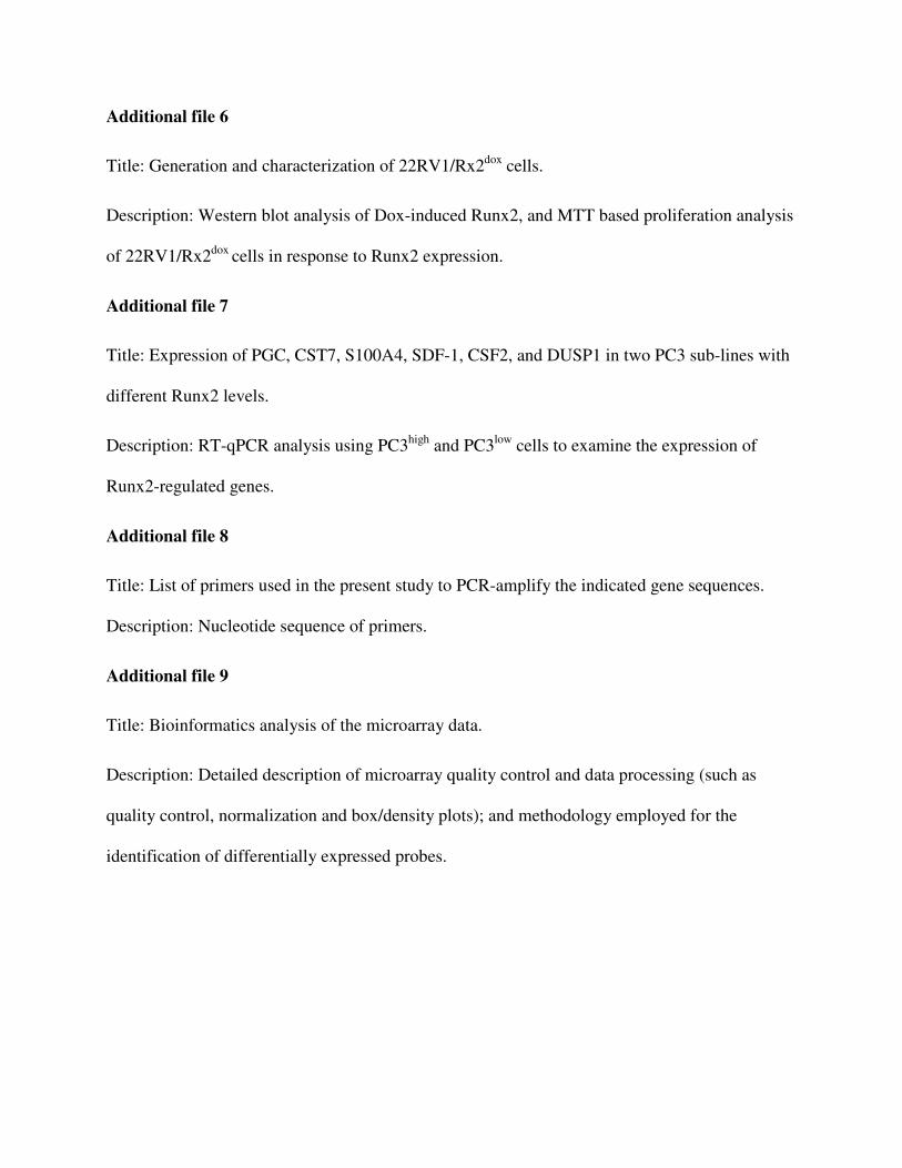
Additional file 7
Title: Expression of PGC, CST7, S100A4, SDF-1, CSF2, and DUSP1 in two PC3 sub-lines with
different Runx2 levels.
Description: RT-qPCR analysis using PC3high
and PC3low
cells to examine the expression of
Runx2-regulated genes.
Additional file 8
Title: List of primers used in the present study to PCR-amplify the indicated gene sequences.
Description: Nucleotide sequence of primers.
Additional file 9
Title: Bioinformatics analysis of the microarray data.
Description: Detailed description of microarray quality control and data processing (such as
quality control, normalization and box/density plots); and methodology employed for the
identification of differentially expressed probes.

Ta
ble
1. G
enes
most
res
pon
sive
to R
un
x2
in
C4
-2B
cel
ls
Gen
e ID
S
ym
bol
Gene N
am
e
Ano
va
Fo
ld c
han
ge
D
esc
rip
tion
of
cancer
rela
ted
or
oth
er
ma
jor
functi
on
R
efe
ren
ce
p
valu
e
I II
q
Tra
nsc
rip
tio
n r
egu
lato
rs
7069
T
HR
SP
T
hyro
id h
orm
on
e re
spon
sive
3
.5E
-06
7
5
Tra
nsc
rip
tion
of
enzy
mes
in
volv
ed i
n t
um
or
lip
id m
etab
oli
sm
2994
7
DN
MT
3L
D
NA
(cyto
sin
e-5
-)-m
eth
ylt
ran
sfer
ase
3-l
ike
5.5
E-0
8
5
6
>50
DN
A m
eth
yla
tion
, n
ucl
eic
acid
met
aboli
c p
roce
ss
4091
S
MA
D6
S
MA
D f
amil
y m
emb
er 6
8
.1E
-10
5
4
Cel
l co
mm
un
icat
ion,
TG
F-b
eta
rece
pto
r si
gn
alin
g p
ath
way
6591
S
NA
I2
Sn
ail
hom
olo
g 2
(D
roso
ph
ila)
8
.8E
-07
4
1
2
6
Pro
mote
s E
MT
by r
epre
ssin
g E
-cad
her
in t
ran
scri
pti
on
[4
4]
6662
S
OX
9
SR
Y (
sex d
eter
min
ing r
egio
n Y
)-b
ox 9
4
.4E
-07
4
6
1
7
Pro
mote
s ch
ond
rocy
te d
iffe
ren
tiat
ion
, ce
ll m
igra
tion,
and
EM
T
[110]
7025
N
R2
F1
N
ucl
ear
rece
pto
r su
bfa
mil
y 2
,
3.5
E-0
7
3
14
2
L
igan
d-d
epen
den
t re
cep
tor,
reg
ula
tion
of
tran
scri
pti
on
ter
min
atio
n
678
Z
FP
36
L2
Z
inc
fin
ger
pro
tein
36
, C
3H
typ
e-li
ke
2
4.1
E-0
9
3
10
Reg
ula
tion o
f R
NA
met
aboli
c p
roce
ss
4088
S
MA
D3
S
MA
D f
amil
y m
emb
er 3
6
.3E
-10
3
7
2
5
Sig
nal
tra
nsd
uce
r d
ow
nst
ream
of
TG
F-β
rec
epto
rs
2832
48
RC
OR
2
RE
ST
core
pre
ssor
2
2.9
E-0
6
3
7
N
egat
ive
regu
lati
on
of
tran
scri
pti
on
1021
7
CT
DS
PL
S
mal
l C
TD
ph
osp
hat
ase
3
1.9
E-0
9
-2
-3
P
hosp
hat
ase,
dep
hosp
hory
late
s R
b, su
pp
ress
es c
ellu
lar
gro
wth
1870
E
2F
2
E2
F t
ran
scri
pti
on f
acto
r 2
8
.2E
-10
-2
-5
-4
T
ran
scri
pti
on
act
ivat
or,
reg
ula
tion o
f ce
ll c
ycl
e p
rogre
ssio
n,
pro
life
rati
on
9039
0
ME
D3
0
Med
iato
r co
mp
lex s
ubu
nit
30
<
1.0
E-
12
-2
-3
T
ran
scri
pti
on
act
ivat
or,
An
dro
gen
rec
epto
r si
gnal
ing p
ath
way
4609
M
YC
V
-myc
myel
ocyto
mat
osi
s vir
al o
nco
gen
e
2.9
E-0
7
-2
-3
-2
Posi
tive
regu
lati
on o
f ce
ll p
roli
fera
tion
, ap
op
tosi
s [1
11
, 11
2]
2989
3
PS
MC
3IP
P
SM
C3
inte
ract
ing p
rote
in
3.5
E-0
8
-2
-4
G
ene
exp
ress
ion
fro
m R
NA
Po
lII
4602
M
YB
V
iral
on
cogen
e h
om
olo
g (
avia
n)
1.6
E-0
8
-3
-5
-3
G1
/S t
ran
siti
on o
f m
itoti
c ce
ll c
ycl
e
4824
N
KX
3-1
N
K3
hom
eob
ox 1
3
.7E
-08
-3
-3
Tra
nsc
rip
tion
rep
ress
or,
loss
indu
ces
pro
stat
e tu
mori
gen
esis
Intr
ace
llu
lar
sig
nali
ng
, ce
ll c
ycle
an
d p
roli
fera
tio
n
5165
5
RA
SD
1
RA
S,
dex
amet
has
on
e-in
duce
d 1
6
.8E
-12
2
6
19
>
50
Ras
rel
ated
, su
pp
ress
es c
ell
pro
life
rati
on
, in
hib
its
tum
or
gro
wth
[9
2]
6446
S
GK
S
eru
m/g
luco
cort
icoid
reg
ula
ted
kin
ase
1
<1
.0E
-
12
19
4
5
C
ellu
lar
Na+
ion h
om
eost
asis
, ce
ll s
urv
ival
5700
7
CX
CR
7
Ch
emokin
e re
cep
tor
7
9.2
E-1
1
8
12
3
0
Rec
epto
r fo
r S
DF
-1,
GP
CR
, en
han
ced i
nte
gri
n a
ctiv
atio
n a
nd t
um
or
gro
wth
[1
13]
1843
D
US
P1
D
ual
sp
ecif
icit
y p
hosp
hat
ase
1
9.1
E-1
3
8
8
10
R
egu
lato
r of
cell
cycl
e, r
esp
on
se t
o s
tres
s [1
14]
5580
P
RK
CD
P
rote
in k
inas
e C
, d
elta
2
.0E
-11
6
5
1
2
Ser
ine-
Th
reon
ine
Kin
ase,
cel
l p
roli
fera
tion
, ce
ll c
on
trac
tion
5589
0
GP
RC
5C
G
pro
tein
-coup
led
rec
epto
r, f
amil
y C
1
.3E
-09
5
5
2
5
GP
CR
, ce
ll c
om
mu
nic
atio
n
1046
1
ME
RT
K
C-m
er p
roto
-on
cogen
e ty
rosi
ne
kin
ase
6.2
E-1
0
4
6
39
P
rote
in a
min
o a
cid p
hosp
hory
lati
on
5920
R
AR
RE
S3
R
etin
oic
aci
d r
ecep
tor
resp
ond
er 3
4
.3E
-08
3
8
4
0
Neg
ativ
e re
gu
lati
on
of
cell
pro
life
rati
on
1903
E
DG
3
Sp
hin
gosi
ne-
1-p
hosp
hat
e re
cep
tor
3
4.8
E-1
0
3
7
G
PC
R,
enh
ance
s tu
mor
gro
wth
, ce
ll m
igra
tion
, an
d a
ngio
gen
esis
[7
7-7
9,
115
, 116
]
8877
S
PH
K1
S
phin
gosi
ne
kin
ase
1
7.5
E-0
9
3
12
3
6
Kin
ase
for
the
pro
duct
ion
of
S1
P,
enhan
ces
tum
or
gro
wth
, ce
ll m
igra
tion
, an
d
angio
gen
esis
, P
Ca
cell
su
rviv
al a
gai
nst
ch
emoth
erap
y,
ost
eocl
ast
chem
ota
xis
[77
-79,
115
, 116
]
7049
T
GF
BR
3
Tra
nsf
orm
ing g
row
th f
acto
r, β
rec
epto
r II
I 7
.1E
-09
3
4
1
2
Neg
ativ
e re
gu
lati
on
of
TG
Fβ
rec
epto
r si
gnal
ing p
ath
way
49
21
D
DR
2
Dis
coid
in d
om
ain
rec
epto
r ty
rosi
ne
kin
ase
2 2
.9E
-05
2
5
Coll
agen
bin
din
g, ce
ll p
roli
fera
tion
1017
C
DK
2
Cycl
in-d
epen
den
t kin
ase
2
1.3
E-0
3
-2
-3
-4
Kin
ase,
mit
oti
c ce
ll c
ycl
e, c
ell
pro
life
rati
on
5503
8
CD
CA
4
Cel
l d
ivis
ion c
ycl
e as
soci
ated
4
7.2
E-0
8
-2
-4
-4
Rep
ress
or
of
E2
F-d
epen
den
t tr
ansc
ripti
onal
act
ivat
ion
and
cel
l p
roli
fera
tion
5997
R
GS
2
Reg
ula
tor
of
G-p
rote
in s
ignal
ing 2
, 24k
Da
9.0
E-0
9
-3
-3
C
ell
cycl
e, G
PC
R s
ignal
ing p
ath
way,
cell
com
mu
nic
atio
n
2580
5
BA
MB
I B
MP
an
d a
ctiv
in m
emb
ran
e-b
oun
d
inh
ibit
or
hom
olo
g (
Xen
op
us
laev
is)
9.7
E-1
0
-2
-3
T
GF
-β s
ignal
ing p
ath
way
Cel
lula
r m
ove
men
t, a
dh
esio
n,
inva
sio
n, cy
tosk
elet
on
rem
odel
ing

8979
7
NA
V2
N
euro
n n
avig
ator
2
6.7
E-0
9
9
10
2
9
Sca
ffold
pro
tein
, ac
tin
cyto
skel
eton
rem
od
elin
g,
ner
vou
s sy
stem
dev
elop
men
t [5
3]
2711
1
SD
CB
P2
S
yn
dec
an b
ind
ing p
rote
in 2
2
.2E
-03
6
1
5
B
ind
s S
DC
2,
regu
late
s ce
ll a
dh
esio
n a
nd c
om
mu
nic
atio
n w
ith
EC
M m
ole
cule
s [8
4, 85]
1134
4
PT
K9
L
Tw
infi
lin
9
.0E
-10
6
5
1
1
Act
in-b
indin
g p
rote
in, cy
tosk
elet
on
org
aniz
atio
n
[57]
8371
5
ES
PN
E
spin
2
.1E
-08
5
8
>
50
A
ctin
-bin
din
g-b
un
dli
ng p
rote
in, ac
tin c
yto
skel
eton
dyn
amic
s, c
ell
loco
moti
on
[5
4, 117
,
118]
5147
4
LIM
A1
E
pli
n,
LIM
dom
ain a
nd a
ctin
bin
din
g 1
3
.0E
-10
4
5
1
5
Cyto
skel
eton
-ass
oci
ated
pro
tein
, re
gula
tion
of
acti
n f
ilam
ent
dep
oly
mer
izat
ion
[5
6]
8829
N
RP
1
Neu
rop
ilin
1
4.1
E-0
8
4
11
4
0
TM
, co
rece
pto
r fo
r V
EG
F,
coll
agen
bin
din
g, ce
ll a
dh
esio
n, in
vas
ion
[1
19]
4582
M
UC
1
Muci
n 1
, ce
ll s
urf
ace
asso
ciat
ed
1.1
E-0
9
4
4
T
M,
cell
ad
hes
ion
, in
vas
ion
[1
20]
4131
M
AP
1B
M
icro
tubu
le-a
ssoci
ated
pro
tein
1B
7
.1E
-09
3
9
1
8
Tra
ns-
mem
bra
ne
gly
cop
rote
in,
cyto
skel
eton
org
aniz
atio
n
[55
, 121]
9644
S
H3
PX
D2
A
SH
3 a
nd
PX
dom
ain
s 2
A
1.4
E-0
9
3
7
A
dap
tor
pro
tein
, E
CM
deg
radat
ion
by a
ctin
bas
ed i
nvad
op
edia
form
atio
n
[122]
6275
S
100
A4
S
100
cal
ciu
m b
indin
g p
rote
in A
4
2.6
E-0
8
2
24
>
50
E
F-h
and c
alci
um
-bin
din
g p
rote
in,
pro
mote
s ce
ll m
oti
lity
, tu
mor
met
asta
sis
[49]
6383
S
DC
2
Syn
dec
an 2
3
.1E
-06
2
5
3
2
TM
, b
ind s
ign
alin
g f
acto
rs,
cell
adh
esio
n, an
gio
gen
esis
, E
CM
deg
radat
ion
[84
, 85]
1013
C
DH
15
C
adh
erin
15,
typ
e 1
, M
-cad
her
in
6.9
E-0
8
2
5
21
T
M,
Ca2
+d
epen
den
t in
terc
ellu
lar
adh
esio
n, sk
elet
al m
usc
le d
evel
op
men
t [1
23]
Sec
rete
d f
act
ors
1437
C
SF
2
Colo
ny s
tim
ula
tin
g f
acto
r 2
/GM
-CS
F
2.3
E-0
9
12
1
7
>50
C
yto
kin
e, o
steo
lyti
c b
on
e m
etas
tasi
s [7
2]
5225
P
GC
P
rogas
tric
sin
(p
epsi
nogen
C)
3.9
E-1
2
11
1
5
A
spar
tyl
pro
tein
ase
[47]
5304
P
IP
Pro
lact
in-i
ndu
ced
pro
tein
<
1.0
E-1
21
0
79
>
50
A
spar
tyl
pro
tein
ase,
lig
and
for
CD
4 r
ecep
tor
T c
ells
[46]
8857
F
CG
BP
F
c fr
agm
ent
of
IgG
bin
din
g p
rote
in
1.5
E-0
9
5
9
C
ell
adh
esio
n,
cell
-cel
l re
cogn
itio
n
6387
C
XC
L1
2
Ch
emokin
e l
igan
d 1
2 (
SD
F-1
) 2
.2E
-06
5
7
2
3
Ch
emokin
e, s
tem
cel
l tr
affi
ckin
g t
o b
on
e, m
igra
tion
, m
etas
tasi
s o
f tu
mor
cell
s
[58
-65]
7422
V
EG
FA
V
ascu
lar
end
oth
elia
l gro
wth
fac
tor
A
4.5
E-1
0
5
3
4
Gro
wth
fac
tor,
an
gio
gen
esis
, ce
llu
lar
morp
hogen
esis
du
rin
g d
iffe
ren
tiat
ion
[88
-91]
1907
E
DN
2
End
oth
elin
2
4.3
E-0
7
5
2
G
row
th f
acto
r, v
asoac
tive,
G-p
rote
in s
ign
alin
g,
smooth
mu
scle
contr
acti
on
[87]
5654
H
TR
A1
H
trA
ser
ine
pep
tid
ase
1
6.9
E-1
0
3
18
Ser
ine
pro
teas
e, d
iffe
ren
tial
ly e
xp
ress
ed i
n o
steo
arth
riti
c ca
rtil
lage
[124]
3817
K
LK
2
Kal
lik
rein
-rel
ated
pep
tid
ase
2
1.5
E-0
8
4
3
S
erin
e p
rote
ase,
pro
tein
met
aboli
c p
roce
ss
4880
N
PP
C
Nat
riu
reti
c p
epti
de
pre
curs
or
C
1.8
E-1
0
4
17
>
50
P
epti
de,
pote
nt
nat
riu
reti
c, d
iure
tic,
an
d v
asodil
atin
g
7057
T
HB
S1
T
hro
mb
osp
ond
in 1
3
.0E
-10
4
1
4
>50
C
ell
adh
esio
n, h
epar
in-b
indin
g, an
ti-a
ngio
gen
ic
3934
L
CN
2
Lip
oca
lin
2
5.1
E-0
8
4
10
5
E
CM
pro
tein
, p
rese
nt
ligan
ds
to c
ell
surf
ace
rece
pto
rs, p
rom
ote
s E
MT
[4
5]
6462
S
HB
G
Sex
horm
on
e-b
ind
ing g
lob
uli
n
8.7
E-1
0
4
10
Ste
roid
-bin
din
g,
sign
al
5764
2
CO
L2
0A
1
Coll
agen
, ty
pe
XX
, α
1
4.9
E-0
7
4
10
>
50
A
dh
esio
n,
esta
bli
shm
ent
of
loca
liza
tion
, tr
ansp
ort
4254
K
ITL
G
KIT
lig
and
2
.3E
-08
4
7
4
1
Hem
atop
oie
tic
gro
wth
fac
tor
and
lig
and f
or
the
KIT
tyro
sin
e k
inas
e re
cep
tor
4884
N
PT
X1
N
euro
nal
pen
trax
in I
2
.5E
-07
3
2
0
C
ell-
cell
sig
nal
ing, t
ran
sport
, est
abli
shm
ent
of
loca
liza
tion
3486
IG
FB
P3
IG
F b
indin
g p
rote
in 3
2
.2E
-08
3
7
Reg
ula
tion o
f ce
ll g
row
th,
com
munic
atio
n, ap
op
tosi
s
9806
S
PO
CK
2
Sp
arc/
ost
eon
ecti
n, cw
cv,
kaz
al-l
ike
dom
ain
s p
rote
ogly
can
(te
stic
an)
2
1.1
E-0
6
3
4
C
ell
dif
fere
nti
atio
n, E
CM
org
aniz
atio
n a
nd
bio
gen
esis
8530
C
ST
7
Cyst
atin
F (
leu
kocy
stat
in)
3.0
E-0
9
2
25
Com
pet
itiv
e in
hib
itors
of
cyst
ein
e p
rote
ases
, m
etas
tasi
s as
soci
ated
pro
tein
[4
8]
4885
N
PT
X2
N
euro
nal
pen
trax
in I
I 7
.6E
-09
2
6
Cel
l-ce
ll s
ign
alin
g,
syn
apti
c tr
ansm
issi
on
1473
81
CB
LN
2
AD
AM
MM
P w
ith
Thb
s-1 m
oti
f 5
.2E
-12
-3
-3
Pep
tid
ase,
cle
avag
e of
pre
cere
bel
lin
in
th
e n
ervou
s sy
stem
67
81
S
TC
1
Sp
ond
in 2
3
.6E
-08
-3
-3
EC
M p
rote
in, in
tegri
n l
igan
d, pat
tern
rec
ogn
itio
n
70
35
T
FP
I T
issu
e fa
ctor
pat
hw
ay i
nhib
itor
3.9
E-0
5
-3
-4
K
un
itz-
typ
e in
hib
itor
of
blo
od
coag
ula
tion
by b
lock
ing f
acto
r X
a
9518
G
DF
15
G
row
th d
iffe
ren
tiat
ion f
acto
r 15
1
.6E
-09
-2
-8
-7
G
row
th f
acto
r, T
GF
-β s
ignal
ing p
athw
ay,
cell
com
mu
nic
atio
n
Fo
ld c
han
ges
on
day
1 a
nd
day
2 a
re l
iste
d i
n c
olu
mn
s I
and
II
resp
ecti
vel
y,
and
colu
mn
q l
ists
fold
chan
ges
bas
ed o
n R
T-q
PC
R a
nal
ysi
s of
the
day
-2 s
amp
les.
Maj
or
gen
e fu
nct
ion
s w
ere
det
erm
ined
usi
ng k
now
led
ge
dat
abas
es f
rom
OM
IM (
On
lin
e M
end
elia
n I
nh
erit
ance
of
Man
), I
PA
™ s
yst
ems
and
th
e in
dic
ated
ref
eren
ced
lit
erat
ure
. A
bb
revia
tion
s: E
MT
, ep
ith
elia
l to
mes
ench
ym
al t
ransi
tion
; T
M,
tran
smem
bra
ne;
GP
CR
, G
-pro
tein
cou
ple
d r
ecep
tor;
EC
M, ex
trac
ellu
lar
mat
rix.
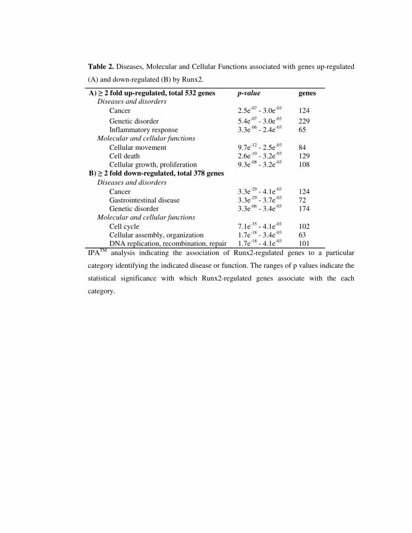
Table 2. Diseases, Molecular and Cellular Functions associated with genes up-regulated
(A) and down-regulated (B) by Runx2.
A) ≥ 2 fold up-regulated, total 532 genes p-value genes Diseases and disorders
Cancer 2.5e-07
- 3.0e-03 124
Genetic disorder 5.4e-07
- 3.0e-03 229
Inflammatory response 3.3e-06
- 2.4e-03 65
Molecular and cellular functions Cellular movement 9.7e
-12 - 2.5e
-03 84 Cell death 2.6e
-10 - 3.2e
-03 129 Cellular growth, proliferation 9.3e
-08 - 3.2e
-03 108 B) ≥ 2 fold down-regulated, total 378 genes
Diseases and disorders Cancer 3.3e
-29 - 4.1e
-03 124 Gastrointestinal disease 3.3e
-29 - 3.7e
-03 72 Genetic disorder 3.3e
-06 - 3.4e
-03 174 Molecular and cellular functions
Cell cycle 7.1e-35
- 4.1e-03 102
Cellular assembly, organization 1.7e-18
- 3.4e-03
63 DNA replication, recombination, repair 1.7e
-18 - 4.1e
-03 101
IPATM
analysis indicating the association of Runx2-regulated genes to a particular
category identifying the indicated disease or function. The ranges of p values indicate the
statistical significance with which Runx2-regulated genes associate with the each
category.
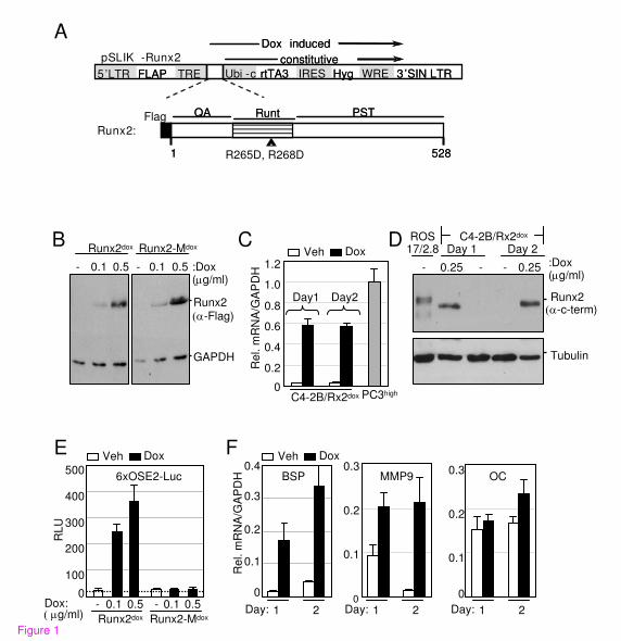
B
GAPDH
Runx2
Runx2-Mdox
0.1 0.5
Runx2dox
*c-Flag)
0.1 0.5--
A
C
0
100
200
300
400
500
Dox: *"og/ml)
6xOSE2-Luc
RLU
E
Runx2-MdoxRunx2dox
0.1 0.5- 0.1 0.5-
:Dox (og/ml)
D
F
Re
l. m
RN
A/G
AP
DH
0
0.1
0.2
0.3OC
Day: 1 2Day: 1 20
0.1
0.2
0.3MMP9
0
0.1
0.2
0.3
0.4BSP
*c-c-term)
Tubulin
-0.25
ROS 17/2.8
C4-2B/Rx2dox
Runx2
- :Dox (og/ml)
0.25-
Day 1 Day 2
Day: 1 2
QA
1 528
PSTRuntQA
1 528
PSTRunt
5’LTR FLAP TRE Ubi -c rtTA3 IRES Hyg WRE 3’SIN LTR5’LTR FLAP TRE Ubi -c rtTA3 IRES Hyg WRE 3’SIN LTR
constitutiveconstitutive
Dox inducedDox induced
Flag
Runx2:
R265D, R268D
pSLIK -Runx2
Re
l. m
RN
A/G
AP
DH
0
0.2
0.4
0.6
0.8
1.0
1.2Veh Dox
Day1 Day2
PC3highC4-2B/Rx2dox
Veh DoxVeh Dox
Figure 1
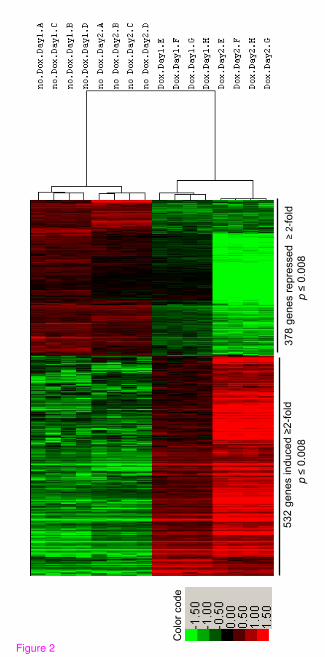
53
2 g
en
es in
du
ce
d œ
2-f
old
3
78
ge
ne
s r
ep
resse
d œ
2-f
old
Co
lor
co
de
pø
0.0
08
pø
0.0
08
Figure 2

Runx2
TGFd-RIII
Myc
PIP
SDF-1
VEGF
- + :Dox
ce
ll e
xtr
act
su
pern
ata
nt
CDK2
Tubulin
C4-2B/Rx2dox
Figure 3
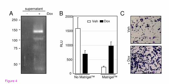
250
150
100
75
50
- + :Dox
supernatant
C
+ D
ox
Ve
h
B
0
500
1000
1500
2000
No Matrigel™ Matrigel™
RLU
A
Veh Dox
Figure 4
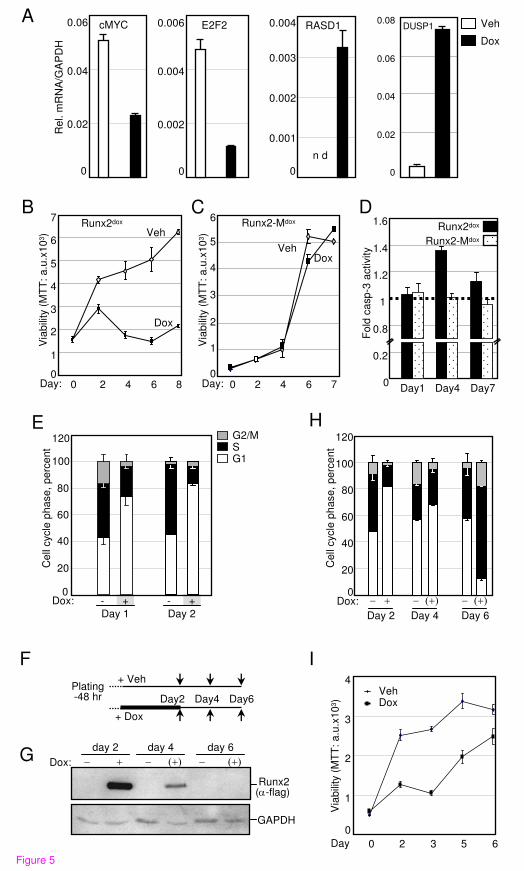
Figure 5
Day 1
- +
Day 2
- +
E
Dox:
Ce
ll cycle
ph
ase
, p
erc
en
t
G2/MSG1
0
20
40
60
80
100
120
B
Via
bili
ty (
MT
T:
a.u
.x1
03)
0
1
2
3
4
5
6
7
0 2 4 6 8
Runx2dox
Veh
Dox
Day:
C
0
1
2
3
4
5
6
0 2 4 6 7
Runx2-Mdox
VehDox
Day:
Via
bili
ty (
MT
T:
a.u
.x1
03)
I
0
1
2
3
4
0 2 3 5 6
VehDox
Day
F
H
Day4 Day7Day10
0.2
0.8
1
1.2
1.4
Fo
ld c
asp
-3 a
ctivity
Runx2dox
Runx2-Mdox
1.6D
Plating-48 hr Day2 Day4 Day6
+ Dox
+ Veh
Ce
ll cycle
ph
ase
, p
erc
en
t
0
20
40
60
80
100
120
Dox: / -Day 2
*-+/Day 4
*-+/Day 6
Via
bili
ty (
MT
T:
a.u
.x1
03)
G
Runx2(c-flag)
GAPDH
Dox: / -day 2
*-+/day 4
*-+/day 6
0
0.001
0.002
0.003
0.004
n d
RASD1
0
0.02
0.04
0.06
0.08DUSP1
0
0.02
0.04
0.06 cMYC
0
0.002
0.004
0.006 E2F2
Re
l. m
RN
A/G
AP
DH
A Veh
Dox
Figure 5

Nucleus
Cytoplasm
Plasma Membrane
Extracellular Space
Figure 6
Additional files provided with this submission:
Additional file 1: Additional file 1.xls, 19906Khttp://www.molecular-cancer.com/imedia/1695821719450985/supp1.xlsAdditional file 2: Additional file 2.doc, 115Khttp://www.molecular-cancer.com/imedia/1643006969450986/supp2.docAdditional file 3: Additional file 3.xls, 79Khttp://www.molecular-cancer.com/imedia/1135951886450986/supp3.xlsAdditional file 4: Additional file 4.pdf, 21Khttp://www.molecular-cancer.com/imedia/7539305334509863/supp4.pdfAdditional file 5: Additional file 5.pdf, 48Khttp://www.molecular-cancer.com/imedia/1113604708450986/supp5.pdfAdditional file 6: Additional file 6.pdf, 28Khttp://www.molecular-cancer.com/imedia/1591460234509864/supp6.pdfAdditional file 7: Additional file 7.pdf, 19Khttp://www.molecular-cancer.com/imedia/7454516794509865/supp7.pdfAdditional file 8: Additional file 8.doc, 80Khttp://www.molecular-cancer.com/imedia/1301714918450986/supp8.docAdditional file 9: Additional file 9.doc, 188Khttp://www.molecular-cancer.com/imedia/1784406338450986/supp9.doc