osteoblast differentiation on collagen scaffold with
TRANSCRIPT

International Journal of Organ Transplantation Medicine
Original Article
*Correspondence: Parichehr Hanachi, PhD, Faculty of Biological Science, Department of Biotechnology, Biochemistry Unit, Alzahra University, Tehran, IranTel: +98-912-542-6316E-mail: [email protected]
Osteoblast Differentiation on Collagen Scaffold with Immobilized Alkaline PhosphataseF. Jafary1, P. Hanachi2*, K. Gorjipour3
1Department of Biochemistry, School of Medicine, Shahid Sadoughi University of Medical Sciences, Yazd, Iran 2Faculty of Biological Science, Department of Biotechnology, Biochemistry Unit, Alzahra University, Tehran, Iran 3Department of Immunology, Faculty of Medicine, Shahid Beheshti University of Medical Sciences, Tehran, Iran
ABSTRACT
Background: In tissue engineering, scaffold characteristics play an important role in the biological inter-actions between cells and the scaffold. Cell adhesion, proliferation, and activation depend on material properties used for the fabrication of scaffolds.
Objective: In the present investigation, we used collagen with proper characteristics including mechani-cally stability, biodegradability and low antigenicity. Optimization of the scaffold was done by immobili-zation of alkaline phosphatase on the collagen surface via cross-linking method, because this enzyme is one of the most important markers of osteoblast, which increases inorganic phosphate concentration and promote mineralization of bone formation.
Methods: Alkaline phosphatase was immobilized on a collagen surface by 1-ethyl-3-(dimethylaminopropyl) carbodiimide hydrochloride, as a reagent. Then, rat mesenchymal stem cells were cultured in osteogenic medium in control and treated groups. The osteogenesis-related genes were compared between treat-ments (differentiated cells with immobilized alkaline phosphatase/collagen scaffold) and control groups (differentiated cells on collagen surface without alkaline phosphatase) on days 3 and 7 by quantitative real-time PCR (QIAGEN software).
Results: Several genes, including alkaline phosphatase, collagen type I and osteocalcine associated with calcium binding and mineralization, showed upregulation in expression during the first 3 days, whereas tumor necrosis factor-α, acting as an inhibitor of differentiation, was down-regulated during osteogen-esis.
Conclusion: Collagen scaffold with immobilized alkaline phosphatase can be utilized as a good candidate for enhancing the differentiation of osteoblasts from mesenchymal stem cells.
KEYWORDS: Collagen scaffold; Alkaline phosphatase; Immobilization; Differentiation; Osteoblast
INTRODUCTION
It has been shown that inorganic phosphate (Pi) has an important role in upregula-tion of osteopontin (OPN) expression in
MC3T3 cells [1]. Osteopontin is a phosphory-lated glycoprotein secreted by osteoblasts and binds to αVβ3 integrin of cells via an RGD (arginine-glycine-aspartic acid) cell adhesion sequence [2]. It is believed that it promotes the attachment of osteoblasts and osteoclasts
to the extracellular matrix. Therefore, OPN may have a key role in composition and re-modeling of the bone [1]. On the other hand, β-glycerophosphate functions as a phosphate group donor in mineralization matrix, which is hydrolyzed by alkaline phosphatase. As a re-sult, Pi can play an important role in osteoblast differentiation and mineralization [3, 5].
The metalloenzyme as famous as alkaline phosphatase (ALP) [phosphate-monster phos-phatase EC 3.1.3.1] is expressed in various tissues. Invertebrate ALP acts as an ectoen-zyme attaching to the plasma membrane via a phosphatidyl inositol glycophospholipid (GPI)

196 Int J Org Transplant Med 2017; Vol. 8 (4) www.ijotm.com
linkage. The catalytic mechanisms entail the formation of a serine phosphate at the active site, which reacts with water at an alkaline pH leading to the release of inorganic phosphate from the enzyme. Four ALP isozymes—intes-tinal, placental, placental-like, and tissue non-specific ALP (TNAP)—are available in hu-man. TNAP is an osteoblastic differentiation marker that is expressed in the initial phases of the process. In addition, the enzyme is neces-sary for mineralization of bone. Furthermore, the greater ALP activity, the great impact it might have on bone formation. Accordingly, ALP has a critical role in bone formation [6].
Many factors such as pore size, porosity, per-meability, and the composition of the scaffold have been indicated to regulate osteoblasts be-havior [7]. Collagen is one of the critical pro-teins in the extracellular matrix of numerous connective tissues. Thus far, different types of collagen (approximately 29) have been identi-fied. The most abundant type of collagen in the body is type I and is found in bones, skins, tendons, vessels, and organs. The structural, physical, chemical, and immunological prop-erties of collagen have been identified, which include its biodegradability and biocompat-ibility, being non-cytotoxic, and having the ability to support cellular growth. Indeed, the collagen can be processed into various forms such as steps, sheets, beads, meshes, fibers, and sponges. Type I collagen can develop mineral to matrix ratio, calcium deposition and osteo-pontin/osteocalcin secretion [8]. These data suggest that collagen has the most important role in promoting osteogenesis and could be an efficient scaffold to be used in bone tissue engineering.
This study aimed to develop a method for immobilization of ALP on collagen fibers scaffolds using covalent bond and 1-ethyl-3-(dimethylaminopropyl) carbodiimide hydro-chloride (EDC) as cross-linkers. Afterwards, the ability of creating scaffolds in mineraliza-tion and osteoblast differentiation were char-acterized in vitro.
MATERIALS AND METHODS
All reagents used in this experimental study were supplied by Sigma (CA, USA) unless in-dicated otherwise. This study was conducted on human bone marrow-derived mesenchymal stem cells (MSCs) that were differentiated on a collagen scaffold affected by alkaline phospha-tase. Various parts of the methods used were approved by Iran National Science Foundation (grant number 90004378).
Activation of the Collagen Fiber and Immobilization of the EnzymeThe collagen matrix (Viscofan cell car-rier) was treated with 10 mg/mL of 1-ethyl-3-(dimethylaminopropyl) carbodiimide hydro-chloride (EDC) (Piers 22980) in phosphate buffer for 30 min [9]. Subsequently, the ma-trix was washed with PBS and incubated in aqueous bovine intestinal mucosa alkaline phosphatase (3 U) (Sigma P6774) for 2 hrs. The collagen matrix was rinsed in PBS four times each for 15 min with shaking.
The Alkaline Phosphatase Activity AssayThe enzyme activity was detected by Taylor, et al, method (2006). The p-nitrophenylphos-phate (pNPP) (Sigma P4744) was selected as the substrate [10]. A stock solution of pump was 350 μM in 25 mm glycine with pH 9.5. The collagen matrix with immobilized en-zyme was treated with 2 mL of substrate at room temperature. Then, every minute, 90 μL of 0.1 M NaOH and 0.1 M EDTA was added as a stop solution and finally the absor-bance was measured at 405 nm. The alkaline phosphatase activity was calculated using the method of Taylor at al, 2005, and the follow-ing equation:
( )405 /reaction aliquotA V Vactivity
β=
Stem Cell Isolation and DifferentiationMSCs were isolated from adipose tissue of three patients who referred to Shahid Sadughi Hospital for abdominal surgery. Adipose tis-sue was digested by enzymatic method (1 mg collagenase per 1 g adipose tissue). The iso-lated cells were then cultured in culture me-dium containing DMEM, 10% fetal bovine
F. Jafary, P. Hanachi, K. Gorjipour
www.ijotm.com Int J Org Transplant Med 2017; Vol. 8 (4) 197
serum, 1% penicillin/streptomycin (Gibco) at 37 °C, and 5% CO2. Cells in the third passage were added to osteogenic medium, including the above-mentioned culture medium and 10 mM β-glycerol phosphate, 50 μg/mL ascorbic acid, and 10-8 M dexamethasone. This medium was used for differentiation of approximately 105 cells into two groups: the treatment group (in presence of ALP/collagen scaffold) and the control group (in the absence of the ALP/col-lagen scaffold).
Total RNA Extraction and Complementary DNA SynthesisThe total RNA was isolated from osteoblasts differentiated from MSCs on days 3 and 7 us-ing RNeasy Mini Kit Qiagen according to the manufacturer’s protocol in the treatment group (in presence of the ALP/collagen scaf-fold) and the control group (in the absence of the ALP/collagen scaffold). The concentration of RNA was determined at 260/280 nm us-ing NanoDrop spectrophotometer. For reverse transcription polymerase chain reaction (RT-PCR) the cDNA was synthesized by Rever-tAid First Strand cDNA Synthesis (Thermo Scientific) following the instructions provided. The synthesized cDNA was stored at -20 °C for later use.
Real-time RT-PCRSimultaneous gene expression level for OCN.COL, TNF, ALP and Runx2 genes along with β-actin house-keeping gene were measured by three steps real-time PCR (Corbett research, Sydney, Australia) using SYBR green method. The primers used for PCR were as follows:
RUNX2:Forward: 5’ CTTCATTTGCACTGGGTCAC 3’ (Tm: 58.4)
Reverse: 5’ CTGCAAATCTCAGCCATGTT 3’ (Tm: 56.4)
TYPE I COLLAGEN:Forward: 5’ ACCAACTGAACGTGACCAAA 3’ (Tm: 56.4)
Reverse: 5’ AGTGGGCAGAAAGGGACTTA 3’ (Tm: 58.4)
β-ACTIN:Forward: 5’ GCTGTGCTATGTTGCCCTAGA 3’ (Tm: 64.0)
Reverse: 5’ GGAACCGCTCATTGCCGATAG 3’ (Tm: 63.3)
ALKALINE PHOSPHATASE:Forward: 5’ CGTCAATTAACGGCTGACAC 3’ (Tm: 58.4)
Reverse: 5’ TCTGGCACAAATGAGTTGGT 3’ (Tm: 56.4)
TNF:Forward: 5’ CTGTGCACTCCTGGTGTTCT 3’ (Tm: 60.5)
Reverse: 5’ TGTCTTCCTCCTGACTGTGC 3’ (Tm: 60.5)
AND, OSTEOCALCIN:Forward: 5’ CTGCATTCTGCCTCTCTGAC 3’ (Tm: 60.5)
Reverse: 5’ CCGGAGTCTATTCACCACCT 3’ (Tm: 60.5).
New Method and Matrix for Osteoblast Differentiation
198 Int J Org Transplant Med 2017; Vol. 8 (4) www.ijotm.com
Real-time PCR reaction was carried out in 20 μL volume including Taq PCR Master Mix 10 μL, forward and reverse primers (0.5 μL for each), 1 μL of cDNA, and 8 μL of dH2O ac-cording to cycling program (Table 1).
The analysis of the results was performed us-ing REST RG (Relative Expression Software Tool) software that enables more sensitive and accurate estimation of the relative gene ex-pression.
RESULTS
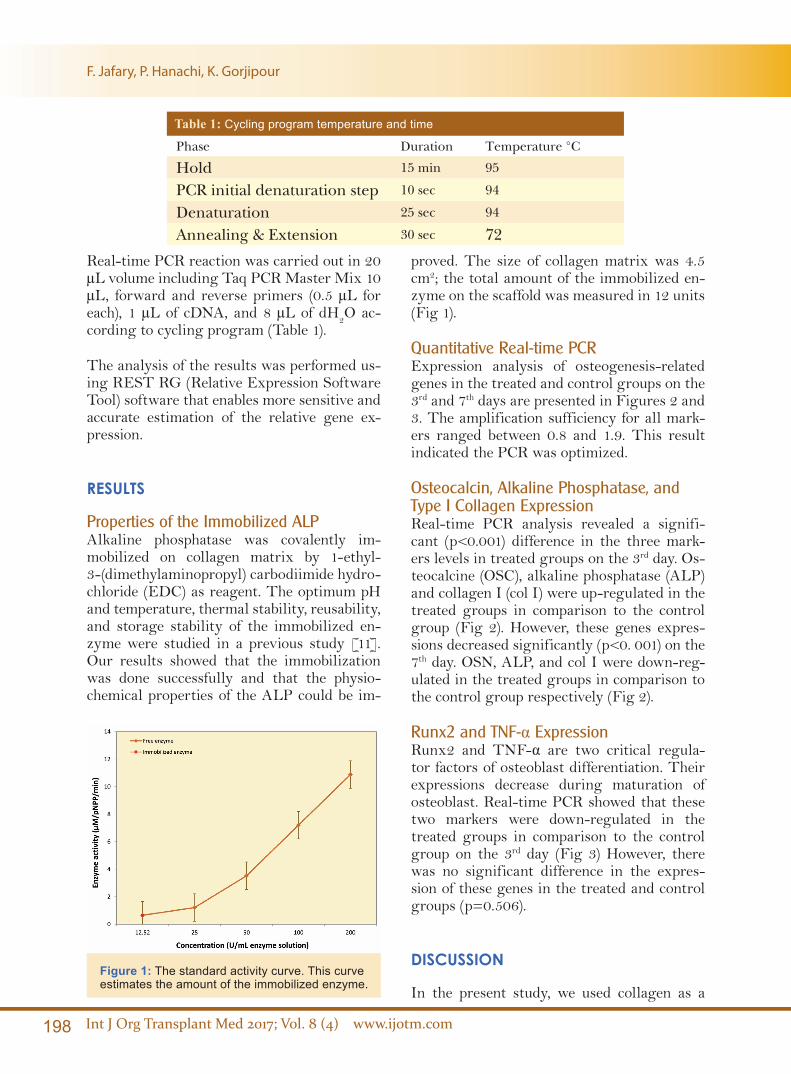
Properties of the Immobilized ALPAlkaline phosphatase was covalently im-mobilized on collagen matrix by 1-ethyl-3-(dimethylaminopropyl) carbodiimide hydro-chloride (EDC) as reagent. The optimum pH and temperature, thermal stability, reusability, and storage stability of the immobilized en-zyme were studied in a previous study [11]. Our results showed that the immobilization was done successfully and that the physio-chemical properties of the ALP could be im-
proved. The size of collagen matrix was 4.5 cm2; the total amount of the immobilized en-zyme on the scaffold was measured in 12 units (Fig 1).
Quantitative Real-time PCRExpression analysis of osteogenesis-related genes in the treated and control groups on the 3rd and 7th days are presented in Figures 2 and 3. The amplification sufficiency for all mark-ers ranged between 0.8 and 1.9. This result indicated the PCR was optimized.
Osteocalcin, Alkaline Phosphatase, and Type I Collagen ExpressionReal-time PCR analysis revealed a signifi-cant (p<0.001) difference in the three mark-ers levels in treated groups on the 3rd day. Os-teocalcine (OSC), alkaline phosphatase (ALP) and collagen I (col I) were up-regulated in the treated groups in comparison to the control group (Fig 2). However, these genes expres-sions decreased significantly (p<0. 001) on the 7th day. OSN, ALP, and col I were down-reg-ulated in the treated groups in comparison to the control group respectively (Fig 2).
Runx2 and TNF-α ExpressionRunx2 and TNF-α are two critical regula-tor factors of osteoblast differentiation. Their expressions decrease during maturation of osteoblast. Real-time PCR showed that these two markers were down-regulated in the treated groups in comparison to the control group on the 3rd day (Fig 3) However, there was no significant difference in the expres-sion of these genes in the treated and control groups (p=0.506).
DISCUSSION
In the present study, we used collagen as a
Table 1: Cycling program temperature and time
Phase Duration Temperature °C
Hold 15 min 95
PCR initial denaturation step 10 sec 94
Denaturation 25 sec 94
Annealing & Extension 30 sec 72
Figure 1: The standard activity curve. This curve estimates the amount of the immobilized enzyme.
F. Jafary, P. Hanachi, K. Gorjipour

www.ijotm.com Int J Org Transplant Med 2017; Vol. 8 (4) 199
scaffold for enzyme immobilization and osteo-blast differentiation. Previously, collagen has been applied as a scaffold in biological systems [12]. The high tensile strength, mechanical stability, and biodegradability of collagen are of great values in biomedical and tissue en-gineering [7]. ALP was immobilized on the collagen surface because the collagen has ap-propriate chemical groups such as carboxyl groups, which can participate in cross-linking reactions. About 12 units of enzymes were im-mobilized on the matrix (4.5 cm2) during the process. The results showed that collagen ma-trix had a satisfactory capability to preserve enzyme and also its activity (48.6%) [11].
Collagen is also an appropriate matrix for proliferation, differentiation, and attachment of different kinds of cells. Various types of col-lagen may have a critical role in cell behavior depending on the organ or tissue of origin [13, 15]. In this study, type I collagen, the critical protein component of bone, was used for os-teoblast differentiation. Stem cells interaction with type I collagen via α2β1 integrin, which is a major signal for the induction of osteoblast differentiation and matrix mineralization [16, 17]. Integrin is a trans-membrane receptor that has an important role in cell interaction with other cells and the surrounding environ-ment. Indeed, α2β1 integrin specific collagen-mimetic surface was reported to supports osteoblastic differentiation [18]. According to the obtained results, ALP immobilization onto type I collagen stimulated in vitro differ-entiation of osteoblasts from MSCs in vitro.
Immobilization of ALP on collagen leads to an increase in the concentration of Pi in cell cul-ture from hydrolysis of β-glycerophosphate. Previous studies showed that modeling the lo-cal concentration of Pi can be useful for bone formation [19].
Different steps of osteoblast differentiation process including proliferative, extracellular matrix synthesis, and mineralization are de-tected by expression of specific genes. Our quantitative Real-time PCR showed induction of the expression of osteoblast markers mRNA through increased concentration of Pi that ex-
plained as follows: ALP enzyme attached to membrane phospholipids of the matrix vesi-cles. Therefore, ALP activity was increased during the matrix maturation phase. In fact, ALP is considered one of the most commonly
Figure 2: Relative gene expression for osteo-calcin, alkaline phosphatase, and collagen I; the three markers levels in treated groups on the 3rd day were more than those on the 7th day in com-parison to the control group. Dashed red line rep-resents a ratio of 1.
Figure 3: Relative gene expression for Runx2 and TNF-α; the two markers were down-regulated on the 3rd day in the treated groups in comparison to those on the 7th day. Dashed red line repre-sents a ratio of 1.
New Method and Matrix for Osteoblast Differentiation

200 Int J Org Transplant Med 2017; Vol. 8 (4) www.ijotm.com
accessible marker to indicate osteoblast differ-entiation [20]. In a previous study, we report-ed that ALP increased during osteogenesis up to three weeks [21]. George, et al, investigated differentiation of mesenchymal stem cells into osteoblast on the honeycomb collagen scaffold. In their research, ALP activity was increased to about three folds on day 28 compared with day 14 [12]. ALP was immobilized on the mi-croporous nanofibrous fibrin scaffold for bone tissue engineering by Osathanon, et al. They showed that ALP/FS scaffold can be helpful in osteogenesis. The expression of ALP was el-evated on the 7th and 14th days compared to the control group [19]. In our study, the expres-sion of ALP was up-regulated at the 3rd day of differentiation and was decreased within 7 days. This result indicated that maturation of the matrix was started as early as three days in the treated cells.
Osteocalcin, also known as bone g-carboxy-glutamic acid-containing protein, is a small noncollagenous protein synthesized by osteo-blasts in the final stage of osteogenic matura-tion. OSN has an important role as a regulator of mineral nucleation via bind to hydroxyapa-tite. The increased expression of OCN, there-fore, shows maturation of osteoblasts will occur. Osteocalcine gene expression was de-tected at the second week by Ducy, et al [22]. The expression of OCN was reported on the 21st day in the study performed by Osathanon, et al [19]. In our research, OCN was up-reg-ulated in the treated groups in comparison to the control group on the 3rd day. However, it was down-regulated on the 7th day. The re-sults could show a high rate of differentiation on ALP/collagen scaffold.
The primary function of differentiated osteo-blasts is collagen synthesis. So collagen is a specific gene of early- and mid-stage of osteo-genesis. COL with unique triple helical struc-ture, plays an important role in the matrix maturation. This protein acts as a substrate for precipitation of hydroxyapatite crystals. Therefore, collagen is an essential component and provides mechanical stability for bone [23]. Osathanon, et al, reported increased col-lagen expression on the 7th day; the expression
decreased on the 21st day. In our research, COL was up-regulated on the 3rd day and declined thereafter [19]. COL is a marker of osteoblast differentiation that elevates in proliferative and matrix maturation phases. Immobilized ALP/collagen scaffold showed significant fold increases in osteogenic markers on day three compared to that on day seven, which suggest-ed its high proliferation and differentiation.
Runt-related transcription factor 2 (Runx2) or osteoblast-specific factor (Osf2) that has been named “a master gene,” is an essential tran-scription factor for the initiation of osteogen-esis. Several osteoblastic gene expressions, in-cluding collagen I, osteopontin and bone sialo protein are regulated by Runx2 at early stag-es of osteoblast differentiation. The level of Runx2 expression in immature osteoblasts is high, but it will be down-regulated in imma-ture osteoblasts during osteogenesis. There-fore, Runx2 plays an essential role in inducing differentiation of multipotent MSCs into im-mature osteoblasts. However, osteoblasts dif-ferentiation and bone formation are inhibited by Runx2. The expression of a critical bone matrix gene depends on Runx2 in early stages of osteoblast differentiation, but it has no ef-fect on the maintenance of the expression of these genes in mature osteoblasts. Normally, the expression level of Runx2 in osteoblasts declines during differentiation process [24]. Hernan Roca, et al, analyzed the Runx2-defi-ciency in mice. They reported complete lack of mature osteoblasts with only a few immature osteoblasts, which expressed ALP weakly, but not OPN and osteocalcin (OCN) [25].
In our research, an expression level of Runx2 in cells on the 3rd day was down-regulated in the treated groups in comparison to the con-trol group. There was no significant differ-ence between treated and control groups on the 7th day.
TNF-α (also known as cachectin) has been identified as a pro-inflammatory cytokine that plays a role in many skeletal diseases. TNF-α arrests expression of osteoblast differentia-tion markers such as Runx2, osteocalcin, and ALP. Indeed, inhibition of collagen and osteo-
F. Jafary, P. Hanachi, K. Gorjipour
www.ijotm.com Int J Org Transplant Med 2017; Vol. 8 (4) 201
calcin synthesis leads to reduced formation of mineralizing nodules and the skeletal matrix. Accordingly, TNF-α decreases bone forma-tion [26]. In our research, TNF-α expression decreased on day three in the treated group. However, no significant difference in expres-sion of this gene was observed between the treated and control groups.
Previous studies demonstrated that collagen can be a perfect scaffold for the differentia-tion and proliferation of MSCs into osteoblast. Collagen with its several properties like low antigenicity, biodegradability, and excellent hemostatic and cell binding can be applied in medical and biomedical industries extensively [27, 28]. However, collagen with ALP immo-bilized has previously not been used as a scaf-fold. In accord with our result, immobilization of ALP onto collagen, makes a suitable scaf-fold for enhancing osteoblast differentiation from MSCs. The expression of genes associat-ed with the final mineralization phase such as OSC was observed on the 3rd day., A previous study showed similar results in the third week. Indeed, the expression of the inhibitor gene for osteoblast differentiation was decreased dur-ing differentiation. Interestingly, osteogenesis was dedifferentiated within seven days. Per-haps this result is related to overexpression of the osteopontine effect of phosphate signaling, because phosphate is a signal for induction of osteopontin gene expression [1]. Huang, et al, found that osteopontin is a negative regulator of proliferation and differentiation in MC3T3-E1 pre-osteoblastic cells. They reported that overexpression of OPN inhibits the effect of bone morphogenic protein-2 (BMP-2) on ALP activity. In addition, overexpression of OPN inhibits mineral deposition and also decreases the expression of osteocalcin and bone sialo-proteins. As a result, ALP immobilized causes an increase in the level of Pi and can lead to increasing OPN expression.
In conclusion, collagen characteristics such as having appropriate groups, being mechanical-ly stable, and its biodegradability, low antige-nicity and cell binding makes type I collagen an excellent candidate for enzyme immobili-zation and also cell culture. ALP is a critical
molecule in mineralization of bone formation and its immobilization on a collagen scaffold leads to increase in the concentration of Pi in cell culture. Our results showed that the col-lagen with immobilized ALP has a positive effect on osteoblast differentiation and via up-regulation of osteogenesis-related genes ex-pression such as ALP, COL, OCN, and down-regulation of inhibitor genes expression like TNF-α. In summary, our results propose that the applied immobilized ALP/collagen matrix is more effective than collagen alone for in-creasing the rate of osteoblast differentiation.
ACKNOWLEDGMENTS
The authors wish to acknowledge all contri-butions of the “Iran National Science Founda-tion” for funding (grant number 90004378) the present research, development, and accom-plishment.
CONFLICTS OF INTEREST: None declared.
REFERENCES1. Beck Gr, Zerler Jr, Moran E. Phosphate is a specific
signal for induction of osteopontin gene expres-sion. Cell Biology 2000;97:8352-57.
2. Denhardt DT, Guo X. Osteopontin. A protein with diverse functions. FASEB J 1993;15:1475-82.
3. Orimo H, Shimada T. The role of tissue-nonspecific alkaline phosphatase in the phosphate-induced activation of alkaline phosphatase and mineraliza-tion in SaOS-2 human osteoblast-like cells. Mol Cell Biochem 2008;3:51-60.
4. Fujita T, Meguro T, Izumo N, et al. Phosphate stim-ulates differentiation and mineralization of the chondroprogenitor clone ATDC5. Jpn J Pharmacol 2001;85:278-81.
5. Chang YL, Stanford CM, Keller JC. Calcium and phosphate supplementation promotes bone cell mineralization: implications for hydroxyapatite (HA)-enhanced bone formation. J Biomed Mater Res 2000;52:270-8.
6. Golub E, Battaglia, K. The role of alkaline phospha-tase in mineralization. Basic science 2007;18:444-8.
7. Tierney C, Jaasma M, Obrien F. Osteoblast activ-ity on collagen –GAG scaffold is affected by col-lagen and GAF concentration. J Biomed Mat Res
New Method and Matrix for Osteoblast Differentiation

202 Int J Org Transplant Med 2017; Vol. 8 (4) www.ijotm.com
2009;1:92-101.8. Gorgieva S, Kokol V. Collagen–vs. Gelatine –Based
Biomaterial and Their Biocompatibility: Review and Perspective. In: Rosario Pignatello, editors. Biomaterials Applications for Nanomedicine; 2011;17-41.
9. Wissink M, Beernink R, Pieper JS, et al. Immobiliza-tion of heparin to EDC/NHS-crosslinked collagen. Characterization and in vitro evaluation. Biomate-rials 2001;22:151-63.
10. Taylor R, Fournier S, Simons B, et al. Covalent protein immobilization on glass surfaces: Appli-cation to alkaline phosphatase. J Biotechnology 2005;118:265-9.
11. George J, Kuboki Y, Miyata T. Differentiation of Mesenchymal stem cells into osteoblast on Hon-eycomb collagen scaffold. Biotechnology and Bio-engineering 2006;95:404-10.
12. Rubin K, Hook M, Obrink B, et al. Substrate adhe-sion of rat hepatocytes: Mechanism of attachment to collagen substrates. Cell 1981;24:463-70.
13. Ruggiero F, Champliaud MF, Garrone R, et al. Inter-actions between cells and collagen Molecules or single chains involve distinct mechanisms. Exp Cell Res 1994;210:215-23.
14. Schor SL, Court J. Different mechanisms in the at-tachment of cells to native and denatured colla-gen. J Cell Sci 1979;38:267-281.
15. Mizuno M, Kuboki Y. Osteoblast-related gene ex-pression of bone marrow cells during the osteo-blastic differentiation induced by type I collagen. J Biochem (Tokyo) 2001:129:133-8.
16. Mizuno M, Fujisawa R, Kuboki Y. Type I collagen-in-duced osteoblastic differentiation of bone-marrow
cells mediated by collagen-alpha2beta1 integrin interaction. J Cell Physiol 2000;184:207-13.
17. Reyes CD, Garcia AJ. Alpha2beta1 integrin-specific collagen-mimetic surfaces supporting osteoblastic differentiation. J Biomed Mater Res 2004;69:591-600.
18. Osathanon T, Giachelli C, Somerman M. Immobi-lization of alkaline phosphatase on microporous nanofibrous fibrin scaffold for bone tissue engi-neering. Biomaterials 2009:30;4513-21.
19. Gaur T, Lengner CJ, Hovhannisyan H. Canonical WNT signaling promote osteogenesis by directly stimulating Runx2 gene expression. J Biol Chem 2005;280:33132-40.
20. Hashemibeni. B, Jafart. F, Esmaeil. N, et al. Com-parison of Phenotypic Characterization between Differentiated Osteoblasts from Stem Cells and Calvaria Osteoblasts in vitro. Int J Prev Med 2013;4:180-6.
21. Ducy P, Desbois C, Boyce B. Increased bone for-mation in osteocalcin-deficient mice. Nature 1996;382:448-52.
22. Overstreet M, Sohrabi A, Polotsky A, et al. Collagen microcarrier spinner culture promotes osteoblast proliferation and synthesis of matrix proteins. Cell Dev Biol 2003;39:228-34.
23. Komori T. Regulation of osteoblast differentiation by Runx2. Adv Exp Med Biol 2010;658:43-9.
24. Roca H, Phimphilai M, Gopalakrishnan R, et al. Co-operative Interactions between RUNX2 and Home-odomain Protein-binding Sites Are Critical for the Osteoblast-specific Expression of the Bone Sialo-protein Gene. J Biol Chem 2005;280:30845-55.
F. Jafary, P. Hanachi, K. Gorjipour