molar in miniature pig morphogenesis and odontogenesis of
TRANSCRIPT

Page 1/21
Spatiotemporal Expression Patterns of CriticalGenes Involved in FGF Signaling duringMorphogenesis and Odontogenesis of DeciduousMolar in Miniature PigWenwen Guo
Capital Medical University School of StomatologyRan Zhang
Peking University Stomatological HospitalLei Hu
Capital Medical University School of StomatologyJiangyi Wang
Capital Medical University School of StomatologyFu Wang
Dalian Medical UniversityJinsong Wang
Capital Medical UniversityChunmei Zhang
Capital Medical University School of StomatologyXiaoshan Wu
Xiangya Hospital Central South UniversitySonglin Wang ( [email protected] )
Capital Medical University School of Stomatology
Research Article
Keywords: miniature pig, tooth development, deciduous molar, �broblast growth factor, dental epithelium,dental mesenchyme
Posted Date: March 5th, 2021
DOI: https://doi.org/10.21203/rs.3.rs-267527/v1
License: This work is licensed under a Creative Commons Attribution 4.0 International License. Read Full License

Page 2/21
Version of Record: A version of this preprint was published at International Journal of Medical Scienceson January 1st, 2022. See the published version at https://doi.org/10.7150/ijms.61798.

Page 3/21
Abstract
BackgroundThe �broblast growth factor (FGF) pathway plays important role in epithelial-mesenchymal interactionsduring tooth development. However, how the ligands, receptors, and inhibitors of the FGF pathway getinvolved into the epithelial-mesenchymal interactions are largely unknown in miniature pigs, which can beused as large animal models for similar tooth anatomy and replacement patterns to humans.
ResultsIn this study, we investigated the spatiotemporal expression patterns of critical genes encoding FGFligands, receptors, and inhibitors in the third deciduous molar of the miniature pig at the cap, early bell,and late bell stages. With the methods of �uorescence in situ hybridization and real time RT-PCR, it wasrevealed that the expression of Fgf3, Fgf4, Fgf7, and Fgf9 mRNAs were located mainly in the dentalepithelium and underlying mesenchyme at the cap stage. The expression levels of Fgf3 and Fgf7 in themesenchyme were upregulated in the early bell stage and then concentrated in the odontoblasts layer inthe late bell stage. In contrast, the expression levels of Fgf4 and Fgf9 in the mesenchyme weredownregulated from the cap to bell stage. Gene expression analysis also suggested that Fgfr1 and Fgfr3were the major receptors regulating dental calci�cation. Furthermore, the inhibitor-coding genes Sprouty 2and Sprouty 4 were expressed in the epithelium and mesenchyme in the three stages, indicating thatelaborate regulation occurred during dental morphogenesis.
ConclusionsThe spatiotemporal expression pattern of FGF signaling provides the foundation for future studiesaiming to �ne-tune dental morphogenesis and odontogenesis by controlling the interactions between thedental epithelium and mesenchyme, thereby promoting tooth regeneration in large mammals.
BackgroundTooth formation is the result of sequential and reciprocal interactions between the ectoderm-derivedepithelium and underlying neural crest-derived mesenchyme [1]. Proteins in the �broblast growth factor(FGF) family mediate inductive interactions between the dental epithelium (DE) and mesenchyme (DM)during several successive stages of tooth formation [2]. According to sequence similarity and functionalproperties (such as receptor speci�city and binding a�nity), FGFs can be subdivided into severalsubfamilies, including FGF1, FGF4, FGF7, FGF8, FGF9, FGF11, and FGF15 [3–6]. Most FGFs mediate theirbiological responses as extracellular proteins via binding to and activating cell surface tyrosine kinaseFGF receptors, known as �broblast boom component receptors (FGFR) 1–4 [3].

Page 4/21
FGF signaling is crucial during development of the tooth epithelium and mesenchyme [7]. Fgf4, Fgf8, andFgf9 are expressed initially in the dental epithelium and later in the enamel knot, thereby affecting cellproliferation during tooth initiation and subsequent morphogenesis to regulate tooth shapeestablishment [8–13]. Additionally, Fgf3, Fgf7, and Fgf10 are expressed in the dental mesenchyme ofmice [14]. Acting as upstream regulators of FGF signaling, sprouty (Spry) genes were �rst identi�ed asinhibitors of FGFR-mediated signaling [15–16]. The inhibitor coding genes Spry1, Spry2, and Spry4 areexpressed and required for proper molar cusp patterning throughout enamel improvement [12, 17–18].However, it remains unclear whether joint FGF and SPRY expression regulates tooth development bymodulating epithelial-mesenchymal interactions in large mammals.
Deciduous molars 3 (DM3) in every jaw quadrant of miniature pig has been used to study toothdevelopment and the cascade initiation of permanent molars [19–21]. This is the �rst study tocharacterize the dynamic expression pro�le of genes related to the FGF signaling pathway in DM3 inminiature pig at the cap, early bell, and late bell (secretory) stages, particularly in relation to cusppatterning and dental calci�cation. Thus, our study improves the understanding of the interactionbetween the epithelium and mesenchyme in tooth development, which may be applicable in human toothregeneration.
ResultsMorphology of DM3 across developmental stages in miniature pig
In miniature pig, the cap, early bell, and late bell (secretory) developmental stages of the DM3 occurred atembryonic day 40 (E40), E50, and E60, respectively. At E40, the tooth bud of DM3 entered the cap stage,characterized by formation of the primary enamel knot, a central region of epithelium which is thesignaling center involved in regulating tooth shape. Further, the epithelial bud that underwent speci�cfolding and was composed of inner enamel epithelium (IEE), outer enamel epithelium (OEE), stellatereticulum (SR), and intermediate cell layer (Fig. 1a-b). At this stage, the cervical loop, formed by the IEEand OEE together with the SR, was intumescent (Fig. 1c). The typical feature of E50 was formation ofsecondary enamel knots and beginning of crown morphology and differentiation with the bell shape (Fig.1d-e). Moreover, the cervical loop elongated and extended towards the future root (Fig. 1f). Finally, at E60,DM3 transited to the later bell stage and exhibited thin DE and bulky DM; additionally, cells at the tips ofthe IEE and DM differentiated into ameloblasts and began to from pre-enamel prisms and pre-odontoblasts regions (Fig. 1g-h). Further, the cervical loop, formed by the IEE and OEE with minorcontribution of the SR, elongated towards the direction of the future root (Fig. 1i). Thus, the study of DM3development at stages E40 to E60 could reveal information on the processes of cusp shaping, crowncalci�cation, and root cytodifferentiation in miniature pig.
Dynamic expression of genes encoding FGF ligands and inhibitors during morphogenesis of DM3
Fgf3, 4, 7 and 9 mRNAs are central components of the typical FGF signaling pathway, whereas Spry2 andSpry4 are key inhibitors [12]. Therefore, we examined these genes expression patterns of in both the

Page 5/21
epithelium and mesenchyme of DM3 at the E40, E50, and E60 stages by �uorescence in situ hybridization(FISH) and immuno�uorescence (IF). Cytokeratin 14 protein was used to mark the margin of the toothepithelium. Fgf3 mRNA mainly presented in the IEE at E40 (Fig. 2a). However, it was expressed mainly inthe dental mesenchyme, speci�cally in the pre-odontoblasts and odontoblast cell layer at E50 and E60(Fig. 2b-c). Fgf3 mRNA was present in the cervical loop in the three stages (Supplementary Fig. 1a-c).Fgf4 mRNA was expressed in the enamel knot and dental mesenchyme at E40 (Fig. 2d) and E50 (Fig. 2e).Interestingly, Fgf4 mRNA was expressed weakly at E60 (Fig. 2f). In addition, Fgf4 mRNA was present onlyin the IEE of the cervical loop at E40 and E50 (Supplementary Fig. 1d-f).
At the cap stage, Fgf7 mRNA was detected in both the DE and DM, with much stronger expressionobserved at E50 (Fig. 2g-h). However, the expression of Fgf7 was restricted in odontoblasts at E60 (Fig.2i). Fgf9 mRNA was mainly expressed in the IEE and dental mesenchyme at E40. However, the expressionlevel was greatly reduced at E50 and E60 (Fig. 2j-l). Additionally, Fgf7 mRNA was present in the cervicalloop in the three stages (Supplementary Fig. 1g-i). Fgf9 mRNA was present in the IEE of the cervicalloop at E40 and E50 (Supplementary Fig. 1j-l). Thus, Fgf3 and Fgf7 mRNAs were upregulated in themesenchyme from E40 to E50, and subsequently found to be concentrated in the odontoblast layer atE60. Conversely, Fgf4 and Fgf9 mRNAs in the mesenchyme were decreased from E40 to E60.
Next, we studied the expression patterns of the inhibitors of FGF signaling. The results showed that Spry2was present in the DE at E40 (Fig. 3a). At E50, Spry2 mRNA was highly expressed in the IEE andunderlying DM (Fig. 3b). Interestingly, Spry2 was mainly expressed in DM at E60 (Fig. 3c). However, Spry4mRNA was present in both the DE and DM at E40 (Fig. 3d). Spry4 mRNA was mainly expressed in the DMat E50 and E60 (Fig. 3e-f). As a result, inhibitors of Spry2 and 4 were downregulated in the DE butupregulated in the DM during odontogenesis. Additionally, Spry2 and Spry4 mRNAs were present in thecervical loop in the three stage (Supplementary Fig. 2).
Distribution of FGF ligand proteins during morphogenesis of DM3
Next, to con�rm the results of FISH, we examined the distribution of several FGF proteins at the threedifferent stages of DM3 development by immuno�uorescence. We found that FGF3 protein was mainlyexpressed in the DE at E40, in the both DE and DM at E50, and in the ameloblast and odontoblast layer atE60 (Fig. 4a-c). FGF4 protein was expressed in the SR of DE and DM at E40, but largely reduced at E50and E60 (Fig. 4d-f). We found that the expression level of FGF7 protein was gradually increased in the DMfrom E40 to E60 (Fig. 4g-i). The expression level of FGF9 protein was gradually decreased both in the DEand DM from E40 to E60 (Fig. 4j-l). The distinct patterns of FGF ligand proteins indicate the elaborate roleof FGF signaling during dental morphogenesis and odontogenesis.
Dynamic expression of genes encoding FGF receptors during morphogenesis of DM3
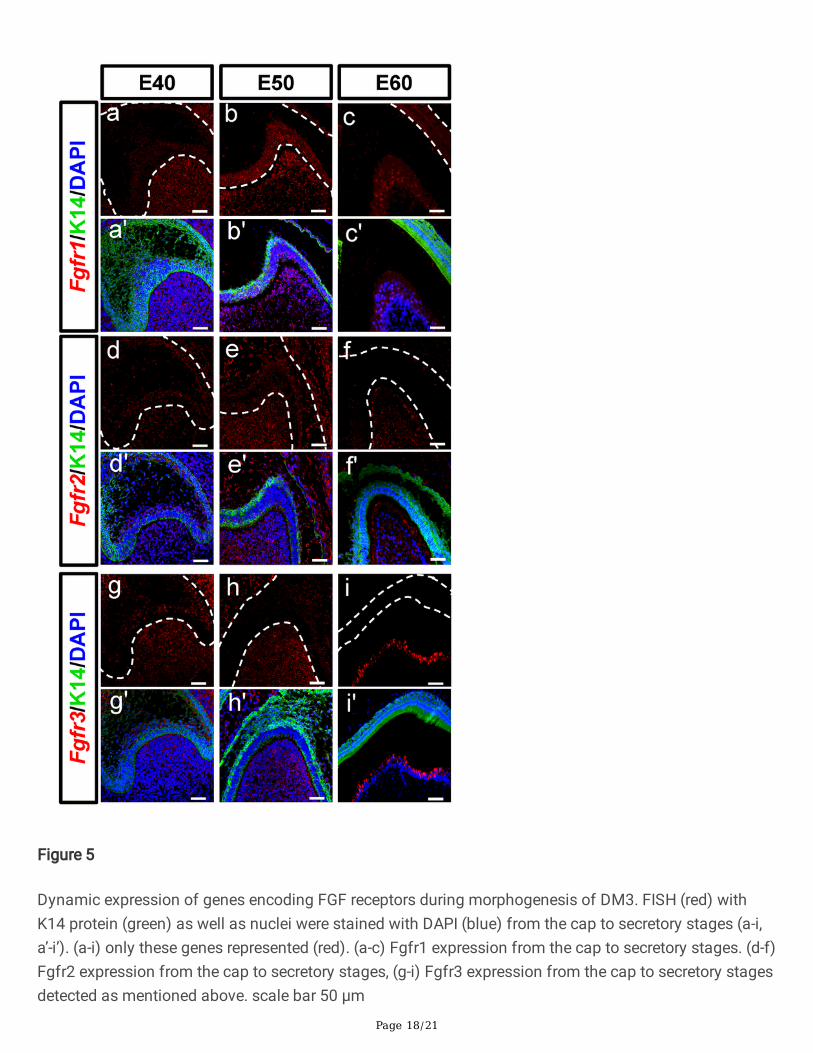
FGF ligands bind FGF receptors on the surface of target cells to transfer signals to the nucleus. Weperformed FISH to examine the expression of Fgfr1, Fgfr2, and Fgfr3 mRNAs in DM3 at the threedevelopmental stages. Fgfr1 mRNA was mainly expressed in the DM at E40, and both in the DE and DM

Page 6/21
at E50 (Fig. 5a-b). However, the expression levels were greatly reduced at E60 (Fig. 5c) except for in theodontoblast cell layer and cervical loop (Supplementary Fig. 3a-c). A similar but weaker expressionpattern was observed for Fgfr2 mRNA at the three stages (Fig. 5d-f). Finally, Fgfr3 was mainly presentin the IEE and underlying DM at E40 (Fig. 5g). Interestingly, Fgfr3 expression was restricted to the DMfrom E50 to E60 (Fig. 5h-i), and only limited to secretory pre-odontoblasts at E60.Additionally, Fgfr2 and Fgfr3 transcripts were present in the cervical loop from E40 andE50 (Supplementary Fig. 3d-i). As a result, the mRNA expression levels of FGF receptors weredownregulated in the DE, upregulated in the DM, and restricted in the odontoblasts during odontogenesis.
Quantitative gene expression dynamics related to the FGF pathway and its inhibitors of DM3
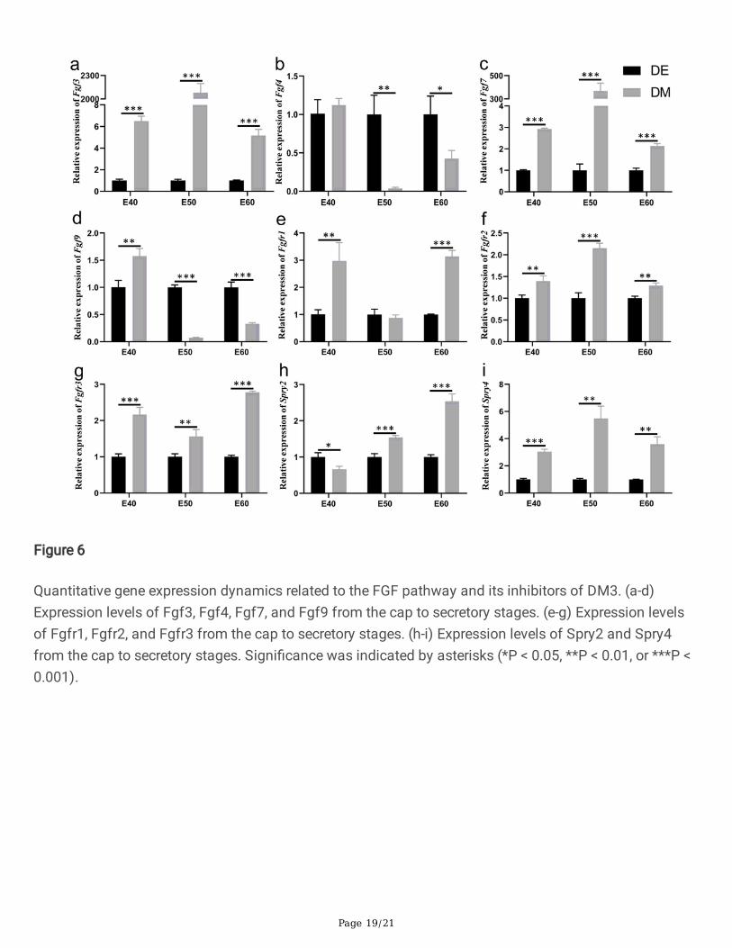
To further investigate the expression dynamics of FGF-related genes and inhibitors, we quanti�ed theirmRNA levels by real time RT-PCR and analyzed the expression differences of each gene in both the DEand DM across the three stages. Tooth germs of DM3 were harvested and the epithelium was separatedfrom the mesenchyme by dispase II treatment before reverse transcription [22]. The results of real timeRT-PCR con�rmed that Fgf3 and Fgf7 showed relatively high expression levels in the DM with respect tothe DE from E40 and E50 (Fig. 6a, c). However, Fgf4 and Fgf9 mRNAs were remarkably more present inthe DE than in the DM from E40 and E50 (Fig. 6b, d). These results were consistent with those of FISHand suggested that these genes were primarily involved in early epithelium growth and formation of theenamel knot. Fgfr1, Fgfr2, Fgfr3 mRNAs were more abundant in the DM than in the DE at the threestages; Fgfr1 and Fgfr3 were highly expressed in the DM at E60 (Fig. 6e-g).
Similar to the expression detected by FISH, the FGF inhibitor Spry2 showed slightly higher expression inthe DE at E40 and markedly higher expression in the DM at E50 and E60 (Fig. 6h). In contrast, Spry4mRNA was more notably accumulated in the DM than in the DE at all three stages (Fig. 6i), with relativelyhigher expression at E60.
DiscussionThe roles of FGF signal in tooth development of miniature pig are not well-understood. In this study, weexamined the dynamic expression of representative FGF ligands and receptors in addition to inhibitors ofDM3 at the E40, E50, and E60. We found that the expression levels of Fgf3, Fgf7, Spry2, Spry4, Fgfr1,Fgfr2, and Fgfr3 were increased in the mesenchyme from E40 to E50, and then concentrated in theodontoblast layer at E60. In contrast, the expression levels of Fgf4 and Fgf9 in the mesenchyme weredecreased from E40 to E60. Most molecules were expressed in the epithelium at the cap stage but weregradually reduced when the tooth bud entered the late bell stage. The related gene pattern diagram wasshown in detail (Fig. 7). Overall, the FGF signaling pathway and sprouty genes may communicatebetween the epithelium and mesenchyme to maintain the tooth morphology and mineralization.
We compared the results with those of the previous studies. We found both the similarities anddifferences among the species of mouse [23], pig and human [24] (Table 1). As an important factor, FGF3has been found to play a signi�cant role in cell proliferation and morphogenesis of tooth development

Page 7/21
[12]. In human, Fgf3 was expressed mainly in the mesenchyme, particularly in the pre-odontoblasts andodontoblast cell layer at the later stage [24]. These �ndings were similar with ours. Additionally, Fgf7expression was detected both in the DE and in the DM at the cap stage of the human tooth [24]. However,Fgf7 may not be involved in mouse tooth development [11]. In our study, the expression of Fgf7 wasremarkable in the DM at the early bell stage but restricted in the odontoblast layer at the secretory stagein the DM3. Fgf4 and Fgf9 mRNAs are indispensable for the DE in mice tooth growth [2]. In our study,Fgf4 and Fgf9 were broadly expressed not only in the DE, but also in the DM from the cap to the early bellstage of DM3, although the functional role in the DM has not been elucidated. According to our results,the ligands of Fgf3, 4, 7, and 9 may play critical roles in the cusp patterning, whereas only Fgf3 and Fgf7may play fundamental role in the cell differentiation of odontoblasts.
Table 1Comparison of the expression of FGF signaling pathway and inhibitors between mouse, pig and humanstage tissues/cells species
Mouse [23] Pig Human [24]
capstage
Enamelknot/Dentalepithelium
Fgf3, Fgf4, Fgf9, Fgf15,Fgf20, Fgf16, Fg17,Fgfr1 b, Fgfr2 b, Fgfr2 c,Spry2, Spry4
Fgf3, Fgf4, Fgf7,Fgf9, Fgfr1,Fgfr2, Fgfr3,Spry2, Spry4
Fgf3, Fgf4, Fgf7,Fgf8, Fgf9,Fgf10, Fgfr1,FGFR2, Fgfr3
dental papilla Fgf3, Fgf10, Fgf16, Fgf17,Fgf18, Fgfr1 c, Fgfr2 c,Spry4
Fgf3, Fgf4, Fgf7,Fgf9, Fgfr1,Fgfr2, Fgfr3,Spry2, Spry4
Fgf3, Fgf4, Fgf7,Fgf8, Fgf9,Fgf10, Fgfr1,Fgfr2, Fgfr3
bellstage
Enamel knot/Dentalepithelium
Fgf4, Fgf9, Fgf16, Fgf20,Fgfr1, Fgfr2 b
Fgf3, Fgf4, Fgf7,Fgf9, Fgfr1,Fgfr2, Spry2
Fgf3, Fgf4, Fgf7,Fgf8, Fgf9,Fgf10, Fgfr1,Fgfr3
dental papilla Fgf3, Fgf10, Fgf15,Fgfr1IIIb, Fgfr1IIIc, Fgfr2IIIc
Fgf3, Fgf4, Fgf7,Fgfr1, Fgfr2,Fgfr3 Spry2,Spry4
Fgf7, Fgf8, Fgf9,Fgfr1
secretorystage
ameloblast/Dentalepithelium
Fgf2, Fgf4, Fgf9, Fgf9,Fgf16, Fgfr1, Fgfr2IIIb
Fgf3, Fgf7, Fgfr1,Spry2
odontoblast/pre-odontoblast
Fgfr1IIIb, Fgfr1IIIc Fgf3, Fgf7,Spry2, Fgfr1,Fgfr2, Fgfr3,
Spry2, Spry4
dental papilla Fgf15 Fgf3, Fgfr1,Fgfr2, Spry2,Spry4

Page 8/21
FGF ligands bind to its receptors and activate intracellular signal transduction pathways by inducingreceptor phosphorylation during tooth growth. In humans, FGF7 binds speci�cally to FGFR2, whereasFGF3 and FGF10 bind to both FGFR1 and FGFR2 [25–26]. With the transgenic mice, Fgfr1 null alleleheterozygosity reduced the frequency of supernumerary tooth formation to 60%, and Fgfr2 null alleleheterozygosity reduced it to 0% [27–28]. In our study, Fgfr1, Fgfr2, and Fgfr3 were mainly expressed inDM cells at the three stages of DM3. However, these receptors related to gene expression and functionnetworks require further analysis in miniature pigs.
Acting as inhibitors of FGF signaling, different sprouty genes cooperate to ensure the correct morphologyand size of developing teeth. In mice, Spry2 expression can be observed throughout the epithelium,whereas Spry4 mRNA accumulates exclusively in the dental mesenchyme to prevent the development ofsupernumerary teeth [12]. In the mouse incisor, which is a continuously growing tooth, Spry2 and Spry4restrict the differentiation of enamel-secreting ameloblasts to the labial side, thereby allowing asymmetricenamel deposition [29]. In this study, we found that, unlike in mice [12], Spry2 was expressed in the DE�rstly, then expressed in DE and DM but expressed mostly in the DM from the cap to the later bell stage,and Spry2 may be transferred from the epithelium to the dental mesenchyme to regulate toothdevelopment. Spry4 expression was enhanced in the DM from the cap stage to the secretory stage. As theinhibitors of FGF signaling, the Sprys may lead to a potential gradient of FGF signaling to regulate toothmorphogenesis and odontogenesis precisely.
The interaction among several FGF family members in the IEE and dental mesenchyme regulates thedifferentiation of dentin and subsequent dentin formation [30]. Particularly, FGF4 and FGF9 expressed inepithelial cells are thought to maintain FGF3 expression in the dental mesenchyme to further regulateepithelial cell proliferation and morphogenesis [31]. Speci�cally, FGF3 stimulates the proliferation of innerenamel epithelial cells, whereas FGF7 stimulates the proliferation of outer enamel epithelial cells [31–32].In contrast, loss of Spry2 function in the epithelium leads to the formation of diastema teeth buds due tothe expression of Fgf3 in the mesenchyme, which was su�cient to control expression of Shh andperhaps also Fgf4. The normal function of Spry4 in the mesenchyme is to prevent the perception of anyepithelial FGF signals, including FGF4 and FGF9 produced in the adjacent M1 tooth germ, to induce ormaintain FGF3 expression [12]. Based on the adverse localization and relevant pattern of Fgf4 and Spry4in the DE as well as Fgf3 and Spry2 in the DM of DM3, we predicted the existence of a feedbackmechanism that precisely regulates FGF signaling to mediate the interaction between the epithelium andmesenchyme during tooth development, which is needed to further exploration (Supplementary Fig. 4).
ConclusionsIn this study, we revealed the spatiotemporal distribution and the potential gradient of FGF and Sproutymolecules during tooth odontogenesis in large mammals. Modulation of the epithelium-mesenchymeinteractions by �ne-tuning the FGF pathway may promote the cusp patterning and dental calci�cation inthe future studies.

Page 9/21
MethodsAnimals
Pregnant miniature pigs were obtained from the Animal Science Institute of Chinese AgricultureUniversity. The gestation age was calculated from the day of insemination. Pregnancy was veri�ed by B-type ultrasonography. All procedures were approved by the Animal Care Use Committee of CapitalMedical University (Beijing, China) (Permit Number: AEEI-T-212). Pregnant miniature pigs wereanesthetized and sacri�ced as previously described [33]. The DM3 and mandible were harvested atembryonic days 40 (E40), E50, and E60, which corresponded to the cap, early, and late bell (secretory)stages.
Tissue preparation for HE and immuno�uorescence
Mandibular samples were �xed with paraformaldehyde, 4% in phosphate-buffered saline (PBS), at 4°Covernight. The samples were rinsed with PBS twice before being decalci�ed with 10% EDTA-PBS for 3–14days according to the degree of calci�cation. The samples were dehydrated in an ethanol series andembedded in para�n. Para�n-embedded samples were sectioned at 7μm thickness for later staining.Sections were stained with hematoxylin and eosin and observed by whole slide image (Pannoramic scan,3DHistech, Budapest, Hungary). The images were processed with Case Viewer software (3DHistech,Budapest, Hungary). Immuno�uorescence (IF) was performed as described previously [34]. Brie�y,depara�nized sections were microwaved for 3 min in citrate buffer (pH 6.0). Then the slides werereheated with adding additional buffer, which processes were repeated for three times for antigenretrieval. When the slides were cooled down to room temperature, non-speci�c antigens were blocked with10% normal goat serum (C-0005, Bioss, Beijing, China) for 1 h. The sections were incubated with primaryantibodies at 4°C overnight. The primary antibodies used in this research were as follows: rabbit anti-FGF3 antibody (bs-1255R, Bioss, Beijing, China), rabbit anti-FGF4 antibody (bs-1256R, Bioss, Beijing,China), rabbit anti-KGF antibody (bs-0734R, Bioss, Beijing, China), rabbit anti-FGF8 antibody (bs-0735R,Bioss, Beijing, China), and rabbit anti-FGF9 antibody (bs-5906R, Bioss, Beijing, China). Donkey anti-RabbitIgG (H+L), Alexa Fluor 594 (A-21207; Thermo, CA, USA) as secondary antibody was subsequently appliedat 37°C for 1 h. The nuclei were stained with DAPI (F6057, Sigma, St. Louis, MO, USA). Finally, thesections were observed using confocal microscopy (TCS SP8, Leica, Wetzlar, Germany). Objective lenseswere used 20× and 40×, N.A. 0.75. The images were processed with LAS AF software.
Fluorescence in situ hybridization
Fluorescence in situ hybridization (FISH) staining was performed using tooth tissues �xed in 4% DEPC-treated paraformaldehyde/PBS, embedded in para�n, and sectioned at 7μm. The procedure for non-radioactive in situ hybridization was described previously [35]. Brie�y, total RNA was extracted from DM3tooth germs at E40–E60. Sequences of the degenerate primers for Fgf3, 4, 7, 9, Fgfr1, 2, 3, and Spry2, 4were listed in Supplementary Table 1. After reverse transcriptase-polymerase chain reaction, the correct-sized bands were extracted from agarose gels and their DNA sequences were determined. Digoxigenin-

Page 10/21
(DIG)-labeled RNA probes were synthesized with DIG-UTP and T7 RNA polymerase (10881767001; Roche,Basel, Switzerland) and DIG RNA labeling mix solution (11277073910; Roche, Basel, Switzerland).Sections were prepared using standard procedures and then incubated in horseradish peroxidase-conjugated, polyclonal sheep anti-digoxigenin antibody (90420, Merck Millipore) at 37°C for 4 h.Immunoreactive cells were visualized with TSA Plus Fluorescence Kits (NEL741001KT, PerkinElmer,Massachusetts, USA). Para�n sections were processed for mucin 2 staining with rabbit anti-Cytokeratin-14 (ab119695, Abcam, Cambridge, England). Donkey anti-Rat IgG (H+L), Alexa Fluor 488 (A-21208;Thermo, CA, USA) as secondary antibody was subsequently applied at 37°C for 1 h. Meanwhile. Slideswere counterstained with DAPI, mounted and imaged on a Leica TCS SP8 confocal microscope. Objectivelenses were used 20× and 40×, N.A. 0.75. The images were processed with LAS AF software.
Real-time RT-PCR
DM3 tooth germs at E40, E50, and E60 were harvested and the dental epithelium was separated from themesenchyme by dispase from miniature pigs described previously [22]. Total RNA was extracted withRNeasy Mini Kit (74106, Qiagen, Hilden, Germany) according to its supplied method. Reversetranscription was performed using the SuperScript III First-Strand Synthesis System (18080051; ThermoFisher Scienti�c). Real-time RT-PCR was conducted in triplicate using SYBR Green PCR Master Mix(A25742; Applied Biosystems, Foster City, CA, USA) on a CFX96 Touch Real-time RT-PCR DetectionSystem (Bio-Rad Laboratories, Hercules, CA, USA), after which melting curve analysis was performed.Expression level of each gene was normalized to that of Gapdh. The relative expression of each gene wasdetermined using the 2-ΔΔCT method. Forward and reverse primers for Fgf3, 4, 7, 9, Fgfr1, 2, 3, and Spry2,4 and Gapdh were listed in Supplementary Table 2.
Statistical analysis
Statistical analysis was performed using GraphPad Prism 8.3 software. One-way analysis of varianceand Newman-Keuls multiple comparison test were used to calculate statistical signi�cance. Differenceswere considered to be statistically signi�cant if p<0.05 (*), p<0.01 (**), or p<0.001 (***).
AbbreviationsFGF: �broblast growth factor; Fgfr: FGF receptors; Spry: Sprouty; DM3: the third deciduous molar; E40:embryonic day 40; E50: embryonic day 50; E60:embryonic day 60; DE: dental epithelium; DM: dentalmesenchyme; IEE: inner enamel epithelium; OEE: outer enamel epithelium; SR: stellate reticulum; O:odontoblasts; E: Enamel; pD: pre-dentin; FISH: �uorescence in situ hybridization; IF: immuno�uorescence;Gapdh: glyceraldehyde-3-phosphate dehy-drogenase; RT-PCR: Reverse Transcription-quantitativePolymerase Chain Reaction; K14: Cytokeratin 14
DeclarationsEthics approval and consent to participate

Page 11/21
All procedures were approved by the Animal Care Use Committee of Capital Medical University (Beijing,China) (Permit Number: AEEI-2018-212).
Consent for publication
Not applicable.
Availability of data and material
All data generated or analyzed during this study are included in this published article and itssupplementary information �les.
Competing interests
Authors declare no con�ict of interests.
Funding
This work was supported by grants from the Chinese Research Unit of Tooth Development andRegeneration, CAMSI Innovation Fund for Medical Sciences, No. 2019-12M-5-031; National NaturalScience Foundation of China (91649124 to S.W.); Beijing Municipal Science & Technology CommissionNo. Z181100001718208; Beijing Municipal Education Commission No. 119207020201; and BeijingHospitals Authority of Hospitals’ Mission Plan, code SML20151401; and Beijing MunicipalityGovernment grants (Beijing Scholar Program- PXM2018_014226_000021;PXM2018_193312_000006_0028S643_FCG; PXM2019_014226_000011; PXM2020_014226_000005;Z181100001718208).
Authors’ contributions
SLW and SXW contributed equally to this work. WWG performed the majority of experiments, withadditional contribution from RZ, FW, JYW, and JSW. WWG, LH and CMZ analyzed the data and preparedthe �rst draft of the manuscript, which was further edited by SLW and SXW. RZ kindly provided thetechnical support of FISH. The author(s) read and approved the �nal manuscript.
Acknowledgements
We acknowledge the excellent technical assistance from the Transgenic Facility, Animal Facility, andImaging Core at the University of Capital Medical and Capital Medical University School of Stomatology.
References1. Zhang YD, Chen Z, Song YQ, Liu C, Chen YP: Making a tooth: growth factors, transcription factors,
and stem cells. Cell Res 2005, 15(5):301–316.

Page 12/21
2. Nie X, Luukko K, Kettunen P: FGF signalling in craniofacial development and developmentaldisorders. Oral Dis 2006, 12(2):102–111.
3. Itoh N, Ornitz DM: Evolution of the Fgf and Fgfr gene families. Trends Genet 2004, 20(11):563–569.
4. Itoh N, Ornitz DM: Fibroblast growth factors: from molecular evolution to roles in development,metabolism and disease. J Biochem 2011, 149(2):121–130.
5. Popovici C, Roubin R, Coulier F, Birnbaum D: An evolutionary history of the FGF superfamily.Bioessays 2005, 27(8):849–857.
�. Maddaluno L, Urwyler C, Werner S: Fibroblast growth factors: key players in regeneration and tissuerepair. Development 2017, 144(22):4047–4060.
7. Liu C, Gu S, Sun C, Ye W, Song Z, Zhang Y, Chen Y: FGF signaling sustains the odontogenic fate ofdental mesenchyme by suppressing beta-catenin signaling. Development 2013, 140(21):4375–4385.
�. Jernvall J, Kettunen P, Karavanova I, Martin LB, Thesleff I: Evidence for the role of the enamel knot asa control center in mammalian tooth cusp formation: non-dividing cells express growth stimulatingFgf-4 gene. Int J Dev Biol 1994, 38(3):463–469.
9. Vaahtokari A, Aberg T, Jernvall J, Keranen S, Thesleff I: The enamel knot as a signaling center in thedeveloping mouse tooth. Mech Dev 1996, 54(1):39–43.
10. Kettunen P, Thesleff I: Expression and function of FGFs-4, -8, and – 9 suggest functional redundancyand repetitive use as epithelial signals during tooth morphogenesis. Dev Dyn 1998, 211(3):256–268.
11. Kettunen P, Laurikkala J, Itaranta P, Vainio S, Itoh N, Thesleff I: Associations of FGF-3 and FGF-10with signaling networks regulating tooth morphogenesis. Dev Dyn 2000, 219(3):322–332.
12. Klein OD, Minowada G, Peterkova R, Kangas A, Yu BD, Lesot H, Peterka M, Jernvall J, Martin GR:Sprouty genes control diastema tooth development via bidirectional antagonism of epithelial-mesenchymal FGF signaling. Dev Cell 2006, 11(2):181–190.
13. Tapaltsyan V, Charles C, Hu J, Mindell D, Ahituv N, Wilson GM, Black BL, Viriot L, Klein OD:Identi�cation of novel Fgf enhancers and their role in dental evolution. Evol Dev 2016, 18(1):31–40.
14. Charles C, Lazzari V, Tafforeau P, Schimmang T, Tekin M, Klein O, Viriot L: Modulation of Fgf3 dosagein mouse and men mirrors evolution of mammalian dentition. Proc Natl Acad Sci U S A 2009,106(52):22364–22368.
15. Hanafusa H, Torii S, Yasunaga T, Nishida E: Sprouty1 and Sprouty2 provide a control mechanism forthe Ras/MAPK signalling pathway. Nat Cell Biol 2002, 4(11):850–858.
1�. Mason JM, Morrison DJ, Basson MA, Licht JD: Sprouty proteins: multifaceted negative-feedbackregulators of receptor tyrosine kinase signaling. Trends Cell Biol 2006, 16(1):45–54.
17. Marangoni P, Charles C, Tafforeau P, Laugel-Haushalter V, Joo A, Bloch-Zupan A, Klein OD, Viriot L:Phenotypic and evolutionary implications of modulating the ERK-MAPK cascade using the dentitionas a model. Sci Rep 2015, 5:11658.
1�. Percival CJ, Marangoni P, Tapaltsyan V, Klein O, Hallgrimsson B: The Interaction of GeneticBackground and Mutational Effects in Regulation of Mouse Craniofacial Shape. G3 (Bethesda) 2017,

Page 13/21
7(5):1439–1450.
19. Wang S, Liu Y, Fang D, Shi S: The miniature pig: a useful large animal model for dental and orofacialresearch. Oral Dis 2007, 13(6):530–537.
20. Xu J, Zheng Z, Fang D, Gao R, Liu Y, Fan ZP, Zhang CM, Wang SL: Early-stage pathogenic sequenceof jaw osteoradionecrosis in vivo. J Dent Res 2012, 91(7):702–708.
21. Wang F, Li Y, Wu X, Yang M, Cong W, Fan Z, Wang J, Zhang C, Du J, Wang S: Transcriptome analysisof coding and long non-coding RNAs highlights the regulatory network of cascade initiation ofpermanent molars in miniature pigs. BMC Genomics 2017, 18(1):148.
22. Wang F, Xiao J, Cong W, Li A, Song T, Wei F, Xu J, Zhang C, Fan Z, Wang S: Morphology andchronology of diphyodont dentition in miniature pigs, Sus Scrofa. Oral Dis 2014, 20(4):367–379.
23. Du W, Du W, Yu H: The Role of Fibroblast Growth Factors in Tooth Development and Incisor Renewal.Stem Cells Int 2018, 2018:7549160.
24. Huang F, Hu X, Fang C, Liu H, Lin C, Zhang Y, Hu X: Expression pro�le of critical genes involved inFGF signaling pathway in the developing human primary dentition. Histochem Cell Biol 2015,144(5):457–469.
25. Miki T, Bottaro DP, Fleming TP, Smith CL, Burgess WH, Chan AM, Aaronson SA: Determination ofligand-binding speci�city by alternative splicing: two distinct growth factor receptors encoded by asingle gene. Proc Natl Acad Sci U S A 1992, 89(1):246–250.
2�. Hoffman MP, Kidder BL, Steinberg ZL, Lakhani S, Ho S, Kleinman HK, Larsen M: Gene expressionpro�les of mouse submandibular gland development: FGFR1 regulates branching morphogenesis invitro through BMP- and FGF-dependent mechanisms. Development 2002, 129(24):5767–5778.
27. Trokovic N, Trokovic R, Mai P, Partanen J: Fgfr1 regulates patterning of the pharyngeal region. GenesDev 2003, 17(1):141–153.
2�. Yu K, Xu J, Liu Z, Sosic D, Shao J, Olson EN, Towler DA, Ornitz DM: Conditional inactivation of FGFreceptor 2 reveals an essential role for FGF signaling in the regulation of osteoblast function andbone growth. Development 2003, 130(13):3063–3074.
29. Klein OD, Lyons DB, Balooch G, Marshall GW, Basson MA, Peterka M, Boran T, Peterkova R, MartinGR: An FGF signaling loop sustains the generation of differentiated progeny from stem cells inmouse incisors. Development 2008, 135(2):377–385.
30. Koussoulakou DS, Margaritis LH, Koussoulakos SL: A curriculum vitae of teeth: evolution, generation,regeneration. Int J Biol Sci 2009, 5(3):226–243.
31. Harada H, Toyono T, Toyoshima K, Yamasaki M, Itoh N, Kato S, Sekine K, Ohuchi H: FGF10 maintainsstem cell compartment in developing mouse incisors. Development 2002, 129(6):1533–1541.
32. Harada H, Ohshima H: New perspectives on tooth development and the dental stem cell niche. ArchHistol Cytol 2004, 67(1):1–11.
33. Li A, Song T, Wang F, Liu D, Fan Z, Zhang C, He J, Wang S: MicroRNAome and expression pro�le ofdeveloping tooth germ in miniature pigs. PLoS One 2012, 7(12):e52256.

Page 14/21
34. Wu X, Hu J, Li G, Li Y, Li Y, Zhang J, Wang F, Li A, Hu L, Fan Z et al: Biomechanical stress regulatesmammalian tooth replacement via the integrin beta1-RUNX2-Wnt pathway. EMBO J 2020,39(3):e102374.
35. Li XB, Yang G, Zhu L, Tang YL, Zhang C, Ju Z, Yang X, Teng Y: Gastric Lgr5(+) stem cells are thecellular origin of invasive intestinal-type gastric cancer in mice. Cell Res 2016, 26(7):838–849.
Figures
Figure 1
Morphology of DM3 across developmental stages in miniature pig. (a-i) H&E staining of different stagesduring tooth germ morphogenesis; Boxed regions in a, d, g were magni�ed in b-c, e-f, h-i. (a-c) Tooth germdeveloped into the cap stage at embryonic day 40 (E40). The epithelium folded into the bud and the innerenamel epithelium (IEE) and outer enamel epithelium (OEE) were separated. The primary enamel knot

Page 15/21
began forming at the tip of IEE. The cervical loop was formed by the IEE and OEE joined with stellatereticulum (SR). (d-f) At E50, the DM3 reached the early bell stage and secondary enamel knots began toform. The outer layer of dental mesenchyme (DM) cells attached to the basement membrane of IEE. (g-i)At E60, the DM3 germ developed into late bell (secretory stage), with parts of the IEE and dental papillacells at the cusp tip differentiated into ameloblasts and odontoblasts (O). Enamel (E) and pre-dentin (pD)were secreted. Scale bars represent 100μm, 200μm, 500 μm (a, d, g) and 50 μm (b, c, e, f, h, i)
Figure 2

Page 16/21
Dynamic expression of genes encoding FGF ligands during morphogenesis of DM3. (a-l, a’-l’)Fluorescence in situ hybridization (FISH) showing the expression of genes (red) withimmuno�uorescence (IF) for Cytokeratin14 (K14) protein (green) to clarify the epithelium, and nuclei werestained with DAPI (blue) from the cap to late bell stages. (a-l) only these genes are represented (red); (a-c)Fgf3 expression from the cap to late bell stages; (d-f) Fgf4 expression from the cap to secretory stages;(g-i) Fgf7 and (j-l) Fgf9 expression from the cap to late bell stages. scale bar 50 μm
Figure 3
Dynamic expression of genes encoding FGF inhibitors during morphogenesis of DM3. FISH (red) withK14 protein (green) as well as nuclei were stained with DAPI (blue) from the cap to secretory stages (a-f,a’-f’). (a-f) only these genes represented (red). (a-c) Spry2 expression from the cap to secretory stages; (d-f) Spry4 expression from the cap to secretory stages. scale bar 50 μm

Page 17/21
Figure 4
Distribution of FGF ligands during morphogenesis of DM3. (a-l, a’-l’) Detection of FGF proteins expressionby IF from the cap to secretory stages. (a-c, a’-c’) FGF3 expression from the cap to secretory stages. (d-f,d’-f’) FGF4 expression from the cap to secretory stages. (g-i, g’-i’) FGF7 and (j-l, j’-l’) FGF9 expression fromthe cap to secretory stages. scale bar 50 μm
Page 18/21
Figure 5
Dynamic expression of genes encoding FGF receptors during morphogenesis of DM3. FISH (red) withK14 protein (green) as well as nuclei were stained with DAPI (blue) from the cap to secretory stages (a-i,a’-i’). (a-i) only these genes represented (red). (a-c) Fgfr1 expression from the cap to secretory stages. (d-f)Fgfr2 expression from the cap to secretory stages, (g-i) Fgfr3 expression from the cap to secretory stagesdetected as mentioned above. scale bar 50 μm
Page 19/21
Figure 6
Quantitative gene expression dynamics related to the FGF pathway and its inhibitors of DM3. (a-d)Expression levels of Fgf3, Fgf4, Fgf7, and Fgf9 from the cap to secretory stages. (e-g) Expression levelsof Fgfr1, Fgfr2, and Fgfr3 from the cap to secretory stages. (h-i) Expression levels of Spry2 and Spry4from the cap to secretory stages. Signi�cance was indicated by asterisks (*P < 0.05, **P < 0.01, or ***P <0.001).

Page 20/21
Figure 7
Pattern diagram of gene expression dynamics of the FGF pathway and its inhibitors of DM3. Lighter colormeans less expression, darker color means higher relative expression. (DM: dental mesenchyme, DE:dental epithelium)
Supplementary Files
This is a list of supplementary �les associated with this preprint. Click to download.
Supplementaltable.docx

Page 21/21
SupplementaryFigure1.tif
SupplementaryFigure2.tif
SupplementaryFigure3.tif
SupplementaryFigure4.tif