down-regulation of wnt10a affects odontogenesis and proliferation
TRANSCRIPT
Biochemical and Biophysical Research Communications 434 (2013) 717–721
Contents lists available at SciVerse ScienceDirect
Biochemical and Biophysical Research Communications
journal homepage: www.elsevier .com/locate /ybbrc
Down-regulation of Wnt10a affects odontogenesis and proliferation inmesenchymal cells
Yang Liu, Dong Han, Lei Wang, Hailan Feng ⇑Department of Prosthodontics, Peking University School and Hospital of Stomatology, 22 Zhongguancun South Avenue, Haidian District, Beijing 100081, PR China
a r t i c l e i n f o
Article history:Received 5 March 2013Available online 18 April 2013
Keywords:Wnt10aTooth developmentCell proliferation
0006-291X/$ - see front matter � 2013 Elsevier Inc. Ahttp://dx.doi.org/10.1016/j.bbrc.2013.03.088
⇑ Corresponding author. Fax: +86 010 62173402.E-mail addresses: [email protected] (Y.
(D. Han), [email protected] (L. Wang), kqfeng
a b s t r a c t
The WNT10a mutation has been found in patients with abnormal odontogenesis. In mice, Wnt10a expres-sion is found in the tooth germ, but its role has not yet been elucidated. We aimed to investigate the roleof Wnt10a in odontogenesis. Mesenchymal cells of the first mandibular molar germ at the bell stage wereisolated, transfected with Wnt10a SiRNA or plasmid, and reassociated with epithelial part of the molargerm. Scrambled SiRNA or empty vector was used in the control group. The reassociated tooth germswere transplanted into mice subrenal capsules. After gene modification, dental mesenchymal cells cul-tured in vitro were checked for cell proliferation and the expression of Dspp was examined. All 12 reas-sociated tooth germs in the control group resumed odontogenesis, while only 5 of 12 in the Wnt10aknockdown group developed into teeth. After Wnt10a knockdown, the mesenchymal cells culturedin vitro presented repressed proliferation. Wnt10a knockdown and overexpression led to both down-and up-regulation of Dspp. We conclude that the down-regulation of Wnt10a impairs odontogensis andcell proliferation, and that Wnt10a regulates Dspp expression in mesenchymal cells. These findings helpto elucidate the mechanism of abnormal tooth development in patients with the WNT10A mutation.
� 2013 Elsevier Inc. All rights reserved.
1. Introduction
The tooth is a critical component of the oral system, whichperforms mastication, assists in pronunciation, and maintains anaesthetic appearance of the face. Abnormalities in odontogenesisinclude changes in the number, morphology, or structure of thetooth, jeopardizing normal oral function and aesthetics.
Adaimy et al. reported a mutation in WNT10A to be associatedwith an auotosomal recessive ectodermal dysplasia, the odonto-onycho-dermal dysplasia (OODD), of which the phenotypeincluded severe hypodotia [1]. Bohring et al. reported that WNT10Amutations caused not only OODD, but also monosymptomatic oli-godontia and Schöpf-Schulz-Passarge syndrome, another form ofectodermal dysplasia that presents tooth abnormality [2]. We alsofound a high prevalence of the WNT10A mutation in patients withnon-syndrome oligodontia.
The Wnt signal pathway plays an important role in develop-ment and disease [3]. Wnt/b-catenin signaling is active at multiplestages of odontogenesis [4]. Deletion of the b-catenin gene in earlytooth mesenchymal cells causes developmental arrest at the bud-to-cap transition [5]. Mouse tooth buds expressing stabilizedb-catenin in the epithelium results in abnormal odontogenesis [6].
ll rights reserved.
Liu), [email protected]@bjmu.edu.cn (H. Feng).
As a member of the canonical Wnt pathway, Wnt10a has beenreported to regulate adipogenesis and osteoblastogenesis in mes-enchymal stem cells [7]. In odontogenesis, Wnt10a expressionwas first detected in the incisor domain, distal to the molar anlagenat embryonic day 12 (E12). Following this, it is expressed at the tipof the epithelial bud at E13.5, and in the enamel knot at E14.5 [8].From the early bell stage (E16), Wnt10a expression shifts from thesecondary enamel knot in the epithelium to the underlying mesen-chymal cells. In the odontoblast layer, Wnt10a shows a similarexpression pattern to dentin sialophosphoprotein (Dspp). Transientoverexpression of Wnt10a in fibroblast cell lines cultured on Matri-gel induces Dspp mRNA [9]. Cell surface heparan sulfate proteogly-can, which is involved in odotogenesis, binds to Wnt10a[10]. Thesereports suggest a potential role of Wnt10a in odontoblast celldifferentiation.
Cell proliferation is commonly regulated by Wnt signaling [3].For example, Wnt9b�/� mice show significantly retardedoutgrowth of the nasal and maxillary processes due to reducedproliferation of mesenchymal cells, leading to cleft lip and palate[11]. Wnt5a, a representative of noncanonical Wnts, affects cellproliferation in the tooth germ; Wnt5a-deficient mice developsmaller and abnormally patterned teeth [12]. Yet, the effect ofWnt10a on proliferation has not been reported to date.
Bioengineered organ germs made by reassociation of isolatedcells from the dental germ resumes odontogenesis and can growinto relatively normal teeth [13,14]. Moreover, after knockdown
718 Y. Liu et al. / Biochemical and Biophysical Research Communications 434 (2013) 717–721
of Msx1 or Dlx2 expression by RNA interference (RNAi) in the den-tal mesenchymal cells, the developmental outcome of reassociatedtooth germ generated from these cells recapitulated the phenotypeof their specific gene knockout mice [15]. This indicated that reas-sociated tooth germ after gene knockdown could serve as a methodto investigate gene function in odontogenesis.
In our study, we applied Wnt10a knockdown and overexpres-sion in dental mesenchymal cells, investigated the influence ofWnt10a down-regulation on odontogenesis and cell proliferation,and the effect of Wnt10a on Dspp expression in dental mesenchy-mal cells.
2. Materials and methods
2.1. Cell preparation
All animal experiments were approved by the Ethics Committeeof Peking University Health Science Center. Embryos at E16 wereobtained from time-mated pregnant ICR mice. In sterile phosphatebuffered saline (PBS) the first mandibular molar germs were dis-sected with a fine needle under a stereomicroscope (JSZ6S, Jian-gnan Novel, China). Isolation of dental mesenchymal cells wasperformed as previously described [14]. Briefly, the tooth germswere treated in 1.5 mg/ml Dispase II (Roche, Mannheim, Germany)at 37 �C for 15 min, after which the epithelium was removed with afine needle. The mesenchyme was then dissociated by gentlepipetting after treatment with 0.25% trypsin (Sigma) at 37 �C for10 min. Cells were resuspended and cultured in Dulbecco’s Modi-fied Eagle’s Medium (DMEM, Gibco) containing 20% fetal bovineserum (FBS, Hyclone, Thermo Scientific) with 100 U/ml penicillin,100 lg/ml streptomycin, and 250 ng/ml amphotericin-B (antibioticantimycotic solution, Sigma, USA) in a humidified incubator at37 �C with 5% CO2. Mesenchymal cells were identified by immuno-histochemistry using anti-vimentin and cytokeratin-14 antibodies(Bioss, China).
2.2. Genetic modification
pBluescript-Wnt10a was kindly provided by Dr. Gregory M.Shackleford of the University of Southern California [16]. Dentalmesenchymal cells were trypsinized and washed. Then 1 � 106 -cells were transfected with 10 lg of pBluescript-Wnt10a or emptyvector by Lipofectamine 2000 (Invitrogen, USA), according to themanufacturer’s instructions. To knockdown Wnt10a, stealth RNAiduplexes (Invitrogen, USA) were introduced using RNAiMAX (Invit-rogen, USA), according to the manufacturer’s instructions. Trans-fection efficiency was assessed by BLOCK-iT Alexa Fluor RedFluorescent Oligo (Invitrogen, USA). Cells transfected with scram-bled SiRNA (Invitrogen, USA) were used for controls. Twenty-fourto 72 h post-transfection, the cells were harvested for RNA andprotein extraction or tooth germ reassociation.
2.3. Tooth germ reassociation and in vitro organ culture
Tooth germ reassociation and in vitro organ culture were per-formed as previously described [13], with a slight modification.Briefly, 24 h after RNAi, primary-cultured dental mesenchymalcells were trypsinized, resuspended, and centrifuged at 3000g for2 min into a cell pellet. The cell pellet was transferred onto a semi-solid medium made from DMEM (Gibco) containing 0.36% agar(Sigma), 20% fetal bovine serum (Hyclone, ThermoScientific),0.18 mg/ml ascorbic acid (Sigma), and 2 mM L-glutamine (Gibco).The pellet was then dissected into small parts, using a fine needleunder a stereomicroscope. Each piece of the cell pellet contained2 � 105 cells. Freshly isolated dental epithelium (enamel organ)
from first mandibular molar at E16 was overlapped with the mese-chymal cell pellet to form a reassociated tooth germ. Twelve reas-sociated tooth germs were made for each of the control andWnt10a-knockdown groups. The reassociated tooth germ was thencultured on the semisolid medium in a humidified incubator at37 �C with 5% CO2 for 24 h before grafting.
2.4. Tooth germ transplantation and hematoxylin-eosin (HE) staining
After 24 h of in vitro culture, the reassociated tooth germs weretransplanted into the subrenal capsules of adult ICR male mice aspreviously described [17]. Briefly, the host mice were anesthetizedby intraperitoneal injection of 0.04 mg/g phentobarbital. Thelumbar region was shaved and disinfected. The left kidney was ex-posed and a small breach was made in the capsule membrane. Thereassociated tooth germs from the control and Wnt10a knockdowngroups were then inserted into the same subrenal capsule. For eachkidney, no more than three germs were grafted. The kidney was re-placed into the abdomen and the incision closed. The grafted tissuewas harvested at 3, 5, 7, and 14 days after transplantation, fixed in4% paraformaldehyde (PFA) for 1 day, and demineralized in 10%ethylenediaminetetraacetic acid (EDTA) for 1 week. Samples werethen paraffin-embedded and sectioned in 4-lm slices. Sectionswere stained with hematoxylin and eosin (HE) for routine histo-logic examination.
2.5. Quantitative real-time polymerase chain reaction (qPCR)
Total RNA was extracted with TRIzol reagent (Invitrogen, Carls-bad, USA), according to the manufacturer’s instructions. Twomicrograms of total RNA was reverse-transcribed to cDNA usingM-MLV reverse transcriptase (Promega, USA) reagents. The DNAresidual was removed by RNase-free DNase (Promega, USA). Quan-titative PCR (qPCR) was performed in an ABI Prism 7000 SequenceDetection System (Applied Biosystems, USA) with SYBR Green re-agent (Roche, USA). The corresponding primer sequences wereWnt10a (sense: 50-CATGCTCGAATGAGACTCCAC-30; anti-sense:50-CCCTACTGTGCGGAACTCAG-30), Dspp (sense: 50-AT TCCGGTT-CCCCAGTTAGTA-30; anti-sense: 50-CTGTTGCTAGTGGTGCTGTT-30),Gapdh (sense: 50-AGGTCGGTGTGAACGGATTTG-30; anti-sense: 50-GGGGTCG TTGATGGCAACA-30). Gapdh served as the endogenousgene control in the samples. The amplification specificity was con-firmed by a melting curve. The relative expression of the targetgenes was calculated using the 2�DDCt method.
2.6. Western blot assay
Cells were harvested and lysed in RIPA buffer containing pro-teinase inhibitors. After concentration measurement, protein sam-ples were separated on a 10% sodium dodecyl sulfate–polyacrylamide gel electrophoresis (SDS–PAGE) and transferredto a polyvinylidene difluoride (PVDF) membrane by western blot-ting. The membranes were blocked in 5% skim milk for 1 h andincubated with antibodies against Wnt10a (Abcam, USA), Dsp(Santa Cruz, USA), and b-actin (Zhongshan Golden Bridge, China)separately at 4 �C overnight. After incubation with peroxidase-linked secondary antibodies, immunoreactive proteins were visu-alized by ECL reagent (Thermo, USA). Relative quantification ofbands in the western blot was performed by Image J software.
2.7. Proliferation assay
Cell proliferation was assessed by Cell Counting Kit-8 (CCK-8,Dojindo, Japan) (an alternative to the MTT assay) and the 5-Ethy-nyl-20-deoxyuridine (EdU, an alternative to BrdU) staining kit (RiboBio, China), according to the manufacturer’s instructions. For the
Y. Liu et al. / Biochemical and Biophysical Research Communications 434 (2013) 717–721 719
CCK-8 test, 5 � 103 cells were seeded into each well of a 96-wellplate. At 6, 24, 48, 72, and 96 h after RNAi, the cells were culturedin a medium containing 10% CCK-8 solution for 2 h. The optic den-sity at 450 nm was measured by an Absorbance Microplate Reader(BioTek, USA). For EdU staining, 4 � 104 cells were seeded into eachwell of a 24-well plate. At 48 and 72 h after RNAi, the cells werecultured in a medium containing 50 lM EdU reagent for 2 h, fixedby 4% PFA, washed, and stained with Apollo staining solution. Nu-clear DNA was stained with DAPI. For each sample, eight indepen-dent zones were randomly chosen and photographed, and positivestaining cells were counted by Image-Pro Plus 6.0 software. In eachgroup, at least 2000 cells were counted and the rate of EdU+/DAPI+
was used to assess cell proliferation.
2.8. Statistical analysis
Statistical significance was determined using the two-tailedStudent’s t-test, assuming equal variances. The chi-squared testwas used to compare rates. The significance was indicated as fol-lows: ⁄P < 0.05; ⁄⁄P < 0.01; ⁄⁄⁄P < 0.001.
3. Results
3.1. Down-regulation of Wnt10a impaired odontogenesis ofreassociated tooth germs
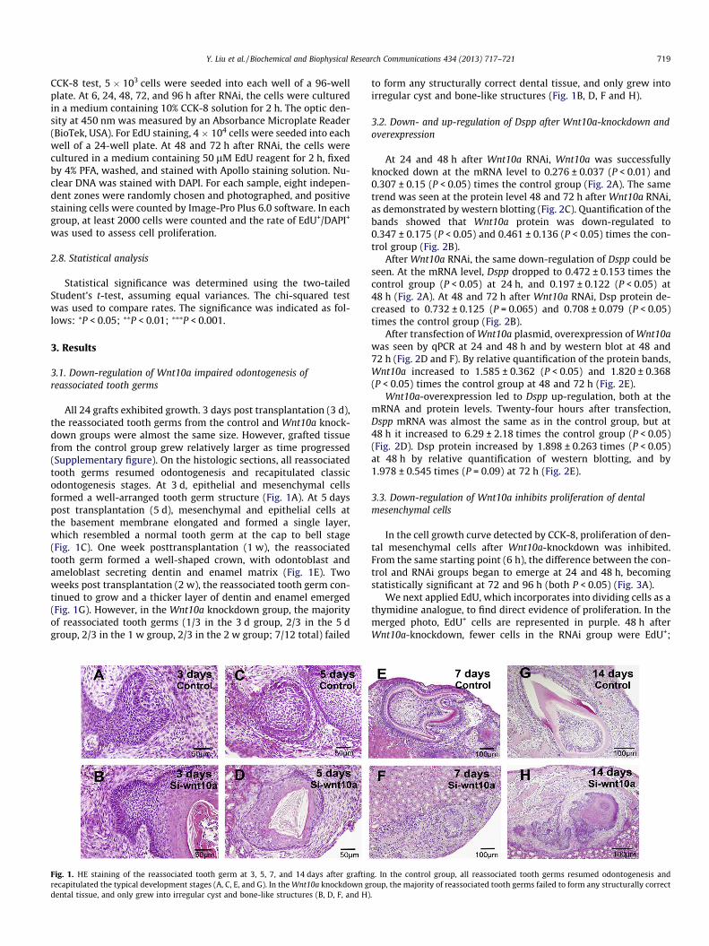
All 24 grafts exhibited growth. 3 days post transplantation (3 d),the reassociated tooth germs from the control and Wnt10a knock-down groups were almost the same size. However, grafted tissuefrom the control group grew relatively larger as time progressed(Supplementary figure). On the histologic sections, all reassociatedtooth germs resumed odontogenesis and recapitulated classicodontogenesis stages. At 3 d, epithelial and mesenchymal cellsformed a well-arranged tooth germ structure (Fig. 1A). At 5 dayspost transplantation (5 d), mesenchymal and epithelial cells atthe basement membrane elongated and formed a single layer,which resembled a normal tooth germ at the cap to bell stage(Fig. 1C). One week posttransplantation (1 w), the reassociatedtooth germ formed a well-shaped crown, with odontoblast andameloblast secreting dentin and enamel matrix (Fig. 1E). Twoweeks post transplantation (2 w), the reassociated tooth germ con-tinued to grow and a thicker layer of dentin and enamel emerged(Fig. 1G). However, in the Wnt10a knockdown group, the majorityof reassociated tooth germs (1/3 in the 3 d group, 2/3 in the 5 dgroup, 2/3 in the 1 w group, 2/3 in the 2 w group; 7/12 total) failed
Fig. 1. HE staining of the reassociated tooth germ at 3, 5, 7, and 14 days after graftinrecapitulated the typical development stages (A, C, E, and G). In the Wnt10a knockdown gdental tissue, and only grew into irregular cyst and bone-like structures (B, D, F, and H)
to form any structurally correct dental tissue, and only grew intoirregular cyst and bone-like structures (Fig. 1B, D, F and H).
3.2. Down- and up-regulation of Dspp after Wnt10a-knockdown andoverexpression
At 24 and 48 h after Wnt10a RNAi, Wnt10a was successfullyknocked down at the mRNA level to 0.276 ± 0.037 (P < 0.01) and0.307 ± 0.15 (P < 0.05) times the control group (Fig. 2A). The sametrend was seen at the protein level 48 and 72 h after Wnt10a RNAi,as demonstrated by western blotting (Fig. 2C). Quantification of thebands showed that Wnt10a protein was down-regulated to0.347 ± 0.175 (P < 0.05) and 0.461 ± 0.136 (P < 0.05) times the con-trol group (Fig. 2B).
After Wnt10a RNAi, the same down-regulation of Dspp could beseen. At the mRNA level, Dspp dropped to 0.472 ± 0.153 times thecontrol group (P < 0.05) at 24 h, and 0.197 ± 0.122 (P < 0.05) at48 h (Fig. 2A). At 48 and 72 h after Wnt10a RNAi, Dsp protein de-creased to 0.732 ± 0.125 (P = 0.065) and 0.708 ± 0.079 (P < 0.05)times the control group (Fig. 2B).
After transfection of Wnt10a plasmid, overexpression of Wnt10awas seen by qPCR at 24 and 48 h and by western blot at 48 and72 h (Fig. 2D and F). By relative quantification of the protein bands,Wnt10a increased to 1.585 ± 0.362 (P < 0.05) and 1.820 ± 0.368(P < 0.05) times the control group at 48 and 72 h (Fig. 2E).
Wnt10a-overexpression led to Dspp up-regulation, both at themRNA and protein levels. Twenty-four hours after transfection,Dspp mRNA was almost the same as in the control group, but at48 h it increased to 6.29 ± 2.18 times the control group (P < 0.05)(Fig. 2D). Dsp protein increased by 1.898 ± 0.263 times (P < 0.05)at 48 h by relative quantification of western blotting, and by1.978 ± 0.545 times (P = 0.09) at 72 h (Fig. 2E).
3.3. Down-regulation of Wnt10a inhibits proliferation of dentalmesenchymal cells
In the cell growth curve detected by CCK-8, proliferation of den-tal mesenchymal cells after Wnt10a-knockdown was inhibited.From the same starting point (6 h), the difference between the con-trol and RNAi groups began to emerge at 24 and 48 h, becomingstatistically significant at 72 and 96 h (both P < 0.05) (Fig. 3A).
We next applied EdU, which incorporates into dividing cells as athymidine analogue, to find direct evidence of proliferation. In themerged photo, EdU+ cells are represented in purple. 48 h afterWnt10a-knockdown, fewer cells in the RNAi group were EdU+;
g. In the control group, all reassociated tooth germs resumed odontogenesis androup, the majority of reassociated tooth germs failed to form any structurally correct.

Fig. 2. Regulation of Dspp expression by Wnt10a. The down-regulation of Dspp after Wnt10a-RNAi was detected by qPCR at the mRNA level (A), and by western blot both fromthe band (C) and relative quantification of the band (B). Up-regulation of Dspp after Wnt10a-transfection could also be seen at the mRNA (D) and protein levels (E and F).
720 Y. Liu et al. / Biochemical and Biophysical Research Communications 434 (2013) 717–721
the same trend was seen at 72 h (Fig. 3B). By determining the rateof EdU+/DAPI+, proliferation in the RNAi group was lower than incontrols at both 48 (18.91 ± 0.99% vs. 23.83 ± 1.17%) and 72 h(19.65 ± 0.76% vs. 25.15 ± 1.78%) (both P < 0.05) (Fig. 3C).
4. Discussion
In this study, we first investigated the effect of Wnt10a down-regulation on odontogenesis in the reassociated tooth germ model.All the reassociated tooth germs in the control group resumedodontogenesis and developed into structurally correct toothcrowns; this was in accordance with other reports [13–15]. Thelow percentage of odontogenesis among reassociated tooth germsin the Wnt10a down-regulation group corresponded to the oligo-dontia in the patient with the WNT10A mutation. Until now,reports of WNT10A focused on cancer research [18] and develop-mental abnormities [1,2]. To the best of our knowledge, this isthe first animal experiment to attribute Wnt10a-loss-of-functionto abnormal odontogenesis.
However, as a rapid preliminary method to investigate genefunction in odontogenesis [15], the reassociated tooth germ modelhas its drawbacks. Although the reassociated tooth germ could de-velop dental crown and root, the morphology of the crown is notidentical to normal ones [19], so it is not appropriate to assess genefunction in determining the crown morphology with this model.Moreover, the subrenal capsule is not the natural physical develop-ment niche as compared to alveolar bone. Thus, our work serves asa preliminary study to unveil the effect of Wnt10a in odntogenesis;it is necessary to generate Wnt10a-knockout mice to further illus-trate the function of Wnt10a in odontogenesis.
During odontogenesis, odontoblast differentiation starts at thelate bell stage in the dental mesenchyme. The expression of Dspp
by odontoblast is pivotal to dentin formation and tooth morpho-genesis [20]. Although Wnt10a and Dspp have been reported toexpress a similar pattern in the developing ondotoblast layer byin situ hybridization [9], there is no direct link between thesetwo genes in primary cultured dental mesenchymal cells. Ourresults demonstrate that Wnt10a knockdown and overexpressionleads to down- and up-regulation of Dspp mRNA and Dsp protein(one of the main translation products of the Dspp gene) in dentalmesenchymal cells. This study provides direct evidence confirmingthat Dspp is the downstream target of Wnt10a in dental mesenchy-mal cells. We thus propose that as an etiological factor, theimpaired WNT10A affects DSPP and odontoblast differentiation,which may jeopardize normal odontogenesis in patients with theWNT10A mutation.
Cell proliferation is necessary for normally sized and patternedorgan development. The Wnt signal often involves proliferation.Deficiency in either Wnt9b or Wnt5a could inhibit cell proliferationand lead to abnormal organ development [11,12]. Small and poorlypatterned teeth can be seen in patients with the WNT10A mutation[21]. To explore the possible cause of this symptom, we first con-ducted a CCK-8 assay, which provided numbers of live cells withwhich to draw a cell proliferation curve. Next, we applied EdU,which incorporates into dividing cells like BrdU, to directly assessthe evidence of proliferation. The results consistently demon-strated repressed proliferation of dental mesenchymal cells afterWnt10a knockdown. To date, this is the first report on the effectof cell proliferation by Wnt10a. This may partially explain thedevelopment of small and poorly patterned teeth in patients withthe WNT10A mutation.
In summary, we have shown impaired odontogenesis afterWnt10a knockdown, confirmed Dspp as the downstream target ofWnt10a in dental mesenchymal cells, and demonstrated repressed

Fig. 3. The effect of Wnt10a on mesenchymal cell proliferation. The cells presented repressed proliferation curves after Wnt10a-knockdown as detected by CCK-8 assay (A). Inthe merged photo of EdU staining (B), the Wnt10a-knockdown group had fewer EdU+ cells (arrowhead). The nucleus was stained with DAPI (arrow). The proliferation rate wasassessed by EdU+/DAPI+; the control group demonstrated a higher level of proliferation 48 and 72 h after Wnt10a-knockdown (C).
Y. Liu et al. / Biochemical and Biophysical Research Communications 434 (2013) 717–721 721
proliferation of dental mesenchymal cells after Wnt10a knock-down. Our results may help to elucidate the mechanism of abnor-mal odontogenesis in patients with the WNT10A mutation.
Acknowledgments
This study was supported by a grant from the National NaturalScience Foundation of China (No. 81271121) and a grant from theConstruction Project for State Key Clinical Discipline. We thank Dr.Gregory M. Shackleford of the University of Southern California forkindly providing Wnt10a plasmid.
Appendix A. Supplementary data
Supplementary data associated with this article can be found, inthe online version, at http://dx.doi.org/10.1016/j.bbrc.2013.03.088.
References
[1] L. Adaimy, E. Chouery, H. Megarbane, S. Mroueh, V. Delague, E. Nicolas, H.Belguith, P. de Mazancourt, A. Megarbane, Mutation in WNT10A is associatedwith an autosomal recessive ectodermal dysplasia: the odonto-onycho-dermaldysplasia, Am. J. Hum. Genet. 81 (2007) 821–828.
[2] A. Bohring, T. Stamm, C. Spaich, C. Haase, K. Spree, U. Hehr, M. Hoffmann, S.Ledig, S. Sel, P. Wieacker, A. Ropke, WNT10A mutations are a frequent cause ofa broad spectrum of ectodermal dysplasias with sex-biased manifestationpattern in heterozygotes, Am. J. Hum. Genet. 85 (2009) 97–105.
[3] C.Y. Logan, R. Nusse, The Wnt signaling pathway in development and disease,Annu. Rev. Cell Dev. Biol. 20 (2004) 781–810.
[4] F. Liu, E.Y. Chu, B. Watt, Y. Zhang, N.M. Gallant, T. Andl, S.H. Yang, M.M. Lu, S.Piccolo, R. Schmidt-Ullrich, M.M. Taketo, E.E. Morrisey, R. Atit, A.A. Dlugosz,S.E. Millar, Wnt/beta-catenin signaling directs multiple stages of toothmorphogenesis, Dev. Biol. 313 (2008) 210–224.
[5] J. Chen, Y. Lan, J.A. Baek, Y. Gao, R. Jiang, Wnt/beta-catenin signaling plays anessential role in activation of odontogenic mesenchyme during early toothdevelopment, Dev. Biol. 334 (2009) 174–185.
[6] E. Jarvinen, I. Salazar-Ciudad, W. Birchmeier, M.M. Taketo, J. Jernvall, I. Thesleff,Continuous tooth generation in mouse is induced by activated epithelial Wnt/beta-catenin signaling, Proc. Natl. Acad. Sci. USA 103 (2006) 18627–18632.
[7] W.P. Cawthorn, A.J. Bree, Y. Yao, B. Du, N. Hemati, G. Martinez-Santibanez, O.A.MacDougald, Wnt6, Wnt10a and Wnt10b inhibit adipogenesis and stimulate
osteoblastogenesis through a beta-catenin-dependent mechanism, Bone 50(2012) 477–489.
[8] H.R. Dassule, A.P. McMahon, Analysis of epithelial-mesenchymal interactionsin the initial morphogenesis of the mammalian tooth, Dev. Biol. 202 (1998)215–227.
[9] T. Yamashiro, L. Zheng, Y. Shitaku, M. Saito, T. Tsubakimoto, K. Takada, T.Takano-Yamamoto, I. Thesleff, Wnt10a regulates dentin sialophosphoproteinmRNA expression and possibly links odontoblast differentiation and toothmorphogenesis, Differentiation 75 (2007) 452–462.
[10] S. Hayano, H. Kurosaka, T. Yanagita, I. Kalus, F. Milz, Y. Ishihara, M.N. Islam, N.Kawanabe, M. Saito, H. Kamioka, T. Adachi, T. Dierks, T. Yamashiro, Roles ofheparan sulfate sulfation in dentinogenesis, J. Biol. Chem. 287 (2012) 12217–12229.
[11] Y.R. Jin, X.H. Han, M.M. Taketo, J.K. Yoon, Wnt9b-dependent FGF signaling iscrucial for outgrowth of the nasal and maxillary processes during upper jawand lip development, Development 139 (2012) 1821–1830.
[12] M. Lin, L. Li, C. Liu, H. Liu, F. He, F. Yan, Y. Zhang, Y. Chen, Wnt5a regulatesgrowth, patterning, and odontoblast differentiation of developing mousetooth, Dev. Dyn. 240 (2011) 432–440.
[13] B. Hu, A. Nadiri, S. Kuchler-Bopp, F. Perrin-Schmitt, H. Peters, H. Lesot, Tissueengineering of tooth crown, root, and periodontium, Tissue Eng. 12 (2006)2069–2075.
[14] K. Nakao, R. Morita, Y. Saji, K. Ishida, Y. Tomita, M. Ogawa, M. Saitoh, Y.Tomooka, T. Tsuji, The development of a bioengineered organ germ method,Nat. Methods 4 (2007) 227–230.
[15] Y. Song, Z. Zhang, X. Yu, M. Yan, X. Zhang, S. Gu, T. Stuart, C. Liu, J. Reiser, Y.Zhang, Y. Chen, Application of lentivirus-mediated RNAi in studying genefunction in mammalian tooth development, Dev. Dyn. 235 (2006) 1334–1344.
[16] J. Wang, G.M. Shackleford, Murine Wnt10a and Wnt10b: cloning andexpression in developing limbs, face and skin of embryos and in adults,Oncogene 13 (1996) 1537–1544.
[17] Y. Zhang, S. Wang, Y. Song, J. Han, Y. Chai, Y. Chen, Timing of odontogenicneural crest cell migration and tooth-forming capability in mice, Dev. Dyn. 226(2003) 713–718.
[18] H. Kirikoshi, S. Inoue, H. Sekihara, M. Katoh, Expression of WNT10A in humancancer, Int. J. Oncol. 19 (2001) 997–1001.
[19] K. Ishida, M. Murofushi, K. Nakao, R. Morita, M. Ogawa, T. Tsuji, The regulationof tooth morphogenesis is associated with epithelial cell proliferation and theexpression of sonic hedgehog through epithelial–mesenchymal interactions,Biochem. Biophys. Res. Commun. 405 (2011) 455–461.
[20] Y. Yamakoshi, Dentinogenesis and dentin sialophosphoprotein (DSPP), J. OralBiosci. 51 (2009) 134.
[21] C. Cluzeau, S. Hadj-Rabia, M. Jambou, S. Mansour, P. Guigue, S. Masmoudi,E. Bal, N. Chassaing, M.C. Vincent, G. Viot, F. Clauss, M.C. Maniere, S.Toupenay, M. Le Merrer, S. Lyonnet, V. Cormier-Daire, J. Amiel, L. Faivre, Y.de Prost, A. Munnich, J.P. Bonnefont, C. Bodemer, A. Smahi, Only four genes(EDA1, EDAR, EDARADD, and WNT10A) account for 90% of hypohidrotic/anhidrotic ectodermal dysplasia cases, Hum. Mutat. 32 (2011) 70–72.