mitochondria, endoplasmic reticulum, golgi apparatus...
TRANSCRIPT
Mitochondria, endoplasmic reticulum, Golgiapparatus, lysosoma, vesicular transport
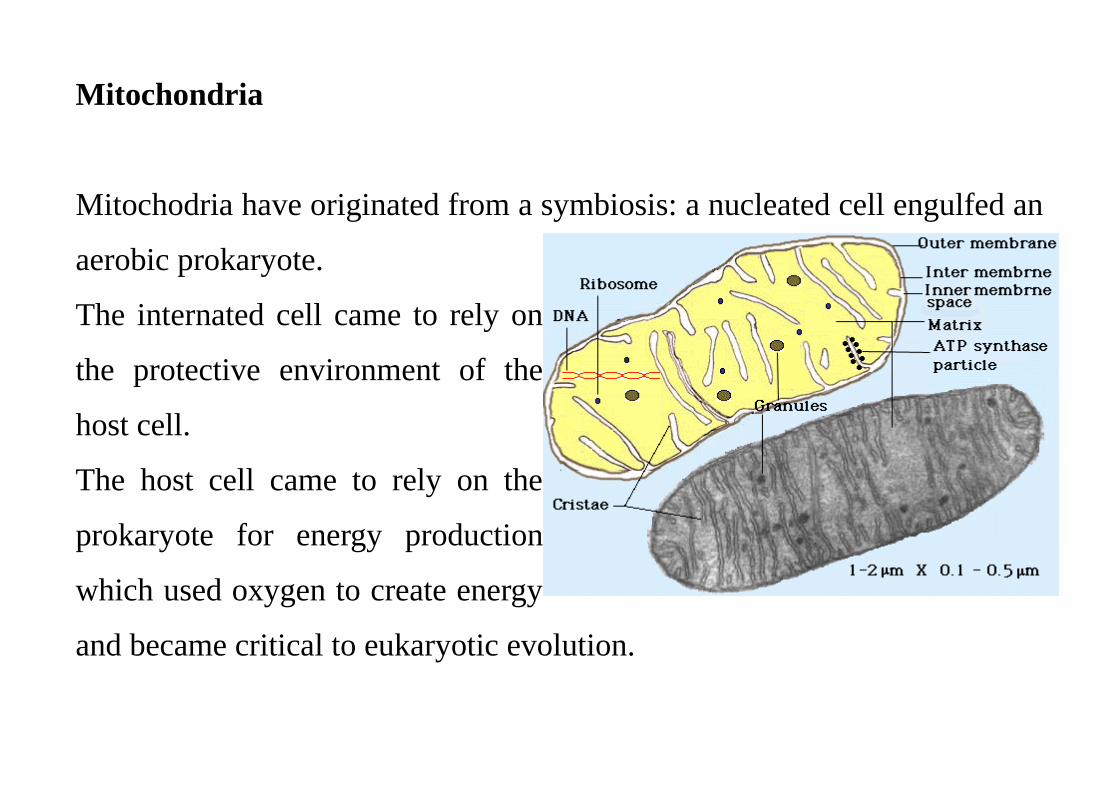
Mitochondria
Mitochodria have originated from a symbiosis: a nucleated cell engulfed an
aerobic prokaryote.
The internated cell came to rely on
the protective environment of the
host cell.
The host cell came to rely on the
prokaryote for energy production
which used oxygen to create energy
and became critical to eukaryotic evolution.
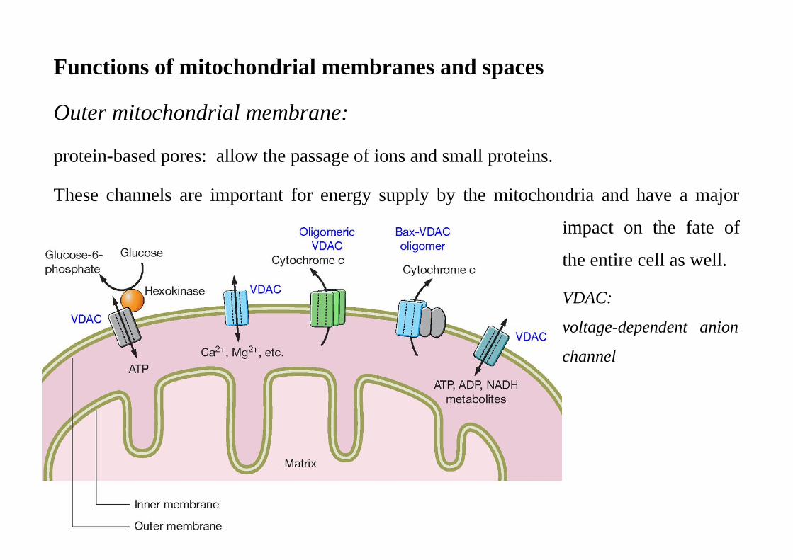
Functions of mitochondrial membranes and spaces
Outer mitochondrial membrane:
protein-based pores: allow the passage of ions and small proteins.
These channels are important for energy supply by the mitochondria and have a major
impact on the fate of
the entire cell as well.
VDAC:
voltage-dependent anion
channel
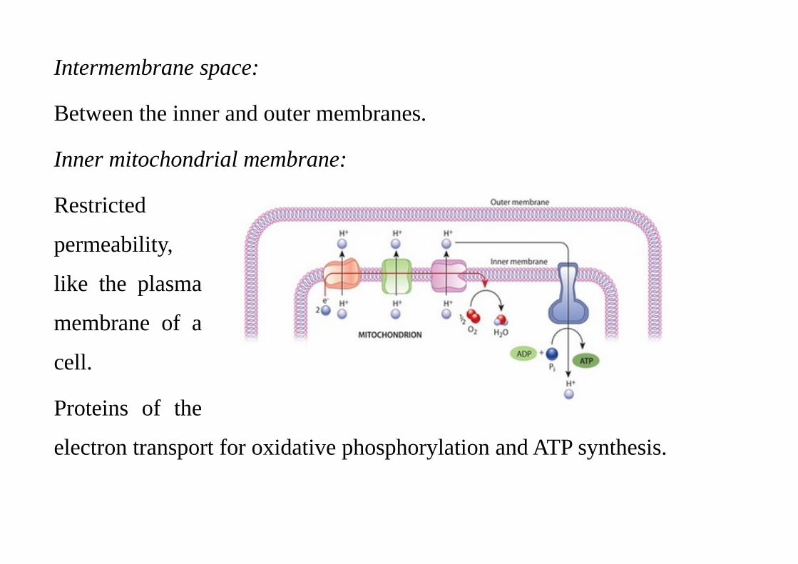
Intermembrane space:
Between the inner and outer membranes.
Inner mitochondrial membrane:
Restricted
permeability,
like the plasma
membrane of a
cell.
Proteins of the
electron transport for oxidative phosphorylation and ATP synthesis.
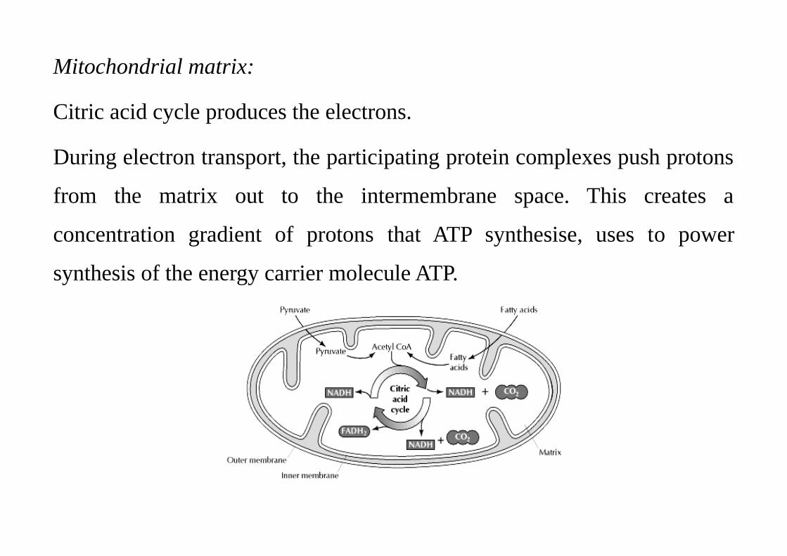
Mitochondrial matrix:
Citric acid cycle produces the electrons.
During electron transport, the participating protein complexes push protons
from the matrix out to the intermembrane space. This creates a
concentration gradient of protons that ATP synthesise, uses to power
synthesis of the energy carrier molecule ATP.
Mithocondrial genom:
genes include rRNA, tRNA genes, genes that encode proteins involved in
electron transport and ATP synthesis.
Mitochondrial proteins are mainly synthesised from nuclear genes:
enzymes required for the citric acid cycle, proteins involved in DNA
replication and transcription, and ribosomal proteins.
The protein complexes of the respiratory chain are a mixture of
mitochondrial proteins and proteins encoded by nuclear genes.
Newly synthesised, unfolded proteins are transported from the cytoplasm
via the two membranes into the matrix, where folding ensues.
Reproduction:
These organelles replicate by dividing in two.
They are constantly dividing, fusing, and changing shape: a single mitochondrion may contain multiple copies of its genome at any given time.
They multiply when a the energy needs of a cell increase: repeatedly stimulating a muscle cell will spur the production of more mitochondria in that cell. Cells with an increased need for energy contain greater numbers of these organelles than cells with lower energy needs.
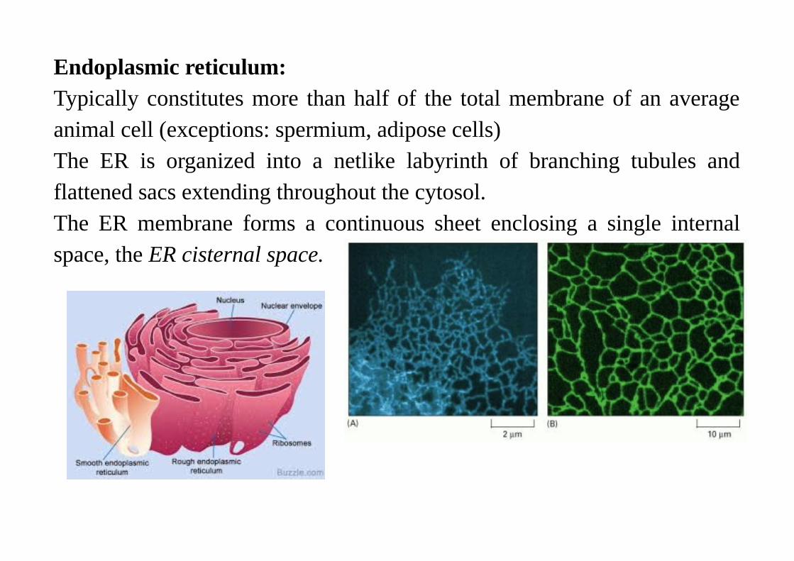
Endoplasmic reticulum:Typically constitutes more than half of the total membrane of an averageanimal cell (exceptions: spermium, adipose cells)The ER is organized into a netlike labyrinth of branching tubules andflattened sacs extending throughout the cytosol.The ER membrane forms a continuous sheet enclosing a single internalspace, the ER cisternal space.
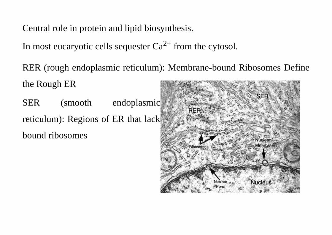
Central role in protein and lipid biosynthesis.
In most eucaryotic cells sequester Ca2+ from the cytosol.
RER (rough endoplasmic reticulum): Membrane-bound Ribosomes Define
the Rough ER
SER (smooth endoplasmic
reticulum): Regions of ER that lack
bound ribosomes
Functions:
The membrane of the RER is the site of production of all the
transmembrane proteins and lipids for most of the cell's organelles (the ER
itself, Golgi apparatus, lysosomes, endosomes, secretory vesicles,
plasmamembrane).
SER: steroid synthesis, production of lipoprotein particles,
Muscle cells have an abundant specialized smooth ER, called the
sarcoplasmic reticulum, which sequesters Ca2+ from the cytosol by means
of a Ca2+-ATPase that pumps in Ca2+ into its lumen. The release and
reuptake of Ca2+ by the sarcoplasmic reticulum trigger the contraction and
relaxation of the myofibrils during muscle contraction.
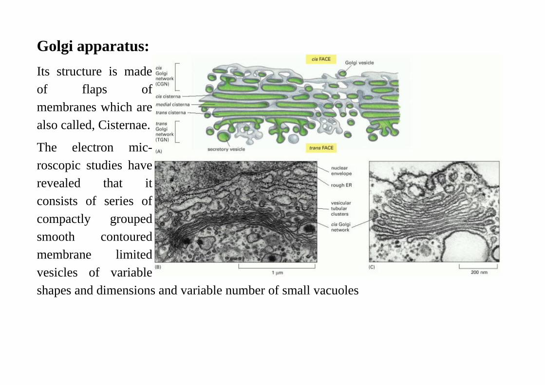
Golgi apparatus:
Its structure is madeof flaps ofmembranes which arealso called, Cisternae.
The electron mic-roscopic studies haverevealed that itconsists of series ofcompactly groupedsmooth contouredmembrane limitedvesicles of variableshapes and dimensions and variable number of small vacuoles
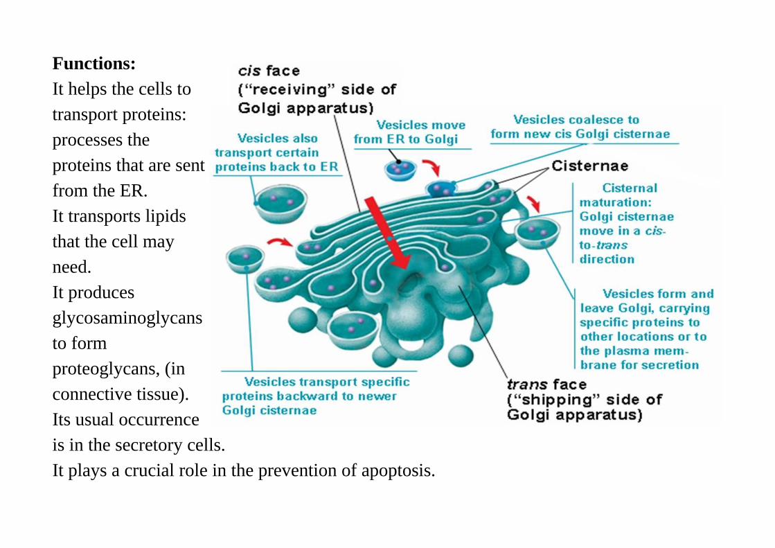
Functions:It helps the cells totransport proteins:processes theproteins that are sentfrom the ER.It transports lipidsthat the cell mayneed. It producesglycosaminoglycansto formproteoglycans, (inconnective tissue).Its usual occurrenceis in the secretory cells.It plays a crucial role in the prevention of apoptosis.
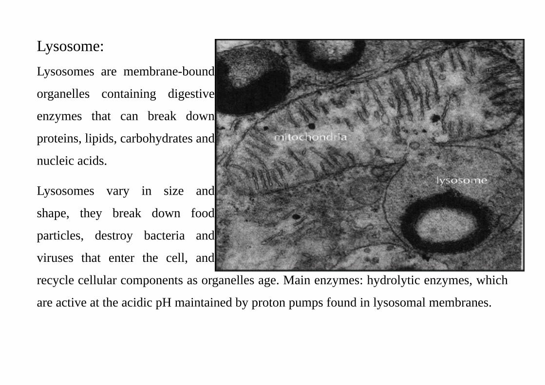
Lysosome:
Lysosomes are membrane-bound
organelles containing digestive
enzymes that can break down
proteins, lipids, carbohydrates and
nucleic acids.
Lysosomes vary in size and
shape, they break down food
particles, destroy bacteria and
viruses that enter the cell, and
recycle cellular components as organelles age. Main enzymes: hydrolytic enzymes, which
are active at the acidic pH maintained by proton pumps found in lysosomal membranes.
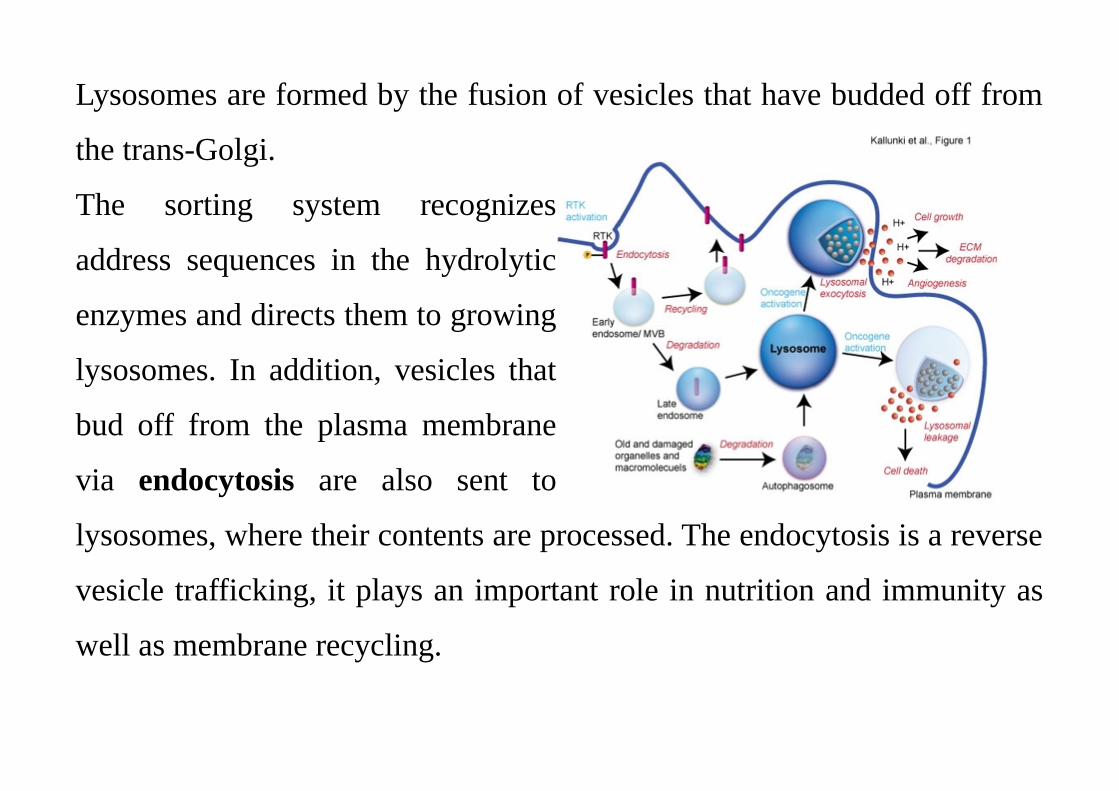
Lysosomes are formed by the fusion of vesicles that have budded off from
the trans-Golgi.
The sorting system recognizes
address sequences in the hydrolytic
enzymes and directs them to growing
lysosomes. In addition, vesicles that
bud off from the plasma membrane
via endocytosis are also sent to
lysosomes, where their contents are processed. The endocytosis is a reverse
vesicle trafficking, it plays an important role in nutrition and immunity as
well as membrane recycling.
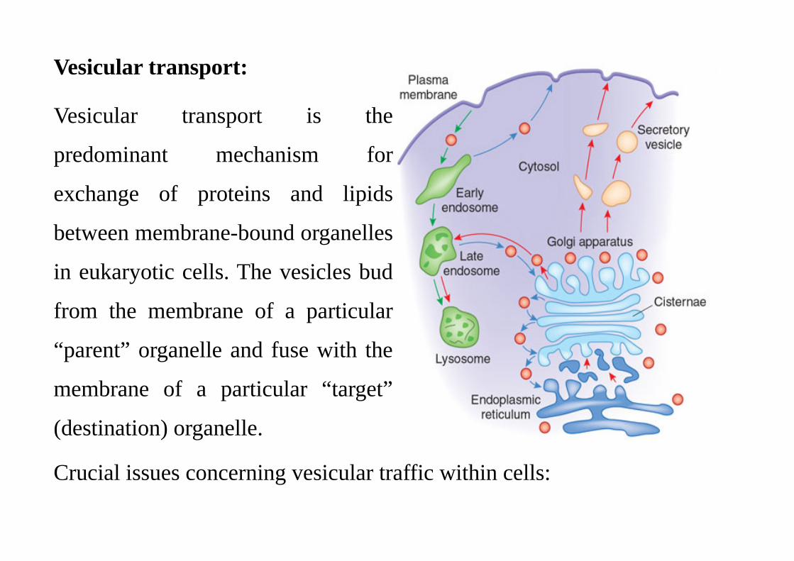
Vesicular transport:
Vesicular transport is the
predominant mechanism for
exchange of proteins and lipids
between membrane-bound organelles
in eukaryotic cells. The vesicles bud
from the membrane of a particular
“parent” organelle and fuse with the
membrane of a particular “target”
(destination) organelle.
Crucial issues concerning vesicular traffic within cells:
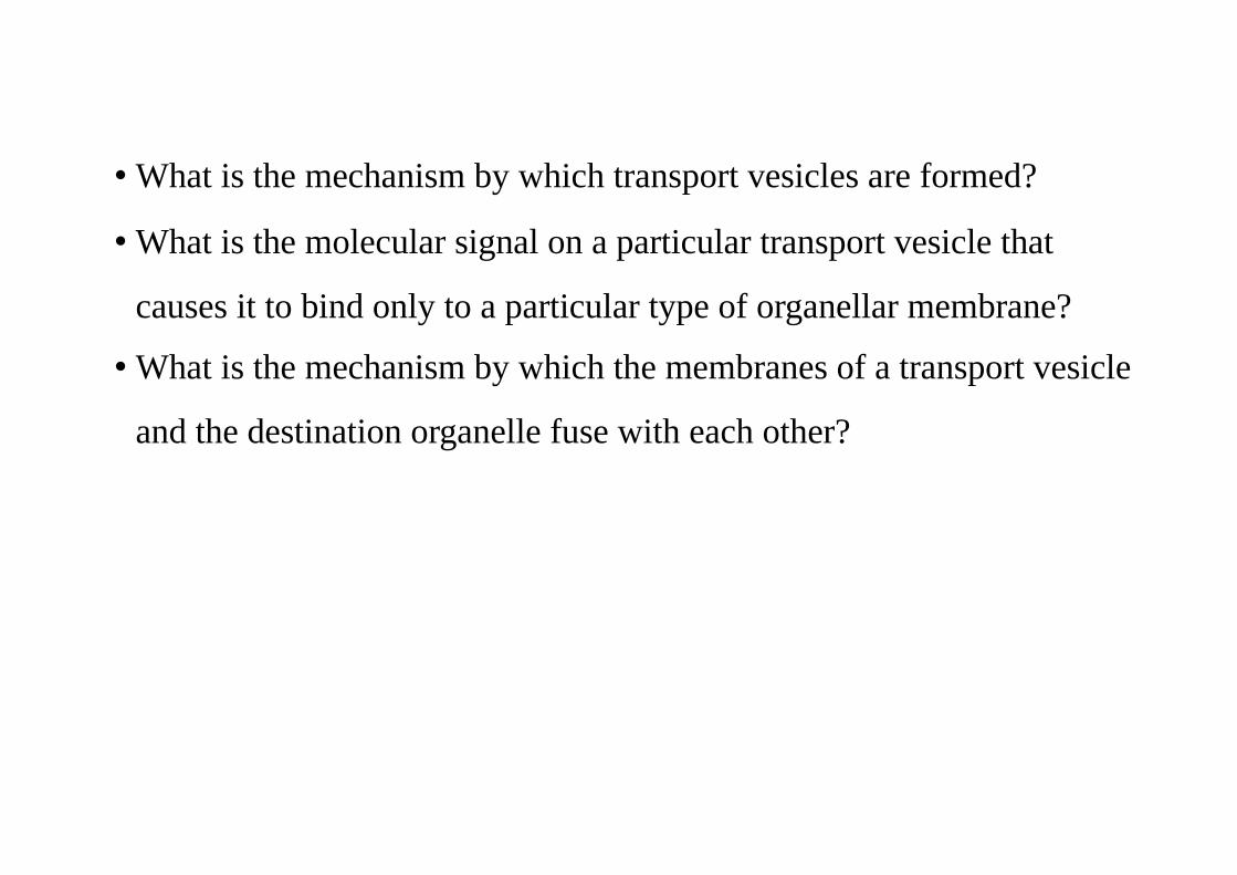
• What is the mechanism by which transport vesicles are formed?
• What is the molecular signal on a particular transport vesicle that
causes it to bind only to a particular type of organellar membrane?
• What is the mechanism by which the membranes of a transport vesicle
and the destination organelle fuse with each other?
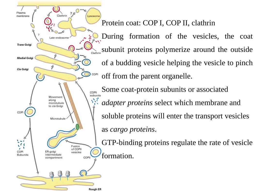
Protein coat: COP I, COP II, clathrin
During formation of the vesicles, the coat
subunit proteins polymerize around the outside
of a budding vesicle helping the vesicle to pinch
off from the parent organelle.
Some coat-protein subunits or associated
adapter proteins select which membrane and
soluble proteins will enter the transport vesicles
as cargo proteins.
GTP-binding proteins regulate the rate of vesicle
formation.
COP I vesicles mediate retrograde transport within the Golgi and from the Golgi
back to the ER.
COP II vesicles mediate transport from the ER to the Golgi.
Clathryn vesicles mediate transport from the plasma membrane and trans-Golgi to
endosomes.
Once vesicles have budded off, the coat is depolymerized, releasing the coat
proteins for reuse.
Fusion of all vesicles with their target membranes exhibits common features:
fusion occurs after the coats have depolymerized, involves a conserved set of
proteins (SNARE SNAP) that mediates targeting of vesicles to the appropriate
fusion partner and triggers the fusion process itself.
Uncoating exposes specific V-SNARE proteins on the surface of the vesicle. V-
SNARE binds to a T-SNARE protein complexed with SNAP25 on the membrane
of the target vesicle.