miscellaneous publications - university of michigan
TRANSCRIPT

MISCELLANEOUS PUBLICATIONS
MUSEUM OF ZOOLOGY, UNIVERSITY OF MICHIGAN, NO. 80
A Comparative Study of the Osteology and Myology of the Cranial and Cervical Regions
of the Shrew, Blarina Brevicauda, and the Mole, Scalopus Aquaticus
BY
GEORGE R. L. GAUGHRAN
ANN ARBOR
UNIVERSITY OF MICHIGAN PRESS
FEBRUARY 26, 19.54

PRICE LIST OF THE MISCELLANEOUS PUBLICATIONS
OF THE MUSEUM OF ZOOLOGY, UNIVERSITY OF MICHIGAN
Address inquiries to the Director of the Museum of Zoology, Ann Arbor, Michigan
Bound in Paper
No. 1. Directions for Collecting and Preserving Specimens of Dragonflies for Museum . . . . . . . . . . . . . . . . . . Purposes. By E. B. Williamson. (1916) Pp. 15, 3 figures $0.25
No. 2. An Annotated List of the Odonata of Indiana. By E. B. Williamson. (1917) Pp. 12, l m a p . . . . . . . . . . . . . . . . . . . . . . . . . . . . . . . . . . . . . . . . . . . . . . . . . . . . $0.25
No. 3. A Collecting Trip to Colombia, South America. By E. B. Williamson. (1918) Pp. 24 (Out of print)
. . . . . . . . . No. 4. Contributions to the Botany of Michigan. By C. K. Dodge. (1918) Pp. 14 $0.25 No. 5. Contrihutions to the Botany of Michigan, II. By C. K. Dodge. (1918) Pp. 44, 1 map . . $0.45 No. 6. A Synopsis of the Classification of the Fresh-water Mollusca of North America,
North of Mexico, and a Catalogue of the More Recently Described Species, with . . . . . . . . . . . . . Notes. By Bryant Walker. (1918) Pp. 213, 1 plate, 233 figures. $3.00
No. 7. The Anculosae of the Alabama River Drainage. By Calvin Goodrich. (1922) Pp. 57, 3 plates . . . . . . . . . . . . . . . . . . . . . . . . . . . . . . . . . . . . . . . . . . . . . . . . . . . $0.75
No. 8. The Amphibians and Reptiles of the Sierra Nevada de Santa Marta, Colombia. By . . . . . . . . . . . . Alexander G. Ruthven. (1922) Pp. 69, 13 plates, 2 figures, 1 map $1.00
No. 9. Notes on American Species of Triacanthagyna and Gynacantha. By E. B. Williamson. (1923) Pp. 67, 7 plates . . . . . . . . . . . . . . . . . . . . . . . . . . . . . . . . . . . . . . . . . $0.75
No. 10. A Preliminary Survey of the Bird Life of North Dakota. By Norman A. Wood. (1923) Pp. 85 ,6 plates, 1 map . . . . . . . . . . . . . . . . . . . . . . . . . . . . . . . . . . . . . . . . . $1.00
No. 11. Notes on the Genus Erythemis with a Description of a New Species (~donata) . By E. B. Williamson.
The Phylogeny and the Distribution of the Genus Erythemis (Odonata). By Clarence H. Kennedy. (1923) Pp. 21, 1 plate. . . . . . . . . . . . . . . . . . . . . . . . . . . . . . . . . $0.50
. . . . . . . . . . . . No. 12. The Genus Gyrotoma. By Calvin Goodrich. (1924) Pp. 29, 2 plates $0.50 No. 13. Studies of the Fishes of the Order Cyprinodontes. By Carl L. Hubbs. (1924) Pp. 23,
4 plates . . . . . . . . . . . . . . . . . . . . . . . . . . . . . . . . . . . . . . . . . . . . . . . . . . . $0.75 No. 14. The Genus Per i les tes (Odonata). By E. B. Williamson and 3. H. Williamson. (1924)
Pp. 3 6 , l plate. . . . . . . . . . . . . . . . . . . . . . . . . . . . . . . . . . . . . . . . . . . . . . . $0.50 No. 15. A Check-List of the Fishes of the Great Lakes and Tributary Waters, with
Nomenclatorial Notes and Analytical Keys. By Carl L. Hubbs. (1926) Pp. 77, 4p la t e s . . . . . . . . . . . . . . . . . . . . . . . . . . . . . . . . . . . . . . . . . . . . . . . . . . . $1.50
No. 16. Studies of the Fishes of the Order Cyprinodontes. VI. By Carl L. Hubbs. (1926) Pp. 79,4 plates . . . . . . . . . . . . . . . . . . . . . . . . . . . . . . . . . . . . . . . . . . . . . . $1.00
No. 17. The Structure and Growth of the Scales of Fishes in Relation to the Interpretation of Their Life-History, with Special Reference to the Sunfish Eupomotis gibbosus.
. . . . . . . . . . . . . . . . By Charles W. Creaser. (1926) Pp. 80, 1 plate, 12 figures. $1.50 No. 18. The Terres t r ia l Shell-hearing Mollusca of Alabama. By Bryant Walker. (1928)
Pp. 180, 278 figures . . . . . . . . . . . . . . . . . . . . . . . . . . . . . . . . . . . . . . . . . . . $1.50 No. 19. The Life History of the Toucan Ramphastos hrevicarinatus. By Josselyn Van Tyne.
(1929) Pp. 43, 8 plates, 1 map . . . . . . . . . . . . . . . . . . . . . . . . . . . . . . . . . . . . $0.75 No. 20. Materials for a Revision of the Catostomid Fishes of Eastern North America. By
Carl L. Hubhs. (1930) Pp. 47, 1 plate. . . . . . . . . . . . . . . . . . . . . . . . . . . . . . . $0.75 No. 21. A Revision of the Libelluline Genus Perithemis (Odonata). By F. Ris. (1930) Pp.
5 0 , g p l a t e s . . . . . . . . . . . . . . . . . . . . . . . . . . . . . . . . . . . . . . . . . . . . . . . . . $0.75 ...... No. 22. The Genus Oligoclada (Odonata). By Donald Borror. (1931) Pp. 42, 7 plates $0.50 No. 23. A Revision of the Puer Group of the North American Genus Melanoplus, with Re-
marks on the Taxonomic Value of the Concealed Male Genitalia in the Q r t a - canthacrinae (Orthoptera, Acrididae). By Theodore H. Hubhell. (1932) Pp. 64, 3 plates, 1 figure, 1 m a p . . . . . . . . . . . . . . . . . . . . . . . . . . . . . . . . . . . . . . . . $0.75
No. 24. A Comparative Life History Study of the Mice of the Genus Peromyscus. By Arthur Svihla. (1932) Pp. 39. . . . . . . . . . . . . . . . . . . . . . . . . . . . . . . . . . . . . . . . . . $0.50
. . . . . . . . . . . No. 25. The Moose of Is le Royale. By Adolph Murie. (1934) Pp. 44, 7 plates $0.70 No. 26. Mammals from Guatemala and British Honduras. By Adolph Murie. (1935) Pp. 30,
1 plate, 1 map . . . . . . . . . . . . . . . . . . . . . . . . . . . . . . . . . . . . . . . . . . . . . . . $0.35 No. 27. The Birds of Northern Peten, Guatemala. By Josselyn Van Tyne. (1935) Pp. 46,
2 plates, 1 map . . . . . . . . . . . . . . . . . . . . . . . . . . . . . . . . . . . . . . . . . . . . . . $0.45 No. 28. Fresh-Water Fishes Collected in British Honduras and Guatemala. By Carl L.
Hubbs. (1935) Pp. 22, 4 plates, 1 map. . . . . . . . . . . . . . . . . . . . . . . . . . . . . . . $0.25 No. 29. A Contribution to a Knowledge of the Herpetology of a Portion of the Savanna Region
of Central Pet&, Guatemala. By L. C. Stuart. (1935) Pp. 56, 4 plates, 1 figure, l m a p . . . . . . . . . . . . . . . . . . . . . . . . . . . . . . . . . . . . . . . . . . . . . . . . . . . . $0.50
(CONTINUED ON LAST PAGES)


The publications of the Museum of Zoology, University of Michigan, consist of two series-the Occasional Papers and the Miscellaneous Pub- lications. Both se r i es were founded by Dr. Bryant Walker, Mr. Brad- shaw H. Swales, and Dr. W. W. Newcomb.
The Occasional Papers, publication of which was begun in 1913, serve a s a medium for original papers based principally upon the collections of the Museum. The papers a r e issued separately to l ibrar ies and special- ists, and, when a sufficient number of pages has been printed to make a volume, a title page, table of contents, and index a r e supplied to l ibrar ies and individuals on the mailing l is t for the entire se r i es .
The Miscellaneous Publications, which include papers on field and museum techniques, monographic studies, and other contributions not within the scope of the Occasional Papers, a r e published separately, and a s i t is not intended they will be grouped into volumes, each number has a title page and, when necessary, a table of contents.

MISCELLANEOUS PUBLICATIONS
MUSEUM OF ZOOLOGY, UNIVERSITY OF MICHIGAN, NO. 80
A Comparative Study of the Osteology and Myology of the Cranial and Cervical Regions
of the Shrew, Blarina Brevicauda, and the Mole, Scalopus Aquaticus
BY
GEORGE R. L. GAUGHRAN
ANN ARBOR
UNIVERSITY OF MICHIGAN PRESS
FEBRUARY 26, 1954


CONTENTS
Page
Introduction . . . . . . . . . . . . . . . . . . . . . . . . . . . . . . . . . . . . . 9
. . . . . . . . . . . . . . . . . . . . . . . . . . . . . . . . . . . . . . . Osteology 10 Blarina brevicauda kirtlandi . . . . . . . . . . . . . . . . . . . . . . . . 11
Cranium . . . . . . . . . . . . . . . . . . . . . . . . . . . . . . . . . . . 11 Ossicula Auditus . . . . . . . . . . . . . . . . . . . . . . . . . . . . . 14
. . . . . . . . . . . . . . . . . . . . . . . . . . . . . . . . . . . Dentary 16 Hyoid and Laryngeal Cartilages . . . . . . . . . . . . . . . . . . . 17 Cervical Vertebrae . . . . . . . . . . . . . . . . . . . . . . . . . . . 18
. . . . . . . . . . . . . . . . . . . . . . . . . . . . . . . . . . . Scapula 19
. . . . . . . . . . . . . . . . . . . . . . . . . . . . . . . . . . . Clavicle 20 . . . . . . . . . . . . . . . . . . . . . . . . . . . . . . . . . Manubrium 20
Scalopus aquaticus machrims . . . . . . . . . . . . . . . . . . . . . . . 20 Cranium . . . . . . . . . . . . . . . . . . . . . . . . . . . . . . . . . . . 20 Ossicula Auditus . . . . . . . . . . . . . . . . . . . . . . . . . . . . . 26
. . . . . . . . . . . . . . . . . . . . . . . . . . . . . . . . . . . Dentary 28 Hyoid and Laryngeal Cartilages . . . . . . . . . . . . . . . . . . . 28 Cervical Vertebrae . . . . . . . . . . . . . . . . . . . . . . . . . . . 29
. . . . . . . . . . . . . . . . . . . . . . . . . . . . . . . . . . . Scapula 31
. . . . . . . . . . . . . . . . . . . . . . . . . . . . . . . . . . . Clavicle 31 Manubrium . . . . . . . . . . . . . . . . . . . . . . . . . . . . . . . . . 32
Myolo gy . . . . . . . . . . . . . . . . . . . . . . . . . . . . . . . . . . . . . . . . 32 . . . . . . . . . . . . . . . . . . . . . . . . Blarina brevicauda kirtlandi 33
Myomeric Musculature . . . . . . . . . . . . . . . . . . . . . . . . . 33 Dorsal Division . . . . . . . . . . . . . . . . . . . . . . . . . . . 33 Lateroventral Division . . . . . . . . . . . . . . . . . . . . . . . 36
Branchiomeric Musculature . . . . . . . . . . . . . . . . . . . . . . 39 Trigeminal Field . . . . . . . . . . . . . . . . . . . . . . . . . . 39 Facial Field . . . . . . . . . . . . . . . . . . . . . . . . . . . . . . 41 Glossopharyngeal Field . . . . . . . . . . . . . . . . . . . . . . 42 Vagus Field . . . . . . . . . . . . . . . . . . . . . . . . . . . . . . 43
. . . . . . . . . . . . . . . . . . . . . . . . . . . Accessory Field 44 . . . . . . . . . . . . . . . . . . . . . . . Scalopus aquaticus machrinus 45
Myomeric Musculature . . . . . . . . . . . . . . . . . . . . . . . . . 45 . . . . . . . . . . . . . . . . . . . . . . . . . . . Dorsal Division 45
Lateroventral Division . . . . . . . . . . . . . . . . . . . . . . . 48 Branchiomeric Musculature . . . . . . . . . . . . . . . . . . . . . . 51
Trigeminal Field . . . . . . . . . . . . . . . . . . . . . . . . . . 51 Facial Field . . . . . . . . . . . . . . . . . . . . . . . . . . . . . . 53
. . . . . . . . . . . . . . . . . . . . . . Glossopharyngeal Field 55 Vagus Field . . . . . . . . . . . . . . . . . . . . . . . . . . . . . . 56
. . . . . . . . . . . . . . . . . . . . . . . . . . . Accessory Field 57
Discussion . . . . . . . . . . . . . . . . . . . . . . . . . . . . . . . . . . . . . . 58 Osteology . . . . . . . . . . . . . . . . . . . . . . . . . . . . . . . . . . . . 58
Cranium . . . . . . . . . . . . . . . . . . . . . . . . . . . . . . . . . . . 58 Auditory Ossicles . . . . . . . . . . . . . . . . . . . . . . . . . . . . 62 Dentary . . . . . . . . . . . . . . . . . . . . . . . . . . . . . . . . . . . 62 Hyoid and Laryngeal Cartilages . . . . . . . . . . . . . . . . . . . 62

Page
Vertebrae . . . . . . . . . . . . . . . . . . . . . . . . . . . . . . . . . . 62 Ligamentum Nuchae . . . . . . . . . . . . . . . . . . . . . . . . . . . 63 Scapula . . . . . . . . . . . . . . . . . . . . . . . . . . . . . . . . . . . 63 Manubrium . . . . . . . . . . . . . . . . . . . . . . . . . . . . . . . . . 63 Clavicle . . . . . . . . . . . . . . . . . . . . . . . . . . . . . . . . . . . 64
Myology . . . . . . . . . . . . . . . . . . . . . . . . . . . . . . . . . . . . . 64 Myomeric Musculature . . . . . . . . . . . . . . . . . . . . . . . . . 64 Branchiomeric Musculature . . . . . . . . . . . . . . . . . . . . . . 66
. . . . . . . . . . . . . . . . . . . . . . . . . . . . . . . . . Special Senses 70
. . . . . . . . . . . . . . . . . . . . . . . . . . . . . . . . . Middle Ea r 70 Eye . . . . . . . . . . . . . . . . . . . . . . . . . . . . . . . . . . . . . . 72
Summary . . . . . . . . . . . . . . . . . . . . . . . . . . . . . . . . . . . . . . . 73
. . . . . . . . . . . . . . . . . . . . . . . . . . . . . . . . . . Literature Cited 75

ILLUSTRATIONS
(F igures 1-67 follow page 82)
F igure
1. Skull of Blarina brevicauda kirt ladi, dorsal view.
2. Skull of Blarina brevicauda kirtlandi, ventral view.
3. Skull of Blarina brevicauda kirtlandi, la teral view.
4. Right malleus of Blarina brevicauda kirtlandi, dorsal view.
5. Right incus of Blarina brevicauda kirtlandi, dorsal view.
6. Right s tapes of Blarina brevicauda kirtlandi, la teral view.
7. Right dentary of Blarina brevicauda kirtlandi, la teral view.
8. Right dentary of Blarina brevicauda kirtlandi, medial view.
9. Cervical vertebrae of Blarina brevicauda kirtlandi, la teral view.
10. Right scapula of Blarina brevicauda kirtlandi, medial view.
11. Right scapula of Blarina brevicauda kirtlandi, la teral view
12. Left clavicle of Blarina brevicauda kirtlandi, anter ior view.
13. Manubrium of Blarina brevicauda kirtlandi, ventral view.
14. Cervical ver tebrae of Blarina brevicauda kirtlandi, ventral view.
15. Medial view of rami showing forward migration of ventral ar t icular facet in Blarina a s compared with Sorex.
16. Hyoid and laryngeal cartilages of Blarina brevicauda kirtlandi, ventral view.
17. Hyoid and laryngeal cartilages of Blarina brevicauda kirtlandi, la teral view.
18. Dorsal aspect of arytenoid cartilages of Blarina brevicauda kirtlandi.
19. Hyoid of Blarina brevicauda kirtlandi, ventral view.
20. Skull of Scalopus aquaticus machrinus, dorsal view.
21. Skull of Scalopus aquaticus rnachrirms, ventral view.
22. Skull of Scalopus aquaticus machrinus, la teral view.
23. Left malleus of Scalopus aquaticus machrinus, dorsal view.
24. Left incus of Scalopus aquaticus machrinus, dorsal view.
25. Left s tapes of Scalopus aquaticus machrinus, la teral view.
26. Anterior aspect of snout of Scalopus aquaticus machrinus.
27. Ventral aspect of snout of Scalopus aquaticus machrinus.
28. Cervical vertebrae of Scalopus aquaticus rnachrirms, la teral view.
29. Left dentary of Scalopus aquatias machrinus, la teral view.
30. Left dentary of Scalopus aquaticus machrinus, medial view.

F i g u r e
31. Cervical ver tebrae of Scalopus aquaticus machrims, ventral view.
32. Ligamentum nuchae of Scalopus aquaticus machrinus.
33. Left scapula of Scalopus aquaticus machrinus, la teral and vertebral surfaces.
34. Manubrium of Scalopus aquaticus machrinus, right side.
35. Clavicles of Scalopus aquaticus machrims, anter ior view.
36. Cricoid, arytenoid, and epiglottis of Scalopus aquaticus machrinus, dorsal view.
37. Hyoid and laryngeal cartilages of Scalopus aquaticus machrinus, ven- t r a l view.
38. Hyoid and laryngeal car t i lages of Scalopus aquaticus m a c h r i m , right side.
39. Superficial musculature of Blarina brevicauda kirtlandi, l a te ra l view.
40. F i r s t depth of musculature of Blarina brevicauda kirtlandi, la teral view.
41. Second depth of musculature of Blarina brevicauda kirtlmuli, la teral view.
42. Third depth of musculature of Blarina brevicuuda kirtlandi, la teral view.
43. Fourth depth of musculature of Blarina breviceuda kirtlandi, la teral view.
44. Fifth depth of musculature of Blarina brevicauda kirtlandi, la teral view.
45A. Superficial layer and f i r s t depth of musculature of Blarina brevicauda kirtlandi, ventral view.
45B. Second and third depths of musculature of Blarina brevicauda kirt- lrmdi, ventral view.
46. Schema of force vectors of masticatory muscles of Blarina brevicauda k i r t l d i .
47. Fourth depth of musculature of Blarina brevicrmda kirtlandi, ventral view.
48. Superficial layer of musculature of Scalopus aquaticus machrinus, dorsal view.
49. Superficial layer of musculature of Scalopus aquaticus machrimcs, ventral view.
50. Superficial layer of musculature of Scalopus aquaticus machrinus, la teral view.
51. F i r s t depth of musculature of Scalopus aquaticus machrinus, la teral view.
52. Second depth of musculature of Scalopus aquaticus m a c h r i m , la teral view.

Figure
53. Third depth of musculature of Scalopus aquaticus machrinus, lateral view.
54. Fourth depth of musculature of Scalopus aquaticus machrinus, la teral view.
55. Fifth depth of musculature of Scalopus aquaticus m a c h r i m , la teral view.
56. Sixth depth of musculature of Scalopus aquaticus machrinus, la teral view.
57. Seventh depth of musculature of Scalopus aquaticus machrinus, la teral view.
58. Eighth depth of musculature of Scalopus aquaticus machrinus, la teral view.
59. Medial, dorsal, and lateral views of the superficial masse te r of Scalopus aquaticus machrinus.
60. Superficial layer and f i r s t depth of musculature of Scalopus aquaticus machrinus, ventral view.
61. Second and third depths of musculature of Scalopus aquaticus machyims, ventral view.
62. Fourth and fifth depths of musculature of Scalopus aquaticus mach~inus, ventral view.
63. Sixth and seventh depths of musculature of Scalopus aquaticus mackrinus, ventral view.
64A and C. Pat tern of distribution of nerves I . , X, XI, and XII in Scalopus aquaticus machrinus .
64B. Pat tern of distribution of cervical nerves in Scalopus aquaticus machrinus .
65. Key t o Figure 64B.
66. Reconstruction of skull of nestling Blarina brevicauda kirtlandi.
67. Reconstruction of skull of nestling Scalofius aquaticus m a c h r i m .


A COMPARATIVE STUDY OF THE OSTEOLOGY AND MYOLOGY OF THE CRANIAL AND CERVICAL
REGIONS OF THE SHREW, BLARINA BRE VICA UDA, AND THE MOLE, SCALOPUSAQUAmCUS*
INTRODUCTION
THE short-tailed shrew Blarina and the North American mole Scalopus a r e members of the superfamily Soricoidea, mammalian order Insectiv- ora. The fo rmer is a representative of the family Soricidae; the lat ter , of the Talpidae. The moles and shrews have probably evolved f rom a common nonfossorial ancestor. The Recent talpids and soricids, how- ever, have evolved along separate courses over a long period of t ime and present many morphological specializations, although retaining certain primitive characters .
The present investigation was initiated for the purpose of making a small contribution to the meager knowledge of the anatomy of North American insectivores. This paper is not a taxonomic study, but i t con- tains a certain amount of morphological data which will prove useful after subsequent analyses of a greater number of species. Although the cranial and cervical regions a r e axial structures, which generally demon- s t r a t e more conservatism in their modifications than is shown by appen- dicular parts, they were chosen for this study because of their greater appeal to me and because no s imilar study of any of the North American insectivores has ever been reported. Whenever possible, an attempt has been made to interpret the functional significance of anatomical modifi- cations. As was anticipated, however, the functional importance of many structural differences could not be ascertained. My decision to use the pra i r ie mole (Scalopus aquaticus) and the short-tailed shrew (Blarina brevicauda) a s comparative fo rms was based on their membership in the two lipotyphlan families of insectivores represented in this country, their differences in habits, and their regional availability. B l a ~ i n a , despite i t s fossorial tendencies, was chosen in preference to Soyex because the small s ize of .Sorex makes i t an extremely difficult subject fo r dissection.
Skeletal material examined consists of five skulls and three skeletons of Talpa europaea, 30 skulls and s ix skeletons of Scalopus aquaticus machrinus, and 30 skulls and eight skeletons of Blarinu brevicauda kirt- landi. One adult pra i r ie mole, one adult short-tailed shrew, two nest young of Blarina, and two nest young of Scalopus were cleared and stained following the Spalteholz technique. In the course of this work I was im- pressed with the value of specimens prepared by this method, for they present a more reliable picture of the relationships of hard par ts than can
* Accepted for publication May 22, 1953. This work was presented in partial fulfillment of the requirements for the degree of
Doctor of Philosophy in the University of Michigan.

10 GEORGE R . L . GAUGHRAN
be obtained from a study of osteological preparations. Eight specimens of the short-tailed shrew and 12 specimens of the prai r ie mole, which were used f o r dissection, were preserved in embalming fluid composed of 3 per cent phenol, 2 per cent formalin, 20 per cent glycerin, and 75 per cent water. All dissections were done under a stereobinocular microscope, and measurements were made with vernier calipers calibrated to 0.1 mm.
I am indebted to Dr. W. H. Burt, Museum of Zoology, University of Michigan, for f ree use of the skeletal material in the Museum of Zoology mammal collection. I wish also to thank Dr . E . T. Hooper, Museum of Zoology, University of Michigan, for alcoholic specimens of Talpa europaea Linnaeus, Dr. G. C. Rinker, Department of Anatomy, University of Michi- gan Medical School, for a preserved specimen of Condylura cr is ta ta Lin- naeus, Dr. C. C. Carpenter, M r . A. Bertoni, and Mr. A . Keiman for speci- mens of Scalopus aquaticus machrinus Rafinesque, and Dr. E . S. Booth, Department of Biological Sciences, Walla Walla College, for specimens of NeZtrotrichus gSibbsi Baird. To Dr. Alfred H. Stockard I owe an immeasur- able debt of gratitude. His invaluable knowledge of anatomy, intelligent criticism, helpful suggestions, and patient understanding have been my support throughout this work. I wish to thank the Board of Governors of the Horace H. Rackham School of Graduate Studies for a grant which made possible the publication of this study.
OSTE OLOGY
Despite the wealth of information on mammalian osteology, gleaned through many years of research by competent men, apparently no one has investigated the development of the skull in North American insectivores. W. K. Parker ' s monograph on the development of the mammalian skull (1885) remains a classic and has been extremely valuable in this work. Other investigations include: G. R. de Beer 's study of Sorex araneus (1929), P. N. van Kampen's work on the tympanic region in insectivores (1905), E . Fischer 's study of Talpa (1901), G. H. Roux's paper on certain Ethiopian insectivores (1947), G. E . Dobson's monograph (1882-go), St. G. Mivart's work on the osteology of the insectivores (1867), C. Giebel's note on Scalopus (1858), and P. M. Butler's study of the Erinaceidae (1948). Though of greater scope, but not to be neglected, a r e H. G. Bronn's erudite tome on mammalian anatomy (1874-1900). M. G. Saint- Hilaire's lessons (1834), G. Cuvier's volumes (1800-37), M. H. M. de Blainville's osteology (1839-64), and M. Weber's text on mammals (1927- 28). Additional, l e s s pertinent, references a re : on Talpa, Bertelli (1909), Brandt (1836), Camerano (1886), Fitzinger (1869), ~ i j ~ ~ e r t (1894), Gregory (1910, 1920), Jacobs (1816), Kober (1885), Muller (1935), van der Klaauw (1924), Wilkie (1925), and Winge (1941); on SC~~OPUS, Baird (1859), Cope (1880), de Beer (1937), Eckhard (1847), Jackson (1915), and True (1897); on Condylura, Jackson (1915) and True (1897); on Blarina, Allen (1894), Baird (1859), and Merriam (1895); on Sorex, Brandt (1836), de ~ e e r (1929, 1937), Jackson (1928), Keen (1942), and Keen and Grobbelaar (1941).
The present investigation is not concerned with embryology; hence, only the bony elements that could be identified in immature specimens,

CRANIAL A N D CERVICAL REGIONS OF THE SHREW A N D MOLE I I
without embryological techniques, a r e considered. In addition, foramina a r e identified, but only the nervous structures which they transmit a r e given, since the blood vascular system was not investigated. A charac- terization of the general form and proportions of the cranium of Blarina introduces more detailed descriptions of the cranium in dorsal, ventrar, lateral, frontal, and occipital views. The auditory ossicles a r e consid- ered under a separate heading, followed by a study of the dentary. The next section describes the hyoid and laryngeal cartilages. A considera- tion of the cervical region of the vertebral column includes a description of the cervical segment a s a unit and a detailed study of certain verte- brae. The scapula, clavicle, and sternum a r e considered only insofar a s they a r e related to cervical and cranial musculature. The discussion of Scalopus follows the same pattern. All references in this paper to the mole o r to the shrew refer to the particular forms under investigation, unless i t is otherwise specifically stated.
Blarina brevicauda kirtlandi
Cranium
General Form.-The cranium of an adult Blarina is a fused, rugose, osseous structure, sagittate in dorsal view, with only a few prominent sutures suggestive of certain of i t s component elements. In comparing i t with that of the long-tailed shrew, one is impressed with the angularity of the posterior region. Its greatest breadth is at the zygomatic processes of the squamosal; this measurement is almost twice the breadth a t the zygomatic processes of the maxillary bones. The mean height of the cranium, a t the intersection of the sagittal and lambdoidal sutures, ap- proximates one-third the greatest length, measured from the tip of the rostrum to the most posterior region of the occipital condyles. The height of the cranium increases gradually from the roof of the rostrum to the posterior edge of the parietals from which the supraoccipital bone de- scends sharply, describing a small arc , to the dorsal r im of the foramen magnum. The height of the posterior region of the cranium is more than twice that of the postnarial rostra1 region. The roof of the cranium is well rounded; the lateral walls of the rostrum descend more abruptly than those of the parietal region, producing a conoid outline rostrally and a cycloid outline in the parietal region. The osseous wall, anterior to the zygomatic processes of the maxillary, is considerably thicker than the wall of the expanded posterior region of the skull. The zygomatic arch is incomplete; the malar (jugal) is absent. There a r e no auditory bullae; the tympanic fo rms only a ring of bone. Two articular facets for the articulation with a double facet on the condylar process of the mandible a r e visible. To aid in the interpretation of the bony elements of the cranium, two nest young of Blarina were cleared and stained for bone (Fig. 66).
Norma Verticalis (Fig. l).-The occipital and parietal regions a r e broad and thin walled, occupying a little l e s s than one-half the length of the skull; in outline they form a rounded contour from the zygomatic
12 GEORGE R. L . GAUGHRAN
process of the squamosal of one side to the same process of the opposite side, then turn sharply mesiad to join the frontal region. Here the walls parallel each other for about 2 mm., then f lare out slightly to the zygo- matic process of the maxillary and taper gradually to the tip of the snout. A median sagittal c res t visibly separates the parietals but disappears in the frontal region of the cranium. A transverse lambdoidal c res t dis- tinguishes the parietals f rom the supraoccipital, and a sharp transverse ridge, just caudal to this suture, suggests the presence of a small inter- parietal. On the lateral surface of the rostrum can be seen the very large infraorbital foramina for branches of the maxillary division of the fifth cranial nerve. At the junction of the rostrum and frontals a pair of small variable foramina is situated beside the most anterior tip of the median sagittal ridge. Of eight specimens, one was without foramina, one had a foramen on the right side only, four had a foramen on the left only, and in two there was a foramen on each side. At the lateral edge of the frontal region, dorsal to the thickened supraglenoid articular surface (not visible in this view), there may be a tiny bilateral foramen. This and the pre- ceding foramina transmit vascular structures. The lateral edge of the parietal continues anteriorly from the lambdoidal c res t a s a prominent ridge separating the squamosal from the parietal and terminating in a small triangular process immediately caudal to the glenoid cavity. Vis- ible on the parietal is a low longitudinal ridge paralleling the sagittal c res t for about 4.5 mm. The nares a r e represented by a single rectangu- l a r opening. A small protuberance, probably the supraorbital process (z~gomat ic process of the frontal), projects laterad in the frontal region. The coronal, sagittal, and lambdoidal sutures can be seen.
Norma Basalis (Fig. 2).-The dentition has been figured by S. F. Baird (1859), F. W. True (1897), and C. H. Merriam (1895). The cleared and stained nest young of Blarina confirmed the dental formula: 3, 1, 3 , 3. A nomenclature of the molar cusps of Sorex is given by Jackson (1928). The palate is narrow anteriorly and becomes wider in the premolar and molar region, terminating in a median palatine lip overhanging the mes- opterygoid fossa. No indication of sutures between the premaxillary, maxillary, and palatines could be discerned. A pair of foramina l ies mesial to the second incisor, followed immediately caudad by a pair of smal ler foramina. The anterior foramina a r e probably incisive, but I could not t race a palatine branch (nasopalatine nerve) f rom the maxillary division of the trigeminal to this opening. The posterior foramina trans- mit a definite structure, but i t was impossible to t race this structure to any main vascular o r nervous trunk. Except for several smal l suture lines, the remainder of the cranium is a fused osseous unit. The only elements to be distinguished with certainty a r e the periotics, which a r e attached loosely to the res t of the cranium. There is a short but promi- nent pair of hamuli of the pterygoids projecting divergently caudad. About 1.5 mm. lateral to the hamuli and immediately ventral to the in- fer ior glenoid articular surface can be seen a pair of large foramina lying in the alisphenoid region, which transmit the mandibular ramus of the trigeminal nerve. In the posterior par t of the orbitotemporal fossa is the superior glenoid articular facet, and a t i t s anterior border is a small

CRANIAL A N D CERVICAL REGIONS OF THE SHREW A N D MOLE '3
foramen for a blood vessel. This foramen is situated immediately caudal to the cribriform plate and is continued craniad by a longitudinal sulcus along the lateral edge of the roof of the orbitotemporal region. On the mesial vertical wall of the orbitotemporal fossa is a thin-walled alis- phenoid canal, the lips of which open into the fossa and ca r ry the ophthal- mic and maxillary branches of the trigeminal nerve and the optic nerve. In the basisphenoidal region, mesial to the anterior tip of the annulus tympanicus, a pair of foramina transmits two small vessels obliquely through the cranial wall. The caudal edge of the squamosal can be seen abutting the annulus tympanicus and the tympanohyal. Here the squamosal forms a thin lamina of bone roofing the pocket in which res t s the capitulum mallei. The lateral lip of the squamosal is a thickened bony c res t con- tinuing craniad a s the short zygomatic process. The annulus tympanicus is an almost f r e e incomplete ring of bone attached laterally to the pro- cessus anterior Folii (processus gracilis) and mesially to the tympano- hyal. The periotic l ies between the squamosal laterally and the basi- occipital and exoccipital mesially; i t is f ree from the latter two bones with exception of a narrow exoccipital s t rut which forms the caudal margin of the jugular foramen (foramen lacerum posterius) for nerves IX, X, and XI. The bony arches of the posterior and horizontal semicircu- l a r canals can be seen in this view. Immediately mesial to this foramen is the condylar (hypoglossal) foramen for the twelfth cranial nerve. The condyles form the most caudal part and extend for about 2.5 mm. on the ventral surface of the cranium. The ossicula auditus and cochlear re- gions will be discussed later in more detail.
Norma Lateralis ( ~ i g . 3).-1n this view the cranium is wedge-shaped in general outline, with a perceptible depression in the dorsal surface several millimeters caudal to the f rontopremaxillary suture. All the teeth a r e visible with the exception of the fifth unicuspid (p2). The sagittal and lambdoidal ridges become increasingly prominent with age. The large infraorbital foramen opens anteriorly, dorsal to M1. Immediately caudal to this foramen is a small opening into a bony canal which passes cranio- dorsad along the lip of the infraorbital foramen before communicating with the nasal cavity. This canal ca r r i e s the lachrymal duct. The orbitotem- poral fossa is triangular, the apex directed ventrad. The convolutions of the ethmoturbinals can be seen through the thin wall of the fossa. Within the orbitotemporal fossa is the sphenopalatine foramen which transmits the palatine twigs of the maxillary branch of the trigeminal nerve. Slightly anterior to the middle of the fossa is a foramen representing the combined inferior orbital f i ssure (foramen lacerum anterius) and foramen rotundum which transmits the optic nerve and the ophthalmic and maxil- lary divisions of the fifth cranial nerve. The mandibular (glenoid) articu- l a r facets can be seen in the posterior half of the fossa. The superior art icular surface is directed cephalomesiad and presents a convex facet; the inferior art icular surface l ies transversely and has a concave articu- l a r facet. The squamosal, forming the most caudal region of the orbito- temporal fossa, is not distinctly limited anteriorly but is separated dor- sally f rom the parietal by a ridge of bone. There is no evidence of the boundaries of the lachrymal bone. The "prooticn ( ~ a r k e r , 1885) extends

' 4 GEORGE R. L . GAUGHRAN
anteriad a s a bony tongue between the parietal and the squamosal. The dorsal region of the posterior wall of the cranium is formed by a supra- occipital bone, limited anteriorly by the lambdoidal crest; i ts ventral part contains the exoccipitals and is separated from the periotics by a narrow cleft.
Norma 0ccipitalis.-In this view the cranium presents an elliptical outline, approximately twice a s broad a s high. Its central part is formed by the supraoccipital and exoccipitals, enclosing an almost circular fora- men magnum, without visible suture. The occipital condyles border the ventral three-fourths of the foramen and a r e coracoidal in outline. The arched roof of the cranium presents a median sagittal ridge and a para- sagittal ridge on each parietal. The supraoccipital is convex centrally and concave lateral to the center, sweeping outward to a prominent lamb- doidal crest which terminates lateroventrally at the posterior end of the ridge formed by the lateral edge of the parietal. The periotic is distinct from the parietal and exoccipital, and the "prootic" extends laterad to terminate sharply between the parietal and the squamosal. A heavy strut of the exoccipital bone extends laterad and abuts the region of the posterior canal of the osseous labyrinth. A small foramen for the facial nerve lies at the posterior border of the tympanohyal. The cochlea appears a s a swollen region anterior to the aforementioned strut.
Norma Frontalis.-The nares form a heart-shaped opening at the apex of the rostrum. Within the nasal cavity can be seen the median perpendicular plate of the ethmoid, extending from the roof to the V- shaped vomer in the floor. Lateral to the plate is a tonguelike bone, the nasoturbinal, extending ventrad from each nasal but not reaching the floor. Adjacent to the lateral walls of the nasal cavity a re the small maxilloturbinals .
Ossicula Auditus (Figs. 4, 5, 6)
The works of Doran (1878), Keen and Grobbelaar (1941), Stroganov (1945), Wilkie (1925; 1929), and Wassif (1948) deal with the auditory os- sicles of insectivores. The periotic and the tympanic ring lie in a hori- zontal plane in the floor of the cranium at about a 30° angle to the sagittal plane. The osseous labyrinth is the most posterior element of this com- plex and consists of the orbiculate cochlea, semicircular canals, and the vestibule. The cochlea is adjacent to the basioccipitals and exoccipitals. A scooplike plate of bone extends cephalad from its anterior end, and the posterior semicircular canal passes caudad from its posterior tip. From the posterolateral surface of the cochlea a bony plate passes laterad to become contiguous with the mesial edge of the tympanohyal. The most caudomesial part of the squamosal, the tympanohyal, and the plate from the cochlea form a tentorium over the stapes, the incus, and the head of the malleus. The tympanic ring is an incomplete oval of bone, with the opening caudad. The mesial a rm expands at i ts posterior extremity, covering the ventral aspect of the orbicular apophysis of the malleus, and i s weakly united to the tympanohyal. The lateral arm, falciform at i ts

CRANIAL A N D CERVICAL REGIONS OF THE SHREW A N D MOLE 15
terminus, is fused solidly with the processus anterior Folii. The manu- brium projects cephalad toward the center of the tympanic ring.
Malleus (Fig. 4).-The malleus is the largest of the three e a r bones. It may be resolved into two prominent anteriorly directed processes, communicating caudally by a mesiolateral plate. The delicate, anteriorly directed manubrium broadens posteriorly a t i t s base, f rom the inferior surface of which the short stout processus lateralis (brevis), wedge- shaped in c ross section, passes craniolaterad. A thin lamina f rom the anterior tip of this process follows the manubrium almost to i t s tip, then disappears into the dorsoventrally compressed, spoonlike terminus of the manubrium. The dorsal edge of the manubrium is also bounded by a very narrow lamina, visible upon examination of the mesial surface. A promi- nent globular projection, the orbicular apophysis, a r i ses imperceptibly from the base of the manubrium and extends mesiad a t an angle of about 65O from i t s base. The bases of the orbicular apophysis and the manu- brium join each other and on the ventral surface of the malleus send a tongue of dense bone into the lamina, whereas on the dorsal surface this tongued projection forms a thin a rch connected with the lamina only a t i t s edges. Arising f rom the posterodorsal aspect of the junction of the neck and the base of the orbicular apophysis and directed slightly posteromes- iad, is a short protuberance, the processus muscularis. F rom the pos- terobasal par t of the orbicular apophysis a r i ses a thin ledge of bone which extends caudolaterad, describes a smooth a r c ventrally, and, flaring out on i ts ventral surface, dips gently craniolaterad to join the mesial edge of the posteriorly directed articular surface of the head of the malleus. On the posteroventral surface of the malleus is a cup-shaped structure, the capitulum mallei, which has a somewhat narrow connection with the orbi- cular apophysis. A long plate passes anteriad from the base of the head, describes a shallow sigmoid curve, and fuses with the dorsal surface of the tympanic ring. This plate, the processus gracilis, has a dorsomesial and a ventrolateral edge; i t has about the same width a s the head of the malleus and the same length a s the manubrium. A thin bony lamina con- nects the neck, the orbicular apophysis, and the posterior region of the processus gracilis. This lamina joins the mesial surface of the processus gracil is longitudinally along the mid-line of the caudal a r c of the sigmoid curve. Immediately below this point of union is a small foramen for the passage of the chorda tympani nerve.
Incus (Fig. 5).-The incus consists of a prominent V-shaped body with the apex directed caudad. The inside of each a r m fo rms a surface f o r articulation with the head of the malleus, the shorter a r m for the dorsal surface and the longer a r m for the ventral surface. The dorsomesial surface of the body constricts into the tapering, cylindrical c r u s longum (processus longus), which, after passing mesiad, bends sharply dorso- caudad a t about a 90° angle and terminates by a mesiolaterally constricted, narrow, terminal peduncle which ends in the center of, and perpendicular to, the flat, circular processus lenticularis (0s orbiculare).
Stapes (Fig. 6) .-The stapes is in the form of a signet ring, the capitu- lum stapedis and the basis stapedis being ra ther delicate structures. The former is an almost circular plate, somewhat larger in diameter than the

1 6 GEORGE R. L . GAUGHRAN
os orbiculare of the incus, and has a roughened, somewhat concave, ven- t r a l art icular surface inclined slightly caudad. F rom the posterior base of the capitulum stapedis a r i s e s a heavy bar, the posterior crus, which passes dorsad in a small, posteriorly convex a r c and unites dorsally with the posterior edge of a thin oval plate, the basis stapedis. The base is contained within the fenestra vestibulae and is thinner a t i t s center than around its margin but is not arched dorsally. The entire stapes l ies a t a slight angle to the longitudinal axis of the skull; the posterior c rus is directed caudolaterad; the anterior crus, anteromesiad. Jus t dorsal to the capitulum stapedis the posterior c rus bears a slight elevation, the stapedial process. The thinner, more highly arched anterior c r u s a r i ses from the anterior base of the head and passes dorsad to join the footplate.
Dentary
Lateral View (Fig. 7) .-The dentary consists of a horizontal body which is joined caudally a t an angle of about 150° by a tr iradiate ramus. The body is relatively heavy, being about twice a s long a s high, and its dorsal and ventral edges a r e parallel posteriorly but converge anteriorly toward the apex. A mental foramen, surrounded by a slight depression, is visible ventral to the f i r s t molar. The margin of the ramus, which forms the anterior edge of the coronoid process, slopes sharply dorso- caudad, whereas i t s posterior edge makes an obtuse angle with the ven- t ra l border of the body, the angle being tipped by a small tuberosity. F rom the short stem of the ramus a r i se a broad coronoid process di- rected dorsad, a weaker cusplike condyloid process directed dorsocaudad, and a thin angular process directed horizontally caudad. A supracondylar (mandibular, sigmoid) notch l ies between the coronoid process and the condyle, and an infracondylar notch l ies between the condyle and the angular process. Each notch is about 1.5 mm. deep. A smal l tubercle projects caudad from the dorsocaudal edge of the coronoid process. Originating from the mid-line of the coronoid process i s a spicular process which sweeps caudad just ventral to the tubercle. Six mandibular teeth-one incisor, one canine, one premolar, and three molars-are vis- ible, capping the alveolar border of the body. The f i r s t incisor is pronate and extends about 3 mm. anterior to the canine, concealing the cephalic extent of the body of the dentary.
Medial View (Fig. 8) .-This view reveals the anterior extent of the body of the dentary, which fo rms a tongue of bone terminating just anterior to the border of the canine. The symphysis is about 2.5 mm. long and lies along the ventral border of the body. The angular process has an incurved spatulate tip. A heavy bony ridge a r i ses just below the apex of the coronoid process and, becoming broader a s i t descends ventrocaudad, merges into the superior edge of the condyle. Originating on the body of the condyle and connected by a lamina of bone with the main mass of the condyle caudal to it, is a well-formed process which projects mesiad. Running transversely along the posterior surface of the condylar lamina and continuing across the main body of the condyle is a concave, bipartite, art icular facet. The dorsal par t of the facet articulates with the

CRANIAL. A N D CERVICAL REGIONS OF THE SHREW A N D MOLE 17
supraglenoid articular surface of the cranium, and the ventral part articu- lates with the infraglenoid articular surface.
At the base of the coronoid process can be seen the r im of the cavern- ous posterointernal ramal fossa which extends vertically for 2 mm. into the ramus of the dentary and accommodates the insertion of some fibers of the massive temporal muscle. The fossa through which these temporal f ibers enter the dentary has been noted by ear l ier investigators. I t has been termed the pterygoid fossa by C. W. Hibbard (1943; 1944) and the internal temporal fossa by Stirton (1930) and Macdonald (1947). The former name is misleading since the pterygoid muscle bears no relation to this fossa, and the latter is confusing because a similar t e rm is used to define a region on the skull. Because of this ambiguity, Jackson pro- posed the t e rm "posterointernal ramal fossan (Hibbard, 1953). Dorsal to this fossa l ies a shallow fossette, and ventral to the mouth of the fossa a r e two small foramina, a dorsal one opening into the fossa, and a ventral one opening into the mandibular canal. Both foramina ca r ry twigs of the inferior alveolar branch of the mandibular ramus of the fifth cranial nerve. These two foramina may be represented by one opening, the greater par t opening into the posterointernal ramal fossa and only the most anterior region, separated by a thin vertical plate of bone from the f ossa, opening into the mandibular canal.
Hyoid and Laryngeal Cartilages
Parker (1885), van Kampen (1905), van der Klaauw (1931), and Sprague (1944) have considered the hyoid region in placental mammals.
Hyoid (Figs. 16- 19) .-The hyoid apparatus consists of eleven ossified elements. A thin, median, transverse bar , the basihyal, articulates pos- terolaterally with the pair of thyrohyals and anterolaterally with the pair of hypohyals. The thyrohyal, arcuate and flattened, articulates by a short cartilaginous stem with the anterior cornu of the thyroid cartilage. The hypohyal is a short flattened element passing anterolaterad and bending sharply upon itself near i t s end to meet a slender cylindrical ceratohyal, which in turn articulates with the caudally directed stylohyal element. The tympanohyal is fused to the cranium anterior to the stylomastoid foramen and is joined with the stylohyal by a tympano-styloid synchon- drosis.
Thyroid (Figs. 16,17) .-The thyroid, the largest of the laryngeal cartilages, is a semilunar calcified arch, open dorsally. The anteroven- t r a l border of the arch has a prominent anterior thyroid notch, and the ventral surface is slightly keeled posteriorly. At the open side of the arch each edge has an anterior and a posterior cornu. The posterior cornu articulates with the lateral surface of the cricoid cartilage, and the anterior cornu joins the thyrohyal by a small bar of cartilage.
Cricoid (Figs. 16, 17, 18) .-The calcified cricoid cartilage consists of a dorsal lamina and a ventral arch. The lamina, concave dorsally, is rectangular in outline; the narrow ventral arch joins the lamina a t an angle of between 30° and 40°.
Arytenoid (Fig. 18) .-The arytenoids a r e small triangular calcified cartilages capping the anterolateral surfaces of the cricoid lamina.

GEORGE R . L . GAUGHRAN
Cervical Vertebrae
General Form.-The cervical ser ies of vertebrae a s a unit describes a very shallow arc, concave dorsally. Sagittal lengths of centra three to seven are almost equal to one another. The centrum of the axis, however, is about 2.8 times longer than that of the other cervicals, mainly because of its large prezygapophyses and odontoid process. The atlas, possessing no centrum, is the smallest cervical vertebra in this dimension. The width of the pedicels and the span of the neural arches increase very slightly and gradually from the axis to the seventh cervical vertebra. The laminae are well developed, but the neural arches, except those of the f i rs t two vertebrae, a re depressed. The vertebral canal i s ovoid in trans- verse section in the atlas and axis, but it is somewhat depressed in the remaining cervicals. The neural spines are poorly developed, being represented by very small tuberosities capping the anteriorly inclined arches on the third and fourth cervical vertebrae, but becoming progres- sively more distinct on the fifth to seventh vertebrae. The dorsal edges of the atlas and axis extend several millimeters above the spines of the remainder of the cervical series, owing in the atlas to the greatly en- larged vertebral canal and in the axis to the well-developed spine. A foramen transversarium perforates each cervical vertebra except the seventh. The wings of the atlas and the transverse processes of the axis a re poorly developed, but those on the remaining cervical vertebrae a re prominent bony struts projecting laterad from the centra. The transverse processes of the more anterior vertebrae are directed caudolaterad, forming an acute angle posteriorly with the lateral edges of the centra. This angle increases gradually in successive vertebrae until the trans- verse process of the seventh cervical is a t right angles to the centrum. The apices of the transverse processes a re the extreme lateral points of the cervical series. Costal processes, present on the fourth to sixth cervical vertebrae, project cephalomesiad. The costal process of the sixth cervical vertebra has a bifid terminus; the posterior tine is more than twice a s large a s the anterior tine and extends caudad to the anterior border of the f i rs t thoracic vertebra. In a ventral view the reniform centra a re concave anteriorly and convex posteriorly. In end view they are three times a s broad a s high and are about half a s wide a s the verte- bral canal. Medially situated hypapophyses project caudoventrad from the second to fourth vertebrae of the series. Prezygapophyses and postzyga- pophyses are well developed with articulation surfaces becoming more oblique in the posterior region.
Atlas (Figs. 9,14).-The width of the atlas i s only slightly greater than its height. The dorsal tubercle (spinal or neural process) is not dis- tinct from the neural arch, being only a small projection craniad from the anterior mid-sagittal line of the arch. The ventral tubercle is a promi- nent hastate process directed caudoventrad. The dorsal arch i s gently convex; the ventral arch is deeply convex, leaving a somewhat oval verte- bral canal. The transverse process is deltoid in shape and projects ven- trocaudad. A foramen transversarium pierces the lateral mass between the transverse process laterally and the posterior articular facet mesial- ly and passes obliquely cephalodorsad through the bone to emerge

CRANIAL AND CERVICAL REGIONS OF THE SHREW AND MOLE ' 9
immediately ventral to the anterior edge of the base of the transverse process. A large atlantal foramen pierces the dorsolateral surface of the neural arch and is connected by a shallow channel with the foramen trans- versarium. The posterior art icular facets, mesially directed reniform concavities on the lateral mass of the atlas, form the ventral border of the vertebral foramen. The anterior art icular facets, which occupy the interval between the dorsal and ventral neural arches and articulate with the occipital condyles, a r e 3 mm. in height and a r e dorsoventrally oblique.
Axis (Figs. 9, 14) .-The length of the centrum is equal to the total height of the vertebra, but the width is considerably less . The floor of the vertebral canal is formed by the flat roof of the centrum, i t s ventrolateral walls a r e formed by the short pedicels, and i t s well-arched roof is formed by the strongly curved laminae which bear a well-developed spinous process. The convex prezygapophyses lie horizontally, their medial edges merge imperceptibly into the walls of the peglike odontoid process, and the ventral edges of their facets a r e recurved caudally below the base of the centrum, forming a small fossette between their surface and the ventral surface of the centrum. A conspicuous dichotomous plate of bone, the hypapophysis, a r i ses from a median longitudinal line of the ventral surface of the body; the anterior part of the plate is a small blunt spur, the posterior part terminates in a ventrocaudally directed falciform blade. The postzygapophyseal facets a r e oval in outline and lie almost horizontally, the art icular surfaces facing ventrad.
Cervical Vertebrae Three to Seven (Figs. 9,14) .-These vertebrae, very similar to one another, have delicate, compressed, neural arches, vertebral canals of modest dimensions, stunted spinous processes, promi- nent intervertebral foramina, and cephalocaudally compressed and trans- versely broadened centra. The transverse processes on cervicals three to seven a r e well developed. Cervical vertebrae four and five bear sickle-shaped costal processes directed craniodorsad, whereas the costal process on the sixth cervical is dichotomous and has a large posterior falciform par t which extends caudad to the anterior border of the f i r s t thoracic vertebra. Projecting ventrocaudad from the mid-ventral line of the centra of the third and fourth cervical vertebrae a r e prominent, scythelike, terminally bifid blades of bone, the hypapophyses.
Scapula (Figs. 10, 11)
The scapulae of insectivores have been considered in the works of de Blainville (1839-64), Bronn (1874-1900), Edwards (1937), Slonaker (1920), Campbell (1939), and Reed (1951). The f i r s t two authors gave excellent illustrations of the scapulae of Talpa and Sorex; Edwards and Slonaker illustrated the scapula of Scalopus, Campbell compared the scapula of Blarina with the scapulae found in various talpids, and Reed considered the scapulae of Scapanus, NeZirotyichus, and Sorex.
The scapula of Blarina is triangular in outline, elongate dorsoven- trally, but narrow anteroposteriorly. The axillary and superior borders gradually approach each other ventrally to form a constricted neck region

20 GEORGE R. L. GAUGHRAN
before expanding to form the lips of the glenoid cavity. The vertebral border of the infraspinatus a rea of the scapular blade is capped by a smoothly convex tuberosity. A prominent suture l ies between this cap and the scapular blade. The spine, 2 mm. a t i t s greatest height, extending from the mid-point of the vertebral border to the anterior edge of the neck of the scapula, fo rms a thin, triangular, anterodorsally situated supra- spinatus fossa and a thicker, rectangular infraspinatus fossa. The latter reaches from the vertebral border to the glenoid cavity; the former oc- cupies only the dorsal third of the blade. A small tubercle is visible a t the apex of the lateral edge of the spine. A Y-shaped osseous process continues ventrally f rom the spine; the anterior horn is the spiculate acromion process which articulates with the clavicle, and the posterior horn, the metacromion process, is the longer of the two and bears an expanded terminus. A prominent ridge which follows the long axis of the medial surface of the neck of the scapula may represent the coracoid process. Reed (1951) stated that the coracoid process is prominent in Sorex, but he did not label i t in his figure.
Clavicle (Fig. 12)
The clavicle has an orthodox mammalian form. It art iculates mesial- ly with the manubrium and laterally with the acromion process of the scapula, and a ligament connects i t s body to the superior lip of the glenoid cavity.
Manubrium (Fig. 13)
The manubrium (presternum), the f i r s t segment of the sternum, repre- sents almost one-fourth of the total length of the sternum. The body is narrow and bears a median ridge along i ts ventral surface. I ts posterior half is about a s wide a s the succeeding sternebrae, but i t expands anteri- orly to form a trefoil. The tip of the manubrium l ies ventral to the articu- lation plane between the centra of cervical vertebrae five and six.
Scalopus aquaticus machrims
Cranium
Literature dealing with the cranial osteology of insectivores contains only a few direct references to Scalopus. The most valuable aids to my interpretations have been Parker ' s monograph (1885), van Kampen's studies of the tympanic region of the European mole (1905), and the work of Giebel (1858) and of Dobson (1882-90).
In my effort to analyze the cranium I have studied thirty crania from adults and two from young specimens, cleared and stained after the Spalteholz method (Williams, 1941). The most immature specimen was a nestling with the following measurements (in mm.): total length, 60;

CRANIAL AND CERVICAL REGIONS OF T H E SHREW AND MOLE 2 1
head length, 30; hind foot, 11; tail, 10 (Fig. 67). Even a t this early stage some synosteosis had already taken place.
General Form.-The cranium of the mole, like that of the shrew, is a fused osseous complex in which only a few of the component elements can be delineated with certainty. It is smoother and more streamlined than the angular skull of the shrew. At f i r s t glance, i t s proportions appear to differ from those of Blarina, but when actual measurements a r e made the two skulls a r e found to be very similar. In the cranium of Scalopus the mean length is three and four-tenths times the greatest height. As in Blarina, the greatest width is a t the base of the zygomatic processes of the squamosal. The greatest height of the cranium, which is more an- ter ior than in the shrew, is a t the point where the temporal lines limiting the posterior edge of the temporal fossa meet a t the suture between the parietal bones. The height of the cranium increases gradually from the rostrum to this point. There is , however, no lambdoidal ridge, s o that the rea r of the brain case is smoothly rounded and does not present the angularity seen in Blarina. In Scalopus the postnarial ros t ra l height is less in relation to the greatest height a t the posterior region of the cra- nium than i t is in Blarina. Here, a s in Blarina, the ros t ra l par t is of a denser bony construction than a r e the frontal and parietal regions. The roofing bones, though thin, a r e by no means fragile, a considerable amount of pressure being required to fracture them. In contrast to the condition in the shrew, a rather thin, bony zygomatic arch is present, stronger in fact than in appearance. The auditory ossicles a r e covered ventrally by tympanic bullae, and only one articular facet appears on the cranium for articulation with the condyle of the dentary.
Norma Verticalis (Fig. 20) .-The occipital and parietal regions a r e broad, but not s o thin walled a s they a r e in the shrew. Posterior to the base of the zygomatic arches the cranium forms a gently curving convex a rc ; anterior to this point i t s walls converge rapidly to form a narrow constriction, f lare out slightly over the turbinals, which a r e visible through the overlying frontals, and then a t the level of the infraorbital foramen i ts walls blend into the sti l l broader maxillary area . From this a rea forward the walls parallel each other and end abruptly a t the bi- furcated narial apex. The zygomatic arches, connecting the base of the squamosal region with the more anterior maxillary, fill out the orbito- temporal constriction and complete the general fusiform shape. Visible in this aspect a r e a median sagittal and two transverse sutures. The sagittal suture marks the denticulate medial margins of the two very large parietal bones, and i t s anterior half is raised to form a slight c res t continuous anteriorly with the more prominent c res t between the frontals. Of the transverse sutures, the anterior, which resembles an inverted W, is the frontoparietal articulation. The posterior suture l ies between the parietal and a large platelike supraoccipital, forming the dorsal border of the foramen magnum.
Parker (1885) has indicated the presence of an interparietal in Talpa, a s well a s in the other insectivores included in his monograph. The presence of an interparietal in Talpa is confirmed by de Beer (1937), though it is not clear whether his statement represents a personal

2 2 GEORGE R. I.. GAUGHRAN
observation o r a repetition of Parker ' s findings. I examined several crania of Talpa europma, all of which showed a very extensive inter- parietal. Concerning the Talpidae, Winge (1941: 169) stated, a s trans- lated by Deichmann and Allen: "Supraoccipitale, together with Inter- garietale, expands in the roof of the braincase ." Giebel (1858), in one of the two reports I have been able to find in which the North American mole is specifically considered, did not mention an interparietal bone. Like- wise, Dobson (1882-go), in his section "Condylura, Scapanus, Scalopus, Talpa, etc." made no statement concerning an interparietal. Jackson (1915), however, under the heading ','Scalofius, " said: "Interparietal short and narrow, somewhat i r regular in outline, but usually narrower anteri- orly." In the adult cranium I find absolutely no evidence to substantiate Jackson's statement. Moreover, in the nestling Scalopus which I exam- ined, if an interparietal was present, i t had already fused with the supra- occipital o r the parietals.
In the immature specimen studied the nasals a r e paired, distinct ele- ments, the anterolateral borders being overlapped slightly by the dorsal edges of the premaxillaries and maxillaries. Their anterior apices reach a s f a r forward a s the anterior tips of the premaxillaries, s o that it is im- possible to be su re that the premaxillaries form the tip of the most antero- dorsal part of the bony rostrum a s reported by Giebel (1858) and Jackson (1915). One skull, however, which I obtained from the casting of a barn owl does show the suture between the premaxillaries and nasals and defi- nitely confirms the observations of these men. The posterior tips of the nasals a r e intercalated between the medial margins of the frontals. No distinct lachrymal was visible, but there was evidence in the nestling that this element had already fused with the maxillary.
The large paired parietals of the mature cranium have already been mentioned. Along their posterolateral borders is a weak indication of the parietosquamosal suture and, far ther posterior, of the suture between the parietal and Parker ' s "prooticw element. The thinnest bone exists in the lateral wall of the maxillaries where i t covers the molar roots. The in- fraorbital foramen transmitting the homonymous branch of the trigeminal nerve can be seen a t the base of the zygomatic process of the maxillary. The base of the zygomatic process of the squamosal forms a broad, tr i- angular, horizontal ledge, from which f ibers of the temporal muscle originate. The squamosal projects laterad a t the lateral surface of the base of the arch, forming a prominent cres t . Medial to the cres t , a t the parietosquamosal suture, is a large venous foramen. The nasal openings a r e vertical and a r e not visible in dorsal view a s they a r e in the cranium of Blarina.
Norma Basalis (Fig. 21) .-The dentition of Scalopus has been figured by de Blainville (1839-64), Baird (1859), and True (1897) and has been described by numerous other authors. The nestling mole which I exam- ined showed both lacteal and permanent dentition, in addition to the pre- maxillary-maxillary suture, and confirmed the correctness of the dental formula: 3, 1, 3, 3.
The is narrow anteriorly between the canines and premolars but widens a t the level of the third premolar, forming laterally a convex

CRANIAL A N D CERVICAL REGIONS O F THE SHREW A N D MOLE
a r c which terminates several millimeters caudal to the third molar in a transverse plate, the posterior edge of which is similar in shape to a brace ( I ) . In the narrow part of the palate mesial to 12 and 13 is a pair of perforations which appear to be the incisive foramina, but I could not identify the structures which they transmit. About 2.3 mm. caudal to these openings, in the mid-line between the canines, is a minute foramen through which a structure passes caudad to emerge on the floor of the nasal cavity by paired foramina on the lateral surface of the mesethmoid. It was impossible to identify this structure in gross dissection. A pair of prominent foramina is seen in the palate a t the region of M2, the dorsal walls of which a r e continued forward on the palate by a pair of furrows. These a re the posterior palatine foramina which transmit the palatine branches of the maxillary division of the trigeminal nerve. At the postero- lateral edges of the palate, caudal to M3, is a pair of foramina, the lesser palatine foramina, which transmit palatal twigs f rom the maxillary divi- sion of the trigeminal nerves. Butler (1948), in his paper on the Erinacei: dae, figured a similar pair of openings which he identified a s the posterior palatine foramina. This term, however, is more properly reserved for the foramina situated a t the maxillopalatine suture. Between the anterior palatine foramina (incisive foramina) and the posterior palatine foramina a re several inconstant perforations for palatal twigs of the trigeminal nerve, which I leave unnamed. In the immature cranium the foramina caudal to M3 a r e merely notches a t the posterolateral margins of the palate.
The premaxillaries a r e small elements bearing a median tonguelike process which passes caudad and intercalates between the extensive maxil- lary bones. A very long zygomatic process of the maxillary projects caudad from a point near the base of M3. This suggests an early fusion of the malar bone with the zygomatic process of the maxillary. The pre- maxillaries surround the palatal part of the palatine bones anteriorly and laterally. The floor of the cranium, posterior to the palate, is rather thin-walled, inflated, and without visible sutures. The boundaries of the pterygoid, palatine, sphenoid, tympanic, and periotic bones cannot be de- termined. A well-developed mesopterygoid fossa is present.
In the nestling the palatines continue caudally f rom the posterior border of the hard palate a s paired plates bounding the mesopterygoid fossa. The sphenoid elements have already started to fuse, forming a median mass with a pair of lateral alae. The pterygoid has fused with the sphenoid complex, and a pair of pterygoid hamuli projects caudoventrad from this region. The basioccipital, a small hexagonal bone notched pos- teriorly, l ies posterior to the sphenoid, i t s posterolateral edges being flanked by the reniform exoccipitals. A large squamosal, having a very short zygomatic process, forms the lateral wall of the brain case, fol- lowed by a platelike part of the periotic which appears to be homologous to Parker ' s "prootic." The remainder of the periotic l ies between these elements.
Several perforations a r e seen in the posterior region of the mature cranium. In the groove between the diploic alisphenoid region and the tympanic bones, about 2 mm. f rom the mid-line, is the foramen for the

24 GEORGE R. L . GAUGHRAN
Eustachian tube. A very small opening l ies a t the anterolateral edge of the tympanic bone, and through it the chorda tympani nerve, a branch of the facial nerve, emerges after its passage through the tympanic chamber to continue anteroventrad and join the lingual nerve. Posterolateral to this foramen, a t the lateral edge of the tympanic, is the flattened, oval, ex- ternal auditory meatus. At the caudal edge of the tympanic is the large carotid foramen through which the internal carotid a r t e ry enters the cranium. In the anterior r im of the carotid foramen is a smal l perfora- tion for the passage of a branch of the superior cervical ganglion of the sympathetic nervous system. Several mill imeters posterolateral to the carotid foramen is the stylomastoid foramen for the exit of the facial nerve. At the posterior r i m of the tympanic, about 2 mm. lateral to the carotid foramen, is a small perforation for the tiny chorda tympani twig of the facialis. This twig passes forward into the tympanic cavity to enter a canal in the lamina of the malleus, emerges from it by a small hole in the processus gracilis, and continues i t s passage through the tympanic chamber. At the anterolateral edge of the condyle a r e two per- forations. The smaller, more posterior of the two, perforating the edge of the condyle, is the hypoglossal (condylar) foramen for the exit of the homonymous nerve; the larger , more anterior perforation, lying be- tween the condyle and the periotic, is the posterior lacerate o r jugular foramen for the exit of the glossopharyngeal, vagus, and spinal accessory nerves. A bony canal, running posterolaterad from the opening for the facial nerve to the posterior lacerate foramen, contains a twig f rom the facial nerve, which passes to the base of the vagus nerve.
Norma Lateralis ( ~ i g . 22) .-In this aspect the cranium presents the same general proportions a s does that of the shrew, but is l e s s angular in the occipital region, more swollen in the sphenoidal and basioccipital regions, and possesses a complete zygomatic arch. The superior alveolar shelf presents a sinuous border. The basal part of the zygomatic process of the maxillary bone, a t the level of M2, is bifurcated by the triangular infraorbital foramen. The dorsal a r m of the yolk is a thin spiculum, whereas the ventral ba r is of heavier proportions. At the base of the zygomatic process of the squamosal is a prominent ledge directed ob- liquely dorsocaudad and terminating dorsally in an anteriorly projecting process. This ledge provides the surface for origin of the snout muscles.
Numerous perforations can be seen in the side of the cranium in the region between the maxillary and squamosal bones. Immediately dorso- caudal to M3 is the very large sphenopalatine foramen for the palatine branches of the maxillary division of the trigeminal nerve. About 3 mm. caudal to this opening is a small bony shelf projecting dorsally and con- cealing a very small foramen which transmits the Vidian nerve, of which only the deep petrosal branch from the superior cervical ganglion of the sympathetic nervous system could be traced. Several mill imeters dorsal to the bony ledge a r e one o r more tiny ethmoidal perforations for homony- mous twigs of the ophthalmic division of the fifth cranial nerve. Postero- dorsal to the bony ledge is the optic foramen for the optic nerve, and fol- lowing this is a very large aperture, the combined foramen rotundum and inferior orbital f issure, transmitting the ophthalmic and maxillary

CRANIAL AND CERVICAL REGIONS OF T H E SHREW A N D MOLE 25
divisions of the trigeminal nerve and the internal maxillary ar tery (ptery- gopalatine artery). Usually a vertical bony s t rut divides the opening into a median foramen for the division of the fifth cranial nerve and a lateral foramen for the artery. About 1.5 mm. far ther caudad is an opening through which the internal maxillary ar tery passes. The bony lateral wall, connecting the last two foramina, caps the dorsal edge of the in- flated part of the alisphenoid bone and fo rms the alisphenoid canal.
Far ther caudad, ventral to the base of the zygomatic arch, is the large foramen ovale for the passage of the third division of the trigeminal nerve. Immediately lateral to this opening, seen only in an anterolateral view, i s the perforation for the exit of the internal maxillary ar tery from a furrow in the floor of the cranial case. Posterolateral to this opening, not visible in a direct lateral view, a t the medial edge of the infraglenoid lip is a foramen through which a vein emerges from the interior of the brain case. Approximately midway between the extremities of the zygo- matic a rch and several millimeters dorsal to the level of the arch is a foramen which pierces the frontal bone. From i t a very thin-walled canal passes within the frontal bone of the skull to a similar perforation on the opposite side. This canal contains a vein which communicates with the median sagittal sinus within the brain case. Visible in this view a re the external auditory meatus, the stylomastoid, the jugular, and the hypo- glossal foramina which have already been discussed. At the base of the zygomatic a rch is the transverse, cup-shaped mandibular fossa which receives the head of the condylar process of the dentary. The flattened, platelike base of the zygomatic process of the squamosal extends several millimeters anterior to the base of the art icular surface and allows for some anteroposterior movement of the mandible.
Norma 0ccipitalis.-The r e a r of the cranium is roughly ovate i11 out- line. The roof is less convex than the floor and presents two ventrolateral hillocks produced by the cancellous basishpenoids and the inflated tym- panic bullae. The median sagittal c res t does not extend f a r enough caudad to be visible in this view. The most lateral margin of the brain case is formed by the heavy flanges of the squamosal, which has been described. In the ventral surface of the cranium is the very large, oval foramen magnum. I ts lateral margins a r e formed by the rather heavy occipital condyles, and i t s dorsal margin is formed by the supraoccipital bone. The supraoccipital has a median convexity and a pair of lateral convexi- t ies and is capped by the large parietals. The posterior wall of the cranium is more delicate than the roof.
Norma Frontalis.-The nares a r e pentagonal in outline; their periph- ery is limited by the premaxillaries except a t their most dorsal point, where the two a r m s of the premaxillaries do not meet, leaving a gap of about .5 mm. to be filled by the interjected tips of the nasals. In the mid- line of the nasal cavity the perpendicular ethmoid plate can be seen about 2 mm. caudal to the apex of the snout, and the V-shaped vomer is visible a t the anterior tip of the floor of the snout, Flanking either side of the ethmoid a r e simple platelike nasoturbinals attached a t their dorsal edges to the nasal bones, and lateral to these nasoturbinals a part of the scroll- like maxilloturbinals can be seen. In this view the flattened, streamlined

26 GEORGE R . L. GAUGHRAN
contour of the cranium can again be appreciated., Also seen in this view a r e the median sagittal crest on the frontal, the ridges delimiting the fossa anteriorly, and the large triangular infraorbital foramina at the base of the zygomatic processes of the maxillaries.
Ossicula Auditus (Figs. 23, 24, 25)
With the removal of the tympanic bulla only the processus gracilis, the manubrium mallei, the lamina, and the collum mallei a r e visible. The caput mallei and the corpus incudis rest in a cavity, the epitympanic re- cess, roofed over by the squamosal and hidden by a vertical bony septum. The crista tympanica forms a complete ring, the posterior rim of which i s barely visible. The malleus i s tilted obliquely dorsolaterally; the manubrium and the processus gracilis parallel each other a s they pass anteriad almost at right angles to the neck. The caput mallei is lateral in position and articulates with the head of the incus. The processus longus of the incus passes posteromesiad to articulate with the head of the stapes. The anterior end of the footplate of the stapes i s more medial than its posterior end, and the head of the stapes i s more lateral than the footplate. The malleus of the prairie mole lacks a processus lateralis (brevis) and a processus muscularis, both of which a r e found in the malleus of the shrew. The orbicular apophysis i s rudimentary, and the lamina i s not so extensive a s in the shrew. The incus possesses a short crus, and the stapedial crura enclose a circular space through which passes a bony tube. The ossicles of Scalopus have been figured by Stroganov (1945).
Malleus (Fig. 23).-When viewed from the ventral surface, the caput mallei i s a flattened, knob-shaped cap bearing a shallow, sigmoid, convex articular surface for the head of the incus. A narrow collum mallei passes anteromesiad from the head a t about 90°, then turns sharply mediad to form an obtuse angle. Also, from the head two bony plates pass forward, one originating from the anterodorsal and the other from the anteroventral part of its base. At about 8 mm. mesiad to i ts origin the dorsal plate turns sharply ventrad to meet the ventral plate, thus enclosing a deep fossa which opens anteriorly. A delicate lamina, triangular in outline, extends from the point of fusion of these two plates to the base of the manubrium mallei. On the basis of Wassif's studies (1948) this lamina i s the processus gracilis. The manubrium is a thin falcate process, flattened laterally and directed anteriad. At the medioventral edge of i ts base is a small rounded prominence which probably represents a much reduced orbicular apophysis. I found no suggestion of a processus cephalicus. The posterior edge of the neck immediately medial to the head bears a deep furrow at the base of which a small foramen can be seen. From this opening a canal passes laterad through the neck to emerge a s a minute hole in the fossa enclosed on the lateral side of the base of the processus gracilis. This bony canal carr ies the chorda tympani twig from the facial nerve. To my knowledge, the passage of this twig through the malleus has not been reported in any previous study of talpid anatomy. Wassif (i948), however, described this condition in Crocidura religiosa (Geoffroy),

CRANIAL A N D CERVICAL REGIONS OF THE SHREW A N D MOLE 27
Hemiechinus a u r i h s (Gmelin), and Paraechinus dorsalis Anderson and De Winton. This perforation of the processus gracilis of the malleus by the chorda tympani is in harmony with the ontogenetic origin of these bony elements. Concerning the anterior process of the malleus de Beer (1937) stated:
The processus anterior (Folli, graci l is) of the malleus may with reasonable c ~ r t a i n t y be regarded a s in par t formed of the homologue of the prearticular (postopercular of Gaupp, 1905b, o r goniale of Gaupp, 1908) of repti les , and b i rds (Voit, 1924). As Gaupp (1911b) has shown, i t i s a membrane-bow situated on the medial s ide of Meckel's cart i - lage, and the chorda tympani nerve runs forward between the two, which is a characteris- tic relation. Later , the preart icular fuses with the a r t icu la r in b i rds (Cords, 1904; Voit, 1924), o r with i t s homologue the malleus in mammals. It i s to be noted that some authors regard the t e r m s "processus anterior (graci l is o r Folii)" and "preart icular" a s equiva- lent. However, i t should be noted that the processus an te r ior i s primari ly a par t of the malleus representing a l l that i s left of i t s connexion with Meckel's cart i lage and ossifying a s cartilage-bone, to which the preart icular membrane-bone subsequently becomes fused.
Incus (Fig. 24) .-The second ossicle in Scalopus, the incus, is mush- room-like in outline; i t s head and the processus brevis represent the pileus, and i t s processus longus represents the stipe. The articular sur- face is convex and projects posterolaterad beyond the medial edge of the processus longus to form a short spur. The processus brevis is a short conical prominence anchored securely a t i t s apex to the wall of the epi- tympanic chamber by a ligament. The long process is a thin triangular plate of bone folded to form a conical gutter, the concavity opening pos- teroventrad. The process passes posteromediad for about 1.5 mm., then turns dorsad and terminates almost immediately in a very delicate liga- mentous thread capped by the disclike lenticular process (Sylvian apophy- s is) . Invariably, this ligament breaks when the ossicles a r e removed, leaving the cartilaginous apophysis attached to the capitulum stapedis. In fact, Stroganov's illustrations of the ossicles of Scalopus figure this un- natural condition. Wilkie noted this unusual condition in Talpa, but did not mention the existence of the delicate ligament between the tip of the long c r u s and the apophysis.
Stapes (Fig. 25) .-The capitulum stapedis in Scalopus is also discoidal, but is slightly greater in diameter than the apophysis and is scarcely separated from the crura . The anterior c r u s is generally heavier than the posterior c r u s and circumscribes a wider a r c than the latter. A small but readily recognizable stapedial process projects caudad from the posterior leg of the stapes. This process is not shown in Stroganov's plates. The two c ru ra widen a s they approach the footplate, their lateral edges end sharply a t the plate, but their medial edges pass centrifugally, gradually to disappear in the substance of the plate, leaving an almost circular in- tercrural canal for the stapedial ar tery . The footplate is an elongate oval and may be flat, o r i t s inner surface may be concave. The par t caudal to the posterior c rus is more extensive than that forward of the anterior crus . The stapes was well ossified in the seven specimens which I ex- amined, a condition unlike that cited by Wilkie for the European mole.

GEORGE R. L . GAUGHRAN
Dentary
Lateral View (Fig. 29).-The length of the body of the dentary is over three and one-half t imes i t s greatest width; i t s ventral margin is marked- ly convex in the molar region. Almost 2 mm. posterior to the base of M, the thickened anterior edge of the coronoid process passes dorsocaudad f rom the body of the dentary to a level several mill imeters dorsal of the zygomatic arch. Here i t fo rms a small posterior spur which meets the sinuous posterior edge of the coronoid to form a dorsomesially sloping triangular plate. Ventrally, separated from the body by a marked ventral concavity and passing caudoventromesiad, is a large, shovel-like, thin- walled, angular process. A ridge, concave dorsally, extends from the inferior edge of the infracondylar notch to the base of the angular process . A triangular condylar process passes dorsocaudad from the ramus, form- ing an obtuse angle with the posterior border of the coronoid process and an acute angle with the superior edge of the angular process. The inferior edge of the condylar process is thickened, forming a ridge which reaches to the base of the thickened anterior lip of the coronoid process, thus causing the greater part of the condyle and coronoid to appear depressed. The mental foramen is about 1 mm. ventral to the alveolar border of P,. The dental formula i s : 2, 0, 3, 3.
Medial View (Fig. 30).-This surface of the body has a greater dorso- ventral dimension than the lateral surface, but appears conspic~ously narrower near the trifid posterior ramus. The symphysis menti, a t the anteroventral region of the body, is about 3 mm. in length. A ridge passes f rom the base of the supracondylar (mandibular) notch to the dorsal edge of the body. The mandibular foramen is approximately 3 mm. anterior to the base of the infracondylar notch, and i t s canal enters the dentary through the ridge. The articular surface capping the condylar process can be seen in this view; i t s surface is convex, and i t s plane extends lateromesioventrad. At the base of i t s medial edge is a small concavity into which f ibers of the external pterygoid muscle a r e inserted. The angle and the coronoid processes a r e more extensive, and the dentary a s a whole is l ess angular in the prai r ie mole than in the shrew.
Hyoid and Laryngeal Cartilages
Hyoid (Figs. 37, 38) .-The hyoid apparatus consists of nine recogniz- able elements. A median unpaired element, the basihyal, l ies transverse- ly between the greater and l esse r cornua. It articulates anterolaterally with the arcuate hypohyals and posterolaterally with the thyrohyal carti- lages. The hypohyal cartilages pass cephalad, and a t their mid-point they bend ventrolaterally to meet the proximal ends of the ceratohyals. The thyrohyals, directed posterolaterally, expand a t their terminals and articu- late with the anterior cornua of the thyroid cartilage. The ceratohyal, a flattened rodlike element, passes posterolaterally to articulate with the anterior extremity of the stylohyal, a thin cylindrical element directed caudolaterad. All of these elements a r e ossified. The caudal tip of the stylohyal does not articulate directly with the cranium, but an arcuate rod of cartilage, showing regional calcification, passes far ther

CRANIAL AND CERVICAL REGIONS OF THE SHREW A N D MOLE 29
caudolaterad f r o m it to attach to the posterolateral margin of the carotid foramen. There is no recognizable tympanohyal element present in the adult; however, in the cranium of the nestling a small, lunate element, which I consider to be the tympanohyal, can be seen between the tympanic annulus and the "prootic ."
Thyroid (Figs. 36, 37, 38) .-This element is calcified and C-shaped. I ts laminae a r e narrow and a r e fused in the ventral mid-line. The thyroid l ies in an oblique plane, i t s leading edge anteroventral and i ts termini posterodorsal. The termini articulate by a pair of posterior cornua with the posterodorsal surface of the lamina of the cricoid cartilage.
Cricoid (Figs. 36, 37, 38).-The cricoid is in the form of a hollow cal- cified cylinder with i t s long axis in the sagittal plane and i ts dorsal wall flattened. The anterior and posterior r i m s of the tube a r e tangential to the dorsal wall, thus producing a quadrangular outline when viewed later- ally. I ts dorsal surface is saddle-shaped, i t s anterior and posterior edges a r e acuminate, and i t s ventral wall is notched both anteriorly and poste- riorly.
Arytenoid (Fig. 36) .-The arytenoid cartilages a r e two small triangular calcified elements which cap the anterior edge of the cricoid cartilage. The base of the triangle is directed cephalad.
Cervical Vertebrae
General Form (Figs. 28, 31).-The cervical segment describes a shallow a rc , convex ventrally, and fo rms an obtuse angle with the thora- cic segment. The sagittal length of the centra decreases from the second to the fifth cervical vertebra, inclusive; the length of the sixth cervical centrum is approximately the same as that of the third. The seventh cer- vical centrum is the shortest of the ser ies . The centrum of the axis is, a s in Blarina, the largest of the ser ies . The interlaminar span of the neural arches increases gradually from the second to the sixth cervical vertebra, and a t the same time the arches become somewhat l e s s convex dorsally. The pedicels, which a r e tangential in the anterior cervicals, become pro- gressively more nearly horizontal in a caudad direction. The very promi- nent spines (hypapophyses), which project caudoventrad from the mid-line of the ventral surfaces of cervical centra two to four in Blarina, a r e represented in Scalopus by very aborted spurs. The spinous processes a r e well developed only on the atlas and axis and reach farther dorsad than do those of the remaining cervical vertebrae. The spine of the atlas is better developed in the mole than in the shrew, but the spine of the axis is relatively l a rge r in the shrew. A rudimentary spine is present on the seventh cervical vertebra. The transverse foramina a r e similar to those in the shrew.
The transverse processes a r e very small on the atlas and axis, but a re recognizable on all of the cervical vertebrae. They a r e shorter and heavier than those in Blarina and, except on the seventh vertebra, do not project beyond the lateral edges of the art icular processes, in sharp con- t r as t to the situation in the shrew where there is a lateral f lare of the transverse processes. These processes on the f i r s t s ix cervical vertebrae

30 GEORGE R. I.. G A U G H R A N
pass directly caudad and their terminals a r e cleft. Costal processes a r e present on cervicals three, four, five, and six. Those on the third, fourth, and fifth vertebrae pass cephalad from the anteroventral surfaces of the centra, whereas the costal process of the sixth cervical vertebra fo rms a T-shaped lamina with one spur projecting craniad and the other caudad and slightly laterad for about 1 mm. beyond the posterior edge of the cen- trum. The dorsal surface of each cervical centrum has a pair of lateral semicircular concavities; the median elevated part of the centrum appears biconcave in outline and has two lateral foramina fo r intraspinal veins. In general, the breadth to height ratio of the centra in Scalopus is s imilar to that in Blarina, and their prezygapophyses and postzygapophyses a r e similar in character.
Atlas (Figs. 28, 31).-The atlas of the prai r ie mole is more nearly c i r - cular in outline than that of the shrew, owing primarily to the greatly in- creased surface of the inferior arch. I ts anterior art icular facets a r e club-shaped concavities occupying approximately three-fourths of the dorsoventral dimension of the atlas. The facets narrow ventrally and occupy almost the entire anterior surface of the ventral arch. The dorsal arch is wider than the ventral a rch and a well-developed dorsal tubercle projects anterodorsad f rom i t s surface. The ventral arch, unlike the chevron-shaped arch in the shrew, is a heavy rectangular plate connecting the ventral terminals of the art icular facets. Laterally, the atlas of the mole fo rms a shallow arc , concave anteriorly. A minute transverse prominence projects caudad from the lateral lip of the foramen transver- sarium and continues anteriorly a s a slight ridge which disappears into the substance of the lateral mass of the atlas. Inferior to the foramen transversarium is a fossette for the insertion of the longus atlantis muscle. Ventral to the atlantal foramen is a diminutive opening which transmits the small ventral ramus of the f i r s t cervical nerve. Posterior- ly, the right and left art icular facets for the atlantoaxial joint a r e con- tinuous ventral to the vertebral canal, forming a U-shaped figure. Im- mediately dorsolateral to each head of this bicipital facet is the circular opening of the transverse foramen through which the vertebral a r t e ry enters the atlas. About 1 mm. dorsolateral to the vertebral foramen is a fossa. A large atlantal foramen for the passage of the f i r s t cervical nerve l ies a t the dorsomesial region of the fossa, and a pair of foramina l ies a t the ventrolateral region of the fossa. The larger, more medial, of the latter two foramina is for the exit of the vertebral a r t e ry from the substance of the atlas; the smaller, more lateral foramen is for the en- trance of the ventral ramus of the f i r s t spinal nerve which passes ventro- laterad, emerging through the opening already mentioned. The ventral spine is not distinct from the arch, and together they form a transverse plate projecting caudoventrad and serving for the insertion of the more anterior fibers of the longus colli muscle.
Axis (Figs. 28,31) .-The axis of the mole is similar in general form and proportions to that of the shrew. Differences to be noted in the mole include the absence of an anterior process of the neural spine, the heav- i e r construction of the neural arch, the l e ss oblique course of the trans- verse canal, and the absence of a hypocentral plate.

CRANIAL AND CERVICAL REGIONS OF THE SHREW A N D MOLE 3 ' Cervical Vertebrae Three to Seven (Figs. 28,31) .-These vertebrae
a r e similar in character to the corresponding vertebrae in the shrew. The centra, however, have a smal ler width-to-length ratio, and the laminae a r e better developed in the prairie mole than in the short-tailed shrew. The transverse processes a r e poorly represented and a r e directed caudad ra ther than laterad. A neural spine is recognizable only on the seventh cervical vertebra. In a ventral view the anterior and posterior edges of the centra form parallel transverse plates.
Scapula (Fig. 33)
The scapula is an extremely elongate bone very unlike the scapula in other mammalian groups. I ts length exceeds four t imes the greatest width of the blade. It is s o oriented that the vertebral border is directed posterodorsad, i t s dorsal border anterodorsomesiad, and i t s axillary border posteroventrolaterad. The glenoid fossa for articulation with the humerus is an oval concavity surrounded by a raised lip with no distinct supraglenoid o r infraglenoid tubercle. At the base of the glenoid border is a prominent acromion process to which a strong and fibrous acromio- clavicular ligament is attached. A small foramen for the passage of the suprascapular nerve pierces i t s base (campbell, 1939). There is no sug- gestion of a coracoid process. The proximal part of the scapula is cylin- drical; the distal part is spatulate and bears on i t s mesial surface a median longitudinal infraspinous fossa enclosed by a horseshoe-shaped ridge which se rves to separate i t from the more lateral extensions of the blade. The tuber is represented by a small spur overhanging the distal extremity of the infraspinous fossa. The supraspinous fossa is not well represented, but is better developed than Campbell's figure would suggest The dorsal surface of the vertebral border is flattened and serves for the insertion of the rhomboideus capitis and rhomboideus cervicis muscles. At the distal region of the dorsal border of the blade is a prominent c res t several millimeters in length, not mentioned by Campbell, Edwards, o r Reed, to which a r e attached the f ibers of insertion of the levator scapulae e t se r ra tus anterior cervicis muscle.
Clavicle (Fig. 35)
This element is a small, compact, cubical bone very unlike i t s homo- logue in the shrew. Its anterior and posterior surfaces a r e rectangular, with the greater dimension directed dorsoventrad; i t s body i s pierced by a canal passing anteroventrad for the passage of a blood vessel. It bears two articular facets. The lateral facet is saddle-shaped in conformation and ovate in outline, i t s dorsoventral measurement being about twice a s great a s i t s anteroposterior measurement; i t articulates with the more expansive clavicular facet of the humerus. The medial facet is an elon- gated concavity, conspicuously narrower than the lateral facet; i t articu- lates with the anterolateral surface of the manubrium. In the nestling

32 G E O R G E R . L . GAUGHRAN
mole examined the clavicles were of the same general proportion, ossified centrally, but surrounded by a peripheral region of unossified cartilage. Fibers of the cleidomastoid muscle and the cleido-occipital muscle origi- nate f rom its anteromedial surface.
Manubrium (Fig. 34)
This anterior element of the sternum is an elongate falciform bone which extends from the level of the second thoracic vertebra to the region of the centrum of the axis. It c a r r i e s the limbs f a r forward to a position immediately caudal to the skull, producing an illusion of the absence of a neck segment. I t is compressed laterally, forming a large keel. The anterior three-fourths of its dorsal surface is flattened and hollowed in the mid-line, with a la r plates extending lateral to the carina. The anterior edge is T-shaped; the s tem of the T l ies in the mid-sagittal plane and the crossbar is directed transversely a t the ventral edge. I ts anterolateral edges bear facets for articulation with the reciprocal surfaces of the clav- icles. The costal cartilages of the f i r s t and second r ibs articulate with the manubrium and the manubriosternal joint, respectively, a s is common in other mammals. The superficial and deep layers of the sternomastoid muscle originate from i ts anterior edge; the sternohyoid and sternothyroid muscles originate f rom i ts dorsal surface between the sternocostal articu- lations of the f i r s t rib.
MYOLOGY
The following descriptions of muscles have been based on the dissec- tion of nine adult Blarina brevicauda kirtlandi, the short-tailed shrew, and twelve adult specimens of Scalopus aquaticus machrinus, the prai r ie mole. Certain regional dissections were made of related fo rms for comparison and confirmation of observations by ear l ier workers. These fo rms in- cluded: one adult Talpa europaea, the common European mole, one adult Condylura cyistata, the star-nosed mole, one adult NeZirotrichus gibbsi, the shrew mole, and two adult Sorex, the long-tailed shrew. All dissec- tions were made with the aid of a stereobinocular microscope of 21X magnification.
Since innervation is sti l l the best cri terion for determining the homology of muscles, I have traced the nerve supply of muscles in the prai r ie mole wherever possible (Figs. 64A, B, C, and 65). The small s ize of the animal, however, placed serious restrictions on the accuracy and completeness with which this could be accomplished. Considerable diffi- culty was experienced in locating and tracing nerve twigs in the mole, and therefore no attempt was made to determine muscle innervations in the short-tailed shrew, a much smal ler animal.
Despite the phylogenetic significance of the insectivores, a careful survey of the rather extensive l i terature on mammalian anatomy reveals a paucity of useful published research on any member of the order . Among the works treating specifically the Soricidae, Talpicae, Scalopinae,

CRANIAL A N D CERVICAL REGIONS OF THE SHREW A N D MOLE 3 3
or Condylurinae are: Camerano's paper (1886), which contains one un- labeled plate of the head muscles of Talpa; Boas and Paulli's monograph on the elephant's head (1908), which contains a colored plate of the head of Talpa showing the masseter, temporal, and snout muscles; Chaine's arti- cle (1914), which considers the digastric and stylohyoid muscles in Talpa and Crociduya; Campbell's work (1939), which deals with the shoulder anatomy of numerous members of the order; Ackert's paper (1912), which has one plate showing some superficial cranial and cervical muscles of Scalopus; Bijvoet's paper (1908), which describes the digastric muscle in Talila; Dobson's work (1882-go), which i s prodigious, but which has a very superficial treatment of the regions here under consideration in the North American forms; Dubecq's paper (1925), which considers the temporal and masseter muscles of Talpa europaea and Crocidura ara?wa; Edwards' paper (1937), concerning the forelimb of Scalopus aquaticus; Fiedler's fine study (1953) on the masticatory muscles of insectivores; Graber's book (1886), which contains illustrations showing some of the musculature of the mole; Jacobs' dissertation (1816), dealing with several muscles in Talpa europaea; M$l1erfs work (1901), which discusses the laryngeal muscles in Sorex vulgaris and Talpa europaea, with supplementary f i g - ures; volume four of Owen's work on the anatomy of vertebrates (1868), in which some of the musculature of Talpa i s described and illustrated; Petit's paper (1927), which considers the temporal and masseter muscles of Talpa europaea and Sorex minutus; Reed's very excellent and recent anatomical study (1951), which deals with the appendicular osteology and myology of Scaflamcs, Ne&otrichus, and Sorex; Rouvie're's (1906) study of the digastric, mylohyoid, and geniohyoid muscles of Talpa; Slijper's work (1946), which has some data on the epaxial musculature of Talpa europaea; Slonaker's paper (1920), dealing with the eye muscles of Scalopus aquati- cus; Sprague's paper (1944), containing a brief discussion of the hyoid muscles of Talpidae and Soricidae; volume two of Todd's work (1836-39), in which the superficial muscles of Talpa are illustrated; Wilkie's article (1925), which considers the stapedius and tensor tympani muscles in Talpa; Winge's study (1941), which discusses some of the cranial musculature of moles and shrews; and 216bek's very detailed analysis (1938) of the mas- seter muscle in Talpa europaea and Crocidura mssu lus .
Whenever possible I have used the terminology of Basle Nomina ~natomica.(Barker , 1907).
Blarina brevicauda kirtlandi
I . Myomeric Musculature
A. Dorsal Division (Epaxial)
1. Ocular .-No ocular muscles were found in gross dissection, though rudimentary slips of them may be present as reported by Cei (1946). Evi- dence of their presence in Blarina i s the following statement by ~ r n b a c k - Christie-Linde (1900): "Ein andrer Unterschied besteht darin, dass, wenn die Untersuchungen Gansers richtig sind, die Nn, oculomotorius, tro- chlearis und abducens bei Talpa fehlen. Bei Sorex sind sie vorhanden."
3.1 GEORGE R. L . GAUGHRAN
2. Transversocostalis .- The epaxial musculature in mammals is a com- plex system of fascicles of varying length connecting elements of the verte- brae and extending from the ilium to the cranium. Originating ontogenet- ically from myomeres dorsal to the horizontal skeletogenous septum, i t is supplied by dorsal r ami of spinal nerves.
M. splenius (Figs. 1,40,41,42).-This muscle, lying deep to the pa rs capi- t i s of the anterior trapezius and rhomboideus capitis muscles, originates along an extensive mid-sagittal raphe in common with i t s antimere and reaches from the level of the third thoracic vertebra to within 3.5 mm. of the lambdoidal ridge. I t is strap-shaped, narrowing a s i t passes antero- laterad; i t spli ts to insert onto the lateral half of the lambdoidal ridge and on the ridge between the parietal and the periotic bones immediately caudal to the insertion of the semispinalis capitis. The mesial insertion is very weak. This muscle covers the deeper rhomboideus cervicis.
M. iliocostalis, pars cervicis (Fig. 43).-Narrower but thicker than the pa rs dorsi, this muscle has fibrous origins mesial to i t s insertion on ribs four to eight. Its anterior f ibers of origin a r e hidden by the rhomboideus thoracis. It passes anterolaterad to insert on the heads of ribs one and two and on the tips of the transverse processes of cervical vertebrae s ix and seven. I ts insertion is intimately fused with that of the longissimus ce rvicis . M. longissimus, pars dorsi et cervicis (Figs. 9,41,43).-This muscle band originates aponeurotically on the anterior tip of the ilium, the anterior third of the sacral spine, and the spines of the last thoracic and all the lumbar vertebrae, by tough tendons on the transverse processes of tho- racic vertebrae three to seven and by fleshy fibers on the bases of the neural arches of the f i r s t and second thoracic and the seventh cervical vertebrae. I ts f ibers pass anterolaterad to insert by fleshy slips lateral- ly on ribs five to thirteen, by deeper sl ips mesially on the transverse processes of thoracic vertebrae three to ten, and on the tips of the trans- verse processes of cervical vertebrae three to six. In the cervical region the insertion is closely blended with the origin of the levator scapulae e t se r ra tus anterior cervicis and is lateral to that of the longus atlantis muscle.
(M. l o ~ s atlantis) (Figs. 9 , 43).-A heavy fusiform muscle originates on the zygapophyseal articulation of cervical vertebrae four and five and passes anterodorsad to inser t on the caudal tip of the atlantal transverse process. If the splenius capitis is bisected and reflected, this muscle can be seen lying in the triangle bounded by the lateral edge of the semispinalis capitis, the mesial edge of the longissimus capitis, and the insertion of the splenius capitis. I ts mesial edge is covered snugly by the lateral edge of the semispinalis capitis and is very easily transected in e r r o r .
M. longissirnus capitis (Figs. 1 , 9 , 4 1,43) .-This element, also known a s the trachelomastoid, is a narrow muscle, elliptical in c ross section; i t s mesial edge is covered by the splenius capitis to which i t is united by a loose fascia. It originates on the neural arches of the las t four cervical vertebrae and the f i r s t two thoracic vertebrae, and f rom the transverse

CRANIAL AND CERVICAL REGIONS OF THE SHREW A N D MOLE 3 5
processes of thoracic vertebrae three and four. Its fibers pass antero- dorsad to insert on the skull along the suture between the squamosal and the "prootic" bones dorsal to the maxillary process of the squamosal. Its attachment lies anterolateral to the insertion of the splenius and covers the insertion of the sternomastoid and cleidomastoid muscles.
M. spinalis.-I was not able to identify fibers which I felt certain belonged to this element.
3 . Transversospinalis
M. semispinalis, pa r s capitis (Figs. 1,9,42,44) .-The pars capitis is a heavy, well-developed muscle originating on the arches of the last five cervical vertebrae and the f i rs t two thoracic vertebrae, and on the trans- verse processes of thoracic vertebrae three to five. It passes antero- dorsad to insert on the supraoccipital region of the skull about .5 mm. posterior to the lambdoidal suture. Its fibers of origin a r e fused with those of the longissimus capitis laterally and the semispinalis cervicis mesially; it is almost completely covered by the more superficial splenius capitis muscle.
M. semispinalis, pa r s dorsi e t ceruicis (Fig. 9).-The semispinalis, multi- fidus, and rotatores muscles a r e not distinct units; accordingly, all three elements a r e described in this section. The origin i s on the mesial edge of the ilium, on the zygapophyseal articulations of the last thoracic and all of the lumbar vertebrae, on the transverse processes of thoracic vertebrae three to twelve, and on the arches of cervical vertebrae three to seven. These fibers pass cephalomesiad, spanning an interval of one, two, three, or more vertebrae, the longest fibers being more cephalad. The fibers insert on the spines and dorsal part of the arches of the last six cervical, all thoracic, and all lumbar vertebrae.
Mm. intertransversarii. -These fibers form distinct fusiform muscles connecting adjacent transverse processes.
Mm. in2ersPinosi.-No cervical interspinous elements were observed.
M. recCus capitis posterior (dorsalis) major ( ~ i g s . 1, 9,44).-This muscle i s a thin triangular element having an acuminate origin on the dorsal edge and the anterior tip of the axial spine. It widens as it passes anteriorly to insert on the back of the skull immediately deep to the insertion of the semispinalis capitis.
M. rectus cwi t i s posterior (dorsalis) minor (Figs. 1,9).-The rectus capitis posterior minor i s a smaller muscle than the major. It lies deep to the major and can not easily be separated from it. The minor origi- nates on the superior edge of the dorsal arch of the atlas and passes cephalad to insert with the major.
M. obliquus capitis superior (anterior) ( ~ i g s . 1,9,43) .-The obliquus capitis superior originates on the posterior tip of the transverse process of the atlas and widens a s it passes anteromesiad to insert on the poste- rior shelf of the lateral part of the lambdoidal crest.

36 GEORGE K. L . GAUGHRAN
M. obliquus capitis inferior (posterior) (Figs. 9,44) .-The obliquus capitis posterior is a heavy muscle having an extensive origin on the lateral sur- face of the spine of the axis. It passes anterolaterad to insert on the su- perior surface of the transverse process of the atlas under cover of the origin of the obliquus capitis superior.
B. Lateroventral Division (Hypaxial)
1. Axial
a . Hyposkeletal
M. recCus capitis anterior (ventralis) (Figs. 2,9,47).-The rectus capitis anterior is a well-developed muscle band originating on the entire inferior surface of the transverse process of the atlas and passing anteroventrad to inser t on the basisphenoidal region of the base of the skull mesial to the periotic.
M. longus capitis (Figs. 2,9,47).-This straplike muscle originates on the costal process of the sixth cervical vertebra and passes craniad to insert on the floor of the cranium anterior to the insertion of the rectus capitis anterior.
M. longus colli (Figs. 14,47) .-This muscle, the mass of which l ies dorsal to the longus capitis muscle, is similar to the epaxial musculature in that i t is composed of a number of fiber groups of various lengths and passing in different directions, which span a variable number of vertebrae. Never- theless, i t ac ts a s a functional unit in the flexion of the vertebral column and is treated a s one muscle. I ts origin is on the tip of the transverse process of the axis, the costal processes of cervical vertebrae three, four, five, and six, and the ventral surface of the seventh cervical centrum. I ts f ibers pass craniad and insert on the tip and lateral edge of the ventral spine of the atlas, on the transverse process, centrum, and ventral spine of the axis, on the ventral spine and centrum of the third cervical verte- bra, on the costal process, ventral spine and centrum of the fourth ce r - vical vertebra, on the ventral surface of the centrum of the fifth cervical vertebra, and on the costal process and ventral surface of the centrum of the sixth cervical vertebra.
b. Lateral Group
M. scalenus (Figs. 9,42).-The scalene is composed of three distinct ele- ments-ventral, mesial, and dorsal-all of which a r e dorsal to the brachial plexus. The most ventral element originates on the f i r s t r ib and inser ts on the transverse processes of the axis and the five posterior cervical vertebrae. The mesial element originates by several sl ips on r ibs three, four, and five. It passes anterodorsad, becoming more constricted in i t s passage to insert by tendons on the tip of the transverse processes of the third and fourth cervical vertebrae and onto the tendon of the dorsal ele- ment. The dorsal element originates on the third and fourth r ibs under cover of the se r ra tus anterior thoracis, and, emerging between the se r ra - tus anterior thoracis and the levator scapulae e t se r ra tus anterior

CRANIAI . A N D C E R V I C A L R E G I O N S OF T H E SHREW A N D MOLE 37
cervicis, i t passes anterodorsad superficial to the latter. It narrows a s i t passes cephalad to be joined by the tendon of the mesial element. These two then join the ventral element of the scalene to insert with i t on the transverse process of the axis. The tendon of the mesial and dorsal ele- ments l ies deep to the superior border of the muscle mass of the ventral element.
c. Ventral Group
M. sternohyoideus (Figs. 13,19,45A).-The sternohyoideus is a wide, well- developed muscle completely concealing the trachea. I ts origin is not separable in Blarina from that of the sternothyroid, though ~ r n b 5 c k - Christie-Linde (1907) apparently considered this origin distinct in Croci- dura. The common origin is on the ventrolateral surfaces of the two-thirds of the second and third sternebrae, whence i t passes cephalad on the ventral surface of the trachea to insert on the posterior edge of the basihyal and thyrohyal cartilages. It is attached by fasciae to i t s antimere and to the surface of the trachea.
M. sternothyroideus (Figs. 16 ,45~) . -Th is muscle fo rms a narrow band running along the ventrolateral surface of the trachea. Anterior to the manubrium, a t the level of tracheal ring four o r five, i t becomes recog- nizable a s a muscle distinct from the sternohyoid. It inser ts on the pos- terolateral edge of the thyroid cartilage under cover of the omohyoid.
M. omohyoidem (Figs. 11,41,42,45A and B) . -~rnb5ck-chr i s t i e -~ inde (1907) stated that the omohyoid is not present in Crocidura murina o r Sorex vulgaris. Sprague (1944) also listed the omohyoid a s absent in Sorex. It i s , however, represented in Blarina by a thin straplike muscle without a tendinous intersection. It originates on the anterior border of the neck 01 the scapula for a vertical distance of about 1.5 mm. from the anterior glenoid tubercle. I ts f ibers pass anteriorly, lateral to the sterno- hyoid muscle, and near the insertion of the latter muscle they turn mesiad to pass between the sternohyoid and sternothyroid muscles and insert on the lateral region of the basihyal bar .
M. gen.iohyoideus (Figs. 8,19,45A, 47).-The geniohyoid is a thin muscle band which i s fused with i t s antiniere along the mid-line. It originates aponeurotically on about 1 mm. of the ventromesial edge of the dentary, immediately caudal to the symphysis, and widens a s i t passes caudad to insert on the basihyal element. This muscle covers the mesial par t of the hyoglossus and the genioglossus muscles and is in turn concealed by the more ventral mylohyoid.
M. hyoglossus (Figs. 19,45B) .-This element is the heaviest of the three tongue muscles. It originates on the basihyal, the thyrohyal, and the joint between them. Its f ibers pass cephalodorsad into the tongue.
M. genioglossus (Figs. 8,45A and B).-Covered by the more ventral genio- hyoid muscle, this muscle fo rms with i t s antimere a triangular mass whose apex is directed caudad. It originates on the ventral edge of the dentary for 3.5 mm. anterior to the tubercle of attachment of the anterior belly of the digastric. I ts f ibers pass posterodorsad into the tongue.

38 GEORGE R . L . GAUGMRAN
M. styloglossus (Figs. 16,45A and B) .-This muscle is the smallest of the three tongue muscles and originates on the lateral surface of the anterior half of the stylohyal. I ts f ibers pass anteromesiad, covering part of the hyoglossus, with the f ibers of which i t intermingles a s i t enters the tongue.
M. thyrohyoideus (Figs. 17,45B, 47) .-The thyrohyoid is a triangular muscle whose fibers have a narrow origin on the anterior and posterior cornua of the thyroid cartilage and fan out a s they pass cephalad to insert on the thyrohyal cartilage. It conceals the origin of the superior part of the thyropharyngeus muscle and is itself partly covered by the sterno- hyoid muscle.
(M. t ransversus hyoideus).-Sprague (1944) stated that this muscle is present in Echinosorex and &inaceus, i t s f ibers passing between the proximal elements of the anterior cornua. It was not found in Blarina.
2. Appendicular
M. levator scapulae e t s e w a t u s anterior cermicis (Figs. 9, 10,42,43) .- This muscle is well developed and originates on the transverse processes of the las t four cervical vertebrae and f rom the f i r s t and second r ibs . It narrows a s i t passes dorsocaudad to insert on the mesial edge of the vertebral border of the scapula.
M. rhomboideus (capitis) (Figs. 1, 10, 11,41) .-The rhomboideus capitis is a large muscle originating deep to the cervical rhomboid on the anterior region of the tubercle capping the vertebral border of the scapula. I t passes cephalad, widening considerably, to insert along the posterior border of the lambdoidal ridge, reaching laterad for 3.5 mm. f rom the mid-sagittal cres t . It covers the deeper splenius muscle and is bound laterally and slightly overlapped cephalically by the cleido-occipital muscle.
2d. rhomboideus (cermicis) (Figs. 11,40,41).-The cervical rhomboid is a triangular muscle originating f rom the posterior region of a roughened tubercle capping the vertebral border of the scapula. I t s f ibers pass anteromesiad to insert in common with i t s antimere in the mid-line along a tendinous raphe terminating anteriorly about 2 mm. caudal to the an- ter ior tip of the cervical part of the anterior trapezius. It covers the posterior part of the rhomboideus capitis.
(M. atlantoscapularis anterior)(ventraliS)(~igs. 9, l l , 4 0 , 4 1,47) .-The atlantoscapularis anterior is a cylindrical muscle whose insertion is found on the posteroventral edge of the metacromion process of the scapula, immediately ventral to the insertion of the posterior trapezius and the origins of the pa rs capitis and pars cervicis of the anterior trapezius. Its f ibers may be traced mesiad, lying against the side of the neck, to their origin on the ventral a rch of the atlas, covering the inser- tion of the atlantoscapularis posterior.
(M. atlantoscapula?-is posterior) (dorsalis) (Figs. 9, 11,40,41,47) .-This muscle, also, is cylindrical in outline, but is of smal ler diameter than the

CRANIAL A N D CERVICAL REGIONS OF T H E SHREW A N D MOLE 39
preceding. I ts insertion is found on the edge of the scapular spine dorsal to the tuber. From here i t may be traced anteroventromesiad along the mesial surface of the preceding muscle to i t s origin on the ventral a rch of the atlas. arnback-christie-Linde (1907) did not mention this element in Crocidura.
11. Branchiomeric Musculature
A . Trigeminal Field
M. temporalis (Figs. 3, 7, 8, 39,40,41,43,45A and B).-The temporal is a massive muscle with an extensive origin covering most of the parietal and frontal regions of the skull. The coronoid process of the dentary produces a prominent elevation immediately caudal to the muscle's anterolateral edge. According to Arnback-Christie-Linde (1907), the temporal muscle is split into three par ts in Crocidura and Sorex. In Blarina separate par ts cannot be delineated with sufficient clarity to warrant subdivision. Super- ficial f ibers originate on the parietosquamosal ridge, the lambdoidal crest , the sagittal crest , and the temporal fascia and converge toward the coronoid prominence, producing two tendinous inscriptions. Deeper fibers originate from the surface of the parietal, the larger part of the frontal, and that part of the squamosal lateral to the tympanic ring. The fibers originating on the parietal and frontal bones insert on the mesial surface of the coronoid, and the fibers which originate along the supraorbital ridge form a cylindrical mass which enters the substance of the ramus to insert within the posterointernal ramal fossa. The par t originating on the squa- mosal passes anteriad, forming a ventrally convex arc , to insert on the lateral surface of the coronoid process and on a prominent spicule project- ing caudolaterad from the lateral surface of this process. The dorso- mesial edge of the masseter is contiguous with, but separable from, the ventrolateral edge of the temporal.
M. masseter (Figs. 3,7, 39,40,41,42,45A and B).-This muscle is a heavy mass originating on the lateral surface and apex of the zygomatic process of the maxillary bone. Its more dorsal f ibers pass caudad, and its more ventral f ibers pass caudoventrad, but the two groups converge toward the posterior region of the mandibular ramus and insert by tough connective tissue on the very narrow angular process.
M. pterygoideus externus (Figs. 3,7,8,44).-The external pterygoid is a bicipital muscle. One of i t s heads originates along a line reaching from the anterior edge of the superior art icular facet of the mandibular fossa of the skull to a point within the r im of the sphenopalatine foramen, and the other head originates on the ventral lip of the sphenopalatine foramen. The optic nerve emerges between them, and these two heads fuse lateral to the large infraorbital branch of the maxillary division of the trigeminal nerve. The fibers then pass caudad to insert on the anterior edge of the superior art icular facet of the dentary, on the anterior edge of the infe- r ior art icular facet of the dentary, and in the fossa between these facets.
M. pterygoideus internus (Figs. 3,8,44).-This muscle has i t s origin from the base of the skull, extending from a point just anterior to the

40 GEORGE R . L . GAUGHRAN
tympanic ring to the posterior border of the palate, with some fibers originating from the floor of the infraorbital foramen dorsal to the last superior molar. The fibers pass posterolaterally to insert on the mesial surface of the reduced angular process of the dentary.
M. digigastricus (Figs. 3,7,40,41,45A and B).-This bicipital muscle ex- hibits a weak tendinous intersection about 2 mm. caudal to the region where the stylohyoid muscle crosses the digastric. The intersection passes obliquely anteromesiad paralleling the slope of the stylohyoid. A superficial head originates on the lateral surface of the periotic region of the skull immediately ventral to the ventral tip of the lambdoidal crest, and a deep head composed of a narrow, flat sheet of fibers originates on the lateral surface of the tympanostyloid synchondrosis. The common muscle mass passes cephalad between the angle of the dentary and the larynx to insert on the prominent tubercle formed at the intersection of the ventral borders of the body and the ramus of the dentary. The ante- rior part of the muscle i s connected with its antimere by a fascia1 sheet which is closely attached to the mylohyoid muscle.
M. mylohyoideus (Figs. 7,8,45A and B).-The mylohyoid is a thin sheet of fibers running transversely between the two dentaries. On the basis of at- tachment of its fibers, the muscle can be divided into two parts, one deep and the other superficial, the posterior fibers of the superficial part and the anterior fibers of the deep part being connected by fasciae.
pa r s superficialis.-This element i s a transverse muscle sheet ex- tending between the ventral borders of the bodies of the dentaries from a point approximately 1 mm. posterior to the symphysis to a prominent tubercle on which the digastric inserts. The mylohyoid branch of the trigeminal nerve runs cephalad along its deep surface and innervates it. There i s no suggestion of a median sagittal tendinous intersection. Its deep surface can be separated without difficulty from the superficial surface of the geniohyoid muscle.
p a r s profundus.-This part i s approximately twice a s large a s the superficial sheet and shows a median sagittal fibrous intersection. It originates on the mesial surface of the dentary along an oblique line pass- ing posteroventrad from the base of the last molar to the base of the angular process. It runs mesiad to form a median sagittal tendinous in- tersection with its antimere and inserts on the median part of the basi- hyal element. The mylohyoid branch of the trigeminal nerve runs along the mesial side of the dentary on the ventral surface of this muscle.
M. tensor veli palatini (Fig. 47) .-The tensor i s a fusiform muscle which appears to originate from the mesial rim of the squamosal. The tendon passes anteromesiad along the lateral border of the tympanic ring and becomes fleshy a s it passes cephalad between the internal pterygoid and the levator palati muscles to become aponeurotic at the level of the ptery- goid hamulus. The aponeurosis bends mesiad and passes over the ventral surface of the hamulus to insert into the soft palate.
M. tensor tympani (Figs. 4,47).-The tensor i s a terete muscle originat- ing on the posterior edge of the squamosal and passing posteromesiad deep

CRANIAL AND CERVICAL REGIONS OF THE SHREW AND MOLE 4 I
to the malleus. As it reaches the anterior wall of the cochlea, i t forms a 90° genu and passes ventrad to become tendinous and insert on the pro- cessus muscularis of the malleus. At its genu this muscle passes under a plate of fasciae which run mesiad from the mesial wall of the promon- torium to the adjacent floor of the periotic.
B. Facial Field
M. zygomaticus major (Figs. 3,30,39,41).-The major is an elongate fusiform muscle,the tendon of which is equal in length to its belly. It originates on the crista of the squamosal and passes anteriorly, super- ficial to the small zygomatic and the levator alae nasi inferioris and superioris, to insert into the fasciae at the base of the snout, immediately anterior to the incisors. Its origin i s concealed by the insertions of the splenius, sternomastoideus, and cleidomastoideus muscles. The anterior part of its belly i s covered by the dorsal border of the masseter muscle. The ventral edge of the belly rests on the masseter and the dorsal r im of the auditory tube. Near the base of its deep surface it receives a twig from the facial nerve.
(M. zygomaticus minor) (Figs. 3, 39,40,41).-The belly of the small zygo- matic i s completely hidden by the large zygomatic muscle. The muscle has its origin on the more ventral part of the crest of the squamosal and passes anteriorly in a valley formed by the masseter. Its ventral edge rests on the superior surface of the auditory tube, and its superior edge i s contiguous with the levator alae nasi inferioris. The long delicate tendon inserts into the fascia of the side of the snout 1 to 2 mm. caudal to the insertion of the great zygomatic muscle. It also receives a twig near its base from the large branch of the seventh cranial nerve which courses forward along its deep surface.
(M. levatm alae m i inferioris) (Figs. 3, 39-41).-The belly and origin of this muscle a r e completely hidden by the inferior margin of the temporal muscle and by the great zygomatic. It i s heavier than the small zygo- matic. Its origin, fused with the inferior surface of the great zygomatic, i s from the dorsal part of the crista. Its tendon i s heavy and passes anteriorly and slightly dorsad to insert in the laterobasal edge of the rhinarium. The large branch of the facial nerve continues along its inferior edge.
(M. levatm alae m i superioris) ( ~ i g s . 3, 39-41) .-This muscle i s of approximately the same size a s the small zygomatic. The larger part of i ts belly is hidden by the levator alae nasi inferioris and the inferior border of the zygomatic muscles. It originates on the dorsal part of the crista, i ts origin being fused with that of the preceding muscle. Just pos- terior to the eye, the belly i s visible and becomes tendinous a s it passes ventral to the eye. This tendon passes anterodorsad, crossing over the infraorbital branch of the trigeminal nerve and inserting into the dorso- lateral edge of the base of the rhinarium. About 4 mm. from its origin it receives a twig of the facial nerve.
(M. levatm labii superioris proprius) (Figs. 3, 39-41) .-This i s a well-

42 GEORGE R. L . GAUGHRAN
developed bicipital muscle. Its dorsal head i s the smaller of the two and originates on the median sagittal crest immediately caudal to the fronto- nasal suture, whence it passes anteroventrad to blend with the superficial fibers of the ventral head. The ventral head originates from the maxil- lary bone directly anterior to the infraorbital foramen and passes antero- dorsad to join the dorsal head. A common tendon runs forward from the region where the two heads unite and inserts into the base of the carti- laginous snout.
M. digastm'cus, venter posterior.-This muscle has been described above a s a single unit of the trigeminal field.
M. stylohyoideus (Figs. 2,19,45A, 47) .-The flattened origin of the stylo- hyoid is on the base of the skull posterior to the stylomastoid foramen and lateral to the origin of the jugulohyoid; i t i s intimately related a t its origin with the fascia of this muscle, and its fibers pass cephalad contiguous with the mesial surface of the facial nerve. At the level of the base of the auditory tube this muscle turns mesiad, looping around the lateral edge of the digastric and, continuing its mesiad course, passes over the ventral surface of the digastric muscle to insert by an aponeurosis at the side of the larynx in the region of the proximal part of the thyrohyal.
(M. jugulohyoideus) (Figs. 2,16,45A, 47).-The jugulohyoid i s represented by a distinct muscle clearly separable from the digastric. It is triangular in shape with its apex directed cephalad; i t originates along the ridge produced by the horizontal semicircular canal and along the lateral border of the posterior lacerate foramen. Its fibers converge a s they pass anteromesiad and insert on the tympanostyloid synchondrosis. Fiedler (1953) called this muscle the M. mastostyloideus.
M. stapedius (Figs. 6,47).-This i s a triangular muscle lying deep to the jugulohyoid, with which it could be confused on superficial observation, It originates along the posterior rim of a fossette formed between the pos- terior semicircular canal, the lateral semicircular canal, and the basal turn of the cochlea. The muscle passes anteromesiad, becomes more constricted, and enters the tympanic chamber through a fissure between the tympanohyal and the periotic. It inserts by a narrow tendon onto the stapedial process of the stapes. The tendon of this muscle gives no evi- dence of a skeletal element of Paauw, the os quartum of de Beer (1937).
C. Glossopharyngeal Field
M. stylopharyngeus (Figs. 16,45B, 47) .-Fibers of this muscle originate along the distal half of the mesial surface of the stylohyal cartilage and pass anteromesiad to insert on the ceratohyal lateral to the insertion of the ceratohyoid muscle. Deeper fibers also pass into the pharyngeal wall.
(M. ceratohyoideus) (Figs. 17,19,47).-The ceratohyoid i s a fan-shaped muscle hidden by the origin of the hyoglossus. It originates about 1 mm. from the base of the thyrohyal cartilage for a distance of about .9 mm. along the lateral surface of this cartilage. Its fibers pass cephalad to

CRANlAL A N D CERVICAL REGIONS OF THE SHREW A N D MOLE 43
insert on the hypohyal and the proximal part of the ceratohyal cartilages. The stylopharyngeus muscle lies immediately lateral to it, and some of its fibers pass deep to the ceratohyoid.
D. Vagus Field
(M. pterygopharyngeus) (Fig. 47).-This muscle is a small terete struc- ture originating on the tip of the hamular process and passing caudad. It turns mesiad in its posterior third to meet its antimere in a middorsal raphe anterior to the chondropharyngeus. The levator veli palatini muscle enters the palate at i ts posteromesial border.
(M. chondropharyngeus) (Figs. 17,47).-The chondropharyngeus is a well-developed muscle originating from the posterior and anterior edges of the more dorsal half of the thyrohyal cartilage. It passes anterodorsad around the pharnyx to meet its antimere in a median dorsal raphe anterior to the thyropharyngeus.
(M. thyropharyngeus) (Figs. 17,4'7) .-This element is represented by two distinct muscle sheets. The more posterior sheet, which is very thin, originates on the dorsal edge of the posterior cornu and passes dorsad around the pharynx partly under cover of the other sheet. This second, more anterior sheet is terete at i ts origin, which is on the lateral sur- face of the thyroid arch where it joins the cornua and on the base of the anterior cornua. This element also passes anterodorsad around the gharynx to meet its antimere in a middorsal raphe.
(M. cricopharyngelcs) .-This element was not seen in the short-tailed shrew.
M. thyroarytenoideus (Figs. 17,18,47) .-This flattened muscle originates on the deep (dorsal) surface of the ventral arch of the thyroid cartilage and narrows as it passes laterad to insert on the arytenoid cartilage. It can best be observed after part of the ventral arch of the thyroid cartilage i s removed.
M. cricoarytenoideus posterior (dorsalis) (Figs. 17,18) .-The posterior cricoarytenoid i s a well-developed muscle covering the entire dorsal surface of the cricoid cartilage. It originates on the posterior edge of this surface of the cricoid and passes cephalad, narrowing a s it ap- proaches its insertion on the arytenoid cartilage.
M. cricoarytenoideus anterior (ventralis) (Figs. 18,47) .-This muscle, covered ventrally by the thyroarytenoideus, originates on the anterior edge of the ventral arch of the cricoid cartilage and passes anterolaterad to insert on the arytenoid cartilage.
M. cricothyroideus (Figs. 16, 17,45B, 47) .-The cricothyroid is a thin platelike muscle, the fibers of which originate on the posteroventral edge of the cricoid cartilage and pass cephalad to insert on the posterior edge of the body of the thyroid cartilage and on the entire ventral edge of the posterior cornu.
M. arytenoideus.-The arytenoid is composed of a meager group of fibers
44 GEORGE R. I.. GAUGHRAN
passing anteroventromesiad from the arytenoid cartilage to meet i t s anti- mere in a median ventral line.
M. thyroepiglotticus.-This muscle was not observed in the short-tailed shrew.
M. aryepig1otticus.-This muscle was not observed in the short-tailed shrew.
E. Accessory Field
M. levator veli palatini (Fig. 47).-This small, fusiform muscle l ies im- mediately mesial to the tensor palati muscle, and i t s origin appears to be fused with that of the tensor, the two muscles becoming distinct a t the anterior border of the tympanic ring. The levator diverges from the ten- s o r a s i t passes cephalomesiad to insert into the soft palate mesial to the pterygopharyngeus.
M. s t e m m a s t o i d e u s (Figs. 1,13, 39-41).-The sternomastoid is larger than the cleido-occipital o r the cleidomastoid. It originates on the antero- lateral edge of the manubrium and along a mid-line raphe which extends 4 mm. anterior to the tip of the manubrium. It passes anterodorsad and fuses with the cleidomastoid, the two having a common aponeurotic inser- tion on the dorsal r im of the "prooticn immediately caudal to the squamo- sal-"prootic" suture. The sternomastoid does not show a dichotomy a s is given by ArnbB'ck-Christie-Linde (1907) for Crocidura.
M. cleidomastoideus (Figs. 1,12, 39-41,45A) .-The cleidomastoid, a heav- i e r muscle than the cleido-occipital, originates along 2 mm. of the antero- ventral surface of the clavicle immediately lateral to the cleido-occipital and passes anterodorsad to fuse with the sternomastoid and enter into a common insertion with it, a s stated above.
(M. cleido-occipitalis) (Figs. 1, 12, 39-41,45A).-This element is a nar- row straplike muscle originating along the anterior and ventral surfaces of the clavicle immediately lateral to the sternoclavicular articulation. I t passes anterodorsad, crossing the splenius and covering the contiguous surfaces of the sternomastoid and cleidomastoid muscles, to inser t by a narrow tendon on the r e a r of the skull about 4 mm. lateral to the rnid- line between the insertions of the temporal and the rhomboideus capitis milscles.
M. t rwez ius (anticus, p a r s capitis) (Figs. 1,11, 39-41).-This element originates on the club-shaped terminus of the metacromion process of the scapula and passes anteromesiad to attach along the lambdoidal c res t lateral to the mid-line. It covers part of the rhomboideus capitis.
M. trapezius (anticus, pars cervicis) (Figs. 11, 39-41).-The pars cervicis is considerably wider than the pa rs capitis. It also originates on the terminus of the metacromion process, but l ies superficial to the origin of the capitis and conceals part of this element. As i t passes dorsad i t widens and becomes conspicuously separated from the pa rs capitis to insert with i t s antimere along a median cervical raphe about 7.5 mm. in
CRANIAL A N D CERVrCAL REGIONS OF T H E SHREW A N D MOLE 45
length, covering the rhomboideus capitis and cervicis muscles. The origins of this and the preceding muscle a r e fused in Crocidura, accord- ing to Arnb~ck-chr is t ie- ~ i n d e (1907).
Scnlopus aquaticus rnachrzrZ12us
I. Myomeric Musculature
A . Dorsal Division (Epaxial)
1 . Ocular.-No ocular muscles were found in gross dissection, nor was there any indication of cranial nerves 111, IV, o r VI.
2. Transversocostalis
M. splenius (Figs. 22, 32,48, 52, 53).- he splenius is a well-developed, fleshy, straplike muscle lying on the surface of the semispinalis capitis and deep to the rhomboideus capitis muscle. It originates along the entire length of the calcified nuchal ligament, on the median raphe immediately anterior and posterior to that ligament, and on the anterior surface of the very heavy, fibrous, interscapular ligament. Passing anterolaterad, the muscle inser ts by an aponeurosis along the oblique ridge a t the squamoso- parietal suture deep to the aponeurosis of the cleido-occipital muscle. This muscle lacks the more medial f ibers of insertion seen in Blarina.
It is supplied by dorsal rami of cervical nerves two and three.
M. iliocostalis, pars cervicis.-This element was not seen in the prai r ie mole.
M. longissimus, Pars cewic i s (Figs. 28, 56) .-This element of the longis- simus system, thin and falcate in outline, l ies along the ventral border of the semispinalis capitis muscle, deep to the levator scapulae e t se r ra tus anterior cervicis. It has fleshy origins on the transverse processes of thoracic vertebrae three to s ix and on the zygapophyseal articulation of thoracic vertebrae one to two and two to three. Each slip is superficial to and conceals the one next anterior. Its insertions a r e fibrous on the zygapophyseal articulation of cervical vertebrae four to five, five to six, and s ix to seven.
The innervation of this muscle could not be found.
(M. longus atlantis) (Figs. 28, 63).-The longus atlantis is a small but well- developed wedge of muscle completely hidden by the longissimus capitis. It originates from the ventral surface of the prezygapophysis of the fourth cervical vertebra and inser ts on the lateral mass of the atlas, concealing the origin of the rectus capitis anterior.
This muscle is supplied by the dorsal r ami of the second and third cervical nerves.
M. longissimus, p a r s capitis (Figs. 22,28, 52, 54,58,60).-The pa rs capitis is a muscle having a narrow tendinous origin, more restricted than in the shrew, on the zygapophyseal articulation of cervical vertebrae three to four. F rom its origin the muscle widens to inser t aponeurotically

46 GEORGE R. L . GAUGHRAN
on the posterior surface of the skull along the squamosal suture deep to the insertion of the splenius, by a fibrous sheet common to it, the deep part of the sternomastoid, and the cleidomastoid. This muscle covers the posterior region of the digastric and i s in turn partly hidden ventrally by the levator scapulae et serratus anterior cervicis and the semispinalis capitis, and dorsally by the splenius, the cleido-occipitalis, and the super- ficial part of the sternomastoideus.
The dorsal rami of cervical nerves two and three were seen to supply this muscle.
M. spinalis.-No definite spinalis fibers were observed. Winckler (19 31) stated that a spinalis dorsi i s present in Talpa euyopaea and that it i s composed of three segments, the composite mass passing from the mam- millary processes of the thoracodorsal vertebrae thirteen, fourteen, and fifteen to the spinous processes of the thoracodorsal vertebrae three to thirteen inclusive. Slijper (1946), however, identified in Talpa europaea an element which passes from the spines of lumbar vertebrae one and two to the spines of thoracic vertebrae one to eleven a s the spinalis muscle and considered that Winckler's element represents part of the semispinalis.
3. Transversospinalis
M. semispinalis, pars capitis (Figs. 20, 28, 54, 56, 58, 60) .-Dobson (1882- 90) did not consider any of the epaxial musculature under his heading, ' Corzdylura, SCQ-, Scalops, Talpa, etc ." He repeatedly called atten- tion, however, to the similarity between the musculature of this group and that of Myogale. In Myogale, Dobson listed both a biventer cervicis and a complexus. In the specimens of Scalopus and the single specimen of Con- dylura which I dissected, an undifferentiated semispinalis capitis element was present without visible separation into these two muscles. A single specimen of Talpa studied showed a distinct division of the semispinalis capitis into a narrow mesial band and a broad lat?ral band. This agrees with the observation made by Winckler (1947) for Talpa europaea. In the prairie mole this muscle originates on the base of the neural arch of each vertebra from cervical three to thoracic three, inclusive. It lies immedi- ately deep to the longissimus cervicis, and extends anterodorsad to insert by a tough aponeurosis on the dorsal part of the occipital bone, the lateral half of i ts area of insertion being covered by the rhomboideus capitis. The mesial edge i s contiguous with that of i ts antimere. The origin of this muscle is more extensive in Blarina.
The dorsal rami of cervical nerves one, two, and three supply this muscle mass.
M. semispinalis, pars dorsi e t cervicis (Figs. 28,56).-The multifidus and the rotatores muscles a re included with the description of the semispinalis. This element, though distinct from the capitis part of the semispinalis system, i s not clearly separable from the more posterior transverso- spinalis system. Its origin i s on the apex and mesial edge of the ilium, the metapophyses of each of the lumbar vertebrae and the last thoracic vertebra, the transverse processes of each of thoracic vertebrae three to

CRANIAL A N D CERVICAL REGIONS OF T H E SHREW A N D MOLE 47
twelve, the base of the neural a rch of each vertebra from the fifth cervical to the second thoracic, and f rom the tendons of the longissimus muscle. I ts f ibers a r e directed anteromesiad, spanning from one to seven verte- brae o r connecting adjacent vertebrae, and inserting on the spinous pro- cesses of each of the f i r s t five lumbar vertebrae (the sixth lumbar lacks a spine), on the spine of each thoracic vertebra, on the caudal edge of the arch of each of the last five cervical vertebrae, and on the caudal tip of the spine of the axis.
A tiny twig, the dorsal ramus, of cervical nerve four was the only nervous element found innervating the semispinalis dors i e t cervicis muscle.
Mm. intertransversarii. -These muscles consist of very short f ibers con- necting adjacent transverse processes in the cervical region. They a r e not clearly separable from the fibers of the longus colli and a r e covered superficially by strong intertransverse ligaments. The f ibers of this muscle a r e shorter and form a smal ler mass in Scalopus than in Blarina.
The innervation of these elements was not seen.
Mm. interspimsi. -Cervical elements of this component were not seen.
M. r e c b capitis posterior (dorsalis) major (Figs. 20, 28, 57).-This is a well-developed muscle deep to the semispinalis capitis with a narrow fleshy origin from the entire dorsal edge of the spine of the axis. It be- comes wider a s i t passes anteriorly, and i t s f ibers insert by a wide apo- neurosis onto the occipital bone deep to the obliquus capitis superior and the semispinalis capitis.
This element is supplied by a twig from the dorsal ramus of the f i r s t cervical nerve.
M. rectus capitis posterior (dorsalis) minor (Figs. 20, 28, 58) .-The rectus capitis posterior minor is a smal ler muscle than the preceding and is s o closely bound to i t by intervening fasciae that i t is not easily observed. I ts f ibers originate from the dorsal spine and a rch of the atlas and insert on the skull deep to the rectus capitis posterior major.
A twig from the dorsal ramus of the f i r s t cervical nerve supplies this muscle.
M. obliquus cwit is superior (anterior) (Figs. 20, 28, 57).-The straplike superior oblique is partly concealed by the longissimus capitis and the semispinalis capitis. It originates from the lateral mass and arch of the atlas immediately ventral and anterior to the atlantal foramen for the dorsal ramus of the f i r s t cervical nerve and runs anteromesiad to insert on the posterior par t of the occipital bone of the skull between the semi- spinalis capitis and the rectus capitis posterior major.
This muscle is innervated by the dorsal ramus of the f i r s t cervical nerve.
M. obliquus capitis inferior (posterior) (F'igs. 28, 57) .-This muscle origi- nates on the spine of the axis ventral to the origin of the rectus capitis posterior major and passes anterolaterad to insert on the dorsal arch of the atlas anterior to the atlantal foramen. It l ies ventral to the rectus

48 GEORGE R. L. GAUGHRAN
capitis posterior major, the edge of which slightly overlaps its superior edge. The origin of this muscle is more extensive in Blarina.
The inferior oblique receives a twig from the dorsal ramus of the first cervical nerve.
B. Lateroventral Division
1. Axial
a . Hyposkeletal
M. rectus c@itis anterior (ventralis) (Figs. 21, 28, 57, 63).-The rectus capitis anterior is a narrow muscle partly hidden ventrally by the longus capitis. Its fibers originate on the lateral mass of the atlas, beneath the insertion of the longus atlantis and immediately caudal to the origin of the obliquus capitis superior, and pass anteromesiad to a fleshy insertion on the base of the skull along the mid-line between the two tympanic bullae.
This muscle is supplied by a twig from the ventral ramus of the f i rs t cervical nerve.
M. lotgus cwit is (Figs. 21,28,63) .-The longus capitis is a long muscle band originating directly from the anterior tip of the costal process of the sixth cervical vertebra and indirectly from that of the fifth by attachment to fibers of the longus colli which originate on the latter. As the muscle passes cephalad it extends toward the median sagittal plane, covering the lateral edge of the rectus capitis anterior. It inserts on the base of the skull in the mid-line at the region of insertion of the rectus capitis anterior.
Twigs from the ventral rami of cervical nerves two and three enter the longus capitis muscle.
M. longus colli (Figs. 31,54, 63).-This muscle, smaller in Scalopus than in Blarim, covers the ventral surfaces of the cervical and anterior tho- racic centra, i ts antimeres forming between themselves a trough for the esophagus. On the basis of fiber direction the muscle i s composed of an anterior and a posterior part. The anterior part originates on the costal processes of cervical vertebrae three to six and inserts on the ventral arch of the atlas, the ventral surface and crest of the axis, and the median ventral crests of cervical vertebrae three to five. The posterior element has its origin on the ventral surfaces of the centra of the seventh cervical and the f i rs t three thoracic vertebrae and the costovertebral articulations of the f i rs t three ribs. Its fibers from the seventh cervical pass craniad to insert on the centra of cervicals five and six, and those from the three thoracic vertebrae insert on the entire mesial surface of the posterior tip of the costal process of the sixth cervical vertebra.
Branches from the ventral rami of the second, third, and fourth cer- vical nerves were found to supply this complex element.
b. Lateral Group
M. scalenus (Figs. 28, 55).-The scalene i s a well-developed falcate muscle, the fibers of which originate on the first, second, third, fourth, and fifth ribs. A heavy superficial bundle of its fibers takes origin on
CRANIAL A N D CERVICAL REGIONS OF THE SHREW A N D MOLE 49
r ibs three, four, and five ventral to the levator scapulae e t se r ra tus anterior cervicis and passes anteriorly into a strong tendon which inser ts on the transverse processes of cervical vertebrae five and six. Another bundle of fibers also takes origin on the third rib, dorsal to the superficial bundle, and passes anteroventrad to join it. A distinct bundle originates lateral to the costovertebral articulation of the f i r s t r ib and spli ts into two parts. The mesial par t l ies deep to the plate of fibers of the super- ficial bundle, which originates from rib five, and joins with this part. The lateral bundle, lying ventrolateral to the mesial part, passes cephalad and unites with fibers originating a t the costovertebral articulation of the f i r s t r ib and inser ts with them on the transverse processes of cervical verte- brae two, three, and four. Separate deep plates of f ibers pass anteriorly from the second and third r ibs to join the main mass a t the level of the f i r s t rib.
The scalene muscle is innervated by branches of the ventral r ami of cervical nerves six, seven, and eight, and thoracic nerve one.
c . Ventral Group
M. sternohyoideus (Figs. 34,37, 53-55,60).-This is a long muscle, nar- rower than in Blarina, covering the ventral surface of the trachea, i t s mesial edge adjacent to that of i t s antimere. It originates f rom the dorsal edge of the manubrium immediately caudal to the articulation a rea of the f i r s t rib and inserts onto the cartilages of the basihyal and the mesial par t of the thyrohyal.
The sternohyoid is innervated by the ansa hypoglossi.
M. sternothyroideus (Figs. 34,37,60).-The sternothyroid is a paired muscle similar to but narrower than the preceding muscle which com- pletely o r partly covers i t ventrally. The muscle originates immediately caudal to the sternohyoid and inserts by a thin aponeurosis on the postero- lateral edge of the thyroid cartilage under cover of the thyrohyoid.
This muscle is supplied by the ansa hypoglossi.
M. omohyoideus.-The omohyoid, which is represented in the shrew, is absent in the prai r ie mole.
M. geniohyoideus (Figs. 30,37,60,61) .- he geniohyoid is a strong muscle closely knit to i t s antimere in the mid-line and taking origin on the mesial surface of the dentary for a distance of several millimeters caudal to the symphysis. Only i t s most anterior fibers a r e visible anterior to the superficial mylohyoid muscle. Posteriorly, i t s f ibers a r e intimately bound to the deep surface of the deep mylohyoid muscle by a dense tough fascia. They insert on the basihyal and mesial part of the thyrohyal carti- lages.
A branch of the hypoglossal nerve supplies the geniohyoid.
M. hyoglossus (Figs. 37, 61, 62) .-This plate of muscle has a broad fleshy origin, deep to the geniohyoid muscle, on the entire anterior surface of the thyrohyal cartilage and the anterior edge of the basihyal, including the basihyal-thyrohyal articulation. The f ibers pass laterally to enter the tongue.
The hypoglossal nerve innervates the hyoglossus muscle.

50 GEORGE R. L . GAUGHRAN
M. genioglossus (Figs. 30,61,62).-The f ibers of the genioglossus originate on the anteromesial surface of the dentary and a r e directed posteromesiad to insert into the base of the tongue. This muscle extends deep to the basihyal cartilage a s f a r caudad a s the epiglottis.
A twig f rom the hypoglossal nerve innervates the genioglossus muscle.
M. styloglossus (Figs. 37, 60-62).-The styloglossus is a narrow muscle originating by fleshy f ibers on the posterolateral three-fourths of the stylohyal cartilage. Passing anteromesiad, i t s f ibers blend with those of the hyoglossus. I ts ventral par t is covered by the geniohyoid muscle.
This element is supplied by cranial nerve XU.
M. thyrohyoideus (Figs. 33,37,60) .-The thyrohyoid is a triangular muscle originating on the lateral side of the larynx f rom the superior cornu of the thyroid cartilage. I ts f ibers fan out anteriorly and inser t on the basihyal and thyrohyal elements of the hyoid apparatus. The origin is covered ventrally by the thyropharyngeus muscle. The thyrohyoid in the shrew has a more extensive origin with deeper fibers arising on the anterior cornu of the thyroid cartilage.
The thyrohyoid muscle is supplied by the ansa hypoglossi.
(M. t r m v e r s u s hyoideus) (interhyoideus) (Figs. 37,62) .-This muscle is a very thin transverse band of fibers interconnecting the hypohyal carti- lages immediately anterior to the basihyal. It is absent in Blarina.
I could not locate the nerve supplying this element. Sprague (1944) stated that i t probably receives a twig from the hypoglossal nerve.
2. Appendicular
M. levator sctl_pulae e t serratus anterior cervicis (Figs. 28, 33, 51) .-This is a large, fan-shaped muscle with a broad origin f rom the t ransverse processes of cervical vertebrae three to seven, the costovertebral articu- lation of the f i r s t rib, and the intertransverse ligaments. I t s f ibers pass obliquely dorsocaudad to insert by a narrow tendon on a prominent ridge a t the dorsovertebral border of the scapula. Reed (1951) identified this element a s the se r ra tus anterior cervicis.
The muscle is innervated by branches of the ventral r ami of cervical nerves four to seven.
M. rhomboideus (capitis) (Figs. 20, 33, 50).-The pa rs capitis is a strong superficial muscle covering the neck dorsally and cupped around the lateral side of the splenius, which l ies beneath it. I ts origin, closely bound with that of the rhomboideus cervicis, is on the posterior edge of the scapula between the tuber of the scapula and the prominent ridge serv- ing for the insertion of the levator scapulae e t se r ra tus anterior cervicis. It fans out a s i t passes cephalad and inser ts by a broad aponeurosis on the dorsal surface of the skull, a slight gap of several mill imeters remaining between the antimeres. The origin of the p a r s capitis in the mole is in the region of the fifth o r sixth rib, whereas i t s origin in the shrew is in the region of the f i r s t o r second rib.
Twigs from the ventral r ami of the second and third cervical nerves supply this muscle.

CRANIAL AND CERVICAL REGIONS OF THE SHREW AND MOLE
M. rhomboideus (cervicis) (Figs. 32, 33,48, 55).-With its antimere this muscle forms a conspicuous median triangle covering the posterior ori- gin of the rhomboideus capitis. Its origin is on the posterior edge of the scapula between the tuber and the levator scapulae ridge, in common with that of the capitis part. Its fibers, however, pass anteromesiad to insert along the full length of the nuchal ligament.
The rhomboideus cervicis receives twigs from the ventral rami of cervical nerves two, three, and four.
(M. atlantoscapularis anterior)(ventralis).-This muscle i s not present in the prairie mole.
(M. atlantosccqpularis posterior) (dorsalis).-This muscle i s not present in the prairie mole.
11. Branchiomeric Musculature
A . Trigeminal Field
The musculature of this field is supplied by the fifth cranial nerve, though this was not observed for all muscles described. For those ele- ments whose innervation was not observed, a specific statement to that effect will be found under the muscle concerned.
M. temporalis (Figs. 20, 29, 30,48).-The temporal is a massive muscle with fibers of origin from the temporal fossa of the skull and from the strong temporal fascia. A temporal line on the bone delimits the temporal fossa. The temporal fascia i s attached to the median sagittal crest and along a line extending laterad to the zygomatic arch. The optic nerve and cutaneous branches of the trigeminal nerve emerge between the antero- lateral edge of the temporal and the adjacent anteromesial edge of the zygomaticus major muscles. A superficial sheet of fibers from the ante- r ior half of the temporal muscle passes ventrad mesial to the zygomatic arch; the fibers converge to insert on the anterior margin of the coronoid process of the dentary. Superficial fibers arising from the inner surface of the base of the zygomatic arch, from the fossa immediately mesial to the base of the arch, and from the posterior 3 mm. of the mesial surface of the zygomatic arch pass cephalad to insert deep to the superficial sheet on the entire lateral surface of the coronoid process. Deeper fibers at- tach to the mesial surface of the platelike coronoid process. These deep fibers a r e not distinct from the deeper part of the masseter muscle. The temporal muscle in the mole has a more restricted origin than in Blarina and lacks the more complicated oblique fascicular arrangement seen in the latter.
M. masseter (Figs. 22, 29,49, 50) .-This is a well-developed muscle, roughly triangular in outline, and lying ventral to the zygomaticus major. Its posterior half is separated superficially into a dorsal and a ventral mass by a longitudinal groove in which the fascia i s thickened. The muscle also is divisible into a superficial layer and a deep layer. The superficial layer, clearly distinct from the deep layer posteriorly but blending

5 2 GEORGE R. L . GAUGHRAN
anteriorly with i ts fibers, originates from the broad basal bridge on the maxillary bone which forms the ventral margin of the infraorbital foramen and from the anterior fourth of the zygomatic arch. I ts fibers pass oblique- ly posterolaterad to insert on the lateral surface of the angular process ventral and anterior to a prominent semilunar groove. The deep layer a r i ses from the inferior surface of the zygomatic arch, from the region below the infraorbital foramen to within 1 o r 2 mm. of the base of the arch. These fibers converge ventrally to insert on the ascending ramus of the dentary along a dorsally concave arc , extending from the condyloid process to the base of the anterior edge of the coronoid process. The deep layer of the masseter is absent in Blarina. Fiedler (1953) recog- nized what I have termed the deep layer of the masseter a s a distinct muscle which he called the M. zygomaticomandibularis.
M. pterygoideus e x t e m s (Figs. 22,30,57).-The external pterygoid is a triangular, bicipital muscle lying deep to the temporal muscle. I ts dorsal head originates from the lateral wall of the frontal bone anterior to the frontoparietal suture. I ts ventral head a r i s e s from the ventral region of the orbitotemporal fossa along a line extending from the sphenopalatine foramen to the alisphenoid canal. The fibers of the two heads converge a s they pass posteriorly and insert on the mesial surface of the condyle of the dentary. A branch of the trigeminal nerve, innervating the masseter and the temporal, emerges between the two heads. According to Toldt (1905), this muscle is single-headed. The fibers of this muscle a r e rela- tively longer in Blarina than in Scalopus.
M. pterygoideus in terns (Figs. 21, 30, 57).-The internal pterygoid is a fleshy, massive muscle between the angle of the dentary and the lateral wall of the skull, visible ventrally after removal of the digastric. It orig- inates from the entire ventral surface of the basisphenoid bone. The fibers a r e directed caudad to insert on the medial surface of the angle of the dentary. The internal pterygoid is better developed and has a greater a rea of insertion in the mole than in the shrew.
M. digastricus, venter anterior (Figs. 30,49, 51, 54).-Since the anterior belly of this muscle is innervated by the trigeminal nerve, and the pos- terior belly is innervated by the facial nerve, the two bellies a r e described separately under the trigeminal and facial fields. There has been con- siderable discussion concerning the condition of the .digastric in Talpa . Dobson (1882-go), in his description of the digastric, based on the s tar- nosed mole, did not mention a venter anterior. In the single specimen of the star-nosed mole which I dissected the digastric showed an anterior belly and a posterior belly of essentially the same form a s those seen in the prairie mole. Dobson's work contains the only description of this muscle in the North American talpids. In Talpa, Rouvi6re (1906) and Chaine (1914) observed only one belly to the digastric. The single speci- men of Talpa europaea which I dissected showed a double-bellied digas- tric, which agrees with the description and figure given by Bijvoet (1908). Neither "inscriptio tendinea" nor a narrow "transverse mylohyoideus" was observed a s shown in Bijvoet's illustration. Toldt (1905) described

CRANIAL AND CERVICAL REGIONS OF THE SHREW AND MOLE 53
two parts to the digastric muscle of Talpa. In Talpa the oblique fibers of the anterior belly do not decussate; instead, the fibers originating from the left tendon pass deep to and cross those of the antimere. A tendinous intersection o r inscription was not observed in any of the forms which I dissected. The anterior belly i s a superficial plate of fibers intimately bound by fibrous connective tissue to the deeper mylohyoid. It originates along the ventral surface of the dentary for a distance of 2 o r 3 mm. caudal to the superficial mylohyoid. It passes posteromesiad, i ts fibers interlacing along the median sagittal line with those of i ts antimere. Its origin is fibrous and is continuous with the tendon of the posterior belly. A tough aponeurotic sheet, concave posteriorly, connects the posterior belly with the anterior belly. The digastric in Scalopus is more primitive than in Blarina.
M. mylohyoideus, pars superficialis (Figs. 30,49,60).-The superficial mylohyoid is a very thin muscle layer, the fibers of which run transverse- ly, connecting the dentaries for a distance of about 4 mm. caudal of the symphysis menti. It l ies ventral to the geniohyoid and is connected by a fascia with the anterior belly of the digastric.
It i s innervated by the mylohyoid branch of the fifth cranial nerve, which runs along its dorsal surface.
M. mylohyoideus, pars profindus (Figs. 30,37,60) .-The deep layer of the mylohyoid is a wide hexagonal band of muscle between the digastric, to which its superficial fibers a re intimately united, and the geniohyoid, to which its deep surface is fused; i ts origin i s on the mesial surface of the ramus of the dentary along the ridge of the bone limiting the inferior alveolar canal. Passing ventromesiad and increasing in width, this muscle terminates with its antimere in a median sagittal raphe. Its most posterior fibers insert on the ventral surface of the basihyal cartilage.
This element also i s innervated by the mylohyoid nerve.
M. tensor veli palatini (Fig. 21) .-The tensor i s a small cuneiform muscle originating from the ventral surface of the skull along the transverse fur- row between the basisphenoid and the auditory bulla. It l ies ventral to the Eustachian tube. The muscle narrows a s it passes cephalad, becomes aponeurotic lateral to the pterygoid hamulus, and turns sharply mesiad, passing over the ventral surface of the hamulus to insert into the fleshy soft palate.
A branch from the trigeminal nerye, passing mesiad in a bony trough immediately anterior to the orifice of the Eustachian tube, supplies the tensor veli palatini muscle.
M. tensor tympani.-No trace of a tensor tympani muscle was discovered in gross dissection. The tensor i s present and is well developed in the shrew.
B. Facial Field
The musculature of this field i s supplied by the facial nerve.
M. zygomaticus major (Figs. 22,48, 50,53).--The zygomaticus major is a well-developed fusiform n~usc le lying between the temporal and masseter
54 GEORGE R . L , GAUGHRAN
muscles. Its fibers originate from a lateral crest which i s directed ob- liquely dorsoventrad. The muscle passes anteriorly, forming at the region of the infraorbital foramen a strong round tendon which continues cephalad through a fascia and inserts into the ventral surface of the tip of the snout. The branch of the facial nerve which innervates all of the snout muscles runs along the ventrolateral edge of the zygomaticus major; the infra- orbital branch of the trigeminal nerve emerges between the tendons of the zygomaticus major and minor. The tendon is attached to the premaxillary bone by two sheaths, the first immediately posterior to the f i rs t incisor and the second immediately lateral to the mid-line at the base of the f i rs t incisor.
M. zygomaticus minor (Figs. 22,53).-This is a narrow fusiform muscle originating from the dorsal half of the aforementioned lateral crest at the base of the zygomatic arch. The muscle narrows a s i t passes anteriad, and at about the level of the infraorbital foramen it forms a small round tendon which i s superior to the levator labii superioris proprius. The tendon runs through the snout fascia and inserts into the ala. This mus- cle is completely hidden by the zygomaticus major and in turn covers the greater part of the levator alae nasi superioris and levator alae nasi inferioris.
(M. levator alae nasi inferioris) (Figs. 22, 53).-This narrow muscle, with i ts origin fused to that of the zygomaticus minor, i s about half a s wide a s the latter. Its tendon i s longer than its belly and runs dorsal to the zygo- maticus minor and the levator alae nasi superioris, inserting into the base of the nasal septum.
(M. Eevator alae nasi superioris) (Figs. 22, 53) .-The levator alae nasi superioris is a narrow fusiform muscle superficial to the inferioris. Its origin is fused with those of the zygomaticus major and the levator alae nasi inferioris. The small rounded tendon passes anteriorly, ventral to the tendon of the levator inferioris, and inserts into the rhinarium dorsal to the nares.
(M. levator labii superioris proprius) (Figs. 22,48, 53) .-This large, well- developed muscle is covered superficially by the zygomaticus major; it lies ventral to the three preceding muscles and is not bicipital a s in the shrew. Its origin is from the dorsal surface of the posterior three-fourths of the zygomatic arch. The fibers of origin from the base of the arch are fused with those of the levator alae nasi superioris, the levator alae nasi inferioris, and the zygomaticus minor. Its tendon is heavy and passes cephalad, dorsal to the infraorbital branch of the trigeminal nerve, joining with its antimere by a tough fibrous sheet which passes over the dorsal surface of the bony rostrum. The united pair of tendons inserts into the dorsal part of the snout near its tip.
The branch of the facial nerve which innervates these muscles runs along the ventral border of the levator labii superioris proprius.
M. d igas t i cus , venter posterior (Figs. 21, 22, 54).-The posterior belly i s a large muscle lying deep to the trachelomastoid muscle and covering the posteroventral part of the masseter muscle. Originating from the

CRANIAL A N D CERVICAL REGIONS OF T H E SHREW A N D MOLE 55
base of the skull, lateral to the occipital condyle, and from the aponeurotic sheet 01 the jugulohyoid, i ts fibers pass anteroventrad and form a strong tendon which attaches to the inferior edge of the dentary and connects apo- neurotically with the venter anterior. The submaxillary duct passes deep to its superior edge.
M. stylohyoi&us (Figs. 21, 22,37,60,61).-This narrow muscle lies on the ventromesial surface of the posterior digastric arid originates by a narrow tendinous plate from the base of the skull, lateral to the occipital condyle. As it passes anteriorly it becomes more clearly separated from the posterior digastric and attaches on the apex of the thyrohyal cartilage.
(M. jugulohyoideus) ( ~ i g s . 37,6l).-The jugulohyoid originates from the base of the skull posterior to the stylomastoid foramen. Its fibers pass cephalad to insert on a ventral aponeurosis, which attaches on the tympano- styloid synchondrosis and on the terminus of the stylohyal cartilage. This muscle represents Fiedler's (1953) M. mastostyloideus.
No special twig of the facial nerve could be found innervatillg this element in the mole.
M. stapedius (Fig. 21).-The stapedius i s a poorly developed muscle, ac- tually and relatively smaller than in Blayina, difficult to locate because of its small size and very light color. Deltoid in outline, i t originates on the base of the skull immediately mesial to the stylomastoid foramen. It. nar- rows rapidly a s its fibers pass cepha1r.d to enter the auditory chamber through a small fissure. Upon removal of the bulla a thin, glistening, cylindrical tendon can be seen extending from the muscle to insert on the stapedial process of the posterior crus of the stapes. Though Wilkie (1929) reported that an extension of this tendon inserts on the head of the stapes in the European mole, this condition was not observed in Scdopus. Nor did I observe an anterior ligament a s illustrated and described for Talpa by Wilkie. In fact, from his illustration it appears that this anterior ligament may actually be the chorda tympani twig of the facial nerve. This twig is not described in his article. An os quartum was not observed.
The branch innervating the stapedius muscle could not be found.
C . Glossopharyngeal Field
The musclulature of this field i s supplied by cranial nerve IX.
M. stylopharyngeus (Figs. 37,63) .-The stylopharyngeus, a well-developed muscle, originates from the mesial surface of the stylohyal cartilage and passes anteromesiad; i ts lateral fibers insert on the posterior edge of the ceratohyal cartilage, and its mesial fibers insert into the pharyngeal roof between the superior and medial pharyngeal constrictors. Hidden ven- trally by the digastric, i t i s tucked between the pharynx and the dentary.
(M. ceratohyoideus) (Figs. 37,62).-This muscle has its origin on the anterior edge of the thyrohyal cartilage, deep to the hyoglossus. Its fibers pass cephalad to insert on the posteromesial edge of the ceratohyal carti- lage, the ceratohyal-hypohyal junction, and the caudal edge of the entire hypohyal cartilage.

GEORGE R . L GAUGHRAN
D. Vagus Field
The musculature of this group receives branches from the tenth cranial nerve through the pharyngeal plexus.
(M. pterygopharyngeus) (Figs. 21, 63) .-This terete muscle originates from the summit of the pterygoid hamulus and passes posteroventrad, meeting its antimere in the roof of the pharynx in a median sagittal raphe.
(M. clzondropkaryngeus) (Figs. 37,6l) .-The chondropharyngeus i s a platelike muscle with its origin from the lateral surface and apex of the thyrohyal cartilage. Its fibers course dorsad around the pharynx to form a median dorsal raphe with its antimere.
(M. tkyropharyngeus) (Figs. 37,61).-The thyropharnygeus i s a heavy band of fibers originating from the dorsolateral surface of the thyroid cartilage and passing cephalodorsad to insert into a median dorsal raphe on the roof of the pharynx.
(M. cricopharyngeus) (Figs. 37,61).-This constrictor i s a narrow band of fibers taking origin from the posterior region of the cricoid cartilage and inserting in a manner similar to that in which the preceding muscle in- ser ts . The cricopharyngeus was not seen in Blarina.
M. tkyroarytenoideus (Figs. 38, 62) .-The thyroarytenoid is a narrow but heavy-bodied muscle. It originates from the posterior surface of the thyroid cartilage and from a mid-line raphe deep to the cricothyroid muscle. Its fibers pass laterad and insert by a broad flat tendon on the base of the arytenoid cartilage.
M. cricoarytenoideus posterior (dorsalis) (Figs. 36,38) .-The posterior cricoarytenoid, a broad muscle, originates from most of the dorsal sur- face of the cricoid cartilage. The fibers pass anteriorly, converge slightly, and insert on the base and mesial edge of the arytenoid cartilage. This muscle and its antimere cover almost completely the dorsal surface of the cricoid cartilage.
M. cricoarytenoideus anterior (ventralis) (Figs. 36,38, 62) .-This muscle i s hidden ventrally by the cricothyroid. Its fibers originate from the lateral oblique edge of the cricoid cartilage and also from the posterior cornu of the thyroid cartilage. The muscle courses anterodorsad, insert- ing on the ventral surface of the arytenoid cartilage.
M. cricothyroideus (Figs. 37, 62) .-Fibers of the cricothyroid pass ceph- alad from the posterior surface of the cricoid cartilage to attach on the caudal border of the thyroid cartilage. The muscle covers the entire ventral surface of the cricoid cartilage and is concealed by the more ven- tral sternothyroid and sternohyoid.
M. arytenoideus (Fig. 36).-The arytenoid forms a very thin band of fibers, originating from the lateral border of the arytenoid cartilage and passing mesiad to insert with its antimere along a median raphe. It covers part of the epiglottis, cricoid, and arytenoid cartilages.
M. thyroepiglotticus .- This muscle was not found.
CRANIAL A N D CERVICAL REGIONS OF THE SHREW A N D MOLE
M. aryepiglotticus .-This muscle was not found.
E. Accessory Field
This musculature i s supplied by branches of the spinal accessory nerve and the ventral rami of cervical nerves two and three.
M. levator ve l i palatini (Fig. 21) .-The levator i s a small terete muscle with fibers arising from the base of the skull mesial to the foramen for the Eustachian tube and, after a ventromesiad course, inserting in the roof of the soft palate.
I was not able to discover the innervation of this element.
M. s termmastoi@us (Figs. 35,51, 53, 54).-Dobson (1882-90) described this muscle fo r Condylura cristata, the star-nosed mole, and assumed that the condition of the musculature in this form and in Scalopus, Sc@ams , and Talpa was sufficiently uniform to merit a single description. My ob- servations do not confirm this assumption. I checked his information on the sternomastoid by dissecting a specimen of Condylura and found that his description of the sternomastoid as a single unit without division into several layers was accurate. In Scalopus, however, this i s not the case, since the sternomastoid i s composed of readily recognizable superficial and deep layers.
pars superficialis .-This part is a well-developed mass with a tough fibrous origin from the extremely heavy sternoclavicular capsule and from the deep surface of the pectoralis superficialis. It widens a s its fibers run dorsad to insert deep to the rectus capitis, on a prominent oblique ridge on the posterolateral surface of the skull between the squa- mosal and the parietal bones, by an aponeurosis common to it and the cleido-occipital muscle.
pays p r o f i d u s .-This deep part i s somewhat heavier than the super- ficial sheet and has a similar origin from the median sagittal plane. The profundus joins the cleidomastoid a s it passes dorsolaterad and narrows to a heavy tendon of insertion on the crest at the base of the zygomatic arch. The sternomastoid i s a smaller, unilaminar muscle in Blarina.
M. cleidomastoideus (Figs. 35, 51, 53).- he origin of the cleidomastoid i s very narrow and tendinous from the anterior surface of the clavicle between the mesial edge of the bone and a large more lateral foramen. This muscle is heavier than the deep sternomastoid which it joins to in- se r t a s has been stated. This element i s smaller in the shrew.
(M. cleido-occipitalis) (Fig. 54) .-This element i s wider than the pre- ceding muscle with an origin several times greater, reaching from the level of the above-mentioned foramen to the dorsal edge of the clavicle. This muscle joins the superficial sternomastoid, the insertion of which has already been given.
M. trapezius (anticus) .-There is a group of very weak fibers arising from the mid-line of the cervical region, which I could not trace a s they pass anterolaterad around the side of the neck. Campbell (1939) called this element the anterior trapezius.

GEORGE R . L . GAUGHRAN
DISCUSSION
Osteology
Cranium
General Form.-The greatest disparity between the crania of Blarina and Scalopus is in size, the maximum length of the cranium of the former being slightly l ess than two-thirds that of the latter. The occipital and parietal regions in Blarina a r e more angular than in Scalofjus, and the posterior wall of the brain case in the shrew slopes anterodorsad, form- ing an acute angle with the horizontal plane of the floor of the brain case, whereas the posterior wall of the cranium in Scalopus l ies in a vertical plane. The general fusiform contour of the cranium in the shrew is inter- rupted by the absence of a malar bone.
A flattening of the brain case of the mole has been claimed by many ear l ier workers to be a fossorial adaptation. Reed (1951) made compara- tive measurements of the length, height, and width of the crania of Sorex, NeUrot~ichus, and Sc@anus and from the resulting data concluded that the cranium of Scapams is no flatter than that of Sorex. I have measured these dimensions in thirty specimens of the prairie mole and thirty speci- mens of the short-tailed shrew with the following results: Scalopus- mean length 36.9 + .18, standard deviation 1; mean height 10.8 .06, standard deviation .31; Blarina-mean length 22.3 + .18, standard devia- tion 1; mean height 7.1 5 .01, standard deviation .03. The ratio of length to height is 3.4 in the mole and 3.1 in the shrew. These proportions agree with Reed's conclusions and indicate that the cranium of Scalopus is no flatter than that of Blayina.
Differences between various cranial bones of the shrew and the mole were noted in immature animals (Figs. 66,67). In the adult mole only the coronal, sagittal, and lambdoidal sutures a r e visible; in the adult shrew the nasofrontal, sagittal, and lambdoidal sutures, and those surrounding the temporal complex can be distinguished.
Premaxil1ary.-This element in the mole lacks a well-developed nasal process, which is present in the shrew; in the mole the premaxillaries extend anterior to the tip of the nasals.
Maxillary .-The zygomatic process is poorly represented in Blarina. The lachrymal canal courses cephalad through the maxillary bone in the shrew, but a lachrymal canal could not be located in the mole.
Palatine.-The pterygoid processes of the palatines form wider plate- like structures in Scalopus than in the shrew. The vertical par t of this bone in both forms assumes a minor role in forming the floor of the orbito- temporal fossa.
Pterygoid.-The boundaries of the pterygoid bones were not distinct in either form, and i t was impossible to determine with certainty the ex- tent to which this element may participate in the pneumaticity of the floor of the skull in Scalopus.
Basisphenoid.-In the mole this element is cancellous and bears a tympanic process which fo rms the anteromesial part of the roof of the

CRANIAL AND CERVICAL REGIONS OF THE SHREW AND MOLE 59
tympanic cavity; in the shrew it i s not cancellous and lacks a tympanic process.
Presphenoid.-This element could not be identified in either form. A1isphenoid.-The alisphenoid in the mole is extremely cancellous and
inflated to form all or part of the large thin-walled prominences lateral to the pterygoid hamuli and probably also a small part of the roof of the tympanic cavity. This pneumatic process adds strength to the skull with little increase in density. The alisphenoid in the shrew lacks a tympanic process and i s not diploic.
0rbitosphenoid.-In the nestlings of both forms this element i s pene- trated by the optic foramen, but in the cranium of the mature shrew the persistence of this foramen i s variable.
Basioccipita1.-This i s a chevron-shaped bone which is partly cancel- lous in Scalopus; in Bla~ina it is Y-shaped and i s not diploic.
Temporal.-The osseous labyrinth i s similar in both genera. The tympanic element i s an incomplete ring in the shrew, but forms a plate- like auditory bulla in the mole. The squamosal in the shrew bears two articular facets for the reception of the dentary, a s compared with the usual mammalian single facet which occurs in the mole. The zygomatic process of the squamosal is well developed in Scdopus but is vestigial in the shrew. A number of investigators have reported the absence of this process in the shrew.
Exoccipita1.-This bone lies in the ventral surface of the cranium in the mole, but it is in the posteroverrtral wall of the cranium in the shrew.
Nasal.-The nasals in the shrew a re rectangular in outline and do not extend so fa r craniad a s the apex 3f the premaxillaries; in the mole the nasals a re triangular and extend craniad almost to the apex of the pre- maxillaries. Thus, the nares face forward in the mole and dorsocephalad in the shrew.
Lachrymal.-The lachrymal was not located with certainty in the mole. In the shrew it forms the lateral wall of the infraorbital canal and is pierced by a lachrymal foramen. Muller (1935) stated that the lachrymal is absent in the Soricidae.
Frontal.-The frontal i s a smaller bone in the shrew than in the mole and at its anterior border i s pierced by a venous foramen which is prob- ably homologous to that seen near the lateral border of the frontal in the mole. Muller (1935) stated that the frontal is inflated in the Talpidae, but I did not find this condition in Scalopus.
Parietal.-The parietals a r e similar in both forms, but the mole lacks a sagittal crest .
Interparieta1.-This element could not be identified in either Blarina or Scalopus.
Supraoccipita1.-The supraoccipital lies in an oblique plane in the shrew and in a vertical plane in the mole. The latter lacks a lambdoidal crest .
Ma1ar.-The malar is absent in the short-tailed shrew, and, though the zygomatic arcade i s complete in the mole, no positive evidence was found of a malar bone in the skull of the nestling. The zygomatic process of the maxillary was very long, however, and probably represents a fused malar .

60 GEORGE R. I.. GAUGHRAN
The loss of the zygomatic a rch in BZarina and in other soricids has never been satisfactorily explained. Winge proposed the following inter- pretation for the loss of the zygomatic arch, a s translated by Deichmann and Allen (1941 : 166) :
In those genera in which the conditions a r e most nearly normal these "lower snout muscles" (as they may briefly be termed without asserting too much in regard to their homologies) have their origin upon the outer side of the zygomatic arch approximately beneath the orbit; the anterior part of the zygomatic arch becomes strong through their origin but i s rather weakened and thin posteriorly; the jugal i s indeed present but much reduced (Gymnura, Hylomys). From this condition development may proceed in two di- rections: Either the origins of the muscles may retain their position below the orbit, becoming thereby more powerful and producing the Prc. zygomaticus of the maxillary, which they chiefly influence, to become stronger and more tubercularly projecting, while the larger posterior part of the zygomatic arch i s reduced and disappears (most cen- tetids); - or the muscles may extend their point of origin backward along the zygomatic arch o r probably along the ligament which takes i ts place until they reach the base of the Squama or Tegmen tympani, where they may give r i se to c r e s t s and tubercles. In the latter case the zygomatic arch i s also weakened and at last completely disappears, prob- ably because it i s made to atrophy by being squeezed and displaced by the long snout muscles which slide along it (Erinaceus a s a transitional form; Talpidae, Soricidae, Hemicentetes and in some respects Chrysochloris).
This explanation for the loss of the zygomatic a rch does not seem probable. It appears more likely that this pressure would cause a re- molding, but hardly a loss, of the arcade. The direct cause of the loss of the a rch may not have been pressure, but rather the loss of the deep layer of the masseter . The arcade still pers is ts in the Talpidae, in which i t se rves for the origin of par t of the masseter and part of the snout muscles. In these fo rms the snout muscles a r e very well developed. Other investigators have hypothesized that the loss of the zygomatic arch is related to a change in the masticatory muscles. In her study of the structure of the Soricidae, xrnE5ck-Christie-Linde (1907) stated that a s a result of the altered origin of the masseter muscle, the zygomatic a rch lost i t s most important reason for existence and therefore has undergone atrophy. Cuvier (1800-37) stated that the composition, force, form, and expanse of the zygomatic arcade has an essential relationship with the movements of the mandible. Giebel, in Bronn's anatomical tomes (1874- 1900), considered the development of the malar and the form of the zygo- matic a rch to be related to the function of the lower jaw. These rela- tionships given by ArnbZck-Christie-Linde, Cuvier, and Giebel appear to be more nearly correct.
In Blarina the origins of the snout muscles have migrated caudad to the base of the a rch s o that none of their elements a r i s e s on the arcade. This movement se rves to lengthen the f ibers and increase the speed of contraction of these muscles, thereby increasing the mobility of the snout. The deep layer of the masseter, which utilized almost the entire length of the zygomatic arcade a s i t s base of origin, has also disappeared in BZarina. The cause for i t s loss has not been explained, but most likely is related to dietary habits; yet I can discern no functional reason for i t s loss . In fo rms retaining this deep layer of the masseter the resultant force on contraction is primarily dorsad and, a s will be discussed later,

CRANIAL AND CERVICAL REGIONS OF T H E SHREW A N D MOLE 61
in Blarina compensation for i t s loss has been achieved by an increase in the mechanical efficiency of the temporal muscle. Whatever the cause for the loss, i t s absence leaves the arch without muscular attachments. Slade (1895) considered the structure of the zygomatic a rch in numerous mammalian o rders in relation to the muscles of mastication, though he did not discuss the significance of i t s absence in certain forms. He did point out, however, that the development of the a rch in mammals is re- lated to the energy and character of the masticatory muscles.
Te1ethmoid.-A cartilaginous snout of considerable s ize projects craniad from the bony rostrum in both the shrew and the mole, and a f i rm, mid-sagittal, cartilaginous nasal septum continues forward into this cartilaginous structure. There a r e regional a r e a s of calcifica- tion in this structure in the mole, which a r e shown in Figures 26 and 27. Reed (1951), in his very fine analysis of insectivore anatomy, wrote:
The role of the mole 's nose in burrowing has been variously, and wrongly interpreted. Cuvier (1817: 137) wrote that there i s a special bone in the nose of the mole, Talpa europaea, which the animals [sic] uses for digging. Mansion (1902) mentioned the s a m e bone in the nose of Talpa, but Shimer (1903) and Winge (1941: 169) stated this a s if gen- e r a l f o r a l l moles; . . . . I can only s a y that there is no special "digging" bone in the nose of Scapanus latimanus o r Neiirotrichus gibbsii, and I doubt if any other mole has one.
My observations on the prai r ie mole agree with Reed's rejection of the idea that the mole's snout is used a s a digging structure. The many ref- erences however, to a prenasal o r telethmoid bone in the mole's nose have caused me considerable concern, for I do not think these references a r e entirely false. Statements similar to those cited by Reed appear in Flower and Lydekker (1891), Flower (1885), Boas and Paulli (1908), Bolk, e t al. (1934), Brandt (1836), and Weber (1927-28). It is impossible to as- certain how many of these reports a r e based on primary observation. A dissection of the snout of the European mole disclosed that the nasal septum, which runs forward in a mid-sagittal plane of the alinasal, is of a cartilaginous nature. This median septum has undergone some degree of calcification a s is shown by i t s sound and resistance in sectioning. The same structure in the prairie mole, however, has the appearance and con- sistency of hyaline cartilage. Concerning Talpa europaea Parker (1885) stated:
This la t te r p a r t [the snoutI] has been effected by the generally intense ossification of the skull, and not only the proper septum nasi (s.n.) has been well ossified on f rom the perpendicular ethmoid, but that foregrowth of the septum (s.nl.) which divides the long alinasal region (al.n.), in front of the premaxil lar ies , has acquired a n endosteal t ract , a lmos t to i t s front end . . . .
It seems probable that this ossification in the nasal septum, which may be better developed in old individuals, has induced ea r l i e r investi- gators to identify a telethmoid bone in the snout of Talpa and that this interpretation has been followed in publications by subsequent authors. The stiffening of the cartilaginous snout se rves not s o much to make the snout rigid for digging o r for hammering, a s B6ker (1935) thought, but to provide the nose with a degree of f i rmness for movement by the snout muscles, the long tendons of which attach to i t s terminus.

GEORGE R . L . GAUGHRAN
Auditory Ossicles
Malleus.-The malleus of the shrew has a well-developed orbicular apophysis, a long processus gracilis and manubrium, an extensive lamina, a processus lateralis, and a processus muscularis. The malleus of the mole has a reduced orbicular apophysis, a shorter processus gracilis and manubrium, a smaller lamina, and lacks a processus lateralis and a processus muscularis.
Incus.-This bone in the short-tailed shrew does not have a processus brevis, but the connection between the lenticular process and the processus longus is heavier in the shrew than in the mole.
Stapes .- The stapes i s similar in both forms.
Dentary
This bone in the shrew has a narrow, spicular, angular process, a short, stout, condylar process with two articular facets, a heavy, rather tuberous coronoid process, and a posterointernal ramal fossa. In the mole the angular process forms a well-developed scooplike plate; the condylar process i s longer and heavier and bears one articular facet; the coronoid process is thin, without tuberosities; and a posterointernal ramal fossa i s absent.
Hyoid and Laryngeal Cartilages
All of these elements a re much the same in Blarina and Scalopus. In Blarina, however, the basihyal is longer, and the ventral arch of the cri- coid i s more delicate than in Scalopus. The tympanohyal i s fused to the cranium in both forms.
Vertebrae
Atlas.-The transverse process of the atlas, though weak in both forms, is more pronounced in Blarina than in Scalopus; the dorsal spine is weaker, but tine ventral spine is stronger in the shrew than in the mole.
Axis.-The vertebral spine and hypapophysis a re better developed in the short-tailed shrew than they are in the mole.
Cervicals Three to Seven.-In the shrew, in contrast to the mole, hypapophyses a re present on cervicals two, three, and four; there is a very noticeable transverse concavity of the anterior articular surfaces of the centra, and there i s an equally pronounced transverse convexity of the posterior articular surfaces. In Scalopus the anterior and pos- terior surfaces of the centra a re almost plane transversely. The pedicels and laminae a re similar in both genera. In Blarina the transverse proc- esses a re more delicate and exhibit a more prominent lateral flare than in Scalopus. The vertebral spines a re very feeble in both forms; they are better developed in the shrew.
One might anticipate finding a distinct shortening ill the cervical seg- ment of the vertebral column in a form co well adapted to fossorial habits a s the mole. The writings of earlier scholars would also suggest a

CRANIAL AND CERVICAL REGIONS OF T H E SHREW AND MOLE 63
shortening of the cervical region. Hisaw (1923) wrote: "The neck i s suf- ficiently shortened to give the fore legs the appearance of articulating with the body alongside of the head." Nelson and Fuertes (1918) discussed the short neck in Scapams townsendi, and Cahalane (1947) spoke of the very short neck of the common and western moles. Superficially, this false neckless appearance i s effected by the forward extension of the manubrium and an anterior migration of the forelimbs. To determine whether o r not the cervical ser ies i s relatively shorter in Scalopus than in Blayina, both this segment and the thoracic central ser ies from thoracic vertebrae two to eleven were measured in six columns in Scalopus and six columns in Blarina, and the mean lengths of the two segments were determined. By comparison of the mean lengths of the two segments, i t was found that the thoracic ser ies of Scalopus is 1.6 times this ser ies inBlarina, whereas the cervical ser ies in the former is 1.96 times the cervical ser ies in the latter. This data would indicate that the neck of Scalopus i s relatively longer than that of Blarina.
Synosteosis has been associated with the f o s s o r i ~ l habit and often has been described in moles. Shimer (1903) regarded fusion of the cervical vertebrae a s a fossorial adaptation. Weber (1927-28) reported that in Talpa the second, third, and fourth cervical vertebrae a r e fused; Bolk, et al. (1934) mentioned the coalescence of the cervical vertebrae in the mole; B6ker (1935) and van der Klaauw (1948) also noted this bony fusion, and most recently Young (1950) stated that the cervical vertebrae in Talpa a r e fused. I found no indication of fusion among the cervical verte- brae of the prairie mole either in osteological preparations or in dis- sected specimens. The obvious conflict between my observations and the statements given above led me to check the condition in Talpa europaea. An examination of three osteological specimens of this species showed no suggestion of fusion between any of the cervical elements, the character of the vertebrae being very much like that of Scalopus.
Hatt (1932), in his study of the vertebral column in ricochetal rodents, noted that ankylosis of the cervical vertebrae occurred in those forms in which the cervical segment was less than 15 per cent of the thoracolumbar ser ies . In Scalopus this ratio i s 29.4, almost twice that critical for fusion.
Ligamentum Nuchae
This structure is a fibrous mid-line raphe in the shrew, but i s heavier and formed of calcified cartilage in the prairie mole.
Scapula
The scapula of the shrew differs from that of the mole in that i t has a coracoid process, a metacromion process, a better developed acromion process, and i s not attenuated in its long axis.
Manubrium
This element in the shrew is very wuch smaller than that in the mole and occupies a more posterior position in the body.

GEORGE R. L . GAUGHRAN
Clavicle
The clavicle of the shrew is rodlike, but that of the mole is cubical and possesses a facet for articulation with the humerus.
Myology
The musculature of both the short-tailed shrew and the prai r ie mole presents a mosaic pattern with regional individualities in the two species. Volumetric analyses of the muscles were not made because of the diffi- culty in determining an accurate standard by which the volume of a par- ticular muscle in the one form can be related with that of i t s homologue in the other fo rm. Another problem was the difficulty of obtaining accu- rate data because of the small s ize and manner of attachment of many of the muscles. I have attempted a functional analysis of certain muscles, but i t is to be borne in mind that these conclusions a r e largely tentative. The effectiveness of muscle action is dependent on many factors, such a s the number, length, and disposition of muscle fibers, the mechanics of the leverage system, and the tonus of muscle f ibers . My theoretical con- siderations a r e based on observations of the living animal, the study of cleared and stained animals, preserved specimens, and cleaned osteologi- cal preparations.
Myomeric Musculature
The splenius muscle has a wider a rea of insertion but is a thinner and relatively shorter element in the shrew than in the mole. Correlated with the development of the splenius, the nuchal ligament in the mole, which serves a s a surface of origin for the fibers of the splenius and rhomboid- eus cervicis muscles, is calcified and offers a greater and more stable surface for muscle origin. The iliocostalis band is relatively and actually wider in the shrew and has a cervical par t which is not present in Scalo- pus. The longissimus cervicis muscle is also more extensive in the shrew. These muscles, members of the transversocostal and transverso- spinal groups, function in extending the vertebral column when acting synergetically, but with unilateral antagonistic action they bend the column laterally. This muscular development in respect to lateral flexion is re- lated to the structure of the transverse processes and of the art icular surfaces of the centra.
The transverse processes a r e directed laterad in the shrew, but in the mole they a r e directed caudad, limiting lateral flexion. In fact, these processes in the mole dovetail on the third and fourth cervical vertebrae with those of the vertebrae next posterior, allowing little la tera l flexion in this region. In Blarina there is a very noticeable t ransverse concavity on the anterior and an equally pronounced t ransverse convexity on the pos- ter ior surfaces of the centra, whereas in the prai r ie mole these surfaces a r e almost plane.
The significance of the better developed cervicis elements in the shrew is probably related to the shrew's more characterist ic cursorial locomotion which involves considerable extension and flexion of the

CRANIAL A N D CERVICAL REGIONS OF T H E SHREW A N D MOLE 65
column. In contrast, if the mole is placed on a firm substrate, one can observe in its locomotion a rapid movement of the limbs with no visible flexion o r extension of the column, much a s a metal toy animal with hinged limbs and a rigid axis would move across the floor. The longissimus capitis has an extensive origin on the cervical vertebrae in the shrew which functions largely in retracting the head, whereas the very restricted origin of this muscle in the mole largely limits the action of the muscle to rotating the head and elevating the snout. The semispinalis dorsi et cervicis is a heavier muscle and is composed of longer fibers in Blarina. The semispinalis capitis has a greater surface of origin in the shrew. The intertransverse musculature i s better developed in Blarina in accord with the larger transverse processes in this form. In the mole the inter- transverse musculature extending between cervicals three to seven i s not well developed and is covered superficially by a strong intertransverse ligament. The rectus capitis posterior muscles a r e similar in Blmina and Scalopus, but, a s a result of the different planes occupied by the occi- pital condyles in the two species and the correlated differences in the angle between the base of the skull and the anterior cervical vertebrae, these muscles have a greater angle of pull in the shrew and hence a greater force vector caudad, whereas the greater force vector in the mole i s ventrad. The longissimus capitis and semispinalis capitis muscles operate on a similar muscle function correlation. The obliquus capitis inferior muscle has a larger surface of origin in the shrew.
The longus colli muscle i s heavier in the shrew and i s related to the strong development of the hypapophyses in this form. By means of these larger processes its fibers a r e afforded a greater angle of pull and an in- creased power a rm with a resultant increase in effective force. The sternohyoid and sternothyroid muscles a r e fused a t their origins in Blayina, the former element being heavier in Blarina and the latter ele- ment being heavier in Scalopus. There i s an omohyoid muscle present in the short-tailed shrew.
The more vertical direction of the fibers of the levator scapulae et serratus anterior cervicis muscle in Blarina, a s compared with their oblique direction in Scalopus, i s correlated with the position of the long axes of the scapulae in the two genera. In the short-tailed shrew the rhomboideus capitis i s a shorter, and probably a thinner, muscle than in Scalopus. The part of the scapula on which it originates i s in the region of the second and third ribs in the former and at the level of the fifth o r sixth rib in the latter. The structure of the rhomboideus capitis and the splenius in Scalopus may be related to the characteristic method by which this animal eats worms. When the mole traps a worm under one of i ts massive hands, it nips various regions of the body a number of times. The worm is then held between the claws of both hands, which a r e placed in front of the snout, palms forward. The free end of the worm i s grasped with the side of the mouth, and the head of the mole i s pulled back sharply with considerable cervical extension, thereby drawing a part of the worm through the claws. That part of the worm thus prepared i s masticated and swallowed, and the operation i s repeated. The part of the worm being chewed remains protruding from the side of the mouth. This rapid

66 GEORGE R. L . GAUGHRAN
retraction of the head, accompanied by cervical extension, i s probably accomplished by the contraction of fibers of the splenius and rhomboideus capitis muscles, their long fibers accommodating movement of greater amplitude. The atlantoscapularis anterior and posterior muscles a re present in the shrew.
Branchiomeric Musculature
In the shrew the sternomastoid, cleidomastoid, and cleido-occipitalis complex appears weaker than in Scalopus, and the cleidomastoid i s bi- laminate in the mole. These three elements in the mole provide a power- ful lateral rotator and depressor of the head. The anterior trapezius is well developed in Blarina, but i s barely represented in Scalopus . The presence of the anterior trapezius and atlantoscapularis anterior and posterior muscles in the shrew is related to the greater mobility of the scapula in this form. The stylohyoid is smaller and probably weaker in the shrew. The stapedius muscle is less developed, and the tensor tym- pani muscle i s absent in Scalopus. The digastric in the two forms i s dis- tinctly different, its structure being more primitive in the prairie mole than in the short-tailed shrew.
The fibers of the external pterygoid muscle have a greater surface of insertion and a more anterior point of origin in Blarina. This element not only protracts the jaw but also opens the mouth, and in this latter action a greater moment of force i s effected by the situation of the ful- crum at the ventral articular facet of the dentary and not at the terminus of the condylar process (Fig. 46). The internal pterygoid muscle is not well developed in the short-tailed shrew and has a smaller origin and in- sertion because of the limited amount of bone in these regions. Though it still functions in closing the jaw, the application of force is very close to the new mechanical axis resulting in a low effort arm and moment of effort (Fig. 46).
The masseter in the short-tailed shrew has its fibers directed ob- liquely and is not distinctly separated into more than one layer. It lacks a deeper part and hence a vertical force component, but the penniform structure of this muscle increases its power. Petit (1927) briefly dis- cussed the masseter of Sorex minutus and mentioned that it also i s an indivisible mass. ArnbKck-Christie- Linde (1907), in her analysis of the masseter of the shrew Crocidura murina, stated that the masseter origi- nates on the zygomatic process of the maxillary and the lower bord$r of the upper jaw and inserts on the angular process of the mandible. ~ l k b e k (1938), for Crocidura russulus, and Dubecq (19 25), for Crockkra aranea, described a similar origin and insertion. Fiedler (1953) described a M. zygomaticomandibularis in Sorex i n d i m . I found a group of fibers in Blarina having a similar origin, insertion, and course, but I consider these to be a part of the temporalis muscle.
Figure 59 rvepresents the fibers and fasciae of the masseter of the prairie mole. ~ l k b e k (1938) gave an excellent detailed analysis of this muscle in Talpa europaea and in eight other insectivores. He reported that the external masseter in Talpa is so twisted that the fibers inserting

CRANIAL AND C ~ V I C A L REGIONS OF THE SHREW AND MOLE 67
most anteriorly originate most posteriorly and ventrally, and those in- serting most posteriorly originate most anteriorly and dorsally, a struc- ture which can be visualized by bending a rectangular card into a semi- circle with the base of the curve along the diagonal of the card. This con- dition, which I was able to observe in Talpa also, i lhbek attributed to the forward migration of the masseter fibers onto the maxillary bone. In both Talpa and Scalopus the anterior fibers of the external masseter have mi- grated a s fa r forward on the maxillary bone a s the f i rs t molar tooth, but in Scalopus the torsion i s not a s easily discerned, and the attachment to the zygomatic arch is more extensive. This origin on the zygomatic arch is 4.5 mm. caudal to the posterior border of the infraorbital foramen in the prairie mole and only 2.5 mm. in the European mole. The masseteric zygomatic fibers-which I saw in Talpa were fleshy, an observation which i s in contrast to Zl6bek's statement. The fascicles of the masseter a r e for the most part short and numerous and form a multipennate structure whit? adds considerably to the power of contraction.
zl6bek analyzed the masseter a s follows: (A) superficial layer, (1) maxillary part composed of anterior, preangular, angular, and supra- angular fascicles, (2) zygomatic part; (B) middle layer; (C) deep layer; and (D) internal layer. Allen (1880), in a general article on the masseter of mammals, also divided this muscle into four layers. I can see no rea- son for applying terms to the various fascicnlar elements of the super- ficial layer of this muscle in the prairie mole since they a r e not separable, but constitute a unit. This objection, however, does not detract from the value of zl&bekfs detailed analysis, and the basic sJructure in Scabpus corresponds to that which he described in Talpa. ~ l i b e k ' s "couche super- ficielle" repcesents Allen's (1880) first layer and Toldt's (1905) super- ficial part. ZlLbek stated that the fleshy fascicles of this layer originate on the anterior part of the zygomatic arch by a large aponeurosis, which he termed the "tetdon superior superficiel," and insert by fleshy fibers on the mandible. zl&bekfs "couche moyenne" i s Allen's second layer and part of Toldt's deep layer of the masseter. It originates on the zygomatic arcade, on the internal surface of the superficial superior tendon, and on the lateral surface of t$e superior deep tendon, and i s bipennate. Its intramuscular tendon, zl&bekrs "inferior tendo? of the masseter," serves to attach the fibers onto the angular process. Zlibek's "couche profonde" is Allen's third layer and a part of Toldt's deep part of the masseter. Its fibers originate on the mesial surface of the superior deep ten_don and on the zygomatic arch and insert on the angle of the mandible. zlkbek's "couche interne" is Allen's fourth layer, probably Toldt's zygomatico- mandibularis muscle, and Fiedler's (1953) M. zygomaticomandibularis. It originates on almost the entire length of the zygomatic arcade and in- se r t s on the lower rim of the fossa occupying the lateral surface of the coronoid and condylar processes. The f i rs t three layers can be seen in Figure 59, illustrating the condition of the masseter in the prairie mole; the fourth layer is illustrated in Figure 54. Because each layer is not a separate sheet divided by clear fascia1 planes but i s an integrated unit, I have not grouped the various fascicles under specific names. In the mas- seter muscle of Talpa europaea, Petit (1927) described an oral and a deep

68 GEORGE R. L . GAUGHRAN
par t which correspond to ~y superficial and deep par ts . His oral o r my superficial part includes Zlhbek's external, middle, and deep layers . Dubecq (1925) described an anterior and a superficial fascicle and a pos- ter ior fascicle for Talpa europaea. The anterior and superficial fascicles correspond to my superficial, and the posterior fascicle corresponds to my deep layer. In the prai r ie mole there is not a c lear separation between the posterior par t of the deep layer of the masseter and the posterior superficial f ibers of the temporal, nor is there any evidence of a change in the course of their f ibers. T?is agrees with Leche's ( ~ r o n n , 1874-1900) observations, and according to Zlgbek a s imilar condition exists in Talpa. In the absence of developmental stages, the plane of cleavage between thesz two muscles must be arbitrari ly designated.
~ l & b e k made a very interesting functional analysis of the masseter muscle, a s applied particulary to Centetoidea, in which the f ibers of the superficial layer have attained an almost horizontal orientation. This tendency is seen in Talpa, Sc_alopus, and Blarina, in which the f ibers as- sume an oblique direction. Zlhbek hypothesizes that the appearance of a tuberculosectorial dentition is associated with this orientation. The re- sultant force of contraction is anterodorsad, protracting the mandible and insuring the shearing action of the anterior blades of the inferior molars against the posterior blades of the superior molars, an action which would be efficient in cutting the chitinous shells of insects.
The functional mechanism of mastication has been studied in various forms and presents different modifications to effect various movements for the attrition of diverse kinds of food. Carnivores, herbivores, and rodents have received considerable attention, yet in no other forms, to my knowledge, is such an interesting modification in the mandibular articula- tion found a s in certain of the insectivores. This unique articulation has been noted and described by Weber (1927-28), Winge (1941), and others. The two articular facets on the dentary of Blarina a r e shown in Figure 8. The interesting point concerning the articulation, and apparently one that has been previously overlooked, is i t s effect upon the mechanicstof move- ment of the lower jaw and, concomitant with this, on the masticatory mus- culature. The net result of this double articulation is that the original hinge joint has become a gliding articulation; the new, more ventral, articulation represents a hinge joint and the mechanical fulcrum of the dentary. Figures 7 and 8 show very well the displacement cephalad of the fulcrum.
The masseter in Blarina lacks a deep layer. I t s f ibers a r e vertical, whereas those of the superficial stratum a r e oblique, resulting in a greater vertical vector and a smal ler horizontal vector in the deeper layer than in the superficial layer. The mechanism of masseter action involves a lever of the third order, i.e., the force is applied between the resistance and the fulcrum, the force and resistance operating in opposite directions (Fig. 46). If the fulcrum is moved closer to the effort, the power a r m is reduced. This is exactly what has happened by the forward migration of the mandibular art icular surface. As a result of these two factors, the masseter acts largely in the rapid protraction of the mandible and, hence, in the speedy operation of the nipping action of the incisors.

CRANIAL A N D CERVICAL REGIONS OF THE SHREW A N D MOLE 69
This action i s supplemented by the internal pterygoid muscle. From this analysis one would conclude that the short-tailed shrew has little power in its bite. Further observation, however, reveals a very interesting com- pensatory mechanism. The temporal muscle i s well developed in the short-tailed shrew and in the prairie mole but cannot be separated into distinct layers in either form. It has a more extensive origin in the shrew than in the mole, covering about 60 per cent of the length of the cranium in the shrew and only about 50 per cent in the mole. The temporal muscle in the shrew originates from the temporal fascia and the parietal, frontal, and squamosal bones; i t inserts on the coronoid process of the dentary. In the mole it originates on the temporal fascia and the surface of the parietal, frontal, and squamosal bones, and inserts on the coronoid process of the dentary, a very low but perceptible temporal ridge demarcating its origin.
According to Allen's (1880) study of the temporal muscle in mammals, there is a deep and a superficial set of fibers. The deep set originates from the floor of the temporal fossa, and the superficial set originates from the temporal aponeurosis; both insert on the coronoid process. ~ rnback -ch r i s t i e -~ inde (1907) considered the temporal muscle of Croci- dura murina to be split into three parts, but i ts origin and insertion are essentially a s in Blarina. Dubecq (1925) did not divide the temporal muscle in Sorex. Petit (1927), in his work on the temporomasseter muscles of insectivores, described a supramasseteric muscle which originates on the anterior extremity of the temporal crest and inserts on the anterior slope of the coronoid process. This element he observed in Sorex minutus, but reported it absent in Talpa europaea. In Blarina a group of fascicles com- prising what I have interpreted a s a part of the temporal muscle may rep- resent the muscle. There i s a similar se t of fibers in the prairie mole. This muscle i s identified by Allen a s the suprazygomatic part of the mas- seter. ArnbPck-christie-Linde considered it the third head of the :em- poral, and Leche described it a s the second part of the temporal. ~ l h b e k gave an illustration of a suprazygomatic part of the temporal in Potamo- gale velox, which appears to be the same element. Most recently, Fiedler (1953) described a suprazygomatic part of the temporal muscle. The consensus of investigators relates these fibers with the temporal muscle, an interpretation which agrees with mine.
The operation of the temporal muscle also involves a lever of the third order (Fig. 46). With the establishment of the fulcrum of the den- tary more anteroventral, the power a rm i s increased because of the very oblique direction of the fibers of the temporal muscle, and the resistance a rm is decreased. Thus, a s a result of the new fulcrum, with any stand- ard effort and resistance the torque i s increased and contributes measur- ably to the power of contraction of the temporal muscle. This condition applies to the bulk of the temporal. Some of the deeper fibers of this muscle, however, a r e inserted on the mesial surface of the dentary close to the fulcrum and have lost power with the development of the new hinge articulation. These fibers have burrowed forward into the substance of the dentary to insert cephalad of the point, thereby operating on the prin- ciple of a third order lever. Actually, their insertion appears to remain too near to the joint for any considerable increase in the moment of the

70 GEORGE R . L GAUGHRAN
force, though there i s the compensating factor of speed. It i s significant that the two articular facets on the dentary a re not
well separated in Sorex (Fig. 15) and that the more ventral surface has moved farther anteriad in Blmina and still farther forward in Crocidura russula. Barrett-Hamilton's (1910-21) illustrations of the mandibles of Sorex araneus and Neomys fodiens show the double facets. Winge (1941) considered the evolutionary development of the two mandibular articular facets and pointed out that a European tertiary soricid Plesiosorex has a single primitive articular surface. M. de Blainville's (1839-64) plates support this statement. There is some doubt, however, concerning the taxonomic position of Pleswsorex. Butler (1948) placed it in the Erina- ceidae, others consider it a soricid, and Simpson (1945) apparently has omitted it in his classification of the mammals. Whatever the position of Plesiosmex, this modification probably has developed a s a mechanism to compensate for the loss of the deep masseter, and one should expect to find various intermediate stages between the single and the double facet.
Special Senses
Middle Ear
The malleus of the prairie mole does not possess a muscular process, nor was I able to discover in the mole the tensor tympani muscle which in other forms inserts on this process. Six careful but fruitless dissections, using various approaches, were made. To my knowledge, the absence of this muscle has been reported only in the pangolin (Weber, 1927-28). The insertion of the tensor tympani muscle in mammals is on the dorsal sur- face of the malleus near the base of the manubrium, and upon contraction it pulls the manubrium and tympanic membrane dorsad. Supposedly, it functions to prevent excessive displacement of the membrane and ossicles particularly in response to low tones, and thus protects the internal ear from injury. Concerning the stapedius and tensor tympani muscles in dogs and rabbits, Beatty (1932) stated: "The animal's hearing is less acute when there i s contraction of either or both of these muscles, and is more acute if the muscles a re put out of action by cutting off their nerve supply." The absence of the tensor tympani muscle in the prairie mole would sug- gest an increased acuteness in auditory reception. The stapedius muscle is present in the prairie mole but is poorly developed and composed of very lightly colored fibers.
One encounters frequent statements and suggestions in respect to the acute auditory sense in moles. Cuvier (1800-37) stated that there i s a large tympanum in the mole. Slonaker (1920) expressed the following view concerning, Scalopus : "Even though no external aural appendage i s present ( ~ u x l e ~ , '90; Herrick, '92), the sense of hearing is apparently rather acute." Cahalane (1947) considered her-ing fairly acute in Scapanus and Scalopus. My observations on the tensur tympani muscle suggest that these statements on hearing a r e valid, but there a r e other factors to be considered.

CRANIAL AND CERVICAL REGIONS OF THE SHREW AND MOLE 7'
Many investigators have called attention to the large diameter of the tympanum in various mammals, with an implication that acute hearing is concomitant with a large tympanic membrane. Howell (1932), in his study of Dipodomys, stated that the tympanic membrane in a certain species of this genus i s 6 mm. in diameter. The measurements of the tympanic membrane of Ta2pa europaea, a s given by Wilkie (1925), a r e 4 mm. by 3 mm. To appreciate this data on s o small a form, Wilkie (1925, 1936) cited the following tympanic drum measurements (in mm.): ox, 12.5 by 11; horse, 11 in length; domestic sheep, 9 by 7. My measurements for the eardrum in the prai r ie mole a r e approximately 4 by 3.5, which agree with Wilkie's data on the s ize of the membrane in Talpa. In man the tym- panic membrane measures only 9 by 8 (Grant, 1948). Despite such im- pressive data on the s ize of the membrane, these authors have neglected a factor which plays a most important part in auditory acuity, namely, that the intensification of vibrations a t the oval window is proportional to the excess in a rea of the tympanic membrane over that of the oval win- dow. As will be shown later, one can obtain a completely erroneous idea of auditory acuity from a consideration of membrane s ize alone. In the prai r ie mole the a rea of the tympanic membrane is approximately 19 t imes the a r e a of the oval window. In man the a r e a of the drum mem- brane is about 20 t imes that of the oval window (Best and Taylor, 1943). Thus, i t appears that if the mole does possess an acute auditory sense, i t is not the result of any great disparity in relative a r e a between the ea r - drum and the oval window.
Another factor which could be responsible for an intensification of vibrations a t the oval window is the leverage action of the auditory ossi- c les . No attempt has been made, however, to determine either the axis of rotation o r the relative lengths of the lever a r m s in the fo rms considered in this study.
Concerning the hearing of the short-tailed shrew, Seton (1909) quoted Kennicott a s saying: "I observed no indication that an acute sense of smell enabled i t to recognize objects a t any considerable distance; but i t s hearing was remarkable." Nelson (1918) wrote about the short-tailed shrew: "While equally active by day and by night, the eyes of these shrews seem to be of little use except to distinguish between light and dark, but their senses of hearing and smell a r e highly deve: ~ p e d , . . ." Approximate measurements of the tympanic membrane and the footplate of the stapes in the short-tailed shrew were made with the following re- sults (in mm.): tympanic membrane, 2.7 by 2; footplate, .4 by .2. By calculating the approximate a r e a s of each f rom these data, the amazingly high tympanic membrane/oval window ratio of 68 to one was obtained. This lends support to observations concerning the auditory acuity of the short-tailed shrew.
In relation to the auditory mechanism of mammals, Keen and Grob- belaar (1941) proposed a different interpretation of sound conduction through the middle e a r . According to them, the auditory ossicles in most small mammals a r e not mechanically constructed to transmit sound waves, since the ossicles of many mammals a r e vestigial structures, and sound reaches the cochlea directly through the a i r in the middle e a r . TO quote f rom their paper :

72 GEORGE R . L . GAUGHRAN
In very small mammals (e.g. Bats, Shrews, Mice) the ossicles a r e such minute structures that i t i s difficult to find them. However carefully one breaks away the floor of the bulla and the walls of the epitympanic recess, the ossicles, if they have not dropped out before, get detached and often become lost among the chips of bone . . . . The minute ossicles in Rodents and Insectivores a r e clearly useless for a complicated sound conduction process, and may be looked upon a s vestigial structures.
These workers have utilized a very poor method of studying the auditory ossicles and the mechanics of their operation. I encountered no difficulty in locating the auditory ossicles in specimens of Blarina, Sorex, Scalopus, and Talpa. In Blarina and Scalopus with careful dissection one can move the handle of the malleus and observe the resultant lever action of this bony chain system, clearly demonstrating a mechanical method of ossicular function which supports the older idea that the ossicles function in sound conduction.
The eyeball of the short-tailed shrew is minute though i t is relatively and actually of a greater diameter than the eyeball of the mole. In gross dissection the eyeball and attenuated optic nerve a r e recognizable, but the ocular muscles and oculomotor nerves could not be discerned. Clark (1932) did not mention the presence o r absence of nerves 11, III, IV, and VI in Blarina brevicauda, but Cei (1946) illustrated a longitudinal section of the eye of Blarina brevicauda and labeled an optic nerve and ocular muscles. The small s ize of the eye, the elongated optic nerve, and the microscopic s ize of the ocular muscles support the conclusion of other workers that the vision of the short-tailed shrew is very poor.
The eye of the prai r ie mole is not visible externally, but one can lo- cate a small, circular, nakedarea of skin which covers i t and which is pierced by a very tiny aperture visible under the dissecting microscope. The eye is a small black sphere capping a very attenuated optic nerve which emerges between the temporal and snout muscles. The sphere does not occupy an orbit, but l ies f a r forward on the maxillary region of the skull. In gross dissection the optic nerve was visible, but neither oculo- motor muscles nor nerves could be identified. Clark (1932) reported that he did not detect optic nerves in Scalopus aquaticus o r in Talpa europaea, but he did not mention cranial nerves III, IV, and VI. Ganser (1858), in an ear l ier but very excellent study of the brain of Talpa europaea, recorded the absence of the oculomotor nerves and identified an optic nerve. Cei (1946) illustrated an optic nerve and oculomotor muscles in Scalopus aquaticus, agreeing with the ear l ier finding of Slonaker (1899, 1902). Winckler (1931-32) was unable through gross dissection to find the oculo- motor muscles and nerves in Talpa europaea, a fact which agrees with my observations. The researches of Ganser, Slonaker, and Cei, founded on histological examination, can be considered the most reliable. I t may be reasonably concluded, therefore, that the ocular muscles a r e present but attenuated and that the oculomotor nerves a r e lacking. All of these data a r e in agreement with the conclusion of ear l ier workers that the eye of the prai r ie mole is of little o r no use in vision. Shimer (1903) attributed

CRANIAL AND CERVICAL REGIONS OF THE SHREW AND MOLE 73
atrophy of the visual apparatus to the assumption of a fossorial Iife. I am inclined, however, to favor the view of Winge (1941: 171) a s translated by Deichmann and Allen: "The eye, which is not used, yields to the pressure of the surrounding parts. From behind it becomes compressed by the enormous temporal muscle; from above and below it is squeezed by the snout muscles; it i s reduced in size and forced out of the primary eye- socket in such a manner that i t becomes placed beyond the foremost edge of the outer wall of the Canalis infraorbitalis; . . . ." This condition is also true of the soricids, despite the fact that they a r e only slightly o r not at all fossorial in habit.
SUMMARY
A comparison of the osteology and myology of Blamna brevicauda kirtlandi and Scalopus aquaticus rnachrinus discloses the following signifi- cant features :
A. Cranium
1. The crania of Blarina and Scalopus have similar general propor- tions, and most of the bones a r e fused; thus many of the sutures a r e ob- literated.
2. The cranium of Blarina has no cancellous bone and has lost the zygomatic arcade, a loss believed to be related to modifications in the mechanism for mastication.
3. The auditory ossicles and tympanic annulus of Blarina a re primi- tive. The auditory ossicles of Scalopus a re similar to those of Blarina, but the mole has a tympanic bulla rather than an annulus.
4 . The validity of the dental formula 5, & 5, 3 for Blwina and Scalopus was confirmed.
5. The cartilaginous median septumof the snout is not calcified in either Blarina o r Scalopus.
B. Visceral Skeleton
1. The dentary of Blarina possesses a double articular condyle and a posterointernal ramal fossa, modifications related to mastication.
2. In both Blarina and Scalopus the hyoid elements a r e primitive in form, and the tympanohyal i s fused to the base of the cranium.
C. Cervical Segment
1. In Blarina the transverse processes a r e better developed and a r e more flared laterally than in Scalopus; prominent hypapophyses occur on certain vertebrae in Blarina, but not in Scalopus. The central articular surfaces a re almost plane in Scalopus, but very markedly concave anteri- orly and convex posteriorly in Blayina.
2. Contrary to expectations in a fossorial form, neither significant shortening nor synosteosis of the cervical vertebrae occurs in Scdopus.
3. Correlated with the more prominent skeletal features in Blarina,

74 GEORGE R. L. GAUGHRAN
the cervical elements of the longitudinal back muscles a r e better devel- oped, the longus colli i s wider, and the intertransverse musculature i s heavier in it than in Scalopus.
4. The stronger cervical musculature, the more effective leverage, and the greater freedom of movement provided by the cervical vertebrae in BZarina agree with the greater agility and strength of axial movement which it possesses over Scalopus.
5. The calcification of the nuchal ligament in Scalopus offers a more stable surface for muscle attachment.
6. The rectus capitis posterior, the longissimus capitis, and the semi- spinalis capitis muscles operate with more power in Scalopus than in Blarina because of the greater angle between the anterior part of the cer- vical vertebrae and the base of the skull in Scalopus. These muscles in Scalopus retract the head in eating, and raise the head to push the soil up- ward in making subsurface galleries.
D. Myomeric Musculature
1. The atlantoscapularis anterior and posterior muscles a re present in Blarina but not in Scalopus. The presence or absence of these elements is related to scapular mobility.
E . Branchiomeric Musculature
1. The digastric muscle is more primitive in form and relationships in Scalopus than in Blarina.
2. The tensor tympani muscle is present in Blarina but not in Scalopzrs. 3. The sternomastoid, cleidomastoid, and cleido-occipital muscles a re
heavier in Scalopus than in Blmina, providing a power for depressing and rotating the head.
4. The anterior trapezius is well developed in Blarina, but i s poorly represented in Scalopus.
5. The external pterygoid i s strong in Blarina. It produces a greater torque, effected by the new fulcrum for the dentary, and increased speed, resulting from the greater length of i ts fibers.
6. The internal pterygoid in Blarina i s small, in agreement with the reduction of bone a t i ts origin and insertion, and its effectiveness a s a jaw levator is diminished because of a reduction in its torque.
7. In Blarina the deep layer of the masseter muscle is lost, and the superficial layer is reduced in size. The mechanical advantage gained by the temporal muscle through the development of a new articular surface on the dentary and skull compensates for these reductions.
8. The effective speed of action of the temporal muscle in Blarina i s enhanced both by an increase in length and by the insertion of some of its fibers closer to the fulcrum and their extension into the substance of the dentary .
F. Sense Organs
1. The tympanum and middle ear appear to be more effective trans- mitters of sound in Blarina than in ScaZopus.

CRANIAL AND CERVICAL REGIONS OF T H E SHREW A N D MOLE 75
2. In both forms the relationship of the auditory ossicles supports the theory of the mechanical conduction of sound.
3. The eyes in both Blarina and Scalopus a r e very poorly developed; little of their anatomy i s discernible in gross dissection in Blarina and even l ess in Scalopus. The eyeball and optic nerve a r e recognizable, but neither any ocular muscles nor their nerves were found. Because of the degree of atrophy of the eyes, vision probably is poor in the shrew and absent in the mole.
LITERATURE CITED
Ackert, J . E . 1912 On a Tactile Organ in the Cheek of the Mole, Scalops Aquaticus.
Anat. Anz., 41(12): 341-47. Allen, H.
1880 On the Temporal and Masseter Muscles of Mammals. Proc. Acad. Nat. Sci. Phila., 3: 385.
1894 Observations on Blarina brevicauda. Bid., pp. 269-70. Anthony, R.
1914 Les ~ o n s e ' ~ u e n c e s morphologiques de l'absence de dents chez les mammiferes (6tude de morphogknie expkrimentale). Journ. anat. e t physiol., 50(2): 93-131.
~ r n b ~ c k - c h r i s t i e - ~ i n d e , A. 1900 Zur Anatomie des Gehirnes niederer Saugetiere. Anat. Anz.,
18: 8-16. 1907 Der Bau der Soriciden und ihre Beziehungen zu andern Sauge-
tieren. Morphol. Jahrb., 36(4): 463-514. Baird, S. F.
1859 Mammals of North America. Philadelphia: J. B. Lippincott Co. Pp. xxiv + 1-735, 60 pls.
Barker, L. F. 1907 Anatomical Terminology with Special Reference to the [B.N.A.] .
Philadelphia: Blakiston Co. Pp. ix + 1-103, 5 illus. Barrett-Hamilton, G. E . H.
1910-21 A History of British Mammals. London: Gurney and Jackson. 21 pts.
Beatty, R. T. 1932 Hearing in Man and Animals. London: Bell and Sons. Pp. xi +
1-227, 94 figs. Bertelli, D.
1909 I condotti ed i forami della mandibola nei mammiferi. Arch. anat. e embriol., 8 : 155-229.
Best, C. H., and N. B. Taylor 1943 The Physiological Basis of Medical Practice. 3d ed.; Balti-
more: Williams and Wilkins Co. Pp. xvi + 1-1942, 497 figs. Bijvoet, W. F.
1908 Zur vergleichenden Morphologie des Musculus Digastricus Mandibulae bei den Saugetieren. Zeitschr. f. Mrophol. u. Anthropol., 11 : 249-316.

76 GEORGE R. L . GAUGHRAN
Blainville, M. H. M., de 1840 Oste'ographie des mammifkres insectivores. In: osteographie
ou description iconographique comparke du squelette e t du sys- thme dentaire des cinq c lasses d'animaux vertebr6s rdcents e t fossiles pour se rv i r de base a l a zoologie e t a l a Ggologie. Pa r i s . Fasc. 6: 1-115, P l s . I, V, VII, VILI, IX, XI.
Boas, J. E . V., and S. Paulli 1908 The Elephant's Head. P a r t I. The Facial Muscles and the
Proboscis. Copenhagen: Nielsen and Lydiche. Pp. 1-80, 17 pls.
BBker, H. 1935 Einfiihrung in die vergleichende biologische Anatomie der
Wirbeltiere. Jena: Gustav Fischer. 1: 1-228, 225 figs. Bolk, L., E . GBppert, E. Kallius, and W. Lubosch
1934 Handbuch der vergleichenden Anatomie de r Wirbeltiere. Berlin: Urban und Schwarzenberg. 2(2d half): vii + 84-1444, Figs. 611- 1279.
Brandt, E . 1836 Bermerkungen iiber den innern Bau des Wuychuchol (Myogale
moschata), im Vergleich mit dem Maulwurfs und der Spitzmaus (Sorex araneus) . Arch. f . Naturgesch., 2(1) : 176-82.
Bronn, H. G. 1874-1900 Klassen und Ordnungen des Thier-Reichs. 6(5th pt.).
Ssugethiere: Mammalia. Leipzig. Pp. 1-1169, 121 pls., 173 figs.
Butler, P . M. 1948 On the Evolution of the Skull and Teeth in the Erinaceidae, with
Special Reference to Fossil Material in the British Museum. Proc. Zool. Soc. Lond., 118: 446-500.
Cabrera, A. 1925 Genera Mammalium. Insectivora, Galeopithecia. Madrid. Pp.
1-232, 18 pis. Cahalane, V. H.
1947 Mammals of North America. N.Y.: Macmillan Co. Pp. x + 1-682.
Camerano, L. 1886 Ricerche intorno alle specie italiane del genere Talpa Linn.
Mem. r. accad. sc i . Torino, s e r . 2, 37: 427-49. Campbell, B.
1939 The Shoulder Anatomy of the Moles. A Study in Phylogeny and Adaptation. Amer. Journ. Anat., 64(1) : 1-39.
Cei, G. 1946 Morfologia degli organi della vista negli insettivori. Arch. ital.
anat. e embriol., 52: 1-42. Chaine, J.
1914 Le Digastrique. Journ. anat. e t physiol., 50(3): 248-319; 50(4): 393-417.
Clark, Le Gros W. E . 1932 The Brain of the Insectivora. Proc. Zool. Soc. Lond., pp. 975-
1013.

CRANIAL AND CERVICAL REGIONS OF T H E SHREW AND MOLE 77
Cope, E . D. 1880 On the Foramina Perforating the Posterior P a r t of the Squa-
mosal Bone of the Mammalia. P roc . Amer . Phil. Soc ., 18 : 452-61.
Cords, E. 1918-19 Der Musculus transversus mandibulae. Anat. Anz ., 51 :
107-17. Cuvier, G.
1800-37 Lesons d'anatomie comparke. 2d. ed.; Pa r i s . Vol. 2: viii + 1-726, 1837; vol. 3: iv + 1-760, 1845.
De Beer, G. R. 1929 The Development of the Skull of the Shrew. Phil. Trans. Royal
Soc. Lond., s e r . B , 217: 411-80. 1937 The Development of the Vertebrate Skull. Oxford: Clarendon
P r e s s . Pp. xxiii + 1-552, 143 pls. Dobson, G. E.
1882 On the Digastric Muscle, i t s Modifications and Functions. Trans. Linn. Soc. Lond., Zool. Ser., 2, 2: 259-644.
1882-90 A Monograph of the Insectivora, Systematic and Anatomical. London: J. Van Voorst. Pp. iv + 1-172, 28 pls.
Doran, A. H. G. 1878 Morphology of the Mammalian Ossicula auditQs. Trans. Linn.
Soc. Lond., Zool. Ser., 2, 1: 371-497. Dubecq, J.
1925 Morphologie comparative de quelques muscles klkvateurs de la mandibule chez l e s vertkbrks. Bordeaux. Pp. 1-185, 96 figs.
Eckhard, C. 1847 Das Zungenbein de r SPugethiere, mit Riicksicht auf das Stim-
morgan und allgemeinen zoologischen Bemerkungen. Arch, f . Anat. Physiol. u. Wiss. Med. Pp. 39-87.
Edwards, L. F . 1937 Morphology of the Forelimb of the Mole (Scalops aquaticus, L.)
in Relation to i t s Fossorial Habits. Ohio Journ. Sci., 37: 20-41. Fiedler, W.
1953 Die Kaumuskulatur der Insectivora. Acta Anatomica, 18: 101- 75.
Fischer, E. 1901 Das Primordialcranium von Talpa europaea. Ein Beitrag zur
Morphologie des S%ugetierscKidels. Anat., 17 : 467-548. Fitzinger, L. J.
1869 Die natiirliche Familie der Maulwiirfe (Talpae) und ihre Arten, nach kritischen Untersuchungen. Sitzb. K. Akad. Wissensch., 59: 1-80.
Flower, W. H. 1885 An Introduction to the Osteology of the Mammalia. 3d ed. rev.
by H. Gadow; London: Macmillan Co. Pp. xi + 1- 382, 134 figs. Flower, W. H., and R. Lydekker
1891 An Introduction to the Study of Mammals Living and Extinct. London: Adam and Black. Pp. xvi + 1-763, 357 figs.

78 GEORGE R. L. GAUGHRAN
Fors ter , A; 1931 Etude de l a morphogenkse de llapophyse mastoi.de. Arch. anat.
hist. e t embryol., 13, fasc. 5/8: 361-429. Ganser, S.
1858 Vergleichend-anatomische Suchen iiber das Gehirn des Maul- wurfs. Morphol. Jahrb., 7 : 591-725.
Giebel, C. 1858 Osteologische Eigenthiimlichkeiten des Nordamerikanischen
Wassermulls. Zeitschr. f . Ges. Naturwiss., 12: 395-405. GSppert, E.
1894 h e r die Herkunft des Wrisberglschen Knorpels. Ein Beitrag zur vergleichenden Anatomie des SHugethierkehlkopfes. Morphol. Jahrb., 21: 68-147.
Graber, D. 1886 Die fuszeren mechanischen Werkzeuge der Wirbeltiere. Leip-
zig. Pp. 1-224, 144 figs. Grant, J. C. B.
1948 A Method of Anatomy, Descriptive and Deductive. 4th ed.; Baltimore: Williams and Wilkins Co. Pp. xxiv + 1-852, 800 figs .
Gregory, W. K. 1910 The Orders of Mammals. Bull. Amer. Mus. Nat. Hist., 27:
1-524. 1920 Studies in Comparative Myology and Osteology. IV. A Review
of the Evolution of the Lacrymal Bone of Vertebrates with Special Reference to that of Mammals. Ibid., 42: 95-263.
Hartman, C. G., and W. L. Straus, J r . 1933 The Anatomy of the Rhesus Monkey (Macaca mulatta) . Balti-
more: Williams and Wilkins Co. Pp. ix + 1-383, 122 figs. Hatt, R. T.
1932 The Vertebral Columns of Ricochetal Rodents. Bull. Amer. Mus. Nat. Hist., 63(Art. 6): 599-738.
Hibbard, C . W. 1943 The Rezabek Fauna, a New Pleistocene Fauna f rom Lincoln
County, Kansas. Sci. Bull. Univ. Kans., 29(2): 235-47. 1944 Stratigraphy and Vertebrate Paleontology of Pleistocene De-
posits of Southwestern Kansas. Bull. Geol. Soc. Amer ., 55: 707- 54.
1953 The Insectivores of the Rexroad Fauna, Upper Pliocene of Kansas. Journ. Paleontol., 27(1) : 21-32.
Hisaw, F . L. 1923 Observations on the Burrowing Habits of Moles (Scalopus
aquaticus machrinoides) . Journ. Mammal. 4(2) : 79- 88. Hogben, L. T.
1919 The Progressive Reduction of the Jugal in the Mammalia. Proc. Zool. Soc. Lond., pp. 71-78.
Howell, A. B. 1932 The Saltatorial Rodent Dipodomys: The Functional and Com-
parative Anatomy of i t s Muscular and Osseous Systems. Proc. Amer. Acad. Arts and Sci., 67(10) : 1-536.
CRANIAL AND CERVICAL REGIONS OF T H E SHREW AND MOLE 79
1936 The Phylogenetic Arrangement of the Muscular System. Anat. Rec., 66(3): 295-316.
Jackson, H. H. T. 1915 A Review of the American Moles. North Amer. Fauna, 38:
1- 100. 1928 A Taxonomic Review of the American Long-tailed Shrews
(Genera Sorex and Microsorex). Ibid., 51 : 1-238. Jacobs, F . G. J.
1816 Talpae Europaeae anatome. Dissertatio. Jena. Pp. xx + 1-68, 3 figs.
Keen, J. A. 1942 The Comparative Anatomy of the Promontory Region and of the
Bony Cochlea in Mammals. Trans. Royal. Soc. S. Africa, 29 : 49-57.
Keen, J. A ., and C. S. Grobbelaar 1941 The Comparative Anatomy of the Tympanic Bulla and Auditory
Ossicles, with a Note Suggesting their Function. Trans. Royal. Soc. S. Africa, 28(4) : 307-29.
Kober, J. 1885 Studien iiber Talpa europaea. Verh. Ges. Basal, 7: 62-119;
465-85. Macdonald, J. R.
1947 A New Shrew from the Niobrara River Upper Miocene of Nebraska. Amer. Journ. Sci., 245: 123-26.
Matthew, W. D. 1909 The Carnivora and Insectivora of the Bridger Basin, Middle
Eocene. Mem. Amer. Mus. Nat. Hist., 9(pt. 6): 291-567. Meckel, J. F.
1825 System der vergleichenden Anatomie. Leipzig: Leopold BHntsch. 2(2) : 1-638.
Merriam, C. H. 1895 Revision of the Shrews of the American Genera Blarina and
Notiosorex. North Amer. Fauna, 10: 1-34. Mivart, St. George
1867 Notes on the Osteology of the Insectivora. Journ. Anat. and Physiol., 1: 281-312; 2: 117-54.
M$ller, J. 1901 Undersagelser over den komparative Anatomi af Larynx-
muskulaturen. Copenhagen: J. Frunodts. Pp. 1-176, 129 figs .
Muller, J. 1935 The Orbitotemporal Region of the Skull of the Mammalia.
Arch. Neerl. Zool., pp. 1-142. Nelson, E. W., and L. A. Fuer tes
1918 Smaller Mammals of North America. Nat. Geog. Mag., 33(5): 371-493.
Owen, R. 1868 On the Anatomy of Vertebrates. Vol. III. Mammals. London:
Longmans, Green and Co. Pp. x + 1-915, 614 figs.

80 GEORGE R. L. GAUGHRAN
Parker , W. K. 1885 On the Structure and Development of the Skull in the Mammalia.
Pt . 111. Insectivora. Phil. Trans. Royal. Soc. Lond., 176: 121-275.
Parsons, F . G . 1899 The Joints of Mammals Compared with Those of Man. Journ.
Anat. and Physiol., 34: 41-68. Petit, G .
1927 Sur l a musculature temporo-masskte'rine d' insectivores e t en particulier d'insectivores de'pourvus d 'arcades zygomatiques. Bull. soc. zool. France, 52: 447-62.
Reed, C. A. 1951 Locomotion and Appendicular Anatomy in Three Soricoid In-
sectivores. Amer. Midl. Nat., 45(3): 513-671. Rouvibre, H.
1906 htudes s u r le d6veloppement phylog6nique de certains muscles sus-hyoidiens. Journ. anat. e t physiol., 42(5) : 487-540.
Roux, G . H. 1947 The Cranial Development of Certain Ethiopian "Insectivores''
and i ts Bearing on the Mutual Affinities of the Group. Acta zool. (Stockholm), 28: 165-397.
Saint-Hilaire, M. G . 1834 Histoire naturelle des mammiferes. Pa r i s . 19 lessons.
Semon, R. 1897-1912 Zoologische Forschungsreisen in Australien und dem
Malayischen Archipel. Denkschr. Medicin-naturwissenschaftl. Gesellsch. Jena. Vol. 6(atlas), 75 pls., 331 figs.; vol. 7(atlas), 46 pls., 279 figs.
Seton, E. T. 1909 Life-histories of Northern Animals: An Account of the Mam-
mals of Manitoba. N.Y.: Charles Scribner's Sons. Vol. 2. Flesh Eaters . Pp. 677-1267, 560 figs., 68 maps.
Shimer, H. W. 1903 Adaptations to Aquatic, Arboreal, Fossorial, and Cursorial
Habits in Mammals. 111. Fossorial Adaptations. Amer . Nat ., 37(444): 819-25.
Shull, A. F . 1907 Habits of the Short-tailed Shrew, Blmina brevicauda (Say).
Amer. Nat., 41: 495-522. Simpson, G. G .
1945 The Principles of Classification and a Classification of Mam- mals. Bull. Amer. Mus. Nat. Hist., 85: 1-350.
Slade, D. D. 1895 The Significance of the Jugal Arch. P roc . Amer. Phil. Soc.,
34(147) : 50-67. Slijper, E. J.
1946 Comparative Biologic-Anatomical Investigations on the Verte- b ra l Column and Spinal Musculature of Mammals. Verhandl. Kon. Nederlandsche Akad. Wetenschappen, Afdeel. Natuurkunde, Amsterdam, 42(5): 1-128.

CRANIAL AND CERVICAL REGIONS OF THE SHREW AND MOLE 8 I
Slonaker, J. R. - 1899 A Preliminary Report on the Eye of the Mole ( ~ c a l o p s aquaticus machrinus). Rep. Indiana Acad. Sci., pp. 146-49.
1902 The Eye of the Common Mole, Scalops aquaticus machrinus. Journ. Comp. Neurol., 12: 335-66.
1920 Some Morphological Changes for Adaptation in the Mole. Journ. Morphol., 34(2) : 335-73.
Sprague, J. M. 1944 The Hyoid Region in the Insectivora. Amer. Journ. Anat., 74
(2): 175-216. Stirton, R. A.
1930 A New Genus of Soricidae from the Barstow Miocene of Cali- fornia. Univ. Calif. Bull. Dept. Geol. Sci., 19(8): 217-28.
Stroganov, S. U. 1945 Morphological Characters of the Auditory Ossicles of Recent
Talpidae. Journ. Mammal., 26(4) : 412-20. Todd, R. B.
1836- 1839 The Cyclopaedia of Anatomy and Physiology. Vol. 2. London: Longmans and Roberts. Pp. 1-1015, 455 figs.
Toldt, C. 1905 Der Winkelfortsatz des Unterkiefers beim Menschen und bei
den SHugetieren und die Beziehungen der Kaumuskeln zu demsel- ben. Sitz. d. K. Akad. d. Wiss., Wien. Math-Naturw. K1. 114(3): 315-476.
True, F . W. 1897 A Revision of the American Moles. Proc. U.S. Nat. Mus., 19:
1-112. van Kampen, P. N.
1905 Die Tympanalgegend des S5ugetierscK5dels. Morphol. Jahrb., 34: 321-722.
van der Klaauw, C. J. 1924 Bau und Entwickelung der Gehijrknb'chelchen. Literatur 1899-
1923. Ergebn. Anat. u. Entwgesch., 25: 565-622. 1931 On the Auditory Bulla in some Fossil Mammals, with a beneral
Introduction to this Region of the Skull. Bull. Amer. Mus. Nat. Hist., 62: 1-352.
194 8 Ecological Studies and Reviews. IV. Ecological Morphology. Bibliotheca Biotheoretica, Ser. D, 4, pt. 2: 27-111.
Wassif, K. 1948 Studies on the Structure of the Auditory Ossicles and Tympanic
Bone in Egyptian Insectivora, Chiroptera, and Rodentia. Bull. Fac. Sci., Fouad I Univ., 27: 177-213.
Weber, M. 1927-28 Die SPugetiere, Einfiihrung in die Anatomie und Systematik
der Recenten und Fossilen Mammalia. 2d ed; Jena. Vol. 1, Anatomischer Teil, pp. xv + 1-444, 316 figs.; Vol. 2, Systema- t ischer Teil, pp. xxiv + 1-898, 573 figs.
Wilkie, H. C. 1925 The Auditory Apparatus of the Common Mole, Talpa europaea.
Proc. Zool. Soc. Lond., pp. 1281-92.

82 GEORGE R. L . GAUGHRAN
Wilkie, H. C. ( ~ o n t . ) 1929 The Attachments of the Auditory Ossicles of the Common Mole .
(Talpa europaea). Ibid., pp. 61-65. 1936 The Auditory Organ of the Ox (Bos taurus). Ibid., pp. 985- 1009.
Williams, T. W., Jr. 1941 Alizarin Red S and Toluidine Blue for Differentiating Adult o r
Embryonic Bone and Cartilage. Stain Tech., 16(1) : 23- 25. Winge, H.
1941 The Interrelationships of the Mammalian Genera. Trans . from Danish by E. Deichmann and G. M. Allen. Copenhagen: C. A. Reitzels. Vol. 1 . Monotremata, Marsupialia, Insectivora, Chiroptera, Edentata. Pp. xii + 1-418.
Winckler, G. 1931 Contribution a l'anatomie comparative des muscles du voile du
palais. Arch. anat. hist. e t embryol., 13: 431-78. 1931-32 Les Nerfs de l 'orbite e t le ganglion ophtalmique dans le
se'rie des mammif2res e t chez l'homme. I-Did., 14: 301-86. 1936 Le Muscle ilio-costal etude de s a structure e t de s a morpho-
logie en fonction des courbures du rachis. Ibid., 21: 143-252. 1939 Contribution a 1'Qtude de l a morphogknkse du muscle spinalis
dorsi. Ibid., 27: 99-142. 1947 Recherches s u r l e syst&me musculaire du transverso-spinalis.
Ibid., 30: 151-231. Young, J. Z .
1950 The Life of Vertebrates. London: Oxford Univ. P r e s s . Pp. xv + 1-767, 497 figs.
glsbek, K. 1938 Le ~ a s s e t e r des Insectivores. Arch. anat. hist. e t embryol.,
25: 183-248.

P L A T E S

GEORGE R. L . GAUGHRAN
PLATE I
Fig. 1 . Skull of Blarina brevicauda Itirtlandi, dorsal view. In this and subsequent skeletal figures muscle origin i s crosshatched and muscle insertion i s stippled.
Fig. 2 . Skull of Blarina brevicauda kirtlandi, ventral view.

PLATE I
I,"
CREST I I
(1) - 1 MM.

GEORGE R. L . GAUGHRAN
PLATE I1
Fig. 3. Skull of Blurina brevicauda kirtlandi, lateral view. Fig. 4 . Right malleus of Blarina brevicauda kirtlandi, dorsal view. Fig. 5 . Right incus of Blarina brevicauda kirtlandi, dorsal view. Fig. 6. Right stapes of Blarina brevicauda kirtlandi, lateral view. Fig. 7 . Right dentary of Blurina brevicauda kirtlandi, lateral view. Fig. 8. Right dentary of Blurina brevicauda kirtlandi, medial view.

PLATE 11
AMBWIDAL CREST
E X T E R N A L V T E R V G ~ ~ D / XMPANIC ANNUL&
AL~SPUENO~O C ~ N A L
I 11 A h+ . '11 MM. L ~ G U S
\M ANUBRIUM
( 7 ) H
IMM

GEORGE R . L . GAUGHRAN
PLATE III
Fig. 9. Cervical vertebraeof Blarina brevicauda kirtlandi, lateral view. Fig. 10. Right scapula of Blarina brevicauda kirtlandi, medial view. Fig. 11. Right scapula of Blarina brevicauda kirtlandi, lateral view. Fig. 12. Left clavicle of Blarina brevicauda kirtlandi, anterior view. Fig. 13. Manubrium of Blarina brevicauda kirtlandi, ventral view.

PLATE 111
LEVATOR SCWVLAE
cORPCOIO hESS %APEZIUS ANT.
MOUWI DEUS
- IMM. ( 1 2 )

GEORGE R. L . GAUGHRAN
PLATE IV
Fig. 14. Cervical vertebrae of Blarina brevicauda kirtlandi, ventral view. Fig. 15. Medial view of rami showing forward migration of ventral articular facet
compared with Sorex. Fig. 16. Hyoid and laryngeal cartilages of Blarina brevicauda kirtlandi, ventral view. Fig. 17. Hyoid and laryngeal cartilages of Blarina brevicauda kirtlandi, lateral view. Fig. 18. Dorsal aspect of arytenoid cartilages of Blarina brevicauda kirtlandi. Fig. 19. Hyoid of Bkrina brevicauda kirtlandi, ventral view.
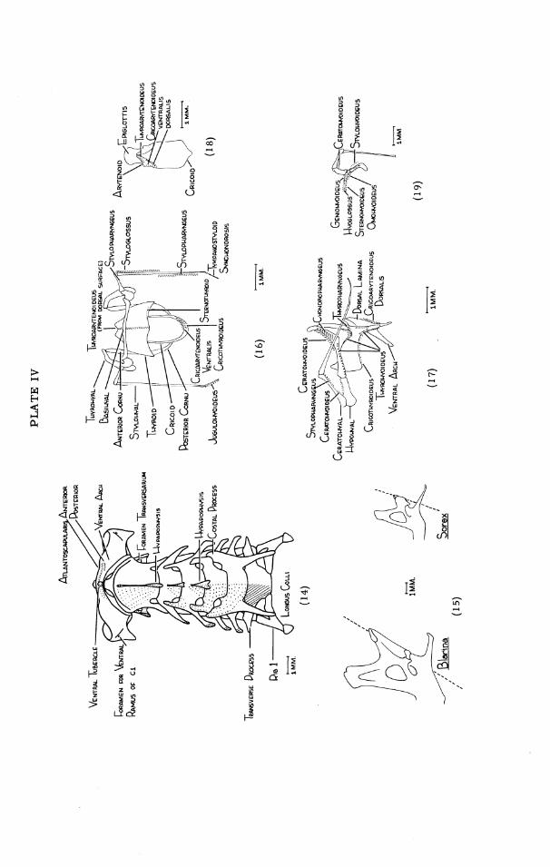

GEORGE R . L . GAUGHRAN
PLATE V
Fig. 20. Skull of Scalopus aquaticus mackrirms, dorsal view. Fig. 21. Skull of Scalopus aquaticus mackrinus, ventral view.


GEORGE R. L . GAUGkIRAN
PLATE VI
Fig. 22. Skull of Scalopus aquaticus machrilacs, lateral view. Fig. 23. Left malleus of Scalopus aquaticus machrinus, dorsal view. Fig. 24. Left incus of Scalopus aquaticus machrinus, dorsal view. Fig. 25. Left stapes of Scalopus aquaticus machrinus, lateral view. Fig. 26. Anterior aspect of snout of Scalopus aquaticus machrinus showing cartilaginous
plates. Fig. 27. Ventral aspect of snout of Scalopus aquaticus machrinus, showing cartilaginous
plates.

PLATE VI
- I M M
( 2 7 )

GEORGE R . L . GAUGHRAN
PLATE VII
Fig. 28. Cervical vertebrae of Scalopus aquaticus machrinzcs, lateral view. Fig. 29. Left dentary of Scalopus aquaticus m a c h r i m , lateral view. Fig. 30. Left dentary of Scalopus aquuticus machr im, medial view.


GEORGE R. L . GAUGHRAN
PLATE WI
Fig. 31. Cervical vertebrae of Scalopus aquaticus machrinus, ventral view. Fig. 32. 'Ligamentum nuchae of Scalopus aquaticus machrinus. Fig. 33. Left scapula of Scalopus aquaticus machrinus, lateral and vertebral surfaces. Fig. 34. Manubrium of Scalopus aquaticus machrim, right side.

PLATE VIII

GEORGE R . L . GAUGHRAN
PLATE M
Fig. 35. Clavicles of Scalopus aquaticus machrinzcs., anterior view. Fig. 36. Cricoid, arytenoid, and epiglottis of Scalopus aquaticus maclminus, dorsal
view. Fig. 37. Hyoid and laryngeal cartilages of Scalqpus aquaticus machrinzcs, ventral view. Fig. 38. Hyoid and laryngeal cartilages of Scalopus aquaticus machrinzcs, right side.

PLATE M
GEORGE R. L. GAUGHRAN
PLATE X
Fig. 39. Superficial musculature of Bhrina brevicauda kirtlandi, lateral view. Fig. 40. F i rs t depth of musculature of Blarina brevicauda kirtlandi, lateral view. Fig. 41. Second depth of musculature of Bhrina brevicauda kirtlandi, lateral view.

PLATE X

GEORGE R. L. GAUGHRAN
PLATE XI
Fig. 42. Third depth of musculature of Blarina brevicauda kirtlandi, lateral view. Fig. 43. Fourth depth of musculature of ~ b r i n a brevicauda kirtlandi, lateral view. Fig. 44. Fifth depth of musculature of Blarina brevicauda Yrtlandi, lateral view.

PLATE XI

GEORGE R. L . G A U G H R A N
PLATE XI1
Fig. 45A. Superficial layer and f i rs t depth of musculature of Blarina brevicauda kirtlandi, ventral view.
Fig. 45B. Second and third depths of musculature of Blarina brevicauda kirtlandi, ventral view.
Fig. 46. Schema of force vectors of masticatory muscles of Blarina brevicauda kirtlandi: ( 1 ) mandible closed, (2) mandible protracted, (3) mandible open.

PLATE XI1

GEORGE R. L. GAUGHRAN
PLATE XIII
Fig. 47. Fourth depth of musculature of Blarina brevicauda kirtlandi, ventral view.

PLATE XI11

GEORGE R . L . GAUGHRAN
PLATE XIV
Fig. 48. Superficial layer of musculature of Scalqpus aquaticus machrinus, dorsal view. Fig. 49. Superficial layer of musculature of Scalopus aquaticus machrinus, ventral view.

PLATE XIV
- 3 MM.
(49)

GEORGE R. L . GAUGHRAN
PLATE XV
Fig. 50. Superficial layer of musculature of Scalopus aquaticus machrinus, lateral view. Fig. 51. First depth of musculature of Scalopzls aquaticus machrinus, lateral view.


GEORGE R. L . GAUGHRAN
PLATE XVI
Fig. 52. Second depth of musculature of Scalopus aquaticus machrim, lateral view. Fig. 53. Third depth of musculature of Scalopus aquaticus machrinus, lateral view.

PLATE XVI

GEORGE R. L . GAUGHRAN
PLATE X M
Fig. 54. Fourth depth of musculature of Scalopus aquaticus machrims, lateral view. Fig. 55. Fifth depth of musculature of Scalopus aquaticus machrinus, lateral view.


GEORGE R. L . GAUGHRAN
PLATE XVIII
Fig. 56. Sixth depth of musculature of Scalopus aquaticus m a c h r i m , lateral view. Fig. 57. Seventh depth of musculature of Scalopus aquaticus machinus, lateral view.

PLATE XVIII

GEORGE R. L . GAUGHRAN
PLATE XM
Fig. 58. Eighth depth of musculature of Scalopzls aquaticus machrinus, lateral view. Fig. 59. Medial, dorsal, and lateral views of the superficial masseter of Scalopus
aqmticus machrinus .

PLATE XIX

GEORGE R . L . GAUGHRAN
PLATE XX
Fig. 60. Superficial layer and first depth of musculature of Scalopzls aquaticus machrims, ventral view.
Fig. 61. Second and third depths of musculature of Scalopus aquatiem machrims, ventral view.
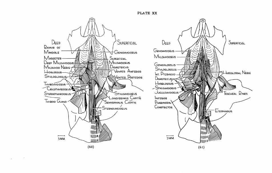
PLATE XX
GEORGE R. L . GAUGHRAN
PLATE XXI
Fig. 62. Fourth and fifth depths of musculature of Scalopus aquaticus machinus, ventral view.
Fig. 63. Sixth and seventh depths of musculature of Scalopus aquaticus machrinus, ventral view.


GEORGE R. L . GAUGHRAN
PLATE XXLI
Fig. 64A and C. Pattern of distribution of nerves IX, X, XI, and XI1 in Scalopus aquaticus m a c h r i m .
Fig. 64B. Pattern of distribution of cervical nerves in Scalopus aquaticus m a c h r i m . Fig. 65. Key to Figure 64B.

PLATE XXII
ENTEIZS SKUL
CONNECTS WlTU C GANGLION
ENTERS T U Y ~ U W I D MEM CRICOTUVBOI OEUS
SYMPATHETIC
DESCENDS TD L ~ E A R + " " ~ ~ ~ ' AND AORTIC ARCH
(64A, B, C )
SEMISDINALIS CAPITIS , RECTUS AND ODLIQUUS MUSCLES PUOMDOIDEUS
SPLENIUS \ CAPITIS
OCClPlTALlS
CERVICIS DORSAL RAM1
(65)
LEVATOR SCAPULAE ET SERQATUS ANT.
CEW. lLONGlSSlMUS CAP.
SERRATUS POST.
DQACUIAL PLEXUS

GEORGE R . L . GAUGHRAN
PLATE XXIII
Fig. 66. Reconstruction of skull of nestling Blarina brevicazida kirtlandi: ventral (left side), dorsal (right side).
Fig. 67. Reconstruction of skull of nestling Scalopus aqzuztincs m a c h r i m : ventral (left side), dorsal (right side).

PLATE XXIII


(CONTINUED FROM INSIDE FRONT COVER)
No. 30. The Darters of ihe Genera Hololepis and Villora. By Carl L. Hubbs and Mott Dwight . . . . . . . . . . . . . . . . . . . . . . . . . . . . Cannon. (1935) Pp. 93, 3 plates, 1 figure $0.50
No. 31. Goniobasis of the Coosa River, Alabama. By Calvin Goodrich. (1936) Pp. 60, 1 plate, 1 figure. . . . . . . . . . . . . . . . . . . . . . . . . . . . . . . . . . . . . . . . . . . . . . $0.35
. . . . . . . No. 32. Following Fox Trails. By Adolph Murie. (1936) Pp. 45, 6 plates, 6 figures $1.00 No. 33. The Discovery of the Nest of the Colima Warbler (Vermivora crissalis). By
. . . . . . . . . . Josselyn Van Tyne. (1936) Pp. 11, colored frontis., 3 plates, 1 map $0.35 No. 34. Mollusca of Peten and North Alta Vera Paz, Guatemala. By Calvin Goodrich and
. . . . . . . . . . . . . . Henry van der Schalie. (1937) Pp. 50, 1 plate, 1 figure, 1 map $0.50 No. 35. A Revision of the Lamprey Genus Ichthyomyzon. By Carl L. Hubbs and Milton B.
. . . . . . . . . . . . . . . . . . . . Trautman. (1937) Pp. 109, 2 plates, 5 figures, 1 map $2.00 No. 36. A Review of the Dragonflies of the Genera Neurocordulia and Platycordulia. By
. . . . . . . . . . . . . . . . . . . . . . . . Francis Byers. (1937) Pp. 36, 8 plates, 4 maps $0.50 No. 37. The Birds of Brewster County, Texas. By Josselyn Van Tyne and George Miksch
. . . . . . . . . . . . . . . . . . Sutton. (1937) Pp. 119, colored frontis., 5 plates, 1 map $1.25 No. 38. Revision of Sciurus variegatoides, a Species of Central American Squirrel. By
. . . . . William P. Harris, J r . (1937) Pp. 39, 3 plates (2 colored), 3 figures, 1 map $0.50 No. 39. Faunal Relationships and Geographic Distribution of Mammals in Sonora, Mexico.
. . . . . . . . . . . . . . . . . . . . . . . . . By William H. Burt. (1938) Pp. 77, 26 maps.. $0.75 No. 40. The Naiad Fauna of the Huron River, in Southeastern Michigan. By Henry van der
. . . . . . . . . . . . . . . . . . . Schalie. (1938) Pp. 83, 12 plates, 28 figures, 18 maps. $1.00 No. 41. The Life History of Henslow's Sparrow, Passerherbulus henslowi (Audubon). By A.
. . . . . . . . . . . . . . . . . . . Sidney Hyde. (1939) Pp. 72, 4 plates, 3 figures, 1 map. $0.75 No. 42. Studies of the Fishes of the Order Cyprinodontes. XVI. A Revision of the Goodeidae.
. . . . . . . . . . . . . . . By Carl L. Huhbs and C. L. Turner. (1939) Pp. 85, 5 plates. $0.90 No. 43. Aquatic Mollusks of the Upper Peninsula of Michigan. By Calvin Goodrich and Henry
. . . . . . . . . . . . . . . . . . . . . . . . . . . . . van der Schalie. (1939) Pp. 45, 2 maps. $0.50 No. 44. The Birds of Buckeye Lake, Ohio. By Milton B. Trautman. (1940) Pp. 466, 15
plates and a frontis., 2 maps . . . . . . . . . . . . . . . . . . . . . . . . . . . . . . . . . . . . . $2.50 No. 45. Territorial Behavior and Populations of Some Small Mammals in Southern Michigan.
. . . . . . . . . . . . . By William H. Burt. (1940) Pp. 58, 2 plates, 8 figures, 2 maps. $0.50 No. 46. A Contribution to the Ecology and Faunal Relationships of the Mammals of the Davis
Mountain Region, Southwestern Texas. By W. Frank Blair. (1940) Pp. 39, 3 plates, 1 map . . . . . . . . . . . . . . . . . . . . . . . . . . . . . . . . . . . . . . . . . . . . . . $0.35
No. 47. A Contribution to the Herpetology of the Isthmus of Tehuantepec. IV. By Norman . . . . . . . . . . . . . . . . . . . . . . . . . . Hartweg and James A. Oliver. (1940) Pp. 31 $0.35
No. 48. A Revision of the Black Basses (Micropterus and Huro) with Descriptions of Four New Forms. By Carl L. Hubbs and Reeve M. Bailey. (1940) Pp. 51, 6 plates, 1 figure, 2 maps ............................................. $0.75
No. 49. Studies of Neotropical Colubrinae. VIII. A Revision of the Genus Dryadophis Stuart, . . . . . . . . . . 1939. By L. C. Stuart. (1941) Pp. 106, 4 plates, 13 figures, 4 maps. $1.15
No. 50. A Contribution to the Knowledge of Variation in Opheodrys vernalis (Harlan), with the Description of a New Subspecies. By Arnold B. Grobman. (1941) Pp. 38, 2 figures, 1 map .............................................. $0.35
No. 51. Mammals of the Lava Fields and Adjoining Areas in Valencia County, New Mexico. . . . . . . . . . . . . . . . . . . . . By Emmet T. Hooper. (1941) Pp. 47, 3 plates, 1 map $0.50
No. 52. Type Localities of Pocket Gophers of the Genus Thomomys. By Emmet T. Hooper. (1941) Pp. 2 6 , l map . . . . . . . . . . . . . . . . . . . . . . . . . . . . . . . . . . . . . . . . . . $0.25
No. 53. The Crane Flies (Tipulidae) of the George Reserve, Michigan. By J. Speed Rogers. (1942) Pp. 128, 8 plates, 1 map . . . . . . . . . . . . . . . . . . . . . . . . . . . . . . . . . . . $1.25
No. 54. The Ecology of the Orthoptera and Dermaptera of the George Reserve, Michigan. . . . . . . . . . . . . . . . . . . By Irving J. Cantrall. (1943) Pp. 182, 10 plates, 2 maps $1.50
No. 55. Birds from the Gulf Lowlands of Southern Mexico. By Pierce Brodkorb. (1943) Pp. 8 8 , l map . . . . . . . . . . . . . . . . . . . . . . . . . . . . . . . . . . . . . . . . . . . . . . . $0.75
No. 56. Taxonomic and Geographic Comments on Guatemalan Salamanders of the Genus . . . . . . . . . . . . . . . . . Oedipus. By L. C. Stuart. (1943) Pp. 33, 2 plates, 1 map. $0.35
No. 57. The Amnicolidae of Michigan: Distribution, Ecology, and Taxonomy. By Elmer G. . . . . . . . . . . . . . . . . . . . . . Berry. (1943) Pp. 68, 9 plates, 10 figures, 10 maps $0.85
No. 58. A Systematic Review of the Neotropical Water Rats of the Genus Nectomys .. (Cricetinae). By Philip Hershkovitz. (1944) Pp. 88, 4 plates, 5 figures, 2 maps. $1.15
No. 59. San Francisco Bay a s a Factor Influencing Speciation in Rodents. By Emmet T. Hooper. (1944) Pp. 89, 5 plates, 18 maps . . . . . . . . . . . . . . . . . . . . . . . . . . . . $1.25
No. 60. The Fresh-Water Triclads of Michigan. By Roman Kenk. (1944) Pp. 44, 7 plates, 5 figures . . . . . . . . . . . . . . . . . . . . . . . . . . . . . . . . . . . . . . . . . . . . . . . . . . $0.50
No. 61. Home Range, Homing Behavior, and Migration in Turtles. By Fred R. Cagle. (1944) Pp. 34, 2 plates, 4 figures, 1 map . . . . . . . . . . . . . . . . . . . . . . . . . . . . . . . . . . $0.35
No. 62. Monograph of the Family Mordellidae (Coleoptera) of North America, North of Mexico. By Emil Liljeblad. (1945) Pp. 229, 7 plates . . . . . . . . . . . . . . . . . . . . $2.00

No. 63. Phylogenetic Position of the Citharidae, a Family of Flatfishes. By Carl L. Hubbs. (1945) Pp. 3 8 , l figure . . . . . . . . . . . . . . . . . . . . . . . . . . . . . . . . . . . . . . . . .
No. 64. Goniobasis livescens of Michigan. By Calvin Goodrich. (1945) Pp. 36, 1 plate, l f i g u r e , l m a p . . . . . . . . . . . . . . . . . . . . . . . . . . . . . . . . . . . . . . . . . . . . . .
No. 65. Endemic Fish Fauna of Lake Waccamaw, North Carolina. By Carl L. Hubbs and . . . . . . . . . . . . . . . . . . . . . . . Edward C. Raney. (1946) Pp. 30, 1 plate, 2 maps
No. 66. Revision of Ceratichthys, a Genus of American Cyprinid Fishes. By Carl L. Hubbs . . . . . . . . . . . . . . . and John D. Black. (1947) Pp. 56, 2 plates, 1 figure, 2 maps
No. 67. A Small Collection of Fishes from Rio Grande do Sul, Brazil. By A. Lourenqo . . . . . . . . . . . . . . . . . . . . . . . . . . . . Gomes. (1947) Pp. 39, 3 plates, 2 figures
No. 68. The Cyprinodont Fishes of the Death Valley System of Eastern California and South- western Nevada. By Robert R. Miller. (1948) Pp. 155, 15 plates, 5 figures, 3 maps
No. 69. The Amphibians and Reptiles of Alta Verapaz, Guatemala. By L. C. Stuart. (1948) Pp. 109,lO figures, 1 m a p . . .....................................
No. 70. The Land and Fresh-water Mollusks of Puerto Rico. By Henry van de r Schalie. (1948) Pp. 134, 15 plates, 4 figures, 64 maps . . . . . . . . . . . . . . . . . . . . . . . . . .
No. 71. The Animal Life of Temporary and Permanent Ponds in Southern Michigan. By ................... Roman Kenk. (1949) Pp. 66, 3 plates, 7 figures, 1 map
No. 72. Faunal Relationships of Recent North American Rodents. By Emmet T. Hooper. . . . . . . . . . . . . . . . . . . . . . . . . . . . . . . . . . . . . . . . . . . . . . . . (1949) Pp. 28
No. 73. A Study of Small Mammal Populations in Northern Michigan. By Richard H. ..................... Manville. (1949) Pp. 83, 4 plates, 6 figures, 1 map.
No. 74. Studies of the Nesting Birds of the Edwin S. George Reserve. Pa r t I. The Vireos. By George M. Sutton. (1949) Pp. 37, 5 plates. ......................... .... No. 75. The Birds of Michigan. By Norman A. Wood. (1951) Pp. 559, 16 plates, 1 map.
No. 76. Studies of the Black Swamp Snake, Seminatrix Pygaea (Cope), with Descriptions of . . . Two New Subspecies. By Herndon G. Dowling. (1950) Pp. 38, 6 figures, 1 map
No. 77. A Systematic Review of the Harvest Mice (Genus Reithrodontomys) of Latin America. .......... By Emmet T. Hooper. (1952) Pp. 255, 9 plates, 24 figures, 12 maps . . . . . . . . . . . . . . . . . . . . . . . . . . . . . . . . . . . . . . . . . . . . . . . . Cloth bound.
No. 78. Studies of Cyprinodont Fishes. XX. A New Subfamily from Guatemala, with Ctenoid Scales and a Unilateral Pectoral Clasper. By Carl L. Hubbs. (1950) Pp. 28, 4 plates, 1 map ..............................................
No. 79. An Analysis of Some Physical Factors Affecting the Local Distribution of the Short- tail Shrew (Blarina brevicauda) in the Northern Pa r t of the Lower Peninsula of Michigan. By William 0. Pruitt , Jr. (1953) Pp. 39, 9 figures. . . . . . . . . . . . . . .
No. 80. A Comparative Study of the Osteology and Myology of the Cranial and Cervical Regions of the Shrew, Blarina brevicauda, and the Mole, Scalopus aquaticus. By George R. L. Gaughran. (1954) Pp. 82, 67 figures . . . . . . . . . . . . . . . . . . . . . .
No. 81. A Systematic Study of the Avian Family Fringillidae, Based on the Structure of the Skull. By Harrison B. Tordoff. (1954) (In press).
No. 82. The Meibomian Glands of Voles and Lemmings (Microtinae). By Wilbur B. Quay. (1954) Pp. 24, 3 plates, 3 figures . . . . . . . . . . . . . . . . . . . . . . . . . . . . . . . . . .
No. 83. The Comparative Myology of the Mammalian Genera Sigmodon, Oryzomys, Neotoma, and Peromyscus (Cricetinae), With Remarks on Their Intergeneric Relationships. By George C. Rinker. (In press).
No. 84. The Burrowing Beetles of the Genus Mycotrupes (Coleoptera: Scarabaeidae: Geotrupinae). By Ada L. Olson, T. H. Hubbell, and H. F. Howden. (1954) Pp. 59, 8 plates, 3 figures ............................................