minimallyinvasiveevaluationandtreatmentof...
TRANSCRIPT

Hindawi Publishing CorporationInternational Journal of Surgical OncologyVolume 2011, Article ID 686030, 12 pagesdoi:10.1155/2011/686030
Review Article
Minimally Invasive Evaluation and Treatment ofColorectal Liver Metastases
Anton L. Gueorguiev,1 Richard Mackey,2 Gopal C. Kowdley,1
Jesus Esquivel,1 and Steven C. Cunningham1
1 Department of Surgery, Saint Agnes Hospital, 900 Caton Avenue, Mailbox no. 207, Baltimore, MD 21229, USA2 Department of Surgery, Saint Joseph Hospital, Baltimore, MD 21231, USA
Correspondence should be addressed to Steven C. Cunningham, [email protected]
Received 7 January 2011; Accepted 5 May 2011
Academic Editor: Abdullah Al Haddad
Copyright © 2011 Anton L. Gueorguiev et al. This is an open access article distributed under the Creative Commons AttributionLicense, which permits unrestricted use, distribution, and reproduction in any medium, provided the original work is properlycited.
Minimally invasive techniques used in the evaluation and treatment of colorectal liver metastases (CRLMs) include ultrason-ography (US), computed tomography, magnetic resonance imaging, percutaneous and operative ablation therapy, standardlaparoscopic techniques, robotic techniques, and experimental techniques of natural orifice endoscopic surgery. Laparoscopictechniques range from simple staging laparoscopy with or without laparoscopic intraoperative US, through intermediate tech-niques including simple liver resections (LRs), to advanced techniques such as major hepatectomies. Hereins, we review minimallyinvasive evaluation and treatment of CRLM, focusing on a comparison of open LR (OLR) and minimally invasive LR (MILR).Although there are no randomized trials comparing OLR and MILR, nonrandomized data suggest that MILR compares favorablywith OLR regarding morbidity, mortality, LOS, and cost, although significant selection bias exists. The future of MILR will likelyinclude expanding criteria for resectability of CRLM and should include both a patient registry and a formalized process forsurgeon training and credentialing.
1. Introduction
As the third commonest cancer in males and females andthe second commonest cause of cancer death [1], colorectalcancer (CRC) is an important health problem in the world.
There are estimated to be 334 000 new cases of CRC inEurope [2] and 142 570 new cases in the United States [1],and 36% of these patients will succumb to their CRC [1].Of all patients with CRC, approximately 65% develop dis-tant metastasis, and the commonest location (40%) is the liv-er [3]. The proportion of patients who presented with syn-chronous versus metachronous colorectal liver metastases(CRLMs) in France was equal in a recent epidemiologic st-udy: the proportion of patients with synchronous CRLM andthe 5-year rate of metachronous CRLM were both 14% [4].Unfortunately, only 25% of patients with CRLM are amena-ble to curative-intent treatment [3, 5–7].
Since the first report of a laparoscopic liver resection (LR)in 1992 (for CRLM) [8, 9], the field of minimally invasive liv-er resection (MILR) has seen tremendous advances, parallel-
ing those of open liver resection (OLR), with increasing safe-ty and efficacy. Although most early MILRs were for benigndisease, the first report notwithstanding, an increasing vol-ume of nonrandomized data suggests no oncologic disadvan-tage to performing MILR compared to OLR.
2. Minimally Invasive Evaluation
Transabdominal US is a widely available, inexpensive, andnoninvasive technique of evaluating for CRLM but, com-pared with other modalities, has the lowest sensitivity andnegative predictive value [10], excepting contrast-enhancedultrasound, which by some studies is as sensitive as CT [11]but which is not available in the United States. Consequently,CT and MRI are among the most commonly employed mo-dalities used to evaluate the liver for CRLM. A recent meta-analysis of diagnostic imaging of CRLM, evaluating 39 arti-cles (3391 patients), found MRI to be the optimal first-linemodality, with a per-patient sensitivity of 88% [12].

2 International Journal of Surgical Oncology
While early (1990s) studies showed staging laparoscopy(SL) to be superior to preoperative imaging in detecting un-resectable or extrahepatic disease, thereby sparing as many as34% of patients a laparotomy [13], when combined withintraoperative ultrasound (IOUS), even in an era of mark-edly improved axial imaging (2000s), SL was able to detectunresectable disease and potentially prevent unnecessary lap-arotomies in 10–25% of patients with primary and secondaryhepatic malignancies [14–16].
Increasing quality in preoperative axial imaging technol-ogy, however, has challenged the use of laparoscopic IOUS.In a prospective study of 194 patients undergoing LR forCRLM published in 2008, Tamandl et al. compared data frompreoperative imaging using multidetector CT (MDCT) andMRI to intraoperative findings using IOUS and bimanualpalpation and found that IOUS provided useful informationregarding additional CRLM in only 2.6% of patients [17].Other groups, however, have consistently found that 10% ofadditional small tumors that were missed by axial imagingmay be detected [18, 19] and the preoperative treatment planmay change in nearly half of cases [15] when a complete eval-uation is performed, including exposure of the entire surfaceof the liver and porta hepatis, an IOUS scan of all 8 liver seg-ments, porta hepatis and the paraceliac nodal bed, and athorough evaluation of the entire peritoneal cavity to detectextrahepatic metastases. Whether done laparoscopically oropen, a complete US evaluation of the liver should includefour steps: (1) an identification of intrahepatic vascular anat-omy, (2) identification and characterization of known le-sions, (3) a search for previously unrecognized lesions, and(4) the planning of a treatment strategy, which may includeresection, ablation, or both. Intrahepatic tumors are evalu-ated for size, number, location, relationship to biliary andvascular structures, and echogenicity (most CRLMs are hy-poechoic (42%) or isoechoic (43%), while a minority (15%)are hyperechoic [18]). Echogenicity should be noted becauseit has been shown to correlate with long-term survival: in aprospective evaluation of 147 patients at Johns Hopkins Hos-pital, the 5-year survival for patients with hypo-, iso-, andhyperechoic CRLM was 14%, 37%, and 46%, respectively[20].
3. Minimally Invasive Treatment Techniques
3.1. Ablative Techniques. Standard ablative techniques in-clude both chemical and thermal ablation and are increas-ingly used, both in isolation for patients with unresectableCRLM and in combination with LR. Chemical ablation withethanol or acetic acid has been performed for CRLM but isless effective for CRLM than for hepatocellular carcinoma[21–23]. Thermal ablation is therefore the preferred treat-ment for CRLM not amenable to surgical resection. Thermalablation includes radiofrequency ablation (RFA), microwave,laser, and cryoablation. In these techniques, focal heating orfreezing of tumor cells causes local tumor destruction whilepreserving surrounding hepatic parenchyma. An emerging
and still poorly studied technique is the nonthermal, non-chemical technique of electrical ablation using irreversibleelectroporation.
Although operative ablation of CRLM is often done incombination with both MILR and OLR, percutaneous ab-lation of CRLM is appropriate in cases in which no resec-tion is planned and offers an attractive minimally invasivetreatment option for such patients. However administered,RFA is relatively safe and less invasive than formal hepaticresection, but several notable complications may occur, in-cluding hepatic failure, hydrothorax, intraperitoneal bleed-ing, hepatic abscess, bile duct leaks, and tumor seeding [24–28]. The reported procedure-related morbidity ranges from2% to 12% and the mortality rate from 0% to 4.3% [24,26–29]. Many of these reports, however, include a varietyof approaches, including percutaneous, laparoscopic, andopen, and include more cases of HCC than CRLM [24, 27–29]. Given the frequent association of HCC with cirrhosis,complications such as bleeding, liver failure, and death maybe more common following ablation of HCC than CRLM[24, 27]. In a recent report of 100 patients undergoing RFAfor CRLM, there was no procedure-related morality and themajor complication rate was 8% [30]. Another study of100 patients undergoing RFA (146 treatments) for CRLMrevealed a major complication rate of 4.8%, including 1 deathfrom liver failure [31]. Gillams and Lees similarly found amajor complication rate of 4.0% in a series of 167 patientsundergoing RFA for CRLM [32].
Microwave ablation (MWA) is an emerging technologythat can also be performed open, percutaneously, or laparo-scopically. Like with RFA, however, no randomized trialssupport its use over other techniques. The primary theoreti-cal advantages of MWA are the ability to ablate larger lesionsand to do so faster. These advantages likely derive from thefact that MWA, unlike RFA, does not rely on electric cur-rent and so is not impeded by tissue desiccation and char-ring, both of which decrease electrical conductivity. In addi-tion, there is no so-called “heat sink” effect, or heat loss fromadjacent blood vessels, which in the case of RFA decreases theeffectiveness and increases the time required for ablation.Most reported experiences have demonstrated safe and ef-fective MWA, with complications and local recurrence ratescomparable to RFA [33–35]. Theoretical disadvantages in-clude the inadvertent injury to adjacent structures and therisk of such collateral damage makes tumor location impor-tant in the decision of which modality (e.g., RFA versusMWA) and route (e.g., open versus laparoscopic) to choose.Ablation of dome lesions or left lateral segment lesions, forinstance, could expose the diaphragm and heart to thermalinjury and serious morbidity, and such lesion may be bet-ter treated with an open or laparoscopic as opposed to per-cutaneous technique.
Cryoablation can similarly be performed via percutane-ous or open approaches. It offers many of the same bene-fits of RFA and MWA, such as preservation of liver paren-chyma, but at a potentially increased cost, given that the com-plication rates may be higher compared with RFA: mor-bidity 10–40% and mortality 0–5% [36]. Complicationsinclude hepatic/iceball fracture (19%), hemorrhage (3.7%),

International Journal of Surgical Oncology 3
coagulopathy (3.8%), biliary fistula (2.9%), and organ failure[36]. Furthermore, animal models have demonstrated thatthere is a more severe systemic response following cryother-apy than following RFA [37].
Irreversible electroporation (IRE) is an emerging, non-thermal, nonchemical technique that uses electrical currentto destroy cells. Its reversible counterpart, reversible electrop-oration, is a common laboratory technique that has beenemployed for decades to transiently render cells widely per-meable to allow entry of large molecules such as drugs andgenes by applying an electrical field to cells that are otherwisenot permeable to the molecules of interest [38]. IRE, pre-viously viewed as an undesireable upper limit of reversibleelectroporation because it rendered the cells permanentlypermeable and therefore nonviable, is now used clinically asa form of nonthermal ablation. While supported by animalmodels [39–41] and available commercially for use in pa-tients, IRE has not been well studied in patients. It doeshave the theoretical advantages, however, of being very fast(micro- to milliseconds), preserving connective tissue archi-tecture thereby allowing ablation close to vital structures inthe liver hilum for example, and unlike thermal techniques itis not likely affected by blood flow [38]. As with many newtechniques, all cases of IRE performed should be registeredto document the role and safety of IRE, as well as to identifyimportant questions for study in clinical trials, and indeedsuch a registry is underway at the University of Louisville[42].
3.2. Ablation and Resection.
(1) Surgical resection is the standard of care for CRLM,but RFA has produced comparable outcomes for lim-ited disease, with some important caveats. AlthoughRFA has demonstrated 5-year survival rates as highas 30% in some studies [31, 32] and numerous otherstudies have attempted to compare resection andablation [43–47], recent analyses using propensityscore methodology [48, 49] have shown that compar-ison of survival rates following RFA and resection isnot reliable, due to major differences in clinicopatho-logic characteristics, RFA technologies and expertise,and analysis of margin status (namely, lack of patho-logic analysis after RFA since no specimen is avail-able). Until randomized controlled trials comparingRFA and surgical resection are available, the questionof their comparability will likely remain unanswered[50]. In a recent systematic review performed bythe American Society of Clinical Oncology in 2009regarding RFA for CRLM [51], over 400 publishedarticles were reviewed, 46 identified as having over10 patients with adequate followup and perioperativedata to analyze. There was a wide variability in thereported 5-year survival (14–55%) and local tumorrecurrence (3.6–60%) [51].
While comparisons to resection may be unreliable andlargely unrevealing, comparisons within RFA series havebeen more fruitful. For instance, it has become clear thatincreasing CRLM size—especially size >3 cm—is directly
proportional to shorter survival and higher rates of recur-rence [52–54]. While some studies have found this to be trueregardless of the approach (open, laparoscopic, or percu-taneous) [53], other studies have found worse local tumorrecurrence and disease-free survival in percutaneously treat-ed patients compared with open cases [55]. Prognostic fac-tors that contribute to overall outcome include node statusof the primary CRC resection, synchronous versus metach-ronous disease, number and size of lesions, margin status ofresected hepatic lesions, CEA levels, the presence of extra-hepatic disease, satellite lesions and systemic treatment [51].RFA is an operator-dependent procedure, whether open, lap-aroscopic, or percutaneous, and requires careful techniqueand patient selection to achieve optimal outcomes.
3.3. Laparoscopic LR, Hand-Assisted Laparoscopic Surgery(HALS), and Hybrid Approaches. Although initial laparo-scopic liver surgery was typically limited to wedge resectionsof benign lesions that were easily accessible [8, 9, 56], moreadvanced liver resections are now performed laparoscopi-cally, including totally laparoscopic right, left, and centralhepatectomies, extended right and left hepatectomies, andposterosuperior resections [57–63].
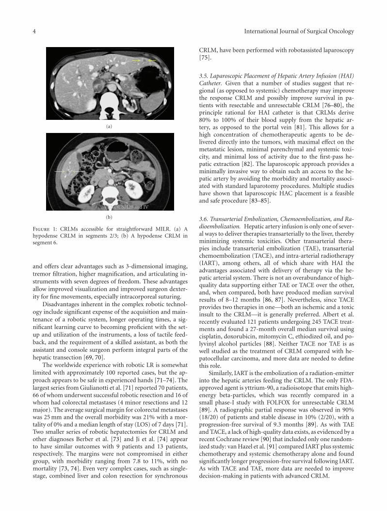
Many surgeons wishing to perform MILR begin a step-wise process of beginning with easy minor resections (Figure1), later using a hand port [64–67] or hybrid technique [68]for complex resections before finally attempting totally lap-aroscopic major resections. However, even when the dissec-tion and transection of the liver is totally laparoscopic incases of extended hepatectomies, the specimen is large andrequires an incision for removal [58, 59]. This incision isoften as long as that required for a hand port, which is onereason that many have advocated for maintaining—and insome cases returning to—a hand-assisted as opposed to atotally laparoscopic approach [66, 67]. The main advantagesof HALS are tactile feedback, including ability to palpate theliver, tumor, and nodes, facile liver mobilization and retrac-tion, quick and easy hemostasis with digital compression incases of unexpected hemorrhage, and the multiple additionaluses of the hand-port incision, including placement ofadditional instruments and removal of the specimen.
The hybrid technique, combining the relatively basic lap-aroscopic skills of liver mobilization with the more advancedbut open dissection of the liver hilum and transection ofthe liver parenchyma through a minilaparotomy, the smallsize of which is made possible by the laparoscopic mobili-zation [68], is another effort to shorten the learning curve(see below) associated with MILR, thereby increasing thenumbers of patients who may benefit from MILR. The hybridapproach, like the HALS approach, is meant to combine theadvantages of laparoscopic surgery (decreased postoperativepain and improved cosmesis, largely due to the avoidance ofa subcostal incision) with the safety, ease, and accessibilityprovided in open procedures.
3.4. Robotic Liver Resection. Robotic surgery overcomescertain limitations encountered with the laparoscopic tech-nique, such as 2-dimensional images and linear instruments,
4 International Journal of Surgical Oncology
(a)
VENOUSH20 and IV
(b)
Figure 1: CRLMs accessible for straightforward MILR. (a) Ahypodense CRLM in segments 2/3; (b) A hypodense CRLM insegment 6.
and offers clear advantages such as 3-dimensional imaging,tremor filtration, higher magnification, and articulating in-struments with seven degrees of freedom. These advantagesallow improved visualization and improved surgeon dexter-ity for fine movements, especially intracorporeal suturing.
Disadvantages inherent in the complex robotic technol-ogy include significant expense of the acquisition and main-tenance of a robotic system, longer operating times, a sig-nificant learning curve to becoming proficient with the set-up and utilization of the instruments, a loss of tactile feed-back, and the requirement of a skilled assistant, as both theassistant and console surgeon perform integral parts of thehepatic transection [69, 70].
The worldwide experience with robotic LR is somewhatlimited with approximately 100 reported cases, but the ap-proach appears to be safe in experienced hands [71–74]. Thelargest series from Giulianotti et al. [71] reported 70 patients,66 of whom underwent successful robotic resection and 16 ofwhom had colorectal metastases (4 minor resections and 12major). The average surgical margin for colorectal metastaseswas 25 mm and the overall morbidity was 21% with a mor-tality of 0% and a median length of stay (LOS) of 7 days [71].Two smaller series of robotic hepatectomies for CRLM andother diagnoses Berber et al. [73] and Ji et al. [74] appearto have similar outcomes with 9 patients and 13 patients,respectively. The margins were not compromised in eithergroup, with morbidity ranging from 7.8 to 11%, with nomortality [73, 74]. Even very complex cases, such as single-stage, combined liver and colon resection for synchronous
CRLM, have been performed with robotassisted laparoscopy[75].
3.5. Laparoscopic Placement of Hepatic Artery Infusion (HAI)Catheter. Given that a number of studies suggest that re-gional (as opposed to systemic) chemotherapy may improvethe response CRLM and possibly improve survival in pa-tients with resectable and unresectable CRLM [76–80], theprinciple rational for HAI catheter is that CRLMs derive80% to 100% of their blood supply from the hepatic ar-tery, as opposed to the portal vein [81]. This allows for ahigh concentration of chemotherapeutic agents to be de-livered directly into the tumors, with maximal effect on themetastatic lesion, minimal parenchymal and systemic toxi-city, and minimal loss of activity due to the first-pass he-patic extraction [82]. The laparoscopic approach provides aminimally invasive way to obtain such an access to the he-patic artery by avoiding the morbidity and mortality associ-ated with standard laparotomy procedures. Multiple studieshave shown that laparoscopic HAC placement is a feasibleand safe procedure [83–85].
3.6. Transarterial Embolization, Chemoembolization, and Ra-dioembolization. Hepatic artery infusion is only one of sever-al ways to deliver therapies transarterially to the liver, therebyminimizing systemic toxicities. Other transarterial thera-pies include transarterial embolization (TAE), transarterialchemoembolization (TACE), and intra-arterial radiotherapy(IART), among others, all of which share with HAI theadvantages associated with delivery of therapy via the he-patic arterial system. There is not an overabundance of high-quality data supporting either TAE or TACE over the other,and, when compared, both have produced median survivalresults of 8–12 months [86, 87]. Nevertheless, since TACEprovides two therapies in one—both an ischemic and a toxicinsult to the CRLM—it is generally preferred. Albert et al.recently evaluated 121 patients undergoing 245 TACE treat-ments and found a 27-month overall median survival usingcisplatin, doxorubicin, mitomycin C, ethiodized oil, and po-lyvinyl alcohol particles [88]. Neither TACE nor TAE is aswell studied as the treatment of CRLM compared with he-patocellular carcinoma, and more data are needed to definethis role.
Similarly, IART is the embolization of a radiation-emitterinto the hepatic arteries feeding the CRLM. The only FDA-approved agent is yttrium-90, a radioisotope that emits high-energy beta-particles, which was recently compared in asmall phase-I study with FOLFOX for unresectable CRLM[89]. A radiographic partial response was observed in 90%(18/20) of patients and stable disease in 10% (2/20), with aprogression-free survival of 9.3 months [89]. As with TAEand TACE, a lack of high-quality data exists, as evidenced by arecent Cochrane review [90] that included only one random-ized study: van Hazel et al. [91] compared IART plus systemicchemotherapy and systemic chemotherapy alone and foundsignificantly longer progression-free survival following IART.As with TACE and TAE, more data are needed to improvedecision-making in patients with advanced CRLM.
International Journal of Surgical Oncology 5
1992
1993
1994
1995
1996
1997
1998
1999
2000
2001
2002
2003
2004
2005
2006
2007
2008
p
1000900800700600500400300200100
0
Total number of laparoscopic liver resections
(a)
1992
1993
1994
1995
1996
1997
1998
1999
2000
2001
2002
2003
2004
2005
2006
2007
2008
p
600
500
400
300
200
100
0
Benign
Benign versus malignant lesions
Malignant
(b)
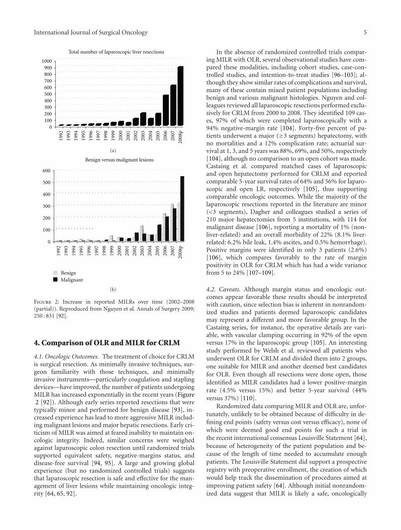
Figure 2: Increase in reported MILRs over time (2002–2008(partial)). Reproduced from Nguyen et al. Annals of Surgery 2009;250 : 831 [92].
4. Comparison of OLR and MILR for CRLM
4.1. Oncologic Outcomes. The treatment of choice for CRLMis surgical resection. As minimally invasive techniques, sur-geon familiarity with those techniques, and minimallyinvasive instruments—particularly coagulation and staplingdevices—have improved, the number of patients undergoingMILR has increased exponentially in the recent years (Figure2 [92]). Although early series reported resections that weretypically minor and performed for benign disease [93], in-creased experience has lead to more aggressive MILR includ-ing malignant lesions and major hepatic resections. Early cri-ticism of MILR was aimed at feared inability to maintain on-cologic integrity. Indeed, similar concerns were weighedagainst laparoscopic colon resection until randomized trialssupported equivalent safety, negative-margins status, anddisease-free survival [94, 95]. A large and growing globalexperience (but no randomized controlled trials) suggeststhat laparoscopic resection is safe and effective for the man-agement of liver lesions while maintaining oncologic integ-rity [64, 65, 92].
In the absence of randomized controlled trials compar-ing MILR with OLR, several observational studies have com-pared these modalities, including cohort studies, case-con-trolled studies, and intention-to-treat studies [96–103]; al-though they show similar rates of complications and survival,many of these contain mixed patient populations includingbenign and various malignant histologies. Nguyen and col-leagues reviewed all laparoscopic resections performed exclu-sively for CRLM from 2000 to 2008. They identified 109 cas-es, 97% of which were completed laparoscopically with a94% negative-margin rate [104]. Forty-five percent of pa-tients underwent a major (≥3 segments) hepatectomy, withno mortalities and a 12% complication rate; actuarial sur-vival at 1, 3, and 5 years was 88%, 69%, and 50%, respectively[104], although no comparison to an open cohort was made.Castaing et al. compared matched cases of laparoscopicand open hepatectomy performed for CRLM and reportedcomparable 5-year survival rates of 64% and 56% for laparo-scopic and open LR, respectively [105], thus supportingcomparable oncologic outcomes. While the majority of thelaparoscopic resections reported in the literature are minor(<3 segments), Dagher and colleagues studied a series of210 major hepatectomies from 5 institutions, with 114 formalignant disease [106], reporting a mortality of 1% (non-liver-related) and an overall morbidity of 22% (8.1% liver-related: 6.2% bile leak, 1.4% ascites, and 0.5% hemorrhage).Positive margins were identified in only 3 patients (2.6%)[106], which compares favorably to the rate of marginpositivity in OLR for CRLM which has had a wide variancefrom 5 to 24% [107–109].
4.2. Caveats. Although margin status and oncologic out-comes appear favorable these results should be interpretedwith caution, since selection bias is inherent in nonrandom-ized studies and patients deemed laparoscopic candidatesmay represent a different and more favorable group. In theCastaing series, for instance, the operative details are vari-able, with vascular clamping occurring in 92% of the openversus 17% in the laparoscopic group [105]. An interestingstudy performed by Welsh et al. reviewed all patients whounderwent OLR for CRLM and divided them into 2 groups,one suitable for MILR and another deemed best candidatesfor OLR. Even though all resections were done open, thoseidentified as MILR candidates had a lower positive-marginrate (4.5% versus 15%) and better 5-year survival (44%versus 37%) [110].
Randomized data comparing MILR and OLR are, unfor-tunately, unlikely to be obtained because of difficulty in de-fining end points (safety versus cost versus efficacy), none ofwhich were deemed good end points for such a trial inthe recent international consensus Louisville Statement [64],because of heterogeneity of the patient population and be-cause of the length of time needed to accumulate enoughpatients. The Louisville Statement did support a prospectiveregistry with preoperative enrollment, the creation of whichwould help track the dissemination of procedures aimed atimproving patient safety [64]. Although initial nonrandom-ized data suggest that MILR is likely a safe, oncologically

6 International Journal of Surgical Oncology
sound means of managing CRLM, ongoing critical review,multidisciplinary teams and participation in prospective datacollection or trials will continue to define optimal approachto CRLM.
4.3. Cost and LOS. Examination of cost has been reported byseveral groups in retrospective series. In a subgroup analysisfrom the University of Louisville, Buell et al. retrospectivelyreviewed and compared 29 laparoscopic and 34 open resec-tions, finding significant actual cost savings for a laparo-scopic approach to major resections ($21,131 versus $36,821;P < 0.01) before, but not after, adjustment for changes inDiagnosis-Related Group coding ($25,457 versus $23,691;P < 0.2) [111]. In a retrospective comparison of open ver-sus laparoscopic left lateral segmentectomy, Vanounou et al.at the University of Pittsburgh used deviation-based costmodeling to analyze open and laparoscopic liver resections(for both benign and malignant diseases) and found that theoverall LOS was 2 days shorter for the laparoscopic cases,which was associated with a per-patient cost reduction of$2939 compared to open cases [112]; when only malignantcases were considered, however, that cost reduction was at-tenuated at $1527 per patient.
Although both the Louisville and the Pittsburgh studiesshowed significantly shorter LOS for MILR versus OLR,other data, derived from comparing standard recovery path-way versus a fast-track recovery pathway following OLR, havefound a similar reduction in LOS (2 days) following fast-track recovery [113]. Indeed, it has been argued [110, 114,115] that LOS is determined more by extent of resection thanby operative approach and may be as little as 2–4 days witheither laparoscopic or open approach.
5. Future Directions and Controversies
5.1. Training and Credentialing. Broad surgical experience,corroborated by many studies [116–118], demonstrates thelearning curve for MILR. Simillis et al., for instance, showedthat only in studies published after 2003 and in studies in-cluding≥20 laparoscopic procedures did the operative bloodloss, LOS, and complications decrease for laparoscopic liversurgery when compared to open LR [117]. As the number ofMILR cases performed increases worldwide at a tremendousrate (Figure 2 [92]) and as the MILR community increasinglyrecognizes the infeasibility of performing randomized trialsof MILR [64], there have been many calls for an internationalregistry [64, 92] of MILR to maintain a record for monitoringoutcomes of efficacy and patient safety, as has already beenachieved for natural orifice transluminal endoscopic surgery[119], an experimental modality much more widely used forcholecystectomy (85% of all NOTES procedures) [119] thanfor MILR [120, 121].
Training and credentialing of MILR are currently per-formed at the local level and left to individual institutions.At a minimum a strong combined expertise in major lapar-oscopic surgery and advanced hepatobiliary techniques, inc-luding knowledge and skill in the use of intraoperativeultrasound, is required of a surgeon who wishes to begin
the learning curve for MILR [64, 117]. Currently, however,there are no clear, widely accepted criteria that define thisrequired expertise [64]. A certification process for MILR hasalso yet to be defined and the American Hepato-Pancreato-Biliary Association does not currently require a specific num-ber of MILR cases for the training of hepatobiliary surgeons[122]. Many surgeons, however, recognize the need for a sys-tematic progression from basic to advanced laparoscopic andhepatobiliary skills then to combined advanced laparoscopicand hepatobiliary skills, and some have published their ex-perience with starting an MILR program from scratch [123].
5.2. Learning Curve Effect.
(1) The learning-curve effect has not been studied exten-sively in laparoscopic liver surgery. Numerous single-institution series have shown an improvement in out-comes when the latter experience is compared to theearly experience [124, 125]. The first detailed analysisevaluating the learning curve effect in MILR waspublished by Vigano et al. in 2009, revealing signif-icantly improved operative time, conversion rate,blood loss, morbidity, and hospital stay progressive-ly over time as experience and volume increased, de-spite an increase in operative complexity over time[126]. The shape of the learning curve was, not sur-prisingly, similar to those reported regarding laparo-scopic colectomy [127]. These results suggest thatMILR is reproducible in selected high-volume cen-ters, by surgeons with advanced laparoscopic and he-patobiliary training.
5.3. Combined Liver and Lung Metastases.
(1) Although the liver is the most common site of col-orectal metastasis [3], another 3.5% of patients withcolon—and 11.5% of patients with rectal—cancerwill develop lung metastasis [128], most of whomhave both liver and lung diseases. Although early(prior to 2001) studies of curative-intent metasta-sectomy in patients with combined hepatic and pul-monary colorectal metastases found widely variable5-year survival rates, ranging from 11% to 44% [129–132], more recent retrospective studies have pro-duced improved 5-year survival rates as high as 64%[133–137] in patients with colorectal pulmonary me-tastases. Although fewer data are available regard-ing minimally invasive treatment of pulmonary—as compared to hepatic—colorectal metastases, largerecent series have shown that pulmonary metastasec-tomies may be performed with minimally invasivevideo-assisted thoracic surgery [137]. Another, evenless invasive option is RFA of pulmonary metastases.Yamakado et al. recently studied a series of 78 patientswith 198 colorectal lung metastases treated with RFA,reporting a 5-year and median survival of 35% and38 months, respectively [138].
International Journal of Surgical Oncology 7
5.4. Combined Liver and Peritoneal Metastases.
(1) Large series of patients with CRC have revealed that4% to 19% of patients have colorectal carcinomatosisat the time of the CRC resection or in follow up, al-though as many as 80% of patients who die of CRChave developed carcinomatosis by the time of theirdeath [139, 140]. In a review of nearly 3000 cases ofCRC in Singapore, 349 (13%) had carcinomatosis,61% (214) of whom had disease that was synchro-nous—and 39% (135) metachronous—with their in-itial presentation of CRC [139]. The proportionsof CRLM patients with no carcinomatosis, withmetachronous carcinomatosis, and with synchro-nous carcinomatosis were 10%, 33%, and 42%, res-pectively, respectively [139]. An increasing body ofdata [141], including randomized controlled data[142], suggests that complete surgical eradicationof metastatic peritoneal disease with cytoreductionand hyperthermic intraperitoneal chemotherapy(HIPEC) is beneficial to selected patients with coloncancer with carcinomatosis, with 5-year survival rates>50% recently reported [143].
Although the presence of CRLM is often considered acontraindication to cytoreduction and HIPEC, and similar-ly the presence of carcinomatosis a contraindication to LR,several recent studies [144–147] have evaluated the com-bination of cytoreduction, HIPEC, and liver resection as anaggressive emerging option for highly selected CRC patientswith both CRLM and peritoneal carcinomatosis. Elias et al.[147] evaluated a series of 24 such patients with a mean Per-itoneal Cancer Index (PCI) [148] of 8.6 (range: 2–25), halfof whom underwent a major hepatectomy, and reportedone postoperative death, a morbidity of 58%, and an over-all 2-year survival of 61%. Patients with ≥3 CRLM had sig-nificantly worse survival compared with patientS who had<3 CRLM [147]. More recently Chua et al. [144] eval-uated a series of 16 patients with both CRLM and carci-nomatosis treated by combined cytoreduction, HIPEC, andliver resection and reported a 2-year survival of 65%; nei-ther survival nor perioperative factors such as morbidity andLOS were different compared with patients who had isolatedcarcinomatosis without CRLM, although those with bothCRLM and carcinomatosis had significantly lower PCIcompared with patients with isolated carcinomatosis [144].Most patients with CRLM and peritoneal carcinomatosis arenot, however, currently candidates for aggressive surgical re-section of their CRLM and these emerging data should beinterpreted with caution.
Minimally invasive techniques have been used to performcytoreduction to palliate metastatic disease to the ovaries[149], to completely remove primary ovarian carcinomaswith limited peritoneal dissemination [150], and to dostaged laparoscopic HIPEC following open cytoreduction[151]. The results of an ongoing protocol on laparoscopiccytoreduction and HIPEC in patients with limited peritonealdissemination appear promising and compare favorably tothose patients having an open cytoreductive procedure [152],
suggesting that combining MILR and minimally invasive cy-toreduction and HIPEC is on the horizon.
6. Summary
The number of cases of MILR performed in the world hasincreased exponentially in recent years, and many centers arenow performing major, complex resections. In the absence ofrandomized trials comparing OLR and MILR, which is notlikely obtainable, nonrandomized data suggest that MILRproduces similar or improved morbidity, mortality, LOS,and cost compared with OLR, although significant selectionbias exists. Because randomized data will be difficult or im-possible to obtain, patient registries should be used to tracksafety and efficacy outcomes. Training of surgeons similarlyshould become more formalized, including an independentprocess for surgeon credentialing.
References
[1] A. Jemal, R. Siegel, J. Xu, and E. Ward, “Cancer statistics,2010,” CA Cancer Journal for Clinicians, vol. 60, no. 5, pp.277–300, 2010.
[2] F. Bray, R. Sankila, J. Ferlay, and D. M. Parkin, “Estimates ofcancer incidence and mortality in Europe in 1995,” EuropeanJournal of Cancer, vol. 38, no. 1, pp. 99–166, 2002.
[3] C. Pestana, R. J. Reitemeier, C. G. Moertel, E. S. Judd, and M.B. Dockerty, “The natural history of carcinoma of the colonand rectum,” The American Journal of Surgery, vol. 108, no. 6,pp. 826–829, 1964.
[4] S. Manfredi, C. Lepage, C. Hatem, O. Coatmeur, J. Faivre,and A. M. Bouvier, “Epidemiology and management of livermetastases from colorectal cancer,” Annals of Surgery, vol.244, no. 2, pp. 254–259, 2006.
[5] G. Steele Jr. and T. S. Ravikumar, “Resection of hepaticmetastases from colorectal cancer: biologic perspectives,”Annals of Surgery, vol. 210, no. 2, pp. 127–138, 1989.
[6] M. A. Choti, “Controversies in the management of hepaticcolorectal metastases,” Annals of Surgical Oncology, vol. 16,no. 9, pp. 2383–2384, 2009.
[7] R. Adam, V. Delvart, G. Pascal et al., “Rescue surgeryfor unresectable colorectal liver metastases downstaged bychemotherapy: a model to predict long-term survival,”Annals of Surgery, vol. 240, no. 4, pp. 644–658, 2004.
[8] M. Gagner, “Pioneers in laparoscopic solid organ surgery,”Surgical Endoscopy, vol. 17, no. 11, pp. 1853–1855, 2003.
[9] M. Gagner, M. Rheault, and J. Dubuc, “Laparoscopic partialhepatectomy for liver tumor,” Surgical Endoscopy, vol. 6, no.99, 1993.
[10] R. Carter, D. Hemingway, T. G. Cooke et al., “A prospectivestudy of six methods for detection of hepatic colorectalmetastases,” Annals of the Royal College of Surgeons ofEngland, vol. 78, no. 1, pp. 27–30, 1996.
[11] S. R. Rafaelsen and A. Jakobsen, “Contrast-enhanced ultra-sound vs multidetector-computed tomography for detectingliver metastases in colorectal cancer: a prospective, blinded,patient-by-patient analysis,” Colorectal Disease, vol. 13, no. 4,pp. 420–425, 2011.
[12] M. C. Niekel, S. Bipat, and J. Stoker, “Diagnostic imaging ofcolorectal liver metastases with CT, MR imaging, FDG PET,and/or FDG PET/CT: a meta-analysis of prospective studies

8 International Journal of Surgical Oncology
including patients who have not previously undergonetreatment,” Radiology, vol. 257, no. 3, pp. 674–684, 2010.
[13] T. J. Babineau, W. D. Lewis, R. L. Jenkins, R. Bleday, G.D. Steele, and R. A. Forse, “Role of staging laparoscopy inthe treatment of hepatic malignancy,” American Journal ofSurgery, vol. 167, no. 1, pp. 151–155, 1994.
[14] W. R. Jarnagin, J. Bodniewicz, E. Dougherty, K. Conlon, L.H. Blumgart, and Y. Fong, “A prospective analysis of staginglaparoscopy in patients with primary and secondary hepato-biliary malignancies,” Journal of Gastrointestinal Surgery, vol.4, no. 1, pp. 34–43, 2000.
[15] K. Thaler, S. Kanneganti, Y. Khajanchee et al., “The evolvingrole of staging laparoscopy in the treatment of colorectalhepatic metastasis,” Archives of Surgery, vol. 140, no. 8, pp.727–734, 2005.
[16] S. R. Grobmyer, Y. Fong, M. D’Angelica, R. P. DeMatteo, L.H. Blumgart, and W. R. Jarnagin, “Diagnostic laparoscopyprior to planned hepatic resection for colorectal metastases,”Archives of Surgery, vol. 139, no. 12, pp. 1326–1330, 2004.
[17] D. Tamandl, B. Herberger, B. Gruenberger et al., “Adequatepreoperative staging rarely leads to a change of intraoperativestrategy in patients undergoing surgery for colorectal cancerliver metastases,” Surgery, vol. 143, no. 5, pp. 648–657, 2008.
[18] M. G. van Vledder, T. M. Pawlik, S. Munireddy, U. Hamper,M. C. De Jong, and M. A. Choti, “Factors determiningthe sensitivity of intraoperative ultrasonography in detectingcolorectal liver metastases in the modern era,” Annals ofSurgical Oncology, vol. 17, no. 10, pp. 2756–2763, 2010.
[19] A. Foroutani, A. M. Garland, E. Berber et al., “Laparoscopicultrasound vs triphasic computed tomography for detectingliver tumors,” Archives of Surgery, vol. 135, no. 8, pp. 933–938,2000.
[20] M. L. DeOliveira, T. M. Pawlik, A. L. Gleisner, L. Assump-caom, G. J. Lopes-Filho, and M. A. Choti, “Echogenicappearance of colorectal liver metastases on intraoperativeultrasonography is associated with survival after hepaticresection,” Journal of Gastrointestinal Surgery, vol. 11, no. 8,pp. 970–976, 2007.
[21] D. Becker, J. M. Hansler, D. Strobel, and E. G. Hahn, “Percu-taneous ethanol injection and radio-frequency ablation forthe treatment of nonresectable colorectal liver metastases—techniques and results,” Langenbeck’s Archives of Surgery, vol.384, no. 4, pp. 339–343, 1999.
[22] B. Yamane and S. Weber, “Liver-directed treatment modal-ities for primary and secondary hepatic tumors,” SurgicalClinics of North America, vol. 89, no. 1, pp. 97–113, 2009.
[23] M. Giovannini and J. F. Seitz, “Ultrasound-guided percuta-neous alcohol injection of small liver metastases: results in 40patients,” Cancer, vol. 73, no. 2, pp. 294–297, 1994.
[24] W. T. Kong, W. W. Zhang, Y. D. Qiu et al., “Major com-plications after radiofrequency ablation for liver tumors:analysis of 255 patients,” World Journal of Gastroenterology,vol. 15, no. 21, pp. 2651–2656, 2009.
[25] E. Buscarini, A. Savoia, G. Brambilla et al., “Radiofrequencythermal ablation of liver tumors,” European Radiology, vol.15, no. 5, pp. 884–894, 2005.
[26] T. de Baere, O. Risse, V. Kuoch et al., “Adverse events duringradiofrequency treatment of 582 hepatic tumors,” AmericanJournal of Roentgenology, vol. 181, no. 3, pp. 695–700, 2003.
[27] T. Livraghi, L. Solbiati, M. F. Meloni, G. S. Gazelle, E.F. Halpern, and S. N. Goldberg, “Treatment of focal livertumors with percutaneous radio-frequency ablation: compli-cations encountered in a multicenter study,” Radiology, vol.226, no. 2, pp. 441–451, 2003.
[28] H. Rhim, K. H. Yoon, J. M. Lee et al., “Major complicationsafter radio-frequency thermal ablation of hepatic tumors:spectrum of imaging findings,” Radiographics, vol. 23, no. 1,pp. 123–136, 2003.
[29] E. Buscarini and L. Buscarini, “Radiofrequency thermal ab-lation with expandable needle of focal liver malignancies:complication report,” European Radiology, vol. 14, no. 1, pp.31–37, 2004.
[30] A. A. Van Tilborg, M. R. Meijerink, C. Sietses et al., “Long-term results of radiofrequency ablation for unresectable col-orectal liver metastases: a potentially curative intervention,”The British Journal of Radiology, vol. 84, no. 1002, pp. 556–565, 2011.
[31] J. Machi, A. J. Oishi, K. Sumida et al., “Long-term outcomeof radiofrequency ablation for unresectable liver metastasesfrom colorectal cancer: evaluation of prognostic factors andeffectiveness in first- and second-line management,” CancerJournal, vol. 12, no. 4, pp. 318–326, 2006.
[32] A. R. Gillams and W. R. Lees, “Radio-frequency ablationof colorectal liver metastases in 167 patients,” EuropeanRadiology, vol. 14, no. 12, pp. 2261–2267, 2004.
[33] C. Boutros, P. Somasundar, S. Garrean, A. Saied, andN. J. Espat, “Microwave coagulation therapy for hepatictumors: review of the literature and critical analysis,” SurgicalOncology, vol. 19, no. 1, pp. e22–e32, 2010.
[34] R. C. G. Martin, C. R. Scoggins, and K. M. McMasters,“Safety and efficacy of microwave ablation of hepatic tumors:a prospective review of a 5-year experience,” Annals ofSurgical Oncology, vol. 17, no. 1, pp. 171–178, 2010.
[35] D. A. Iannitti, R. C. G. Martin, C. J. Simon et al., “Hepatictumor ablation with clustered microwave antennae: the USphase II trial,” HPB—Journal of the International HepatoPancreato Biliary Association, vol. 9, no. 2, pp. 120–124, 2007.
[36] J. K. Seifert and T. Junginger, “Cryotherapy for liver tumors:current status, perspectives, clinical results, and review ofliterature,” Technology in Cancer Research and Treatment, vol.3, no. 2, pp. 151–163, 2004.
[37] K. K. Ng, C. M. Lam, R. T. Poon et al., “Comparison ofsystemic responses of radiofrequency ablation, cryotherapy,and surgical resection in a porcine liver model,” Annals ofSurgical Oncology, vol. 11, no. 7, pp. 650–657, 2004.
[38] B. Rubinsky, “Irreversible electroporation in medicine,”Technology in Cancer Research and Treatment, vol. 6, no. 4,pp. 255–260, 2007.
[39] K. P. Charpentier, F. Wolf, L. Noble, B. Winn, M. Resnick,and D. E. Dupuy, “Irreversible electroporation of the liverand liver hilum in swine,” HPB—Journal of the InternationalHepato Pancreato Biliary Association, vol. 13, no. 3, pp. 168–173, 2011.
[40] E. W. Lee, C. Chen, V. E. Prieto, S. M. Dry, C. T. Loh, andS. T. Kee, “Advanced hepatic ablation technique for creatingcomplete cell death: irreversible electroporation,” Radiology,vol. 255, no. 2, pp. 426–433, 2010.
[41] R. E. Brown, M. R. Bower, X. Li et al., “Nonthermal liver abla-tion by irreversible electroporation: results in a preclinicalporcine model,” in Proceedings of the 9th World Congress of theInternational Hepato-Pancreato-Biliary Association, BuenosAires, Argentina, May 2010.
[42] Registry, “Ablation Registry for Unresectable Soft TissueTumors,” http://www.ablationregistry.com/.
[43] T. A. Aloia, J. N. Vauthey, E. M. Loyer et al., “Solitarycolorectal liver metastasis: resection determines outcome,”Archives of Surgery, vol. 141, no. 5, pp. 460–467, 2006.

International Journal of Surgical Oncology 9
[44] W. S. Lee, S. H. Yun, H. K. Chun et al., “Clinical outcomesof hepatic resection and radiofrequency ablation in patientswith solitary colorectal liver metastasis,” Journal of ClinicalGastroenterology, vol. 42, no. 8, pp. 945–949, 2008.
[45] E. K. Abdalla, J. N. Vauthey, L. M. Ellis et al., “Recurrenceand outcomes following hepatic resection, radiofrequencyablation, and combined resection/ablation for colorectal livermetastases,” Annals of Surgery, vol. 239, no. 6, pp. 818–827,2004.
[46] T. J. White, S. H. Roy-Choudhury, D. J. Breen et al.,“Percutaneous radiofrequency ablation of colorectal hepaticmetastases—initial experience. An adjunct technique tosystemic chemotherapy for those with inoperable colorectalhepatic metastases,” Digestive Surgery, vol. 21, no. 4, pp. 314–320, 2004.
[47] G. Otto, C. Duber, M. Hoppe-Lotichius, J. Konig, M. Heise,and M. B. Pitton, “Radiofrequency ablation as first-linetreatment in patients with early colorectal liver metastasesamenable to surgery,” Annals of Surgery, vol. 251, no. 5, pp.796–803, 2010.
[48] S. Tsai and T. M. Pawlik, “Outcomes of ablation versusresection for colorectal liver metastases: are we comparingapples with oranges?” Annals of Surgical Oncology, vol. 16,no. 9, pp. 2422–2428, 2009.
[49] A. L. Gleisner, M. A. Choti, L. Assumpcao, H. Nathan, R.D. Schulick, and T. M. Pawlik, “Colorectal liver metastases:recurrence and survival following hepatic resection, radiofre-quency ablation, and combined resection-radiofrequencyablation,” Archives of Surgery, vol. 143, no. 12, pp. 1204–1212,2008.
[50] S. Mulier, Y. Ni, J. Jamart, L. Michel, G. Marchal, andT. Ruers, “Radiofrequency ablation versus resection forresectable colorectal liver metastases: time for a randomizedtrial?” Annals of Surgical Oncology, vol. 15, no. 1, pp. 144–157,2008.
[51] S. L. Wong, P. B. Mangu, M. A. Choti et al., “AmericanSociety of Clinical Oncology 2009 clinical evidence reviewon radiofrequency ablation of hepatic metastases fromcolorectal cancer,” Journal of Clinical Oncology, vol. 28, no.3, pp. 493–508, 2010.
[52] E. Berber, R. Pelley, and A. E. Siperstein, “Predictors ofsurvival after radiofrequency thermal ablation of colorectalcancer metastases to the liver: a prospective study,” Journal ofClinical Oncology, vol. 23, no. 7, pp. 1358–1364, 2005.
[53] F. F. Amersi, A. McElrath-Garza, A. Ahmad et al., “Long-term survival after radiofrequency ablation of complexunresectable liver tumors,” Archives of Surgery, vol. 141, no.6, pp. 581–588, 2006.
[54] L. Solbiati, T. Livraghi, S. N. Goldberg et al., “Percutaneousradio-frequency ablation of hepatic metastases from colorec-tal cancer: long-term results in 117 patients,” Radiology, vol.221, no. 1, pp. 159–166, 2001.
[55] R. M. Eisele, U. Neumann, P. Neuhaus, and G. Schu-macher, “Open surgical is superior to percutaneous accessfor radiofrequency ablation of hepatic metastases,” WorldJournal of Surgery, vol. 33, no. 4, pp. 804–811, 2009.
[56] M. Gagner, T. Rogula, and D. Selzer, “Laparoscopic liverresection: benefits and controversies,” Surgical Clinics ofNorth America, vol. 84, no. 2, pp. 451–462, 2004.
[57] J. Y. Cho, H. S. Han, Y. S. Yoon, and S. H. Shin, “Feasibilityof laparoscopic liver resection for tumors located in theposterosuperior segments of the liver, with a special referenceto overcoming current limitations on tumor location,”Surgery, vol. 144, no. 1, pp. 32–38, 2008.
[58] A. A. Gumbs, B. Bar-Zakai, and B. Gayet, “Totally laparo-scopic extended left hepatectomy,” Journal of GastrointestinalSurgery, vol. 12, no. 7, p. 1152, 2008.
[59] A. A. Gumbs and B. Gayet, “Multimedia article. Totallylaparoscopic extended right hepatectomy,” SurgicalEndoscopy, vol. 22, no. 9, pp. 2076–2077, 2008.
[60] C. G. S. Huscher, M. M. Lirici, S. Chiodini, and A. Recher,“Current position of advanced laparoscopic surgery of theliver,” Journal of the Royal College of Surgeons of Edinburgh,vol. 42, no. 4, pp. 219–225, 1997.
[61] G. Samama, L. Chiche, J. L. Brefort, and Y. Le Roux,“Laparoscopic anatomical hepatic resection: report of fourleft lobectomies for solid tumors,” Surgical Endoscopy, vol. 12,no. 1, pp. 76–78, 1998.
[62] N. O’Rourke and G. Fielding, “Laparoscopic right hepatec-tomy: surgical technique,” Journal of Gastrointestinal Surgery,vol. 8, no. 2, pp. 213–216, 2004.
[63] A. A. Gumbs and B. Gayet, “Totally laparoscopic centralhepatectomy,” Journal of Gastrointestinal Surgery, vol. 12, no.7, p. 1153, 2008.
[64] J. F. Buell, D. Cherqui, D. A. Geller et al., “The internationalposition on laparoscopic liver surgery: the Louisville state-ment, 2008,” Annals of Surgery, vol. 250, no. 5, pp. 825–830,2009.
[65] A. J. Koffron, G. Auffenberg, R. Kung, and M. Abecassis,“Evaluation of 300 minimally invasive liver resections at asingle institution: less is more,” Annals of Surgery, vol. 246,no. 3, pp. 385–392, 2007.
[66] A. Cuschieri, “Laparoscopic hand-assisted hepatic surgery,”Seminars in Laparoscopic Surgery, vol. 8, no. 2, pp. 104–113,2001.
[67] Y. Fong, W. Jarnagin, K. C. Conlon, R. DeMatteo, E.Dougherty, and L. H. Blumgart, “Hand-assisted laparoscopicliver resection: lessons from an initial experience,” Archives ofSurgery, vol. 135, no. 7, pp. 854–859, 2000.
[68] A. J. Koffron, R. D. Kung, G. B. Auffenberg, and M. M.Abecassis, “Laparoscopic liver surgery for everyone: thehybrid method,” Surgery, vol. 142, no. 4, pp. e461–e468, 2007.
[69] A. Amodeo, A. L. Quevedo, J. V. Joseph, E. Belgrano, and H.R. H. Patel, “Robotic laparoscopic surgery: cost and training,”Minerva Urologica e Nefrologica, vol. 61, no. 2, pp. 121–128,2009.
[70] D. G. Murphy, R. Hall, R. Tong, R. Goel, and A. J. Costello,“Robotic technology in surgery: current status in 2008,” ANZJournal of Surgery, vol. 78, no. 12, pp. 1076–1081, 2008.
[71] P. C. Giulianotti, A. Coratti, F. Sbrana et al., “Robotic liversurgery: results for 70 resections,” Surgery, vol. 149, no. 1, pp.29–39, 2011.
[72] S. B. Choi, J. S. Park, J. K. Kim et al., “Early experiences ofrobotic-assisted laparoscopic liver resection,” Yonsei MedicalJournal, vol. 49, no. 4, pp. 632–638, 2008.
[73] E. Berber, H. Y. Akyildiz, F. Aucejo, G. Gunasekaran, S. Cha-likonda, and J. Fung, “Robotic versus laparoscopic resectionof liver tumours,” HPB—Journal of the International HepatoPancreato Biliary Association, vol. 12, no. 8, pp. 583–586,2010.
[74] W. B. Ji, H. G. Wang, Z. M. Zhao, W. D. Duan, F. Lu, and J. H.Dong, “Robotic-assisted laparoscopic anatomic hepatectomyin China: initial experience,” Annals of Surgery, vol. 253, no.2, pp. 342–348, 2010.
[75] A. Patriti, G. Ceccarelli, A. Bartoli, A. Spaziani, L. M.Lapalorcia, and L. Casciola, “Laparoscopic and robot-assistedone-stage resection of colorectal cancer with synchronous

10 International Journal of Surgical Oncology
liver metastases: a pilot study,” Journal of Hepato-Biliary-Pancreatic Surgery, vol. 16, no. 4, pp. 450–457, 2009.
[76] A. E. Chang, P. D. Schneider, P. H. Sugarbaker, C. Simp-son, M. Culnane, and S. M. Steinberg, “A prospectiverandomized trial of regional versus systemic continuous5-fluorodeoxyuridine chemotherapy in the treatment ofcolorectal liver metastases,” Annals of Surgery, vol. 206, no.6, pp. 685–693, 1987.
[77] N. Kemeny, Y. Huang, A. M. Cohen et al., “Hepatic arterialinfusion of chemotherapy after resection of hepatic metas-tases from colorectal cancer,” The New England Journal ofMedicine, vol. 341, no. 27, pp. 2039–2048, 1999.
[78] T. G. Allen-Mersh, S. Earlam, C. Fordy, K. Abrams, andJ. Houghton, “Quality of life and survival with continuoushepatic-artery floxuridine infusion for colorectal liver metas-tases,” The Lancet, vol. 344, no. 8932, pp. 1255–1260, 1994.
[79] P. Rougier, A. Laplanche, M. Huguier et al., “Hepatic arterialinfusion of floxuridine in patients with liver metastasesfrom colorectal carcinoma: long-term results of a prospectiverandomized trial,” Journal of Clinical Oncology, vol. 10, no. 7,pp. 1112–1118, 1992.
[80] D. C. Hohn, R. J. Stagg, M. A. Friedman et al., “A randomizedtrial of continuous intravenous versus hepatic intraarterialfloxuridine in patients with colorectal cancer metastatic tothe liver: the Northern California Oncology Group trial,”Journal of Clinical Oncology, vol. 7, no. 11, pp. 1646–1654,1989.
[81] C. Breedis and G. Young, “The blood supply of neoplasms inthe liver,” American Journal of Pathology, vol. 30, no. 5, pp.969–977, 1954.
[82] H. S. G. Chen and J. F. Gross, “Intra-arterial infusion ofanticancer drugs: theoretic aspects of drug delivery andreview of responses,” Cancer Treatment Reports, vol. 64, no.1, pp. 31–40, 1980.
[83] F. Feliciotti, A. Paganini, M. Guerrieri, R. Chan, R. Campag-nacci, and E. Lezoche, “Laparoscopic intra-arterial catheterimplantation for regional chemotherapy of liver metastasis,”Surgical Endoscopy, vol. 10, no. 4, pp. 449–452, 1996.
[84] D. R. Urbach, D. M. Herron, Y. S. Khajanchee, L. L.Swanstrom, and P. D. Hansen, “Laparoscopic hepatic arteryinfusion pump placement,” Archives of Surgery, vol. 136, no.6, pp. 700–704, 2001.
[85] D. R. Urbach and P. D. Hansen, “Laparoscopic placementof a continuous hepatic artery infusion pump,” Seminars inLaparoscopic Surgery, vol. 7, no. 2, pp. 140–147, 2000.
[86] D. J. Martinelli, S. Wadler, C. W. Bakal et al., “Utility ofembolization or chemoembolization as second-line treat-ment in patients with advanced or recurrent colorectalcarcinoma,” Cancer, vol. 74, no. 6, pp. 1706–1712, 1994.
[87] H. S. Salman, J. Cynamon, M. Jagust et al., “Randomizedphase II trial of embolization therapy versus chemoemboliza-tion therapy in previously treated patients with colorectalcarcinoma metastatic to the liver,” Clinical Colorectal Cancer,vol. 2, no. 3, pp. 173–179, 2002.
[88] M. Albert, M. V. Kiefer, W. Sun et al., “Chemoembolizationof colorectal liver metastases with cisplatin, doxorubicin,mitomycin C, ethiodol, and polyvinyl alcohol,” Cancer, vol.117, no. 2, pp. 343–352, 2010.
[89] R. A. Sharma, G. A. Van Hazel, B. Morgan et al., “Radioem-bolization of liver metastases from colorectal cancer usingyttrium-90 microspheres with concomitant systemic oxali-platin, fluorouracil, and leucovorin chemotherapy,” Journalof Clinical Oncology, vol. 25, no. 9, pp. 1099–1106, 2007.
[90] A. Townsend, T. Price, and C. Karapetis, “Selective internalradiation therapy for liver metastases from colorectal cancer,”Cochrane Database of Systematic Reviews, no. 4, articeCD007045, 2009.
[91] G. Van Hazel, A. Blackwell, J. Anderson et al., “Randomisedphase 2 trial of SIR-Spheres plus fluorouracil/leucovorinchemotherapy versus fluorouracil/leucovorin chemotherapyalone in advanced colorectal cancer,” Journal of SurgicalOncology, vol. 88, no. 2, pp. 78–85, 2004.
[92] K. T. Nguyen, T. C. Gamblin, and D. A. Geller, “Worldreview of laparoscopic liver resection-2,804 patients,” Annalsof Surgery, vol. 250, no. 5, pp. 831–841, 2009.
[93] N. Katkhouda and E. Mavor, “Laparoscopic management ofbenign liver disease,” Surgical Clinics of North America, vol.80, no. 4, pp. 1203–1211, 2000.
[94] J. Fleshman, D. J. Sargent, E. Green et al., “Laparoscopiccolectomy for cancer is not inferior to open surgery basedon 5-year data from the COST Study Group trial,” Annals ofSurgery, vol. 246, no. 4, pp. 654–662, 2007.
[95] Y. Liang, G. Li, P. Chen, and J. Yu, “Laparoscopic versus opencolorectal resection for cancer: a meta-analysis of resultsof randomized controlled trials on recurrence,” EuropeanJournal of Surgical Oncology, vol. 34, no. 11, pp. 1217–1224,2008.
[96] L. Aldrighetti, C. Pulitano, M. Catena et al., “A prospectiveevaluation of laparoscopic versus open left lateral hepaticsectionectomy,” Journal of Gastrointestinal Surgery, vol. 12,no. 3, pp. 457–462, 2008.
[97] M. Abu Hilal, M. J. W. McPhail, B. Zeidan et al., “Laparo-scopic versus open left lateral hepatic sectionectomy: acomparative study,” European Journal of Surgical Oncology,vol. 34, no. 12, pp. 1285–1288, 2008.
[98] I. Dagher, G. Di Giuro, J. Dubrez, P. Lainas, C. Smadja, andD. Franco, “Laparoscopic versus open right hepatectomy: acomparative study,” American Journal of Surgery, vol. 198, no.2, pp. 173–177, 2009.
[99] K. Ito, H. Ito, C. Are et al., “Laparoscopic versus openliver resection: a matched-pair case control study,” Journal ofGastrointestinal Surgery, vol. 13, no. 12, pp. 2276–2283, 2009.
[100] M. Tsinberg, G. Tellioglu, C. H. Simpfendorfer et al., “Com-parison of laparoscopic versus open liver tumor resection: acase-controlled study,” Surgical Endoscopy, vol. 23, no. 4, pp.847–853, 2009.
[101] B. Topal, S. Fieuws, R. Aerts, H. Vandeweyer, and F.Penninckx, “Laparoscopic versus open liver resection of hep-atic neoplasms: comparative analysis of short-term results,”Surgical Endoscopy, vol. 22, no. 10, pp. 2208–2213, 2008.
[102] F. M. Polignano, A. J. Quyn, R. S. M. de Figueiredo, N. A.Henderson, C. Kulli, and I. S. Tait, “Laparoscopic versus openliver segmentectomy: prospective, case-matched, intention-to-treat analysis of clinical outcomes and cost effectiveness,”Surgical Endoscopy, vol. 22, no. 12, pp. 2564–2570, 2008.
[103] K. T. Nguyen, J. W. Marsh, A. Tsung, J. J. Steel, T. C. Gamblin,and D. A. Geller, “Comparative benefits of laparoscopicvs open hepatic resection: a critical appraisal,” Archives ofSurgery, vol. 146, no. 3, pp. 348–356, 2010.
[104] K. T. Nguyen, A. Laurent, I. Dagher et al., “Minimallyinvasive liver resection for metastatic colorectal cancer: amulti-institutional, international report of safety, feasibility,and early outcomes,” Annals of Surgery, vol. 250, no. 5, pp.842–848, 2009.
[105] D. Castaing, E. Vibert, L. Ricca, D. Azoulay, R. Adam, andB. Gayet, “Oncologic results of laparoscopic versus open

International Journal of Surgical Oncology 11
hepatectomy for colorectal liver metastases in two specializedcenters,” Annals of Surgery, vol. 250, no. 5, pp. 849–855, 2009.
[106] I. Dagher, N. O’Rourke, D. A. Geller et al., “Laparoscopicmajor hepatectomy: an evolution in standard of care,” Annalsof Surgery, vol. 250, no. 5, pp. 856–860, 2009.
[107] R. J. de Haas, D. A. Wicherts, E. Flores, D. Azoulay, D.Castaing, and R. Adam, “R1 resection by necessity forcolorectal liver metastases: is it still a contraindication tosurgery?” Annals of Surgery, vol. 248, no. 4, pp. 626–637,2008.
[108] M. A. Choti, J. V. Sitzmann, M. F. Tiburi et al., “Trendsin long-term survival following liver resection for hepaticcolorectal metastases,” Annals of Surgery, vol. 235, no. 6, pp.759–766, 2002.
[109] T. M. Pawlik, C. R. Scoggins, D. Zorzi et al., “Effect of surgicalmargin status on survival and site of recurrence after hepaticresection for colorectal metastases,” Annals of Surgery, vol.241, no. 5, pp. 714–722, 2005.
[110] F. K. S. Welsh, P. P. Tekkis, T. G. John, and M. Rees,“Open liver resection for colorectal metastases: better short-and long-term outcomes in patients potentially suitable forlaparoscopic liver resection,” HPB—Journal of the Interna-tional Hepato Pancreato Biliary Association, vol. 12, no. 3, pp.188–194, 2010.
[111] J. F. Buell, M. T. Thomas, S. Rudich et al., “Experiencewith more than 500 minimally invasive hepatic procedures,”Annals of Surgery, vol. 248, no. 3, pp. 475–485, 2008.
[112] T. Vanounou, J. L. Steel, K. T. Nguyen et al., “Comparingthe clinical and economic impact of laparoscopic versus openliver resection,” Annals of Surgical Oncology, vol. 17, no. 4, pp.998–1009, 2010.
[113] R. M. van Dam, P. O. Hendry, M. M. E. Coolsen et al.,“Initial experience with a multimodal enhanced recoveryprogramme in patients undergoing liver resection,” BritishJournal of Surgery, vol. 95, no. 8, pp. 969–975, 2008.
[114] J. Khan, T. M. Pawlik, and S. C. Cunningham, “No smallissue: hepatectomies-minimally invasive but more-than-minimally biased toward minor resections?” Journal of theAmerican College of Surgerons, vol. 212, no. 1, pp. 134–135,2010.
[115] S. Gagandeep and R. Selby, “Laparoscopic liver resections:extent of resection defines length of stay,” Journal Gastroin-testinal Surgery, vol. 10, no. 8, pp. 1188–1189, 2006.
[116] T. van Gulik, “Open versus laparoscopic resection forliver tumours,” HPB—Journal of the International HepatoPancreato Biliary Association, vol. 11, no. 6, pp. 465–468,2009.
[117] C. Simillis, V. A. Constantinides, P. P. Tekkis et al.,“Laparoscopic versus open hepatic resections for benign andmalignant neoplasms—a meta-analysis,” Surgery, vol. 141,no. 2, pp. 203–211, 2007.
[118] J. S. Barkun, J. K. Aronson, L. S. Feldman, G. J. Maddern,and S. M. Strasberg, “Evaluation and stages of surgicalinnovations,” The Lancet, vol. 374, no. 9695, pp. 1089–1096,2009.
[119] K. S. Lehmann, J. P. Ritz, A. Wibmer et al., “The Germanregistry for natural orifice translumenal endoscopic surgery:report of the first 551 patients,” Annals of Surgery, vol. 252,no. 2, pp. 263–270, 2010.
[120] S. J. Phee, K. Y. Ho, D. Lomanto et al., “Natural orificetransgastric endoscopic wedge hepatic resection in an exper-imental model using an intuitively controlled master andslave transluminal endoscopic robot (MASTER),” SurgicalEndoscopy, vol. 24, no. 9, pp. 2293–2298, 2010.
[121] J. F. Noguera, C. Dolz, A. Cuadrado, J. M. Olea, and A.Vilella, “Transvaginal liver resection (NOTES) combinedwith minilaparoscopy,” Revista Espanola de EnfermedadesDigestivas, vol. 100, no. 7, pp. 411–415, 2008.
[122] AHPBA, “Advanced GI Surgery Curriculum for Hepato-Pancreato-Biliary Surgery Fellowship; Appendix, VersionApril 21, 2010,” http://www.fellowshipcaselog.org/fellows/FC Guidelines for Logging HPB Cases.pdf.
[123] E. Wang, A. W. C. Kow, C. Y. Chan, K. H. Liau, and C.K. Ho, “Starting a laparoscopic hepatectomy programme,”Singapore Medical Journal, vol. 50, no. 4, pp. 354–359, 2009.
[124] S. Chang, A. Laurent, C. Tayar, M. Karoui, and D. Cherqui,“Laparoscopy as a routine approach for left lateral sectionec-tomy,” British Journal of Surgery, vol. 94, no. 1, pp. 58–63,2007.
[125] I. Dagher, J. M. Proske, A. Carloni, H. Richa, H. Tranchart,and D. Franco, “Laparoscopic liver resection: results for 70patients,” Surgical Endoscopy, vol. 21, no. 4, pp. 619–624,2007.
[126] L. Vigano, A. Laurent, C. Tayar, M. Tomatis, A. Ponti, and D.Cherqui, “The learning curve in laparoscopic liver resection:improved feasibility and reproducibility,” Annals of Surgery,vol. 250, no. 5, pp. 772–782, 2009.
[127] P. P. Tekkis, A. J. Senagore, C. P. Delaney, and V. W. Fazio,“Evaluation of the learning curve in laparoscopic colorectalsurgery: comparison of right-sided and left-sided resections,”Annals of Surgery, vol. 242, no. 1, pp. 83–91, 2005.
[128] E. Pihl, E. S. R. Hughes, and F. T. McDermott, “Lungrecurrence after curative surgery for colorectal cancer,”Diseases of the Colon and Rectum, vol. 30, no. 6, pp. 417–419,1987.
[129] J. R. Headrick, D. L. Miller, D. M. Nagorney et al., “Surgicaltreatment of hepatic and pulmonary metastases from coloncancer,” Annals of Thoracic Surgery, vol. 71, no. 3, pp. 975–980, 2001.
[130] K. Kobayashi, M. Kawamura, and T. Ishihara, “Surgicaltreatment for both pulmonary and hepatic metastases fromcolorectal cancer,” Journal of Thoracic and CardiovascularSurgery, vol. 118, no. 6, pp. 1090–1096, 1999.
[131] S. Murata, Y. Moriya, T. Akasu, S. Fujita, and K. Sugihara,“Resection of both hepatic and pulmonary metastases inpatients with colorectal carcinoma,” Cancer, vol. 83, no. 6,pp. 1086–1093, 1998.
[132] J. F. Regnard, D. Grunenwald, L. Spaggiari et al., “Surgicaltreatment of hepatic and pulmonary metastases from col-orectal cancers,” Annals of Thoracic Surgery, vol. 66, no. 1, pp.214–219, 1998.
[133] R. Marudanayagam, K. Ramkumar, V. Shanmugam et al.,“Long-term outcome after sequential resections of liver andlung metastases from colorectal carcinoma,” HPB—Journalof the International Hepato Pancreato Biliary Association, vol.11, no. 8, pp. 671–676, 2009.
[134] S. Limmer, E. Oevermann, C. Killaitis, P. Kujath, M. Hoff-mann, and H. P. Bruch, “Sequential surgical resection ofhepatic and pulmonary metastases from colorectal cancer,”Langenbeck’s Archives of Surgery, vol. 395, no. 8, pp. 1129–1138, 2010.
[135] J. Pfannschmidt, H. Hoffmann, and H. Dienemann,“Reported outcome factors for pulmonary resection inmetastatic colorectal cancer,” Journal of Thoracic Oncology,vol. 5, supplement 2, no. 6, pp. S172–S178, 2010.
[136] D. R. Carpizo and M. D’Angelica, “Liver resection formetastatic colorectal cancer in the presence of extrahepaticdisease,” Annals of Surgical Oncology, vol. 16, no. 9, pp. 2411–2421, 2009.

12 International Journal of Surgical Oncology
[137] M. Riquet, C. Foucault, A. Cazes et al., “Pulmonary resectionfor metastases of colorectal adenocarcinoma,” Annals ofThoracic Surgery, vol. 89, no. 2, pp. 375–380, 2010.
[138] K. Yamakado, Y. Inoue, M. Takao et al., “Long-term results ofradiofrequency ablation in colorectal lung metastases: singlecenter experience,” Oncology Reports, vol. 22, no. 4, pp. 885–891, 2009.
[139] D. G. Jayne, S. Fook, C. Loi, and F. Seow-Choen, “Peritonealcarcinomatosis from colorectal cancer,” British Journal ofSurgery, vol. 89, no. 12, pp. 1545–1550, 2002.
[140] M. J. Koppe, O. C. Boerman, W. J. G. Oyen, and R.P. Bleichrodt, “Peritoneal carcinomatosis of colorectal ori-gin: incidence and current treatment strategies,” Annals ofSurgery, vol. 243, no. 2, pp. 212–222, 2006.
[141] L. Maggiori and D. Elias, “Curative treatment of colorectalperitoneal carcinomatosis: current status and future trends,”European Journal of Surgical Oncology, vol. 36, no. 7, pp. 599–603, 2010.
[142] V. J. Verwaal, S. van Ruth, E. de Bree et al., “Randomizedtrial of cytoreduction and hyperthermic intraperitonealchemotherapy versus systemic chemotherapy and palliativesurgery in patients with peritoneal carcinomatosis of colorec-tal cancer,” Journal of Clinical Oncology, vol. 21, no. 20, pp.3737–3743, 2003.
[143] D. Elias, J. H. Lefevre, J. Chevalier et al., “Complete cytore-ductive surgery plus intraperitoneal chemohyperthermiawith oxaliplatin for peritoneal carcinomatosis of colorectalorigin,” Journal of Clinical Oncology, vol. 27, no. 5, pp. 681–685, 2009.
[144] T. C. Chua, T. D. Yan, J. Zhao, and D. L. Morris, “Peritonealcarcinomatosis and liver metastases from colorectal cancertreated with cytoreductive surgery perioperative intraperi-toneal chemotherapy and liver resection,” European Journalof Surgical Oncology, vol. 35, no. 12, pp. 1299–1305, 2009.
[145] D. Elias, P. Dube, S. Bonvalot et al., “Treatment of livermetastases with moderate peritoneal carcinomatosis by hep-atectomy and cytoreductive surgery followed by immediatepost-operative intraperitoneal chemotherapy: feasibility andpreliminary results,” Hepato-Gastroenterology, vol. 46, no. 25,pp. 360–363, 1999.
[146] C. P. Carmignani, G. Ortega-Perez, and P. H. Sugarbaker,“The management of synchronous peritoneal carcinomatosisand hematogenous metastasis from colorectal cancer,” Euro-pean Journal of Surgical Oncology, vol. 30, no. 4, pp. 391–398,2004.
[147] D. Elias, E. Benizri, M. Pocard, M. Ducreux, V. Boige, and P.Lasser, “Treatment of synchronous peritoneal carcinomatosisand liver metastases from colorectal cancer,” European Jour-nal of Surgical Oncology, vol. 32, no. 6, pp. 632–636, 2006.
[148] P. Jacquet and P. H. Sugarbaker, “Clinical research method-ologies in diagnosis and staging of patients with peritonealcarcinomatosis,” Cancer Treatment and Research, vol. 82, pp.359–374, 1996.
[149] P. A. van Dam, P. J. H. van Dam, L. Verkinderen, P.Vermeulen, F. Deckers, and L. Y. Dirix, “Robotic-assistedlaparoscopic cytoreductive surgery for lobular carcinoma ofthe breast metastatic to the ovaries,” Journal of MinimallyInvasive Gynecology, vol. 14, no. 6, pp. 746–749, 2007.
[150] Y. L. He, L. Y. Zhang, and D. X. Peng, “[Laparoscopiccytoreductive surgery for ovarian carcinoma: report of 4cases],” Di Yi Jun Yi Da Xue Xue Bao, vol. 24, no. 4, pp. 479–480, 2004.
[151] A. Knutsen, T. D. Sielaff, E. Greeno, and T. M. Tuttle,“Staged laparoscopic infusion of hyperthermic intraperi-toneal chemotherapy after cytoreductive surgery,” Journal ofGastrointestinal Surgery, vol. 10, no. 7, pp. 1038–1043, 2006.
[152] J. Esquivel, T. C. Chua, and A. Averbach, “Laparoscopiccytoreductive surgery and HIPEC in patients with limitedperitoneal surface malignancies,” Annals of Surgery, vol. 253,no. 4, pp. 764–768, 2011.

Submit your manuscripts athttp://www.hindawi.com
Stem CellsInternational
Hindawi Publishing Corporationhttp://www.hindawi.com Volume 2014
Hindawi Publishing Corporationhttp://www.hindawi.com Volume 2014
MEDIATORSINFLAMMATION
of
Hindawi Publishing Corporationhttp://www.hindawi.com Volume 2014
Behavioural Neurology
EndocrinologyInternational Journal of
Hindawi Publishing Corporationhttp://www.hindawi.com Volume 2014
Hindawi Publishing Corporationhttp://www.hindawi.com Volume 2014
Disease Markers
Hindawi Publishing Corporationhttp://www.hindawi.com Volume 2014
BioMed Research International
OncologyJournal of
Hindawi Publishing Corporationhttp://www.hindawi.com Volume 2014
Hindawi Publishing Corporationhttp://www.hindawi.com Volume 2014
Oxidative Medicine and Cellular Longevity
Hindawi Publishing Corporationhttp://www.hindawi.com Volume 2014
PPAR Research
The Scientific World JournalHindawi Publishing Corporation http://www.hindawi.com Volume 2014
Immunology ResearchHindawi Publishing Corporationhttp://www.hindawi.com Volume 2014
Journal of
ObesityJournal of
Hindawi Publishing Corporationhttp://www.hindawi.com Volume 2014
Hindawi Publishing Corporationhttp://www.hindawi.com Volume 2014
Computational and Mathematical Methods in Medicine
OphthalmologyJournal of
Hindawi Publishing Corporationhttp://www.hindawi.com Volume 2014
Diabetes ResearchJournal of
Hindawi Publishing Corporationhttp://www.hindawi.com Volume 2014
Hindawi Publishing Corporationhttp://www.hindawi.com Volume 2014
Research and TreatmentAIDS
Hindawi Publishing Corporationhttp://www.hindawi.com Volume 2014
Gastroenterology Research and Practice
Hindawi Publishing Corporationhttp://www.hindawi.com Volume 2014
Parkinson’s Disease
Evidence-Based Complementary and Alternative Medicine
Volume 2014Hindawi Publishing Corporationhttp://www.hindawi.com