migrating gossypioma: unforeseen challengesapexjournal.org/irms/archive/2014/dec/fulltext/malhotra...
TRANSCRIPT

International Research on Medical Sciences Vol.2(6), pp. 075-080, December, 2014 Available online at http://www.apexjournal.org
ISSN 2315-8845© 2014 Apex Journal International
Case Study
Migrating gossypioma: Unforeseen challenges
Parveen Malhotra*, Naveen Malhotra, Vani Malhotra, Ajay Chugh, and Abhisekh, Jai Karan
Department of Medical Gastroenterology, Anaesthesiology, Gynaecology and Obstetrics and Surgery, PGIMS Rohtak.
Accepted 6 November, 2014
One of the common medico-legal problems is retained sponge or any surgical instrument following an intra-abdominal surgery and is called Gossypioma. The condition can have various kind of manifestations ranging from asymptomatic stage to severe gastrointestinal complications like vomiting, pain abdomen, obstruction, perforation, peritonitis and even death. Transmural migration of gossypiboma is a very rare entity which leads to bowel or visceral perforation, obstruction or fistula formation. Transmural migration of an intra-abdominal gossypiboma has been reported to occur in stomach, ileum, colon, bladder, vagina and diaphragm. As per medical literature, very few cases have been reported. However, we report the first case of the largest gossypiboma to date: a surgical mop measuring 26 × 23 cm which was successfully removed endoscopically. A 60 year-old man underwent six months back, open cholecystectomy for symptomatic gall stones. He became symptomatic after two weeks of operative interference and developed complaints of pain abdomen and persistent vomiting. The barium swallow and abdominal ultrasonogram revealed a mass located in the distal part of stomach and duodenum which gave suspicion of gastric carcinoma. On upper gastro-intestinal endoscopy, a surgical mop that had totally migrated into the stomach and duodenum was seen. The surgical mop was successfully removed by endoscopy through the esophagus. The recovery of the patient was uneventful. Transmural migration of gossypiboma into the stomach should be considered in the differential diagnosis of any postoperative patient with unexplained persistent complaints of pain abdomen and vomiting. Endoscopy becomes handy for both diagnostic as well as therapeutic modality. However, surgery may be considered in endoscopically failed cases or in cases of incomplete migration of gossypiboma located into the stomach. Key words: Endoscopy, Gossypiboma; Intraluminal migration; Retained surgical sponge.
INTRODUCTION Gossypiboma is the term used to describe a retained non-absorbable surgical material that is composed of cotton matrix which leads to serious surgical complications for both patient and surgeon (Sozutek et al., 2010). Clinical symptoms related to intra-abdominal gossypiboma may vary from mild abdominal pain to major complications including bowel or visceral perfora-tion, obstruction, fistula formation or sepsis (Gawande et al., 2003). Despite its rarity, transmural migration of gossypiboma is one of the possible causes of these gastrointestinal complications. Transmural migration of an intra-abdominal gossypiboma has been reported to occur in stomach, ileum, colon, bladder, vagina and diaphragm (Erdil et al., 2008). To our knowledge, this is *Corresponding author. Email: [email protected]
the sixth reported case of transgastric migration of a gossypiboma in the medical literature. However, all of them were of smaller size of surgical sponge and three of them were removed endoscopically (Erdil et al., 2008; Mentes et al. 1997). We report the largest gastro-duodenal gossypiboma to date which was first diagnosed and then successfully treated endoscopically. CASE PRESENTATION
A 60 year-old man underwent six months back, open cholecystectomy for symptomatic gall stones. He became symptomatic after two weeks of operative interference and developed complaints of pain abdomen and persistent vomiting. Abdominal pain was persistent and mainly in the epigastrium and right hypochondrium. He took symptomatic treatment from various health

076 Int. Res. Med. Sci
Figure 1. Barium swallow showing mass like lesion at Antro-duodenal area.
practitioners but had no relief. He had a bout of fever which was associated with rigor and chills and remained for one week. He was treated with oral antibiotics and got relief. There was no relief in pain abdomen and vomiting and the intensity increased in last two weeks, before he reported in our institute. In these last two weeks he was not able to accept anything orally and was being maintained on intravenous fluids. Laboratory parameters revealed mild anemia, normal liver and renal function tests. The barium swallow (Figure 1) and abdominal ultrasonogram revealed a mass located in the distal part of stomach and duodenum which gave suspicion of gastric carcinoma. Hence, endoscopy was performed for proper diagnosis. A written informed consent including surgical risks was obtained from the patient and
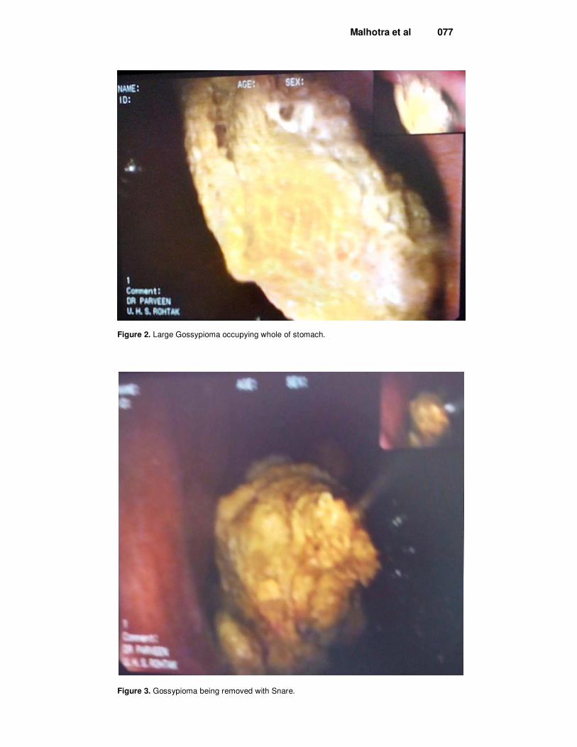
procedure was performed under sedation. On the endoscopy, a large surgical sponge which totally migrated and was found partially in pyloric antrum and rest half was stucked in duodenal bulb (Figure 2). The surgical sponge was loosened up with saline. Subsequently, it was grasped with saw-tooth forceps and pulled into the stomach. The compress was grasped again with a snare followed by releasing from the bulbus, then pulled out through the esophagus to the posterior region of the tongue (Figure 3). The sponge was then removed with gentle round motions from the mouth. The pyloric antrum was hugely dilated due to prolonged stucking of surgical sponge at that place (Figure 4). The size of removed surgical sponge was 26 cm × 23 cm and is of the largest size which has been successfully
Malhotra et al 077
Figure 2. Large Gossypioma occupying whole of stomach.
Figure 3. Gossypioma being removed with Snare.

078 Int. Res. Med. Sci
Figure 4. Hugely dilated Antrum after removal of stucked Gossypioma.
Figure 5. The removed surgical sponge of size 26 cm × 23 cm.
removed endoscopically, till date (Figure 5). No bleeding, fistula or injury was observed. The next day, all laboratory parameters, check endoscopy and abdominal
ultrasonogram were found to be normal hence patient was started on per- oral feeds which he accepted well, hence was discharged under hemodynamically condition

after two days of observation. DISCUSSION Retained surgical instrument or sponge following an intra-abdominal surgery is a potentially dangerous medico-legal problem. Despite a published incidence of 1:1000 to 1:1500 after intra-abdominal surgeries, it is encountered more commonly than reported (Cheng et al. 2007). The fear of litigation, disclosing the error by other clinicians or asymptomatic gossypiboma may mask the real incidence.
Gossypiboma induces two types of foreign body reactions; the first type is an aseptic fibrinous response that creates adhesions and encapsulation while the second type is an exudative reaction which leads to inflammatory reaction with abscess formation (Erbay et al., 2012; Düx et al., 2002). The first type reaction causes mild clinical symptoms like a painless abdominal mass, even asymptomatic, exudative reactions may manifest as a severe clinical course resulting in intestinal perforation, obstruction, fistula formation or sepsis (Sozutek et al., 2010; Gawande et al., 2003; Erdil et al., 2008; Mentes et al., 1997; Cheng et al., 2007; Erbay et al., 2012; Düx et al., 2002; Kundan et al., 2010). Migration of a retained sponge is a rare condition compared to abscess formation. It is a result of bodily response to extrude the foreign material by developing a fistula externally or into a hollow viscus. Transmural migration occurs as a result of inflammation in the intestinal wall that evolves to necrosis (Mentes et al., 1997; Kundan et al., 2010; Sarda et al., 2007). The migration site closes after complete migration of the surgical sponge. The small intestine is the most affected site due to its thin wall and large outer surface. Compared with the intestines, the stomach is an unusual site for transmural migration due to its higher localization and thick wall (Erbay et al., 2012; Sarda et al., 2007). The same thing occurred in our case as patient developed gradually progressive features of obstruction and had an episode of fever associated with rigor and chills which may had been due to mild peritonitis, at the time of migration of surgical sponge into gastrointestinal tract. The patient developed features of complete gastro-duodenal outlet obstruction, due to complete stucking of sponge at pyloric antro-duodenal level. Until now, this condition has been previously reported only in five cases (Erdil et al., 2008; Mentes et al., 1997; Erbay et al., 2012; Sarda et al., 2007). Interestingly, all of them occurred after acute open cholecystectomy operations, as occurred in our case. Hence, we emphasize that acute cholecystectomy is a major factor that leads to this kind of complication.
Imaging procedures such as plain X-ray, USG, CT and/or magnetic resonance (MR) may usually be helpful for diagnosis. Basically, a “whorl-like” mass imaging on plain X-ray is usually enough for diagnosis. In addition,
Malhotra et al 079 imaging of a hyperechogenic mass with hypoechoic rim on USG or a rounded mass with a dense central part and enhancing wall on CT are the basic signs of gossypiboma (Kopka et al., 1996; Cheng et al., 2007). It is frequently misdiagnosed as intra-abdominal hematoma, abscess or neoplasm which leads to unnecessary radical surgical interventions. Hence, gossypiboma should be considered in the differential diagnosis of any postoperative patient who presents with such suspicious radiological findings.
Gossypiboma should be removed as soon as possible to avoid further surgical complications and legal problems (Sozutek et al., 2010). Although open surgery is the most common approach in the treatment of gossypiboma, according to the localization of gossypiboma and skills of the clinician, removal can be easily performed by minimally invasive techniques such as endoscopy or laparoscopy (Sozutek et al., 2010; Mentes et al., 1997; Erbay et al., 2012). Although successful removals of surgical sponges by endoscopy has been reported before, the feasibility of endoscopy in removal of such a large surgical sponge compress was challenging. To our knowledge, herein we report the first case of the largest gossypiboma published to date successfully treated endoscopically. Hence, we emphasize that endoscopy may be a good option in the removal of such a large sponge located in the stomach and duodenum. However, surgery should be considered when fixed reaction and/or partial migration has occurred.
Patients undergoing emergency surgery, those with high body mass index, lengthy operations, inexperienced staff or unexpected change in surgical procedure are major risk factors for retained surgical materials (Sozutek et al., 2010; Gawande et al., 2003; Erdil et al., 2008; Mentes et al., 1997; Cheng et al., 2007; Erbay et al., 2012; Düx et al., 2002; Kundan et al., 2010). Simple precautions like educating the staff, tagging the sponges with markers or peroperative multiple counts of sponges and materials should reduce the incidence of gossypiboma (Kundan et al., 2010). In addition, new technologies like electronic tagging of sponges may be helpful in decreasing the incidence (Fabian, 2005) However, the feasibility of the procedure for developing country like ours, is not clear. CONCLUSION Transmural migration of gossypiboma into the stomach or small intestine should be considered in the differential diagnosis of any postoperative patient with unexplained pain abdomen and vomiting. Endoscopy becomes handy for both diagnostic as well as therapeutic modality. However, surgery may be considered in endoscopically failed/ refractory cases or in cases of incomplete migration of gossypiboma.

080 Int. Res. Med. Sci REFERENCES Cheng, T.C., Chou, A.S., Jeng, C.M., Chang, P.Y., Lee,
C.C. (2007). Computed tomography findings of gossypiboma. J. Chin. Med. Assoc., 70: 565-569.
Düx, M., Ganten, M., Lubienski, A., Grenacher, L. (2002). Retained surgical sponge with migration into the duodenum and persistent duodenal fistula. Eur. Radiol., 12: 74-77.
Erbay, G., Koc, Z., Caliskan, K., Araz, F., Ulusan, S. (2010). Imaging and clinical findings of a gossypiboma migrated into the stomach. Turk. J. Gastroenterol., 23: 54-57.
Erdil, A., Kilciler, G., Ates, Y., Tuzun, A., Gulsen, M., Karaeren, N., Dagalp, K. (2008). Transgastric migration of retained intraabdominal surgical sponge: gossypiboma in the bulbus. Inter. Med., 47: 613-615.
Fabian, C.E. (2005). Electronic tagging of surgical sponges to prevent their accidental retention. Surgery, 137: 298-301.
Gawande, A.A., Studdert, D.M., Orav, E.J., Brennan, T.A., Zimmer, M.J. (2003). Risk factors for retained instruments and sponges after surgery. N. Eng. J. Med., 348: 229-235.
Kopka, L., Fischer, U., Gross, A.J., Funke, M.,
Oestmann, J.W., Grabbe, E. (1996). CT of retained surgical sponges (textilomas): pitfalls in detection and evaluation. J. Comput. Assist Tomogr. 20: 919-923.
Kundan, K.K., Patil, S.K., Gorad, K.P., Panchal, A.H., Arora, S.S., Gautam, R.P. (2010). Intraluminal migration of surgical sponge: gossypiboma. Saudi J. Gastroenterol. 16: 221-222.
Mentes, B.B., Yilmaz, E., Sen, M., Kayhan, B., Gorgul, A., Tatlicioglu, E. (1997). Transgastric migration of a surgical sponge. J. Clin. Gastroenterol., 12: 55-57.
Sarda, A.K., Pandey, D., Neogi, S., Dhir, U. (2007). Postoperative complications due to a retained surgical sponge. Singapore Med. J., 48: 160-164.
Sozutek, A., Karabuga, T., Bozdag, A.D., Derici, H. (2010). Asymptomatic gossypiboma mimicking a liver mass. Turk. J. Surg., 26: 225-228.