microparticle shedding from neural progenitor cells and vascular
TRANSCRIPT

RESEARCH ARTICLE
Microparticle Shedding from NeuralProgenitor Cells and Vascular CompartmentCells Is Increased in Ischemic StrokeGemma Chiva-Blanch1,2, Rosa Suades1,2, Javier Crespo1,2, Esther Peña1,2,Teresa Padró1,2, Elena Jiménez-Xarrié2,3, Joan Martí-Fàbregas2,3, Lina Badimon1,2*
1 Cardiovascular Research Center (CSIC-ICCC), Barcelona, Spain, 2 Biomedical Research Institute SantPau (IIB-Sant Pau), Barcelona, Spain, 3 Department of Neurology, Hospital de la Santa Creu i Sant Pau,Barcelona, Spain
Abstract
Purpose
Ischemic stroke has shown to induce platelet and endothelial microparticle shedding, but
whether stroke induces microparticle shedding from additional blood and vascular compart-
ment cells is unclear. Neural precursor cells have been shown to replace dying neurons at
sites of brain injury; however, if neural precursor cell activation is associated to microparticle
shedding, and whether this activation is maintained at long term and associates to stroke
type and severity remains unknown. We analyzed neural precursor cells and blood and vas-
cular compartment cells microparticle shedding after an acute ischemic stroke.
Methods
Forty-four patients were included in the study within the first 48h after the onset of stroke.
The cerebral lesion size was evaluated at 3–7 days of the stroke. Circulating microparticles
from neural precursor cells and blood and vascular compartment cells (platelets, endothelial
cells, erythrocytes, leukocytes, lymphocytes, monocytes and smooth muscle cells) were
analyzed by flow cytometry at the onset of stroke and at 7 and 90 days. Forty-four age-
matched high cardiovascular risk subjects without documented vascular disease were used
as controls.
Results
Compared to high cardiovascular risk controls, patients showed higher number of neural
precursor cell- and all blood and vascular compartment cell-derived microparticles at the
onset of stroke, and after 7 and 90 days. At 90 days, neural precursor cell-derived micropar-
ticles decreased and smooth muscle cell-derived microparticles increased compared to lev-
els at the onset of stroke, but only in those patients with the highest stroke-induced cerebral
lesions.
PLOS ONE | DOI:10.1371/journal.pone.0148176 January 27, 2016 1 / 14
OPEN ACCESS
Citation: Chiva-Blanch G, Suades R, Crespo J, PeñaE, Padró T, Jiménez-Xarrié E, et al. (2016)Microparticle Shedding from Neural Progenitor Cellsand Vascular Compartment Cells Is Increased inIschemic Stroke. PLoS ONE 11(1): e0148176.doi:10.1371/journal.pone.0148176
Editor: Valery Combes, University of TechnologySydney, AUSTRALIA
Received: October 8, 2015
Accepted: January 13, 2016
Published: January 27, 2016
Copyright: © 2016 Chiva-Blanch et al. This is anopen access article distributed under the terms of theCreative Commons Attribution License, which permitsunrestricted use, distribution, and reproduction in anymedium, provided the original author and source arecredited.
Data Availability Statement: All relevant data arewithin the paper and its Supporting Information files.
Funding: GC-B is a Sara Borrell Postdoctoral Fellow(CD13/00023) from Instituto de Salud Carlos III andreceived the ImmunoTools special Award 2014. Thiswork has been possible thanks to funding receivedfrom Spanish Ministry of Economy andCompetitiveness (PNS-SAF2013-42962-R, to LB)and from RETICS of Instituto Carlos III (RIC andTERCEL to LB, and RETICS (INVICTUS-RD12/014/0002 and FEDER and FIS PI071062 to JM-F). Noneof the funders played any role in study design, data

Conclusions
Stroke increases blood and vascular compartment cell and neural precursor cell microparti-
cle shedding, an effect that is chronically maintained up to 90 days after the ischemic event.
These results show that stroke induces a generalized blood and vascular cell activation and
the initiation of neuronal cell repair process after stroke. Larger cerebral lesions associate
with deeper vessel injury affecting vascular smooth muscle cells.
IntroductionCirculating microparticles (cMPs) are phospholipid blebs sized 0.1–1.0μm shed from theplasma membrane of eukaryotic cells when injured, activated, or undergoing apoptosis. cMPsare shed from several cell types, and have been shown to reflect cellular activation and/or tissuedegeneration occurring in vivo [1]. High endothelial and platelet MP levels have been observedin patients who have had an acute ischemic stroke [2], severe hypertension and increased riskof coronary heart disease [3–5]. Due to their molecular cargo cMPs seem to contribute to bothvascular disease initiation and progression and also be involved in its clinical outcomes. cMPsmay have relevant clinical applications including their potential use both as biomarkers of dis-ease for improving cardiovascular risk prediction [6] and as novel therapeutic targets [7].
Neural precursor cells (NPCs) express CD34, a progenitor cell biomarker, and CD56 (neuralcell adhesion molecule -NCAM-), a neural surface marker present in both neural stem andmature cells. NPCs have been shown to increase after stroke and migrate to the lesion sitewhere they differentiate to mature neural cells in the rodent [8] and the human brain [9]. Thus,it seems plausible that NPCs also release MPs after brain injury. Nevertheless, NPC-derivedcMPs after a stroke have never been analyzed.
Stroke is the second leading cause of death worldwide, responsible for the 9 percent of thetotal 50.5 million deaths each year, and the principal cause of disability in the elderly. In acuteischemic stroke the mechanisms of brain ischemia are critically dependent on endothelial, vas-cular and inflammatory factors [10,11]. In fact, levels of endothelial-derived MPs directly cor-relate with clinical disease severity and infarct volume and are markers of vascular pathology[12,13]. Nevertheless, if stroke is involved in increased MP shedding of other cells of the bloodand vascular compartment (BVCCs) and NPCs, and if this activation is maintained at the longterm and correlates to stroke severity still remains unclear. Therefore, we aimed to determineMP shedding from different cells at the onset of stroke and at 7 and 90 days and compare it tosubjects at high risk but without cardiovascular disease. We also aimed to analyze the associa-tion between cMPs and cerebral infarction size, etiology and level of chronic disability inpatients with ischemic stroke.
Materials and Methods
PatientsForty-four patients with a suspected ischemic stroke were included in the study. Patients wereadmitted to the Neurology Department at Hospital de la Santa Creu i Sant Pau (Barcelona,Spain) and included within the first 48h after the onset of stroke. The Ethics Committee atHospital de la Santa Creu i Sant Pau approved the study, and all patients or their legal represen-tatives gave written informed consent. The study was conducted according to the Declarationof Helsinki.
Ischemic Stroke Increases Microparticle Shedding
PLOS ONE | DOI:10.1371/journal.pone.0148176 January 27, 2016 2 / 14
collection and analysis, decision to publish orpreparation of the manuscript.
Competing Interests: The authors have declaredthat no competing interests exist.

After inclusion in the study, a medical record was administered to obtain demographic fac-tors, medical and therapeutic data, and stroke etiological subtype, according to the SSS-TOASTclassification and NIHSS score at admission. All patients underwent magnetic resonance imag-ing (MRI, n = 30) or non-contrast computed tomography (CT, n = 14). MRI and CT were per-formed on patients at a mean of 3–7 days after the presentation of stroke as part of thestandard clinical care protocol. Lesion volumes were evaluated by an observer blinded to cMPquantification and calculated as (AxBxC)/2, where A is the maximum lesion diameter, B is theperpendicular of A, and C is the coronal diameter. Old lesions were not included as lesion vol-ume. Patients were categorized in tertiles of lesion volume (1st tertile: 0 to 0.08cm3, n = 14; 2nd
tertile: from 0.081 to 4.40cm3; n = 15 and 3rd tertile: from 4.41 to 88cm3, n = 15). Blood sampleswere taken within the first 48h after the onset of stroke, and then after 7 and 90 days.
Subjects without cardiovascular diseaseThe 44 control subjects included in this study were at high cardiovascular risk but free of car-diovascular disease or cancer and belong to the SAFEHEART cohort [14], an open, multicen-tre, long-term prospective study. Demographic and clinical characteristics data, cardiovascularhistory, classic cardiovascular risk factors and current treatment for hypercholesterolemia wereobtained from all subjects using a standardized report form at inclusion. The study wasapproved by the local ethics committee and was conducted according to the Declaration ofHelsinki. A written informed consent was obtained from all participants prior to the study.
Blood samplingVenous blood was withdrawn after 10–14 hours of fasting into 3.8% sodium citrate tubes.Blood cells were removed by low-speed centrifugation (250xg, 15min) at room temperature(RT) in order to avoid in vitro platelet activation. Platelet-rich plasma (PRP) was carefully aspi-rated, leaving about 1mm undisturbed layer on top of the cells. A second centrifugation step(11000×g, 10min, RT) and a third centrifugation step (11000xg, 3min, RT) were performed toensure the complete removal of platelets and to obtain the platelet-free plasma (PFP). All sam-ples were processed identically and within 30min after extraction. PFP aliquots of 250μL wereimmediately frozen in liquid nitrogen and stored at -80°C until processing for isolation andquantification of cMPs.
Circulating microparticles isolation and quantificationTo isolate cMPs, 225μL of frozen PFP aliquots were thawed on melting ice and centrifuged at20000×g for 30min. The supernatants (200μL) were discarded and the cMP-enriched pelletwas washed once with 200μL citrate-phosphate buffered saline solution (citrate-PBS; 1.4mMphosphate, 154mMNaCl, 10.9mM trisodium citrate, pH 7.4). A second equal centrifugationstep was made and 200μL of the supernatant were discarded to resuspend the remaining cMPpellets in 75μL citrate-PBS.
Triple-label flow cytometric analysis was performed as described by Suades et al [15].Briefly, 5μL of washed cMP suspensions were diluted in 30μL PBS buffer containing 2.5mMCaCl2 (Annexin Binding Buffer, ABB, BD Biosciences, San Jose). Thereafter, combinations of5μL of CF405M-conjugated AV (Immunostep, Salamanca, Spain) with two specific monoclo-nal antibodies (mAb, 5μL each, S1 Table) labeled with fluorescein isothiocyanate (FITC) andphycoerythrin (PE), or the isotype-matched control antibodies were added. Samples were incu-bated 20min at RT in the dark and diluted with ABB before being immediately analyzed on aFACSCantoIITM flow cytometer (except for MPs from smooth muscle cells, SMC). SMC-derived cMPs were quantified according to the methodology described by Leroyer et al [16].
Ischemic Stroke Increases Microparticle Shedding
PLOS ONE | DOI:10.1371/journal.pone.0148176 January 27, 2016 3 / 14

Briefly, 5μL of the MP suspension were incubated 20min at RT in the dark with 5μLAV-CF405M and 5μL CD142-FITC (tissue factor, TF) in a final volume of 50μL ABB. cMPswere fixed with 450μL ABB/PFA 2% during 30min and centrifuged at 20 000×g for 30min topellet cMPs. After removing the supernatant, cMPs were permeabilized with 25μL of ABB/saponin 0.1% 20min at RT in the dark. After permeabilizing, 5μL of smooth muscle actin(SMA)-α-PE were added to the cMP suspension and incubated 20min at RT in the dark andfinally diluted in ABB prior to flow cytometer analyses.
Acquisition was performed at 1min per sample at low flow. Flow rate was measured beforeeach experiment (mean of 17 ± 0.5μL/min). Forward scatter (FSC), side scatter (SSC) and fluo-rescence data were obtained with the settings in the logarithmic scale. Gate limits were estab-lished following the criteria previously described [15,17]. The upper threshold for FSC to 1μmwas set with the Flow Check YG Size Range Calibration Kit (Polysciences, Warrington, PA,USA) with beads of 1μm in diameter, and with the Megamix-Plus FSC beads (BioCytex, Mar-seille, France). Megamix-Plus FSC beads for cytometer settings in microparticle analysis are amix of beads of the following bead-equivalent diameters: 0.1μm, 0.3μm, 0.5μm and 0.9μm (S1Fig). According to beads signal, the lower detection limit was placed as a threshold above theelectronic background noise of the flow cytometer for FSC and at the second logarithm forSSC. cMPs within the established gate limits (>0.1 to 1μm) were identified and quantifiedbased on their binding to Annexin V and reactivity to cell-specific mAb (Fig 1).
To identify positive marked events, thresholds of fluorescence were also set based on samplesincubated with the same final concentration of isotype-matched control antibodies after titrationexperiments. AV binding level was corrected for autofluorescence using fluorescence signalsobtained with microparticles in a calcium-free buffer (PBS). To reduce background noise, bufferswere prepared on the same day and filtered through 0.2μm pore size filters under vacuum.
We quantified AV+ cMPs from NPC (CD56/CD34), platelets (CD61), endothelial cells(CD146), erythrocytes (CD235ab), leukocytes (CD45), lymphocytes (CD3), monocytes(CD14) and SMC (SMA-α) carrying markers of cell activation as shown in S1 Table. Other leu-kocyte-derived cMPs were inferred by subtracting agranulocytes cMPs (lymphocytes plusmonocytes) from total leukocyte-derived cMPs instead of labeling with specific mAbs.
Data were analyzed with the FACSDivaTM software (version 6.1.3, Becton Dickinson).cMP concentration (number of cMPs per μL of PFP) was determined according to Nieuwland’s
Fig 1. Gating and acquisition strategy for the detection of circulatingmicroparticles by flowcytometry.Gate limits were established before analyses using the Megamix-Plus FSC beads for cytometersettings in microparticle analysis (S1 Fig). G1 was set according to cMPs size and granularity (defined as<1μm). Annexin V-CF405M+ cMPs (P1) were selected from G1. cMPs binding FITC+ (P2) or PE+ (P3) labeledantibodies were selected from P1 and quantified. Double staining with FITC- and PE- labeled antibodies fromP1 (Annexin V+ cMPs) was quantified from Q2 region. Pacific blue is the channel for CF405M quantification.CF405M is a blue fluorescent dye. FITC indicates fluorescein isothiocyanate; PE, phycoerythrin.
doi:10.1371/journal.pone.0148176.g001
Ischemic Stroke Increases Microparticle Shedding
PLOS ONE | DOI:10.1371/journal.pone.0148176 January 27, 2016 4 / 14

formula [18], based on sample’s volume, flow cytometer’s flow rate and the number of fluores-cence-positive events (N), as follows: cMPs/μL = N x (Vf/Va) x (Vt/FR) x (1/Vi) [where Vf(μL)= final volume of washed cMP suspension, Va(μL) = volume of washed cMP suspension usedfor each labelling analysis, Vt(μL) = total volume of cMP suspension before fluorescence-acti-vated cell sorting analysis, FR(μL/min) = flow rate of the cytometer at low mode (the averagevolume of microparticle suspension analyzed in one minute), 1 is the μL unit of volume, andVi(μL) = original volume of plasma used for microparticle isolation].
Statistical analysisSample size was determined assuming a loss of 0% participants (ENE 3.0, GlaxoSmithKline,Brentford, United Kingdom). To detect mean differences in the number of CD56+/CD34+/AV+ cMP of 10 units with a conservative SD of 15, 26 subjects would be needed to completethe study (α risk = 0.05, power = 0.9). However, to obtain greater statistical power, the samplesize was nearly doubled. The number of CD56+/CD34+/AV+ cMP was used to determine thesample size but all cMPs were considered primary outcomes.
Statistical analyses were performed using the SPSS Statistical Analysis System (version22.0). Descriptive statistics [mean ± sd, mean ± sem, or n (%)] were used to describe the base-line characteristics of the patients and the outcome variables. Variables with a skewed distribu-tion were transformed to their natural logarithms for analyses.
To analyze the changes in cMPs after 7 and 90 days of the onset of stroke, repeated measuresANOVA with the Bonferroni post-hoc test was used. One-way ANOVA and the Bonferronipost-hoc test were used to compare the differences of the outcome variables in response to theetiology of stroke and other pathological conditions. Repeated measures ANCOVA and theBonferroni post-hoc test were used to compare the differences of changes in outcome variablesin response to the etiology of stroke and other pathological conditions. ROC-Curve analysesfor predicted probabilities were performed to identify the threshold concentration of cMPsable to discriminate between patients and controls, and the corresponding area under thecurve (AUC) with its 95% confidence interval (CI) was calculated. A cut-off level of cMPs wasdetermined with the shortest distance from upper left corner of the ROC curve, minimizing[(1-sensitivity)2 + (1-specificity)2]. Correlation analyses were performed with the Spearman’srho correlation coefficient. P was considered significant when<0.05.
Results
Baseline characteristicsBaseline characteristics of control patients at high cardiovascular risk and stroke patients canbe found on S1 Appendix and S2 Fig.
According to the SSS-TOAST classification, 6 patients had suffered a large artery atheroscle-rosis stroke; 13 suffered a cardioembolic stroke; 7 patients had a small vessel occlusion stroke; 2suffered a stroke of uncommon etiology and 16 subjects suffered a stroke of undeterminedetiology.
Levels and changes of neural precursor cell-derived circulatingmicroparticlesCompared to high risk controls, patients had increased number of NPC-derived cMPs at thestroke onset, and after 7 and 90 days (P<0.001). A ROC-curve analysis (S3 Fig) showed thatCD56+/CD34+/AV+ cMPs at a cut-off point of 2.8 cMPs/μl of PFP, P<0.0001, properly dis-criminated between controls and stroke patients with a 81.8% sensitivity and 83.3% specificity
Ischemic Stroke Increases Microparticle Shedding
PLOS ONE | DOI:10.1371/journal.pone.0148176 January 27, 2016 5 / 14

[area under de curve (AUC) = 0.894 (95% CI 0.823, 0.965)]. As depicted in Fig 2, NPC-MPswere found decreased compared to baseline levels at 7 (P = 0.050) and 90 days (P = 0.008) ofthe onset of stroke in overall patients. In addition, patients in the upper tertile of lesion volumespresented lower NPC-originated cMP concentration at 90 days than patients in the lower ter-tile (P = 0.007 for the comparison between the 1st and 3rd tertile, one-way ANOVA with theBonferroni post-hoc test). Moreover, NPC-derived cMPs at 90 days after the onset of strokenegatively correlated to lesion volumes (P = 0.001, -0.478, Spearman correlation coefficient).Patients with any level of disability (modified Rankin Scale�1 at 90 days after the onset ofstroke) presented 2.9 fold lower levels of NPC-derived cMPs (P = 0.001) than patients withoutany level of disability. No significant correlation was found between NPC-derived cMPs andthe stroke etiology according to the SSS-TOAST classification, and between NPC-derivedcMPs and the NIHSS score at any time.
Levels of circulating microparticles from the cells of the blood andvascular compartmentAs shown in Table 1, compared to high cardiovascular risk subjects, patients showed higherlevels of cMPs originated from platelets, endothelial cells, erythrocytes and leukocytes, includ-ing monocytes, lymphocytes and other leukocytes. Moreover, these differences still remainedafter 7 and 90 days of the onset of stroke (data not shown). No significant differences werefound between patients and controls in cMPs derived from SMC at the onset of stroke andafter 7 days. At 90 days after the onset of stroke, SMA-α+/AV+ cMPs were increased in strokepatients compared to controls (P = 0.046, one-way ANOVA).
In addition, patients also presented higher levels of cMPs carrying markers of cell activation(CD142+, CD62L+, CD62E+, CD62P+, CD11-α+, CD29+, CD15+, CD63+ and CD11b+) thannon-CVD. Again, these differences still persisted after 7 and 90 days of the onset of stroke(data not shown).
Association between circulating microparticles and stroke etiologyWe observed that the etiological subtype of stroke, according to the SSS-TOAST classification,was associated to differential MP shedding at 90 days (Fig 3), but not at the onset of stroke orafter 7 days. Compared with the other etiologies, patients with large-artery atherosclerosisstroke (n = 6), presented higher P-Selectin (CD62P+/AV+)- carrying and other leukocyte-
Fig 2. Circulating microparticles CD56+/CD34+/AV+ of patients at the onset of stroke and at 7 and 90days by tertiles of lesion volume.Results are represented as mean ± sem. Different letters within tertiles oflesion volume denote statistical differences, measured by repeated measures ANCOVA with the lesionvolume as the covariate and the Bonferroni post-hoc test. cMPs denotes circulating microparticles; PFP,platelet free plasma and AV, Annexin V.
doi:10.1371/journal.pone.0148176.g002
Ischemic Stroke Increases Microparticle Shedding
PLOS ONE | DOI:10.1371/journal.pone.0148176 January 27, 2016 6 / 14

Table 1. cMP levels in the 44 non-CVD controls and the 44 patients at the onset of stroke.
cMPs (cMP/μL PFP) CONTROLS (n = 44) PATIENTS (n = 44) P
MP from all cell origins
AV+ 343.10 ± 173.70 693.56 ± 324.83 <0.001
MP from neural progenitor cells
CD34+/CD56+/AV+ 1.70 ± 2.46 24.48 ± 25.73 <0.001
MP from platelets
CD61+/AV+ 104.69 ± 66.90 176.72 ± 113.03 <0.001
CD61+/CD142+/AV+ 5.57 ± 11.52 22.96 ± 28.13 <0.001
CD62P+/AV+ 10.12 ± 8.79 39.61 ± 36.00 <0.001
MP from endothelial cells
CD146+/AV+ 0.01 ± 0.01 2.82 ± 4.30 <0.001
CD62E+/AV+ 39.42 ± 31.05 102.70 ± 81.15 <0.001
CD146+/CD62E+/AV+ 0.06 ± 0.39 2.21 ± 3.93 0.002
MP from erythrocytes
CD235ab+/AV+ 75.27 ± 59.75 149.37 ± 84.51 <0.001
MP from leukocytes
CD45+/AV+ 44.39 ± 30.71 120.57 ± 68.63 <0.001
CD62L+/AV+ 31.43 ± 17.83 87.80 ± 79.16 <0.001
CD11-α+/AV+ 48.22 ± 49.61 120.67 ± 112.53 <0.001
MP from lymphocytes
CD3+/AV+ 2.58 ± 4.26 8.75 ± 10.73 <0.001
MP from other leukocytes
CD45+/ CD3-/CD14-/AV+ 35.76 ± 28.34 80.48 ± 57.78 <0.001
MP from monocytes
CD14+/AV+ 4.86 ± 4.69 20.05 ± 24.13 0.001
CD11-α+/CD14+/AV+ 0.32 ± 1.29 3.48 ± 5.74 0.003
CD142+/CD14+/AV+ 0.82 ± 2.20 4.61 ± 7.69 0.005
MP from smooth muscle cells
SMA-α +/AV+ 21.81 ± 27.78 23.70 ± 25.12 0.835
CD142+/SMA-α +/AV+ 1.10 ± 1.92 7.79 ± 10.17 <0.001
MP from activated cells
CD29+/AV+ 47.12 ± 40.34 87.88 ± 61.74 <0.001
CD15+/AV+ 18.06 ± 18.13 60.44 ± 84.61 <0.001
CD29+/CD15+/AV+ 1.65 ± 2.80 16.13 ± 26.89 0.001
CD63+/AV+ 2.94 ± 3.27 11.05 ± 14.79 <0.001
CD11b+/AV+ 17.26 ± 15.78 39.02 ± 36.38 0.001
CD63+/CD11b+/AV+ 0.23 ± 0.71 3.54 ± 8.22 0.005
CD142+/AV+ 54.65 ± 56.23 115.01 ± 107.00 <0.001
Results are expressed as mean ± sd. Used controls were patients at high cardiovascular disease who have never suffered a stroke. Selected markers
were CD56/CD34 for neural progenitor cells, CD61 for platelets, CD146 for endothelial cells, CD235ab for erythrocytes, CD45 for total leukocytes, and
CD3 for lymphocyte, CD14 for monocyte origins accounting for agranulocytes and SMA-α for smooth muscle cells. Other leukocytes were inferred
subtracting agranulocytes subpopulation from leukocytes fraction. The other CDs were used as biomarkers of cell activation (see S1 Table). P value from
the one-way ANOVA.
doi:10.1371/journal.pone.0148176.t001
Ischemic Stroke Increases Microparticle Shedding
PLOS ONE | DOI:10.1371/journal.pone.0148176 January 27, 2016 7 / 14

derived (CD45+/CD14-/CD3-/AV+) cMPs (P = 0.004 and 0.002 respectively), and lower plate-let-derived cMPs carrying TF (CD61+/CD142+/AV+, P = 0.033) at day 90 after the stroke (one-way ANOVA). Nevertheless, the differences in cMPs between the onset of stroke and after 90days in these patients (with large-artery atherosclerosis stroke) only reached statistical signifi-cance for CD62P+/AV+ and other leukocyte-derived cMPs (P = 0.049 and 0.047, respectively,repeated-measures ANCOVA with the Bonferroni posthoc test).
No differences in the concentration of cMPs at the onset of stroke or after 7 and 90 days orin time-course changes of the other cMPs quantified were observed between different strokeetiologies.
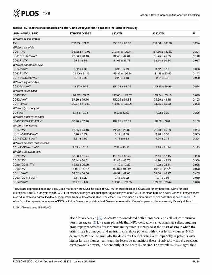
Changes in circulating microparticles after 7 and 90 days of the onset ofstrokeChanges in circulating microparticles after 7 and 90 days of the onset of stroke are shown inTable 2. CD63+/AV+ (platelet- and/or leukocyte-derived cMPs) decreased (P = 0.023) andCD63+/CD11b+/AV+ (leukocyte-derived cMPs) tended to decrease (P = 0.050) at 90 days. Onthe opposite, as depicted in Fig 4, patients in the 1st and 2nd tertile of ischemic brain sizeshowed unaltered concentrations of SMA-α+/AV+ cMPs while patients in the highest tertile ofcerebral lesion volumes showed increased SMC-derived cMPs after 90 days of the onset ofstroke (P = 0.025 for the differences between 90 days and baseline, repeated-measuresANCOVA with the Bonferroni posthoc test).
No differences were observed in the levels of the other cMPs after 7 or 90 days after theonset of stroke. Notwithstanding, after 7 days of the onset of stroke, intravenous thrombolysis(IVT)-treated patients (n = 10) had 59% less platelet-originated cMPs (CD61+/AV+ cMPs,P = 0.014). Nevertheless, after 90 days of the onset of stroke, these differences disappeared.
DiscussionTo our knowledge, little is known about chronic consequences of stroke in cell activation. Weobserved that the number of NPC-derived cMPs is increased in stroke patients with respect tohigh cardiovascular risk controls, and declines with time post-event only in those patients withhigher lesion volumes.
NPC-derived (CD56+/CD34+/AV+) cMPs have not been measured before. NPCs can secreteseveral factors that regulate neurogenesis and modulate inflammatory responses after braindamage, and they are mobilized at sites of injury to replace dead neurons [8,9] and repair the
Fig 3. Circulating microparticles at 90 days after the onset of stroke according to the SSS-TOASTclassification. A, CD62P+ (P-Selectin) cMPs; B, Other leukocyte-derived cMPs (CD45+/CD3-/CD14-) and C,Platelet-derived tissue factor positive (CD142+/CD61+) cMPs. *Significantly different from the other types ofstroke (P from the one-way ANOVA with the Bonferroni posthoc test). cMPs denotes circulatingmicroparticles; PFP, platelet free plasma and AV, Annexin V. Type I (n = 6) large artery atherosclerosisstroke; type II (n = 13) cardioembolic stroke; type III (n = 7) small vessel occlusion stroke; type IV (n = 2)stroke of uncommon etiology and type V (n = 16) stroke of undetermined etiology (SSS-TOASTclassification).
doi:10.1371/journal.pone.0148176.g003
Ischemic Stroke Increases Microparticle Shedding
PLOS ONE | DOI:10.1371/journal.pone.0148176 January 27, 2016 8 / 14
blood-brain barrier [19]. As cMPs are considered both biomarkers and cell-cell communica-tion messengers [20], it seems plausible that NPC-derived MP shedding may reflect ongoingbrain repair processes after ischemic injury since is increased at the onset of stroke when thebrain tissue is damaged, and maintained in those patients with lower lesion volumes. NPC-derived cMPs decline gradually the days after the ischemic event (especially in patients withhigher lesion volumes), although the levels do not achieve those of subjects without a previouscerebrovascular event, independently of the brain lesion size. The overall results suggest that
Table 2. cMPs at the onset of stoke and after 7 and 90 days in the 44 patients included in the study.
cMPs (cMP/μL PFP) STROKE ONSET 7 DAYS 90 DAYS P
MP from all cell origins
AV+ 702.86 ± 63.00 758.12 ± 80.86 838.68 ± 100.97 0.224
MP from platelets
CD61+/AV+ 176.72 ± 113.03 213.34 ± 109.74 187.66 ± 139.69 0.301
CD61+/CD142+/AV+ 22.96 ± 28.13 32.48 ± 44.64 31.75 ± 45.85 0.140
CD62P+/AV+ 39.61 ± 36 51.65 ± 36.71 52.54 ± 50.14 0.087
MP from endothelial cells
CD146+/AV+ 2.82 ± 4.30 3.99 ± 5.99 3.62 ± 5.17 0.096
CD62E+/AV+ 102.70 ± 81.15 155.35 ± 166.34 111.18 ± 83.53 0.142
CD146+/CD62E+/AV+ 2.21 ± 3.93 2.25 ± 4.13 2.31 ± 3.6 0.980
MP from erythrocytes
CD235ab+/AV+ 149.37 ± 84.51 154.09 ± 92.05 143.15 ± 99.96 0.884
MP from leukocytes
CD45+/AV+ 120.57 ± 68.63 157.06 ± 119.07 139.54 ± 83.15 0.099
CD62L+/AV+ 87.80 ± 79.16 100.29 ± 91.86 75.39 ± 48.16 0.103
CD11-α+/AV+ 120.67 ± 112.53 116.82 ± 102.26 83.55 ± 55.53 0.293
MP from lymphocytes
CD3+/AV+ 8.75 ± 10.73 9.92 ± 12.99 7.22 ± 9.29 0.295
MP from other leukocytes
CD45+/ CD3-/CD14-/AV+ 80.48 ± 57.78 104.85 ± 78.19 96.68 ± 66.8 0.159
MP from monocytes
CD14+/AV+ 20.05 ± 24.13 22.44 ± 25.39 21.00 ± 25.89 0.234
CD11-α+/CD14+/AV+ 3.48 ± 5.74 5.17 ± 8.73 3.28 ± 6.57 0.365
CD142+/CD14+/AV+ 4.61 ± 7.69 4.71 ± 6.83 4.24 ± 7.76 0.304
MP from smooth muscle cells
CD142+/SMA-α +/AV+ 7.79 ± 10.17 7.36 ± 13.13 12.85 ± 21.74 0.193
MP from activated cells
CD29+/AV+ 87.88 ± 61.74 115.15 ± 86.75 92.44 ± 87.15 0.253
CD15+/AV+ 60.44 ± 84.61 51.46 ± 48.75 40.96 ± 42.73 0.368
CD29+/CD15+/AV+ 16.13 ± 26.89 11.12 ± 19.32 11.32 ± 23.41 0.986
CD63+/AV+ 11.05 ± 14.79a 16.16 ± 19.62a 9.34 ± 12.72b 0.023
CD11b+/AV+ 39.02 ± 36.38 46.39 ± 47.08 38.80 ± 40.17 0.455
CD63+/CD11b+/AV+ 3.54 ± 8.22 3.46 ± 6.02 1.31 ± 3.96 0.050
CD142+/AV+ 115.01 ± 107 112.09 ± 109.65 105.57 ± 99.44 0.876
Results are expressed as mean ± sd. Used markers were CD61 for platelet, CD146 for endothelial cell, CD235ab for erythrocytes, CD45 for total
leukocytes, and CD3 for lymphocyte, CD14 for monocyte origins accounting for agranulocytes and SMA-α for smooth muscle cells. Other leukocytes were
inferred subtracting agranulocytes subpopulation from leukocytes fraction. The other CDs were used as biomarkers of cell activation (see S1 Table). P
value from the repeated measures ANOVA with the Bonferroni post-hoc test. Values in rows with different superscript letters are significantly different.
doi:10.1371/journal.pone.0148176.t002
Ischemic Stroke Increases Microparticle Shedding
PLOS ONE | DOI:10.1371/journal.pone.0148176 January 27, 2016 9 / 14

NPC-originated cMPs may be a novel biomarker of stroke. These MP may be involved in neu-rorepairing process as their parental cells, considering the negative correlation between lesionvolumes and NPC-derived cMPs and also considering the fact that patients with any level ofdisability (modified Rankin Scale�1 at 90 days after the onset of stroke) have decreased NPC-derived cMPs at 90 days after the onset of stroke compared to patients without disability andsmall cerebral lesions.
Except for SMC-derived MPs, we observed that MPs from all cell origins and activationmarkers are increased in stroke patients compared to the non-CVD population (in a meanrange of 2 to 15-fold), and this is maintained at 90 days. It suggests that increased MP sheddingis secondary to both cell activation and also cell apoptosis due to vascular injury, primarily inthose patients with a lesion volume>4.41cm3. On the other hand, increased MP shedding inacute stroke has been previously observed, especially from endothelial cells [2,13,17,21,22]. Infact, we observed that activated endothelial cell-derived MPs (CD146+/AV+ and CD146+/CD62E+/AV+) are the most increased cMPs (by ~300 fold, compared to the mean range of 2-to 15-fold increase in the other cMPs) in stroke patients compared to high-CV risk controls.No correlation was found between E-Selectin (CD62E)-carrying cMPs and the time of strokeonset, supporting the idea that stroke promotes chronic cell activation. These observations sug-gest that endothelial cMPs are biomarkers of stroke, and further suggest that activated endothe-lial cMPs are major players in stroke induction and vascular insult rather than in vascularrepair as previously suggested [23]. We found higher levels of endothelial-derived cMPs carry-ing CD62E (a biomarker of endothelial activation) than CD146 (one of the most specificmarker of endothelial cell lineage). It has been previously described [24] that that the expres-sion and proportion of cell surface molecules on endothelial-derived MPs may differ fromtheir parental cells. Therefore, it can be possible that endothelial cells, when activated, releasehigher amounts of MP loaded with CD62E than MP carrying CD146.
Although endothelial MPs have been previously reported to correlate with lesion volume[12,13], we only found that SMC-derived cMPs at 90 days correlate with infarction size. Corre-lations of cMPs with ischemic lesion volumes at presentation may suggest a correlation withthe degree of endothelial apoptosis and inflammation within the ischemic lesion, while correla-tion of ischemic lesion with cMPs at 90 days after the presentation of stroke may reflect thelong-term vascular damage after the cerebrovascular event.
Fig 4. Changes in smoothmuscle cell derived circulatingmicroparticles after 90 days of stroke bytertiles of lesion size. Results are represented as mean ± sd. Different letters within tertiles of lesion volumedenote statistical differences, measured by repeated measures ANCOVA with the lesion volume as thecovariate and the Bonferroni post-hoc test. cMPs denotes circulating microparticles; PFP, platelet freeplasma and AV, Annexin V. SMA-α (smooth muscle actin-α) was used as a biomarker of smooth musclecells.
doi:10.1371/journal.pone.0148176.g004
Ischemic Stroke Increases Microparticle Shedding
PLOS ONE | DOI:10.1371/journal.pone.0148176 January 27, 2016 10 / 14

TF is crucial for the initiation of coagulation, and platelet-derived MPs have 50- to 100-foldhigher procoagulant activity than activated platelets [25]. Therefore, elevated levels of circulat-ing MPs and TF-loaded cMPs are associated with an increased risk of thrombosis and throm-boembolic events. Recently it has been observed that in 73 patients with an acute ischemicstroke, TF-carrying cMPs were elevated compared to healthy individuals [26], in agreementwith our findings. Besides, SMC shed TF-loaded MP with thrombogenic properties in human[27] and rat aorta [28]. Along this line, platelet and SMC-derived TF+-cMPs were significantlyelevated compared to controls in the three time points, partially explaining the increased riskof recurrent stroke in those patients.
SMC in blood vessel wall play a key role in cerebral blood flow control, brain perfusion main-tenance and regulation. Surprisingly, we observed that at the onset of stroke and after 7 days,SMC-derived cMPs (but not SMC-derived MPs carrying TF) were similar to the levels observedin non-CVD controls. At 90 days however, these MP were increased compared to both controlsand patients at the onset of stroke, but only in those patients with the highest lesion volumes.These results indicate that larger cerebral lesions associate with deeper vessel injury affectingsmooth muscle cells function. Although SMC represent the major cell type in blood vessels, verylittle is known about SMC-derived MPs [16,27,28]. It is known that stroke induces SMC toswitch to a phenotype through different mechanisms that will be either detrimental or beneficialto brain repair [29,30], which may be also related to MP shedding, considering that the fluctua-tion observed in SMC-derived MP shedding was determined by the infarct size.
In view of the results, further studies are required to elucidate if MP in stroke are procoagu-latory or vasoconstrictive (one of the causes), or if they are merely the consequence of tissuedamage.
This study is not exempt of limitations. Although there is increasing evidence that some MPmay be formed without externalization of PS [31], in our study, MP carrying cell-specific anti-gens but not binding AV (a PS ligand) only accounted for<1% of the total MP population.Therefore, we only analyzed MP containing AV in their surface. Blood samples were takenwithin the 48h of the onset of stroke, sometimes after IVT or other medication administrationbecause informed consent had to be obtained first. Thus, correlations between clinical parame-ters and MP levels could reflect delayed apoptotic cell injury and inflammatory stimulationafter ischemic stroke. During the first days after the onset of stroke, patients were polymedi-cated. This circumstance may partially explain the fact that the main differences were observedafter 90 days of the onset of stroke. Although the number of patients is relatively small, thiscohort has a representative distribution of clinical characteristics, and the incidence of the dif-ferent types of stroke was comparable to previously reported data [32]. Finally, this is an obser-vational study and the meaning and pathophysiological consequences of the increase of cMPsare unclear and further research is warranted.
ConclusionsIschemic stroke increases MP shedding and is chronically maintained, which may in partexplain the increased risk of recurrent stroke in these patients, since a previous cerebral ische-mic event is one of the major risk factors for a further one. To our knowledge, this is the firsttime that MP shedding from several cellular origins after stroke is measured. The specificmechanism underlying the involvement of elevated MPs in stroke (cause or consequence) andtheir relationship with repair of ischemic tissue remains to be established to better understandstroke pathophysiology. Finally, whether specific MPs can help to clinically differentiate stroketype and affected cerebral lesion size deserve further investigation in a larger number ofpatients.
Ischemic Stroke Increases Microparticle Shedding
PLOS ONE | DOI:10.1371/journal.pone.0148176 January 27, 2016 11 / 14

Supporting InformationS1 Appendix. Baseline characteristics of the patients included in the study.(PDF)
S1 Fig. Gate limits for microparticle analysis with the Megamix-Plus FSC beads for cytome-ter settings in microparticle analysis. A) Gate limits were established before analyses usingthe Megamix-Plus FSC beads for cytometer settings in microparticle analysis (BioCytex, Mar-seille, France). According to Megamix-Plus FSC beads signal, the lower limit of quantificationis>0.1μm, as beads of 0.1μmwere negative for FITC signal. B) Gate limits for microparticlequantification (G1) were set according to beads signal.(PDF)
S2 Fig. Cell sources of MP for controls and patients at the onset of stroke. Pie-charts show-ing distribution of cMPs from controls (n = 44) and patients at the onset of stroke (n = 44) bymajor cell origins, indicated by percentages of each marker relative to cell lineage. Used con-trols were patients at high cardiovascular disease who have never suffered a stroke. Selectedmarkers were CD61 for platelets, CD146 for endothelial cells, CD45 for total leukocytes, CD3for lymphocytes, CD14 for monocytes and SMA-α for smooth muscle cells origins. Other leu-kocyte cMPs were positive for CD45 but negative for CD3 or CD14.(PDF)
S3 Fig. ROC curve analysis to determine the threshold of NPC-derived cMPs that discrimi-nates between patients and controls. CD56+/CD34+/AV+ cMPs at a cut-off point of 2.8 MP/μL of PFP, P<0.0001, properly discriminated between controls and stroke patients with a81.8% sensitivity and 83.3% specificity [area under de curve (AUC) = 0.894 (95% CI 0.823,0.965)]. Used controls were patients at high cardiovascular disease who have never suffered astroke. cMPs denotes circulating microparticles; PFP, platelet free plasma and AV, Annexin V.(PDF)
S1 Table. Cell surface molecules for circulating microparticle identification and characteri-zation.mAb indicates monoclonal antibody; PS, phosphatidylserine; FITC, fluorescein isothio-cyanate; PE, phycoerythrin; LPS, lipopolysaccharide.(PDF)
S2 Table. Baseline characteristics of subjects devoid of cardiovascular disease (n = 44) andpatients at the onset of stroke (n = 44). Results are expressed as mean ± sd or n (%) whenindicated. Used controls were patients at high cardiovascular disease who have never suffered astroke. P value from one-way ANOVA for quantitative variables and from Chi-square analysisfor qualitative variables.(PDF)
AcknowledgmentsWe are indebted to stroke patients for their participation in the study.
Author ContributionsConceived and designed the experiments: LB JM-F EP TP. Performed the experiments: GC-BRS JC EJ-X. Analyzed the data: GC-B RS JC LB. Contributed reagents/materials/analysis tools:GC-B RS JC. Wrote the paper: GC-B RS LB.
Ischemic Stroke Increases Microparticle Shedding
PLOS ONE | DOI:10.1371/journal.pone.0148176 January 27, 2016 12 / 14

References1. Morel O, Jesel L, Freyssinet JM, Toti F. Cellular mechanisms underlying the formation of circulating
microparticles. Arteriosclerosis, thrombosis, and vascular biology. 2011; 31: 15–26. doi: 10.1161/ATVBAHA.109.200956 PMID: 21160064
2. Cherian P, Hankey GJ, Eikelboom JW, Thom J, Baker RI, McQuillan A, et al. Endothelial and plateletactivation in acute ischemic stroke and its etiological subtypes. Stroke. 2003; 34: 2132–7. PMID:12907813
3. Preston RA, Jy W, Jimenez JJ, Mauro LM, Horstman LL, Valle M, et al. Effects of severe hypertensionon endothelial and platelet microparticles. Hypertension. 2003; 41: 211–7. PMID: 12574084
4. Koga H, Sugiyama S, Kugiyama K, Watanabe K, Fukushima H, Tanaka T, et al. Elevated levels of VE-cadherin-positive endothelial microparticles in patients with type 2 diabetes mellitus and coronary arterydisease. Journal of the American College of Cardiology. 2005; 45: 1622–30. PMID: 15893178
5. Amabile N, Guerin AP, Leroyer A, Mallat Z, Nguyen C, Boddaert J, et al. Circulating endothelial micro-particles are associated with vascular dysfunction in patients with end-stage renal failure. Journal of theAmerican Society of Nephrology. 2005; 16: 3381–8. PMID: 16192427
6. Sinning JM, Losch J, Walenta K, BohmM, Nickenig G, Werner N. Circulating CD31+/Annexin V+ micro-particles correlate with cardiovascular outcomes. European heart journal. 2011; 32: 2034–41. doi: 10.1093/eurheartj/ehq478 PMID: 21186238
7. Viera AJ, Mooberry M, Key NS. Microparticles in cardiovascular disease pathophysiology and out-comes. Journal of the American Society of Hypertension. 2012; 6: 243–52. doi: 10.1016/j.jash.2012.06.003 PMID: 22789878
8. Arvidsson A, Collin T, Kirik D, Kokaia Z, Lindvall O. Neuronal replacement from endogenous precursorsin the adult brain after stroke. Nat Med. 2002; 8: 963–70. PMID: 12161747
9. Marti-Fabregas J, Romaguera-Ros M, Gomez-Pinedo U, Martinez-Ramirez S, Jimenez-Xarrie E, MarinR, et al. Proliferation in the human ipsilateral subventricular zone after ischemic stroke. Neurology.2010; 74: 357–65. doi: 10.1212/WNL.0b013e3181cbccec PMID: 20054008
10. Lindsberg PJ, Carpen O, Paetau A, Karjalainen-Lindsberg ML, Kaste M. Endothelial ICAM-1 expres-sion associated with inflammatory cell response in human ischemic stroke. Circulation. 1996; 94: 939–45. PMID: 8790029
11. Wang PY, Kao CH, Mui MY, Wang SJ. Leukocyte infiltration in acute hemispheric ischemic stroke.Stroke. 1993; 24: 236–40. PMID: 8421825
12. Jung KH, Chu K, Lee ST, Park HK, Bahn JJ, Kim DH, et al. Circulating endothelial microparticles as amarker of cerebrovascular disease. Annals of neurology. 2009; 66: 191–9. doi: 10.1002/ana.21681PMID: 19743467
13. Simak J, Gelderman MP, Yu H, Wright V, Baird AE. Circulating endothelial microparticles in acuteischemic stroke: a link to severity, lesion volume and outcome. Journal of thrombosis and haemostasis.2006; 4: 1296–302. PMID: 16706974
14. Alonso R, Andres E, Mata N, Fuentes-Jimenez F, Badimon L, Lopez-Miranda J, et al; SAFEHEARTInvestigators. Lipoprotein(a) levels in familial hypercholesterolemia: an important predictor of cardio-vascular disease independent of the type of LDL receptor mutation. Journal of the American College ofCardiology. 2014; 63: 1982–9. PMID: 24632281
15. Suades R, Padro T, Alonso R, Lopez-Miranda J, Mata P, Badimon L. Circulating CD45+/CD3+ lympho-cyte-derived microparticles map lipid-rich atherosclerotic plaques in familial hypercholesterolaemiapatients. Thrombosis and haemostasis. 2014; 111: 111–21. doi: 10.1160/TH13-07-0612 PMID:24085382
16. Leroyer AS, Isobe H, Leseche G, Castier Y, Wassef M, Mallat Z, et al. Cellular origins and thrombo-genic activity of microparticles isolated from human atherosclerotic plaques. Journal of the AmericanCollege of Cardiology. 2007; 49: 772–7. PMID: 17306706
17. Suades R, Padro T, Vilahur G, Badimon L. Circulating and platelet-derived microparticles in humanblood enhance thrombosis on atherosclerotic plaques. Thrombosis and haemostasis. 2012; 108:1208–19. doi: 10.1160/TH12-07-0486 PMID: 23138460
18. Nieuwland R, Berckmans RJ, McGregor S, Boing AN, Romijn FP, Westendorp RG, et al. Cellular originand procoagulant properties of microparticles in meningococcal sepsis. Blood. 2000; 95: 930–5. PMID:10648405
19. Obermeier B, Daneman R, Ransohoff RM. Development, maintenance and disruption of the blood-brain barrier. Nat Med. 2013; 19: 1584–96. doi: 10.1038/nm.3407 PMID: 24309662
20. Loyer X, Vion AC, Tedgui A, Boulanger CM. Microvesicles as cell-cell messengers in cardiovasculardiseases. Circ Res. 2014; 114: 345–53. doi: 10.1161/CIRCRESAHA.113.300858 PMID: 24436430
Ischemic Stroke Increases Microparticle Shedding
PLOS ONE | DOI:10.1371/journal.pone.0148176 January 27, 2016 13 / 14

21. Lackner P, Dietmann A, Beer R, Fischer M, Broessner G, Helbok R, et al. Cellular microparticles as amarker for cerebral vasospasm in spontaneous subarachnoid hemorrhage. Stroke. 2010; 41: 2353–7.doi: 10.1161/STROKEAHA.110.584995 PMID: 20814009
22. Li P, Qin C. Elevated circulating VE-cadherin+CD144+ endothelial microparticles in ischemic cerebro-vascular disease. Thromb Res. 2015; 135: 375–81. doi: 10.1016/j.thromres.2014.12.006 PMID:25523345
23. Dignat-George F, Boulanger CM. The many faces of endothelial microparticles. Arteriosclerosis, throm-bosis, and vascular biology. 2011; 31: 27–33. doi: 10.1161/ATVBAHA.110.218123 PMID: 21160065
24. Abid Hussein MN, Meesters EW, Osmanovic N, Romijn FP, Nieuwland R, Sturk A. Antigenic character-ization of endothelial cell-derived microparticles and their detection ex vivo. Journal of Thrombosis andHaemostasis. 2003; 1: 2434–43. PMID: 14629480
25. Sinauridze EI, Kireev DA, Popenko NY, Pichugin AV, Panteleev MA, Krymskaya OV, et al. Plateletmicroparticle membranes have 50- to 100-fold higher specific procoagulant activity than activatedplatelets. Thrombosis and haemostasis. 2007; 97: 425–34. PMID: 17334510
26. Switonska M, Slomka A, Sinkiewicz W, Zekanowska E. Tissue-factor-bearing microparticles (MPs-TF)in patients with acute ischaemic stroke: the influence of stroke treatment on MPs-TF generation. Euro-pean journal of neurology. 2015; 22: 395–401, e28–9. doi: 10.1111/ene.12591 PMID: 25370815
27. Schecter AD, Spirn B, Rossikhina M, Giesen PL, Bogdanov V, Fallon JT, et al. Release of active tissuefactor by human arterial smooth muscle cells. Circulation research. 2000; 87: 126–32. PMID:10903996
28. Brisset AC, Terrisse AD, Dupouy D, Tellier L, Pech S, Navarro C, et al. Shedding of active tissue factorby aortic smooth muscle cells (SMCs) undergoing apoptosis. Thrombosis and haemostasis. 2003; 90:511–8. PMID: 12958621
29. Poittevin M, Lozeron P, Hilal R, Levy BI, Merkulova-Rainon T, Kubis N. Smooth muscle cell phenotypicswitching in stroke. Translational stroke research. 2014; 5: 377–84. doi: 10.1007/s12975-013-0306-xPMID: 24323725
30. Starke RM, Chalouhi N, Ding D, Raper DM, McKisic MS, Owens GK, et al. Vascular smooth musclecells in cerebral aneurysm pathogenesis. Translational stroke research. 2014; 5: 338–46. doi: 10.1007/s12975-013-0290-1 PMID: 24323713
31. Latham SL, Tiberti N, Gokoolparsadh N, Holdaway K, Olivier Couraud P, Grau GE, Combes V.Immuno-analysis of microparticles: probing at the limits of detection. Scientific Reports. 2015; 5:16314. doi: 10.1038/srep16314 PMID: 26553743
32. Kolominsky-Rabas PL, Weber M, Gefeller O, Neundoerfer B, Heuschmann PU. Epidemiology of ische-mic stroke subtypes according to TOAST criteria: incidence, recurrence, and long-term survival inischemic stroke subtypes: a population-based study. Stroke. 2001; 32: 2735–40. PMID: 11739965
Ischemic Stroke Increases Microparticle Shedding
PLOS ONE | DOI:10.1371/journal.pone.0148176 January 27, 2016 14 / 14