membrane potential · 2020-04-30 · 2019. 12. 07. 1 membrane potential madarász tamás pte-Áok...
TRANSCRIPT

2019. 12. 07.
1
Membrane potential
Madarász Tamás
PTE-ÁOK Department of Biophysics
Structure of cell membrane
Transport
Active transport is the movement of molecules across a membrane from a region of their lower concentration to a region of their higher concentration.primary active transport that uses ATP.secondary active transport that uses an electrochemical gradient.Carrier proteins:- Uniporter is an integral membrane protein that transports a single
type of substrate species (charged or uncharged) across the cell membrane.
- Symporter: transports two different type substrate across the cellmembrane.
- Antiporter: transports two or more different molecules or ions across the membrane.
Passive transport: It does not require an input of cellular energy because it is instead driven by the tendency of the system to grow in entropy.
Diffusion will continue until this gradient has been eliminated.Passive osmosis and diffusion: Some substances (small molecules, ions) such as carbon dioxide (CO2) and oxygen (O2), can move across the plasma membrane by diffusion, which is a passive transport process.
Passive transport
106 ions per second or greater
http://physiology.elte.hu/eloadas/kiegtanar_elettan/potencial_neuro_2009.pdf
Channel Channel
Membrane
Filter region
Gate
Intracellular fluid
•Calcium-activated potassium channel - open in response to the presence of calcium ions or other signalling molecules.•Inwardly rectifying potassium channel - passes current (positive charge) more easily in the inward direction (into the cell).•Tandem pore domain potassium channel - are constitutively open or possess high basal activation, such as the "resting potassium channels" or "leak channels" that set the negative membrane potential of neurons.•Voltage-gated potassium channel - are voltage-gated ion channels that open or close in response to changes in the transmembrane voltage.
Potassium ion channels

2019. 12. 07.
2
Active transport
Three sodium ions are exported and two potassium ions are imported
Main components
•water
•Ions
– Cations (K+, Na+, Ca2+)
– Anions (Cl-, H2PO4− and
HPO42−)
•Proteins–with negative charge (pH! – isoelectric point)
Membrane potential
• Membrane potential is the difference in electric potentialbetween the interior and the exterior membrane side.
Nerve impulse transmission
http://physiology.elte.hu/eloadas/kiegtanar_elettan/potencial_neuro_2009.pdf
Muscle and nerve tissue are electrically excitable
Muscle and nerve tissue are electrically conductive
Donnan potential
2019. 12. 07.
3
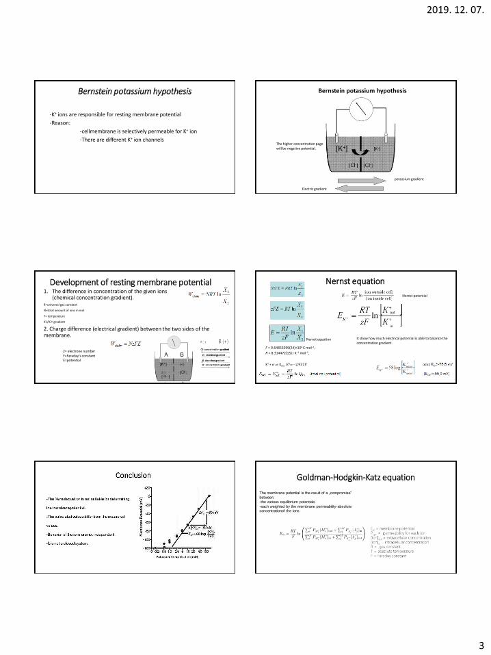
Bernstein potassium hypothesis
-K+ ions are responsible for resting membrane potential
-Reason:
-cellmembrane is selectively permeable for K+ ion
-There are different K+ ion channels
Bernstein potassium hypothesis
Electric gradient
potassium gradient
The higher concentration page will be negative potential.
Development of resting membrane potential1. The difference in concentration of the given ions
(chemical concentration gradient).R=universal gas constant
N=total amount of ions in mol
T= temperature
X1/X2=gradient
2. Charge difference (electrical gradient) between the two sides of the membrane.
Z= electrone numberF=Faraday’s constantE=potential
Nernst equation
F = 9.64853399(24)×104 C mol−1,
R = 8.314472(15) J K−1 mol−1,
Nernst potential
K+ + e- ⇌ K(S) E0=−2.931V
Nernst equation It show how much electrical potential is able to balance the concentration gradient.
out
in
Goldman-Hodgkin-Katz equation
The membrane potential is the result of a „compromise” between:-the various equilibrium potentials
-each weighted by the membrane permeability-absolute concentrationof the ions

2019. 12. 07.
4

2019. 12. 07.
5
Types of receptors
Internal receptors that respond to changes inside the body are known as interoceptors. Example: baroreceptor in the vascular wall
Exteroceptor: a sense organ receiving stimuli from the external environment, as the eye or the heat receptors in the skin
Proprioceptor: Proprioception is mediated by proprioceptors, mechanosensory neurons located within muscles, tendons, and joints.