mature luffa leaves (luffa cylindrica l.) as a tool for ... · tissues. our research has shown that...
TRANSCRIPT

fpls-08-00228 February 17, 2017 Time: 18:4 # 1
ORIGINAL RESEARCHpublished: 21 February 2017
doi: 10.3389/fpls.2017.00228
Edited by:Joachim Hermann Schiemann,
Julius Kühn-Institut, Germany
Reviewed by:Christina Kühn,
Humboldt University of Berlin,Germany
Taras P. Pasternak,Albert Ludwigs University of Freiburg,
Germany
*Correspondence:Izabela A. Chincinska
Specialty section:This article was submitted to
Plant Biotechnology,a section of the journal
Frontiers in Plant Science
Received: 25 October 2016Accepted: 06 February 2017Published: 21 February 2017
Citation:Błazejewska K, Kapusta M,
Zielinska E, Tukaj Z andChincinska IA (2017) Mature Luffa
Leaves (Luffa cylindrica L.) as a Toolfor Gene Expression Analysis by
Agroinfiltration.Front. Plant Sci. 8:228.
doi: 10.3389/fpls.2017.00228
Mature Luffa Leaves(Luffa cylindrica L.) as a Tool forGene Expression Analysis byAgroinfiltrationKamila Błazejewska1, Małgorzata Kapusta2, Elzbieta Zielinska1, Zbigniew Tukaj1 andIzabela A. Chincinska1*
1 Department of Plant Physiology and Biotechnology, Faculty of Biology, University of Gdansk, Gdansk, Poland, 2 Departmentof Plant Cytology and Embryology, Faculty of Biology, University of Gdansk, Gdansk, Poland
We exploited the potential of cucurbits for ectopic gene expression. Agroinfiltration isa simple and commonly used method to obtain transient expression of foreign genesin plants. In contrast to in vitro transformation techniques, agroinfiltration can be usedfor genetic modification of mature plant tissues. Although the cucurbits are commonlyused as model plants for molecular biology and biotechnology studies, to date there areno literature sources on the possibility of transient gene expression in mature cucurbittissues. Our research has shown that mature leaves of Luffa cylindrica L. (luffa), incontrast to other cucurbit species, can be successfully transiently transformed withAgrobacterium tumefaciens. We efficiently transformed luffa leaves with a reporter geneencoding β-glucuronidase (GUS). The GUS activity in transiently transformed leaf tissueswas detected within 24 h after the infiltration with bacteria. Additionally, we have shownthat the activity of a transiently expressed the GUS gene can be monitored directlyin the EDTA-exudates collected from the cut petioles of the agroinfiltrated leaves. Theresults suggest that luffa leaves can be useful as a plant expression system for studiesof physiological and biochemical processes in cucurbits.
Keywords: cucurbits, agroinfiltration, luffa, phloem exudate, plant overexpression system, foliar application oftracers
INTRODUCTION
The Cucurbitaceae species (cucurbits) are a significant source of food and substances of medicalimportance (Manamohan et al., 2011). Several species of Cucurbitaceae, for example Cucurbitamaxima, Cucurbita pepo, Cucurbita ficifolia or Cucumis sativus, are widely used as model plants forresearch, especially in studies of phloem functions and biochemistry (Golecki et al., 1999; Zhanget al., 2010, 2012).
The use of cucurbits in phloem research was primarily associated with the ease of exudatesampling from severed stems and petioles (Richardson et al., 1982; Lin et al., 2009). The fluidexuding from the cucurbit vascular tissue contains phloem sap derived from the fascicular phloemas well as from the extrafascicular phloem (Zhang et al., 2010, 2012; Zimmermann et al., 2013).This is a spatially distinct system of sieve elements occurring specifically in Cucurbitaceae species(Turgeon and Oparka, 2010). Cucurbit exudates are rich in proteins (their concentration canreach up to 60–100 mg/mL) (Richardson et al., 1982; Walz et al., 2004). It was shown, that the
Frontiers in Plant Science | www.frontiersin.org 1 February 2017 | Volume 8 | Article 228

fpls-08-00228 February 17, 2017 Time: 18:4 # 2
Błazejewska et al. Agroinfiltration of Mature Cucurbit Plants
exudate proteins applied to C. maxima cotyledons traffickedsymplastically through mesophyll plasmodesmata andtranslocate over long distance in the phloem (Balachandranet al., 1997). Various techniques, like the intergeneric graftingexperiments or the studies on systemic movement of plant virusparticles and phloem tracers, e.g., 5(6)-carboxyfluorescein orfluorescein diacetate, were used to investigate the phloem loadingmechanisms and the long-distance trafficking of molecules incucurbits (Grignon et al., 1989; Golecki et al., 1999; Pradel et al.,1999; Zhang et al., 2010). To date, there are only a few reportsin which transgenic cucurbit species were used for investigationof phloem function. In contrast, the techniques enabling theinduction of heterological expression in plant tissues are widelyused as research tools for phloem studies of many non-cucurbitspecies, especially the plants defined as apoplastic loaders(Chincinska et al., 2008; Krügel et al., 2012).
Agroinfiltration belongs to the most popular planttransformation techniques. In this method, the suspensionof Agrobacterium, carrying a binary vector with a targetedtransgene, is pressed into the intercellular spaces of a selectedplant tissue, usually a leaf. Thus, the successful introduction ofbacterial suspension is one of the most important requirementsaffecting the efficiency of transient transformation usingagroinfiltration (Bhaskar et al., 2009; Leuzinger et al., 2013). Theagroinfiltration technique was well-developed for model plantssuch as Nicotiana benthamiana (Wydro et al., 2006; Goodin et al.,2008), Nicotiana tabacum or Arabidopsis thaliana (Cazzonelli andVelten, 2006; Kim et al., 2009). This method was also applied inseveral crops, e.g., tomato (Lycopersicon esculentum Mill.), lettuce(Lactuca L.) (Joh et al., 2005), potato (Solanum tuberosum L.)(Bhaskar et al., 2009), and grape (Vitis L.) (Santos-Rosa et al.,2008). Nevertheless, to date, there is no agroinfiltration protocolfor generation of plant expression systems based on maturecucurbit tissues. Therefore, the potential application of variouscucurbit species for a gene functions assay using agroinfiltrationwas tested in this study. We showed that mature leaves ofLuffa cylindrica L., in contrast to other cucurbits tested, can beefficiently agroinfiltrated. We detected the expression of a geneencoding GUS driven by the CaMV 35S promoter in luffa leavesa few hours after agroinfiltration. Moreover, the GUS activitywas also detected in the EDTA-exudates collected from the cutpetioles of the transiently transformed luffa leaves.
MATERIALS AND METHODS
Plant MaterialThe Cucurbitaceae species were cultivated from seeds (LegutkoBreeding and Seed Company, Ltd) and grown in a greenhousein 30 cm diameter pots. All plants were grown at 270 µmolphotons/m2/s with a light/dark cycle of 16 h/8 h at 25/16◦C and40–50% relative humidity.
MicroscopyParts of leaf blades of examined cucurbit species were fixed in 8%formaldehyde and 0.25% glutaraldehyde in piperazine buffer at4◦C overnight then embedded in Steedman’s Wax and sectioned
at 10 µm (Krawczyk et al., 2016). The chromatin of the nucleiwas visualized with 1 µg/mL Propidium Iodide and cell wallswere stained with 0.1% Calcofluor White M2R. Specimens wereclosed in PBS buffer and viewed in epifluorescence with LeicaDM6000 B supported by LAS AF software. Images of leaf bladesare maximum projections of taken Z-stacks, deconvolved usingfive iterations of a 3D Non-blind algorithm (AutoQuantTM) tomaximize spatial resolution.
Agroinfiltration ProceduresFor agroinfiltration analysis in luffa plants we usedAgrobacterium tumefaciens strain LBA 4404 transformedwith a commercially available vector pRI 201-AN-GUS (Takara,Clontech Laboratories, Inc.) containing β-glucuronidase gene(uidA) sequence under the control of the CaMV 35S promoter.The untransformed A. tumefaciens strain LBA 4404 was used asa negative control. The Agrobacterium pre-culture conditionsand the preparation of the bacteria suspension were preparedaccording to the procedure described previously and optimizedfor agroinfiltration of N. benthamina (Wydro et al., 2006).Mature leaves were harvested from the luffa plants growing inthe greenhouse. The leaves were cut at the petiole bases, thentransported to the laboratory and stored in a closed container.High humidity in the containers was held by placing wet papertowels at the bottom. Immediately before the infiltration theleaf was weighed, then placed adaxial side down on a layer ofsoft paper towels designed to protect the leaf from mechanicaldamage during the syringe infiltration. Subsequent fragmentsof the leaf were infiltrated using a 1 mL syringe. The leafsaturated with the bacterial suspension was gently dried witha paper towel and weighed. Immediately after infiltrationthe leaves were placed in plant propagators (the dimensions:58 cm× 40 cm× 22 cm, Supplementary Figure S1). To maintainthe optimal physiological condition of the leaves as well as tostabilize their turgor, propagator bottoms were coated with a1 cm thick layer of water and covered with clear plastic covers.The propagators with leaves were then placed under the LEDlamps.
Phloem ExudationThe petioles of the leaves previously infiltrated with the bacteriaas well as the control leaves were submersed under 2.5 mM EDTAsolution (to prevent the contact with atmospheric oxygen) andrecut by 2–3 mm. The leaves were immediately transferred todark plastic tubes containing 5 mL of 2.5 mM EDTA solutionto facilitate exudation. To protect against evaporation of EDTAsolution the tubes were carefully sealed with parafilm.
Protein AnalysisLeaves without petioles were ground in 30 mL of proteinextraction buffer [50 mM NaHPO4, pH 7.0, 10 mMβ-mercaptoethanol, 10 mM EDTA, 0.1% (w/v) sodium laurylsarcosine, and 0.1% (w/v) Triton X-100]. The leaf extracts andthe EDTA-exudates were used for the total soluble protein (TSP)content measurement using the Bradford (1976) method and forGUS activity assays.
Frontiers in Plant Science | www.frontiersin.org 2 February 2017 | Volume 8 | Article 228

fpls-08-00228 February 17, 2017 Time: 18:4 # 3
Błazejewska et al. Agroinfiltration of Mature Cucurbit Plants
Electrophoresis and Western BlottingSDS-PAGE was performed according to Brunelle and Green(2014), with the modification that acrylamide/bisacrylamidesolution mix was in ratio (29:1) in both stacking and resolvinggels. Exudate samples were separated in a 12% polyacrylamidegel and stained with Coomassie brilliant blue. Western blottingwas performed as described previously (Brunelle and Green,2014). The anti-β-glucuronidase (C-Terminal) primary antibody(Sigma–Aldrich) and the goat anti-IgG rabbit coupled with HRP(horseradish peroxidase) secondary antibody were used for GUSdetection. A recombinant GUS protein from Escherichia coli(Sigma–Aldrich) was used as a reference.
GUS Activity AnalysisThe histochemical GUS staining and fluorogenic GUS assayswere performed as described by Jefferson et al. (1987),using the 5-bromo-4-chloro-3-indolyl glucuronide (X-gluc) and4-methylumbelliferyl-β-D-glucuronide hydrate (4-MUG) (BioBasic, Inc.) as substrates, respectively. Because the fluorescenceintensity of 4-methylumbelliferone (4-MU) is influenced bythe presence of plant extracts, the 4-MU concentrationcurves (4-MU standard curves) for the GUS activity assayswere generated separately in the exudates and in the leafextracts using the leaf extracts and exudates, respectively,for the standard curve preparation. Spectrophotometric andfluorescence measurements were done using Varioscan FlashMultimode Reader (Thermo Scientific).
RESULTS
Leaves of Different Cucurbit SpeciesShow Different Infiltration SusceptibilityWe selected 11 different cucurbit species (Table 1) to test asusceptibility of their leaves to infiltration. The mature leaves
(source leaves from the middle part of the stem) were collectedfrom 2-month old plants and then infiltrated with 1 mL pureinfiltration medium to test their absorption properties. Weanticipated that the difference in the leaf weight before and afterinfiltration would correspond to the volume of resuspensionmedium absorbed by the leaf tissue. The infiltration area, visibleafter the injection as a darker stain on the leaf surface, was alsoevaluated (Table 1). The results showed that L. cylindrica leavescould absorb significantly more fluid than the other cucurbits.A single application of the medium into the luffa leaf coveredan average leaf area of 8.22 ± 1.23 cm2 ( ± SD; n = 8) and wassignificantly higher in comparison to all other species. Becausethe fluid introduced into the luffa leaves was easily absorbed andquickly diffused in the adjacent tissue around the injection site,it became possible after a few injection repetitions to bring theleaves to the complete saturation with the fluid.
A relatively high leaf absorption capacity was also shown inthe case of the Lagenaria species. However, the infiltration ofLagenaria leaves proved to be very time consuming due to thedelicate structure of their leaves. The infiltration of Lagenarialeaves required very careful handling to prevent mechanicaldamage of the tissues while the luffa leaves showed relative highresistance to damage. In contrast, the infiltration of the matureleaves of Cucurbita and Cucumis species appeared to be extremelydifficult.
Luffa Leaves Anatomy Can Explain theirExcellent AbsorptivityStructure of leaf tissues, especially cuticle thickness, stomataanatomy and density, significantly affect the susceptibility ofvarious plant species to foliar infections by pathogenic bacteria(Melotto et al., 2008). Since the agroinfiltration procedureinvolves injection of bacteria suspension into the air spacesof leaf mesophyll, it can be assumed that these leaf structureshave a significant impact on the introduction of bacteria by
TABLE 1 | Comparison of cucurbit leaves absorption capacity.
Difference in the leaf weight before and after infiltration Infiltration surface area
Cucurbit varieties Mean [g] SD Homogenous groups Mean [cm2] SD Homogenous groups
a b a b c d
Cucurbita maxima Duch. cv. Bambino 0.097 0.070 ∗ 0.47 0.12 ∗
Cucurbita pepo L. cv. Makaronowa Warszawska 0.138 0.115 ∗ 0.48 0.13 ∗
Cucurbita maxima Duch. cv. Melonowa Zółta 0.096 0.025 ∗ 0.64 0.96 ∗
Lagenaria sinceraria Standl. cv. Kobra 0.139 0.040 ∗ 0.88 0.18 ∗
Cucumis metuliferus Mey. 0.060 0.052 ∗ 1.00 1.05 ∗
Lagenaria sinceraria Standl. cv. Birdhouse 0.179 0.122 ∗ 1.17 2.81 ∗
Cucurbita moschata Duch. 0.149 0.091 ∗ 1.36 0.44 ∗ ∗
Cucurbita maxima Duch. cv. Atlantic Giant 0.168 0.138 ∗ 1.52 0.57 ∗ ∗
Lagenaria sinceraria Standl. cv. Marenka 0.218 0.099 ∗ ∗ 3.24 0.49 ∗ ∗
Lagenaria sinceraria Standl. cv. Snake 0.113 0.039 ∗ 4.24 2.23 ∗
Luffa cylindrica L. 0.354 0.221 ∗ 8.22 1.23 ∗
We measured the amount of the medium absorbed by the leaf and the infiltration surface area. The leaves were infiltrated with 1 mL pure infiltration medium. One-wayANOVA and the Scheffé’s post hoc method was used for statistical analysis. Homogeneous groups are marked with asterisks under the same letter. Total n = 6 leavesfrom at least two individual plants of each species were used. The experiment was repeated two times independently.
Frontiers in Plant Science | www.frontiersin.org 3 February 2017 | Volume 8 | Article 228
fpls-08-00228 February 17, 2017 Time: 18:4 # 4
Błazejewska et al. Agroinfiltration of Mature Cucurbit Plants
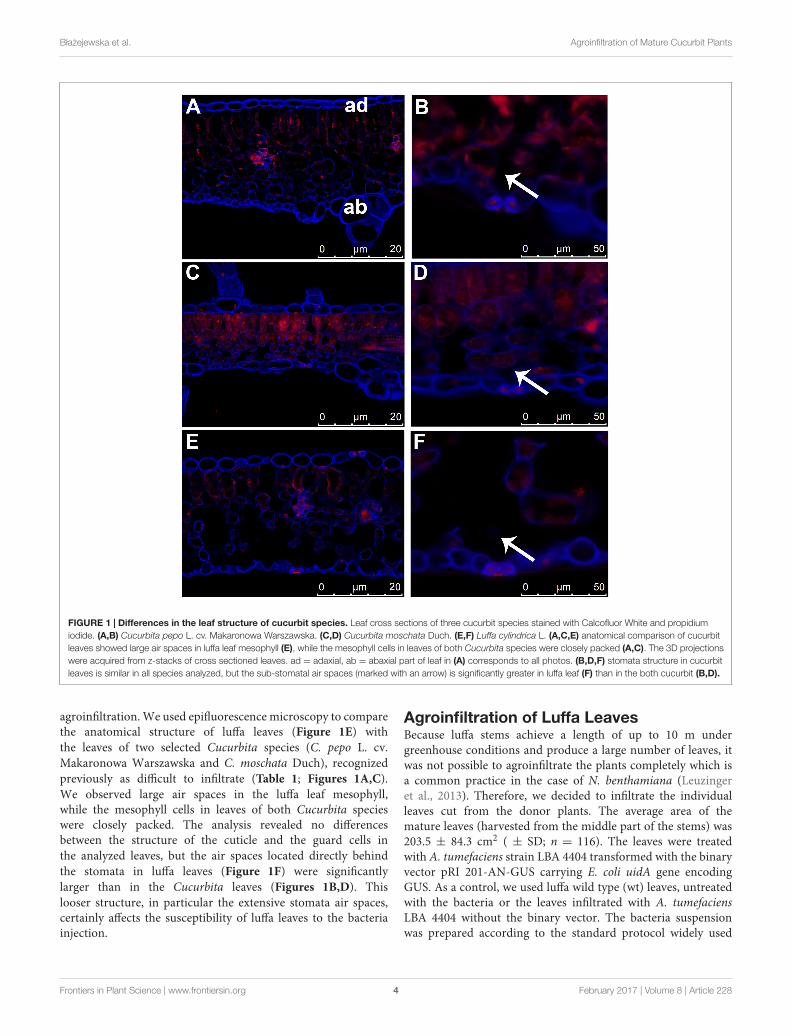
FIGURE 1 | Differences in the leaf structure of cucurbit species. Leaf cross sections of three cucurbit species stained with Calcofluor White and propidiumiodide. (A,B) Cucurbita pepo L. cv. Makaronowa Warszawska. (C,D) Cucurbita moschata Duch. (E,F) Luffa cylindrica L. (A,C,E) anatomical comparison of cucurbitleaves showed large air spaces in luffa leaf mesophyll (E), while the mesophyll cells in leaves of both Cucurbita species were closely packed (A,C). The 3D projectionswere acquired from z-stacks of cross sectioned leaves. ad = adaxial, ab = abaxial part of leaf in (A) corresponds to all photos. (B,D,F) stomata structure in cucurbitleaves is similar in all species analyzed, but the sub-stomatal air spaces (marked with an arrow) is significantly greater in luffa leaf (F) than in the both cucurbit (B,D).
agroinfiltration. We used epifluorescence microscopy to comparethe anatomical structure of luffa leaves (Figure 1E) withthe leaves of two selected Cucurbita species (C. pepo L. cv.Makaronowa Warszawska and C. moschata Duch), recognizedpreviously as difficult to infiltrate (Table 1; Figures 1A,C).We observed large air spaces in the luffa leaf mesophyll,while the mesophyll cells in leaves of both Cucurbita specieswere closely packed. The analysis revealed no differencesbetween the structure of the cuticle and the guard cells inthe analyzed leaves, but the air spaces located directly behindthe stomata in luffa leaves (Figure 1F) were significantlylarger than in the Cucurbita leaves (Figures 1B,D). Thislooser structure, in particular the extensive stomata air spaces,certainly affects the susceptibility of luffa leaves to the bacteriainjection.
Agroinfiltration of Luffa LeavesBecause luffa stems achieve a length of up to 10 m undergreenhouse conditions and produce a large number of leaves, itwas not possible to agroinfiltrate the plants completely which isa common practice in the case of N. benthamiana (Leuzingeret al., 2013). Therefore, we decided to infiltrate the individualleaves cut from the donor plants. The average area of themature leaves (harvested from the middle part of the stems) was203.5 ± 84.3 cm2 ( ± SD; n = 116). The leaves were treatedwith A. tumefaciens strain LBA 4404 transformed with the binaryvector pRI 201-AN-GUS carrying E. coli uidA gene encodingGUS. As a control, we used luffa wild type (wt) leaves, untreatedwith the bacteria or the leaves infiltrated with A. tumefaciensLBA 4404 without the binary vector. The bacteria suspensionwas prepared according to the standard protocol widely used
Frontiers in Plant Science | www.frontiersin.org 4 February 2017 | Volume 8 | Article 228

fpls-08-00228 February 17, 2017 Time: 18:4 # 5
Błazejewska et al. Agroinfiltration of Mature Cucurbit Plants
FIGURE 2 | Changes in the weight of agroinfiltrated leaves. The averageweight of the leaves infiltrated with Agrobacterium tumefaciens carrying pRI201-AN-GUS (n > 6) as well as the leaves infiltrated with A. tumefaciens(n = 6) without binary vector increased significantly directly after introductionof bacteria suspension (marked with an asterisks; P < 0.01) but at 1 dpidecreased the average weight significantly and then remained stable in theconsecutive days after infiltration. The analysis was repeated independentlythree times.
for N. benthamiana agroinfiltration (Wydro et al., 2006). Toprevent the loss of turgor, the leaves immediately before andafter agroinfiltration were kept under high humidity conditions(Supplementary Figure S1).
We observed that the leaves incubated in high humidityconditions showed excellent susceptibility to the infiltration formany hours after harvesting. Moreover, the introduction ofbacteria (OD600 = 1.0) into the leaves stored in high humiditywas much easier than in the case of leaves that had recentlybeen cut from the host plants. The increased agroinfiltrationefficiency at high humidity growth conditions was reportedpreviously in Arabidopsis (Kim et al., 2009). High humiditypromotes the opening of the stomata, which in consequence,increases the access of the externally applied fluid to the sub-stomatal air spaces, as well as to the air spaces located inthe deeper parts of the spongy and palisade mesophyll. Theincreased humidity conditions did not significantly affect theagroinfiltration susceptibility of the other cucurbit species wetested. We suppose, that the compact structure (Figure 1) ofthe leaf tissues in many cucurbit species efficiently prevents thepenetration of the externally applied liquids into the deepertissues despite the stomata opening.
The leaves were weighed before and immediately after theinfiltration to estimate the amount of the bacterial suspensionabsorbed. The biomass of leaves was also measured up to threepost-infiltration days to monitor a turgor loss in the leaves.Directly after saturation of the leaf with the bacterial suspension,the average mass of luffa leaves increased by 22.7± 12.4% (± SD;n = 14), relative to the leaf biomass before the agroinfiltration(Figure 2). However, 1 day post-infiltration (1 dpi) the massof the infiltrated leaves decreased significantly, returning tothe values similar to those measured immediately before theinfiltration. The leaf mass then remained stable for at least3 days (2 and 3 dpi) (Figure 2). The comparison of the biomasschanges in the wt leaves with the leaves infiltrated with thebacteria suspension (A. tumefaciens with or without the binaryvector) in the days following their harvesting from the donorplants, demonstrates that the infiltration procedures we used
FIGURE 3 | The histochemical X-gluc staining of the GUS activity inluffa leaves and EDTA-exudates. (A) Leaf disks (10 mm in diameter)randomly collected from: the leaves infiltrated with A. tumefaciens carrying pRI201-AN-GUS, the leaves infiltrated with A. tumefaciens LBA 4404 without thebinary vector, wt leaves. (B) Petiole cross section of the leaf infiltrated withA. tumefaciens carrying pRI 201-AN-GUS and from luffa wt leaf. The bluestained vascular bundles are shown. (C) GUS activity in EDTA-exudatesamples detected with X-gluc on microtiter plate. The 100 µL samples werecollected at 1 dpi from wt leaves (wells A1–A9 and B1–B9), from the leavesinfiltrated with untransformed A. tumefaciens LBA 4404 (wells C1–C9 andD1–D9) and from the leaves infiltrated with A. tumefaciens carrying pRI201-AN-GUS (wells C10–C12, D10–D12, E1–E12, F1–F12, G1–G2, H1–H2).The histochemical analysis of leaf tissues and exudates were repeatedindependently at least three times.
are insignificant for the transpiration intensity in luffa leaves(Figure 2).
GUS Activity in Leaves and ExudatesThe histochemical detection revealed the high GUS activity in allrandom samples collected from the leaves agroinfiltrated with thevector pRI 201-AN-GUS, whereas the wt leaves and the leavesinfiltrated with Agrobacterium without the binary vector did notshow the GUS activity (Figure 3A, Supplementary Figure S2).The staining pattern in the leaf fragments was non-uniformindicating the heterogeneous GUS activity in the agroinfiltratedtissues. Therefore, we decided to sample the whole leaf bladesfor further GUS analysis. Fluorometric assays estimated theGUS activity in the extracts from the whole pRI 201-AN-GUStransformed leaf blades as 31.7± 13.2 (± SD; n= 6); 34.2± 16.9(± SD; n= 6) and 50.4± 27.4 (± SD; n= 5) [µM 4-MU/mg totalsoluble proteins/min], at 1, 2, and 3 dpi, respectively. When theGUS activity in leaves extracts was normalized to the total surface
Frontiers in Plant Science | www.frontiersin.org 5 February 2017 | Volume 8 | Article 228
fpls-08-00228 February 17, 2017 Time: 18:4 # 6
Błazejewska et al. Agroinfiltration of Mature Cucurbit Plants
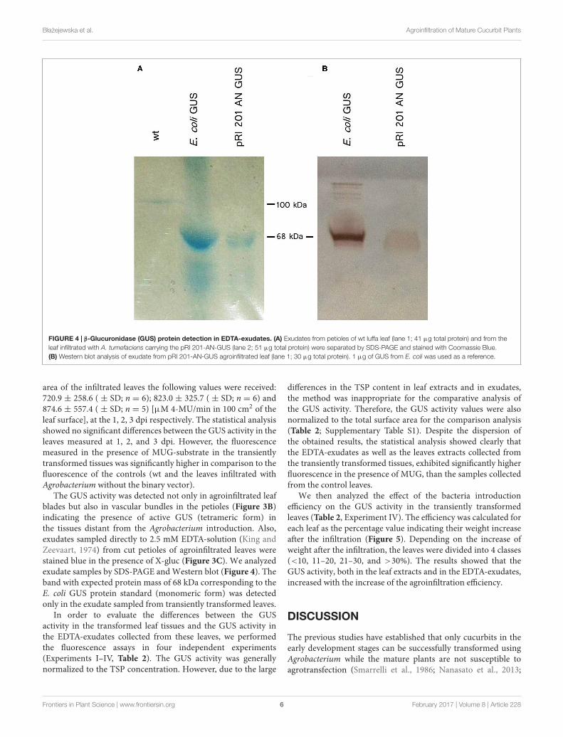
FIGURE 4 | β-Glucuronidase (GUS) protein detection in EDTA-exudates. (A) Exudates from petioles of wt luffa leaf (lane 1; 41 µg total protein) and from theleaf infiltrated with A. tumefaciens carrying the pRI 201-AN-GUS (lane 2; 51 µg total protein) were separated by SDS-PAGE and stained with Coomassie Blue.(B) Western blot analysis of exudate from pRI 201-AN-GUS agroinfiltrated leaf (lane 1; 30 µg total protein). 1 µg of GUS from E. coli was used as a reference.
area of the infiltrated leaves the following values were received:720.9 ± 258.6 ( ± SD; n = 6); 823.0 ± 325.7 ( ± SD; n = 6) and874.6 ± 557.4 ( ± SD; n = 5) [µM 4-MU/min in 100 cm2 of theleaf surface], at the 1, 2, 3 dpi respectively. The statistical analysisshowed no significant differences between the GUS activity in theleaves measured at 1, 2, and 3 dpi. However, the fluorescencemeasured in the presence of MUG-substrate in the transientlytransformed tissues was significantly higher in comparison to thefluorescence of the controls (wt and the leaves infiltrated withAgrobacterium without the binary vector).
The GUS activity was detected not only in agroinfiltrated leafblades but also in vascular bundles in the petioles (Figure 3B)indicating the presence of active GUS (tetrameric form) inthe tissues distant from the Agrobacterium introduction. Also,exudates sampled directly to 2.5 mM EDTA-solution (King andZeevaart, 1974) from cut petioles of agroinfiltrated leaves werestained blue in the presence of X-gluc (Figure 3C). We analyzedexudate samples by SDS-PAGE and Western blot (Figure 4). Theband with expected protein mass of 68 kDa corresponding to theE. coli GUS protein standard (monomeric form) was detectedonly in the exudate sampled from transiently transformed leaves.
In order to evaluate the differences between the GUSactivity in the transformed leaf tissues and the GUS activity inthe EDTA-exudates collected from these leaves, we performedthe fluorescence assays in four independent experiments(Experiments I–IV, Table 2). The GUS activity was generallynormalized to the TSP concentration. However, due to the large
differences in the TSP content in leaf extracts and in exudates,the method was inappropriate for the comparative analysis ofthe GUS activity. Therefore, the GUS activity values were alsonormalized to the total surface area for the comparison analysis(Table 2; Supplementary Table S1). Despite the dispersion ofthe obtained results, the statistical analysis showed clearly thatthe EDTA-exudates as well as the leaves extracts collected fromthe transiently transformed tissues, exhibited significantly higherfluorescence in the presence of MUG, than the samples collectedfrom the control leaves.
We then analyzed the effect of the bacteria introductionefficiency on the GUS activity in the transiently transformedleaves (Table 2, Experiment IV). The efficiency was calculated foreach leaf as the percentage value indicating their weight increaseafter the infiltration (Figure 5). Depending on the increase ofweight after the infiltration, the leaves were divided into 4 classes(<10, 11–20, 21–30, and >30%). The results showed that theGUS activity, both in the leaf extracts and in the EDTA-exudates,increased with the increase of the agroinfiltration efficiency.
DISCUSSION
The previous studies have established that only cucurbits in theearly development stages can be successfully transformed usingAgrobacterium while the mature plants are not susceptible toagrotransfection (Smarrelli et al., 1986; Nanasato et al., 2013;
Frontiers in Plant Science | www.frontiersin.org 6 February 2017 | Volume 8 | Article 228

fpls-08-00228 February 17, 2017 Time: 18:4 # 7
Błazejewska et al. Agroinfiltration of Mature Cucurbit Plants
TABLE 2 | The GUS activity in the agroinfiltrated luffa leaves and exudates.
GUS activity [µM 4-MU/min/100 cm2 leaf]
EDTA-exudates
No. experiment Leaf extracts 1 dpi 2 dpi 3 dpi
Mean SD n Mean SD n Mean SD n Mean SD n
Experiment I 79.8 58.5 6 60.4 36.5 6 63.5 38.4 6 7.7 2.3 6
Experiment II 244.2 146.3 10 87.2 61.8 4 14.9 8.5 4 10.3 14.6 4
Experiment III nc nc nc 164.5 64.7 4 24.8 11.9 4 27.0 17.3 4
Experiment IV 710.4 369.3 14 50.3 54.5 14 nc nc nc nc nc nc
The GUS activity were measured fluorescently in leaves extracts and EDTA-exudates and then normalized to the 100 cm2 leaf surface area. In the Experiments I–II theleaves extracts were collected at 3 dpi and in the Experiment IV at 1 dpi. In the Experiments I–III the exudates were collected at 1–3 dpi and in the Experiment IV at 1 dpi,nt = not collected, dpi = day post-infiltration.
Ramírez-Ortega et al., 2014, 2015). However, our work hasshown that L. cylindrica L. (Cucurbitaceae) can be successfullytransformed by agroinfiltration.
One of the most important factors affecting agroinfiltrationefficiency in several plant species is the susceptibility oftheir tissues to the introduction of bacterial suspension(Bhaskar et al., 2009; Leuzinger et al., 2013). N. benthamiana,whose leaves have excellent absorption properties enabling theoptimization of the agroinfiltration procedure, has become themost popular transient expression system (Wydro et al., 2006;Goodin et al., 2008). Our analysis showed that among 11species of cucurbits, only luffa leaves revealed an excellentabsorption property. This absorption property allows theinfiltration of the mesophyll tissue with relative large volumesof liquid containing high density suspensions of Agrobacteriumcells.
The susceptibility of luffa leaves to infiltration can beexplained by the loose structure of the mesophyll tissue withthe large air spaces, especially revealed by the microscopicexamination sub-stomatal air spaces. In contrast to luffa themesophyll cells in other Cucurbita leaves are closely packedand the stomata air spaces are slight. High humidity promotesstomatal opening, which in consequence, increases access tothe air spaces located in the deeper leaf tissues (Melotto et al.,2008; Kim et al., 2009). It was observed that high humidityconditions increase the agroinfiltration susceptibility of the luffaleaves. However, the compact structure of Cucurbita leaf tissuesefficiently prevents the penetration of the externally appliedbacteria into the deeper tissues despite the stomata opening. Thiscould explain the low effectivity of transformation of Cucurbitaplants.
The volume of intercellular spaces in leaves depends onplant species, leaf maturity as well as the growth conditions.Anatomical and physiological changes in leaves, in particularthe reduction of intercellular space volume and thus theformation of more compact leaf tissue, were observed underabiotic and biotic stress in various plant species (Gausmanet al., 1975; Beattie and Lindow, 1995). Luffa species aretropical and subtropical plants known as tolerant to variousenvironmental stress conditions (Liao and Linn, 1996; Filipowiczet al., 2014; Li et al., 2014; Liu et al., 2016). The loose
FIGURE 5 | Effect of agroinfiltration efficiency on the GUS activity inleaf blade and exudates. To estimate the efficiency of bacteria introductiona leaf weight increase after infiltration was calculated as percentage valuesrelative to the weight before the infiltration. We divided the infiltrated leaves(n = 14) into four classes (<10, 11–20, 21–30, and >30%) depending on theleaf weight (the X-axis). GUS activity (the Y-axis) in the whole leaf bladesextracts as well as in the EDTA-exudate samples were measured at 1 dpi andthen normalized relative to the 100 cm2 of leaf surface area [µM 4-MU/min in100 cm2].
mesophyll structure in the leaves of luffa plants reported hereindicates indirectly that the plants were grown under non-stressconditions.
Intercellular spaces and substomatal cavities in leaves providealso a habitat for endophytic phyllosphere microorganisms,especially for foliar pathogens (Beattie and Lindow, 1995).Foliar pathogenic bacteria can reach the leaf interiorthrough natural surface openings (e.g., stomata, hydathodes)or through surface wounds (Wilson et al., 1999; Hiranoand Upper, 2000). Numerous bacteria species use activemechanisms to colonize intercellular spaces, for examplePseudomonas syringae can use coronatine to actively openstomata (Melotto et al., 2008). Although, A. tumefaciens isa soil bacterium which infects underground plant organs
Frontiers in Plant Science | www.frontiersin.org 7 February 2017 | Volume 8 | Article 228

fpls-08-00228 February 17, 2017 Time: 18:4 # 8
Błazejewska et al. Agroinfiltration of Mature Cucurbit Plants
it is commonly used as a tool for transformation of plantleaves (Cazzonelli and Velten, 2006; Goodin et al., 2008;Bhaskar et al., 2009; Leuzinger et al., 2013). Probably, itwill be possible to increase the agroinfiltration efficiencyusing some of the colonization strategies described for foliarendophytes. One particularly interesting possibility will beto use different extracellular polysaccharides isolated fromsuitable Pseudomonas species as adjuvants to support thepenetration of Agrobacterium into the interior of the leaves(El-Banoby and Rudolph, 1979).
Our results suggest the possibility to use luffa leaves as atool for studies requiring the application of liquids into the leaftissue. Such techniques are already used in plant physiology andbiotechnology studies, especially for evaluating phloem functionsin different plant species (Grignon et al., 1989; Pradel et al.,1999; Zhang et al., 2010; Knoblauch et al., 2015). In the case ofcucurbits the problem with the application of liquid substancesinto mature leaves generally required some damage of leaf surfaceby using, e.g., fine sandpaper or carborundum as an abrasive(Grignon et al., 1989; Pradel et al., 1999; Shalitin and Wolf,2000; Arazi et al., 2001; Zhang et al., 2010; Dunham et al.,2014; Kang et al., 2016). In contrast, the infiltration of luffaleaves is a more delicate method as it uses the natural abilityof leaves to absorb water. The absorbance properties can beadditionally elevated by pretreatment of luffa leaves in highhumidity conditions.
The plant transformation techniques including transienttransformation methods are used as a popular research toolfor the elucidation of phloem functions and biochemistry inplant species known as apoplastic phloem loaders, such asN. benthamiana, S. tuberosum, or A. thaliana (Chincinska et al.,2008; Krügel et al., 2012). There are the distinct differences in thephloem structure between cucurbits and other plants (Turgeonand Medville, 2004). Although many species of Cucurbitaceae arewidely used in research of phloem functions and biochemistry,only a few reports provide data obtained using transgeniccucurbits (Smarrelli et al., 1986; Nanasato et al., 2013; Ramírez-Ortega et al., 2014, 2015; Cheng et al., 2015). Many questionsabout cucurbit phloem functions (Slewinski et al., 2013), suchas regulation of symplastic loading or long-distance movementof macromolecules, as well as phloem exudation and sealingmechanisms, have not been elucidated so far. The system forrapid and easy expression of foreign genes in mature luffaleaves provides an additional strategy for cucurbit phloemresearch.
It would be especially interesting to use the luffa leavesexpression system as a tool for monitoring of macromoleculeslong distance trafficking in the phloem. Our observation indicatesthat GUS molecules can move from the agroinfiltrated leaftissues, where they are produced, along the non-transformedpetioles to the EDTA solution, in which the leaves were immersedafter the infiltration. Phloem trafficking of recombinant proteinshas already been described, but the mechanisms regulating thesephenomenon are still not explained. For instance, Imlau et al.(1999) observed the long-distance trafficking of non-phloem27 kDa GFP protein in transgenic A. thaliana and in tobaccoplants expressing GFP driven by the AtSUC 2 phloem-specific
promoter. The molecular weight of GUS protein in a monomericform is 68 kDa and it is higher as GFP weight. Moreover, theGUS protein is active only in tetrameric form. Although, it waspreviously estimated that proteins greater than 200 kDa can movein the cucurbit phloem (Balachandran et al., 1997; Walz et al.,2004), it seems unlikely that the GUS protein is transportedover long distances as an active tetrameric protein. This issueneeds to be clarified, because the process of GUS tetramerizationis reversible (Matsuura et al., 2011). We suppose that GUScan be transported as monomeric subunits, whose assemblyinto tetramers take place away from the protein productionsite.
The luffa leaves expression system can be used for avariety of cucurbit studies, including luffa itself. There arenumerous reports on a wide range of luffa plants applications(Sujatha et al., 2013). It was shown that a number ofsubstances identified in luffa seeds and leaf extracts hastherapeutic potential, for example, a novel class of smallmolecule ribosome-inactivating peptides, which in additionto a strong inhibitory activity on cell-free protein synthesisshow also antifungal, antimicrobial, and anti-tumorial properties(Ling et al., 1993; Parkash et al., 2002; Li et al., 2003).Presumably, transformed luffa leaves can become a morecompatible expression system for the production of these luffaproteins (as well as other cucurbit-derived recombinant proteins)than expression systems based on bacteria or yeast (Liu et al.,2010).
The L. cylindrica leaves can be transiently transformed in acomparatively simple manner as in the case of N. benthamianamodel plants. However, it can be assumed that the furtheroptimization of the agroinfiltration procedure will lead toimprovement of the expression system based on transientlytransformed luffa leaves. It will be of particular interest tomonitor the presence of recombinant proteins in exudate.Furthermore, the optimization of heterological expression inthe luffa system enables the production and purification ofrecombinant proteins not only in transiently transformed leavesbut also in exudates.
This study is the first case report about the application ofcucurbits in the study of gene expression by agroinfiltration ofmature leaves. The possibility to ease the genetic modificationof mature luffa leaves will contribute to the development of ourknowledge about Cucurbitaceae biology, especially in regards tothe regulation of phloem functions. This will be the case not onlyin cucurbit research but also among scientists looking for a simpleand reliable method for the heterologous expression in eukaryoticsystems.
AUTHOR CONTRIBUTIONS
IC conceived the research plans. IC supervised the experiments.KB, MK, EZ, and IC performed the experiments andanalyzed the data. IC conceived the project and wrotethe article with contributions of all the authors. KB,MK, EZ, ZT, and IC supervised and complemented thewriting.
Frontiers in Plant Science | www.frontiersin.org 8 February 2017 | Volume 8 | Article 228

fpls-08-00228 February 17, 2017 Time: 18:4 # 9
Błazejewska et al. Agroinfiltration of Mature Cucurbit Plants
FUNDING
This research was supported by the Foundation for Polish Science(FNP, POMOST/2011-4/4) co-financed from the EuropeanUnion under the European Regional Development Fund.
ACKNOWLEDGMENTS
We thank Dr. hab. Christina Kühn, Prof. Dr. hab.Renata Ochocka, Dr. hab. Agnieszka Szalewska-Pałasz,
Prof. Dr. hab. Julian Swierczynski, and Dr. Natalia Filipowiczfor the sincere support. We are very thankful forcritical reading and English correction by Dr. MagdalenaMiklaszewska.
SUPPLEMENTARY MATERIAL
The Supplementary Material for this article can be found onlineat: http://journal.frontiersin.org/article/10.3389/fpls.2017.00228/full#supplementary-material
REFERENCESArazi, T., Slutsky, S. G., Shiboleth, Y. M., Wang, Y., Rubinstein, M., Barak, S.,
et al. (2001). Engineering zucchini yellow mosaic potyvirus as a non-pathogenicvector for expression of heterologous proteins in cucurbits. J. Biotechnol. 87,67–82. doi: 10.1016/S0168-1656(01)00229-2
Balachandran, S., Xiang, Y., Schobert, C., Thompson, G. A., and Lucas, W. J.(1997). Phloem sap proteins from Cucurbita maxima and Ricinus communishave the capacity to traffic cell to cell through plasmodesmata. Proc. Natl. Acad.Sci. U.S.A. 94, 14150–14155. doi: 10.1073/pnas.94.25.14150
Beattie, G. A., and Lindow, S. E. (1995). The secret life of foliar bacterial pathogenson leaves. Annu. Rev. Phytopathol. 33, 145–172. doi: 10.1146/annurev.py.33.090195.001045
Bhaskar, P. B., Venkateshwaran, M., Wu, L., Ané, J.-M., and Jiang, J. (2009).Agrobacterium-mediated transient gene expression and silencing: a rapid toolfor functional gene assay in potato. PLoS ONE 4:5812. doi: 10.1371/journal.pone.0005812
Bradford, M. M. (1976). A rapid and sensitive method for the quantitation ofmicrogram quantities of protein utilizing the principle of protein-dye binding.Anal. Biochem. 72, 248–254. doi: 10.1016/0003-2697(76)90527-3
Brunelle, J. L., and Green, R. (2014). “One-dimensional SDS-polyacrylamidegel electrophoresis (1D SDS-PAGE),” in Methods in Enzymology. LaboratoryMethods in Enzymology: Protein Part C, ed. J. Lorsch (Amsterdam: ElsevierInc.), 151–159. doi: 10.1016/B978-0-12-420119-4.00012-4
Cazzonelli, C. I., and Velten, J. (2006). An in vivo, luciferase-based,Agrobacterium-infiltration assay system: implications for post-transcriptional gene silencing. Planta 224, 582–597. doi: 10.1007/s00425-006-0250-z
Cheng, J.-T., Li, X., Yao, F.-Z., Shan, N., Li, Y.-H., Zhang, Z.-X., et al. (2015).Functional characterization and expression analysis of cucumber (Cucumissativus L.) hexose transporters, involving carbohydrate partitioning and phloemunloading in sink tissues. Plant Sci. 237, 46–56. doi: 10.1016/j.plantsci.2015.05.006
Chincinska, I. A., Liesche, J., Krügel, U., Michalska, J., Geigenberger, P., Grimm, B.,et al. (2008). Sucrose transporter StSUT4 from potato affects flowering,tuberization, and shade avoidance response. Plant Physiol. 146, 515–528. doi:10.1104/pp.107.112334
Dunham, J. P., Simmons, H. E., Holmes, E. C., and Stephenson, A. G. (2014).Analysis of viral (zucchini yellow mosaic virus) genetic diversity duringsystemic movement through a Cucurbita pepo vine. Virus Res. 191, 172–179.doi: 10.1016/j.virusres.2014.07.030
El-Banoby, F. E., and Rudolph, K. (1979). A polysaccharide from liquid culturesof Pseudomonas phaseolicola which specifically induces water-soaking in beanleaves (Phaseolus vulgaris L.). J. Phytopath. 95, 38–50. doi: 10.1111/j.1439-0434.1979.tb01576.x
Filipowicz, N., Schaefer, H., and Renner, S. S. (2014). Revisiting Luffa(Cucurbitaceae) 25 years after C. Heiser: species boundaries and application ofnames tested with plastid and nuclear DNA sequences. Syst. Bot. 39, 205–215.doi: 10.1105/tpc.11.1.127215
Gausman, H. W., Heald, C. M., and Escobar, D. E. (1975). Effect of Rotylenchulusreniformis on reflectance of cotton plant leaves. J. Nematol. 7, 368–374.
Golecki, B., Schulz, A., and Thompson, G. A. (1999). Translocation of structural Pproteins in the phloem. Plant Cell 11, 127–140. doi: 10.1105/tpc.11.1.127
Goodin, M. M., Zaitlin, D., Naidu, R. A., and Lommel, S. A. (2008). Nicotianabenthamiana: its history and future as a model for plant-pathogen interactions.Mol. Plant Microbe Interact. 21, 1015–1026. doi: 10.1094/MPMI-21-8-1015
Grignon, N., Touraine, B., and Durand, M. (1989). 6(5)Carboxyfluorescein asa tracer of phloem sap translocation. Am. J. Bot. 76, 871–877. doi: 10.2307/2444542
Hirano, S. S., and Upper, C. D. (2000). Bacteria in the leaf ecosystem with emphasison Pseudomonas syringae—a pathogen, ice nucleus, and epiphyte. Microbiol.Mol. Biol. Rev. 64, 624–653. doi: 10.1128/MMBR.64.3.624-653.2000
Imlau, A., Truernit, E., and Sauer, N. (1999). Cell-to-cell and long-distancetrafficking of the green fluorescent protein in the phloem and symplasticunloading of the protein into sink tissues. Plant Cell 11, 309–322. doi: 10.2307/3870862
Jefferson, R. A., Kavanagh, T. A., and Bevan, M. W. (1987). GUS fusions:β-glucuronidase as a sensitive and versatile gene fusion marker in higher plants.EMBO J. 6, 3901–3907.
Joh, L. D., Wroblewski, T., Ewing, N. N., and VanderGheynst, J. S. (2005). High-level transient expression of recombinant protein in lettuce. Biotechnol. Bioeng.91, 861–871. doi: 10.1002/bit.20557
Kang, M., Seo, J.-K., Choi, H., Choi, H.-S., and Kim, K.-H. (2016). Establishmentof a simple and rapid gene delivery system for cucurbits by using engineered ofZucchini yellow mosaic virus. Plant Pathol. J. 32, 70–76. doi: 10.5423/PPJ.NT.08.2015.0173
Kim, M. J., Baek, K., and Park, C.-M. (2009). Optimization of conditions fortransient Agrobacterium-mediated gene expression assays in Arabidopsis. PlantCell Rep. 28, 1159–1167. doi: 10.1007/s00299-009-0717-z
King, R. W., and Zeevaart, J. A. D. (1974). Enhancement of phloem exudationfrom cut petioles by chelating agents. Plant Physiol. 53, 96–103. doi: 10.1104/pp.53.1.96
Knoblauch, M., Vendrell, M., de Leau, E., Paterlini, A., Knox, K., Ross-Elliot, T.,et al. (2015). Multispectral phloem-mobile probes: properties and applications.Plant Physiol. 167, 1211–1220. doi: 10.1104/pp.114.255414
Krawczyk, E., Rojek, J., Kowalkowska, A. K., Kapusta, M., Znaniecka, J., andMinasiewicz, J. (2016). Evidence for mixed sexual and asexual reproduction inthe rare European mycoheterotrophic orchid Epipogium aphyllum, Orchidaceae(ghost orchid). Ann. Bot. 118, 159–172. doi: 10.1093/aob/mcw084
Krügel, U., He, H.-X., Gier, K., Reins, J., Chincinska, I., Grimm, B., et al. (2012). Thepotato sucrose transporter StSUT1 interacts with a DRM-associated proteindisulfide isomerase. Mol. Plant 5, 43–62. doi: 10.1093/mp/ssr048
Leuzinger, K., Dent, M., Hurtado, J., Stahnke, J., Lai, H., Zhou, X., et al.(2013). Efficient agroinfiltration of plants for high-level transient expression ofrecombinant proteins. J. Vis. Exp. 77:50521. doi: 10.3791/50521
Li, F., Yang, X.-X., Xia, H.-C., Zeng, R., Hu, W.-G., Li, Z., et al. (2003). Purificationand characterization of Luffin P1, a ribosome-inactivating peptide from theseeds of Luffa cylindrica. Peptides 24, 799–805. doi: 10.1016/S0196-9781(03)00173-6
Li, H., Wang, F., Chen, X.-J., Shi, K., Xia, X.-J., Considine, M. J., et al. (2014).The sub/supra-optimal temperature-induced inhibition of photosynthesis andoxidative damage in cucumber leaves are alleviated by grafting onto figleafgourd/luffa rootstocks. Physiol. Plant. 152, 571–584. doi: 10.1111/ppl.12200
Liao, C. T., and Linn, C. H. (1996). Photosynthetic response of grafted bitter melonseedlings to flood stress. Environ. Exp. Bot. 36, 167–172. doi: 10.1016/0098-8472(96)01009-X
Frontiers in Plant Science | www.frontiersin.org 9 February 2017 | Volume 8 | Article 228
fpls-08-00228 February 17, 2017 Time: 18:4 # 10
Błazejewska et al. Agroinfiltration of Mature Cucurbit Plants
Lin, M.-K., Lee, Y.-J., Lough, T. J., Phinney, B. S., and Lucas, W. J. (2009). Analysisof the pumpkin phloem proteome provides insights into angiosperm sievetube function. Mol. Cell. Proteomics 8, 343–356. doi: 10.1074/mcp.M800420-MCP200
Ling, M.-H., Qi, H.-Y., and Chi, C.-W. (1993). Protein, cDNA, and genomic DNAsequences of the towel gourd trypsin inhibitor. A squash family inhibitor. J. Biol.Chem. 268, 810–814.
Liu, L., Wang, R., He, W., He, F., and Huang, G. (2010). Cloning and solubleexpression of mature α - luffin from Luffa cylindrica and its antitumor activitiesin vitro. Acta Biochim. Biophys. Sin. 42, 585–592. doi: 10.1093/abbs/gmq056
Liu, S., Li, H., Lv, X., Ahammed, G. J., Xia, X., Zhou, J., et al. (2016).Grafting cucumber onto luffa improves drought tolerance by increasing ABAbiosynthesis and sensitivity. Sci. Rep. 6:20212. doi: 10.1038/srep20212
Manamohan, M., Prakash, N., and Sharath Chandra, G. (2011). “Cucurbits,” inAdvances In Horticulture Biotechnology - Gene Cloning and Transgenics, ed.H. P. Singh (New Delhi: Westville Publishing House), 227–259.
Matsuura, T., Hosoda, K., Ichihashi, N., Kazuta, Y., and Yomo, T. (2011). Kineticanalysis of β-galactosidase and β-glucuronidase tetramerization coupled withprotein translation. J. Biol. Chem. 286, 22028–22034. doi: 10.1074/jbc.M111.240168
Melotto, M., Underwood, W., and He, S. Y. (2008). Role of stomata in plant innateimmunity and foliar bacterial diseases. Annu. Rev. Phytopathol. 46, 101–122.doi: 10.1146/annurev.phyto.121107.104959
Nanasato, Y., Konagaya, K., Okuzaki, A., Tsuda, M., and Tabei, Y. (2013).Improvement of Agrobacterium-mediated transformation of cucumber(Cucumis sativus L.) by combination of vacuum infiltration and co-cultivation on filter paper wicks. Plant Biotechnol. Rep. 7, 267–276.doi: 10.1007/s11816-012-0260-1
Parkash, A., Ng, T. B., and Tso, W. W. (2002). Isolation and characterization ofluffacylin, a ribosome inactivating peptide with anti-fungal activity from spongegourd (Luffa cylindrica) seeds. Peptides 23, 1019–1024. doi: 10.1016/S0196-9781(02)00045-1
Pradel, K. S., Ullrich, C. I., Santa Cruz, S., and Oparka, K. J. (1999). Symplasticcontinuity in Agrobacterium tumefaciens-induced tumours. J. Exp. Bot. 50,183–192. doi: 10.1093/jexbot/50.331.183
Ramírez-Ortega, F. A., Herrera-Pola, P. S., Toscano-Morales, R., Xoconostle-Cázares, B., and Ruiz-Medrano, R. (2014). Overexpression of the pumpkin(Cucurbita maxima) 16 kDa phloem protein CmPP16 increases tolerance towater deficit. Plant Signal. Behav. 9, 973823. doi: 10.4161/15592324
Ramírez-Ortega, F. A., Xoconostle-Cázares, B., Toscano-Morales, R., and Ruiz-Medrano, R. (2015). A simple method for transient transformation of pumpkin(Cucurbita maxima) seedlings. POJ 8, 37–46.
Richardson, P. T., Baker, D. A., and Ho, L. C. (1982). The chemical composition ofcucurbit vascular exudates. J. Exp. Bot. 33, 1239–1247.
Santos-Rosa, M., Poutaraud, A., Merdinoglu, D., and Mestre, P. (2008).Development of a transient expression system in grapevine via agro-infiltration.Plant Cell Rep. 27, 1053–1063. doi: 10.1007/s00299-008-0531-z
Shalitin, D., and Wolf, S. (2000). Cucumber mosaic virus infection affects sugartransport in melon plants. Plant Physiol. 123, 597–604. doi: 10.1104/pp.123.2.597
Slewinski, T. L., Zhang, C., and Turgeon, R. (2013). Structural and functionalheterogeneity in phloem loading and transport. Front. Plant Sci. 4:244. doi:10.3389/fpls.2013.00244
Smarrelli, J., Watters, M. T., and Diba, L. H. (1986). Response of various cucurbitsto infection by plasmid-harboring strains of Agrobacterium. Plant Physiol. 82,622–624.
Sujatha, D., Chithakari, R., Raghuvardhan, L., Prasad, B., Gulab Khan, R.,Sadanandam, A., et al. (2013). In vitro plantlet regeneration and genetictransformation of sponge gourd (Luffa cylindrica L.). Afr. J. Plant Sci. 7,244–252. doi: 10.5897/AJPS12.196
Turgeon, R., and Medville, R. (2004). Phloem loading. A reevaluationof the relationship between plasmodesmatal frequencies and loadingstrategies. Plant Physiol. 136, 3795–3803. doi: 10.1104/pp.104.042036
Turgeon, R., and Oparka, K. (2010). The secret phloem of pumpkins. Proc. Natl.Acad. Sci. U.S.A. 107, 13201–13202. doi: 10.1073/pnas.1008134107
Walz, C., Giavalisco, P., Schad, M., Juenger, M., Klose, J., and Kehr, J. (2004).Proteomics of curcurbit phloem exudate reveals a network of defenceproteins. Phytochemistry 65, 1795–1804. doi: 10.1016/j.phytochem.2004.04.006
Wilson, M., Hirano, S. S., and Lindow, S. E. (1999). Location and survival of leaf-associated bacteria in relation to pathogenicity and potential for growth withinthe leaf. Appl. Environ. Microbiol. 65, 1435–1443.
Wydro, M., Kozubek, E., and Lehmann, P. (2006). Optimization of transientAgrobacterium-mediated gene expression system in leaves of Nicotianabenthamiana. Acta Biochim. Pol. 53, 289–298.
Zhang, B., Tolstikov, V., Turnbull, C., Hicks, L. M., and Fiehn, O. (2010). Divergentmetabolome and proteome suggest functional independence of dual phloemtransport systems in cucurbits. Proc. Natl. Acad. Sci. U.S.A. 107, 13532–13537.doi: 10.1073/pnas.0910558107
Zhang, C., Yu, X., Ayre, B. G., and Turgeon, R. (2012). The origin and compositionof cucurbit “phloem” exudate. Plant Physiol. 158, 1873–1882. doi: 10.1104/pp.112.194431
Zimmermann, M. R., Hafke, J. B., van Bel, A. J. E., and Furch, A. C. U. (2013).Interaction of xylem and phloem during exudation and wound occlusion inCurcurbita maxima. Plant Cell Environ. 36, 237–247. doi: 10.1111/j.1365-3040.2012.02571.x
Conflict of Interest Statement: The authors declare that the research wasconducted in the absence of any commercial or financial relationships that couldbe construed as a potential conflict of interest.
Copyright © 2017 Błazejewska, Kapusta, Zielinska, Tukaj and Chincinska.This is an open-access article distributed under the terms of theCreative Commons Attribution License (CC BY). The use, distributionor reproduction in other forums is permitted, provided the originalauthor(s) or licensor are credited and that the original publication in thisjournal is cited, in accordance with accepted academic practice. No use,distribution or reproduction is permitted which does not comply with theseterms.
Frontiers in Plant Science | www.frontiersin.org 10 February 2017 | Volume 8 | Article 228