mastcells synthesize, store, and release nervegrowthfactor · mastcells synthesize, store, ... was...
TRANSCRIPT
Proc. Nati. Acad. Sci. USAVol. 91, pp. 3739-3743, April 1994Neurobiology
Mast cells synthesize, store, and release nerve growth factor(plasticity/inammation/hyperalgesia/neuroimmune interactions)
A. LEON*t, A. BUIUANI*, R. DAL Toso*, M. FABRIS*, S. ROMANELLO*, L. ALOEO,AND R. LEVI-MONTALCINIt*Researchlife, c/o Centro di Ricerca Biomedica, Ospedale Civile, 31033 Castelfranco Veneto, Italy; and hInstitute of Neurobiology, National ResearchCouncil, 00137 Rome, Italy
Contributed by R. Levi-Montalcini, December 20, 1993
ABSTRACT Mast cells and nerve growth factor (NGF)have both been reported to be involved in neuroimmuneinteractions and tissue inflammation. In many peripheraltissues, mast cells interact with the innervating fibers. Changesin the behaviors of both of these elements occur after tissueunury/inflammation. As such conditions are typically associ-ated with rapid mast cell activation and NGF accumulation ininflammatory exudates, we hypothesized that mast cells may becapable ofproducing NGF. Here we report that (i) NGFmRNAis expressed in adult rat peritoneal mast cells; (i) anti-NGFantibodies clearly stain vesicular compartments of purifiedmast cells and mast cells in histological sections of adult rodentmesenchymal tissues; and (iii) medium conditioned by perito-neal mast cells contains biologically active NGF. Mast cells thusrepresent a newly recognized source of NGF. The knownactions of NGF on peripheral nerve fibers and immune cellssuggest that mast cell-derived NGF may control adap-tive/reactive responses of the nervous and immune systemstoward noxious tissue perturbations. Conversely, alterations innormal mast cell behaviors may provoke maladaptive neuroim-mune tissue responses whose consequences could have pro-found implications in inflammatory disease states, includingthose of an autoimmune nature.
Mast cells are involved in inflammatory and hypersensitivityreactions (1) and occur in many peripheral tissues, in perivas-cular regions in close apposition to innervating sensory orautonomic nerve fibers (2), and also within the peripheral andcentral nervous systems (3). Nervous and immunologicalmediators such as neuropeptides or IgE can affect the stateof mast cell activation (4). Secretory products of activatedmast cells can stimulate or facilitate axon reflexes, therebyinducing positive feedback loops (5). Activated mast cellsalso secrete a wide array of pluripotent cytokines and otherinflammatory mediators (6) and may thus act as bidirectionalcarriers of information between the nervous and immunesystems, suggesting profound implications for tissue homeo-static mechanisms.Neurotrophic and/or tropic cues could be produced di-
rectly by mast cells. Nerve growth factor (NGF) reportedlyaccumulates in inflammatory sites or exudates (7, 8) causedby various noxious stimuli, including those of autoimmuneorigin (9, 10). NGF is the prototype of target-derived neuro-trophic factors critical for development and maintenance ofspecific peripheral and central neuronal populations (11, 12).Evidence also points to an action for NGF on cells of theimmune system (13, 14), including mast cells (15). NGF maythus play a key role not only within the nervous system but,more importantly, in cross-talk between cells of the nervousand the immune systems (16).
NGF accumulation in acute inflammatory exudates hasbeen attributed to cellular infiltrates or to the induction oftissue NGF expression. However, the rapid appearance(within 2 h) and degree of NGF accumulation in rat skinblister fluid (8) is more compatible with release ofNGF fromresident tissue cells containing the stored protein. Rapidinduction of preprotachykinin mRNA expression, which is aNGF-sensitive process, in dorsal root ganglia (DRG) of ratswith adjuvant monoarthritis (17) supports this hypothesis. Inaddition, the remodeling of intestinal mucosal nerve fibersduring intestinal inflammation is correlated with changes inmast cell density (18). Mast cells and sympathetic neurons inculture form contacts (19), suggesting a mast cell-nerveending chemotactic NGF-like gradient effect.Given the potential involvement of mast cells and NGF in
neuroimmune interactions and the close microanatomicalassociations between mast cells and sensory or autonomicfibers in several tissues (20, 21) mast cells may, in fact,produce NGF. This study investigates the capability ofpurified rat peritoneal mast cells to synthesize and releasebiologically active NGF.
MATERIALS AND METHODSImmunocytology and Histochemistry. Rat peritoneal mast
cells (RPMCs) were prepared from male Wistar rats (150-200g) (Charles River Breeding Laboratories) as described (22).Over 90%o cell purity was indicated by toluidine blue orsafranin staining (23). Immunocytochemistry for NGF wasperformed with an affinity-purified rabbit polyclonal anti-body to mouse NGF (1-0.1 ,&g/ml) and a peroxidase-conjugated goat anti-rabbit polyclonal antibody (Vectastain,Vector Laboratories). Cryostat sections (15 gam) from adultrat ear pinna were similarly processed. To control for stainingspecificity, primary antibody was preincubated with excessNGF, substituted with nonspecific rabbit IgG (2 pg/ml;Sigma), or omitted.
Biological Assays and Quantitation of NGF Activity. NGFactivity in RPMC conditioned medium was tested on disso-ciated cultures of chicken embryonic day 8 (E8) DRG andE10 sympathetic neurons (11). Conditioned medium wasobtained by incubating 106 RPMCs per ml in Dulbecco'smodified Eagle's medium (DMEM) plus 10% fetal calf serum(FCS) for 24 h, at which time cells were still viable bymicroscopic appearance. Sympathetic and DRG neuronswere cultured (24) for 24 and 48 h, respectively, without orwith various amounts of the conditioned medium. The per-centage of cells bearing neurites of >4 somal diameters wasdetermined in random fields, and NGF was quantitated asdescribed (24). Biospecificity was evaluated by adding a goatanti-mouse NGF polyclonal antibody (10 pg/ml) or a mouse
Abbreviations: NGF, nerve growth factor; DRG, dorsal root ganglia;RPMC, rat peritoneal mast cell; E8, embryonic day 8; BDNF,brain-derived neurotrophic factor.tTo whom reprint requests should be addressed.
3739
The publication costs of this article were defrayed in part by page chargepayment. This article must therefore be hereby marked "advertisement"in accordance with 18 U.S.C. §1734 solely to indicate this fact.

Proc. Natl. Acad. Sci. USA 91 (1994)
monoclonal anti-NGF antibody (1 ,ug/ml; clone 27/21; Boeh-ringer Mannheim). A two-site ELISA was used to quantitateNGF in appropriate dilutions of mast cell conditioned mediaversus mouse 2.5S NGF as described (25). Blanks consistedof samples added to microwells coated with mouse myelomaIgG (Calbiochem) instead of anti-NGF antibody.RNA Extraction and PCR. Total RNA was extracted as
described (26) from RPMCs, rat basophilic leukemia (RBL-2H3) cells (P. Ghezzi, Mario Negri Institute, Milan), mouseneuroblastoma neuro-2a cells (ATCC-CCL 131), or malemouse submaxillary glands. The RNA was then precipitatedwith 4 M sodium acetate (40C overnight) and separated bycentrifugation (8000 x g for 15 min). PCR primers and aninternal hybridization probe for rat NGF were from SevernBiotech (Kidderminster, U.K.). Their sequences were asfollows: 5' primer, 5'-TCA TCC ACC CAC CCA GTC TTC-3', 5' corresponding to residue 654; 3' primer, 5'-GGC AGCCTGTTTGTC GTC TGT-3', 3' corresponding to residue 946;internal probe, 5'-CGC CTT GAC AAA GGT GTG AGTCGT-3', corresponding to residues 896-919 of the NGF gene
sequence (27). Primer sequence identity with other membersofthe NGFgene family was <50%o and PCR ofa brain-derivedneurotrophic factor (BDNF) cDNA clone with the chosenprimers gave negative results. First-strand cDNA synthesiswas performed with the 3' antisenseNGF primerand Moloneymurine leukemia virus reverse transcriptase (United StatesBiochemical). The volume was then increased 5-fold withwater and 25 ,ul of the reverse transcriptase products wereused for PCR in a final vol of 100 ,u containing 4 mM MgCl2,0.5 mM dNTPs, 0.5 ,uM primers, and 10 units of Taq DNApolymerase Stoffel fragment (Perkin-Elmer/Cetus). The re-action cycle consisted of 1 min each at 940C, 500C, and 72°C.Mast cell, neuro-2a, and submaxillary gland cDNAs wereamplified for 23 cycles, while 35 cycles were used for RBL-2H3 cDNA. Samples were then subjected to electrophoresisand a 4% agarose gel and transferred to a nylon filter. Theinternal probe was 3'-end-labeled with [a-32P]dCTP (3000Ci/mmol; 1 Ci = 37 GBq) (New England Nuclear) usingterminal deoxynucleotidyl transferase (United States Bio-chemical). Hybridization was carried out in standard solutions
I*
t..
A. - B -c
ft4
D FFIG. 1. NGF-like immunoreactivity in purified RPMCs (A-C) and rat ear pinna sections (D-F). (A and D) Secondary antibody only. (B and
E) Polyclonal anti-NGF antibody. Higher magnifications of an anti-NGF immunostained isolated mast cell (C) or from a tissue section (F) arealso shown. (Bars = 10 gm.)
3740 Neurobiology: Leon et al.
.j&fffl!F .1 .:
!.,.;:. :. ;l
%. r
Proc. Natl. Acad. Sci. USA 91 (1994) 3741
(28) with 10%o formamide (250C overnight) and filters werewashed in 1x standard saline citrate at room temperaturebefore autoradiography (Hyperfilm-MP; Amersham).
RESULTSNGF-Like Immunoreactivity in RPMCs. All mast cells,
identified by safranin or toluidine blue staining, were clearlylabeled by affinity-purified anti-NGF antibodies (Fig. 1B);highest levels of immunoreactivity appeared to be localizedwithin granules (Fig. 1C). Omission of primary antibody orabsorption with excess NGF yielded no or barely visiblestaining (Fig. 1A). Another primary rabbit anti-NGF poly-clonal antibody (Sigma) gives positive labeling. No stainingwas seen in the non-mast cell resident cell populationspresent in the peritoneal cavity washes before mast cellpurification.NGF immunoreactivity was also observed in the mast cell
line RBL-2H3 but with a lower intensity of staining (data notshown). Specific NGF-like immunoperoxidase staining wasdetected in mast cells of histological sections from rat earpinna (Fig. 1 D-F).RPMCs Contain NGF mRNA. The presence of NGF
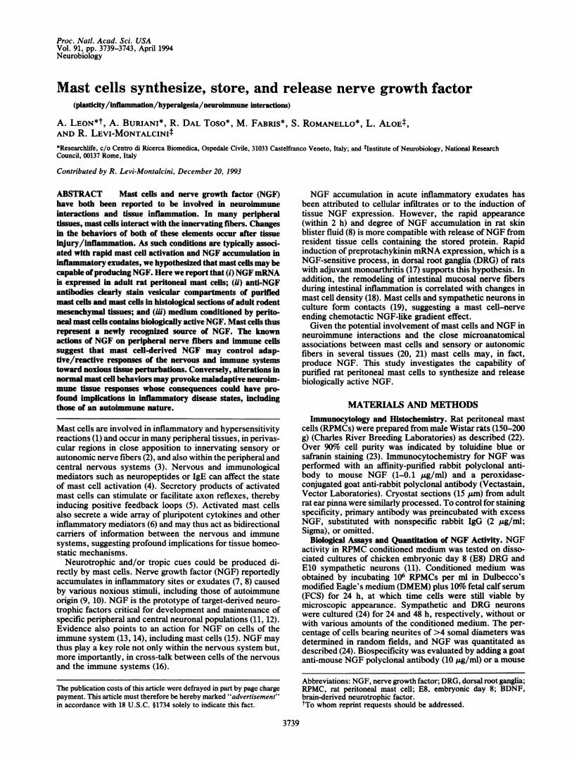
mRNA in RPMCs was examined by PCR, as no NGFhybridizing band was observed by Northern blot analysis.PCR amplification ofmast cell reverse-transcribed total RNA(1 ,ug) with NGF-specific primers yielded a 292-bp band,which hybridized with a NGF-specific internal probe (Fig. 2),indicating the expression of the NGF gene in RPMCs. Mousesubmaxillary gland total RNA served as a positive control(Fig. 2). No hybridizing band was found in PCR-processedmast cell RNA without the reverse transcription step. Al-though only a very minor proportion of non-mast cells waspresent in the purified RPMC preparation, PCR amplificationcould have resulted in a contribution to the NGF signal by thenon-RPMCs. When total RNA (1 ,ug) from the rat mast cellline RBL-2H3 was reverse transcriptase PCR amplified, aNGF hybridizing band was clearly detected (Fig. 2), albeit athigher amplification values. No NGF hybridizing signal wasevident in the PCR amplification products of mouse neuro-blastoma neuro-2a cells (Fig. 2).
Biological Activity of Spontaneously Released NGF fromMast Cells. Chicken embryo DRG and sympathetic neuronsin in vitro culture were used to measure NGF in mediumconditioned for 24 h with RPMCs (Fig. 3). Mast cell viabilitywas assessed by quantifying the histamine content of ran-domly selected conditioned medium using an enzyme immu-noassay kit (Immunotech, Marseilles, France). Mast cellsreleased 1.65 ± 0.2 ,ug of histamine per 106 cells (mean ± SD;n = 4) over the same 24-h period, being equivalent to 9.6%± 1.2% of total histamine content and only slightly aboveroutinely determined levels of basal histamine release (22).
C.)2 2
CO cr:
292 bp _~!:+.4 ,*le...
< -jCM 3mz a:
FIG. 2. NGF oligonucleotide probe hybridization of PCR ampli-fied products from male mouse submaxillary glands (SMG), RPMCs,mouse neuro-2a cells (N2A), and rat basophil leukemia cells (RBL).Autoradiograms of hybridization bands of SMG (positive control),N2A (negative control), and RPMCs were obtained after 23 cycles ofamplification. The RBL cDNA was amplified 35 cycles. No NGFhybridization band was observed in N2A cDNA amplification prod-ucts even after 35 cycles.
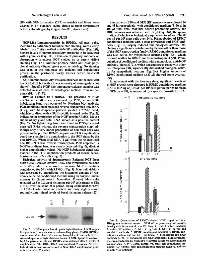
Sympathetic (E10) and DRG (E8) neurons were cultured 24and 48 h, respectively, with conditioned medium (3-50 gl in100-plI final vol). Maximal neurite-promoting activity forDRG neurons was obtained with 12 1ul (Fig. 3B), the quan-titation ofwhich was biologically equivalent to -2 ng ofNGFper ml per 106 mast cells over 24 h. Preincubation of RPMCconditioned medium with a goat polyclonal anti-NGF anti-body (Fig. 3B) largely reduced this biological activity, ex-cluding a significant contribution by factors other than thoseof the NGF neurotrophin family. RPMC conditioned mediumwas also active for sympathetic neurons (Fig. 3A), whichrespond neither to BDNF nor to neurotrophin 4 (29). Prein-cubation ofconditioned medium with a monoclonal anti-NGFantibody (clone 27/21), which does not cross-react with otherneurotrophins (30), significantly diminished biological activ-ity for sympathetic neurons (Fig. 3A). Higher amounts ofRPMC conditioned medium (.25 1.d) showed some cytotox-icity.
In agreement with the bioassay data, significant levels of3-NGF protein were detected in RPMC conditioned medium
(2.56 ± 0.65 ng of (3-NGF per 106 cells per ml per 24 h; mean+ SEM; n = 10), as measured by a specific two-site ELISA.
100 A
Lnw
I-wz
-
-Jw0
SYMPATHETIC NEURONS
80
60
40
20
....
(A P,:,-j
w
I-
2z
I-
-J-j
0
FIG. 3. Quantitation of RPMC-released NGF trophic activity.Histograms represent mean ± SEM of the percentage of neurite-bearing cells (A, n = 6; B, n = 10). Bars: 1, control culture medium;2, anti-NGF antibody; 3, NGF (1 ng/ml); 4, NGF (1 ng/ml) andanti-NGF antibody; 5, RPMC conditioned medium; 6, RPMC con-ditioned medium and anti-NGF antibody. (A) Monoclonal anti-NGFantibody 27/21. (B) Polyclonal anti-NGF antibodies. Statistical anal-ysis was conducted by Student's-Newman-Keuls t test for multiplecomparisons. *, P < 0.001, control vs. mast cell conditioned me-dium; **, P < 0.001, mast cell conditioned medium alone vs. additionof anti-NGF antibody.
Neurobiology: Leon et al.

Proc. Natl. Acad. Sci. USA 91 (1994)
DISCUSSION
The present study shows that RPMCs express the genetranscript for NGF and display a NGF-like immunoreactivitylargely associated with granule-containing compartments.The RBL-2H3 cell line behaved similarly, indicating that cellsof the mastocytic-basophilic lineage are NGF-producingcells. Furthermore, a two-site ELISA detected abundant3-NGF protein levels in RPMC conditioned medium. NGF-
like neurotrophic activity was also observed when the sameconditioned medium was added to cultured avian embryonicsensory and sympathetic neurons, the latter of which respondto neither BDNF nor neurotrophin 4 (29). A monoclonalantibody recognizing NGF but not BDNF or neurotrophin 3(30) effectively blocked mast cell-derived trophic'bioactivity.Together with the occurrence of NGF-like immunopositivemast cells in histological sections of adult rat ear pinna, thesedata show that rodent connective tissue-type mast cellssynthesize, store, and release biologically active NGF. Thepossibility that other mast cell types or basophils have similarproperties, or that mast cells produce neurotrophic factorsother than NGF, remains to be investigated.Numerous cell types synthesize NGF including other he-
matopoietic cells (31). NGF expression and/or productionhas been observed in vivo either during development (32, 33)or during tissue injury (34) and in vitro following stimulation(e.g., with inflammatory agents) (31, 34, 35).- Despite evi-dence of NGF in peripheral adult tissues such as skin, thecellular element(s) contributing to NGF production in normalor pathological conditions remains to be clearly identified (36,37). Our results indicate NGF-like immunoreactivity in mastcells of mesenchymal adult rodent tissues or RPMCs. Al-though some cross-reactivity of the polyclonal antibodytoward other neurotrophins cannot be excluded (38), theexpression of NGF mRNA favors this immunostaining toreflect reactivity with one or more differently processedforms of the NGF protein. In addition, the vesicular local-ization of anti-NGF immunoreactivity indicates that connec-tive tissue-type mast cells may store, in analogy to tumornecrosis factor (39), preformed (3-NGF and/or specializedprecursor storage forms. Independent of the intracellularNGF form(s), these experiments show that RPMCs releaserelatively large amounts of authentic biologically active-3-NGF in the absence of any overt cellular stimulation.Preliminary studies with two-site ELISA indicate that,fNGF also occurs within unstimulated RPMCs (unpub-lished data). Given their defensive role, mast cells mayrepresent a continuous source ofNGF for tissue homeostaticfunctions and/or a prompt NGF supply for their reequilibra-tion after tissue inflammatory insults; other cell types maycontribute to tissue NGF levels at later stages of the inflam-mation process (7, 8). Mast cells could, by releasing cyto-kines such as tumor necrosis factor, also indirectly upregu-late NGF production in surrounding cells (35).
Inflammatory insults to a variety of tissues, includingperipheral nerves, are often accompanied by mast cell acti-vation, hyperalgesia (40), and extracellular NGF accumula-tion (7, 8). Mast cells are frequently found closely apposed tosmall caliber or unmyelinated fibers (20), which may containsubstance P and/or other neuropeptides (21). Small pepti-dergic fibers are generally assumed to be nociceptive sensoryfibers, whose dependence on NGF for survival and differ-entiation is developmentally restricted. However, in the adultthese neurons continue to express high-affinity NGF recep-tors and to respond to NGF with enhanced neuropeptideexpression (12), while anti-NGF antibodies modify theirmorphology (41). Induction of neuropeptide synthesis in
sensory neurons is also found in many models of tissueinflammation. In some instances, neuropeptide inductionoccurs within hours of initiation of inflammation (17, 40) and
is abolished by anti-NGF antibodies (42). The increasedlevels of neuropeptides that accompany tissue inflammationmay, upon primary afferent stimulation, result in their in-creased release from the sensory terminals in the spinal cord(43) and likely underlie centrally mediated prolonged behav-ioral hyperalgesia (40). Interestingly, a single high systemicdose of NGF in adult rats caused long-lasting thermal andmechanical hyperalgesia (44). These data, together with ours,support the notion that activated mast cells may, by releasingNGF, directly control plastic modifications in the centralprocessing of sensory nociceptive inputs after tissue injuryand inflammation. At the same time, NGF-induced neu-ropeptide upregulation may cause increased neuropeptiderelease at peripheral nociceptor terminals, and proteolyticproducts of NGF in inflamed tissues may indirectly modulatetheir excitation (45). Given the potential NGF effects oncollateral sprouting or growth of sensory and sympatheticfibers (11, 36), mast cells, via release of NGF, could controlperipheral short- and long-term modifications in the sensi-tivity and/or amplitude of sensory terminal fields towardpersistent or subsequent, even subthreshold, noxious tissuestimuli. The degree of mast cell -activation may thus contrib-ute significantly to the hyperalgesic and pathological painstates frequently accompanying tissue inflammation, espe-cially in peripheral nerves.Mast cell progenitors migrate from the bloodstream to
those tissues where they undergo terminal phenotypic dif-ferentiation, proposing that these cells represent potentialmobile quanta for replenishment of tissue NGF. NGF itselfcan increase tissue mast cell number in vivo (15, 46) and affectmast cell survival, differentiation, and mediator release invitro (47-50). RPMCs express the functional NGF receptortyrosine kinase (51), indicating possible autocrine and para-crine actions of NGF on mast cells. Given that NGF canaffect not only neurons but also hematopoietic cells, mastcells may, through the release of NGF, convey informationto the nervous system as well as modulate their own behaviorand the reactivity of tissue-infiltrating cells.The capacity ofat least connective tissue-type mast cells to
produce NGF points to their having a pathophysiologicalpotential far beyond that currently recognized. The ability ofmast cells to synthesize and release NGF may represent awell-integrated tissue defense mechanism for maintainingand/or restoring homeostatic functions after noxious pertur-bations (16). On the other hand, mast cell hyerplasia andnerve remodeling are found in some chronic inflammatorystates (18, 52-54). Such tissue modifications may be triggeredand/or sustained by mast cell-derived NGF and lead tolong-lasting changes in tissue reactivity and behavioral re-sponses toward persistent or recurrent inflammatory stimuli.Accordingly, alterations in mast cell properties-e.g., in theentity and duration of their activation-could play a criticalrole in the progression and/or secondary complications ofinflammatory tissue responses. A more complete under-standing of their local stimulatory and inhibitory regulation(55) might open avenues to the management of inflammatorydisease states, including those of autoimmune origin.
The authors thank Dr. S. D. Skaper for useful discussions andcomments, Dr. L. Facci for the histamine measurements, and Ms. P.Lentola for expert secretarial assistance. A special note of thanks tothe late Prof. Angelo Burlina, whose continuous effort and initiativein biomedical research helped to make this work possible.
1. Galli, S. J., Dvorak, A. M. & Dvorak, H. F. (1984) Prog.Allergy 34, 1-141.
2. Bienenstock, J., Tomioka, M., Stead, R. M., Quinonez, G.,Simon, G. T., Coughlin, M. D. & Denburg, J. A. (1987) Int.Arch. Allergy Appl. Immunol. 82, 238-243.
3. Johnson, D. & Krenger, W. (1992) Neurochem. Res. 17,939-951.
3742 Neurobiology: Leon et al.

Proc. Natl. Acad. Sci. USA 91 (1994) 3743
4. Olsson, Y. (1968) Int. Rev. Cytol. 24, 27-70.5. Ratzlaff, R. E., Cavanaugh, V. J., Miller, G. W. & Oakes,
S. G. (1992) J. Neuroimmunol. 41, 89-96.6. Gordon, J. R., Burd, P. R. & Galli, S. J. (1990) Immunol.
Today 11, 458-464.7. Aloe, L., Tuveri, M. A., Carcassi, U. & Levi-Montalcini, R.
(1992) Arthritis Rheum. 35, 351-355.8. Weskamp, G. & Otten, U. (1987) J. Neurochem. 118, 1779-
1786.9. Bracci-Laudiero, L., Aloe, L., Levi-Montalcini, R., Buttinelli,
C., Schilter, D., Gillessen, S. & Otten, U. (1992) Neurosci.Lett. 147, 9-12.
10. Bracci-Laudiero, L., Aloe, L., Levi-Montalcini, R., Galeazzi,M., Schilter, D., Scully, J. L. & Otten, U. (1993) Neuroreport4, 563-565.
11. Levi-Montalcini, R. (1987) Science 237, 1154-1162.12. Lindsay, R. M. & Hamar, A. J. (1989) Nature (London) 337,
362-364.13. Otten, U., Ehrhard, P. & Peck, R. (1989) Proc. Natl. Acad. Sci.
USA 86, 10059-10063.14. Bischoff, S. C. & Dahinden, C. A. (1992) Blood 79, 2662-2669.15. Aloe, L. & Levi-Montalcini, R. (1977) Brain Res. 133, 358-366.16. Levi-Montalcini, R., Aloe, L. & Alleva, E. (1990) Prog. Neu-
rol. Endocrinol. Immunol. 3, 1-10.17. Donaldson, L. F., Harmar, A. J., McQueen, D. S. & Seckl,
J. R. (1992) Mol. Brain Res. 16, 143-149.18. Stead, R. H., Kosecka-Janiszewska, U., Oestreicher, A. B.,
Dixon, M. F. & Bienenstock, J. (1991) J. Neurosci. 11, 3809-3821.
19. Blennerhassett, M. G., Tomioka, M. & Bienenstock, J. (1991)Cell Tissue Res. 265, 121-128.
20. Newson, B., Dahlstrbm, A., Enerbdck, L. & Ahlman, H. (1983)Neuroscience 10, 565-570.
21. Skofitsh, G., Savitt, J. M. & Jacobowitz, D. M. (1985) His-tochemistry 82, 5-8.
22. Mousli, M., Bronner, C., Bueb, J. L., Tschirhart, E., Gies,J. P. & Landry, Y. (1989) J. Pharmacol. Exp. Ther. 250,329-335.
23. Mayrhofer, G. (1980) Histochem. J. 12, 513-526.24. Skaper, S. D., Facci, L., Milani, D., Leon, A. & Toffano, G.
(1990) in Methods and Neurosciences, ed. Conn, P. M. (Aca-demic, New York), Vol. 2, pp. 17-33.
25. Naher-Noe, M., Gnahn, H., Grundler, A., Klingelhnfer, J.,Weindl, A. & Conrad, B. (1993) Eur. J. Clin. Chem. Clin.Biochem. 31, 375-380.
26. Chomczynski, P. & Sacchi, N. (1987) Anal. Biochem. 162,156-159.
27. Whittemore, S. R., Friedman, P. L., Larhammer, D., Persson,H., Gonzales-Carvajal, M. & Holets, V. R. (1988) J. Neurosci.Res. 20, 403-410.
28. Sambrook, J., Fritsch, E. F. & Maniatis, T. (1989) MolecularCloning: A Laboratory Manual (Cold Spring Harbor Lab.Press, Plainview, NY), Vols. 1-3.
29. Korsching, S. (1993) J. Neurosci. 13, 2739-2748.
30. SWdestrom, S., Hallbo6k, F., Ibifiez, C. F., Persson, H. &Ebendal, T. (1990) J. Neurosci. Res. 27, 665-677.
31. Mallat, M., Houlgatte, R., Brachet, P. & Prochiantz, A. (1989)Dev. Biol. 133, 309-311.
32. Davies, A. M., Bandtlow, C., Heumann, R., Korsching, S.,Rohrer, H. & Thoenen, H. (1987) Nature (London) 326, 353-358.
33. Whittemore, S. R., LArkfors, L., Ebendal, T., Holets, V. R.,Ericsson, A. & Persson, H. (1987) J. Neurosci. 7, 244-251.
34. Friedman, W. S., Larkfors, L., Ayer-Le Lievre, C., Ebendal,T., Olson, L. & Persson, H. (1990) J. Neurosci. Res. 27,374-382.
35. Hattori, A., Tanaka, E., Murase, K., Ishida, N., Chatani, Y.,Tsujimoto, M., Hayashi, K. & Kohno, M. (1993) J. Biol. Chem.268, 2577-2582.
36. Diamond, J., Coughlin, M., Macintyre, L., Holmes, M. &Visneau, B. (1987) Proc. NatI. Acad. Sci. USA 84, 6596-6600.
37. Scarisbrick, I. A., Jones, E. G. & Isackson, P. J. (1993) J.Neurosci. 13, 875-893.
38. Murphy, R. A., Acheson, A., Hodges, R., Haskins, J., Rich-ards, C., Reklow, E., Chlumecky, V., Barker, P. A., Alderson,R. F. & Lindsay, R. M. (1993) J. Neurosci. 13, 2853-2862.
39. Young, J. D., Liu, C., Butler, G., Cohn, Z. A. & Galli, S. J.(1987) Proc. Natl. Acad. Sci. USA 84, 9175-9179.
40. Dubner, R. & Ruda, A. (1992) Trends Neurosci. 15, 96-103.41. Gold, B. G., Mobley, W. C. & Matheson, S. F. (1991) J.
Neurosci. 11, 943-955.42. Donnerer, J., Schuligoi, R. & Stein, C. (1992) Neuroscience 49,
693-698.43. Garry, M. C. & Margreaves, K. M. (1992) Brain Res. 582,
139-142.44. Lewin, G. R., Ritter, A. M. & Mendell, L. M. (1993) J. Neu-
rosci. 13, 2136-2148.45. Taiwo, Y. O., Levine, J. D., Burch, R. M., Woo, J. E. &
Mobley, W. C. (1991) Proc. Natl. Acad. Sci. USA 88, 5144-5148.
46. Marshall, J. S., Stead, R. H., McSharry, C., Nielsen, L. &Bienenstock, J. (1990) J. Immunol. 144, 1886-1892.
47. Aloe, L. & De Simone, R. (1989) Int. J. Dev. Neurosci. 7,565-573.
48. Bruni, A., Bigon, E., Boarato, E., Mietto, L., Leon, A. &Toffano, G. (1982) FEBS Lett. 138, 140-193.
49. Tomioka, M., Stead, R. H., Nielsen, L., Coughlin, M. D. &Bienenstock, J. (1988) J. Clin. Immunol. 82, 599-607.
50. Matsuda, H., Kannah, Y., Ushio, H., Kiso, Y., Kenemoto, T.,Suzuki, H. & Kitamura, Y. (1991) J. Exp. Med. 174, 7-14.
51. Horigome, K., Pryor, J. C., Bullock, E. D. & Johnson, E. M.(1993) J. Biol. Chem. 268, 14881-14887.
52. Nennesmo, I. & Reinholt, F. (1986) Neurosci. Lett. 69, 2%-301.
53. Dvorak, A. M. & Silen, W. (1985) Ann. Surg. 201, 53-63.54. Naukkarinen, A., Harvima, I., Paukkonen, K., Aalto, M.-L. &
Horsmanheimo, M. (1993) Arch. Dermatol. Res. 285, 341-346.55. Aloe, L., Leon, A. & Levi-Montalcini, R. (1993) Agents
Actions 39, C145-C147.
Neurobiology: Leon et al.