magnetic 3-d ordered macroporous silica templated from ...magnetic 3-d ordered macroporous silica...
TRANSCRIPT

Microporous and Mesoporous Materials 130 (2010) 26–31
Contents lists available at ScienceDirect
Microporous and Mesoporous Materials
journal homepage: www.elsevier .com/locate /micromeso
Magnetic 3-D ordered macroporous silica templated from binary colloidalcrystals and its application for effective removal of microcystin
Jia Liu, Yue Cai, Yonghui Deng *, Zhenkun Sun, Dong Gu, Bo Tu, Dongyuan Zhao *
Department of Chemistry, Shanghai Key Laboratory of Molecular Catalysis and Innovative Materials, Advanced Materials Laboratory, Fudan University, Shanghai 200433, PR China
a r t i c l e i n f o a b s t r a c t
Article history:Received 17 August 2009Received in revised form 30 September 2009Accepted 9 October 2009Available online 8 November 2009
Keywords:Macroporous materialsColloidal crystalMagnetic propertyMicrocystin
1387-1811/$ - see front matter � 2009 Elsevier Inc. Adoi:10.1016/j.micromeso.2009.10.008
* Corresponding authors. Tel.: +86 21 51630205; faE-mail addresses: [email protected] (Y. Den
Zhao).
Magnetic 3-D ordered macroporous siliceous materials were conveniently fabricated by combination of asimple co-sedimentation of binary colloidal system of PMMA spheres and magnetite nanoparticles andthe infiltration of silica precursors. The SEM and TEM observations indicate that the obtained magnetic3-D ordered macroporous silica materials have face-centered cubic (fcc) packing structure with orderedmacropore of �200 nm and interconnected windows of about 50 nm. Magnetic property characterizationshows that the magnetic 3-D ordered macroporous silica materials possess high magnetization(19.2 emu/g) and superparamagentism. By utilization of their ordered macroporous structure with largeentrances and good affinity for biomacromolecules as well as high magnetization, a fast and efficientremoval of microcystin in water with high removal efficiency (>93%) and removal capacity (3.3 ng MC/mg) was achieved with the help of an applied magnetic field.
� 2009 Elsevier Inc. All rights reserved.
1. Introduction
Ordered porous materials with tailorable pore size and func-tionalities have recently attracted great attention for their wide po-tential applications in photonic crystals, bio-separation, catalysis,drug delivery and electrochemical cells [1–3]. Particularly, orderedmacroporous materials, due to periodically large nanostructure,tunable pore size, large entrance and easily functionalizable sur-face show great promise in various applications involving largebiomolecules [4–7]. Many methods have been reported to fabricateordered macroporous materials, including soft lithograph, emul-sion templating synthesis and so on [8,9]. One of the most commonapproaches is the hard-templating process with colloidal crystalsof monodisperse spheres as the sacrificial templates [10–12]. Theguest molecules can be introduced into the interstitial voids ofthe colloidal crystals through different strategies, such as solutionor melt infiltration, sol–gel chemistry, in situ polymerization, elec-trodeposition and chemical vapor deposition (CVD) [12,13]. Thesubsequent removal of the templates through thermal or chemicaletching leads to ordered macroporous materials. In the past dec-ades, ordered macroporous materials with various compositionshave been fabricated, including ceramics oxides, metals, polymers,semiconductors [14–18].
Magnetic nanomaterials, due to their unique magnetic behav-ior, have recently attracted increasing attention for their potential
ll rights reserved.
x: +86 21 65641740.g), [email protected] (D.
applications in biomedicine, separation and enrichment, magneticnanodevice [19–21]. Functionalization of the ordered macroporousmaterials with magnetic property is of great interest to their appli-cations, because the additional magnetic property can greatly facil-itate the manipulation of macroporous materials. By usingcolloidal crystal templates, Eagleton and Searson synthesized mag-netic ordered macroporous nickels by an electrodeposition [22].Stein and co-workers synthesized magnetic ordered macroporousnickels, cobalts and their oxides [23]. Galloro et al. synthesized or-dered macroporous maghemites (c�Fe2O3) through an infiltratingcolloidal crystal with polyferrocenylsilane followed by calcinationto remove the template [24]. The above mentioned magnetic mac-roporous materials are difficult to further modify by functionalgroups, which may limit their applications. Usually, for bio-relatedapplications, the magnetic ordered macroporous materials shouldhave easily functionalizable surface, high magnetic responsivenessand good bio-affinity, however, to our best knowledge, most of thereported magnetic ordered macroporous materials fail to meetthese requirements.
Herein, we report a synthesis of magnetic 3-D ordered macro-porous siliceous materials templated from binary colloidal crystalsby combination of a simple co-sedimentation of binary colloidalsystem of PMMA spheres and magnetite nanoparticles and theinfiltration of silica precursors. The obtained magnetic 3-D orderedmacroporous silica materials possess face-centered cubic (fcc)packing structure with ordered macropore of �200 nm, intercon-nected windows of about 50 nm, high magnetization (19.2 emu/g) and superparamagentism. Due to their ordered macroporousstructure with large entrances and good affinity for biomacromol-

J. Liu et al. / Microporous and Mesoporous Materials 130 (2010) 26–31 27
ecules as well as high magnetization, they show a fast and efficientremoval of microcystin in water with high removal efficiency(>93%) and removal capacity (3.3 ng MC/mg) with the help of anapplied magnetic field.
2. Experimental
2.1. Chemicals
Monodisperse PMMA microspheres were prepared via a surfac-tant-free emulsion polymerization process by using potassiumpersulfate as the initiator according to previous report [25]. Themicrospheres were negatively charged (with SO2�
4 groups on thesurface) which were dispersed into the deionized water producingconcentration of 9.1 wt.%. Tetraethyl orthosilicate (TEOS), ethanol,aqueous ammonia, ferrous chloride, trisodium citrate and sodiumnitrite were analytical reagents and purchased from ShanghaiChemical Company. All chemicals were used as received withoutfurther purification. Deionized water was used in all experiments.For microcystin removal experiments, a-cyano-4-hydroxycin-namic acid (CHCA), acetonitrile (ACN) and trifluoroacetic acid(TFA) were purchased from Merck (Darmstadt, Germany) andaqueous solutions were prepared using Milli-Q water by Milli-Qsystem (Millipore, Bedford, MA). Microcystin-LR (purity P 96%)was purchased from Alexis Corporation (San Diego, USA).
2.2. Synthesis of magnetite nanoparticles
Magnetite nanoparticles were synthesized by redox reaction[26] between nitrite and ferrous (Fe2+) salts in a basic aqueoussolution at 25 �C. Typically, 5.8 mmol of sodium nitrite was addedto an aqueous solution containing 13 mmol FeCl2�4H2O, 20 mL ofHCl (2.0 M) and 800 mL of water. After stirring for 5 min, 80 mLof concentrated ammonia aqueous solution (28 wt.%) was addedto the solution, resulting in a black dispersion. After vigorousstirring for 0.5 h, the magnetite nanoparticles were separated byusing a magnet (1000 Oe) and washed with deionized water. Toenhance the colloidal dispersibility, the magnetite nanoparticleswere treated in 50 mL of nitric acid solution (2.0 M) for 5 minand then separated from the dispersion for redispersion in 100 mLof trisodium citrate aqueous solution (0.5 M) with ultrasonictreatment. After vibration for 10 min, the magnetite nanoparticleswere separated and redispersed in deionized water to obtain stablemagnetic fluid with solid content of 2.0 wt.%.
Scheme 1. The synthesis procedure for the magnetic 3-D ordered macroporoussilica materials.
2.3. Synthesis of macroporous silica–magnetite composites
The magnetic fluid (20 g, 2 wt.%) and aqueous dispersion ofPMMA colloids with the size of 250 nm (5.0 g, 5 wt.%) were mixedin an glass under ultrasonication. Then, the obtained solution wasplaced in an oven at 70 �C for fast solvent evaporation for 5–8 h,and the gray PMMA-magnetite binary colloidal crystals were ob-tained. After further sintering treatment at 90 �C for 24 h, the col-loidal crystals were soaked in a silica precursor ethanol solution for3 h at 25 �C, herein, the silica precursor was pre-hydrolyzed bymixing 0.9 g of TEOS, 2.0 mL of H2O, 10 mL of ethanol and 0.1 mLof HCl (2.0 M). Afterward, the silica impregnated colloidal crystalswere withdrawn from the solution and allowed to exposure to airfor evaporation of ethanol and hydrolysis of the silica precursor.The above impregnation procedure was repeated for 3 times to en-sure the fully filling of all the interstitial voids in the binary colloidcrystals by the silica species. Finally, the composites were pyro-lyzed in a tube furnace at 400 �C for 5 h in N2 with a heating rateof 2 �C min�1.
2.4. Removal of microcystins (MCs)
A certain volume of aqueous suspension of ordered macropo-rous magnetic-silica composites (100 mg L�1) was added into acomparison tube, and the water was decanted with the help of amagnet. Then the magnetic material was redispersed with 200 lLof MC-LR (10 lg/L). The solution containing the magnetic-silicacomposites and MC-LR was shaken at 25 �C for 10 min. Subse-quently, the magnetic composites with absorbed MC-LR were sep-arated from the solution with a magnet, and 1.0 lL of thesupernatant liquor was deposited on the plate, and then 5.0 lL ofa mixture of 0.5 mg mL�1 CHCA (in 50% (v/v) acetonitrile and0.1% (v/v) TFA aqueous solution) was introduced. After dried, thesamples were applied for matrix-assisted laser desorption/ioniza-tion time-of-flight mass spectrometry (MALDI-TOF MS) analysis.
2.5. Characterizations and measurements
Scanning electron microscopy (SEM) images were recorded on aPhilips XL30 electron microscope (Netherlands) operating at 20 kV.A thin gold film was sputtered on the sample before the character-ization. Transmission electron microscopy (TEM) images were ta-ken with a JEOL 2011 microscope (Japan) operating at 200 kV.For the TEM measurements, the samples were dispersed in ethanoland then dried on a holey carbon film Cu grid. The wide-angleX-ray diffraction (XRD) patterns were performed on a GermanBruker D4 X-ray diffractometer (Germany) with Ni-filtered Cu Karadiation (40 kV, 40 mA). Fourier-transform infrared (FT-IR) spectrawere collected on a Nicolet Fourier spectrophotometer (USA) usingKBr pellets. Magnetic characterization was carried out on a super-conducting quantum interference device (SQUID) magnetometry.MALDI-TOF MS experiments were performed in positive ion modeon a 4700 Proteomics Analyzer (Applied Biosystems, USA) withthe Nd-YAG laser at 355 nm, a repetition rate of 200 Hz and anacceleration voltage of 20 kV. Analysis of MC was performed inthe reflector TOF detection mode.
3. Results and discussion
The fabrication of magnetic 3-D macroporous silica materials isillustrated in Scheme l. Firstly, a homogeneous and stable aqueousdispersion containing monodisperse negatively charged poly(methylmethacrylate) (PMMA) colloids and Fe3O4 nanoparticles stabi-lized with citrate groups was subject to heating at 70 �C for thebinary assembly of PMMA and Fe3O4 colloids. Secondly, the result-ing composite crystals were heated at 90 �C and then impregnatedwith a pre-hydrolyzed tetraethyl orthosilicate (TEOS) ethanol solu-

Fig. 2. TEM of magnetite nanoparticles prepared at room temperature of 25 �C. Theinset (a) shows the photograph of the aqueous dispersion of the magnetitenanoparticles. The inset (b) is the high-resolution TEM image of a single magnetitenanoparticle.
28 J. Liu et al. / Microporous and Mesoporous Materials 130 (2010) 26–31
tion followed with drying in air to completely fill the voids in thebinary colloid crystals with silica species. Finally, calcination inN2 to remove the PMMA template leads to magnetic ordered mac-roporous silica.
The monodisperse PMMA colloids were synthesized throughthe soap-free emulsion polymerization method. The scanning elec-tron microscopy (SEM) image shows that the PMMA colloids havea uniform size of 250 nm with a deviation <5% (Fig. 1a). The mag-netite nanoparticles stabilized with citrate groups were preparedby a simple redox method (Fig. 2). Due to the stabilization effectof citrate groups, the obtained magnetite nanoparticles can be welldispersed in aqueous solution (Fig. 2, inset). TEM observation indi-cates they have small particle size of about 6–10 nm (Fig. 2). Afterthe co-sedimentation and impregnation with silica oligomer spe-cies, the obtained composite colloidal crystals (Fig. 1b) possess typ-ical face-centered cubic (fcc) packing structure with (1 1 1) planeparallel to the substrate, and the interstitial voids were found tobe fully filled (Fig. 1b, inset). It indicates that the silica is success-fully introduced into the colloidal crystals and solidified the or-dered macrostructure. After calcination in N2, highly ordered 3-Dmacroporous structure was obtained (Fig. 1c). The macropore sizemeasured by SEM images is about 200 nm, less than the diameter(250 nm) of the PMMA colloid templates, suggesting that a consid-erable structure shrinkage occurs during the calcination process.The windows of the macropores are clearly visible and the diame-ter is calculated to be about 50 nm (Fig. 1d). The large window isoriginated from the large contact surface between the neighboringPMMA colloids formed by slightly melting the colloids during heat-ing at near their glass transition temperature (Tg) of 90 �C prior tothe silica impregnation [27–29]. The large window size is benefi-cial to the fast mass transportation for applications involving mac-romoelcules. Additionally, as shown in the SEM image (Fig. 1d), theobtained macroporous material has a rough framework, implyingthat the Fe3O4 nanoparticles are incorporated in the silica matrix.
The removal of PMMA template was verified by fourier-trans-form infrared (FT-IR) spectra. The absorption bands of theFe3O4ASiO2APMMA composites at 2800–3000 and 1730 cm�1 are
Fig. 1. SEM images of (a) PMMA colloids, (b) silica impregnated PMMAAF
attributed to ACH2 and AC@O of PMMA, respectively (Curve b,Fig. 3). The peaks at 1090 and 560 cm�1 are from the SiAOASi ofsilica and FeAO of Fe3O4 particles, respectively. After calcination,only absorption peaks for Fe3O4 and silica are observed in the mag-netic macroporous silica (Curve b, Fig. 3), suggesting that PMMAtemplates can be fully decomposed in N2 at 400 �C [30].
The transmission electron microscopy (TEM) image (Fig. 4a) ofthe obtained magnetite-silica composites shows highly ordered
e3O4 colloidal crystals, and (c, d) magnetic 3-D ordered porous silica.

3500 3000 2500 2000 1500 1000 500
b
% T
rans
mitt
ance
Wavenumber (nm)
a
C=OC-H
Si-O-Si Fe-O
Fig. 3. FT-IR spectra of (a) as-made magnetite-silica-PMMA composite colloidalcrystals and (b) 3-D ordered macroporous magnetic-silica composites aftercalcination in N2 at 400 �C for 5 h.
J. Liu et al. / Microporous and Mesoporous Materials 130 (2010) 26–31 29
macropore arrays viewed along the [2 1 1] direction of an fcc lat-tice, further confirming the replication of ordered macrostructureof the binary colloidal crystals. The TEM image with large magnifi-cation (Fig. 4b) clearly shows that the magnetite nanoparticleswith diameter of 6–10 nm are homogeneously distributed in thesilica matrix around the macropores. The silica component of theframeworks is very important. Firstly, it serves as ‘‘glue” for bind-
Fig. 4. TEM (a and b) and HRTEM (c) images of magnetic macroporous silica materials,location of magnetic nanoparticles.
ing the magnetite nanoparticles and supporting the 3-D orderedmacrostructure during the impregnation and calcination process.Secondly, it acts as protective coating for the nanoparticles to pre-vent them from being etched in practical applications. Thirdly, itprovides silica-like surface for binding the objects or for furthersurface functionalization with organic groups. The HRTEM imagesclearly reveal that the magnetite nanoparticles are well crystal-lized (Fig. 4c). Selected area electron diffraction (SAED) patterns(Fig. 4d) show spotty diffraction cycles assigned to magnetite, indi-cating a polycrystalline feature.
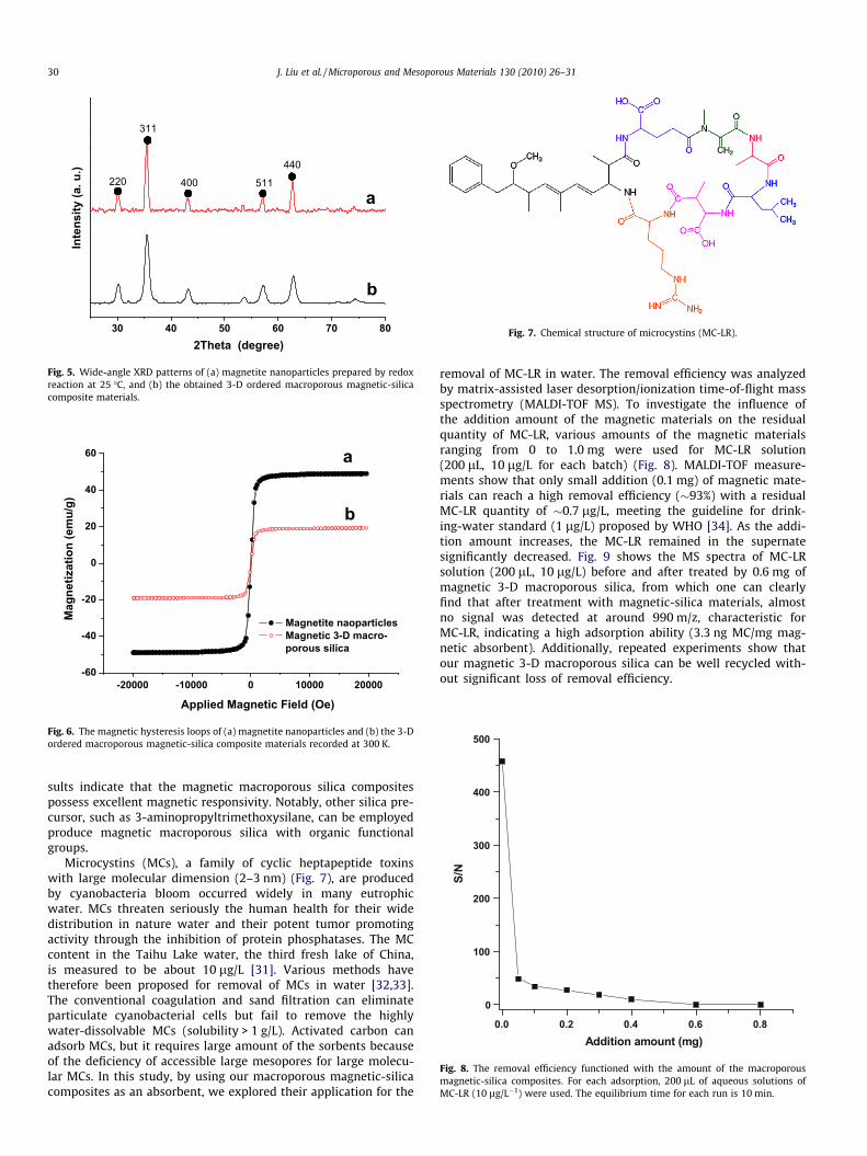
X-ray diffraction (XRD) patterns (Fig. 5) of the ordered macro-porous magnetic silicas show characteristic diffraction peaks ofmagnetite, suggesting that the Fe3O4 nanoparticles are well re-tained in the silica matrix. Compared to pure magnetite nanoparti-cles (Curve a in Fig. 5), the diffraction peaks of magnetic 3-Dordered macroporous silica are somewhat wider, which is probablycaused by the coating of silica (Curve b in Fig. 5). Magnetic charac-terization with a superconducting quantum interference device(SQUID) magnetometer at 300 K indicates that the magnetitenanoparticles and the 3-D macroporous silica composites havemagnetization saturation values of 49.0 and 19.2 emu/g, respec-tively (Fig. 6), indicating a high magnetization. Accordingly, themagnetite content in the macroporous composites is calculatedto be as high as 39 wt.%, indicating a high magnetization. Addition-ally, no remanence is detected for both of them, indicating a super-paramagnetism feature due to the nanosized Fe3O4. As a result ofgood magnetic property, the magnetic 3-D ordered macroporoussilica composites suspended in water (0.05 g/mL) can be quicklyseparated from its dispersion with a magnet (1000 Oe). These re-
and SAED pattern of the macroporous frameworks. The white arrows indicate the
30 40 50 60 70 80
b
a
Inte
nsity
(a. u
.)
2Theta (degree)
220
311
400 511
440
Fig. 5. Wide-angle XRD patterns of (a) magnetite nanoparticles prepared by redoxreaction at 25 �C, and (b) the obtained 3-D ordered macroporous magnetic-silicacomposite materials.
-20000 -10000 0 10000 20000-60
-40
-20
0
20
40
60
b
Mag
netiz
atio
n (e
mu/
g)
Applied Magnetic Field (Oe)
Magnetite naoparticles Magnetic 3-D macro-
porous silica
a
Fig. 6. The magnetic hysteresis loops of (a) magnetite nanoparticles and (b) the 3-Dordered macroporous magnetic-silica composite materials recorded at 300 K.
Fig. 7. Chemical structure of microcystins (MC-LR).
0.0 0.2 0.4 0.6 0.8
0
100
200
300
400
500
S/N
Addition amount (mg)
Fig. 8. The removal efficiency functioned with the amount of the macroporousmagnetic-silica composites. For each adsorption, 200 lL of aqueous solutions ofMC-LR (10 lg/L�1) were used. The equilibrium time for each run is 10 min.
30 J. Liu et al. / Microporous and Mesoporous Materials 130 (2010) 26–31
sults indicate that the magnetic macroporous silica compositespossess excellent magnetic responsivity. Notably, other silica pre-cursor, such as 3-aminopropyltrimethoxysilane, can be employedproduce magnetic macroporous silica with organic functionalgroups.
Microcystins (MCs), a family of cyclic heptapeptide toxinswith large molecular dimension (2–3 nm) (Fig. 7), are producedby cyanobacteria bloom occurred widely in many eutrophicwater. MCs threaten seriously the human health for their widedistribution in nature water and their potent tumor promotingactivity through the inhibition of protein phosphatases. The MCcontent in the Taihu Lake water, the third fresh lake of China,is measured to be about 10 lg/L [31]. Various methods havetherefore been proposed for removal of MCs in water [32,33].The conventional coagulation and sand filtration can eliminateparticulate cyanobacterial cells but fail to remove the highlywater-dissolvable MCs (solubility > 1 g/L). Activated carbon canadsorb MCs, but it requires large amount of the sorbents becauseof the deficiency of accessible large mesopores for large molecu-lar MCs. In this study, by using our macroporous magnetic-silicacomposites as an absorbent, we explored their application for the
removal of MC-LR in water. The removal efficiency was analyzedby matrix-assisted laser desorption/ionization time-of-flight massspectrometry (MALDI-TOF MS). To investigate the influence ofthe addition amount of the magnetic materials on the residualquantity of MC-LR, various amounts of the magnetic materialsranging from 0 to 1.0 mg were used for MC-LR solution(200 lL, 10 lg/L for each batch) (Fig. 8). MALDI-TOF measure-ments show that only small addition (0.1 mg) of magnetic mate-rials can reach a high removal efficiency (�93%) with a residualMC-LR quantity of �0.7 lg/L, meeting the guideline for drink-ing-water standard (1 lg/L) proposed by WHO [34]. As the addi-tion amount increases, the MC-LR remained in the supernatesignificantly decreased. Fig. 9 shows the MS spectra of MC-LRsolution (200 lL, 10 lg/L) before and after treated by 0.6 mg ofmagnetic 3-D macroporous silica, from which one can clearlyfind that after treatment with magnetic-silica materials, almostno signal was detected at around 990 m/z, characteristic forMC-LR, indicating a high adsorption ability (3.3 ng MC/mg mag-netic absorbent). Additionally, repeated experiments show thatour magnetic 3-D macroporous silica can be well recycled with-out significant loss of removal efficiency.
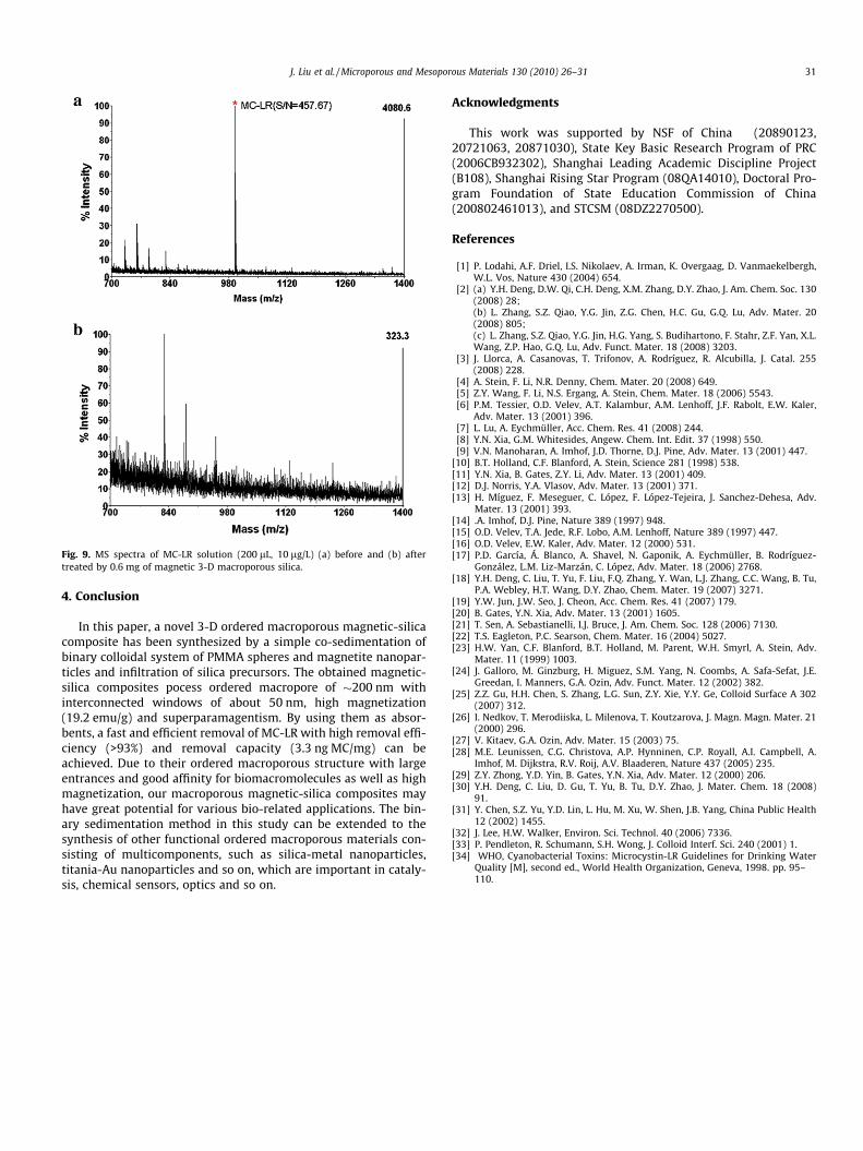
Fig. 9. MS spectra of MC-LR solution (200 lL, 10 lg/L) (a) before and (b) aftertreated by 0.6 mg of magnetic 3-D macroporous silica.
J. Liu et al. / Microporous and Mesoporous Materials 130 (2010) 26–31 31
4. Conclusion
In this paper, a novel 3-D ordered macroporous magnetic-silicacomposite has been synthesized by a simple co-sedimentation ofbinary colloidal system of PMMA spheres and magnetite nanopar-ticles and infiltration of silica precursors. The obtained magnetic-silica composites pocess ordered macropore of �200 nm withinterconnected windows of about 50 nm, high magnetization(19.2 emu/g) and superparamagentism. By using them as absor-bents, a fast and efficient removal of MC-LR with high removal effi-ciency (>93%) and removal capacity (3.3 ng MC/mg) can beachieved. Due to their ordered macroporous structure with largeentrances and good affinity for biomacromolecules as well as highmagnetization, our macroporous magnetic-silica composites mayhave great potential for various bio-related applications. The bin-ary sedimentation method in this study can be extended to thesynthesis of other functional ordered macroporous materials con-sisting of multicomponents, such as silica-metal nanoparticles,titania-Au nanoparticles and so on, which are important in cataly-sis, chemical sensors, optics and so on.
Acknowledgments
This work was supported by NSF of China (20890123,20721063, 20871030), State Key Basic Research Program of PRC(2006CB932302), Shanghai Leading Academic Discipline Project(B108), Shanghai Rising Star Program (08QA14010), Doctoral Pro-gram Foundation of State Education Commission of China(200802461013), and STCSM (08DZ2270500).
References
[1] P. Lodahi, A.F. Driel, I.S. Nikolaev, A. Irman, K. Overgaag, D. Vanmaekelbergh,W.L. Vos, Nature 430 (2004) 654.
[2] (a) Y.H. Deng, D.W. Qi, C.H. Deng, X.M. Zhang, D.Y. Zhao, J. Am. Chem. Soc. 130(2008) 28;(b) L. Zhang, S.Z. Qiao, Y.G. Jin, Z.G. Chen, H.C. Gu, G.Q. Lu, Adv. Mater. 20(2008) 805;(c) L. Zhang, S.Z. Qiao, Y.G. Jin, H.G. Yang, S. Budihartono, F. Stahr, Z.F. Yan, X.L.Wang, Z.P. Hao, G.Q. Lu, Adv. Funct. Mater. 18 (2008) 3203.
[3] J. Llorca, A. Casanovas, T. Trifonov, A. Rodríguez, R. Alcubilla, J. Catal. 255(2008) 228.
[4] A. Stein, F. Li, N.R. Denny, Chem. Mater. 20 (2008) 649.[5] Z.Y. Wang, F. Li, N.S. Ergang, A. Stein, Chem. Mater. 18 (2006) 5543.[6] P.M. Tessier, O.D. Velev, A.T. Kalambur, A.M. Lenhoff, J.F. Rabolt, E.W. Kaler,
Adv. Mater. 13 (2001) 396.[7] L. Lu, A. Eychmüller, Acc. Chem. Res. 41 (2008) 244.[8] Y.N. Xia, G.M. Whitesides, Angew. Chem. Int. Edit. 37 (1998) 550.[9] V.N. Manoharan, A. Imhof, J.D. Thorne, D.J. Pine, Adv. Mater. 13 (2001) 447.
[10] B.T. Holland, C.F. Blanford, A. Stein, Science 281 (1998) 538.[11] Y.N. Xia, B. Gates, Z.Y. Li, Adv. Mater. 13 (2001) 409.[12] D.J. Norris, Y.A. Vlasov, Adv. Mater. 13 (2001) 371.[13] H. Míguez, F. Meseguer, C. López, F. López-Tejeira, J. Sanchez-Dehesa, Adv.
Mater. 13 (2001) 393.[14] .A. Imhof, D.J. Pine, Nature 389 (1997) 948.[15] O.D. Velev, T.A. Jede, R.F. Lobo, A.M. Lenhoff, Nature 389 (1997) 447.[16] O.D. Velev, E.W. Kaler, Adv. Mater. 12 (2000) 531.[17] P.D. García, Á. Blanco, A. Shavel, N. Gaponik, A. Eychmüller, B. Rodríguez-
González, L.M. Liz-Marzán, C. López, Adv. Mater. 18 (2006) 2768.[18] Y.H. Deng, C. Liu, T. Yu, F. Liu, F.Q. Zhang, Y. Wan, L.J. Zhang, C.C. Wang, B. Tu,
P.A. Webley, H.T. Wang, D.Y. Zhao, Chem. Mater. 19 (2007) 3271.[19] Y.W. Jun, J.W. Seo, J. Cheon, Acc. Chem. Res. 41 (2007) 179.[20] B. Gates, Y.N. Xia, Adv. Mater. 13 (2001) 1605.[21] T. Sen, A. Sebastianelli, I.J. Bruce, J. Am. Chem. Soc. 128 (2006) 7130.[22] T.S. Eagleton, P.C. Searson, Chem. Mater. 16 (2004) 5027.[23] H.W. Yan, C.F. Blanford, B.T. Holland, M. Parent, W.H. Smyrl, A. Stein, Adv.
Mater. 11 (1999) 1003.[24] J. Galloro, M. Ginzburg, H. Miguez, S.M. Yang, N. Coombs, A. Safa-Sefat, J.E.
Greedan, I. Manners, G.A. Ozin, Adv. Funct. Mater. 12 (2002) 382.[25] Z.Z. Gu, H.H. Chen, S. Zhang, L.G. Sun, Z.Y. Xie, Y.Y. Ge, Colloid Surface A 302
(2007) 312.[26] I. Nedkov, T. Merodiiska, L. Milenova, T. Koutzarova, J. Magn. Magn. Mater. 21
(2000) 296.[27] V. Kitaev, G.A. Ozin, Adv. Mater. 15 (2003) 75.[28] M.E. Leunissen, C.G. Christova, A.P. Hynninen, C.P. Royall, A.I. Campbell, A.
Imhof, M. Dijkstra, R.V. Roij, A.V. Blaaderen, Nature 437 (2005) 235.[29] Z.Y. Zhong, Y.D. Yin, B. Gates, Y.N. Xia, Adv. Mater. 12 (2000) 206.[30] Y.H. Deng, C. Liu, D. Gu, T. Yu, B. Tu, D.Y. Zhao, J. Mater. Chem. 18 (2008)
91.[31] Y. Chen, S.Z. Yu, Y.D. Lin, L. Hu, M. Xu, W. Shen, J.B. Yang, China Public Health
12 (2002) 1455.[32] J. Lee, H.W. Walker, Environ. Sci. Technol. 40 (2006) 7336.[33] P. Pendleton, R. Schumann, S.H. Wong, J. Colloid Interf. Sci. 240 (2001) 1.[34] WHO, Cyanobacterial Toxins: Microcystin-LR Guidelines for Drinking Water
Quality [M], second ed., World Health Organization, Geneva, 1998. pp. 95–110.