local delivery of tgf beta2 can substitute for placode …cmchuong/1996tgfb_skin.pdf · tgf b2...
TRANSCRIPT
DEVELOPMENTAL BIOLOGY 179, 347–359 (1996)ARTICLE NO. 0266
Local Delivery of TGF b2 Can Substitutefor Placode Epithelium to InduceMesenchymal Condensation duringSkin Appendage Morphogenesis
Sheree A. Ting-Berreth and Cheng-Ming Chuong1
Department of Pathology, University of Southern California, HMR 204,2011 Zonal Avenue, Los Angeles, California 90033
Development of skin appendages requires interactions between the epithelium and mesenchyme. Without the epithelium,dermal condensations cannot develop, and those already formed will disintegrate. Here we explored the molecular basisof this epithelial requirement and tried to identify the molecule(s) responsible by using the chick feather bud developmentas a model. TGF b2 is a likely candidate because its message is predominantly expressed in the feather bud epithelium,and the protein is enriched in the dermal–epidermal junction within the bud. We tested this hypothesis by placing TGFb-soaked beads on skin explants. We found that TGF b2, but not TGF b1, beads placed on top of epithelially strippedmesenchymes can induce dermal condensations. NCAM and tenascin-C (Tn-C) are expressed and protein kinase C issuppressed in the normal feather bud domain. This molecular organization is lost in denuded mesenchyme but can berestored by TGF b2-coated beads. Subsequently, the TGF b2-induced dermal condensations can induce nascent epitheliumto form skin appendages. Together with our recent findings that ectopic Sonic hedgehog (Shh) expression causes widerTGF b expression and larger dermal condensation, these results strongly suggest that TGF b2 produced by epithelialplacode is downstream to Shh and plays a key role in the induction of dermal condensation by activating the expressionof NCAM and Tn-C, and by suppressing PKC expression. q 1996 Academic Press, Inc.
INTRODUCTION Vaino et al., 1993). It was further suggested that E-M inter-actions occur through a series of dialogues between the epi-thelium and mesenchyme using different growth factorsSecondary inductions are important for the orderly ar-(Davidson, 1993).rangement of functional tissues during organogenesis.
We have used the development of chicken feather as aMany of these processes, such as the induction of limb,model to explore the molecular basis of E-M interactions dur-tooth, kidney, and skin appendage (i.e., hair, feather, etc.)ing induction and morphogenesis (Chuong, 1993). Studies us-formation, require specific and complex epithelial –mesen-ing E-M recombination and transplantation of chick embry-chymal (E-M) interactions throughout their developmentonic skins have demonstrated that interactions between the(reviewed in Sawyer and Fallon, 1983; Sengel, 1976). For theepithelium and the underlying mesenchyme are essential dur-most part, the genes and molecules underlying the basis ofing skin appendage formation. Although the phenotypic deter-secondary inductions remain elusive. However, research onminants specifying the type of appendage (i.e., feather or scale)primary induction in early Xenopus embryogenesis (re-reside in the mesenchyme, that alone is not enough for suc-viewed in Jessell and Melton, 1992; Green and Smith, 1991)cessful development without proper reciprocal responses fromand secondary inductions of limb and tooth developmentthe epithelium (Rawles, 1963). Removal of the epithelium atrevealed the importance of growth factors in these processesEmbryonic Day 7 will suppress the process of feather germ(reviewed in Tabin, 1991; Nieswander and Martin, 1993;formation, and feather germs already formed will disintegrateinto a flat sheet of fibroblasts without any feather-specificmolecular and cellular organization. Furthermore, reciprocal1 To whom correspondence should be addressed.
347
0012-1606/96 $18.00Copyright q 1996 by Academic Press, Inc.All rights of reproduction in any form reserved.
AID DB 8371 / 6x14$$$$41 10-11-96 15:53:07 dbas AP: Dev Bio
348 Ting-Berreth and Chuong
E-M recombination using the scaleless mutant and normal MATERIALS AND METHODSskin have demonstrated that the defect of the scalelesschicken resides in the epithelium (McAleese and Sawyer, Chicken embryos. Fertilized White Leghorn chicken eggs were1981; Sawyer, 1983). These results demonstrate that even obtained from K & R Farms (Westminster, CA) and incubated atthough the mesenchyme has the capacity to form dermal con- 387C (Humidare) under humid conditions. Embryos for skin ex-
plants and immunostaining were staged according to Hamburgerdensations, the epithelium is essential for its initiation andand Hamilton (1951).maintenance (reviewed in Cairns and Saunders, 1954; Rawles,
Skin explant cultures and epithelial–mesenchymal recombina-1963; Sengel, 1976).tion. The dorsal skins of White Leghorn chicken embryos atWhat are the epithelial factors required to organize der-stages 31–32 (Embryonic Day 7) were dissected in Hank’s bufferedmal condensations in the mesenchyme? Previously, we re-saline solution (HBSS, Gibco/BRL). When E-M separation was re-ported the involvement of adhesion molecules, such as ten-quired, the skin was incubated in 21 calcium-magnesium-free sa-
ascin (Tn) in mediating the process of dermal condensations line (274 mM NaCl, 8 mM KCl, 0.8 mM NaH2PO4, 0.4 mM(Jiang and Chuong, 1992). In addition, there are differences KH2PO4 , 24 mM NaHCO3, 20 mM glucose, pH 7.3) containingin the distribution and function of intracellular signaling 0.25% EDTA (weight/volume) on ice for 5–10 min. The epitheliummolecules as feather germs develop. We found that protein was then dissected and dissociated using watchmaker tweezers. E-
M recombinant, mesenchyme, or skin explants were transferred tokinase A (PKA) is enriched in the feather germ mesenchymeculture inserts in six-well culture dishes (Falcon) and cultured inand PKC is enriched in the interfeather germ mesenchyme.DMEM media (Gibco/BRL) with 10% fetal calf serum (Irvine Scien-PKA agonists favor feather bud formation while PKC ago-tific). Media were placed both in the outside well and the innernists favor the expansion of interbud domain (Noveen etchamber. In the inner chamber, a thin layer of the media was leftal., 1995). What are the possible factors that regulate theto keep the explant moist and provided an air–liquid interphase.spatial and temporal expression of these molecules inCultures were carried under 100% humidity to avoid drying. When
feather mesenchyme? We hypothesized that certain growth evaporation occurred, media from the outside well were drawn intofactor(s) might mediate the epithelial effect in regulating the inner chamber. The explants were incubated at 377C at anthese molecules, thus keeping the mesenchymal cells con- atmosphere of 5% CO2 and 95% air for 3 to 4 days. Developmentdensed specifically beneath each placode. We have exam- of the explants during culture was observed with an inverted micro-
scope (Olympus IMT-2) and recorded with Olympus OM-2 camera.ined several growth factors in the induction of skin append-Growth factors and the preparation of beads. Human plate-ages. In this paper we focus on the roles of TGF b.
let TGF b1 and porcine TGF b2 proteins were gifts from Dr.TGF b have been shown to play a regulatory role on theSonia Jakowlew (NIH). The proteins were activated under acidicexpressions of adhesion molecules and extracellular matrixconditions and stored at 0607C until use. They were dissolvedproteins (Chiquet-Ehrismann et al., 1989; Nakashima et al.,in DMEM or used to coat Affi-Gel Blue beads as described in1994), as well as the induction of mesenchymal cell move-Hayamizu et al. (1991) with some minor modifications. Approxi-
ment (Lucas and Caplan, 1988; Yang and Moses, 1990). TGF mately 100 Affi-Gel Blue beads (Bio-Rad, 100–250 mm in diame-b also has been shown to act via PKC signaling (Wrenn et ter) were washed twice in sterile PBS and added to 5-ml aliquotsal., 1993). TGF bmRNA was detected in chicken embryonic of TGF b solution (final concentration range from 1 to 10 mg/mlskin (Jakowlew et al., 1994). In Drosophila, decapentaplegic dissolved in 4 mM HCl/PBS containing 0.1% BSA) and incubated
at 377C for 1 hr. If treated beads were not immediately used, they(dpp), a homologue of vertebrate TGF b superfamily mem-were stored at 47C for up to 1 week. Beads stored for longer thanber, is downstream to hedgehog (hh) (Heberlein et al., 1993).1 week exhibited less TGF b activity in our skin explant cultureIn vertebrates, one of the hh homologue, sonic hedgehogassay. The treated beads were maneuvered with fine tweezers(Shh) is present in feather placodes and we recently showedonto the skin explants, mesenchyme explants, or sandwichedthat retroviral-mediated Shh overexpression induces dermalbetween the epithelium and mesenchyme of recombined ex-condensation and TGF b2 expression (Ting-Berreth andplants. Control beads were treated identically using the same
Chuong, 1996). These data strongly suggest the need to ex- concentration of BSA or no protein at all.plore the roles of TGF b in epithelial –mesenchymal interac- Nonradioactive in situ hybridization. Nonfixed stage 34 andtions during feather formation. 35.5 embryos were embedded in O.C.T. cryosection compound and
In this study, we examined the expression patterns of frozen on dry ice. Longitudinal sections of the embryos were col-lected on poly-L-lysine-coated slides and fixed in 4% paraformalde-TGF b2 mRNA, TGF b2 protein, and TGF b type II receptorhyde in PBS at pH 7.0 for 30 min at room temperature. The fixedprotein in feather morphogenesis. We also used Affi-Gelsections were washed twice in PBS containing 0.1% Tween-20Blue beads as the vehicle (Hayamizu et al., 1991) to deliver(PBT) and dehydrated through a series of methanol in PBT (25, 50,TGF b to the mesenchyme in the presence and absence70, and 100%). The sections were then rehydrated through theof epithelium. Our results suggest that enhanced TGF b2methanol series and treated with 10 mg/ml proteinase K in PBT atactivity in the placode region may be one of the endogenousroom temperature for 7 min. After proteinase K treatment, the
factors provided by the epithelium that is required by the sections were washed once in freshly prepared PBT containing 2mesenchyme during the formation of dermal condensa- mg/ml of glycine and then washed twice with PBT. The sectionstions. This hypothesis is supported by the ability of TGFb2, were refixed with 0.2% glutaraldehyde in 4% paraformaldehyde/not TGF b1, to substitute for the epithelium in organizing PBS for 20 min and washed twice in PBT. Following the PBT washes
the sections were treated with hybridization buffer (50% for-mesenchymal condensations.
Copyright q 1996 by Academic Press, Inc. All rights of reproduction in any form reserved.
AID DB 8371 / 6x14$$$$42 10-11-96 15:53:07 dbas AP: Dev Bio
349TGF b2 in Feather Morphogenesis
mamide, 51 SSC, 1% SDS, 50 mg/ml heparin, 50 mg/ml tRNA) E staining was performed on skin explant sections to determinethe cell density. The sections were deparaffined as described abovecontaining 3 mg/ml digoxigenin-labeled riboprobes and hybridized
overnight in a humidified chamber at 507C. Chick TGF b2 cDNA and the nuclei were stained with hematoxylin.in pUC-19 (Jakowlew et al., 1994) was digested with SphI andBamHI to liberate the 1.9-kb cDNA fragment. The 1.9-kb fragmentwas subcloned into pGEM 7Z. Riboprobes in the sense or antisenseorientation were synthesized by linearizing with SphI and tran-
RESULTSscribing with SP6, or with BamHI and transcribing with T7, respec-tively. After hybridization, the sections were washed twice in solu-tion I (50% formamide, 51 SSC) at 607C for 30 min each, once in
TGF b2 and Its Receptors Are Expressed in1:1 mixture of solution I and solution II (0.5 M NaCl; 10 mM Trisat pH 7.5; 0.1% Tween 20) at 607C for 10 min and three washes in Specific Temporal–Spatial Patternssolution II at 377C for 5 min each. The sections were then incubated during Feather Germ Inductionwith 50 mg/ml RNase A in solution II for 30 min at 377C. After theRNase A treatment, the sections were washed once in solution II One of the earliest morphological events during featherat 377C and twice in solution III (50% formamide, 21 SSC pH. 5.2) development is the formation of epithelial placodes from aat 657C for 30 min each. The sections were washed three times in flat and apparently homogenous ectoderm. It is under theseTris-buffered saline containing 0.1% Tween 20 (TBST) and pre- placodes that the mesenchymal cells condense and featherblocked with TBST containing 10% normal goat serum for at least growth occurs. During early feather bud formation, TGF b21 hr. In order to prevent nonspecific binding of the anti-digoxigenin
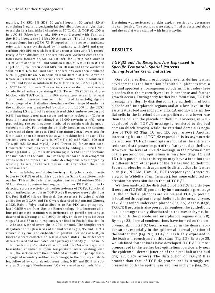
message is uniformly distributed in the epithelium of bothFab conjugated with alkaline phosphatase (Boehringer Mannheim),placode and interplacode regions and at a low level in thethe antibody was preabsorbed by diluting it 1:2000 in the TBSTearly feather bud mesenchyme (Figs. 1A and 1B). The epithe-containing 1.5 mg/ml of heat-inactivated chick embryo powder andlial cells in the interbud domain proliferate at a lower rate0.1% heat-inactivated goat serum and gently rocked at 47C for atthan the cells in the placode epithelium. However, in well-least 1 hr and then centrifuged at 15,000 rev/min at 47C. After
preblocking, the sections were incubated overnight with the preab- developed buds, TGF b2 message is restricted to the budsorbed antibody. Following the antibody incubation, the sections domain (black arrows), while the interbud domain is nega-were washed three times in TBST containing 2 mM levamisole for tive of TGF b2 (Figs. 1C and 1D, open arrows). Another5 min each, then six more washes with rocking for 1 hr each. The interesting feature of TGF b2 expression is its asymmetricsections were washed twice in NTMT (100 mM NaCl, 100 mM distribution. TGF b2 transcripts are found in the entire an-Tris, pH 9.5, 50 mM MgCl2, 0.1% Tween 20) for 20 min each. terior and distal posterior part of the feather bud epithelium.Colorimetric reactions were performed by adding 4.5 ml/ml NBT
However, the level of TGF b2 message in the proximal part(Promega) and 3.5 ml/ml BCIP (Promega) in NTMT to the sectionsof the posterior bud epithelium is very low (Figs. 1C andand incubated in the dark. The time required for color development1D). It is possible that this region may have a function thatvaries with the probes used. Color development was stopped byis different from other parts of the feather bud epithelium.washing the sections three times in PBT, after which the slidesSeveral molecules with asymmetric distributions in featherwere mounted.buds (i.e., NCAM, Hox C6, FGF receptor type 3) were re-Immunostaining and histochemistry. Polyclonal rabbit anti-
bodies to TGF b2 used in this study is from Santa Cruz Biotechnol- viewed in Widelitz et al. (in press), but none exhibited ex-ogy Inc. This antibody is raised against amino acid residues 352 to pression patterns similar to that of TGF b2.377 in the carboxy-terminal region of human TGF b2 and lacks We then analyzed the distribution of TGF b2 and its typedetectable cross reactivity with other isoforms of TGFb. Polyclonal II receptor (TGFbR II) proteins by immunostaining. At stagerabbit antibodies to human TGF b type II receptor were a gift from 31, the epithelial placodes are visible and TGF b2 proteinDr. Fred Hall (Children Hospital, Los Angeles). Polyclonal rabbit is localized throughout the epithelium. In the mesenchyme,antibodies to NCAM and Tn-C were described in Jiang and Chuong
TGF b2 is found under each placode (Fig. 2A). At this stage,(1992). Rabbit Polyclonal antibodies to Pan-PKC and phosphory-TGFbR II protein is also present throughout the epithelium,lated-CREB were from Upstate Biotechnology, Inc. Immuno-alka-but is homogeneously distributed in the mesenchyme, be-line phosphatase staining was performed on paraffin sections asneath both the placode and interplacode regions (Fig. 2B).described in Chuong et al. (1990). Briefly, chick embryos between
stages 31 and 37 or skin explants were fixed in Bouin’s fixative for By stage 33, dermal condensations have formed on the em-1–2 hr and washed overnight in 70% ethanol. The samples were bryonic skin. TGF b2 became enriched in the dermal con-dehydrated through a series of ethanol washes (80, 95, and 100%), densation, especially in the epidermal–dermal junction ofcleared in xylene, and embedded in paraffin. Sections at 6–8 mm the feather bud (Fig. 2C). TGFbR II is highly expressed inthickness were collected on gelatin-coated slides. The slides were the feather mesenchyme at this stage (Fig. 2D). By stage 35,deparaffinized and incubated with primary antibody (diluted in 11 well-defined feather buds have developed. TGF b2 is mostTBST containing 5% fetal calf serum and 1% BSA) overnight in a
pronounced in the feather bud epithelium, particularly nearhumidified chamber at room temperature. After washing withthe epidermal–dermal junction of the distal half of the budTBST, the sections were then incubated with alkaline phosphatase-(Fig. 2E, black arrows). The distribution of TGFbR II isconjugated secondary antibodies (Promega) to the primary antibod-broader than that of TGF b2 protein and is strongly ex-ies, followed by color development using NBT and BCIP as sub-
strates (Promega). Nonimmune IgGs were used as controls. H and pressed in both the epithelium and mesenchyme (Fig. 2F).
Copyright q 1996 by Academic Press, Inc. All rights of reproduction in any form reserved.
AID DB 8371 / 6x14$$$$42 10-11-96 15:53:07 dbas AP: Dev Bio
350 Ting-Berreth and Chuong
TGF b2 Can Induce Dermal Condensations congregation of feather buds around the condensation waswithout the Epithelium much more obvious than when the bead was placed on top
of the skin. The bead itself occupies the mesenchymal re-Classical embryology studies have utilized in vitro cul- gion with compact mesenchymal cells condensing around
ture systems of embryonic skins to study skin appendage the bead (e.g., see Fig. 4 for cross-sections). Feather budsformation (Sengel, 1978; Novel, 1973; Rawles, 1963). Skin
cannot form directly on top of the bead itself but the controlexplants of stage 30–31 dorsal skins were able to form nor-
bead does not perturb local feather growth (Fig. 3A).mal feather buds after a few days in culture. The continuousWe then asked whether TGF b2 can substitute for theinteractions between both the epithelium and mesenchyme
epithelium in organizing dermal condensations. The epithe-are required for feather formation. If the mesenchyme islium was peeled off from half of the skin explant, and thestripped of its epithelium and cultured alone, dermal con-remainder served as a positive control. TGF b2 beads weredensations will not form, and existing condensations willplaced on top of the naked mesenchymal region and cul-dissolve into unorganized fibroblasts.tured for 3 days. The control beads produced no mesenchy-We utilized the skin explant culture system to test themal reaction (Fig. 3E). In contrast, beads soaked in TGF b2roles of TGF b2. We began by adding TGF b2 ranging frominduced the accumulation of dermal cells, as judged by3 to 30 ng/ml in the culture media. There was no apparenttrans-illumination (Fig. 3F). To test whether this accumula-effect on feather growth after 4 days in culture. An exem-tion of dermal cells was really a ‘‘dermal condensation’’ orplary culture containing 20 ng/ml is shown in Fig. 3B. How-simply a tissue reaction elicited by TGF b2, the TGF b2ever, in many developmental processes, the gradient distri-bead was removed from the dermal condensation after 24bution of growth factors appears to be important (Gurdonhr in culture, and a stage 31 epithelium was placed on top ofet al., 1994). To create a local gradient, we placed TGF b2-the condensation (Fig. 3G). After culturing this recombinedcoated Affi-Gel Blue beads (Hayamizu et al., 1991; Vaino etskin for an additional 4 days, large and fused feather budsal., 1993) on top of skin explants. Control beads (see Tabledeveloped from the TGF b2-induced dermal condensation1) did not produce any suppressive or stimulatory effect due(Fig. 3H). The size of these feathers reflects the size of der-to the physical or chemical properties of the bead (Fig. 3A,mal condensation from the increased dose of TGF b2 inbead is indicated by the arrow). When a TGF b2-coated beadthat area. Some feather buds occasionally form in areas fur-(soaked in 7.5 mg/ml) was placed on top of a skin explant,ther away from the induced condensation, but these feath-a small condensation was induced around the bead, and aers are sparse and not as robust as those around the conden-ring of closely packed feather buds surrounds the condensa-sation. The background feather buds probably represent thetion (Fig. 3C). TGF b1, at the equivalent dosage, had noeffect of some dermal condensations that have not beensignificant effect (Table 1).completely dissolved. Although the newly induced featherSince our immunostaining data showed that TGF b2 isbuds did elongate, we did not observe the formation ofenriched in the dermal–epidermal junction of feather budsfeather filaments after 7 days in this culture condition.in vivo, we reasoned that the delivery of TGF b2 directlyWhen an epithelium was recombined with TGF b2-inducedto the dermal–epidermal junction may be more effective indermal condensations after 48 hours in culture, feathersour in vitro assays. We applied the TGF b2 bead by sand-were not able to grow from these condensations (data notwiching it between the epithelium and mesenchyme. Thisshown). These results demonstrate that TGF b2 is able toindeed led to a more pronounced effect. A lower concentra-substitute for the effect of the epithelium to allow the for-tion of TGF b2 (5 mg/ml) was able to induce a central zone
of condensation surrounding the bead (Fig. 3D), and the mation of condensations in the absence of the epithelium,
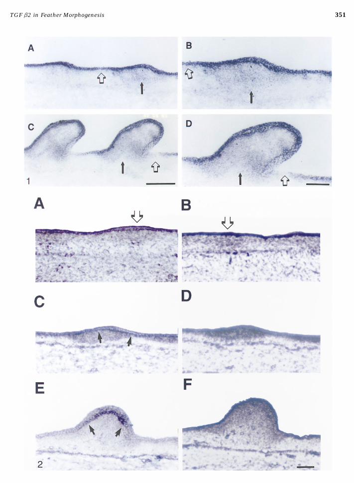
FIG. 1. TGF b2 message is expressed in epithelium. Nonradioactive in situ hybridization of longitudinal frozen sections through thedorsal regions of chick embryos. (A, B) Stage 34 where the skin has developed feather placodes. TGF b2 message is expressed uniformlyin the epithelium in the feather germ (black arrows) and intergerm (open arrows) regions and at a low level in the feather bud mesenchyme.(C, D) Stage 35.5 embryo exhibits well-formed feather buds. TGF b2 message is still predominant in the epithelium. Some muscle tissue(near bottom of the C) and the feather bud mesenchyme also express low levels of TGF b2. In the epithelium, TGF b2 is limited to thebud region (black arrows) and posterior interbud domain (open arrows). Bar for A,C, 250 mm; B,D, 100 mm.FIG. 2. Distribution of TGF b2 and TGF b receptor type II proteins in the developing skin. Immunoalkaline phosphatase staining oflongitudinal sections through the dorsal regions of the chick embryo. (A, C, E) TGF b2; (B, D, F) TGF b receptor, type II. (A, B) Stage 31,representing the early epidermal placode stage. (C, D) Stage 34, representing epidermal placode stage. (E, F) Stage 35, representing theearly feather bud stage. (A) TGF b2 is already preferably distributed beneath the placode. (B) TGF b receptor is homogeneously distributedat this stage. (C) TGF b2 began to be highly expressed in the epidermal–dermal junction of the feather bud. (D) TGF b receptor is enrichedin the entire stage 34 feather bud. (E) TGF b2 is expressed in the epidermal–dermal junction of the stage 35 bud. (F) TGF b receptor isexpressed in the entire feather bud. Open arrows indicate feather placodes, and black arrows mark the dermal–epidermal junction. Theanterior is toward the left side of the picture. Bar, 100 mm.
Copyright q 1996 by Academic Press, Inc. All rights of reproduction in any form reserved.
AID DB 8371 / 6x14$$$$43 10-11-96 15:53:07 dbas AP: Dev Bio
351TGF b2 in Feather Morphogenesis
10-11-96 15:53:07 dbas AP: Dev Bio

352 Ting-Berreth and Chuong
and this condensation is able to proceed with feather forma-tion when a competent epithelium is placed on top of itwithin a specific time window. After this period, feathergrowth cannot be rescued.
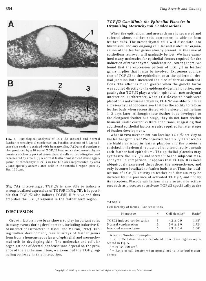
Histological examination showed that the TGF b2-in-duced condensation is due to an accumulation of mesenchy-mal cells (Fig. 4A). There is a significant increase in cellnumber in the TGF b2-induced condensation when com-pared to the interbud mesenchyme (Fig. 4B, Table 2). Nucleicount on the histological sections showed that the cell den-sity of the TGF b2-induced condensation is significantlyhigher than the interbud mesenchyme and similar to thenormal bud mesenchyme (Table 2).
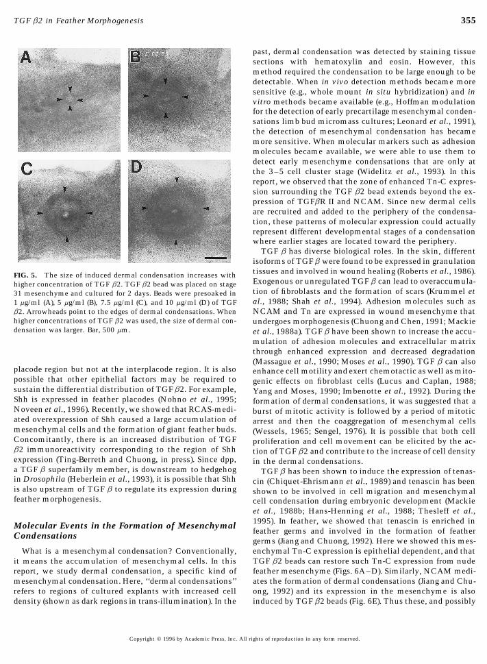
It is difficult to determine the absolute concentration ofTGF b2 in the tissue because it involves the equilibriumbetween the protein solution to the bead and the bead tothe tissue. To test whether the effect of the bead can bedescribed by the behavior of a diffusible factor, we testedits dose response as a relative comparison. Affi-Gel Bluebeads were presoaked in 1.0–10.0 mg/ml TGF b2 and wereplaced on stage 31 skin mesenchyme (Figs. 5A–5D). At 1mg/ml TGF b2, the condensation is barely visible. From 5to 10 mg/ml, there is a gradual increase in the size of thedermal condensations induced. Although the size of dermalcondensations at different concentrations varies slightlywith each set of experiments, there is a consistent correla-tion of dosage to the size of condensation. The size of theinduced condensation increased as the concentration ofTGF b2 beads increased. Another interesting observation isthat a ring of condensed dermal cells formed outside thebead, suggesting that dermal cells may condense at an opti-mal TGF b2 concentration. Although these results do notrule out the possibility that dermal condensations can bemediated by cell–cell interactions as well as diffusible fac-tors, the simplest explanation is that TGF b2 itself diffusesout of the bead and mediates the biological effect.
Effect of TGF b2 on Mesenchymal Expression ofTn, PKC, P-CREB, and TGF b Receptors
To explore the possible downstream events followingTGF b2 stimulation, we examined the expression of severalrelevant molecules. We have previously shown that adhe-sion molecules are expressed in early feather development.
FIG. 3. Effect of TGF b2 on skin explants. (A–D) Skin explantcultures from stage 31 embryo. (E–H) Stage 31 skins explants withtheir epithelium dissected off before culture. (A) Four-day cultureof a control skin explant containing a noncoated Affi-Gel Blue bead(arrow). (B) Stage 31 skin cultured with 20 ng/ml TGF b2 in the culture. A piece of epithelium (E) was left toward the left side ofmedia. After 4 days, no apparent difference in feather development the picture for comparison. (F) A bead presoaked in 10 mg/ml TGF(compare A and B). (C) When the bead was precoated with a 7.5 b2 induced a large dermal condensation on the skin mesenchymemg/ml TGF b2 and placed on top of the skin, a small condensation after 2 days in culture. (G) Stage 31 epithelium was reconstitutedaround the bead formed after 3 days in culture. (D) When the bead on a specimen similar to that in B with bead removed. (H) Four(5 mg/ml) was sandwiched between the epithelium and mesen- days later, many large feather buds formed from the induced con-chyme, a larger condensation and a ring of feather buds surrounded densation. Further away, background feathers occasionally form.that condensation. (E) A control bead (arrow) placed on the naked Arrowheads delineate boundaries of the replace epithelium. Bar,mesenchyme (M) did not induce any condensation after 2 days in 500 mm.
Copyright q 1996 by Academic Press, Inc. All rights of reproduction in any form reserved.
AID DB 8371 / 6x14$$$$43 10-11-96 15:53:07 dbas AP: Dev Bio
353TGF b2 in Feather Morphogenesis
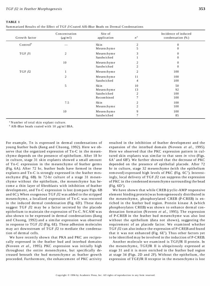
TABLE 1Summarized Results of the Effect of TGF b-Coated Affi-Blue Beads on Dermal Condensations
Concentration Site of Incidence of inducedGrowth factor (mg/ml) application na condensation (%)
Controlb — Skin 2 0Mesenchyme 5 0
TGF b1 2 Mesenchyme 3 0Sandwiched 1 0
10 Mesenchyme 2 0Sandwiched 2 0
TGF b2 1 Mesenchyme 2 100
2 Mesenchyme 11 100Sandwiched 4 100
5 Skin 10 50Mesenchyme 13 92Sandwiched 2 100Recombined 7 100
7.5 Skin 2 100Mesenchyme 2 100
10 Mesenchyme 9 100Sandwiched 7 85
a Number of total skin explant culture.b Affi-Blue beads coated with 10 mg/ml BSA.
For example, Tn is expressed in dermal condensations of resulted in the inhibition of feather development and theexpansion of the interbud domain (Noveen et al., 1995).young feather buds (Jiang and Chuong, 1992). Here we ob-
serve that the organized expression of Tn-C in the mesen- Here we observed that the PKC expression pattern in cul-tured skin explants was similar to that seen in vivo (Figs.chyme depends on the presence of epithelium. After 18 hr
in culture, stage 31 skin explants showed a small amount 6A* and 6B*). We further showed that the decrease of PKCdepended on the presence of epithelial placode. After 72of Tn-C expression in the mesenchyme of feather germs
(Fig. 6A). After 72 hr, feather buds have formed in these hr in culture, stage 32 mesenchyme (with the epitheliumremoved) expressed high levels of PKC (Fig. 6C*). Interest-explants and Tn-C is strongly expressed in the feather mes-
enchyme (Fig. 6B). In 72-hr culture of a stage 31 mesen- ingly, local delivery of TGF b2 can suppress the expressionof PKC in the condensed mesenchyme surrounding the beadchyme without the epithelium, the mesenchyme has be-
come a thin layer of fibroblasts with inhibition of feather (Fig. 6D*).We have shown that while CREB (cyclic-AMP responsivedevelopment, and Tn-C expression is lost (compare Figs. 6B
and 6C). When exogenous TGF b2 was added to the stripped element binding protein) was homogeneously distributed inthe mesenchyme, phosphorylated CREB (P-CREB) is en-mesenchyme, a localized expression of Tn-C was restored
in the induced dermal condensation (Fig. 6D). These data riched in the feather bud region. Protein kinase A (whichphosphorylates CREB) was shown to enhance dermal con-suggest TGF b2 may be a factor secreted by the placode
epithelium to maintain the expression of Tn-C. NCAM was densation formation (Noveen et al., 1995). The expressionof P-CREB in the feather bud mesenchyme was also lostalso shown to be expressed in dermal condensations (Jiang
and Chuong, 1992) and a similar expression was observed without the epithelium (data not shown), suggesting therequirement of an placode factor. We examined whetherin response to TGF b2 (Fig. 6E). These adhesion molecules
may act downstream of TGF b2 to mediate the condensa- TGF b2 can also induce the expression of P-CREB and foundthat it was not enhanced (Fig. 6E*). Thus other factors yettion of dermal cells.
We have recently shown that PKA and PKC are recipro- to be identified may be involved in the induction of P-CREB.Another molecule we examined is TGFbR II protein. Incally expressed in the feather bud and interbud domains
(Noveen et al., 1995). PKC expression was initially high the mesenchyme, TGFbR II is ubiquitously expressed atstage 31 and it is more enriched in the feather bud regionthroughout the entire mesenchyme, but specifically de-
creased beneath the bud mesenchyme as feather growth at stage 34 (Figs. 2D and 2F). Without the epithelium, theexpression of TGFbR II receptor in the mesenchyme is lostproceeded. Furthermore, the enhancement of PKC activity
Copyright q 1996 by Academic Press, Inc. All rights of reproduction in any form reserved.
AID DB 8371 / 6x14$$$$43 10-11-96 15:53:07 dbas AP: Dev Bio
354 Ting-Berreth and Chuong
TGF b2 Can Mimic the Epithelial Placodes inOrganizing Mesenchymal Condensations
When the epithelium and mesenchyme is separated andcultured alone, neither skin component is able to formfeather buds. The mesenchymal cells will dissociate intofibroblasts, and any ongoing cellular and molecular organi-zation of the feather germs already present, at the time ofepithelium removal, will gradually be lost. We have exam-ined many molecules for epithelial factors required for theinduction of mesenchymal condensation. Among them, wefound that the expression pattern of TGF b2 in feathergerms implies that it may be involved. Exogenous applica-tion of TGF b2 to the epithelium or at the epidermal–der-mal junction both increased the size of dermal condensa-tions. The effect is much greater when the growth factorwas applied directly to the epidermal–dermal junction, sug-gesting that TGF b2 plays a role in epithelial–mesenchymalinteraction. Furthermore, when TGF b2-coated beads wereplaced on a naked mesenchymes, TGF b2 was able to inducea mesenchymal condensation that has the ability to reformfeather buds when reconstituted with a piece of epithelium1–2 days later. Although these feather buds developed tothe elongated feather bud stage, they do not form featherfilament under current culture conditions, suggesting thatadditional epithelial factors are also required for later stagesof feather development.
What in vivo mechanism can localize TGF b2 activity toFIG. 4. Histological analysis of TGF b2 induced and normal the feather germ area? We observed that TGF b2 transcriptsfeather mesenchymal condensation. Paraffin sections of 3-day cul- are highly enriched in feather placodes and the protein isture skin explants stained with hematoxylin. (A) Dermal condensa- enriched in the dermal–epidermal junction directly beneathtion produced by a 10 mg/ml TGF b2 bead on a naked mesenchyme each feather bud epithelium. The epithelial placodes mayconsists of closely packed mesenchymal cells surrounding the bead synthesize the TGF b2 and secrete it to the subjacent mes-represented by area 1. (B) A normal feather bud showed dense aggre-
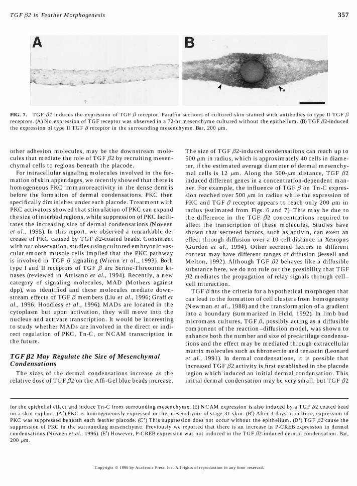
enchyme. In comparison, it appears that TGFbR II is moregation of mesenchymal cells in the bud area (represented by areaubiquitously expressed throughout the mesenchyme, and2) and sparsely accumulated cells in the interbud region (area 3).only becomes localized to feather buds later. Thus the local-Bar, 100 mm.ization of TGF b2 activity to feather bud domain may bedictated by the presence of activated TGF b2, and not byits receptors. Placode epithelium may also provide activa-tors such as proteases to activate TGF b2 specifically at the(Fig. 7A). Interestingly, TGF b2 is also able to induce a
strong localized expression of TGFbR II (Fig. 7B). It is possi-ble that TGF b2 also induces TGFbR II in vivo and thusamplifies the TGF b response in the feather germ region.
TABLE 2Cell Density of Dermal Condensations
DISCUSSION Phenotype n Cell densitya Ratiob
TGFb2-induced condensation 5 4.2 { 0.9 1.451Growth factors have been shown to play important rolesNormal condensation 5 5.0 { 1.0 1.722in many events during development, including inductive E-Inter-bud mesenchyme 5 2.9 { 0.4 1.003M interactions (reviewed in Jessell and Melton, 1992). Dur-
ing feather development, regular arrays of feather germsNote. n, Number of samples.
form from a homogeneous layer of epithelial and mesenchy- 1, 2, 3, Cell densities are calculated from these regions repre-mal cells in developing skin. The molecular and cellular sented in Fig. 5.organizations of dermal condensations depend on the pres- a Å cells/1000 mm3.ence of the epithelium. Here, we examined the TGF b sig- bÅ Ratio of cell density when normalized to inter-bud mesen-
chyme.naling pathway in this interaction.
Copyright q 1996 by Academic Press, Inc. All rights of reproduction in any form reserved.
AID DB 8371 / 6x14$$$$43 10-11-96 15:53:07 dbas AP: Dev Bio
355TGF b2 in Feather Morphogenesis
past, dermal condensation was detected by staining tissuesections with hematoxylin and eosin. However, thismethod required the condensation to be large enough to bedetectable. When in vivo detection methods became moresensitive (e.g., whole mount in situ hybridization) and invitro methods became available (e.g., Hoffman modulationfor the detection of early precartilage mesenchymal conden-sations limb bud micromass cultures; Leonard et al., 1991),the detection of mesenchymal condensation has becamemore sensitive. When molecular markers such as adhesionmolecules became available, we were able to use them todetect early mesenchyme condensations that are only atthe 3–5 cell cluster stage (Widelitz et al., 1993). In thisreport, we observed that the zone of enhanced Tn-C expres-sion surrounding the TGF b2 bead extends beyond the ex-pression of TGFbR II and NCAM. Since new dermal cellsare recruited and added to the periphery of the condensa-tion, these patterns of molecular expression could actuallyrepresent different developmental stages of a condensationwhere earlier stages are located toward the periphery.
TGF b has diverse biological roles. In the skin, differentisoforms of TGFb were found to be expressed in granulationtissues and involved in wound healing (Roberts et al., 1986).FIG. 5. The size of induced dermal condensation increases withExogenous or unregulated TGF b can lead to overaccumula-higher concentration of TGF b2. TGF b2 bead was placed on stagetion of fibroblasts and the formation of scars (Krummel et31 mesenchyme and cultured for 2 days. Beads were presoaked inal., 1988; Shah et al., 1994). Adhesion molecules such as1 mg/ml (A), 5 mg/ml (B), 7.5 mg/ml (C), and 10 mg/ml (D) of TGF
b2. Arrowheads point to the edges of dermal condensations. When NCAM and Tn are expressed in wound mesenchyme thathigher concentrations of TGF b2 was used, the size of dermal con- undergoes morphogenesis (Chuong and Chen, 1991; Mackiedensation was larger. Bar, 500 mm. et al., 1988a). TGF b have been shown to increase the accu-
mulation of adhesion molecules and extracellular matrixthrough enhanced expression and decreased degradation(Massague et al., 1990; Moses et al., 1990). TGF b can also
placode region but not at the interplacode region. It is also enhance cell motility and exert chemotactic as well as mito-possible that other epithelial factors may be required to genic effects on fibroblast cells (Lucus and Caplan, 1988;sustain the differential distribution of TGFb2. For example, Yang and Moses, 1990; Imbenotte et al., 1992). During theShh is expressed in feather placodes (Nohno et al., 1995; formation of dermal condensations, it was suggested that aNoveen et al., 1996). Recently, we showed that RCAS-medi- burst of mitotic activity is followed by a period of mitoticated overexpression of Shh caused a large accumulation of arrest and then the coaggregation of mesenchymal cellsmesenchymal cells and the formation of giant feather buds. (Wessels, 1965; Sengel, 1976). It is possible that both cellConcomitantly, there is an increased distribution of TGF proliferation and cell movement can be elicited by the ac-b2 immunoreactivity corresponding to the region of Shh tion of TGF b2 and contribute to the increase of cell densityexpression (Ting-Berreth and Chuong, in press). Since dpp, in the dermal condensations.a TGF b superfamily member, is downstream to hedgehog TGF b has been shown to induce the expression of tenas-in Drosophila (Heberlein et al., 1993), it is possible that Shh cin (Chiquet-Ehrismann et al., 1989) and tenascin has beenis also upstream of TGF b to regulate its expression during shown to be involved in cell migration and mesenchymalfeather morphogenesis. cell condensation during embryonic development (Mackie
et al., 1988b; Hans-Henning et al., 1988; Thesleff et al.,1995). In feather, we showed that tenascin is enriched inMolecular Events in the Formation of Mesenchymalfeather germs and involved in the formation of featherCondensationsgerms (Jiang and Chuong, 1992). Here we showed this mes-enchymal Tn-C expression is epithelial dependent, and thatWhat is a mesenchymal condensation? Conventionally,
it means the accumulation of mesenchymal cells. In this TGF b2 beads can restore such Tn-C expression from nudefeather mesenchyme (Figs. 6A–D). Similarly, NCAM medi-report, we study dermal condensation, a specific kind of
mesenchymal condensation. Here, ‘‘dermal condensations’’ ates the formation of dermal condensations (Jiang and Chu-ong, 1992) and its expression in the mesenchyme is alsorefers to regions of cultured explants with increased cell
density (shown as dark regions in trans-illumination). In the induced by TGF b2 beads (Fig. 6E). Thus these, and possibly
Copyright q 1996 by Academic Press, Inc. All rights of reproduction in any form reserved.
AID DB 8371 / 6x14$$$$43 10-11-96 15:53:07 dbas AP: Dev Bio
356 Ting-Berreth and Chuong
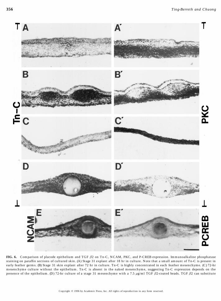
FIG. 6. Comparison of placode epithelium and TGF b2 on Tn-C, NCAM, PKC, and P-CREB expression. Immunoalkaline phosphatasestaining on paraffin sections of cultured skin. (A) Stage 31 explant after 18 hr in culture. Note that a small amount of Tn-C is present inearly feather germs. (B) Stage 31 skin explant after 72 hr in culture. Tn-C is highly concentrated in each feather mesenchyme. (C) 72-hrmesenchyme culture without the epithelium. Tn-C is absent in the naked mesenchyme, suggesting Tn-C expression depends on thepresence of the epithelium. (D) 72-hr culture of a stage 31 mesenchyme with a 7.5 mg/ml TGF b2-coated beads. TGF b2 can substitute
Copyright q 1996 by Academic Press, Inc. All rights of reproduction in any form reserved.
AID DB 8371 / 6x14$$8371 10-11-96 15:53:07 dbas AP: Dev Bio
357TGF b2 in Feather Morphogenesis
FIG. 7. TGF b2 induces the expression of TGF b receptor. Paraffin sections of cultured skin stained with antibodies to type II TGF breceptors. (A) No expression of TGF receptor was observed in a 72-hr mesenchyme cultured without the epithelium. (B) TGF b2-inducedthe expression of type II TGF b receptor in the surrounding mesenchyme. Bar, 200 mm.
other adhesion molecules, may be the downstream mole- The size of TGF b2-induced condensations can reach up tocules that mediate the role of TGF b2 by recruiting mesen- 500 mm in radius, which is approximately 40 cells in diame-chymal cells to regions beneath the placode. ter, if the estimated average diameter of dermal mesenchy-
For intracellular signaling molecules involved in the for- mal cells is 12 mm. Along the 500-mm distance, TGF b2mation of skin appendages, we recently showed that there is induced different genes in a concentration-dependent man-homogeneous PKC immunoreactivity in the dense dermis ner. For example, the influence of TGF b on Tn-C expres-before the formation of dermal condensations. PKC then sion reached over 500 mm in radius while the expression ofspecifically diminishes under each placode. Treatment with PKC and TGF b receptor appears to reach only 200 mm inPKC activators showed that stimulation of PKC can expand radius (estimated from Figs. 6 and 7). This may be due tothe size of interbud regions, while suppression of PKC facili- the difference in the TGF b2 concentrations required totates the increasing size of dermal condensations (Noveen affect the transcription of these molecules. Studies haveet al., 1995). In this report, we observed a remarkable de- shown that secreted factors, such as activin, can exert ancrease of PKC caused by TGF b2-coated beads. Consistent effect through diffusion over a 10-cell distance in Xenopuswith our observation, studies using cultured embryonic vas- (Gurdon et al., 1994). Other secreted factors in differentcular smooth muscle cells implied that the PKC pathway context may have different ranges of diffusion (Jessell andis involved in TGF b signaling (Wrenn et al., 1993). Both Melton, 1992). Although TGF b2 behaves like a diffusibletype I and II receptors of TGF b are Serine-Threonine ki- substance here, we do not rule out the possibility that TGFnases (reviewed in Attisano et al., 1994). Recently, a new b2 mediates the propagation of relay signals through cell–category of signaling molecules, MAD (Mothers against cell interaction.dpp), was identified and these molecules mediate down- TGF b fits the criteria for a hypothetical morphogen thatstream effects of TGF b members (Liu et al., 1996; Graff et can lead to the formation of cell clusters from homogeneityal., 1996; Hoodless et al., 1996). MADs are located in the (Newman et al., 1988) and the transformation of a gradientcytoplasm but upon activation, they will move into the into a boundary (summarized in Held, 1992). In limb budnucleus and activate transcription. It would be interesting micromass cultures, TGF b, possibly acting as a diffusibleto study whether MADs are involved in the direct or indi- component of the reaction–diffusion model, was shown torect regulation of PKC, Tn-C, or NCAM transcription in enhance both the number and size of precartilage condensa-the future. tions and the effect may be mediated through extracellular
matrix molecules such as fibronectin and tenascin (LeonardTGF b2 May Regulate the Size of Mesenchymal et al., 1991). In dermal condensations, it is possible thatCondensations increased TGF b2 activity is first established in the placode
region which induced an initial dermal condensation. ThisThe sizes of the dermal condensations increase as therelative dose of TGF b2 on the Affi-Gel blue beads increase. initial dermal condensation may be very small, but TGF b2
for the epithelial effect and induce Tn-C from surrounding mesenchyme. (E) NCAM expression is also induced by a TGF b2 coated beadon a skin explant. (A*) PKC is homogeneously expressed in the mesenchyme of stage 31 skin. (B*) After 3 days in culture, expression ofPKC was suppressed beneath each feather placode. (C*) This suppression does not occur without the epithelium. (D*) TGF b2 cause thesuppression of PKC in the surrounding mesenchyme. Previously we reported that there is an increase in P-CREB expression in dermalcondensations (Noveen et al., 1996). (E*) However, P-CREB expression was not induced in the TGF b2-induced dermal condensation. Bar,200 mm.
Copyright q 1996 by Academic Press, Inc. All rights of reproduction in any form reserved.
AID DB 8371 / 6x14$$$$44 10-11-96 15:53:07 dbas AP: Dev Bio
358 Ting-Berreth and Chuong
H.-M., and De Robertis, E. (1990). Gradients of homeoproteinscan augment its response through the positive autoregula-in developing feather buds. Development 110, 1021–1030.tion of TGF bs (van Obberghen-Shilling et al., 1988) and/
Davidson, E. H. (1993). Later embryogenesis: Regulatory circuitryor by stimulating the synthesis of TGF b receptors in thein morphogenetic fields. Development 118, 665–690.surrounding mesenchymal cells (Fig. 7). Thus the response
Graff, J. M., Bansal, A., and Melton, D. A. (1996). Xenopus Madto TGF b is amplified and the propagation of dermal conden-proteins transduce distinct subsets of signals for the TGF b super-
sations facilitated. This propagation has to stop at some family. Cell 85, 479–487.point and mesenchymal cells outside of its influence will Green, J. B. A., and Smith, J. C. (1991). Growth factors as morpho-remain as fibroblasts. By drawing analogy to other models gens: Do gradients and thresholds establish body plan? Trends(reviewed in Held, 1992), we postulate that there may be a Genet. 7, 245–250.threshold above which TGF b receptor expressions is en- Gurdon, J. B., Harger, P., Mitchell, A., and Lemaire, P. (1994). Ac-
tivin signaling and response to a morphogen gradient. Naturehanced and TGF b2 signaling is amplified. Below that371, 487–492.threshold, TGF b2 signals becomes insignificant. Hence, a
Hamburger, V., and Hamilton, H. (1951). A series of normal stagesgradient is transformed into a boundary and a sharp borderin the development of the chick embryo. J. Morphol. 88, 49 –92.of dermal condensations is established. The corollary is that
Hans-Henning, E., Halfter, W., and Tucker, R. P. (1988). The distri-by adjusting the concentration of TGF b2, the proportionbution of fibronectin and tenascin along migratory pathways ofof activated TGF b2, or the amount of TGF b receptors, itthe neural crest in the trunk of amphibian embryos. Develop-
is possible to regulate the size of dermal condensations, ment 103, 743 –756.which in turn will determine the size of the feather. Hayamizu, T. F., Sessions, S. K., Wanek, N., and Bryant, S. V. (1991).
In summary, regularly spaced feather germs in developing Effects of localized application of transforming growth factor b1chick skin form from homogenous epithelial and mesen- on developing chick limbs. Dev. Biol. 145, 164–173.chymal cells. This cellular reorganization requires E-M in- Heberlein, U., Wolff, T., and Rubin, G. M. (1993). The TGF beta
homologue dpp and the segment polarity gene hedgehog are re-teractions and a molecular signaling network. In this report,quired for propagation of a morphogenetic wave in the Drosophilawe showed that TGF b2 mRNA is synthesized predomi-retina. Cell 75, 913–926.nantly in the feather placode epithelium. TGF b2 protein is
Held, L. I. (1992). ‘‘Models for Embryonic Periodicity,’’ pp. 16 –24.enriched in the dermal–epidermal junction and can induceKarger, New York.dermal condensations without the epithelium. In the sig-
Hoodless, P. A., Haerry, T., Abdollah, S., Stapleton, M., O’Connor,naling cascade, TGF b2 may be downstream to Shh andM. B., Attisano, L., and Wrana, J. L. (1996). MADR1, a MAD-
upstream to TGF b receptor, PKC, Tn-C, and NCAM. related protein that functions in BMP2 signaling pathways. Cell85, 489–500.
Imbenotte, J., Liu, L., Desauty, G., and Harel, L. (1992). StimulationACKNOWLEDGMENTS by TGF b of chick embryo fibroblasts - inhibition by an IGFBP-
3. Exp. Cell Res. 199, 229–233.Jakowlew, S. B., Ciment, G., Tuan, R. S., Sporn, M. B., and Roberts,We thank Dr. Sonia Jakowlew (NIH) for providing us with TGF
A. B. (1994). Expression of transforming growth factor-b2 and b3b2 protein and cDNA probe for in situ hybridization and Dr. FredmRNAs and proteins in the developing chick embryo. Differenti-Hall for antibodies to type II TGF b receptor. We also thank Dr.ation 55, 105–118.YiPing Chen for critical reading of this manuscript and Dr. Randall
Jessell, T. M., and Melton, D. A. (1992). Diffusible factors in verte-Widelitz for helpful discussions. This work is supported by grantsbrate embryonic induction. Cell 68, 257–270.from NIH, NSF, and Council for Tobacco Research.
Jiang, T.-X., and Chuong, C.-M. (1992). Mechanism of Feather Mor-phogenesis: I. Analyses with antibodies to adhesion moleculestenascin, N-CAM and integrin. Dev. Biol. 150, 82–98.REFERENCES
Krummel, P. M., Michna, B. A., Thomas, B. L., Sporn, M. B., Nelson,J. M., Salzberg, A. M., Cohen, I. K., and Diegelmann, R. F. (1988).Attisano, L., Wrana, J. L., Lopez-Casillas, F., and Massague, J. (1994).Transforming growth factor b (TGF-b) induces fibrosis in fetalTGF-b receptors and actions. Biochim. Biophys. Acta 1222, 71–wound model. J. Paediatr. Surg. 23, 647–652.80.
Leonard, C. M., Fuld, H. M., Frenz, D. A., Downie, S. A., Massague,Cairns, J. M., and Saunders, J. W. (1954). The influence of embry-J., and Newman, S. A. (1991). Role of transforming growth factor-onic mesoderm on the regional specification of epidermal deriva-b in chondrogenic pattern formation in the embryonic limb:tives in the chick. J. Exp. Zool. 127, 221–248.Stimulation of mesenchymal condensation and fibronectin geneChiquet-Ehrismann, R., Kalla, P., and Pearson, C. A. (1989). Partici-expression by exogenous TGF-b and evidence for endogenouspation of tenascin and transforming growth factor-b in reciprocalTGF-b like activity. Dev. Biol. 145, 99 –109.epithelial-mesenchymal interactions of MCF7 cells and fibro-
Liu, F., Hata, A., Baker, J. C., Doody, J., Carcamo, J., Harland,blasts. Cancer Res. 49, 4322–4325.R. M., and Massague, J. (1996). A human Mad protein acting asChuong, C.-M. (1993). The making of a feather. Homeoproteinsa BMP-regulated transcriptional activator. Nature 381, 620–623.and retinoids and adhesion molecules. BioEssays 15, 513–521.
Lucus, P. A., and Caplan, A. I. (1988). Chemotactic response ofChuong, C.-M., and Chen, H.-M. (1991). Enhanced expression ofembryonic limb bud mesenchymal cells and muscle-derived fi-neural cell adhesion molecules and tenascin (cytotactin) duringbroblasts to transforming growth factor-b. Connect. Tissue Res.wound healing. Am. J. Pathol. 138, 1–14.
Chuong, C.-M., Guillermo, O., Ting, S. A., Jegalian, B. G., Chen, 18, 1–7.
Copyright q 1996 by Academic Press, Inc. All rights of reproduction in any form reserved.
AID DB 8371 / 6x14$$$$44 10-11-96 15:53:07 dbas AP: Dev Bio
359TGF b2 in Feather Morphogenesis
Mackie, E. J., Halfter, W., Liverani, D. (1988a). Induction of tenascin ment’’ (R. H. Sawyer and J. F. Fallon, Eds.), pp. 115–146. Praeger,New York.in healing wounds. J. Cell Biol. 107, 2569–2579.
Sawyer, R. H., and Fallon, J. F., Eds. (1983). ‘‘Epithelial-Mesenchy-Mackie, E. J., Tucker, R. P., Halfter, W., and Chiquet-Ehrismann,mal Interactions in Development.’’ Praeger, New York.R. (1988b). The distribution of tenascin coincides with pathways
Sengel, P. (1976). ‘‘Morphogenesis of Skin’’ (M. Abercrombie, D. R.of neural crest cell migration. Development 102, 237–250.Newth, and J. G. Torrey, Eds.). Cambridge Univ. Press, Cam-Massague, J., Cheifetz, S., Boyd, F. T., and Andres, J. L. (1990). TGF-bridge.b receptors and TGF-b binding proteoglycans: Recent progress
Sengel, P. (1978). Feather pattern development. In ‘‘Ciba Founda-in identifying their functional properties. Ann. N.Y. Acad. Sci.tion Symposium 29,’’ pp. 51–70. Elsevier Science, New York.593, 59–72.
Shah, M., Foreman, D. M., and Ferguson, M. W. J. (1994). Neutraliz-McAleese, S. R., and Sawyer, R. H. (1981). Correcting the phenotypeing antibody to TGF-b1,2 reduces cutaneous scarring in adultof the epidermis from chick embryos homozygous for the generodents. J. Cell Sci. 107, 1137–1157.scaleless (sc/sc). Science 214, 1033–1034.
Tabin, C. J. (1991). Retinoids, homeoboxes, and growth factors:Moses, H. L., Yang, E. Y., and Pietenpol, J. A. (1990). TGF-b stimula-Toward molecular models for limb development. Cell 66, 199–tion and inhibition of cell proliferation: New mechanistic in-217.sights. Cell 63, 245 –247.
Thesleff, I., Vaahtokari, A., and Partanen, A. M. (1995). RegulationNakashima, M., Nagasawa, H., Yamada, Y., and Reddi, H. (1994).of organogenesis. Common molecular mechanisms regulatingRegulatory role of transforming growth factor-b, bone morpho-the development of teeth and other organs. Internat. J. Dev. Biol.genetic protein-2, and protein-4 on gene expression of extracellu-39, 35 –50.lar matrix proteins and differentiation of dental pulp cells. Dev.
Ting-Berreth, S. A., and Chuong, C.-M. (1996). Sonic hedgehog inBiol. 162, 18–28.feather morphogenesis: Induction of mesenchymal condensationNewman, S. A., Frisch, H. L., and Percus, J. K. (1988). On the sta-and association with cell death. Dev. Dynam. 207, 157.tionary state analysis of reaction-diffusion mechanisms for bio-
Vaino, S., Karavanova, I., Jowett, A., and Thesleff, I. (1993). Identi-logical pattern formation. J. Theor. Biol. 134, 183–197.fication of BMP-4 as a signal mediating secondary induction be-Nieswander, L., and Martin, G. R. (1993). FGF-4 and BMP-2 havetween epithelial and mesenchymal tissues during early toothopposite effects on limb growth. Nature 361, 68–71.development. Cell 75, 45–58.Nohno, T., Kawakami, Y., Ohuchi, H., Fujiwara, A., Yoshioka, H.,
van Obberghen-Schilling, E., Roche, N. S., Flanders, K. C., Sporn,and Noji, S. (1995). Involvement of the Sonic hedgehog gene inM. B., and Roberts, A. B. (1988). Transforming growth factor-b1chick feather development. Biochem. Biophys. Res. Commun.positively regulates its own expression in normal and trans-206, 33–39.formed cells. J. Biol. Chem. 263, 8569–8572.Noveen, A., Jiang, T.-X., and Chuong, C.-M. (1995). Protein kinase
Wessels, N. K. (1965). Morphology and proliferation during earlyA and protein kinase C modulators have reciprocal effects onfeather development. Dev. Biol. 12, 131–153.mesenchymal condensation during skin appendage morphogene-
Widelitz, R. B., Jiang, T.-X., Murray, B. A., and Chuong, C.-M.sis. Dev. Biol. 171, 677–693.(1993). Adhesion molecules in skeletogenesis: II. Neural cell ad-Noveen, A., Jiang, T.-X., and Chuong, C.-M. (1996). CAMP, anhesion molecules mediate precartilaginous mesenchymal con-activator of protein kinase A, suppresses the expression of sonicdensations and enhance chondrogenesis. J. Cell. Physiol. 156,hedgehog. Biochem. Biophys. Res. Commun. 219, 180–185.399–411.Novel, G. (1973). Feather pattern stability and reorganization in
Widelitz, R. B., Jiang, T.-X., Noveen, A., Ting-Berreth, S. A., Yin,cultured skin. J. Embryol. Exp. Morph. 30, 605–633.E., and Chuong, C.-M. Molecular histology in skin appendageRawles, M. E. (1963). Tissue interactions in scale and feather devel-morphogenesis. Microscopy Res. Technol., in press.opment as studied in dermal-epidermal recombination. J.
Wrenn, R. W., Raeuber, C. L., Herman, L. E., Walton, W. J., andEmbryol. Exp. Morph. 11, 765–789.Rosenquist, T. H. (1993). Transforming growth factor-b: SignalRoberts, A. B., Sporn, M. B., Assoian, R. K., Smith, J. M., Roche,transduction via protein kinase C in cultured embryonic vascularN. S., Wakefield, L. M., Heine, U. L., Liotta, L. A., Falanga, V.,smooth muscle cells. In Vitro Cell. Dev. Biol. 29A, 73–78.Kehrl, J. H., and Fauci, A. S. (1986). Transforming growth factor
Yang, E. Y., and Moses, H. L. (1990). Transforming growth factortype b: rapid induction of fibrosis and angiogenesis in vivo andb1-induced changes in cell migration, proliferation, and angio-stimulation of collagen formation in vitro. Proc. Natl. Acad. Sci.genesis in the chick chorioallantoic membrane. J. Cell Biol. 111,USA 83, 4167–4171.731–741.Sawyer, R. H. (1983). The role of epithelial-mesenchymal interac-
tions in regulating gene expression during avian scale morpho- Received for publication July 25, 1996Accepted August 19, 1996genesis. In ‘‘Epithelial-Mesenchymal Interactions in Develop-
Copyright q 1996 by Academic Press, Inc. All rights of reproduction in any form reserved.
AID DB 8371 / 6x14$$$$45 10-11-96 15:53:07 dbas AP: Dev Bio